WO2013180201A1 - Antigen-binding molecule for eliminating aggregated antigens - Google Patents
Antigen-binding molecule for eliminating aggregated antigens Download PDFInfo
- Publication number
- WO2013180201A1 WO2013180201A1 PCT/JP2013/064979 JP2013064979W WO2013180201A1 WO 2013180201 A1 WO2013180201 A1 WO 2013180201A1 JP 2013064979 W JP2013064979 W JP 2013064979W WO 2013180201 A1 WO2013180201 A1 WO 2013180201A1
- Authority
- WO
- WIPO (PCT)
- Prior art keywords
- amino acid
- antigen
- tyr
- thr
- binding
- Prior art date
Links
Images


Classifications
-
- C—CHEMISTRY; METALLURGY
- C07—ORGANIC CHEMISTRY
- C07K—PEPTIDES
- C07K16/00—Immunoglobulins [IGs], e.g. monoclonal or polyclonal antibodies
- C07K16/18—Immunoglobulins [IGs], e.g. monoclonal or polyclonal antibodies against material from animals or humans
-
- C—CHEMISTRY; METALLURGY
- C07—ORGANIC CHEMISTRY
- C07K—PEPTIDES
- C07K16/00—Immunoglobulins [IGs], e.g. monoclonal or polyclonal antibodies
-
- A—HUMAN NECESSITIES
- A61—MEDICAL OR VETERINARY SCIENCE; HYGIENE
- A61P—SPECIFIC THERAPEUTIC ACTIVITY OF CHEMICAL COMPOUNDS OR MEDICINAL PREPARATIONS
- A61P43/00—Drugs for specific purposes, not provided for in groups A61P1/00-A61P41/00
-
- C—CHEMISTRY; METALLURGY
- C07—ORGANIC CHEMISTRY
- C07K—PEPTIDES
- C07K16/00—Immunoglobulins [IGs], e.g. monoclonal or polyclonal antibodies
- C07K16/18—Immunoglobulins [IGs], e.g. monoclonal or polyclonal antibodies against material from animals or humans
- C07K16/28—Immunoglobulins [IGs], e.g. monoclonal or polyclonal antibodies against material from animals or humans against receptors, cell surface antigens or cell surface determinants
- C07K16/2866—Immunoglobulins [IGs], e.g. monoclonal or polyclonal antibodies against material from animals or humans against receptors, cell surface antigens or cell surface determinants against receptors for cytokines, lymphokines, interferons
-
- C—CHEMISTRY; METALLURGY
- C07—ORGANIC CHEMISTRY
- C07K—PEPTIDES
- C07K16/00—Immunoglobulins [IGs], e.g. monoclonal or polyclonal antibodies
- C07K16/42—Immunoglobulins [IGs], e.g. monoclonal or polyclonal antibodies against immunoglobulins
- C07K16/4283—Immunoglobulins [IGs], e.g. monoclonal or polyclonal antibodies against immunoglobulins against an allotypic or isotypic determinant on Ig
-
- G—PHYSICS
- G01—MEASURING; TESTING
- G01N—INVESTIGATING OR ANALYSING MATERIALS BY DETERMINING THEIR CHEMICAL OR PHYSICAL PROPERTIES
- G01N33/00—Investigating or analysing materials by specific methods not covered by groups G01N1/00 - G01N31/00
- G01N33/48—Biological material, e.g. blood, urine; Haemocytometers
- G01N33/50—Chemical analysis of biological material, e.g. blood, urine; Testing involving biospecific ligand binding methods; Immunological testing
- G01N33/68—Chemical analysis of biological material, e.g. blood, urine; Testing involving biospecific ligand binding methods; Immunological testing involving proteins, peptides or amino acids
- G01N33/6854—Immunoglobulins
-
- A—HUMAN NECESSITIES
- A61—MEDICAL OR VETERINARY SCIENCE; HYGIENE
- A61K—PREPARATIONS FOR MEDICAL, DENTAL OR TOILETRY PURPOSES
- A61K39/00—Medicinal preparations containing antigens or antibodies
- A61K2039/505—Medicinal preparations containing antigens or antibodies comprising antibodies
-
- C—CHEMISTRY; METALLURGY
- C07—ORGANIC CHEMISTRY
- C07K—PEPTIDES
- C07K2317/00—Immunoglobulins specific features
- C07K2317/10—Immunoglobulins specific features characterized by their source of isolation or production
- C07K2317/14—Specific host cells or culture conditions, e.g. components, pH or temperature
-
- C—CHEMISTRY; METALLURGY
- C07—ORGANIC CHEMISTRY
- C07K—PEPTIDES
- C07K2317/00—Immunoglobulins specific features
- C07K2317/20—Immunoglobulins specific features characterized by taxonomic origin
- C07K2317/24—Immunoglobulins specific features characterized by taxonomic origin containing regions, domains or residues from different species, e.g. chimeric, humanized or veneered
-
- C—CHEMISTRY; METALLURGY
- C07—ORGANIC CHEMISTRY
- C07K—PEPTIDES
- C07K2317/00—Immunoglobulins specific features
- C07K2317/50—Immunoglobulins specific features characterized by immunoglobulin fragments
- C07K2317/52—Constant or Fc region; Isotype
-
- C—CHEMISTRY; METALLURGY
- C07—ORGANIC CHEMISTRY
- C07K—PEPTIDES
- C07K2317/00—Immunoglobulins specific features
- C07K2317/90—Immunoglobulins specific features characterized by (pharmaco)kinetic aspects or by stability of the immunoglobulin
-
- C—CHEMISTRY; METALLURGY
- C07—ORGANIC CHEMISTRY
- C07K—PEPTIDES
- C07K2317/00—Immunoglobulins specific features
- C07K2317/90—Immunoglobulins specific features characterized by (pharmaco)kinetic aspects or by stability of the immunoglobulin
- C07K2317/94—Stability, e.g. half-life, pH, temperature or enzyme-resistance
-
- G—PHYSICS
- G01—MEASURING; TESTING
- G01N—INVESTIGATING OR ANALYSING MATERIALS BY DETERMINING THEIR CHEMICAL OR PHYSICAL PROPERTIES
- G01N2333/00—Assays involving biological materials from specific organisms or of a specific nature
- G01N2333/435—Assays involving biological materials from specific organisms or of a specific nature from animals; from humans
- G01N2333/46—Assays involving biological materials from specific organisms or of a specific nature from animals; from humans from vertebrates
- G01N2333/47—Assays involving proteins of known structure or function as defined in the subgroups
-
- G—PHYSICS
- G01—MEASURING; TESTING
- G01N—INVESTIGATING OR ANALYSING MATERIALS BY DETERMINING THEIR CHEMICAL OR PHYSICAL PROPERTIES
- G01N2333/00—Assays involving biological materials from specific organisms or of a specific nature
- G01N2333/435—Assays involving biological materials from specific organisms or of a specific nature from animals; from humans
- G01N2333/705—Assays involving receptors, cell surface antigens or cell surface determinants
- G01N2333/70503—Immunoglobulin superfamily, e.g. VCAMs, PECAM, LFA-3
- G01N2333/70535—Fc-receptors, e.g. CD16, CD32, CD64 (CD2314/705F)
Definitions
- the present invention relates to the use of an antigen-binding molecule for eliminating an associated antigen from plasma, a method for eliminating the associated antigen from plasma, comprising administering the antigen-binding molecule, and an antigen associated from plasma.
- a method for producing a molecule is provided.
- amyloidosis A group of diseases characterized by extracellular accumulation of various protein aggregates is called amyloidosis, and according to the classification of the Ministry of Health, Labor and Welfare's specific disease research team, there are 10 types of systemic amyloidosis and 10 types of localized amyloidosis. There are reported types of disease.
- ⁇ -synuclein disease is known as a disease in which ⁇ -synuclein aggregated in nerve cells is deposited.
- Some hereditary Parkinson's diseases are caused by the deposition of ⁇ -synuclein in brain neurons and are considered a type of ⁇ -synuclein disease.
- diseases that are known to be caused by protein aggregates, but the mechanism of protein aggregation has not yet been clarified. Does not exist.
- Non-patent Documents 1 and 2 are on the market, and many antibody drugs have been developed.
- the amount of antigen that can be neutralized per antibody molecule depends on the affinity. By increasing the affinity, it is possible to neutralize the antigen with a small amount of antibody, and the affinity of the antibody can be increased by various methods. (Non-patent Document 3). Furthermore, if it is possible to covalently bind to an antigen and make the affinity infinite, it is possible to neutralize one molecule of antigen (two antigens in the case of bivalence) with one molecule of antibody. However, the conventional methods limit the stoichiometric neutralization reaction of one molecule of antigen (two antigens in the case of bivalent) with one molecule of antibody. It was impossible to neutralize. That is, there is a limit to the effect of increasing affinity (Non-Patent Document 4).
- a neutralizing antibody in order to maintain the neutralizing effect for a certain period of time, it is necessary to administer an amount of antibody equal to or greater than the amount of antigen produced in vivo during that period.
- the affinity maturation technique alone has a limit in reducing the required antibody dose. Therefore, in order to maintain the antigen neutralizing effect for a target period with an amount of antibody equal to or less than the amount of antigen, it is necessary to neutralize a plurality of antigens with one antibody.
- Non-Patent Document 5 A pH-dependent antigen-binding antibody that binds strongly to antigen under neutral conditions in plasma and dissociates from antigen under acidic conditions within endosomes can dissociate from antigen within endosomes. Since a pH-dependent antigen-binding antibody can bind to an antigen again when the antibody is recycled into the plasma by FcRn after dissociating the antigen, a single molecule of pH-dependent antigen-binding antibody can bind multiple antigens. It becomes possible to combine repeatedly. Such a recycling antibody is extremely useful as a pharmaceutical because one molecule of antibody can repeatedly bind to a plurality of antigens.
- the retention of the antigen in plasma is very short compared to the antibody that is recycled by binding to FcRn.
- the antibody antigen complex has a long plasma retention property as in the case of an antibody. Therefore, when a normal antibody is administered, the antibody binds to the antigen, and the antigen binds to the antibody, so that the retention in the plasma becomes longer (it is less likely to disappear from the plasma), and the antigen concentration in the plasma increases. End up.
- the pH-dependent antigen-binding antibody can suppress an increase in plasma antigen concentration by dissociating from the antigen in the endosome. However, even with such a pH-dependent antigen-binding antibody, there is a possibility that the antigen concentration in plasma will be increased by antibody administration compared to before antibody administration.
- Patent Document 2 Recycled antibodies, such as normal antibodies and pH-dependent antigen-binding antibodies, increase the concentration of antigen in the plasma upon administration of the antibody, whereas pH dependence has enhanced binding to FcRn under neutral conditions
- Antigen-binding antibodies can reduce the antigen concentration in plasma by antibody administration. Such an antibody is extremely useful as a pharmaceutical because it can actively remove an antigen from plasma.
- a pH-dependent antigen-binding antibody with enhanced binding to FcRn can remove antigen from plasma, but in diseases caused by aggregates, it is a single substance with normal function in plasma.
- a pH-dependent antigen-binding antibody with enhanced FcRn binding can be considered.
- the removal of the monomer is preferentially performed, and it may be difficult to remove the aggregate. Conceivable.
- the present invention has been made in view of such a situation, and an object of the present invention is to provide an antigen-binding molecule capable of disappearing from plasma in favor of the pathogenic protein aggregates over monomers. It is to provide a pharmaceutical composition containing a binding molecule and a method for producing the antigen-binding molecule.
- the present inventors have conducted intensive research to solve the above problems. As a result, the present inventors have included an antigen-binding molecule and an Fc region whose antigen-binding activity changes depending on the ion concentration conditions in an antigen-binding molecule that binds to an antigen that forms an aggregate, thereby allowing protein association. We succeeded in producing an antigen-binding molecule that can be eliminated from the plasma in preference to the monomer.
- the present invention relates to the following.
- An antigen-binding molecule comprising an antigen-binding domain and an Fc region that bind to an associated antigen and whose binding activity to the antigen varies depending on the ionic concentration conditions.
- the antigen-binding molecule according to [1] wherein the binding activity to an associated antigen is higher than the binding activity to an unassociated antigen.
- the binding activity of the complex of the associated antigen and antigen-binding molecule to FcRn or Fc ⁇ receptor is higher than the binding activity of the complex of the unassociated antigen and antigen-binding molecule to FcRn or Fc ⁇ receptor.
- the clearance ratio of the associated antigen in the presence of the antigen-binding molecule to the clearance of the associated antigen in the absence of the antigen-binding molecule is equal to the clearance ratio of the unassociated antigen.
- the antigen-binding molecule according to any one of [1] to [9], wherein the antigen is an antigen that associates in plasma.
- the antigen is huntingtin, ataxin-1, ataxin-2, Ca channel ⁇ 1A, ataxin-7, TATA binding protein, MDJ, DRPLA, androgen receptor, ⁇ 1-antitrypsin, ⁇ 1-antichymotrypsin, neuroserpin, C1 inhibitor, antithrombin III, A ⁇ , L-ch, transthyretin, SAA, ⁇ 2M, H-ch, cystatin C, ⁇ -synuclein, amylin, hemoglobin, crystallin, IgA, Tau protein, TAR DNA-binding protein 43kDa (TDP -43), Superoxide dismutase (SOD1), FUS (Fused in Sarcoma gene), Prion, PHOX2B, ARX, poly-adenylate binding protein nuclear 1 (PABPN1),
- the antigen-binding molecule according to any one of [1] to [11], wherein the Fc region is an Fc region represented by any one of SEQ ID NOs: 9, 10, 11, or 12, respectively.
- the FcRn-binding activity of the Fc region under acidic pH conditions is enhanced as compared to the FcRn-binding activity of the Fc region represented by any of SEQ ID NOs: 9, 10, 11, or 12.
- the Fc region is represented by EU numbering at positions 238, 244, 245, 249 , 250, 251, 252, 253, 254, 255, 255, 256, 257, 258, 260, 262, 265, 270, 272, 279, 283, 285, 286, 288, 293, 303, 305, 307, 308, 309, 311, 312, 314, 316, 317, 318, 332, 332, 339 340, 341, 343, 356, 360, 360, 362, 375, 376, 377, 378, 380, 382, 385, 386, 387, 388, 388, 389 , 400, 413, 415, 423, 424, 427, 428, 430, 431, 433, 434, 435, 436, 438, 439, 440, The antigen-binding molecule according to [13], which is an Fc region in which at least
- the Fc region is represented by EU numbering among the amino acid sequences of the Fc region represented by any of SEQ ID NOs: 9, 10, 11, or 12; Leu for amino acid at position 238 Leu for the amino acid at position 244 Arg for the amino acid of position 245; 249 of the amino acid is Pro,
- the amino acid at position 250 is either Gln or Glu, or 251 of the amino acid is Arg, Asp, Glu, or Leu, 252 of the amino acid is Phe, Ser, Thr, or Tyr, 254 of the amino acid is either Ser or Thr,
- the 255th amino acid is Arg, Gly, Ile, or Leu, 256 of the amino acid is Ala, Arg, Asn, Asp, Gln, Glu, Pro, or Thr, 257 of the amino acid is Ala, Ile, Met, Asn, Ser, or Val, Asp for the amino acid at position 258
- the amino acid at position 260 is Ser, Leu for the amino acid at position 262
- 288 of the amino acid is either Asn or Pro
- 293 of the amino acid is Val
- 307 of the amino acid is one of Ala, Glu, Gln, or Met
- the amino acid at position 311 is Ala, Glu, Ile, Lys, Leu, Met, Ser, Val, or Trp
- 309 amino acid is Pro
- the amino acid at position 312 is either Ala, Asp, or Pro
- 314 of the amino acid is either Ala or Leu
- Lys for the amino acid at position 316 317 amino acid is Pro
- 318 of the amino acid is either Asn or Thr
- 332 of the amino acid is Phe
- 339 of the amino acid is either Asn, Thr, or Trp
- the amino acid at position 341 is Pro
- 343 of the amino acid is Glu, His, Lys, Gln, Arg, Thr, or Tyr, Arg for the amino acid at position 375
- the FcRn binding activity of the Fc region under neutral pH conditions is enhanced as compared with the FcRn binding activity of the Fc region represented by any of SEQ ID NOs: 9, 10, 11, or 12.
- the Fc region is represented by EU numbering at positions 237, 248, 250, 252 254, 255, 256, 257, 258, 265, 286, 289, 297, 298, 303, 305, 307, 308, 308, 309, 311, 312rd, 314th, 315th, 317th, 332th, 334th, 360th, 376th, 380th, 382th, 384th, 385th, 386th, 387th, 389th, 389th, 424th, 428th
- the antigen-binding molecule according to [16] which is an Fc region in which at least one amino acid selected from the group of positions 433, 434, or 436 is substituted.
- the Fc region is represented by EU numbering among the amino acid sequences of the Fc region represented by any of SEQ ID NOs: 9, 10, 11, or 12; Met is the amino acid at position 237.
- Ile the amino acid at position 248 250 of the amino acid is Ala, Phe, Ile, Met, Gln, Ser, Val, Trp, or Tyr, 252 of the amino acid is Phe, Trp, or Tyr, Thr, the amino acid at position 254 Glu for the 255th amino acid
- the 256th amino acid is Asp, Asn, Glu, or Gln
- 257 of the amino acid is one of Ala, Gly, Ile, Leu, Met, Asn, Ser, Thr, or Val
- the amino acid at position 258 is His, Ala for the amino acid at position 265 286 of the amino acid is either Ala or Glu
- the amino acid at position 289 is His, 297 of the amino acid is Ala, Ala at position 303 Ala for the amino acid at position 305
- the amino acid sequence of the Fc region is represented by EU numbering; 221 of the amino acid is either Lys or Tyr, 222 of the amino acid is Phe, Trp, Glu or Tyr, 223 of the amino acid is Phe, Trp, either Glu or Lys, 224 of the amino acid is Phe, Trp, Glu or Tyr, 225 of the amino acid is Glu, one of the Lys or Trp, 227 of the amino acid is Glu, Gly, one of Lys or Tyr, 228 of the amino acid is Glu, Gly, one of Lys or Tyr, 230 of the amino acid is one of Ala, Glu, Gly or Tyr, 231 of the amino acid is Glu, Gly, Lys, Pro or Tyr, 232 of the amino acid is Glu, Gly, one of Lys or Tyr, 233 of the amino acid is Ala, Asp, Phe, Gly, His, Ile, Lys, Leu, Met, Asn, Gln, Arg, Ser, Thr, Val
- the amino acid at position 315 317 of the amino acid is Glu or Gln
- 318 of the amino acid is His, Leu, Asn, Pro, Gln, Arg, Thr, one of Val or Tyr
- the amino acid at position 320 is Asp, Phe, Gly, His, Ile, Leu, Asn, Pro, Ser, Thr, Val, one of Trp or Tyr
- 322 of the amino acid is Ala, Asp, Phe, Gly, His, Ile, Pro, Ser, Thr, Val, one of Trp or Tyr
- the amino acid at position 323 is Ile
- 324 of the amino acid is Asp, Phe, Gly, His, Ile, Leu, Met, Pro, Arg, Thr, Val, one of Trp or Tyr
- 325 of the amino acid is Ala, Asp, Glu, Phe, Gly, His, Ile, Lys, Leu, Met, Pro, Gln, Arg, Ser, Thr, Val, Trp or Tyr,
- the antigen-binding molecule according to [20] comprising at least one amino acid selected from the group of [20].
- the antigen-binding molecule according to [22] wherein the inhibitory Fc ⁇ receptor is human Fc ⁇ RIIb.
- the amino acid sequence of the Fc region is represented by EU numbering; Asp for the amino acid at position 233 234 of the amino acid is either Trp or Tyr, 237 of the amino acid is Ala, Asp, Glu, Leu, Met, Phe, one of Trp or Tyr, Asp for the amino acid at position 239 267 of the amino acid is one of Ala, Gln or Val, 268 of the amino acid is either Asn, Asp, or Glu, Gly is the 271st amino acid 326 of the amino acid is Ala, Asn, Asp, Gln, Glu, Leu, Met, one of Ser or Thr, 330 of the amino acid is Arg, Lys, or Met, 323 of the amino acid is Ile, Leu, or Met, or Asp for the 296th
- a pharmaceutical composition comprising the antigen-binding molecule according to any one of [1] to [27] as an active ingredient.
- a method for screening an antigen-binding molecule having a function of binding to an associated antigen and eliminating the associated antigen from plasma comprising the following step (a): (a) A step of selecting an antigen-binding molecule having an antigen-binding activity with respect to an associated antigen of the antigen-binding molecule under intracellular ion concentration conditions lower than the antigen-binding activity under extracellular ion concentration conditions.
- a method for producing an antigen-binding molecule having a function of binding to an associated antigen and eliminating the associated antigen from plasma comprising the following steps (a) to (c): (a) selecting an antigen-binding molecule that has an antigen-binding activity against an associated antigen of the antigen-binding molecule under intracellular ionic concentration conditions lower than the antigen-binding activity under extracellular ionic concentration conditions; (b) culturing a host cell containing a vector containing a gene encoding the antigen-binding molecule selected in step (a), and (c) A step of isolating an antigen-binding molecule from the culture solution obtained in the step (b).
- a method for producing an antigen-binding molecule having a function of binding to an associated antigen and eliminating the associated antigen from plasma comprising the following steps (a) to (c): (a) selecting an antigen-binding molecule that has an antigen-binding activity against an associated antigen of the antigen-binding molecule under intracellular ionic concentration conditions lower than the antigen-binding activity under extracellular ionic concentration conditions; (b) (i) selecting an antigen-binding molecule whose binding activity to an associated antigen of the antigen-binding molecule under extracellular ion concentration conditions is higher than the binding activity to an unassociated antigen, and / or (ii) The binding activity of the complex of the associated antigen and antigen-binding molecule to the FcRn or Fc ⁇ receptor under extracellular ion concentration conditions is the binding of the complex of the unassociated antigen and antigen-binding molecule to the FcRn or Fc ⁇ receptor.
- FIG. 4 is a graph showing the transition of “human IgA concentration when human IgA + GA2-IgG1 is administered / human IgA concentration when human IgA is administered alone”.
- multiple antibodies bind to associated antigens in a multivalent form to form a large immune complex, and are efficiently incorporated into cells by binding firmly and multivalently to the receptor. It is a figure to do.
- an unassociated antigen since it does not form a large immune complex, it is a diagram illustrating that the binding to the receptor is weak and the uptake into cells is not efficient.
- Associated antigen in the present invention, it means a molecule in which two or more molecules (monomers) present in a normal biological fluid are associated or increased in number.
- the associated antigen may be a molecule in which monomers having the same three-dimensional structure (protein secondary structure, tertiary structure) are associated or increased in number compared to the antigen normally present in biological fluid.
- it may be a molecule in which molecules are associated or increased in quantity due to partial or total modification as compared with a monomer.
- the molecule may be a molecule in which the mixture of both is associated or multiplied.
- another type of antigen to which the antigen-binding molecule does not bind may be present in the associated antigen.
- the target antigen of the antigen-binding molecule of the present invention is not particularly limited as long as it is an antigen that associates, but an antigen that is associated during a disease state is preferable, and an antigen in which the associated antigen is a causative agent of the disease is more preferable.
- an antigen include the antigens described in Table 7 below.
- an associated antigen may be described as an associated antigen or a multimeric antigen.
- the biological fluid in the present invention means all fluids that fill between the blood vessels or tissues / cells in the living body in which the antigen that is associated during the pathological condition exists, specifically, plasma, interstitial fluid Cerebrospinal fluid, spinal fluid, puncture fluid, joint fluid, alveolar fluid (bronchoalveolar lavage fluid), lymph fluid, ascites, pleural effusion, pericardial effusion, cyst fluid, aqueous humor (aqueous humor) and the like.
- Examples of a method for preparing such an associated antigen include the following methods. (1) The associated antigen is purified from the plasma containing the associated antigen by a chromatography method or the like. (2) The monomer antigen is chemically cross-linked with a chemical cross-linking agent such as SPDP (N-Succinimidyl 3- (2-pyridyldithio) propionate), and the associated antigen is purified by a chromatography method or the like. (3) The associated antigen is purified by a chromatography method or the like by chemically or completely denaturing the monomer antigen by heat treatment or acid treatment.
- a chemical cross-linking agent such as SPDP (N-Succinimidyl 3- (2-pyridyldithio) propionate
- Modification of amino acids For modification of amino acids in the amino acid sequence of an antigen-binding molecule, site-directed mutagenesis (Kunkel et al. (Proc. Natl. Acad. Sci. USA (1985) 82, 488-492)) or Overlap A known method such as extension PCR can be appropriately employed.
- a method for modifying an amino acid to be substituted with an amino acid other than a natural amino acid a plurality of known methods can also be employed (Annu. Rev. Biophys. Biomol. Struct. (2006) 35, 225-249, Proc. Natl Acad. Sci. USA (2003) 100 (11), 6353-6357).
- a cell-free translation system (Clover Direct (Protein Express) in which a tRNA in which an unnatural amino acid is bound to a complementary amber suppressor tRNA of a UAG codon (amber codon), which is one of the stop codons, is also suitable. Used.
- the meaning of the term “and / or” used in representing an amino acid modification site includes any combination in which “and” and “or” are appropriately combined.
- “the amino acid at positions 33, 55, and / or 96 is substituted” includes the following amino acid modification variations; (a) 33rd, (b) 55th, (c) 96th, (d) 33rd and 55th, (e) 33rd and 96th, (f) 55th and 96th, (g) 33rd And 55th and 96th.
- an epitope which refers to an antigenic determinant present in an epitope antigen, refers to a site on an antigen to which an antigen binding domain in an antigen binding molecule disclosed herein binds.
- an epitope can be defined by its structure.
- the epitope can also be defined by the binding activity to the antigen in the antigen-binding molecule that recognizes the epitope.
- the antigen is a peptide or polypeptide
- the epitope can be specified by the amino acid residues constituting the epitope.
- the epitope is a sugar chain
- the epitope can be specified by a specific sugar chain structure.
- a linear epitope is an epitope including an epitope whose primary amino acid sequence is recognized.
- Linear epitopes typically include at least 3, and most commonly at least 5, such as about 8 to about 10, 6 to 20 amino acids in a unique sequence.
- a conformational epitope is, in contrast to a linear epitope, an epitope in which the primary sequence of the amino acid containing the epitope is not a single defining component of the recognized epitope (eg, the primary sequence of amino acids does not necessarily define the epitope).
- a conformational epitope may include an increased number of amino acids relative to a linear epitope.
- the antibody recognizes the three-dimensional structure of the peptide or protein. For example, when a protein molecule is folded to form a three-dimensional structure, certain amino acid and / or polypeptide backbones that form a conformational epitope are juxtaposed to allow the antibody to recognize the epitope.
- Methods for determining the conformation of an epitope include, but are not limited to, for example, X-ray crystallography, two-dimensional nuclear magnetic resonance spectroscopy, and site-specific spin labeling and electromagnetic paramagnetic resonance spectroscopy. See, for example, Epitope® Mapping® Protocols in Methods Methods in Molecular Biology (1996), Vol. 66, Morris (ed.).
- the structure of an antigen binding domain that binds to an epitope is called a paratope.
- the epitope and paratope are stably bound by hydrogen bond, electrostatic force, van der Waals force, hydrophobic bond, etc. acting between the epitope and the paratope.
- the binding force between this epitope and the paratope is called affinity.
- the sum of the binding forces when multiple epitopes and multiple paratopes bind is called avidity.
- Binding activity A method for confirming binding to an epitope by a test antigen-binding molecule containing an antigen-binding domain for an antigen is exemplified below, but is not limited thereto.
- a test antigen-binding molecule containing an antigen-binding domain for a certain antigen recognizes a linear epitope present in the antigen molecule.
- a linear peptide consisting of an amino acid sequence constituting the antigen is synthesized.
- the peptide can be chemically synthesized.
- it can be obtained by genetic engineering techniques using cDNA encoding the antigen.
- the binding activity between the linear peptide and a test antigen-binding molecule containing an antigen-binding domain for the antigen is evaluated.
- the binding activity of the antigen-binding molecule to the peptide can be evaluated by ELISA using an immobilized linear peptide as an antigen.
- the binding activity to the linear peptide can be revealed based on the level of inhibition by the linear peptide in the binding of the antigen-binding molecule to the cell where the antigen is expressed on the cell surface. These tests can reveal the binding activity of the antigen-binding molecule to the linear peptide.
- test antigen-binding molecule containing an antigen-binding domain for a certain antigen recognizes a three-dimensional epitope.
- host cells eg, animal cells
- Insect cells, yeast cells are transduced with the recombinant gene encoding the antigen.
- An antigen containing a conformational epitope is prepared from the culture solution of the recombinant cell thus prepared.
- a test antigen-binding molecule containing an antigen-binding domain for an antigen recognizes a conformational epitope means that the test antigen-binding molecule binds strongly to the antigen when it comes into contact with the immobilized conformational epitope, Examples include a case where the antigen-binding molecule does not substantially bind to a linear peptide consisting of an amino acid sequence constituting the amino acid sequence of the immobilized antigen. Instead of the above linear peptides, disulfide bonds such as dithiothreitol, dithioerythritol ⁇ -mercaptoethanol, phosphine derivatives, sodium borohydride, etc. are cleaved.
- test antigen binding molecule to IgA modified with a reducing agent and / or a chaotropic agent such as a surfactant such as guanidine hydrochloride, urea, sodium lauryl sulfate may also be used.
- substantially not binding refers to a binding activity of 80% or less, usually 50% or less, preferably 30% or less, particularly preferably 15% or less of the binding activity to human IgA.
- test antigen-binding molecule containing an antigen-binding domain for a certain antigen to the antigen for example, the method described in AntibodiesAnA Laboratory Manual (Ed Harlow, David Lane, Cold Spring Harbor Laboratory (1988) 359- 420). That is, it can be evaluated by the principle of ELISA or EIA using IgA as an antigen.
- the binding activity of a test antigen-binding molecule containing an antigen-binding domain for a certain antigen to the antigen is quantitatively evaluated by comparing the signal level generated by the enzyme reaction. That is, a test antigen-binding molecule is added to an ELISA plate on which an antigen is immobilized, and the test antigen-binding molecule bound to the antigen immobilized on the plate is detected using an enzyme-labeled antibody that recognizes the test antigen-binding molecule.
- the binding activity of the test antigen-binding molecule to the antigen can be compared by preparing a dilution series of the test antigen-binding molecule and determining the antibody binding titer (titer) for the antigen.
- the binding of the test antigen-binding molecule to the antigen expressed on the cell surface suspended in a buffer or the like can be detected by a flow cytometer.
- a flow cytometer For example, the following devices are known as flow cytometers. FACSCantoTM II FACSAriaTM FACSArrayTM FACSVantageTM SE FACSCaliburTM (Brand name of BD Biosciences) EPICS ALTRA HyPerSort Cytomics FC 500 EPICS XL-MCL ADC EPICS XL ADC Cell Lab Quanta / Cell Lab Quanta SC (both are trade names of Beckman Coulter)
- the following method can be mentioned as an example of a suitable method for measuring the binding activity of a test antigen-binding molecule containing an antigen-binding domain for a certain antigen.
- staining is performed with a FITC-labeled secondary antibody that recognizes a test antigen-binding molecule reacted with a cell expressing a certain antigen.
- the antigen-binding molecule is adjusted to a desired concentration and used. For example, it can be used at any concentration between 10 ⁇ g / ml and 10 ng / ml.
- fluorescence intensity and cell number are measured by FACSCalibur (BD).
- the amount of antibody bound to the cells is reflected in the fluorescence intensity obtained by analysis using CELL QUEST Software (BD), that is, the value of Geometric Mean. That is, by obtaining the value of Geometric Mean, the binding activity of the test antigen binding molecule represented by the binding amount of the test antigen binding molecule can be measured.
- BD CELL QUEST Software
- test antigen-binding molecule containing an antigen-binding domain for a certain antigen shares an epitope with a certain antigen-binding molecule can be confirmed by competition for the same epitope.
- Competition between antigen-binding molecules is detected by a cross-blocking assay or the like.
- a competitive ELISA assay is a preferred cross-blocking assay.
- the antigen coated on the well of a microtiter plate is preincubated in the presence or absence of a candidate competitive antigen binding molecule, and then the test antigen binding molecule is Added.
- the amount of test antigen binding molecule bound to the antigen in the well is indirectly correlated with the binding ability of a candidate competing antigen binding molecule that competes for binding to the same epitope. That is, the greater the affinity of the competitive antigen-binding molecule for the same epitope, the lower the binding activity of the test antigen-binding molecule to the well coated with the antigen.
- the amount of the test antigen-binding molecule bound to the well via the antigen can be easily measured by labeling the antigen-binding molecule in advance.
- biotin labeled antigen binding molecules are measured by using an avidin peroxidase conjugate and an appropriate substrate.
- a cross-blocking assay using an enzyme label such as peroxidase is particularly referred to as a competitive ELISA assay.
- Antigen-binding molecules can be labeled with other labeling substances that can be detected or measured. Specifically, radiolabels or fluorescent labels are known.
- the competing antigen binding molecule has at least 20%, preferably at least 20 ⁇ , binding of the test antigen binding molecule to the antigen. If it is possible to block 50%, more preferably at least 50%, the test antigen binding molecule is an antigen binding molecule that binds to substantially the same epitope as the competitive antigen binding molecule or competes for binding to the same epitope. is there.
- the fact that the test antigen-binding molecule and the control antigen-binding molecule share the epitope constitutes the epitope It can be evaluated by comparing the binding activities of both antigen-binding molecules to a peptide having an amino acid mutation introduced into the peptide.
- binding activity for example, it can be measured by comparing the binding activity of a test antigen-binding molecule and a control antigen-binding molecule against a linear peptide into which a mutation has been introduced in the above-mentioned ELISA format.
- the binding activity to the mutant peptide bound to the column is quantified, and the antigen-binding molecule eluted in the eluate after the test antigen-binding molecule and the control antigen-binding molecule are allowed to flow through the column.
- a method for adsorbing a mutant peptide on a column as a fusion peptide with GST, for example, is known.
- the fact that the test antigen-binding molecule and the control antigen-binding molecule share the epitope can be evaluated by the following method, for example. .
- a cell expressing an antigen of interest and a cell expressing an antigen in which a mutation is introduced into an epitope are prepared.
- a test antigen-binding molecule and a control antigen-binding molecule are added to a cell suspension in which these cells are suspended in an appropriate buffer such as PBS.
- an FITC-labeled antibody capable of recognizing the test antigen-binding molecule and the control antigen-binding molecule is added to the cell suspension washed with an appropriate buffer.
- the fluorescence intensity and the number of cells stained with the labeled antibody are measured by FACSCalibur (BD).
- the concentration of the test antigen-binding molecule and the control antigen-binding molecule is adjusted to a desired concentration by appropriately diluting with a suitable buffer and used. For example, it is used at any concentration between 10 ⁇ g / ml and 10 ng / ml.
- the amount of the labeled antibody bound to the cells is reflected in the fluorescence intensity obtained by analysis using CELL
- BD CELL
- Antigen-binding domain in the present specification, a domain having any structure can be used as long as it binds to an antigen of interest.
- examples of such domains include, for example, a variable domain of antibody heavy and light chains, a module called an A domain of about 35 amino acids contained in Avimer, a cell membrane protein present in vivo (International Publication WO2004 / 044011, WO2005 / 040229), Adnectin (international publication WO2002 / 032925) containing 10Fn3 domain that binds to the protein in fibronectin, a glycoprotein expressed on the cell membrane, and a bundle of three helices consisting of 58 amino acids of ProteinA Affibody having IgG binding domain constituting scaffold as a scaffold (International Publication WO1995 / 001937), ankyrin repeat having a structure in which a subunit containing 33 amino acid residues and two antiparallel helices and loop subunits are repeatedly stacked.
- DARPins Designed Ankyrin Repeat proteins
- NGAL neutrophil gelatinase-associated lipocalin
- VLR variable lymphocyte receptor
- leucine leucine
- LRR lampreys and eels -Rich-repeat
- antigen-binding domain of the present invention include antigen-binding domains comprising antibody heavy and light chain variable regions.
- antigen binding domains include “scFv (single chain Fv)”, “single chain antibody”, “Fv”, “scFv2 (single chain Fv 2)”, “Fab” or “F ( ab ′) 2 ”and the like are preferable.
- Immune complex is at least one unassociated antigen, or when an associated antigen and at least one antigen-binding molecule bind to each other to form a complex of a larger molecular weight antigen and an antigen-binding molecule. Means the structure that occurs in
- an antibody refers to an immunoglobulin that is naturally occurring or produced by partial or complete synthesis.
- the antibody can be isolated from natural resources such as plasma and serum in which it naturally exists, or from the culture supernatant of hybridoma cells producing the antibody, or partially or completely by using techniques such as genetic recombination Can be synthesized.
- Preferred examples of antibodies include immunoglobulin isotypes and subclasses of those isotypes.
- human immunoglobulins IgG1, IgG2, IgG3, IgG4, IgA1, IgA2, IgD 9 classes (isotypes) of IgE and IgM are known.
- the antibody of the present invention may include IgG1, IgG2, IgG3, and IgG4.
- human IgG1, human IgG2, human IgG3, and human IgG4 constant regions multiple allotype sequences due to gene polymorphisms are described in Sequences of proteins of immunological interest, NIH Publication No.91-3242. Either of them may be used.
- sequence of human IgG1 the amino acid sequence at positions 356 to 358 represented by EU numbering may be DEL or EEM.
- human Ig ⁇ (Kappa) constant region and human Ig ⁇ (Lambda) constant region multiple allotype sequences due to gene polymorphisms are described in Sequences of proteins of immunological interest, NIH Publication No.91-3242. Any of them may be used in the present invention. Methods for producing antibodies having the desired binding activity are known to those skilled in the art.
- the antibody can be obtained as a polyclonal or monoclonal antibody using known means.
- Mammal-derived monoclonal antibodies include those produced by hybridomas and those produced by host cells transformed with expression vectors containing antibody genes by genetic engineering techniques.
- the monoclonal antibodies of the present invention include “humanized antibodies” and “chimeric antibodies”.
- Monoclonal antibody-producing hybridomas can be prepared, for example, as follows by using known techniques. That is, a mammal is immunized using a sensitizing antigen according to a normal immunization method. The resulting immune cells are fused with known parental cells by conventional cell fusion methods. Next, a hybridoma producing a desired antibody can be selected by screening monoclonal antibody-producing cells by a usual screening method.
- the mammal immunized with the sensitizing antigen is not limited to a specific animal, but is preferably selected in consideration of compatibility with the parent cell used for cell fusion. In general, rodent animals such as mice, rats, hamsters, rabbits, monkeys and the like are preferably used.
- the above animals are immunized with a sensitizing antigen.
- immunization is performed by administering a sensitizing antigen intraperitoneally or subcutaneously to a mammal.
- a sensitized antigen diluted with PBS (Phosphate-Buffered Saline) or physiological saline at an appropriate dilution ratio is mixed with a normal adjuvant, for example, Freund's complete adjuvant, and emulsified, if desired.
- the sensitizing antigen is administered to the mammal several times every 4 to 21 days.
- an appropriate carrier can be used during immunization with the sensitizing antigen.
- a partial peptide having a low molecular weight when used as a sensitizing antigen, it may be desirable to immunize the sensitizing antigen peptide bound to a carrier protein such as albumin or keyhole limpet hemocyanin.
- a carrier protein such as albumin or keyhole limpet hemocyanin.
- a hybridoma that produces a desired antibody can be prepared as follows using DNA immunization.
- DNA immunization a sensitized antigen is expressed in vivo in the immunized animal to which the vector DNA constructed in such a manner that the gene encoding the antigen protein can be expressed in the immunized animal.
- This is an immunization method in which immune stimulation is given.
- DNA immunization is expected to have the following advantages. -If the antigen is a membrane protein, the structure of the membrane protein can be maintained to provide immune stimulation-No need to purify the immune antigen
- DNA immunization In order to obtain the monoclonal antibody of the present invention by DNA immunization, first, DNA expressing a protein serving as an antigen is administered to an immunized animal.
- DNA encoding a protein serving as an antigen can be synthesized by a known method such as PCR.
- the obtained DNA is inserted into an appropriate expression vector and administered to an immunized animal.
- the expression vector for example, a commercially available expression vector such as pcDNA3.1 can be suitably used.
- a method for administering a vector to a living body a generally used method can be used. For example, DNA immunization is performed by introducing gold particles adsorbed with an expression vector into cells of an immunized animal individual using a gene gun.
- immune cells are collected from the mammal and subjected to cell fusion. Spleen cells can be used as preferred immune cells.
- Mammalian myeloma cells are used as the cells fused with the immune cells.
- the myeloma cell is preferably provided with an appropriate selection marker for screening.
- a selectable marker refers to a trait that can (or cannot) survive under certain culture conditions.
- Known selection markers include hypoxanthine-guanine-phosphoribosyltransferase deficiency (hereinafter abbreviated as HGPRT deficiency) or thymidine kinase deficiency (hereinafter abbreviated as TK deficiency).
- HGPRT deficiency hypoxanthine-guanine-phosphoribosyltransferase deficiency
- TK deficiency thymidine kinase deficiency
- Cells having HGPRT or TK deficiency have hypoxanthine-aminopterin-thymidine sensitivity (hereinafter abbreviated as HAT sensitivity).
- HGPRT-deficient or TK-deficient cells can be selected in a medium containing 6 thioguanine, 8 azaguanine (hereinafter abbreviated as 8AG), or 5 'bromodeoxyuridine, respectively.
- 8AG 8 azaguanine
- 5 'bromodeoxyuridine normal cells that incorporate these pyrimidine analogs into DNA die.
- cells deficient in these enzymes that cannot take up these pyrimidine analogs can survive in selective media.
- G418 resistance confers resistance to 2-deoxystreptamine antibiotics (gentamicin analogs) by a neomycin resistance gene.
- gentamicin analogs gentamicin analogs
- myeloma cells suitable for cell fusion are known.
- Examples of such myeloma cells include P3 (P3x63Ag8.653) (J.JImmunol. (1979) 123 (4), 1548-1550), P3x63Ag8U.1 (Current Topics in Microbiology and Immunology (1978) 81, 1- 7), NS-1 (C. Eur. J. Immunol. (1976) 6 (7), 511-519), MPC-11 (Cell (1976) 8 (3), 405-415), SP2 / 0 ( Nature (1978) 276 (5685), 269-270), FO (J. Immunol. Methods (1980) 35 (1-2), 1-21), S194 / 5.XX0.BU.1 (J. Exp. Med. (1978) 148 (1), 313-323), R210 (Nature (1979) 277 (5692), 131-133) and the like can be suitably used.
- P3x63Ag8.653 J.JImmunol. (1979) 123 (4)
- cell fusion between the immune cells and myeloma cells is performed according to a known method such as the method of Köhler and Milstein et al. (Methods Enzymol. (1981) 73, 3-46).
- the cell fusion can be carried out in a normal nutrient culture medium in the presence of a cell fusion promoter.
- a cell fusion promoter for example, polyethylene glycol (PEG), Sendai virus (HVJ) or the like is used, and an auxiliary agent such as dimethyl sulfoxide is optionally added to increase the fusion efficiency.
- the usage ratio of immune cells and myeloma cells can be set arbitrarily.
- the number of immune cells is preferably 1 to 10 times that of myeloma cells.
- the culture medium used for the cell fusion for example, RPMI1640 culture medium suitable for the growth of the myeloma cell line, MEM culture medium, and other normal culture liquids used for this type of cell culture are used. Serum replacement fluid such as fetal serum (FCS) can be suitably added.
- FCS fetal serum
- a predetermined amount of the immune cells and myeloma cells are mixed well in the culture solution, and a PEG solution (for example, an average molecular weight of about 1000 to 6000) preheated to about 37 ° C. is usually 30 to 60%. It is added at a concentration of (w / v).
- a desired fused cell is formed by gently mixing the mixture.
- cell fusion agents and the like that are undesirable for the growth of hybridomas can be removed by repeating the operation of adding the appropriate culture solution listed above and removing the supernatant by centrifugation.
- the hybridoma thus obtained can be selected by culturing in a normal selective culture solution, for example, a HAT culture solution (a culture solution containing hypoxanthine, aminopterin and thymidine).
- a HAT culture solution a culture solution containing hypoxanthine, aminopterin and thymidine.
- the culture using the HAT culture solution can be continued for a time sufficient for cells other than the desired hybridoma (non-fused cells) to die (usually, sufficient time is several days to several weeks).
- screening and single cloning of hybridomas producing the desired antibody are performed by the usual limiting dilution method.
- the hybridoma thus obtained can be selected by using a selective culture solution corresponding to the selection marker possessed by the myeloma used for cell fusion.
- a selective culture solution corresponding to the selection marker possessed by the myeloma used for cell fusion.
- cells having HGPRT or TK deficiency can be selected by culturing in a HAT culture solution (a culture solution containing hypoxanthine, aminopterin and thymidine). That is, when HAT-sensitive myeloma cells are used for cell fusion, cells that have succeeded in cell fusion with normal cells can selectively proliferate in the HAT medium.
- the culture using the HAT culture solution is continued for a time sufficient for cells other than the desired hybridoma (non-fusion cells) to die.
- a desired hybridoma can be selected by culturing for several days to several weeks. Subsequently, screening and single cloning of hybridomas producing the desired antibody can be performed by conventional limiting
- Desired antibody screening and single cloning can be suitably performed by a screening method based on a known antigen-antibody reaction.
- the binding activity of an antibody to an immobilized antigen can be evaluated based on the principle of ELISA.
- the antigen is immobilized on the well of an ELISA plate.
- the culture supernatant of the hybridoma is brought into contact with IgA in the well, and an antibody that binds to IgA is detected.
- the monoclonal antibody is derived from a mouse, the antibody bound to the cell can be detected by an anti-mouse immunoglobulin antibody.
- a hybridoma that produces a desired antibody having an ability to bind to an antigen selected by these screenings can be cloned by a limiting dilution method or the like.
- the hybridoma producing the monoclonal antibody thus produced can be subcultured in a normal culture solution.
- the hybridoma can be stored for a long time in liquid nitrogen.
- the hybridoma is cultured according to a usual method, and a desired monoclonal antibody can be obtained from the culture supernatant.
- a hybridoma can be administered to a mammal compatible therewith and allowed to proliferate, and a monoclonal antibody can be obtained from the ascites.
- the former method is suitable for obtaining a highly pure antibody.
- An antibody encoded by an antibody gene cloned from antibody-producing cells such as the hybridoma can also be suitably used.
- An antibody encoded by the gene is expressed by incorporating the cloned antibody gene into a suitable vector and introducing it into a host. Methods for isolation of antibody genes, introduction into vectors, and transformation of host cells are already established by, for example, Vandamme et al. (Eur. J. Biochem. (1990) 192 (3), 767- 775). As described below, methods for producing recombinant antibodies are also known.
- cDNA encoding a variable region (V region) of the antibody is obtained from a hybridoma cell that produces the desired antibody.
- V region variable region
- RNA is extracted from the hybridoma.
- the following method can be used. -Guanidine ultracentrifugation (Biochemistry (1979) 18 (24), 5294-5299) -AGPC method (Anal. Biochem. (1987) 162 (1), 156-159)
- Extracted mRNA can be purified using mRNA “Purification” Kit (manufactured by GE Healthcare Bioscience) or the like.
- kits for extracting total mRNA directly from cells such as QuickPrep mRNA Purification Kit (manufactured by GE Healthcare Bioscience) are also commercially available.
- mRNA can be obtained from the hybridoma.
- CDNA encoding the antibody V region can be synthesized from the obtained mRNA using reverse transcriptase.
- cDNA can be synthesized by AMV Reverse Transcriptase First-strand cDNA Synthesis Kit (manufactured by Seikagaku Corporation).
- the desired cDNA fragment is purified from the obtained PCR product and then ligated with vector DNA.
- a recombinant vector is produced, introduced into Escherichia coli or the like and a colony is selected, a desired recombinant vector can be prepared from Escherichia coli that has formed the colony. Then, whether or not the recombinant vector has the target cDNA base sequence is confirmed by a known method such as the dideoxynucleotide chain termination method.
- cDNA is synthesized using RNA extracted from hybridoma cells as a template to obtain a 5′-RACE cDNA library.
- a commercially available kit such as SMART® RACE® cDNA® amplification kit is appropriately used.
- the antibody gene is amplified by PCR using the obtained 5′-RACE® cDNA library as a template.
- Primers for amplifying mouse antibody genes can be designed based on known antibody gene sequences. These primers have different nucleotide sequences for each immunoglobulin subclass. Therefore, it is desirable to determine the subclass in advance using a commercially available kit such as IsoIStrip mouse monoclonal antibody isotyping kit (Roche Diagnostics).
- primers capable of amplifying genes encoding ⁇ 1, ⁇ 2a, ⁇ 2b, ⁇ 3 as heavy chains and ⁇ and ⁇ chains as light chains are provided. Can be used.
- a primer that anneals to a portion corresponding to a constant region close to the variable region is generally used as the 3 ′ primer.
- the primer attached to the 5 ′ RACE cDNA library preparation kit is used as the 5 ′ primer.
- an immunoglobulin comprising a combination of a heavy chain and a light chain
- a desired antibody can be screened using the binding activity of the reconstituted immunoglobulin to the antigen as an index.
- the binding of the antibody to the antigen is more preferably specific.
- Antibodies that bind to the antigen can be screened, for example, as follows; (1) contacting an antibody containing a V region encoded by cDNA obtained from a hybridoma with a desired antigen; (2) detecting the binding between the antigen and antibody, and (3) selecting an antibody that binds to the antigen-expressing cell.
- a method for detecting the binding between an antibody and an antigen is known. Specifically, the binding between the antibody and the antigen-expressing cell can be detected by a technique such as ELISA described above.
- a panning method using a phage vector is also preferably used as an antibody screening method using binding activity as an index.
- an antibody gene is obtained from a polyclonal antibody-expressing cell group as a heavy chain and light chain subclass library
- a screening method using a phage vector is advantageous.
- Genes encoding the variable regions of the heavy chain and the light chain can form a single chain Fv (scFv) by ligating with an appropriate linker sequence.
- scFv single chain Fv
- the phage encoding the antigen can be recovered to recover the DNA encoding scFv having the desired binding activity. By repeating this operation as necessary, scFv having a desired binding activity can be concentrated.
- the cDNA is digested with a restriction enzyme that recognizes restriction enzyme sites inserted at both ends of the cDNA.
- a preferred restriction enzyme recognizes and digests a base sequence that appears infrequently in the base sequence constituting the antibody gene.
- a restriction enzyme that gives a sticky end.
- An antibody expression vector can be obtained by inserting a cDNA encoding the V region of the antibody digested as described above into an appropriate expression vector.
- a chimeric antibody is obtained.
- the chimeric antibody means that the origin of the constant region and the variable region are different.
- a heterologous chimeric antibody such as mouse-human
- a human-human homologous chimeric antibody is also included in the chimeric antibody of the present invention.
- a chimeric antibody expression vector can be constructed by inserting the V region gene into an expression vector having a constant region in advance.
- a restriction enzyme recognition sequence for a restriction enzyme that digests the V region gene can be appropriately arranged on the 5 ′ side of an expression vector holding a DNA encoding a desired antibody constant region (C region).
- a chimeric antibody expression vector is constructed by fusing both digested with the same combination of restriction enzymes in-frame.
- an antibody gene is incorporated into an expression vector so as to be expressed under the control of an expression control region.
- An expression control region for expressing an antibody includes, for example, an enhancer and a promoter.
- An appropriate signal sequence can also be added to the amino terminus so that the expressed antibody is secreted extracellularly.
- the expressed polypeptide can be cleaved at the carboxyl terminal portion of the sequence, and the cleaved polypeptide can be secreted extracellularly as a mature polypeptide.
- a suitable host cell is transformed with this expression vector, whereby a recombinant cell expressing a DNA encoding the desired antibody can be obtained.
- DNAs encoding antibody heavy chains (H chains) and light chains (L chains) are incorporated into separate expression vectors.
- An antibody molecule having an H chain and an L chain can be expressed by co-transfecting the same host cell with a vector in which the H chain and the L chain are incorporated.
- host cells can be transformed by incorporating DNAs encoding H and L chains into a single expression vector (see International Publication WO 1994/011523).
- host cells Many combinations of host cells and expression vectors for producing antibodies by introducing an isolated antibody gene into a suitable host are known. Any of these expression systems can be applied to isolate the antigen-binding molecule of the present invention.
- animal cells When eukaryotic cells are used as host cells, animal cells, plant cells, or fungal cells can be used as appropriate. Specifically, the following cells can be exemplified as animal cells.
- Mammalian cells CHO (Chinese hamster ovary cell line), COS (Monkey kidney cell line), myeloma (Sp2 / O, NS0, etc.), BHK (baby hamster kidney cell line), HEK293 (human embryonic kidney cell line) with sheared adenovirus (Ad) 5 DNA), PER.C6 cell (human embryonic retinal cell line transformed with the Adenovirus Type 5 (Ad5) E1A and E1B genes), Hela, Vero, etc. (Current Protocols in Protein Science (May, 2001 , Unit 5.9, Table 5.9.1)).
- Amphibian cells Xenopus oocytes and the like.
- Insect cells sf9, sf21, Tn5, etc.
- an antibody gene expression system using cells derived from the genus Nicotiana such as Nicotiana tabacum is known.
- Callus cultured cells can be used as appropriate for transformation of plant cells.
- the following cells can be used as fungal cells.
- Saccharomyces genus such as Saccharomyces serevisiae
- Pichia genus such as methanol-utilizing yeast (Pichia pastoris)
- Filamentous fungi Aspergillus genus such as Aspergillus niger
- antibody gene expression systems using prokaryotic cells are also known.
- bacterial cells such as E. coli (E. coli) and Bacillus subtilis can be used as appropriate.
- An expression vector containing the target antibody gene is introduced into these cells by transformation. By culturing the transformed cells in vitro, a desired antibody can be obtained from the culture of the transformed cells.
- transgenic animals can also be used for the production of recombinant antibodies. That is, the antibody can be obtained from an animal into which a gene encoding a desired antibody has been introduced.
- an antibody gene can be constructed as a fusion gene by inserting it in-frame into a gene encoding a protein that is uniquely produced in milk.
- a protein secreted into milk for example, goat ⁇ casein can be used.
- the DNA fragment containing the fusion gene into which the antibody gene has been inserted is injected into a goat embryo, and the injected embryo is introduced into a female goat.
- the desired antibody can be obtained as a fusion protein with milk protein from milk produced by a transgenic goat (or its progeny) born from a goat that has received the embryo.
- hormones can be administered to the transgenic goats to increase the amount of milk containing the desired antibody produced from the transgenic goats (Bio / Technology (1994), 12 (7), 699-702). .
- the recombinant type artificially modified for the purpose of, for example, reducing the heterologous antigenicity against human as an antigen-binding domain in the antigen-binding molecule
- An antigen-binding domain derived from an antibody can be appropriately employed.
- the recombinant antibody includes, for example, a humanized antibody. These modified antibodies are appropriately produced using known methods.
- variable region of an antibody used to generate an antigen binding domain in an antigen binding molecule described herein is usually composed of three complementarity determining regions (complementarity) sandwiched between four framework regions (FR). -determining (region); (CDR).
- CDRs are regions that substantially determine the binding specificity of an antibody.
- the amino acid sequence of CDR is rich in diversity.
- the amino acid sequences constituting FR often show high identity even among antibodies having different binding specificities. Therefore, it is generally said that the binding specificity of a certain antibody can be transplanted to another antibody by CDR grafting.
- Humanized antibodies are also referred to as reshaped human antibodies.
- non-human animals for example, humanized antibodies to which CDR grafting technology (CDR grafting technology) for grafting CDRs of mouse antibodies onto human antibodies are known.
- CDR grafting technology CDR grafting technology
- General genetic recombination techniques for obtaining humanized antibodies are also known.
- Overlap-Extension-PCR is known as a method for transplanting mouse antibody CDRs into human FRs.
- PCR extension the base sequence which codes CDR of the mouse antibody which should be transplanted is added to the primer for synthesize
- a human FR comprising an amino acid sequence having high identity with the FR amino acid sequence adjacent to the mouse CDR to be transplanted.
- the base sequences to be linked are designed to be connected to each other in frame.
- Human FRs are synthesized individually by each primer.
- a product in which DNA encoding mouse CDR is added to each FR is obtained.
- the base sequences encoding mouse CDRs of each product are designed to overlap each other.
- the overlapping CDR portions of the products synthesized using the human antibody gene as a template are annealed with each other to perform a complementary chain synthesis reaction. By this reaction, human FRs are linked via the mouse CDR sequence.
- a human-type antibody expression vector can be prepared by inserting the DNA obtained as described above and a DNA encoding the human antibody C region into an expression vector so as to be fused in frame. After introducing the integration vector into a host and establishing a recombinant cell, the recombinant cell is cultured, and a DNA encoding the humanized antibody is expressed, whereby the humanized antibody becomes a culture of the cultured cell. (See European Patent Publication EP239400, International Publication WO1996 / 002576).
- the CDR forms a favorable antigen-binding site when linked via CDR.
- a human antibody FR can be suitably selected.
- FR amino acid residues can be substituted so that the CDR of the reshaped human antibody forms an appropriate antigen-binding site.
- amino acid sequence mutations can be introduced into FRs by applying the PCR method used for transplantation of mouse CDRs into human FRs.
- partial nucleotide sequence mutations can be introduced into primers that anneal to the FR.
- a nucleotide sequence mutation is introduced into the FR synthesized by such a primer.
- a mutant FR sequence having a desired property can be selected by measuring and evaluating the antigen-binding activity of a mutant antibody substituted with an amino acid by the above method (Cancer Res., (1993) 53, 851-856). .
- transgenic animals having all repertoires of human antibody genes are used as immunized animals, and DNA immunization is performed. Desired human antibodies can be obtained.
- the V region of a human antibody is expressed as a single chain antibody (scFv) on the surface of the phage by the phage display method.
- Phages expressing scFv that bind to the antigen can be selected.
- the DNA sequence encoding the V region of the human antibody that binds to the antigen can be determined.
- the V region sequence is fused in-frame with the sequence of the desired human antibody C region, and then inserted into an appropriate expression vector, whereby an expression vector can be prepared.
- the human antibody is obtained by introducing the expression vector into a suitable expression cell as described above and expressing the gene encoding the human antibody.
- These methods are already known (see International Publications WO1992 / 001047, WO1992 / 020791, WO1993 / 006213, WO1993 / 011236, WO1993 / 019172, WO1995 / 001438, WO1995 / 015388).
- the amino acid positions assigned to CDRs and FRs of antibodies are defined according to Kabat (Sequences of Proteins of Immunological Interest (National Institute of Health, Bethesda, Md. , 1987 and 1991.
- Kabat Sequences of Proteins of Immunological Interest
- the amino acids in the variable region are in accordance with Kabat numbering
- the amino acids in the constant region are EU numbering according to the amino acid position of Kabat. It is expressed according to
- the ion concentration refers to the metal ion concentration.
- Metal ion means group I such as alkali metal and copper group excluding hydrogen, group II such as alkaline earth metal and zinc group, group III excluding boron, group IV excluding carbon and silicon, It refers to ions of elements belonging to Group A, Group VIII, Group V, Group VI and Group VII, such as Iron and Platinum, and metal elements such as antimony, bismuth and polonium. Metal atoms have the property of releasing valence electrons and becoming cations, which is called ionization tendency. A metal having a large ionization tendency is considered to be chemically active.
- An example of a metal ion suitable for the present invention is calcium ion.
- Calcium ions are involved in the regulation of many life phenomena, such as contraction of muscles such as skeletal muscle, smooth muscle and myocardium, activation of leukocyte movement and phagocytosis, activation of platelet deformation and secretion, lymphocytes, etc. , Activation of mast cells such as histamine secretion, cellular response via catecholamine alpha receptor and acetylcholine receptor, exocytosis, release of transmitter from neuronal terminals, neuronal axon flow, etc. Ions are involved.
- intracellular calcium ion receptors are troponin C, calmodulin, parvalbumin, myosin light chain, etc., which have multiple calcium ion binding sites and are thought to be derived from a common origin in molecular evolution. Many of its binding motifs are also known. For example, cadherin domain, EF hand contained in calmodulin, C2 domain contained in Protein kinase C, Gla domain contained in blood clotting protein FactorIX, C-type lectin contained in asialoglycoprotein receptor and mannose binding receptor, LDL receptor The included A domain, annexin, thrombospondin type 3 domain and EGF-like domain are well known.
- the calcium ion concentration condition when the metal ion is calcium ion, the calcium ion concentration condition includes a low calcium ion concentration condition and a high calcium ion concentration condition.
- the binding activity changes depending on the calcium ion concentration condition means that the binding activity of the antigen-binding molecule to the antigen changes depending on the difference between the low calcium ion concentration and the high calcium ion concentration.
- the binding activity of an antigen-binding molecule to an antigen under a high calcium ion concentration condition is higher than the binding activity of the antigen-binding molecule to an antigen under low calcium ion concentration conditions.
- the high calcium ion concentration is not particularly limited to a unique numerical value, but may preferably be a concentration selected from 100 ⁇ M to 10 ⁇ m. In another embodiment, the concentration may be selected between 200 ⁇ M and 5 ⁇ mM. In another embodiment, the concentration may be selected between 400 ⁇ M and 3 ⁇ mM, and in another embodiment, the concentration may be selected between 200 ⁇ M and 2 ⁇ mM. Furthermore, it may be a concentration selected from between 400 ⁇ M and 1 ⁇ mM. Particularly preferred is a concentration selected from 500 ⁇ M to 2.5 ⁇ mM close to the calcium ion concentration in plasma (blood) in vivo.
- the low calcium ion concentration is not particularly limited to a unique numerical value, but may preferably be a concentration selected from 0.1 ⁇ M to 30 ⁇ M. In another embodiment, it may also be a concentration selected from between 0.2 ⁇ M and 20 ⁇ M. In another embodiment, the concentration may be selected from 0.5 ⁇ M to 10 ⁇ M, and in another embodiment, the concentration may be selected from 1 ⁇ M to 5 ⁇ M. It can also be a concentration selected between 2 ⁇ M and 4 ⁇ M. Particularly preferred is a concentration selected from 1 ⁇ M to 5 ⁇ M close to the ionized calcium concentration in the early endosome in vivo.
- the binding activity to the antigen under the condition of low calcium ion concentration is lower than the binding activity to the antigen under the condition of high calcium ion concentration is a calcium ion concentration selected from 0.1 ⁇ M to 30 ⁇ M of the antigen binding molecule.
- the binding activity to the antigen is weaker than the binding activity to the antigen at a calcium ion concentration selected between 100 ⁇ M and 10 ⁇ mM.
- the antigen-binding molecule has a weaker binding activity against an antigen at a calcium ion concentration selected between 0.5 ⁇ M and 10 ⁇ M than the binding activity against an antigen at a calcium ion concentration selected between 200 ⁇ M and 5 ⁇ mM.
- the antigen binding activity at the calcium ion concentration in the early endosome in the living body is weaker than the antigen binding activity at the calcium ion concentration in the plasma in the living body, specifically, the antigen
- the binding activity of the binding molecule to the antigen at a calcium ion concentration selected between 1 ⁇ M and 5 ⁇ M is weaker than the binding activity to the antigen at a calcium ion concentration selected between 500 ⁇ M and 2.5 ⁇ M.
- Whether or not the binding activity of the antigen-binding molecule to the antigen is changed depending on the condition of the metal ion concentration can be determined, for example, by using a known measurement method as described in the above-mentioned binding activity section. For example, in order to confirm that the binding activity of an antigen-binding molecule to an antigen under a high calcium ion concentration condition changes higher than the binding activity of the antigen-binding molecule to an antigen under a low calcium ion concentration condition, The binding activity of the antigen binding molecule to the antigen under conditions of low calcium ion concentration and high calcium ion concentration is compared.
- the expression “the binding activity to the antigen under the condition of low calcium ion concentration is lower than the binding activity to the antigen under the condition of high calcium ion concentration” means that the antigen binding molecule binds to the antigen under the high calcium ion concentration condition. It can also be expressed that the activity is higher than the binding activity to the antigen under low calcium ion concentration conditions.
- “the binding activity to the antigen under the condition of low calcium ion concentration is lower than the binding activity to the antigen under the condition of high calcium ion concentration” is referred to as “the antigen binding ability under the low calcium ion concentration condition is high calcium ion concentration”.
- Conditions other than the calcium ion concentration when measuring the binding activity to the antigen can be appropriately selected by those skilled in the art and are not particularly limited.
- measurement can be performed under the conditions of HEPES buffer and 37 ° C.
- it can be measured using Biacore (GE Healthcare).
- the antigen is a soluble antigen
- the binding activity to the soluble antigen can be measured by flowing the antigen as an analyte to the chip on which the antigen-binding molecule is immobilized.
- the antigen is a membrane-type antigen
- it is possible to evaluate the binding activity to the membrane-type antigen by flowing the antigen-binding molecule as an analyte to the chip on which the antigen is immobilized. It is.
- the binding activity to the antigen under the condition of low calcium ion concentration is weaker than the binding activity to the antigen under the condition of high calcium ion concentration
- the binding activity to the antigen under the low calcium ion concentration condition and high calcium is not particularly limited, but is preferably the ratio of KD (Dissociation constant: dissociation constant) to the antigen and KD under the high calcium ion concentration condition.
- the value of (Ca 3 ⁇ M) / KD (Ca 2 mM) is 2 or more, more preferably the value of KD (Ca 3 ⁇ M) / KD (Ca 2 mM) is 10 or more, more preferably KD (Ca 3 ⁇ M)
- the value of / KD (Ca 2 mM) is 40 or more.
- the upper limit of the value of KD (Ca 3 ⁇ M) / KD (Ca 2 mM) is not particularly limited, and may be any value such as 400, 1000, 10000, etc., as long as it can be produced by a person skilled in the art.
- KD dissociation constant
- apparent KD ApparentKDdissociation constant
- KD dissociation constant
- apparent KD apparent dissociation constant
- constant dissociation rate constant
- kd dissociation rate constant
- KD dissociation constant
- the ratio of kd (low calcium concentration condition) / kd (high calcium concentration condition), which is the ratio of rate constants, is preferably 2 or more, more preferably 5 or more, and even more preferably 10 or more. More preferably, it is 30 or more.
- the upper limit of the value of Kd (conditions for low calcium concentration) / kd (conditions for high calcium concentration) is not particularly limited, and may be any value such as 50, 100, 200, etc. as long as it can be produced by the common general knowledge of those skilled in the art.
- kd dissociation rate constant
- apparent kd Apparent dissociation rate constant: apparent dissociation
- kd (dissociation rate constant) and apparent kd (apparent dissociation rate constant) can be measured by methods known to those skilled in the art. For example, Biacore (GE healthcare), a flow cytometer or the like can be used. Is possible.
- Biacore GE healthcare
- a flow cytometer or the like can be used. Is possible.
- the conditions other than the calcium concentration are preferably the same.
- an antigen-binding domain (or antigen-binding molecule) whose binding activity to an antigen under conditions of low calcium ion concentration, which is one embodiment provided by the present invention, is lower than the binding activity to antigen under conditions of high calcium ion concentration is as follows: It can be obtained by screening an antigen-binding domain (or antigen-binding molecule) comprising the steps (a) to (c).
- an antigen-binding domain (or antigen-binding molecule) whose binding activity to an antigen under conditions of a low calcium ion concentration, which is one embodiment provided by the present invention, is lower than the binding activity to an antigen under conditions of a high calcium ion concentration is as follows: It can be obtained by screening an antigen-binding domain (or antigen-binding molecule) comprising these steps (a) to (c) or a library thereof.
- step (a) contacting an antigen-binding domain (or antigen-binding molecule) or a library thereof in a high calcium concentration condition with an antigen; (b) placing the antigen-binding domain (or antigen-binding molecule) bound to the antigen in step (a) under low calcium concentration conditions; (c) A step of isolating the antigen-binding domain (or antigen-binding molecule) dissociated in the step (b).
- the antigen-binding domain (or antigen-binding molecule) whose binding activity to an antigen under conditions of low calcium ion concentration, which is one embodiment provided by the present invention, is lower than the binding activity to antigen under conditions of high calcium ion concentration is as follows: It can be obtained by screening an antigen-binding domain (or antigen-binding molecule) comprising these steps (a) to (d) or a library thereof.
- step (a) contacting a library of antigen-binding domains (or antigen-binding molecules) with an antigen under low calcium concentration conditions; (b) selecting an antigen-binding domain (or antigen-binding molecule) that does not bind to an antigen in the step (a), (c) binding the antigen-binding domain (or antigen-binding molecule) selected in step (b) to an antigen under high calcium concentration conditions; (d) A step of isolating the antigen-binding domain (or antigen-binding molecule) bound to the antigen in the step (c).
- an antigen-binding domain (or antigen-binding molecule) whose binding activity to an antigen under conditions of a low calcium ion concentration, which is one embodiment provided by the present invention, is lower than the binding activity to an antigen under conditions of a high calcium ion concentration is as follows: Can be obtained by a screening method comprising the steps (a) to (c).
- step (a) contacting an antigen-binding domain (or antigen-binding molecule) library with a column on which an antigen is immobilized under high calcium concentration conditions; (b) eluting the antigen-binding domain (or antigen-binding molecule) bound to the column in the step (a) from the column under low calcium concentration conditions; (c) A step of isolating the antigen-binding domain (or antigen-binding molecule) eluted in the step (b).
- an antigen-binding domain (or antigen-binding molecule) whose binding activity to an antigen under conditions of a low calcium ion concentration, which is one embodiment provided by the present invention, is lower than the binding activity to an antigen under conditions of a high calcium ion concentration is as follows: Can be obtained by a screening method comprising the steps (a) to (d).
- step (a) passing an antigen-binding domain (or antigen-binding molecule) library through an antigen-immobilized column under a low calcium concentration condition; (b) recovering the antigen-binding domain (or antigen-binding molecule) eluted in the step (a) without binding to the column; (c) a step of binding the antigen-binding domain (or antigen-binding molecule) recovered in the step (b) to an antigen under a high calcium concentration condition, (d) A step of isolating the antigen-binding domain (or antigen-binding molecule) bound to the antigen in the step (c).
- an antigen-binding domain (or antigen-binding molecule) whose binding activity to an antigen under conditions of a low calcium ion concentration, which is one embodiment provided by the present invention, is lower than the binding activity to an antigen under conditions of a high calcium ion concentration is as follows: Can be obtained by a screening method comprising the steps (a) to (d).
- step (a) contacting a library of antigen binding domains (or antigen binding molecules) with an antigen under high calcium concentration conditions; (b) obtaining an antigen-binding domain (or antigen-binding molecule) bound to the antigen in the step (a), (c) placing the antigen-binding domain (or antigen-binding molecule) obtained in the step (b) under a low calcium concentration condition; (d) A step of isolating an antigen-binding domain (or antigen-binding molecule) whose antigen-binding activity is weaker than the criterion selected in the step (b) in the step (c).
- the above process may be repeated twice or more. Therefore, according to the present invention, in the screening method described above, the conditions for the low calcium ion concentration obtained by the screening method further comprising the step of repeating steps (a) to (c) or (a) to (d) twice or more.
- An antigen-binding domain (or antigen-binding molecule) is provided that has a lower binding activity to the antigen in the antigen than binding activity to the antigen under conditions of high calcium ion concentration.
- the number of times the steps (a) to (c) or (a) to (d) are repeated is not particularly limited, but is usually within 10 times.
- the antigen-binding activity of the antigen-binding domain (or antigen-binding molecule) under low calcium concentration conditions is not particularly limited as long as it is an antigen-binding activity having an ionized calcium concentration of 0.1 ⁇ M to 30 ⁇ M.
- a preferred ionized calcium concentration can include antigen binding activity between 0.5 ⁇ M and 10 ⁇ M. More preferable ionized calcium concentration includes an ionized calcium concentration in cells in a living body, especially an ionized calcium concentration in an early endosome, and specifically includes an antigen binding activity at 1 ⁇ M to 5 ⁇ M.
- the antigen-binding activity of the antigen-binding domain (or antigen-binding molecule) under high calcium concentration conditions is not particularly limited as long as the ion-binding calcium concentration is between 100 ⁇ M and 10 ⁇ m. Mention may be made of antigen binding activity between 200 ⁇ M and 5 ⁇ mM. More preferable ionized calcium concentration includes extracellular ionized calcium concentration in the living body, in particular, ionized calcium concentration in plasma, specifically, antigen binding activity at 0.5 to 2.5 ⁇ m. .
- the antigen-binding activity of the antigen-binding domain can be measured by a method known to those skilled in the art, and conditions other than the ionized calcium concentration can be appropriately determined by those skilled in the art.
- the antigen-binding activity of an antigen-binding domain (or antigen-binding molecule) is KD (Dissociation constant), apparent KD (Apparent dissociation constant), and dissociation rate kd (Dissociation rate: dissociation rate constant) ), Or apparent kd (Apparent dissociation: apparent dissociation rate constant).
- KD Dissociation constant
- apparent KD Apparent dissociation constant
- dissociation rate kd Dissociation rate: dissociation rate constant
- apparent kd Apparent dissociation: apparent dissociation rate constant
- the step of selecting an antigen-binding domain (or antigen-binding molecule) whose antigen-binding activity under a high calcium concentration condition is higher than that under a low calcium concentration condition is that the antigen-binding activity under a low calcium concentration condition is This is the same meaning as the step of selecting an antigen-binding domain (or antigen-binding molecule) lower than the antigen-binding activity under high calcium concentration conditions.
- the difference between the antigen binding activity under the high calcium concentration condition and the antigen binding activity under the low calcium concentration condition is not particularly limited,
- the antigen binding activity under a high calcium concentration condition is 2 times or more of the antigen binding activity under a low calcium concentration condition, more preferably 10 times or more, and more preferably 40 times or more.
- the antigen-binding domain (or antigen-binding molecule) of the present invention to be screened by the above screening method may be any antigen-binding domain (or antigen-binding molecule), for example, screening the antigen-binding domain (or antigen-binding molecule) described above Is possible.
- an antigen-binding domain (or antigen-binding molecule) having a natural sequence may be screened, or an antigen-binding domain (or antigen-binding molecule) having an amino acid sequence substituted may be screened.
- the antigen-binding domain (or antigen-binding molecule) of the present invention contains at least one amino acid residue that changes the binding activity of the antigen-binding molecule to the antigen depending on the condition of ion concentration. It can be obtained from a library mainly composed of a plurality of antigen-binding molecules having different sequences.
- a metal ion concentration and a hydrogen ion concentration are preferably exemplified.
- library refers to a plurality of antigen-binding molecules or a plurality of fusion polypeptides containing antigen-binding molecules, or nucleic acids and polynucleotides encoding these sequences.
- sequences of a plurality of antigen-binding molecules or a plurality of fusion polypeptides comprising antigen-binding molecules contained in the library are not single sequences but are fusion polypeptides comprising antigen-binding molecules or antigen-binding molecules having different sequences from each other.
- the term “different in sequence from each other” in the description of a plurality of antigen binding molecules having different sequences from each other means that the sequences of individual antigen binding molecules in the library are different from each other. That is, the number of different sequences in the library reflects the number of independent clones having different sequences in the library, and is sometimes referred to as “library size”. In a normal phage display library, the number is from 10 6 to 10 12 , and the library size can be increased to 10 14 by applying a known technique such as a ribosome display method. However, the actual number of phage particles used during phage library panning selection is typically 10 to 10,000 times larger than the library size.
- the term “different from each other” in the present invention means that the sequences of individual antigen-binding molecules in the library from which the number of library equivalents is excluded are different from each other, more specifically, antigen-binding molecules having different sequences from each other.
- the term “plurality” in the description of a library mainly composed of a plurality of antigen-binding molecules of the present invention is, for example, an antigen-binding molecule, a fusion polypeptide, a polynucleotide molecule, a vector or a virus of the present invention.
- antigen-binding molecules of the invention there are two or more antigen-binding molecules of the invention that are substantially the same, preferably the same sequence, except for flexible residues or specific variant amino acids at very diverse amino acid positions exposed on the surface
- the sequences are substantially the same, preferably the same except for the base encoding the flexible residue, or the base encoding a particular variant amino acid at a very diverse amino acid position exposed on the surface. If there are two or more polynucleotide molecules of the present invention, there are a plurality of polynucleotide molecules of the present invention.
- the term “consisting mainly of” in the description of the library mainly composed of a plurality of antigen-binding molecules of the present invention means that the antigen binding to the antigen depends on the condition of ion concentration among the number of independent clones having different sequences in the library. It reflects the number of antigen-binding molecules with different molecular binding activities. Specifically, it is preferable that at least 10 4 antigen-binding molecules exhibiting such binding activity exist in the library. More preferably, the antigen-binding domain of the present invention can be obtained from a library having at least 10 5 antigen-binding molecules exhibiting such binding activity.
- the antigen-binding domain of the present invention can be obtained from a library in which at least 10 6 antigen-binding molecules exhibiting such binding activity are present.
- the antigen-binding domain of the present invention can be obtained from a library in which at least 10 7 antigen-binding molecules exhibiting such binding activity are present.
- the antigen-binding domain of the present invention can be obtained from a library in which at least 10 8 antigen-binding molecules exhibiting such binding activity are present.
- it can also be suitably expressed as a ratio of antigen-binding molecules whose antigen-binding molecule-binding activity differs depending on ion concentration conditions.
- the antigen-binding domain of the present invention comprises 0.1% to 80%, preferably 0.5% to 60%, more preferably 0.5% to 60% of the number of independent clones having different binding sequences in the library. Preferably from 1% to 40%, more preferably from 2% to 20%, particularly preferably from 4% to 10%.
- a fusion polypeptide a polynucleotide molecule or a vector
- it can be expressed by the number of molecules or a ratio in the whole molecule as described above.
- a virus it can be expressed by the number of virus individuals or the ratio of the whole individual as described above.
- the antigen-binding domain (or antigen-binding molecule) of the present invention to be screened by the above screening method may be prepared in any way, for example, When the metal ion has a calcium ion concentration, it is prepared from pre-existing antibodies, pre-existing libraries (such as phage libraries), hybridomas obtained from immunization of animals, and B cells from immunized animals.
- Antibodies or libraries, amino acids capable of chelating calcium to these antibodies or libraries for example, aspartic acid or glutamic acid
- antibodies or libraries into which unnatural amino acid mutations have been introduced amino acids capable of chelating calcium (for example, aspartic acid or glutamic acid) Minic acid) or a library with a high content of unnatural amino acid, an amino acid capable of chelating calcium at a specific location (for example, a library containing aspartic acid or glutamic acid or a non-natural amino acid mutation), etc.
- amino acids capable of chelating calcium for example, aspartic acid or glutamic acid
- Minic acid amino acids capable of chelating calcium
- a library with a high content of unnatural amino acid an amino acid capable of chelating calcium at a specific location
- an amino acid that changes the binding activity of an antigen-binding molecule to an antigen depending on ion concentration conditions as described above for example, when the metal ion is a calcium ion, the amino acid that forms a calcium-binding motif Any type.
- Calcium binding motifs are well known to those skilled in the art and have been described in detail (eg, Springer et al. (Cell (2000) 102, 275-277), Kawasaki and Kretsinger (Protein Prof. (1995) 2, 305-490) Moncrief et al. (J. Mol. Evol. (1990) 30, 522-562), Chauvaux et al. (Biochem. J.
- any known calcium-binding motif such as C-type lectin such as ASGPR, CD23, MBR, DC-SIGN and the like can be included in the antigen-binding molecule of the present invention.
- a calcium binding motif contained in the antigen binding domain described in SEQ ID NO: 2 can also be mentioned.
- an amino acid having a metal chelate action can be suitably used as an example of an amino acid that changes the binding activity of an antigen-binding molecule to an antigen depending on the condition of calcium ion concentration.
- amino acids having a metal chelating action include, for example, serine (Ser (S)), threonine (Thr (T)), asparagine (Asn (N)), glutamine (Gln (Q)), aspartic acid (Asp (D) ) And glutamic acid (Glu (E)) and the like.
- the position of the antigen-binding domain containing the amino acid is not limited to a specific position. As long as the binding activity of the antigen-binding molecule to the antigen is changed depending on the condition of the calcium ion concentration, the heavy chain variable region that forms the antigen-binding domain or It can be at any position in the light chain variable region. That is, the antigen-binding domain of the present invention is mainly composed of antigen-binding molecules having different sequences from each other, the amino acid that changes the binding activity of the antigen-binding molecule to the antigen depending on the calcium ion concentration condition. Can be obtained from a library.
- the antigen-binding domain of the present invention can be obtained from a library mainly composed of antigen-binding molecules having different sequences from each other, wherein the amino acid is contained in CDR3 of the heavy chain.
- the antigen-binding domain of the present invention comprises antigens having different sequences from each other, wherein the amino acids are contained in positions 95, 96, 100a and / or 101 represented by the Kabat numbering of heavy chain CDR3. It can be obtained from a library consisting primarily of binding molecules.
- the antigen-binding domain of the present invention has an amino acid sequence that changes the binding activity of an antigen-binding molecule to an antigen depending on the condition of calcium ion concentration. It can be obtained from a library consisting primarily of different antigen binding molecules. In another embodiment, the antigen-binding domain of the present invention can be obtained from a library mainly composed of antigen-binding molecules having different sequences from each other, wherein the amino acid is contained in CDR1 of the light chain. In another aspect, the antigen-binding domain of the present invention is an antigen-binding domain comprising antigen-binding molecules having different sequences from each other, which are contained in positions 30, 31, and / or 32 represented by Kabat numbering of light chain CDR1. It can be obtained from the main library.
- the antigen-binding domain of the present invention can be obtained from a library mainly composed of antigen-binding molecules having different sequences from each other, the amino acid residues of which are contained in the light chain CDR2.
- the antigen-binding domain of the present invention can be obtained from a library mainly composed of antigen-binding molecules having different sequences from each other, the amino acid residues of which are contained in the light chain CDR3.
- the antigen-binding domain of the present invention is obtained from a library mainly composed of antigen-binding molecules having different sequences, wherein the amino acid residue is contained in position 92 represented by Kabat numbering of light chain CDR3. obtain.
- the antigen-binding domain of the present invention has an antigen-binding sequence in which the amino acid residues are different in sequence from each other contained in two or three CDRs selected from CDR1, CDR2 and CDR3 of the light chain described above. It can be obtained as a different embodiment of the present invention from a library consisting mainly of molecules. Furthermore, the antigen-binding domain of the present invention includes the amino acid residue at any one or more of positions 30, 31, 32, 50, and / or 92 represented by the Kabat numbering of the light chain. It can be obtained from a library mainly composed of antigen-binding molecules having different sequences.
- the framework sequence of the light and / or heavy chain variable region of the antigen binding molecule comprises a human germline framework sequence.
- the antigen binding molecule of the invention will exhibit little or no immunogenic response when administered to a human (eg, treatment of disease). It is thought not to cause.
- the phrase “an moth containing a germline sequence” of the present invention means that a part of the framework sequence of the present invention is identical to a part of any human germline framework sequence. Means.
- the heavy chain FR2 sequence of the antigen-binding molecule of the present invention is a sequence in which the heavy chain FR2 sequences of a plurality of different human germline framework sequences are combined
- the "germline sequence of the present invention” Is an antigen binding molecule.
- V-Base http://vbase.mrc-cpe.cam.ac.uk/
- a preferred arrangement is the work area.
- These framework region sequences can be appropriately used as germline sequences contained in the antigen-binding molecule of the present invention. Germline sequences can be classified on the basis of their similarity (Tomlinson et al. (J. Mol. Biol. (1992) 227, 776-798) Williams and Winter (Eur. J. Immunol. (1993) 23, 1456 -1461) and Cox et al. (Nat. Genetics (1994) 7, 162-168)).
- a suitable germline sequence can be appropriately selected from V ⁇ classified into 7 subgroups, V ⁇ classified into 10 subgroups, and VH classified into 7 subgroups.
- VH1 subgroups eg, VH1-2, VH1-3, VH1-8, VH1-18, VH1-24, VH1-45).
- VH1-46, VH1-58, VH1-69) VH2 subgroup (eg, VH2-5, VH2-26, VH2-70)
- VH3 subgroup VH3-7, VH3-9, VH3-11, VH3) -13, VH3-15, VH3-16, VH3-20, VH3-21, VH3-23, VH3-30, VH3-33, VH3-35, VH3-38, VH3-43, VH3-48, VH3-49 , VH3-53, VH3-64, VH3-66, VH3-72, VH3-73, VH3-74), VH4 subgroup (VH4-4, VH4-28, VH4-31, VH4-34, VH4-39, V
- the fully human V ⁇ sequence is not limited to the following, for example, A20, A30, L1, L4, L5, L8, L9, L11, L12, L14, L15, which are classified into the Vk1 subgroup, L1, L19, L22, L23, L24, O2, O4, O8, O12, O14, O18, Vk2 subgroup A1, A2, A3, A5, A7, A17, A18, A19, A23, O1, O11 A11, A27, L2, L6, L10, L16, L20, L25, B3 classified into Vk4 subgroup, B2 classified into Vk5 subgroup (in this specification, Vk5-2 A10, A14, A26 etc. (Kawasaki et al. (Eur. J.
- the fully human V ⁇ sequence is not limited to the following, but for example, V1-2, V1-3, V1-4, V1-5, V1-7, V1- 9, V1-11, V1-13, V1-16, V1-17, V1-18, V1-19, V1-20, V1-22, VL1 subgroup V2-1, V2-6, V2 -7, V2-8, V2-11, V2-13, V2-14, V2-15, V2-17, V2-19, V3-2, V3-3, V3-4, classified into VL3 subgroup V4-1, V4-2, V4-3, V4-4, V4-6 classified into VL4 subgroup, V5-1, V5-2, V5-4, V5-6 etc. classified into VL5 subgroup (Kawasaki et al. (Genome Res. (1997) 7, 250-261)) is preferable.
- framework sequences differ from each other by the difference in one or more amino acid residues.
- These framework sequences can be used together with the “at least one amino acid residue that changes the binding activity of an antigen-binding molecule to an antigen depending on ion concentration conditions” of the present invention.
- Examples of fully human frameworks used in conjunction with the “at least one amino acid residue that alters the binding activity of an antigen binding molecule to an antigen under ion concentration conditions” of the present invention are not limited to this.
- Others include KOL, NEWM, REI, EU, TUR, TEI, LAY, POM, etc. (for example, Kabat et al. (1991) and Wu et al. (J. Exp. Med. (1970) 132, 211-250)).
- germline sequences are expected to eliminate adverse immune responses in most individuals. It has been. Affinity maturation steps that occur during normal immune responses frequently result in somatic mutations in the variable regions of immunoglobulins. These mutations occur mainly around CDRs whose sequences are hypervariable, but also affect residues in framework regions. These framework mutations are not present in germline genes and are unlikely to be immunogenic in patients. On the other hand, the normal human population is exposed to the majority of framework sequences expressed by germline genes, and as a result of immune tolerance, these germline frameworks are less immunogenic in patients Alternatively, it is expected to be non-immunogenic. In order to maximize the potential for immune tolerance, the gene encoding the variable region can be selected from a set of functional germline genes that normally exist.
- an antigen-binding molecule in which the amino acid that changes the binding activity of an antigen-binding molecule to an antigen according to the condition of the calcium ion concentration of the present invention is contained in the framework sequence, a site-directed mutagenesis method (Kunkel et al. (Proc (Natl. Acad. Sci. USA (1985) (82), (488-492)) and Overlap extension (PCR) can be used as appropriate.
- a light chain variable region selected as a framework sequence that contains at least one amino acid residue that changes the binding activity of an antigen-binding molecule to an antigen depending on calcium ion concentration conditions and a randomized variable region sequence library
- a library containing a plurality of antigen-binding molecules having different sequences from each other according to the present invention can be prepared by combining the heavy chain variable region prepared as above.
- the ion concentration is a calcium ion concentration
- a light chain variable region sequence and a randomized variable region sequence library described in SEQ ID NO: 2 (Vk5-2) A library combining the prepared heavy chain variable region is preferable.
- the amino acid sequence is added to the light chain variable region sequence selected as a framework sequence that contains at least one amino acid residue that changes the binding activity of the antigen-binding molecule to the antigen depending on the calcium ion concentration conditions. It is also possible to design such that various amino acids are included as residues other than the residues. In the present invention, such residues are referred to as flexible residues. As long as the binding activity of the antigen-binding molecule of the present invention to the antigen varies depending on the condition of ion concentration, the number and position of the flexible residues are not limited to a specific embodiment. That is, the heavy and / or light chain CDR and / or FR sequences can include one or more flexible residues.
- the ion concentration is a calcium ion concentration
- the ion concentration is a calcium ion concentration
- Table 1 or Examples include the amino acid residues listed in Table 2.
- the 94th position can be Leu (L) instead of Ser (S).
- the 94th position can be Leu (L) instead of Ser (S).
- flexible residues are light and heavy chains having several different amino acids that are presented at that position when comparing amino acid sequences of known and / or natural antibodies or antigen binding domains.
- Kabat Sequences of Proteins of Immunological Interest (National Institute of Health Bethesda Md.) (1987 and 1991) provides in determining the highly diverse positions of known and / or natural antibodies. The data to be used is valid.
- the amino acids are preferably from about 2 to about 20, preferably from about 3 to about 19, preferably from about 4 to about 18, preferably from 5 to 17, preferably from 6 to 16, preferably from 7 to 7 at certain positions. If it has a diversity of 15, preferably 8 to 14, preferably 9 to 13, preferably 10 to 12 possible different amino acid residues, the position is very diverse.
- an amino acid position preferably has at least about 2, preferably at least about 4, preferably at least about 6, preferably at least about 8, preferably about 10, preferably about 12 possible different amino acids. Can have residue diversity.
- the ion concentration is a calcium ion concentration
- SEQ ID NO: 3 (Vk1), SEQ ID NO: 4 (Vk2), SEQ ID NO: 5 (Vk3), SEQ ID NO:
- a library combining a heavy chain variable region prepared as a randomized variable region sequence library is preferable.
- the amino acid residues include amino acid residues contained in the light chain CDR1.
- non-limiting examples of the amino acid residues include amino acid residues contained in the light chain CDR2.
- an amino acid residue contained in CDR3 of the light chain is also exemplified.
- amino acid residues in which the amino acid residue is contained in CDR1 of the light chain positions 30, 31 and / or represented by EU numbering in the CDR1 of the light chain variable region
- An amino acid residue at position 32 is mentioned.
- a non-limiting example of the amino acid residue that is included in the light chain CDR2 is the amino acid residue at position 50 represented by Kabat numbering in the light chain variable region CDR2.
- the amino acid residue is contained in the light chain CDR3, and a non-limiting example of the amino acid residue is the amino acid residue at position 92 represented by Kabat numbering in the light chain variable region CDR3.
- these amino acid residues may be included alone as long as these amino acid residues form a calcium-binding motif and / or the binding activity of the antigen-binding molecule to the antigen varies depending on the calcium ion concentration conditions.
- two or more of these amino acids may be included in combination.
- troponin C, calmodulin, parvalbumin, myosin light chain, etc. which have multiple calcium ion binding sites and are thought to be derived from a common origin in molecular evolution, are known to contain the binding motif. It is also possible to design light chain CDR1, CDR2 and / or CDR3.
- cadherin domain for the above purpose, EF hand contained in calmodulin, C2 domain contained in Protein kinase C, Gla domain contained in blood coagulation protein FactorIX, C-type lectin contained in asialoglycoprotein receptor and mannose binding receptor, The A domain, annexin, thrombospondin type 3 domain and EGF-like domain contained in the LDL receptor can be appropriately used.
- a light chain variable region into which at least one amino acid residue that changes the binding activity of an antigen-binding molecule to an antigen is changed according to the above ion concentration conditions and a heavy chain variable region prepared as a randomized variable region sequence library are combined. Even in such a case, it is possible to design such that the flexible residue is included in the sequence of the light chain variable region, as described above.
- the number and position of the flexible residues are not limited to a specific embodiment. That is, the heavy and / or light chain CDR and / or FR sequences can include one or more flexible residues.
- the ion concentration is a calcium ion concentration
- non-limiting examples of flexible residues introduced into the light chain variable region sequence include amino acid residues described in Table 1 or Table 2.
- An example of a heavy chain variable region to be combined is preferably a randomized variable region library.
- Known methods are appropriately combined with the method for producing the randomized variable region library.
- the originally constructed immune library can be suitably used as a randomized variable region library.
- the V gene in genomic DNA ⁇ and the reconstructed functional V gene CDR sequence are replaced with a synthetic oligonucleotide set including a sequence encoding a codon set of an appropriate length.
- the synthesized library can also be suitably used as a randomized variable region library. In this case, since the diversity of the heavy chain CDR3 gene sequence is observed, it is also possible to replace only the CDR3 sequence.
- a criterion for generating amino acid diversity in the variable region of an antigen-binding molecule is to provide diversity at amino acid residues at positions exposed on the surface of the antigen-binding molecule.
- a surface exposed position is a position that can be exposed and / or contacted with an antigen based on the structure, structural ensemble, and / or modeled structure of the antigen-binding molecule. Generally speaking, it is the CDR.
- the position exposed on the surface is determined using coordinates from a three-dimensional model of the antigen binding molecule using a computer program such as the Insight II program (Accelrys).
- the position exposed on the surface can be determined using algorithms known in the art (eg, Lee and Richards (J. Mol. Biol. (1971) 55, 379-400), Connolly (J. Appl. Cryst. (1983) 16, 1983). 548-558)).
- the determination of the position exposed on the surface can be performed using software suitable for protein modeling and three-dimensional structural information obtained from antibodies.
- SYBYL biopolymer module software (Tripos Associates) is preferably exemplified as software that can be used for such a purpose.
- the “size” of the probe used in the calculation is set to a radius of about 1.4 angstroms or less.
- Pacios Comput. Chem. (1994) 18 (4), 377-386 and J. Mol. Model. (1995) 1 , 46-53).
- a naive library comprising a naive sequence that is an antibody sequence constructed from an antibody gene derived from a lymphocyte of a healthy person and having no bias in its repertoire is also a randomized variable region library.
- a naive sequence that is an antibody sequence constructed from an antibody gene derived from a lymphocyte of a healthy person and having no bias in its repertoire is also a randomized variable region library.
- the antigen-binding domain of the present invention can be obtained from a library comprising a plurality of antigen-binding molecules of different sequences of the present invention by combining with a light chain variable region prepared as a randomized variable region sequence library.
- the ion concentration is a calcium ion concentration
- SEQ ID NO: 7 (6RL # 9-IgG1) or SEQ ID NO: 8 (6KC4-1 # 85-IgG1)
- the light chain variable region prepared as a randomized variable region sequence library it can be prepared by appropriately selecting from light chain variable regions having germline sequences.
- a heavy chain variable region sequence described in SEQ ID NO: 7 (6RL # 9-IgG1) or SEQ ID NO: 8 (6KC4-1 # 85-IgG1) and a light chain variable region having a germline sequence A combined library is preferred.
- the sequence of the heavy chain variable region selected as a framework sequence containing in advance “at least one amino acid residue that changes the binding activity of an antigen-binding molecule to an antigen depending on ion concentration conditions” is flexible. It can also be designed to include residues. As long as the binding activity of the antigen-binding molecule of the present invention to the antigen varies depending on the condition of ion concentration, the number and position of the flexible residues are not limited to a specific embodiment. That is, the heavy and / or light chain CDR and / or FR sequences can include one or more flexible residues.
- a non-limiting example of a flexible residue introduced into the heavy chain variable region sequence described in SEQ ID NO: 7 is examples include all amino acid residues of the chains CDR1 and CDR2, as well as amino acid residues of CDR3 other than positions 95, 96 and / or 100a of the heavy chain CDR3.
- flexible residues introduced into the heavy chain variable region sequence described in SEQ ID NO: 8 (6KC4-1 # 85-IgG1), all amino acid residues of heavy chain CDR1 and CDR2
- Other amino acid residues of CDR3 other than positions 95 and / or 101 of heavy chain CDR3 are also mentioned.
- the light chain variable region prepared as a heavy chain variable region and a randomized variable region sequence library introduced with the above-described “at least one amino acid residue that changes the binding activity of an antigen-binding molecule to an antigen depending on ion concentration conditions”
- a library containing a plurality of antigen binding molecules with different sequences can also be generated.
- the ion concentration is the calcium ion concentration
- a specific residue of the heavy chain variable region exhibits the binding activity of the antigen-binding molecule to the antigen depending on the condition of the calcium ion concentration.
- a library combining a heavy chain variable region sequence substituted with at least one amino acid residue to be changed and a light chain variable region prepared as a randomized variable region sequence library or a light chain variable region having a germline sequence Preferably mentioned.
- the amino acid residues include amino acid residues contained in heavy chain CDR1.
- non-limiting examples of the amino acid residues include amino acid residues contained in heavy chain CDR2.
- an amino acid residue contained in heavy chain CDR3 is also exemplified.
- amino acid residue is included in CDR3 of the heavy chain, and non-limiting examples of amino acid residues include those at positions 95, 96, 100a and / or 101 represented by Kabat numbering in CDR3 of the heavy chain variable region.
- amino acids include amino acids.
- these amino acid residues may be included alone as long as these amino acid residues form a calcium-binding motif and / or the binding activity of the antigen-binding molecule to the antigen varies depending on the calcium ion concentration conditions.
- two or more of these amino acids may be included in combination.
- Light chain variable region or reproductive region prepared as a heavy chain variable region and randomized variable region sequence library introduced with at least one amino acid residue that changes the binding activity of the antigen-binding molecule to the antigen depending on the ion concentration conditions.
- a flexible residue is included in the sequence of the heavy chain variable region as described above.
- the number and position of the flexible residues are not limited to a specific embodiment. That is, one or more flexible residues may be included in the heavy chain CDR and / or FR sequences.
- a randomized variable region library can also be suitably used as the CDR1, CDR2 and / or CDR3 amino acid sequence of the heavy chain variable region other than the amino acid residue that changes the binding activity of the antigen-binding molecule to the antigen depending on the ion concentration conditions.
- a germline sequence is used as the light chain variable region, for example, SEQ ID NO: 3 (Vk1), SEQ ID NO: 4 (Vk2), SEQ ID NO: 5 (Vk3), SEQ ID NO: 6 (Vk4)
- Non-limiting examples include germline sequences such as
- any amino acid can be preferably used as long as it forms a calcium-binding motif.
- An amino acid having an electron donating property may be mentioned.
- Preferred examples of such an electron-donating amino acid include serine, threonine, asparagine, glutamine, aspartic acid, and glutamic acid.
- the ion concentration conditions refer to hydrogen ion concentration conditions or pH conditions.
- the condition of the concentration of protons that is, the nuclei of hydrogen atoms
- the pH is defined as -log10aH +. If the ionic strength in the aqueous solution is low (eg, less than 10 ⁇ 3 ), aH + is approximately equal to the hydrogen ion strength. For example, since the ion product of water at 25 ° C.
- an aqueous solution having a pH lower than 7 is acidic
- an aqueous solution having a pH higher than 7 is alkaline.
- the pH condition when used as the ion concentration condition, is a high hydrogen ion concentration or low pH, that is, a pH acidic range condition, and a low hydrogen ion concentration or high pH, that is, a neutral pH range. These conditions are listed.
- the binding activity changes depending on the pH condition.
- the binding activity of the antigen-binding molecule to the antigen depends on the difference in conditions between high hydrogen ion concentration or low pH (pH acidic range) and low hydrogen ion concentration or high pH (pH neutral range). That changes.
- the binding activity of the antigen-binding molecule to the antigen in the neutral pH condition is higher than the binding activity of the antigen-binding molecule to the antigen in the acidic pH condition.
- the binding activity of the antigen-binding molecule to the antigen in the acidic pH range is higher than the binding activity of the antigen-binding molecule to the antigen in the neutral pH range.
- the neutral pH range is not particularly limited to an unambiguous numerical value, but can preferably be selected from pH 6.7 to pH 10.0.
- the pH can be selected between pH 6.7 and pH 9.5.
- the pH can be selected from between pH 7.0 and pH 9.0, and in other embodiments, the pH can be selected from between pH 7.0 and pH 8.0.
- Particularly preferred is pH 7.4 which is close to the extracellular pH in the living body, particularly close to the pH in plasma (blood).
- the acidic pH range is not particularly limited to an unambiguous numerical value, but can be preferably selected from pH 4.0 to pH 6.5.
- the pH can be selected between pH 4.5 and pH 6.5.
- it can be selected from between pH 5.0 and pH 6.5, and in other embodiments it can be selected from between pH 5.5 and pH 6.5.
- Particularly preferred is an extracellular pH in the living body, particularly pH 5.8 which is close to the pH in the early endosome.
- the antigen-binding activity of the antigen-binding molecule at a high hydrogen ion concentration or low pH is lower than that at a low hydrogen ion concentration or high pH (pH neutral range).
- the antigen-binding activity of an antigen-binding molecule at a pH selected between pH 4.0 and pH 6.5 is greater than the antigen-binding activity at a pH selected between pH 6.7 and pH 10.0. It means weak.
- the antigen-binding molecule has an antigen-binding activity at a pH selected between pH 4.5 and pH 6.5, compared to an antigen-binding activity at a pH selected between pH 6.7 and pH 9.5.
- the binding activity of the antigen-binding molecule to the antigen at a pH selected from between pH 5.0 and pH 6.5 is selected from between pH 7.0 and pH 9.0. It means that it is weaker than the binding activity to the antigen.
- the antigen-binding molecule has an antigen-binding activity at a pH selected between pH 5.5 and pH 6.5, and has an antigen-binding activity at a pH selected between pH 7.0 and pH 8.0. Means weaker. Particularly preferably, it means that the antigen-binding activity at the pH in the early endosome in vivo is weaker than the antigen-binding activity at the pH in plasma in vivo, specifically, the antigen-binding molecule at pH 5.8. It means that the binding activity to the antigen is weaker than the binding activity to the antigen at pH 7.4.
- Whether or not the binding activity of the antigen-binding molecule to the antigen is changed depending on the pH condition can be determined by using, for example, a known measurement method as described in the above-mentioned section of binding activity. That is, the binding activity is measured under different pH conditions in the measurement method. For example, in order to confirm that the binding activity of an antigen-binding molecule to an antigen under neutral pH conditions changes higher than the binding activity of the antigen-binding molecule to an antigen under acidic pH conditions, The binding activity of the antigen-binding molecule to the antigen under acidic and pH neutral conditions is compared.
- the expression “the binding activity to an antigen under conditions of high hydrogen ion concentration or low pH, ie pH acidic range is lower than the binding activity to antigen under conditions of low hydrogen ion concentration or high pH ie pH neutral” is Expressing that the binding activity of an antigen-binding molecule to an antigen under conditions of low hydrogen ion concentration or high pH, ie pH neutral range, is higher than that to antigen under conditions of high hydrogen ion concentration or low pH, ie pH acidic range You can also.
- the binding activity to an antigen under conditions of high hydrogen ion concentration or low pH, ie, pH acidic range is lower than the binding activity to antigen under conditions of low hydrogen ion concentration or high pH, ie pH neutral”.
- the binding activity to the antigen under conditions of high hydrogen ion concentration or low pH, ie pH acidic range is weaker than the ability to bind to antigen under conditions of low hydrogen ion concentration or high pH, ie pH neutral pH.
- High hydrogen ion concentration or low pH or pH acidic binding activity to antigen is lower than low hydrogen ion concentration or high pH or pH neutral pH binding activity" Or binding activity to antigens under conditions of low pH, ie acidic pH range, at low hydrogen ion concentration or at high pH, ie pH Sometimes referred to as weakly than the binding affinity to the antigen under the condition of frequency. "
- the conditions other than the hydrogen ion concentration or pH at the time of measuring the binding activity to the antigen can be appropriately selected by those skilled in the art, and are not particularly limited.
- measurement can be performed under the conditions of HEPES buffer and 37 ° C.
- it can be measured using Biacore (GE Healthcare).
- the antigen is a soluble antigen
- the binding activity to the soluble antigen can be measured by flowing the antigen as an analyte to the chip on which the antigen-binding molecule is immobilized.
- the antigen is a membrane-type antigen
- it is possible to evaluate the binding activity to the membrane-type antigen by flowing the antigen-binding molecule as an analyte to the chip on which the antigen is immobilized. It is.
- the antigen-binding molecule of the present invention as long as the binding activity to the antigen in the condition of high hydrogen ion concentration or low pH, ie pH acidic range is weaker than the binding activity to the antigen in the condition of low hydrogen ion concentration or high pH, pH neutral range
- the ratio of the binding activity to the antigen under conditions of high hydrogen ion concentration or low pH or pH acidic range and the binding activity to the antigen under conditions of low hydrogen ion concentration or high pH or pH neutral range is not particularly limited, but preferably Is the ratio of KD (dissociation constant) in the condition of high hydrogen ion concentration or low pH, ie pH acidic range, to KD (pH 5.
- the value of / KD (pH 7.4) is 2 or more, more preferably the value of KD (pH 5.8) / KD (pH 7.4) is 10 or more, more preferably KD (pH 5.8)
- the value of / KD (pH7.4) is 40 or more.
- the upper limit of the value of KD (pH5.8) / KD (pH7.4) is not particularly limited, and may be any value such as 400, 1000, 10000, etc., as long as it can be produced by a person skilled in the art.
- KD dissociation constant
- apparent KD ApparentKDdissociation constant
- KD dissociation constant
- apparent KD apparent dissociation constant
- the ratio of the binding activity of the antigen-binding molecule of the present invention to the antigen in the condition of high hydrogen ion concentration or low pH, that is, pH acidic range, and the binding activity to the antigen in the condition of low hydrogen ion concentration or high pH, pH neutral range As another index to be shown, for example, kd (Dissociation rate constant) which is a dissociation rate constant can also be suitably used.
- kd dissociation rate constant
- KD dissociation constant
- the value of hydrogen ion concentration or the ratio of kd (dissociation rate constant) at high pH, i.e. neutral pH condition is preferably the value of kd (in acidic pH condition) / kd (in neutral pH condition) It is 2 or more, more preferably 5 or more, further preferably 10 or more, more preferably 30 or more.
- Kd (in the acidic pH range) / kd (in the neutral pH range) value is not particularly limited, and any value such as 50, 100, 200, etc., as long as it can be prepared by the common general knowledge of those skilled in the art But you can.
- kd dissociation rate constant
- apparent kd Apparent dissociation rate constant: apparent dissociation
- kd (dissociation rate constant) and apparent kd (apparent dissociation rate constant) can be measured by methods known to those skilled in the art. For example, Biacore (GE healthcare), a flow cytometer or the like can be used. Is possible.
- Biacore GE healthcare
- a flow cytometer or the like can be used. Is possible.
- the conditions other than the hydrogen ion concentration, that is, pH are preferably the same.
- the binding activity to an antigen under conditions of high hydrogen ion concentration or low pH, that is, pH acidic range is binding to antigen under conditions of low hydrogen ion concentration or high pH, that is, pH neutral range.
- An antigen-binding domain (or antigen-binding molecule) having a lower activity can be obtained by screening an antigen-binding domain (or antigen-binding molecule) comprising the following steps (a) to (c).
- the binding activity to the antigen under conditions of high hydrogen ion concentration or low pH, ie pH acidic range which is one aspect provided by the present invention, is binding to antigen under conditions of low hydrogen ion concentration or high pH, ie pH neutral range.
- An antigen-binding domain (or antigen-binding molecule) having a lower activity can be obtained by screening an antigen-binding domain (or antigen-binding molecule) or a library thereof including the following steps (a) to (c).
- step (a) contacting an antigen-binding domain (or antigen-binding molecule) or a library thereof in a neutral pH range with an antigen; (b) placing the antigen-binding domain (or antigen-binding molecule) bound to the antigen in step (a) under conditions of an acidic pH range; (c) A step of isolating the antigen-binding domain (or antigen-binding molecule) dissociated in the step (b).
- the binding activity to an antigen under conditions of high hydrogen ion concentration or low pH, ie pH acidic range which is one aspect provided by the present invention, is binding to antigen under conditions of low hydrogen ion concentration or high pH, ie pH neutral range.
- An antigen-binding domain (or antigen-binding molecule) having a lower activity can be obtained by screening an antigen-binding domain (or antigen-binding molecule) or a library thereof including the following steps (a) to (d).
- step (a) contacting the antigen-binding domain (or antigen-binding molecule) library with the antigen under acidic pH conditions; (b) selecting an antigen-binding domain (or antigen-binding molecule) that does not bind to an antigen in the step (a), (c) a step of binding the antigen-binding domain (or antigen-binding molecule) selected in the step (b) to an antigen under neutral pH conditions; (d) A step of isolating the antigen-binding domain (or antigen-binding molecule) bound to the antigen in the step (c).
- the binding activity to the antigen under conditions of high hydrogen ion concentration or low pH, ie pH acidic range which is one aspect provided by the present invention, is binding to antigen under conditions of low hydrogen ion concentration or high pH, ie pH neutral range.
- An antigen-binding domain (or antigen-binding molecule) having a lower activity can be obtained by a screening method including the following steps (a) to (c).
- step (a) contacting a library of antigen-binding domains (or antigen-binding molecules) with a column immobilized with an antigen under conditions of a neutral pH range; (b) a step of eluting the antigen-binding domain (or antigen-binding molecule) bound to the column in the step (a) from the column under pH acidic conditions; (c) A step of isolating the antigen-binding domain (or antigen-binding molecule) eluted in the step (b).
- the binding activity to the antigen under conditions of high hydrogen ion concentration or low pH, ie pH acidic range which is one aspect provided by the present invention, is binding to antigen under conditions of low hydrogen ion concentration or high pH, ie pH neutral range.
- An antigen-binding domain (or antigen-binding molecule) having a lower activity can be obtained by a screening method including the following steps (a) to (d).
- step (a) passing an antigen-binding domain (or antigen-binding molecule) library through an antigen-immobilized column under acidic pH conditions; (b) recovering the antigen-binding domain (or antigen-binding molecule) eluted in the step (a) without binding to the column; (c) a step of binding the antigen-binding domain (or antigen-binding molecule) collected in the step (b) to an antigen under a neutral pH condition; (d) A step of isolating the antigen-binding domain (or antigen-binding molecule) bound to the antigen in the step (c).
- the binding activity to the antigen under conditions of high hydrogen ion concentration or low pH, ie pH acidic range which is one aspect provided by the present invention, is binding to antigen under conditions of low hydrogen ion concentration or high pH, ie pH neutral range.
- An antigen-binding domain (or antigen-binding molecule) having a lower activity can be obtained by a screening method including the following steps (a) to (d).
- step (a) contacting a library of antigen-binding domains (or antigen-binding molecules) with an antigen under neutral pH conditions; (b) obtaining an antigen-binding domain (or antigen-binding molecule) bound to the antigen in the step (a), (c) placing the antigen-binding domain (or antigen-binding molecule) obtained in the step (b) under conditions of an acidic pH range; (d) A step of isolating an antigen-binding domain (or antigen-binding molecule) whose antigen-binding activity is weaker than the criterion selected in the step (b) in the step (c).
- the above process may be repeated two or more times. Therefore, according to the present invention, in the above-described screening method, in the conditions in the acidic pH range obtained by the screening method further comprising the step of repeating steps (a) to (c) or (a) to (d) twice or more.
- An antigen-binding domain (or antigen-binding molecule) is provided that has an antigen-binding activity that is lower than the antigen-binding activity under neutral pH conditions.
- the number of times the steps (a) to (c) or (a) to (d) are repeated is not particularly limited, but is usually within 10 times.
- the antigen-binding activity of the antigen-binding domain (or antigen-binding molecule) in a high hydrogen ion concentration condition or at low pH, that is, in the acidic pH range is particularly suitable if the antigen-binding activity is between pH 4.0 and 6.5.
- preferred pH includes antigen binding activity between pH 4.5 and 6.6.
- an antigen binding activity between pH 5.0 and 6.5, and further an antigen binding activity between pH 5.5 and 6.5 can be mentioned.
- More preferable pH includes the pH in the early endosome in the living body, specifically, the antigen binding activity at pH 5.8.
- the antigen-binding activity of the antigen-binding domain (or antigen-binding molecule) at low hydrogen ion concentration conditions or high pH, ie, neutral pH range, is not particularly limited as long as the antigen-binding activity is between pH 6.7 and 10,
- a preferable pH is an antigen binding activity having a pH of 6.7 to 9.5.
- an antigen binding activity between pH 7.0 and 9.5, and further an antigen binding activity between pH 7.0 and 8.0 can be mentioned.
- a more preferable pH can be a pH in plasma in a living body, specifically, an antigen binding activity at a pH of 7.4.
- the antigen-binding activity of an antigen-binding domain can be measured by a method known to those skilled in the art, and conditions other than the ionized calcium concentration can be appropriately determined by those skilled in the art.
- the antigen-binding activity of an antigen-binding domain (or antigen-binding molecule) is KD (Dissociation constant), apparent KD (Apparent dissociation constant), and dissociation rate kd (Dissociation rate: dissociation rate constant) ), Or apparent kd (Apparent dissociation: apparent dissociation rate constant).
- KD Dissociation constant
- apparent KD Apparent dissociation constant
- dissociation rate kd Dissociation rate: dissociation rate constant
- apparent kd Apparent dissociation: apparent dissociation rate constant
- the antigen-binding domain (or antigen-binding molecule) has higher antigen-binding activity under conditions of low hydrogen ion concentration or high pH, ie, pH neutral range than antigen binding activity under conditions of high hydrogen ion concentration or low pH, ie pH-pH region. ) In the antigen binding domain (or lower antigen binding activity in conditions of high hydrogen ion concentration or low pH or pH acidic range than antigen binding activity in conditions of low hydrogen ion concentration or high pH or pH neutral pH (or This means the same as the step of selecting (antigen-binding molecule).
- the antigen binding activity at low hydrogen ion concentration or high pH or pH neutral conditions is higher than the antigen binding activity at high hydrogen ion concentration or low pH or pH acidic conditions, in low hydrogen ion concentration or high pH or pH
- the antigen binding activity in is at least 2 times the antigen binding activity under the conditions of high hydrogen ion concentration or low pH, ie, pH acidic range, more preferably 10 times or more, and more preferably 40 times or more.
- the antigen-binding domain (or antigen-binding molecule) of the present invention to be screened by the above screening method may be any antigen-binding domain (or antigen-binding molecule), for example, screening the above-mentioned antigen-binding domain (or antigen-binding molecule) Is possible.
- an antigen-binding domain (or antigen-binding molecule) having a natural sequence may be screened, or an antigen-binding domain (or antigen-binding molecule) having an amino acid sequence substituted may be screened.
- the antigen-binding domain (or antigen-binding molecule) of the present invention to be screened by the above screening method may be prepared in any way, for example, a pre-existing antibody, a pre-existing library (such as a phage library) Antibodies or libraries prepared from hybridomas obtained from immunization of animals or B cells from immunized animals, amino acids (for example, histidine and glutamic acid) whose side chain has a pKa of 4.0-8.0 Antibodies or libraries with natural amino acid mutations (amino acids with side chain pKa of 4.0-8.0 (for example, histidine or glutamic acid) or unnatural amino acid content or side chain pKa of 4.0-8.0 at specific locations) Leading to amino acid mutations such as histidine and glutamic acid or unnatural amino acid mutations It is possible to use an input library.
- a pre-existing antibody such as a phage library
- Antigen-binding activity under conditions of low hydrogen ion concentration or high pH, ie pH neutral range, from antigen-binding domains (or antigen-binding molecules) prepared from hybridomas obtained from immunization to animals and B cells from immunized animals As a method for obtaining an antigen-binding domain (or antigen-binding molecule) higher than the antigen-binding activity under conditions of high hydrogen ion concentration or low pH, ie, pH acidic range, for example, an antigen-binding domain as described in International Publication WO2009 / 125825
- a person skilled in the art can appropriately determine the number of amino acids to be substituted with amino acids (for example, histidine and glutamic acid) whose side chain has a pKa of 4.0-8.0, or unnatural amino acids, or the number of amino acids to be inserted.
- amino acids for example, histidine and glutamic acid
- side chain has a pKa of 4.0-8.0, or unnatural amino acids, or the number of amino acids to be inserted.
- amino acid eg histidine or glutamic acid
- amino acid eg histidine or glutamic acid
- one amino acid eg histidine or glutamic acid
- one amino acid eg histidine or glutamic acid
- two or more amino acids with side chain pKa of 4.0-8.0 (eg histidine or glutamic acid) or unnatural amino acids
- side chain pKa of 4.0-8.0 The above amino acids (for example, histidine and glutamic acid) and unnatural amino acids can be inserted.
- substitution of amino acids with side chain pKa of 4.0-8.0 for example, histidine or glutamic acid
- non-natural amino acids for example, amino acids with side chain pKa of 4.0-8.0
- insertion of amino acids with side chain pKa of 4.0-8.0 for example, histidine or glutamic acid
- unnatural amino acids for example, amino acids with side chain pKa of 4.0-8.0
- other amino acid deletions, additions, insertions and / or substitutions can be performed simultaneously.
- Substitution with amino acids with side chain pKa of 4.0-8.0 or non-natural amino acids or insertion of amino acids with side chain pKa of 4.0-8.0 (eg histidine or glutamic acid) or unnatural amino acids
- An amino acid (such as histidine or glutamic acid) whose side chain pKa is 4.0-8.0 or an unnatural amino acid, which can be randomly performed by a method such as histidine or the like in which alanine in alanine scanning is replaced by histidine or the like KD (pH acidic range) / KD (pH neutral range) or kd (pH acidic range) / kd from antigen-binding domains or antibodies in which substitution or insertion mutations are randomly introduced compared to before the mutation
- An antigen-binding molecule having an increased (pH neutral range) value can be selected.
- an amino acid having a side chain pKa of 4.0-8.0 for example, histidine or glutamic acid
- a non-natural amino acid is mutated, and the antigen binding activity in the acidic pH range is an antigen in the neutral pH range.
- antigen-binding molecules with lower than binding activity include, for example, antigen binding in the neutral pH range after mutation to amino acids (eg histidine or glutamic acid) whose side chain has a pKa of 4.0-8.0 or unnatural amino acids
- antigen-binding molecules whose activity is equivalent to that of an amino acid whose side chain has a pKa of 4.0-8.0 for example, histidine or glutamic acid
- an antigen-binding activity in the neutral pH range before mutation to an unnatural amino acid is done.
- an amino acid having a side chain pKa of 4.0-8.0 (for example, histidine or glutamic acid) or an antigen-binding molecule after mutation of a non-natural amino acid is an amino acid having a side chain pKa of 4.0-8.0 (for example, histidine Or glutamic acid) or an antigen-binding molecule equivalent to the antigen-binding molecule before the mutation of the unnatural amino acid means that the amino acid whose side chain has a pKa of 4.0-8.0 (for example, histidine or glutamic acid) or before the mutation of the unnatural amino acid
- the antigen-binding activity of an antigen-binding molecule is 100%
- the antigen-binding activity of the antigen-binding molecule after mutation of an amino acid (for example, histidine or glutamic acid) whose side chain is pKa 4.0-8.0 or an unnatural amino acid is It means at least 10% or more, preferably 50% or more, more preferably 80% or more, more
- an antigen-binding molecule When the antigen-binding activity of an antigen-binding molecule is reduced by substitution or insertion into an amino acid (for example, histidine or glutamic acid) whose side chain has a pKa of 4.0-8.0 or an unnatural amino acid, Antigen binding activity due to substitution, deletion, addition and / or insertion of multiple amino acids, etc. before substitution or insertion of amino acids (eg histidine or glutamic acid) whose side chain has a pKa of 4.0-8.0 or unnatural amino acids It can be made equivalent to antigen binding activity.
- amino acid for example, histidine or glutamic acid
- substitution, deletion, addition and / or insertion of one or more amino acids after substitution or insertion of an amino acid such as histidine or glutamic acid
- an amino acid such as histidine or glutamic acid
- side chain has a pKa of 4.0-8.0 or an unnatural amino acid
- antigen-binding molecules whose binding activity is equivalent by performing.
- the antigen-binding domain (or antigen-binding molecule) of the present invention to be screened by the aforementioned screening method may be prepared in any way, for example,
- the ion concentration condition is a hydrogen ion concentration condition or pH condition
- a pre-existing antibody such as a phage library
- Antibodies or libraries made from B cells from animals antibodies or libraries introduced with amino acid mutations such as histidine or glutamate in the side chain of 4.0-8.0 or unnatural amino acids into these antibodies or libraries (sides)
- Amino acids with a pKa of the chain of 4.0-8.0 eg histidine or gluta Acid
- non-natural amino acid content in the library amino acids whose side chain has a pKa of 4.0-8.0 (such as histidine and gluta
- a light chain variable region and a randomized variable region sequence library introduced with “at least one amino acid residue that changes the binding activity of an antigen-binding molecule to an antigen depending on hydrogen ion concentration conditions” are prepared.
- a library containing a plurality of antigen-binding molecules having different sequences from each other according to the present invention can also be prepared by combining with the prepared heavy chain variable region.
- amino acid residue is an amino acid residue contained in the light chain CDR1.
- amino acid residues include amino acid residues contained in the light chain CDR2.
- amino acid residues contained in CDR3 of the light chain is also exemplified.
- amino acid residues that is included in the light chain CDR1 of the amino acid residue position 24, 27, 28 represented by Kabat numbering in the CDR1 of the light chain variable region
- amino acid residues at positions 31, 32 and / or 34 examples include amino acid residues at positions 31, 32 and / or 34.
- the amino acid residue that is included in the light chain CDR2 of the amino acid residue the 50-position, 51-position, 52-position, 53-position represented by Kabat numbering in the CDR2 of the light chain variable region Examples include amino acid residues at positions 54, 55 and / or 56.
- amino acid residue is included in the CDR3 of the light chain, and as a non-limiting example of the amino acid residue, positions 89, 90, 91, 92, represented by Kabat numbering in the CDR3 of the light chain variable region, Examples include amino acid residues at positions 93, 94 and / or 95A.
- these amino acid residues can be included alone, or two or more of these amino acid residues can be combined as long as the binding activity of the antigen-binding molecule to the antigen changes depending on the hydrogen ion concentration conditions. Can be included.
- a flexible residue is included in the sequence of the light chain variable region.
- the binding activity of the antigen-binding molecule of the present invention to the antigen varies depending on the conditions of the hydrogen ion concentration
- the number and position of the flexible residues are not limited to a specific mode. That is, the heavy and / or light chain CDR and / or FR sequences can include one or more flexible residues.
- non-limiting examples of flexible residues introduced into the light chain variable region sequence include the amino acid residues listed in Table 3 or Table 4.
- the amino acid sequence of the light chain variable region other than the amino acid residue or flexible residue that changes the binding activity of the antigen-binding molecule to the antigen depending on the condition of hydrogen ion concentration is, as a non-limiting example, Vk1 (SEQ ID NO: 3).
- Germline sequences such as Vk2 (SEQ ID NO: 4), Vk3 (SEQ ID NO: 5), Vk4 (SEQ ID NO: 6) can be preferably used.
- any amino acid residue can be suitably used as the amino acid residue that changes the binding activity of the antigen-binding molecule to the antigen depending on the condition of the hydrogen ion concentration.
- amino acid residues include And amino acids having a pKa of the side chain of 4.0-8.0.
- amino acids having such electron donating properties in addition to natural amino acids such as histidine or glutamic acid, histidine analogs (US2009 / 0035836) or m-NO2-Tyr (pKa 7.45), 3,5-Br2-Tyr (pKa 7.21)
- non-natural amino acids such as 3,5-I2-Tyr (pKa 7.38) (Bioorg. Med. Chem.
- a particularly preferred example is an amino acid having a side chain pKa of 6.0 to 7.0, and histidine is a preferred example of such an amino acid having an electron donating property.
- a cell-free translation system (Clover Direct (Protein Express) in which a tRNA with a non-natural amino acid linked to a complementary amber suppressor tRNA of the UAG codon (amber codon), which is one of the stop codons, is also suitable. Used.
- a randomized variable region library is preferably mentioned.
- Known methods are appropriately combined with the method for producing the randomized variable region library.
- the originally constructed immune library can be suitably used as a randomized variable region library.
- the synthesis of the V gene in the genomic DNA or the CDR sequence of the reconstructed functional V gene includes a sequence encoding a codon set of an appropriate length.
- Synthetic libraries substituted with oligonucleotide sets can also be suitably used as randomized variable region libraries.
- a criterion for generating amino acid diversity in the variable region of an antigen-binding molecule is to provide diversity at amino acid residues at positions exposed on the surface of the antigen-binding molecule.
- a surface exposed position is a position that can be exposed to the surface and / or contacted with an antigen based on the structure, structural ensemble, and / or modeled structure of the antigen-binding molecule. Generally speaking, it is its CDR.
- the position exposed on the surface is determined using coordinates from a three-dimensional model of the antigen binding molecule using a computer program such as the Insight II program (Accelrys).
- the position exposed on the surface can be determined using algorithms known in the art (eg, Lee and Richards (J. Mol. Biol. (1971) 55, 379-400), Connolly (J. Appl. Cryst. (1983) 16, 548-558)).
- the determination of the position exposed on the surface can be performed using software suitable for protein modeling and three-dimensional structural information obtained from antibodies.
- SYBYL biopolymer module software (Tripos Associates) is preferably exemplified as software that can be used for such a purpose.
- the “size” of the probe used in the calculation is set to a radius of about 1.4 angstroms or less.
- Pacios Comput. Chem. (1994) 18 (4), 377-386 and J. Mol. Model. , 46-53).
- a naive library consisting of a naive sequence that is an antibody sequence constructed from an antibody gene derived from a lymphocyte of a healthy person and having no bias in its repertoire is also a randomized variable region library.
- Fc region Fc region comprises an amino acid sequence derived from the constant region of the antibody heavy chain.
- the Fc region is a part of the heavy chain constant region of the antibody including the hinge, CH2 and CH3 domains from the N-terminus of the hinge region at the papain cleavage site at the approximately amino acid position 216 represented by EU numbering.
- the Fc region can be obtained from human IgG1, but is not limited to a particular subclass of IgG.
- Preferable examples of the Fc region include an Fc region having binding activity to FcRn in the acidic pH range as described later.
- an Fc region having binding activity to Fc ⁇ receptor can be mentioned as described later.
- an Fc region is represented by human IgG1 (SEQ ID NO: 9), IgG2 (SEQ ID NO: 10), IgG3 (SEQ ID NO: 11), or IgG4 (SEQ ID NO: 12).
- the Fc region is exemplified.
- human IgG1, human IgG2, human IgG3, and human IgG4 constant regions multiple allotype sequences due to gene polymorphisms are described in Sequences of proteins of immunological interest, NIH Publication No.91-3242. Any of them may be used.
- the amino acid sequence at positions 356 to 358 represented by EU numbering may be DEL or EEM.
- the Fc region need not be an Fc region derived from the above-mentioned human IgG constant region as long as it contains a domain that binds to Fc ⁇ R and / or FcRn.
- FcRn Unlike the Fc ⁇ receptor, which belongs to the immunoglobulin superfamily, human FcRn is structurally similar to a major tissue incompatibility complex (MHC) class I polypeptide, and has a class I MHC molecule and 22-29% sequence. Have identity (Ghetie et al., Immunol. Today (1997) 18 (12), 592-598). FcRn is expressed as a heterodimer consisting of a transmembrane ⁇ or heavy chain complexed with a soluble ⁇ or light chain ( ⁇ 2 microglobulin).
- MHC tissue incompatibility complex
- the ⁇ chain of FcRn consists of three extracellular domains ( ⁇ 1, ⁇ 2, ⁇ 3), and the short cytoplasmic domain tethers the protein to the cell surface.
- the ⁇ 1 and ⁇ 2 domains interact with the FcRn binding domain in the Fc region of antibodies (Raghavan et al. (Immunity (1994) 1, 303-315).
- FcRn is expressed in the maternal placenta or yolk sac of mammals, which is involved in the transfer of IgG from the mother to the fetus. In addition, in the small intestine of rodent neonates that express FcRn, it is involved in the movement of maternal IgG across the brush border epithelium from colostrum or milk ingested FcRn. FcRn is expressed in many other tissues as well as various endothelial cell lines across many species. It is also expressed on human adult vascular endothelium, muscle vasculature, and liver sinusoidal capillaries. FcRn is thought to play a role in maintaining the plasma concentration of IgG by binding to IgG and recycling it to serum. The binding of FcRn to IgG molecules is usually strictly pH dependent, with optimal binding observed in the acidic pH range below 7.0.
- Human FcRn having a polypeptide containing a signal sequence represented by SEQ ID NO: 13 as a precursor is human ⁇ 2-microglobulin in vivo (SEQ ID NO: 14 describes the polypeptide containing the signal sequence). And form a complex. Soluble human FcRn forming a complex with ⁇ 2-microglobulin is produced by using ordinary recombinant expression techniques. The binding activity of the Fc region of the present invention to soluble human FcRn forming a complex with such ⁇ 2-microglobulin can be evaluated.
- human FcRn refers to a form that can bind to the Fc region of the present invention, and examples include a complex of human FcRn and human ⁇ 2-microglobulin.
- Binding activity of Fc region to FcRn, particularly human FcRn is determined by a method known to those skilled in the art as described in the above-mentioned binding activity section. It is possible to measure, and conditions other than pH can be appropriately determined by those skilled in the art.
- Antigen binding activity and human FcRn binding activity of an antigen binding molecule are KD (Dissociation constant), apparent KD (Apparent dissociation constant), dissociation rate kd (Dissociation rate), Or it can be evaluated as apparent kd (Apparent dissociation). These can be measured by methods known to those skilled in the art. For example, Biacore (GE healthcare), Scatchard plot, flow cytometer, etc. can be used.
- the conditions other than pH for measuring the binding activity of the Fc region of the present invention to human FcRn can be appropriately selected by those skilled in the art and are not particularly limited. For example, it can be measured under conditions of MES buffer and 37 ° C. as described in International Publication WO2009 / 125825.
- the binding activity of the Fc region of the present invention to human FcRn can be measured by methods known to those skilled in the art, and can be measured using, for example, Biacore (GE Healthcare).
- the measurement of the binding activity between the Fc region of the present invention and human FcRn was carried out by using the Fc region or Fc region containing the human FcRn, Fc region or Fc region, respectively. It can be evaluated by flowing the antigen-binding molecule of the invention as an analyte.
- the pH neutral range as a condition having the binding activity between the Fc region and FcRn contained in the antigen-binding molecule of the present invention usually means pH 6.7 to pH 10.0.
- the pH neutral range is preferably a range indicated by any pH value from pH 7.0 to pH 8.0, preferably pH 7.0, 7.1, 7.2, 7.3, 7.4, 7.5, 7.6, 7.7, 7.8, It is selected from 7.9 and 8.0, and particularly preferably has a pH of 7.4 which is close to the pH of plasma (blood) in vivo.
- pH 7.0 can be used instead of pH 7.4.
- the acidic pH range as a condition having binding activity between the Fc region and FcRn contained in the antigen-binding molecule of the present invention usually means pH 4.0 to pH 6.5. Preferably, it means pH 5.5 to pH 6.5, and particularly preferably means pH 5.8 to pH 6.0 which is close to the pH in the early endosome in vivo.
- the temperature used for the measurement conditions the binding affinity between the human FcRn-binding domain and human FcRn may be evaluated at any temperature from 10 ° C to 50 ° C. Preferably, a temperature of 15 ° C. to 40 ° C. is used to determine the binding affinity between the human FcRn binding domain and human FcRn. More preferably from 20 ° C.
- 35 ° C. such as any one of 20, 21, 22, 23, 24, 25, 26, 27, 28, 29, 30, 31, 32, 33, 34, and 35 ° C. are also used to determine the binding affinity between the human FcRn binding domain and human FcRn.
- a temperature of 25 ° C. is a non-limiting example of an embodiment of the present invention.
- the human FcRn binding activity of natural human IgG1 is KD 1.7 ⁇ M in the acidic pH range (pH 6.0), but almost no activity in the neutral pH range It cannot be detected. Therefore, in a preferred embodiment, an antigen-binding molecule containing an Fc region with a binding activity to human FcRn in the acidic pH range of KD 20 ⁇ M or stronger can be screened. In a more preferred embodiment, an antigen-binding molecule containing an Fc region with a binding activity to human FcRn in the acidic pH range of KD 2.0 ⁇ M or higher can be screened.
- an antigen-binding molecule comprising an Fc region with a binding activity to human FcRn in the acidic pH range of KD 0.5 ⁇ M or stronger can be screened.
- the KD value is determined by the method described in The Journal of Immunology (2009) 182: 7663-7671 (an antigen-binding molecule is immobilized on a chip and human FcRn is flowed as an analyte).
- an Fc region having an activity of binding to FcRn under acidic pH conditions is preferable.
- the domain can be used as it is as long as it is an Fc region having an activity of binding to FcRn in advance under acidic pH conditions. If the domain has no or weak binding activity to FcRn under acidic pH conditions, an Fc region having the desired binding activity to FcRn can be obtained by modifying the amino acid in the antigen-binding molecule. An Fc region having or enhanced binding activity to a desired FcRn under the acidic pH condition can be suitably obtained by modifying an amino acid in the region.
- An amino acid modification of the Fc region that brings about such a desired binding activity can be found by comparing the binding activity to FcRn under conditions of acidic pH range before and after the amino acid modification.
- Those skilled in the art can appropriately modify amino acids using a known technique similar to the technique used for modifying the binding activity to the Fc ⁇ receptor.
- the Fc region having binding activity to FcRn under the acidic pH condition contained in the antigen-binding molecule of the present invention can be obtained by any method.
- human IgG-type immunoglobulin used as the starting Fc region By modifying these amino acids, an FcRn-binding domain having or enhanced binding activity to FcRn under acidic pH conditions can be obtained.
- Preferred Fc regions of IgG type immunoglobulin for modification include, for example, Fc regions of human IgG (IgG1, IgG2, IgG3, or IgG4, and variants thereof).
- the amino acid at any position can be modified as long as it has binding activity to FcRn under acidic pH conditions, or can enhance binding activity to human FcRn under acidic conditions. If the antigen-binding molecule contains the Fc region of human IgG1 as the Fc region, modifications that have the effect of enhancing the binding to FcRn over the binding activity of the starting Fc region of human IgG1 under conditions of acidic pH It is preferable.
- amino acids that can be modified for example, as described in International Publication WO1997 / 034631, 252 position, 254 position, 256 position, 309 position, 311 position, 311 position, 315 position, 433 represented by EU numbering.
- Preferred examples include at least one amino acid selected from the group of position, 413, 415, 424, 433, 434, 435, 436, 439 or 447.
- amino acids capable of such modification for example, as described in International Publication WO2002 / 060919, positions 251 252 254 255 255 256 308 represented by EU numbering , 309, 311, 312, 385, 386, 386, 387, 389, 428, 433, 434 or 436 are also preferably mentioned. . Furthermore, as described in International Publication WO2004 / 092219, amino acids at positions 250, 314, and 428 represented by EU numbering can also be mentioned as amino acids capable of such modification.
- amino acids that can be modified as described in, for example, International Publication WO2006 / 020114, positions 238, 244, 245, 249, 252, 256, 257, 258, 258 260, 262, 270, 272, 279, 283, 285, 286, 288, 293, 307, 311, 312, 316, 317, 318, 332 , 339, 341, 343, 375, 376, 377, 378, 380, 380, 382, 423, 427, 430, 431, 434, 436, 438, Preferable examples also include at least one amino acid selected from the group of positions 440 or 442.
- amino acids that can be modified as described above, EU numbering 251 position, 252 position, 307 position, 308 position, 378 position, 428 position, Amino acids at positions 430, 434 and / or 436 are also preferred. By modification of these amino acids, the binding to FcRn is enhanced under the acidic pH range of the Fc region of IgG type immunoglobulin.
- 251 of the amino acid is either Arg or Leu
- 252 of the amino acid is Phe
- 254 of the amino acid is either Ser or Thr
- the 255th amino acid is Arg, Gly, Ile, or Leu
- 256 of the amino acid is Ala
- 308 of the amino acid is either Ile or Thr
- 309 amino acid is Pro
- the amino acid at position 311 is either Glu, Leu, or Ser
- the amino acid at position 312 is either Ala or Asp
- 314 of the amino acid is either Ala or Leu
- 385 of the amino acid is Ala, Arg, Asp, G
- one non-limiting embodiment of the modification that has the effect of enhancing the binding to FcRn over the binding activity of the starting Fc region of human IgG1 under conditions of acidic pH is as follows: It may be a modification represented by EU numbering in which the amino acid at position 308 is Ile, the amino acid at position 309 is Pro, and / or the amino acid at position 311 is Glu. Another non-limiting embodiment of the modification is that the amino acid at position 308 is Thr, the amino acid at position 309 is Pro, the amino acid at position 311 is Leu, the amino acid at position 312 is Ala, and / or the amino acid at position 314 is It can be a modification involving Ala.
- the amino acid at position 308 is Ile or Thr
- the amino acid at position 309 is Pro
- the amino acid at position 311 is Glu, Leu, or Ser
- the amino acid at position 312 is Ala
- One non-limiting variation of the modification includes Thr at position 308, Pro at position 309, Ser at position 311, Asp at position 312 and / or Leu at position 314. It can be a modification.
- one non-limiting embodiment of the modification that has the effect of enhancing the binding to FcRn over the binding activity of the starting Fc region of human IgG1 under conditions of acidic pH is as follows: EU numbering modification that includes Leu at amino acid position 251, Tyr at amino acid position 252, Ser at amino acid position 254, or Thr, Arg at amino acid position 255, and / or Glu at amino acid position 256 It can be.
- one non-limiting embodiment of the modification that has the effect of enhancing the binding to FcRn over the binding activity of the starting Fc region of human IgG1 under conditions of acidic pH is as follows: As represented by EU numbering, the amino acid at position 428 is either Leu, Met, Phe, Ser, or Thr, and the amino acid at position 433 is Arg, Gln, His, Ile, Lys, Pro, or Ser, 434 A modification wherein the amino acid at position is any of His, Phe, or Tyr and / or the amino acid at position 436 is at least one selected from the group consisting of Arg, Asn, His, Lys, Met, or Thr It can be.
- Another non-limiting embodiment of the modification may be a modification in which the amino acid at position 428 contains His or Met, and / or the amino acid at position 434 contains His or Met.
- one non-limiting aspect of the modification that has the effect of enhancing the binding to FcRn over the binding activity of the starting Fc region of human IgG1 under conditions of acidic pH is as follows: It may be a modification represented by EU numbering in which the amino acid at position 385 is Arg, the amino acid at position 386 is Thr, the amino acid at position 387 is Arg, and / or the amino acid at position 389 is Pro.
- Another non-limiting embodiment of the modification may be a modification in which the amino acid at position 385 includes Asp, the amino acid at position 386 includes Pro, and / or the amino acid at position 389 includes Ser.
- a non-limiting embodiment of a modification that has the effect of enhancing the binding to FcRn over the binding activity of the starting Fc region of human IgG1 under conditions of acidic pH represented by EU numbering
- the amino acid at position 250 is either Gln or Glu, or 428 of the amino acid is either Leu or Phe
- the number of amino acids to be modified is not particularly limited, and only one amino acid can be modified, or two amino acids can be modified.
- the Fc region of human IgG1 When the Fc region of human IgG1 is included as the Fc region, one non-limiting embodiment of the modification that has the effect of enhancing the binding to FcRn over the binding activity of the starting Fc region of human IgG1 under conditions of acidic pH is as follows:
- the amino acid at position 250 may be Gln, and / or the amino acid at position 428 may contain either Leu or Phe.
- another non-limiting embodiment of the modification may be a modification in which the amino acid at position 250 includes Glu, and / or the amino acid at position 428 includes either Leu or Phe.
- the Fc region of human IgG1 is included as the Fc region, in one non-limiting embodiment of the modification that has an effect of enhancing the binding to FcRn over the binding activity of the starting Fc region of human IgG1 under conditions of acidic pH range, Represented by EU numbering, 251 of the amino acid is either Asp or Glu, Tyr for the amino acid of position 252; Gln is the amino acid at position 307 The amino acid at position 308 is Pro, Val for the amino acid of position 378; 380th amino acid is Ala, Leu, the amino acid at position 428 430 of the amino acid is either Ala or Lys, 434 of the amino acid is one of Ala, His, Ser, or Tyr, or Ile, the amino acid at position 436 Modification of at least two or more amino acids selected from the group of The number of amino acids to be modified is not particularly limited, and only two amino acids can be modified, or three or more amino acids can be modified.
- the modification represented by EU numbering may include an amino acid at position 307 containing Gln, and an amino acid at position 434 containing either Ala or Ser.
- Another non-limiting embodiment of the modification may be a modification in which the amino acid at position 308 includes Pro and the amino acid at position 434 includes Ala.
- Still another non-limiting embodiment of the modification may be a modification in which the amino acid at position 252 includes Tyr and the amino acid at position 434 includes Ala.
- One non-limiting embodiment of the modification may be a modification in which the amino acid at position 378 includes Val and the amino acid at position 434 includes Ala.
- Another different, non-limiting embodiment of the modification can be a modification in which the amino acid at position 428 contains Leu and the amino acid at position 434 contains Ala.
- another different non-limiting embodiment of the modification may be a modification in which the amino acid at position 434 includes Ala and the amino acid at position 436 includes Ile.
- another non-limiting embodiment of the modification may be a modification in which the amino acid at position 308 includes Pro and the amino acid at position 434 includes Tyr.
- another non-limiting embodiment of the modification can be a modification in which the amino acid at position 307 contains Gln and the amino acid at position 436 contains Ile.
- one non-limiting aspect of the modification that has the effect of enhancing the binding to FcRn over the binding activity of the starting Fc region of human IgG1 under conditions of acidic pH is as follows: It may be a modification represented by EU numbering, including any one of Gln at the 307th amino acid, Ala at the 380th amino acid, and Ser at the 434th amino acid.
- Another non-limiting embodiment of the modification may be a modification in which the amino acid at position 307 includes Gln, the amino acid at position 380 includes Ala, and the amino acid at position 434 includes Ala.
- another non-limiting embodiment of the modification may be a modification in which the amino acid at position 252 includes Tyr, the amino acid at position 308 includes Pro, and the amino acid at position 434 includes Tyr.
- One non-limiting embodiment of the modification may be a modification in which the amino acid at position 251 includes Asp, the amino acid at position 307 includes Gln, and the amino acid at position 434 includes His.
- the Fc region of human IgG1 is included as the Fc region, in one non-limiting embodiment of the modification that has an effect of enhancing the binding to FcRn over the binding activity of the starting Fc region of human IgG1 under conditions of acidic pH range, Represented by EU numbering, Leu for amino acid at position 238 Leu for the amino acid at position 244 Arg for the amino acid of position 245; 249 of the amino acid is Pro, Tyr for the amino acid of position 252; The 256th amino acid is Pro, 257 of the amino acid is Ala, Ile, Met, Asn, Ser, or Val, Asp for the amino acid at position 258 The amino acid at position 260 is Ser, Leu for the amino acid at position 262 Lys for amino acid at position 270 272 of the amino acid is either Leu or Arg, 279 of the amino acid is Ala, Asp, Gly, His, Met, Asn, Gln, Arg, Ser, Thr, Trp, or Tyr, 283 of the amino
- 288 of the amino acid is either Asn or Pro
- 293 of the amino acid is Val
- 307 of the amino acid is one of Ala, Glu, or Met
- the amino acid at position 311 is Ala, Ile, Lys, Leu, Met, Val, or Trp
- the amino acid at position 312 is Pro
- Lys for the amino acid at position 316 317 amino acid is Pro
- 318 of the amino acid is either Asn or Thr
- 332 of the amino acid is Phe
- 339 of the amino acid is either Asn, Thr, or Trp
- the amino acid at position 341 is Pro
- 343 of the amino acid is Glu, His, Lys, Gln, Arg, Thr, or Tyr, Arg for the amino acid at position 375
- 376 of the amino acid is Gly, Ile, Met, Pro, Thr, or Val
- one non-limiting embodiment of the modification that has the effect of enhancing the binding to FcRn over the binding activity of the starting Fc region of human IgG1 under conditions of acidic pH is as follows:
- the amino acid at position 257 may contain Ile and the amino acid at position 311 may contain Ile.
- Another non-limiting embodiment of the modification may be a modification in which the amino acid at position 257 contains Ile and the amino acid at position 434 contains His.
- Still another non-limiting embodiment of the modification may be a modification in which the amino acid at position 376 includes Val and the amino acid at position 434 includes His.
- an antigen comprising an Fc region having characteristics of binding activity to human FcRn in the neutral pH range. Binding molecules can also be screened. In a more preferred embodiment, an antigen-binding molecule containing an Fc region having a human FcRn binding activity in the neutral pH range of KD 40 ⁇ M or higher can be screened. In an even more preferred embodiment, an antigen-binding molecule comprising an Fc region with a binding activity to human FcRn in the neutral pH range of KD 15 ⁇ M or stronger can be screened.
- an antigen comprising an Fc region having characteristics of binding activity to human FcRn in the neutral pH range Binding molecules can also be screened.
- an antigen-binding molecule containing an Fc region having a human FcRn binding activity in the neutral pH range of KD 40 ⁇ M or higher can be screened.
- an antigen-binding molecule comprising an Fc region with a binding activity to human FcRn in the neutral pH range of KD 15 ⁇ M or stronger can be screened.
- an Fc region having binding activity to human FcRn in the acidic pH range and / or neutral pH range is preferable.
- the Fc region can be used as it is if it is an Fc region that has a binding activity to human FcRn in advance in the acidic pH range and / or neutral pH range.
- the binding activity to the desired human FcRn is altered by modifying the amino acid in the Fc region contained in the antigen-binding molecule.
- An antigen-binding molecule containing an Fc region can be obtained, but an Fc region having binding activity for a desired human FcRn in the acidic pH range and / or neutral pH range by modifying amino acids in the human Fc region is also suitable. Can be obtained.
- an antigen-binding molecule containing an Fc region having a desired binding activity to human FcRn is obtained by modifying amino acids in the Fc region having human FcRn binding activity in the acidic pH range and / or neutral pH range in advance. Can be done.
- the amino acid modification of the human Fc region that brings about such a desired binding activity can be found by comparing the binding activity to human FcRn in the acidic pH range and / or neutral pH range before and after the amino acid modification. Those skilled in the art can appropriately modify amino acids using known techniques.
- “modification of amino acid” or “amino acid modification” of the Fc region includes modification to an amino acid sequence different from the amino acid sequence of the starting Fc region.
- the modified variant of the starting Fc region can bind to human FcRn in the acidic pH range (thus, the starting Fc region does not necessarily require binding activity to human FcRn under pH neutral conditions)
- Any Fc region can be used as the starting domain.
- an Fc region of an IgG antibody that is, a natural type Fc region is preferably exemplified.
- a modified Fc region further modified by using an Fc region that has already been modified as a starting Fc region can also be suitably used as the modified Fc region of the present invention.
- the starting Fc region can mean the polypeptide itself, a composition comprising the starting Fc region, or the amino acid sequence encoding the starting Fc region.
- the starting Fc region can include the Fc regions of known IgG antibodies produced recombinantly as outlined in the antibody section.
- the origin of the starting Fc region can be obtained from any organism or person, including but not limited to a non-human animal.
- any organism suitably includes an organism selected from mice, rats, guinea pigs, hamsters, gerbils, cats, rabbits, dogs, goats, sheep, cows, horses, camels, and non-human primates.
- the starting Fc region can also be obtained from a cynomolgus monkey, marmoset, rhesus monkey, chimpanzee, or human.
- the starting Fc region can be obtained from human IgG1, but is not limited to a particular subclass of IgG.
- the Fc region represented by human IgG1 (SEQ ID NO: 9), IgG2 (SEQ ID NO: 10), IgG3 (SEQ ID NO: 11), or IgG4 (SEQ ID NO: 12) is appropriately used as the starting Fc region. Means that you can. Similarly, herein, it means that the Fc region of any class or subclass of IgG from any of the aforementioned organisms can preferably be used as the starting Fc region. Examples of naturally occurring variants or engineered forms of IgG are known in the literature (Curr. Urr Opin. Biotechnol. (2009) 20 (6), 685-91, Curr. Opin. Immunol. (2008) 20 (4 ), 460-470, Protein Eng. Des. Sel. (2010) 23 (4), 195-202, International Publications WO2009 / 086320, WO2008 / 092117, WO2007 / 041635, and WO2006 / 105338) It is not limited to.
- the modification include one or more mutations, for example, a mutation substituted with an amino acid residue different from the amino acid of the starting Fc region, or insertion of one or more amino acid residues with respect to the amino acid of the starting Fc region or the starting Fc region. Deletion of one or more amino acids from these amino acids.
- the amino acid sequence of the modified Fc region includes an amino acid sequence including at least a part of the Fc region that does not occur in nature.
- Such variants necessarily have less than 100% sequence identity or similarity with the starting Fc region.
- the variant has about 75% to less than 100% amino acid sequence identity or similarity with the amino acid sequence of the starting Fc region, more preferably about 80% to less than 100%, more preferably about 85% to 100%.
- amino acid difference between the starting Fc region and the modified Fc region of the invention there is at least one amino acid difference between the starting Fc region and the modified Fc region of the invention.
- the amino acid difference between the starting Fc region and the modified Fc region can also be suitably specified by the amino acid difference in which the position of the amino acid residue represented by the EU numbering described above is specified.
- a cell-free translation system (Clover Direct (Protein Express) in which a tRNA with a non-natural amino acid linked to a complementary amber suppressor tRNA of the UAG codon (amber codon), which is one of the stop codons, is also suitable. Used.
- the Fc region having the binding activity to human FcRn in the acidic pH range contained in the antigen-binding molecule of the present invention can be obtained by any method. Specifically, the amino acid of human IgG type immunoglobulin used as the starting Fc region The Fc region has an activity of binding to human FcRn in the acidic pH range of KD 20 ⁇ M or higher by modification, and in a more preferred embodiment, the Fc region has an activity of binding to human FcRn in the acidic pH range of KD 2.0 ⁇ M or higher.
- an antigen-binding molecule containing an Fc region having a binding activity to human FcRn in the acidic pH range of KD ⁇ ⁇ ⁇ ⁇ 0.5 ⁇ M or higher can be screened.
- Preferred IgG immunoglobulin Fc regions for modification include, for example, IgG1, IgG2, IgG3, or IgG4 represented by SEQ ID NO: 9, SEQ ID NO: 10, SEQ ID NO: 11, or SEQ ID NO: 12, respectively. Human IgG and their modified Fc regions.
- the antigen-binding molecule contains the Fc region of human IgG1 as the Fc region
- binding to FcRn under the conditions of acidic pH range can be achieved by modifying the amino acid of the human IgG type immunoglobulin used as the starting Fc region.
- amino acids that can be modified to bring about the effects of, for example, as described in International Publication WO2000 / 042072, 238, 252, 253, 254, 255, 256, 265 represented by EU numbering 272nd, 286th, 288th, 303th, 305th, 307th, 309th, 309th, 311st, 312th, 317th, 340th, 356th, 360th, 362th, 376th, 378th, At least one selected from the group of 380, 382, 386, 388, 400, 413, 415, 424, 433, 434, 435, 436, 439 or 447 The above amino acids are preferred.
- amino acids capable of such modification for example, as described in International Publication WO2002 / 060919, positions 251 252 254 255 255 256 308 represented by EU numbering , 309, 311, 312, 385, 386, 386, 387, 389, 428, 433, 434 or 436 are also preferably mentioned. . Furthermore, as described in International Publication WO2004 / 092219, amino acids at positions 250, 314, and 428 represented by EU numbering can also be mentioned as amino acids capable of such modification.
- amino acids that can be modified as described above EU numbering 251 position, 252 position, 307 position, 308 position, 378 position, 428 position
- Preferable examples also include at least one amino acid selected from the group of positions 430, 434 or 436. By modification of these amino acids, the binding to FcRn is enhanced under the acidic pH condition of the Fc region of IgG type immunoglobulin.
- an Fc region having binding activity to human FcRn in the neutral pH range can also be obtained.
- Preferred IgG immunoglobulin Fc regions for modification include, for example, IgG1, IgG2, IgG3, or IgG4 represented by SEQ ID NO: 9, SEQ ID NO: 10, SEQ ID NO: 11, or SEQ ID NO: 12, respectively.
- Human IgG and their modified Fc regions As long as the modification to other amino acids has a binding activity to human FcRn in the neutral range of pH, or can enhance the binding activity to human FcRn in the neutral range, the amino acid at any position can be modified.
- the antigen-binding molecule contains the Fc region of human IgG1 as the human Fc region, modifications that have the effect of enhancing the binding to human FcRn in the neutral pH range over the binding activity of the starting Fc region of human IgG1 are included. It is preferable.
- amino acids that can be modified include EU numbering 221 to 225, 227, 228, 230, 232, 233 to 241, 243 to 252, 254 to 260, 262-272, 274, 276, 278-289, 291-312, 315-320, 324, 325, 327-339, 341, 343, 345 , 360, 362, 370, 375-378, 380, 382, 385-387, 389, 396, 414, 416, 423, 424, 426, 438 And at least one amino acid selected from the group of positions 440 and 442. These amino acid modifications enhance binding to human FcRn in the neutral pH range of the Fc region of IgG type immunoglobulins.
- a modification that enhances binding to human FcRn even in the neutral pH range is appropriately selected.
- Fc region variant amino acids for example, represented by EU numbering 237, 248, 250, 252, 254, 255, 256, 257, 258, 265, 265, 286, 289, 297, 298, 303, 305, 307, 308, 309, 311, 312, 314, 315, 317, 332, 334, 360, 360, 376 , 380, 382, 384, 385, 386, 386, 387, 389, 424, 428, 433, 434 and 436.
- a particularly preferable modification is represented by, for example, EU numbering of the Fc region.
- Met is the amino acid at position 237. Ile, the amino acid at position 248 250 of the amino acid is Ala, Phe, Ile, Met, Gln, Ser, Val, Trp, or Tyr, 252 of the amino acid is Phe, Trp, or Tyr, Thr, the amino acid at position 254 Glu for the 255th amino acid
- the 256th amino acid is Asp, Asn, Glu, or Gln
- 257 of the amino acid is one of Ala, Gly, Ile, Leu, Met, Asn, Ser, Thr, or Val
- the amino acid at position 258 is His, Ala for the amino acid at position 265 286 of the amino acid is either Ala or Glu
- Table 5-2 is a continuation of Table 5-1.
- Table 5-3 is a continuation table of Table 5-2.
- Table 5-4 is a continuation table of Table 5-3.
- Table 5-5 is a continuation of Table 5-4.
- Table 5-6 is a continuation table of Table 5-5.
- Table 5-7 is a continuation table of Table 5-6.
- Table 5-8 is a continuation table of Table 5-7.
- Table 5-9 is a continuation table of Table 5-8.
- Table 5-10 is a continuation table of Table 5-9.
- Table 5-11 is a continuation table of Table 5-10.
- Table 5-12 is a continuation table of Table 5-11.
- Table 5-13 is a continuation table of Table 5-12.
- Table 5-14 is a continuation table of Table 5-13.
- Table 5-15 is a continuation table of Table 5-14.
- Table 5-16 is a continuation table of Table 5-15.
- Table 5-17 is a continuation table of Table 5-16.
- Table 5-18 is a continuation table of Table 5-17.
- Table 5-19 is a continuation table of Table 5-18.
- Table 5-20 is a continuation table of Table 5-19.
- Table 5-21 is a continuation table of Table 5-20.
- Table 5-22 is a continuation table of Table 5-21.
- Table 5-23 is a continuation table of Table 5-22.
- Table 5-24 is a continuation table of Table 5-23.
- Table 5-25 is a continuation table of Table 5-24.
- Table 5-26 is a continuation table of Table 5-25.
- Table 5-27 is a continuation of Table 5-26.
- Table 5-28 is a continuation of Table 5-27.
- Table 5-29 is a continuation table of Table 5-28.
- Table 5-30 is a continuation table of Table 5-29.
- Table 5-31 is a continuation table of Table 5-30.
- Table 5-32 is a continuation table of Table 5-31.
- Fc ⁇ receptor Fc ⁇ receptor refers to a receptor that can bind to the Fc region of IgG1, IgG2, IgG3, and IgG4 monoclonal antibodies, and virtually any member of the family of proteins encoded by the Fc ⁇ receptor gene Also means.
- this family includes Fc ⁇ RI (CD64), including isoforms Fc ⁇ RIa, Fc ⁇ RIb and Fc ⁇ RIc; isoforms Fc ⁇ RIIa (including allotypes H131 and R131, ie Fc ⁇ RIIa (H) and Fc ⁇ RIIa (R)), Fc ⁇ RIIb (Fc ⁇ RIIb ⁇ 1 and Fc ⁇ RIIb-2) and Fc ⁇ RIIc containing Fc ⁇ RII (CD32); and isoforms Fc ⁇ RIIIa (including allotypes V158 and F158, ie Fc ⁇ RIIIa (V) and Fc ⁇ RIIIa (F)) and Fc ⁇ RIIIb (allotypes Fc ⁇ RIIIb-NA1 and Fc ⁇ R Including, but not limited to, Fc ⁇ RIII (CD16), including -NA2, as well as any undiscovered human Fc ⁇ Rs or Fc ⁇ R isoforms or allotypes.
- Fc ⁇ RIIa including allotype
- Fc ⁇ R may be derived from any organism, including but not limited to humans, mice, rats, rabbits and monkeys.
- Mouse Fc ⁇ Rs include Fc ⁇ RI (CD64), Fc ⁇ RII (CD32), Fc ⁇ RIII (CD16) and Fc ⁇ RIII-2 (Fc ⁇ RIV, CD16-2), as well as any undiscovered mouse Fc ⁇ Rs or Fc ⁇ R isoforms or allotypes However, it is not limited to these.
- Suitable examples of such Fc ⁇ receptors include human Fc ⁇ RI (CD64), Fc ⁇ RIIa (CD32), Fc ⁇ RIIb (CD32), Fc ⁇ RIIIa (CD16) and / or Fc ⁇ RIIIb (CD16).
- the polynucleotide sequence and amino acid sequence of human Fc ⁇ RI are SEQ ID NO: 15 (NM_000566.3) and 16 (NP_000557.1), respectively.
- the polynucleotide sequence and amino acid sequence of human Fc ⁇ RIIa are respectively SEQ ID NO: 17 (BC020823). .1) and 18 (AAH20823.1) (allotype R131 is a sequence in which the 166th amino acid of SEQ ID NO: 18 is substituted with Arg), the polynucleotide sequence and amino acid sequence of Fc ⁇ RIIb are SEQ ID NO: 19 respectively.
- the polynucleotide sequence and amino acid sequence of Fc ⁇ RIIIa are SEQ ID NO: 21 (BC033678.1) and 22 (AAH33678.1), respectively, and the polynucleotide sequence and amino acid sequence of Fc ⁇ RIIIb.
- the sequences are described in SEQ ID NOs: 23 (BC128562.1) and 24 (AAI28563.1), respectively (the RefSeq registration numbers are shown in parentheses).
- allotype V158 allotype F158 is used unless otherwise specified, as indicated as Fc ⁇ RIIIaV, but the allotype of isoform Fc ⁇ RIIIa described in this application is not particularly limited. Absent.
- Fc ⁇ RIa including Fc ⁇ RIa, Fc ⁇ RIb and Fc ⁇ RIc (CD64) and isoforms Fc ⁇ RIIIa (including allotypes V158 and F158) and Fc ⁇ RIIIb (including allotypes Fc ⁇ RIIIb-NA1 and Fc ⁇ RIIIb-NA2) and Fc ⁇ RIII (CD16) of IgG
- ITAM is contained in its own cytoplasmic domain of isoforms Fc ⁇ RIIa (including allotypes H131 and R131) and Fc ⁇ RIIc (CD32) including Fc ⁇ RIIc.
- Fc ⁇ receptor having the ability to transmit an activation signal as described above is also referred to as an active Fc ⁇ receptor in the present invention.
- the cytoplasmic domain of Fc ⁇ RIIb contains ITIM that transmits inhibitory signals.
- the activation signal from BCR is suppressed by cross-linking of Fc ⁇ RIIb and B cell receptor (BCR), resulting in suppression of BCR antibody production.
- BCR B cell receptor
- macrophages phagocytic ability and ability to produce inflammatory cytokines are suppressed by cross-linking of Fc ⁇ RIII and Fc ⁇ RIIb.
- the Fc ⁇ receptor having the ability to transmit an inhibitory signal as described above is also referred to as an inhibitory Fc ⁇ receptor in the present invention.
- Fc region for Fc [gamma] R As avidity aforementioned Fc region for Fc [gamma] R, as the Fc region contained in the antigen-binding molecules of the present invention include Fc region with binding activity for Fc ⁇ receptor.
- Fc region is represented by human IgG1 (SEQ ID NO: 9), IgG2 (SEQ ID NO: 10), IgG3 (SEQ ID NO: 11), or IgG4 (SEQ ID NO: 12).
- the Fc region is exemplified.
- Fc ⁇ receptor has binding activity to the Fc region of IgG1, IgG2, IgG3, or IgG4 monoclonal antibodies, in addition to the FACS and ELISA formats described above, ALPHA screen (Amplified Luminescent Proximity Homogeneous Assay) and surface plasmon It can be confirmed by the BIACORE method using the resonance (SPR) phenomenon (Proc. Natl. Acad. Sci. USA (2006) 103 (11), 4005-4010).
- SPR resonance
- ALPHA screen is implemented based on the following principle by ALPHA technology using two beads of donor and acceptor.
- a molecule bound to the donor bead interacts biologically with the molecule bound to the acceptor bead, and a luminescent signal is detected only when the two beads are in close proximity.
- a photosensitizer in the donor bead excited by the laser converts ambient oxygen into excited singlet oxygen. Singlet oxygen diffuses around the donor bead, and when it reaches the adjacent acceptor bead, it causes a chemiluminescence reaction in the bead, and finally light is emitted.
- the chemiluminescence reaction does not occur because the singlet oxygen produced by the donor bead does not reach the acceptor bead.
- an antigen-binding molecule containing a biotin-labeled Fc region is bound to a donor bead, and an Fc ⁇ receptor tagged with glutathione S-transferase (GST) is bound to an acceptor bead.
- GST glutathione S-transferase
- polypeptide aggregates with wild-type Fc region interact with Fc ⁇ receptors to produce a signal of 520-620 nm.
- Antigen binding molecules comprising untagged Fc region variants compete for interaction between an antigen binding molecule having a native Fc region and an Fc ⁇ receptor. Relative binding affinity can be determined by quantifying the decrease in fluorescence that results from competition.
- biotinylate antigen-binding molecules such as antibodies using Sulfo-NHS-biotin or the like.
- a method for tagging Fc ⁇ receptor with GST it is expressed in a cell or the like held in a vector in which a fusion gene in which a polynucleotide encoding Fc ⁇ receptor and a polynucleotide encoding GST are fused in frame is operably linked.
- a purification method using a glutathione column can be appropriately employed.
- the obtained signal is suitably analyzed by fitting to a one-site competition model using nonlinear regression analysis using software such as GRAPHPAD PRISM (GraphPad, San Diego).
- the Biacore system takes the shift amount, that is, the mass change at the sensor chip surface on the vertical axis, and displays the time change of mass as measurement data (sensorgram).
- Kinetics association rate constant (ka) and dissociation rate constant (kd) are obtained from the sensorgram curve, and affinity (KD) is obtained from the ratio of the constants.
- an inhibition measurement method is also preferably used. Examples of inhibition assays are described in Proc. Natl. Acad. Sci. USA (2006) 103 (11), 4005-4010.
- Fc region represented by human IgG1 (SEQ ID NO: 9), IgG2 (SEQ ID NO: 10), IgG3 (SEQ ID NO: 11), or IgG4 (SEQ ID NO: 12) as the Fc region included in the present invention
- An Fc ⁇ R-binding modified Fc region that has a higher binding activity to the Fc ⁇ receptor than the binding activity to the Fc ⁇ receptor of the Fc region of natural human IgG can also be used as appropriate.
- the “Fc region of natural human IgG” binds to the EU numbering position 297 of the Fc region of human IgG1, IgG2, IgG3 or IgG4 exemplified by SEQ ID NO: 9, 10, 11 or 12.
- Such an Fc ⁇ R-binding modified Fc region can be produced by modifying amino acids in the Fc region of natural human IgG. Whether the binding activity of the Fc ⁇ R-binding modified Fc region to Fc ⁇ R is higher than the binding activity of the Fc region of natural human IgG to Fc ⁇ R can be appropriately determined using the method described in the above-mentioned section of binding activity.
- “modification of amino acid” or “amino acid modification” of the Fc region includes modification to an amino acid sequence different from the amino acid sequence of the starting Fc region.
- Any Fc region can be used as the starting Fc region so long as the modified variant of the starting Fc region can bind to the human Fc ⁇ receptor in the neutral pH range.
- an Fc region that has been further modified with an Fc region that has already been modified as a starting Fc region can also be suitably used as the Fc region of the present invention.
- the starting Fc region can mean the polypeptide itself, a composition comprising the starting Fc region, or the amino acid sequence encoding the starting Fc region.
- the starting Fc region may include known Fc regions produced by recombination outlined in the antibody section.
- the origin of the starting Fc region can be obtained from any organism or person, including but not limited to a non-human animal.
- any organism suitably includes an organism selected from mice, rats, guinea pigs, hamsters, gerbils, cats, rabbits, dogs, goats, sheep, cows, horses, camels, and non-human primates.
- the starting Fc region can also be obtained from a cynomolgus monkey, marmoset, rhesus monkey, chimpanzee, or human.
- the starting Fc region can be obtained from human IgG1, but is not limited to a particular class of IgG.
- the Fc region of any class or subclass of IgG from any of the aforementioned organisms can preferably be used as the starting Fc region. Examples of naturally occurring variants or engineered forms of IgG are known in the literature (Curr. Urr Opin. Biotechnol. (2009) 20 (6), 685-91, Curr. Opin. Immunol. (2008) 20 (4 ), 460-470, Protein Eng. Des. Sel. (2010) 23 (4), 195-202, International Publications WO2009 / 086320, WO2008 / 092117, WO2007 / 041635, and WO2006 / 105338) It is not limited to.
- the modification include one or more mutations, for example, a mutation substituted with an amino acid residue different from the amino acid of the starting Fc region, or insertion of one or more amino acid residues with respect to the amino acid of the starting Fc region or the starting Fc region. Deletion of one or more amino acids from these amino acids.
- the amino acid sequence of the modified Fc region includes an amino acid sequence including at least a part of the Fc region that does not occur in nature.
- Such variants necessarily have less than 100% sequence identity or similarity with the starting Fc region.
- the variant has about 75% to less than 100% amino acid sequence identity or similarity with the amino acid sequence of the starting Fc region, more preferably about 80% to less than 100%, more preferably about 85% to 100%.
- amino acid difference between the starting Fc region and the Fc ⁇ R binding modified Fc region of the invention there is at least one amino acid difference between the starting Fc region and the Fc ⁇ R binding modified Fc region of the invention.
- the amino acid difference between the starting Fc region and the Fc ⁇ R-binding modified Fc region of the present invention can be suitably specified, particularly by the amino acid difference specified at the position of the amino acid residue specified by the aforementioned EU numbering.
- a cell-free translation system (Clover Direct (Protein Express) in which a tRNA with a non-natural amino acid linked to a complementary amber suppressor tRNA of the UAG codon (amber codon), which is one of the stop codons, is also suitable. Used.
- the Fc ⁇ R-binding modified Fc region (Fc ⁇ R-binding modified Fc region), which has higher Fc ⁇ receptor-binding activity than the Fc ⁇ -receptor binding activity of the natural human IgG Fc region contained in the antigen-binding molecule of the present invention, can be obtained by any method.
- the Fc ⁇ R-binding modified Fc region can be obtained by modifying the amino acid of a human IgG type immunoglobulin used as the starting Fc region.
- Preferred IgG immunoglobulin Fc regions for modification include, for example, human IgG (IgG1, IgG2, IgG3, or IgG4, and variants thereof) exemplified by SEQ ID NO: 9, 10, 11, or 12 Examples include the Fc region.
- the amino acid at any position can be modified.
- the antigen-binding molecule contains the Fc region of human IgG1 as the human Fc region, the binding to the Fc ⁇ receptor of the Fc region of natural human IgG whose sugar chain bound at position 297 of the EU number is a fucose-containing sugar chain It is preferable that a modification that brings about an effect of high binding activity to the Fc ⁇ receptor is also included.
- Examples of such amino acid modifications include international publications WO2007 / 024249, WO2007 / 021841, WO2006 / 031370, WO2000 / 042072, WO2004 / 029207, WO2004 / 099249, WO2006 / 105338, WO2007 / 041635, WO2008 / 092117, WO2005 / 070963, It is reported in WO2006 / 020114, WO2006 / 116260, WO2006 / 023403 and the like.
- amino acids that can be modified include, for example, EU positions, 221nd, 222th, 223rd, 224th, 225th, 227th, 228th, 230th, 231st, 232nd, 233th , 234, 235, 236, 237, 238, 239, 240, 241, 243, 244, 245, 246, 247, 249, 250, 251, 254 , 255, 256, 258, 260, 262, 263, 264, 265, 266, 267, 268, 269, 270, 271, 272, 273, 274, 275, 276, 278, 279, 280, 281, 282, 283, 284, 285, 286, 288, 290, 291, 292, 293 , 294, 295, 296, 297, 298, 299, 299, 300, 301, 302, 303, 304, 305, 311, 313, 315, 315, 317, 318 , 320, 322, 323, 324, 325, 326, 327,
- the amino acid at position 315 317 of the amino acid is Glu or Gln
- 318 of the amino acid is His, Leu, Asn, Pro, Gln, Arg, Thr, one of Val or Tyr
- the amino acid at position 320 is Asp, Phe, Gly, His, Ile, Leu, Asn, Pro, Ser, Thr, Val, one of Trp or Tyr
- 322 of the amino acid is Ala, Asp, Phe, Gly, His, Ile, Pro, Ser, Thr, Val, one of Trp or Tyr
- the amino acid at position 323 is Ile
- 324 of the amino acid is Asp, Phe, Gly, His, Ile, Leu, Met, Pro, Arg, Thr, Val, one of Trp or Tyr
- 325 of the amino acid is Ala, Asp, Glu, Phe, Gly, His, Ile, Lys, Leu, Met, Pro, Gln, Arg, Ser, Thr, Val, Trp or Tyr,
- Trp for the amino acid of position 434; 436th amino acid is Ile, or 440 of the amino acid is Gly, His, Ile, Leu or Tyr, Modification of at least one amino acid selected from the group of Further, the number of amino acids to be modified is not particularly limited, and only one amino acid can be modified, or two or more amino acids can be modified. Examples of combinations of amino acid modifications at two or more sites include combinations described in Table 6 (Tables 6-1 to 6-3).
- Table 6-2 is a continuation table of Table 6-1.
- Table 6-3 is a continuation table of Table 6-2.
- the pH condition for measuring the binding activity between the Fc region and the Fc ⁇ receptor contained in the antigen-binding molecule of the present invention can be appropriately selected from pH acidic range or pH neutral range.
- the pH neutral range as a condition for measuring the binding activity between the Fc region and Fc ⁇ receptor contained in the antigen-binding molecule of the present invention usually means pH 6.7 to pH 10.0. Preferably it is in the range indicated by any pH value from pH 7.0 to pH 8.0, preferably selected from pH 7.0, 7.1, 7.2, 7.3, 7.4, 7.5, 7.6, 7.7, 7.8, 7.9, and 8.0. Particularly preferred is pH 7.4, which is close to the pH in plasma (blood) in vivo.
- the acidic pH range as a condition having the binding activity between the Fc region and Fc ⁇ receptor contained in the antigen-binding molecule of the present invention usually means pH 4.0 to pH 6.5. Preferably, it means pH 5.5 to pH 6.5, and particularly preferably means pH 5.8 to pH 6.0 which is close to the pH in the early endosome in vivo.
- the temperature used for the measurement conditions the binding affinity between the Fc region and the Fc ⁇ receptor can be evaluated at any temperature between 10 ° C. and 50 ° C. Preferably, a temperature of 15 ° C. to 40 ° C. is used to determine the binding affinity between the Fc region and the Fc ⁇ receptor. More preferably, from 20 ° C.
- 35 ° C. such as any one of 20, 21, 22, 23, 24, 25, 26, 27, 28, 29, 30, 31, 32, 33, 34, and 35 ° C. are also used to determine the binding affinity between the Fc region and the Fc ⁇ receptor.
- a temperature of 25 ° C. is a non-limiting example of an embodiment of the present invention.
- the binding activity of the Fc ⁇ R-binding modified Fc region to the Fc ⁇ receptor is higher than the binding activity of the native Fc region to the Fc ⁇ receptor.
- the binding activity to any human Fc ⁇ receptor is higher than the binding activity of the natural Fc region to these human Fc ⁇ receptors.
- the binding activity of an antigen-binding molecule containing an Fc ⁇ R-binding modified Fc region is 105% or more compared to the binding activity of an antigen-binding molecule containing a natural Fc region of human IgG as a control.
- the natural Fc region a starting Fc region can be used, and a natural Fc region of an antibody of the same subclass can also be used.
- an Fc region of natural human IgG in which the sugar chain bound to the amino acid at position 297 represented by EU numbering is a fucose-containing sugar chain is preferably used.
- the technique described in Non-Patent Document 6 can be used to determine whether or not the sugar chain bonded to the amino acid at position 297 represented by EU numbering is a fucose-containing sugar chain. For example, it is possible to determine whether or not the sugar chain bound to the Fc region of natural human IgG is a fucose-containing sugar chain by the following method.
- an antigen-binding molecule having an Fc region of an IgG monoclonal antibody can be used as appropriate.
- the structure of the Fc region is as follows: SEQ ID NO: 9 (A added to the N terminus of RefSeq registration number AAC82527.1), 10 (A added to the N terminus of RefSeq registration number AAB59393.1), 11 (RefSeq registration number CAA27268.1) ), And 12 (A is added to the N end of RefSeq registration number AAB59394.1).
- an antigen-binding molecule containing an Fc region of an antibody of a specific isotype as a test substance
- the antigen-binding molecule having the Fc region of the IgG monoclonal antibody of the specific isotype as a control
- the effect of the binding activity on the Fc ⁇ receptor by the antigen-binding molecule containing the test Fc region is verified.
- an antigen-binding molecule containing an Fc region verified to have a high binding activity to the Fc ⁇ receptor is appropriately selected.
- an Fc region having selective Fc ⁇ receptor binding activity As an example of an Fc region preferably used in the present invention, an Fc region having a property that binding activity to a specific Fc ⁇ receptor is higher than binding activity to other Fc ⁇ receptors ( Preferable examples also include an Fc region having a selective Fc ⁇ receptor-binding activity.
- an antibody When an antibody is used as an antigen-binding molecule, one molecule of antibody can only bind to one molecule of Fc ⁇ receptor, so one molecule of antigen-binding molecule binds to the inhibitory Fc ⁇ receptor to another active Fc ⁇ R. It cannot bind, and cannot bind to other active Fc ⁇ receptors or inhibitory Fc ⁇ receptors in the state of being bound to active Fc ⁇ receptors.
- active Fc ⁇ receptors include Fc ⁇ RIa (including CD64) and Fc ⁇ RIIIa (including allotypes V158 and F158) and Fc ⁇ RIIIb (including allotypes Fc ⁇ RIIIb-NA1 and Fc ⁇ RIIIb-NA2) including Fc ⁇ RIa, Fc ⁇ RIb and Fc ⁇ RIc.
- Fc ⁇ RIII CD16
- Fc ⁇ RIIa including allotypes H131 and R131
- Fc ⁇ RIIb is a preferred example of the inhibitory Fc ⁇ receptor.
- the binding activity to a specific Fc ⁇ receptor is higher than the binding activity to other Fc ⁇ receptors
- the binding activity to an inhibitory Fc ⁇ receptor is higher than the binding activity to an active Fc ⁇ receptor. It is done.
- the binding activity of the Fc region to Fc ⁇ RIIb is higher than the binding activity of any one of Fc ⁇ RIa, Fc ⁇ RIIa, Fc ⁇ RIIIa and / or Fc ⁇ RIIIb to the human Fc ⁇ receptor.
- the binding activity of the antigen-binding molecule containing the Fc region to Fc ⁇ RIIb is 105% or more of the binding activity of any one of Fc ⁇ RIa, Fc ⁇ RIIa, Fc ⁇ RIIIa and / or Fc ⁇ RIIIb to the human Fc ⁇ receptor, preferably 110% or more, 120% or more, 130% or more, 140% or more, particularly preferably 150% or more, 160% or more, 170% or more, 180% or more, 190% or more, 200 %% or more, 250% or more, 300 %, 350%, 400%, 450%, 500%, 750%, 10x, 20x, 30x, 40x, 50x, 60x, 70x, 80x , 90-fold, 100-fold or more binding activity.
- an Fc region having a binding activity to an inhibitory Fc ⁇ receptor higher than a binding activity to an active Fc ⁇ receptor having selective binding activity to an inhibitory Fc ⁇ receptor
- an Fc region in which the amino acid at position 238 or 328 represented by EU numbering is modified to an amino acid different from the natural Fc region is preferable.
- an Fc region having a binding activity to an inhibitory Fc ⁇ receptor higher than a binding activity to an active Fc ⁇ receptor (having selective binding activity to an inhibitory Fc ⁇ receptor)
- Preferred examples include Fc regions that are amino acids represented by EU numbering of the Fc region, wherein the amino acid at position 238 represented by EU numbering is modified to Asp, or the amino acid at position 328 is modified to at least one of Glu.
- the Fc region having selective binding activity to the inhibitory Fc ⁇ receptor the Fc region or modification described in US2009 / 0136485 can be appropriately selected.
- the amino acid at position 238 represented by EU numbering in the Fc region is Asp, and the amino acid at position 328 is at least one of Glu.
- Preferred examples include Fc regions that are modified in the above.
- substitution of Asp at position 238 represented by EU numbering with Asp and amino acid at position 237 represented by EU numbering is at position 237 represented by Trp and EU numbering.
- Amino acid is Phe
- amino acid at position 267 represented by EU numbering is Val
- amino acid at position 267 represented by EU numbering is Gln
- amino acid at position 268 represented by EU numbering is Asn
- 271 is represented by EU numbering
- the amino acid at the position is Gly
- the amino acid at position 326 represented by EU numbering is Leu
- the amino acid at position 326 represented by EU numbering is Gln
- the amino acid at position 326 represented by EU numbering is represented by Glu
- EU numbering is represented by EU numbering.
- the amino acid at position 326 represented by Met is Asp
- the amino acid at position 267 represented by EU numbering is Ala
- the amino acid at position 234 represented by EU numbering is Trp
- EU numbering is EU numbering.
- the amino acid at position 234 is Tyr, the amino acid at position 237 represented by EU numbering is Ala, the amino acid at position 237 represented by EU numbering is Asp, the amino acid at position 237 represented by EU numbering is Glu, and EU numbering
- the amino acid at position 237 represented by Leu the amino acid at position 237 represented by EU numbering is Met
- the amino acid at position 237 represented by EU numbering is Tyr
- the amino acid at position 330 represented by EU numbering is Lys
- the amino acid at position 330 represented by EU numbering is Arg
- the amino acid at position 233 represented by EU numbering is Asp
- the amino acid at position 268 represented by EU numbering is Asp
- the amino acid at position 268 represented by EU numbering is Glu
- amino acid at position 326 represented by EU numbering is Asp
- amino acid at position 326 represented by EU numbering is Ser
- amino acid at position 326 represented by EU numbering is Thr
- amino acid at position 323 represented by EU numbering A The amino acid
- an antigen-binding molecule is used in the broadest sense of a molecule containing an antigen-binding domain and an Fc region.
- the type is included.
- an antibody is an example of a molecule in which an antigen-binding domain is bound to an Fc region.
- Antibodies can include single monoclonal antibodies (including agonist and antagonist antibodies), human antibodies, humanized antibodies, chimeric antibodies, and the like. Further, when used as an antibody fragment, an antigen-binding domain and an antigen-binding fragment (for example, Fab, F (ab ′) 2, scFv and Fv) can be preferably mentioned.
- a scaffold molecule in which a three-dimensional structure such as an existing stable ⁇ / ⁇ barrel protein structure is used as a scaffold and only a part of the structure is made into a library for the construction of an antigen-binding domain is also an antigen of the present invention. It can be included in the binding molecule.
- the antigen-binding molecule of the present invention can include at least a part of an Fc region that mediates binding to FcRn and binding to Fc ⁇ receptor.
- the antigen binding molecule can be an antibody or an Fc fusion protein.
- a fusion protein refers to a chimeric polypeptide comprising a polypeptide comprising a first amino acid sequence linked to a polypeptide having a second amino acid sequence that is not naturally linked in nature.
- the fusion protein may comprise an amino acid sequence encoding at least part of an Fc region (eg, part of an Fc region conferring binding to FcRn or part of an Fc region conferring binding to an Fc ⁇ receptor), and, for example, ligand binding of a receptor
- a non-immunoglobulin polypeptide comprising an amino acid sequence encoding the receptor or the receptor binding domain of the ligand can be included.
- the amino acid sequences can be in separate proteins that are brought together into the fusion protein, or they can usually be in the same protein, but are put into a new rearrangement in the fusion polypeptide. Fusion proteins can be made, for example, by chemical synthesis or by recombinant techniques that produce and express a polynucleotide in which the peptide region is encoded in the desired relationship.
- Each domain such as an antigen-binding domain and Fc region of the present invention can be directly linked by a polypeptide bond or can be linked via a linker.
- the linker any peptide linker that can be introduced by genetic engineering, or a synthetic compound linker (for example, the protein disclosed in ProteinProEngineering (1996) 9 (3), 299-305) can be used. In this case, a peptide linker is preferable.
- the length of the peptide linker is not particularly limited and can be appropriately selected by those skilled in the art according to the purpose. However, the preferred length is 5 amino acids or more (the upper limit is not particularly limited, but usually 30 amino acids or less, preferably Is 20 amino acids or less), particularly preferably 15 amino acids.
- Synthetic chemical linkers are commonly used for cross-linking peptides such as N-hydroxysuccinimide (NHS), disuccinimidyl suberate (DSS), bis (sulfosuccinimidyl) Suberate (BS3), dithiobis (succinimidyl propionate) (DSP), dithiobis (sulfosuccinimidyl propionate) (DTSSP), ethylene glycol bis (succinimidyl succinate) (EGS), ethylene Glycol bis (sulfosuccinimidyl succinate) (sulfo-EGS), disuccinimidyl tartrate (DST), disulfosuccinimidyl tartrate (sulfo-DST), bis [2- (succinimideoxycarbonyloxy ) Ethyl] sulfone (BSOCOES), bis [2- (sulfosuccinimidooxycarbonyloxy
- linkers for linking each domain When a plurality of linkers for linking each domain are used, the same type of linker can be used, or a heterogeneous linker can also be used.
- a linker having a peptide tag such as a His tag, an HA tag, a myc tag, or a FLAG tag can be used as appropriate.
- bonds together by a hydrogen bond, a disulfide bond, a covalent bond, an ionic interaction, or the combination of these bonds can also be utilized suitably.
- the affinity between CH1 and CL of an antibody is used, or the Fc region originating from the above-mentioned bispecific antibody is used in association with a hetero Fc region.
- a disulfide bond formed between domains can also be suitably used.
- the polynucleotides encoding the domains are linked in frame.
- Methods for ligating polynucleotides in-frame include known methods such as ligation of restriction fragments, fusion PCR, and overlap PCR. These methods can be used alone or in combination as appropriate for the production of the antigen-binding molecule of the present invention. Can be used.
- the terms “linked”, “fused”, “linked” or “fused” are used interchangeably. These terms refer to linking elements or components such as two or more polypeptides so as to form one structure by all means including the above-described chemical bonding means or recombinant techniques.
- In-frame fusion means that when two or more elements or components are polypeptides, two or more open reading frames are used to form a continuous longer reading frame so as to maintain the correct reading frame of the polypeptide.
- a bimolecular Fab is used as an antigen-binding domain
- the antibody that is an antigen-binding molecule of the present invention in which the antigen-binding domain and the Fc region are linked in-frame by a peptide bond without using a linker is suitable for the present application.
- Antigen-binding molecule in which the associated antigen disappears preferentially over the unassociated antigen in plasma has an associated antigen as compared to the binding activity against the unassociated antigen. If a higher binding activity is selected, the disappearance effect can be further enhanced.
- Such an antigen-binding molecule has, for example, a difference between the binding activity to an associated antigen and the binding activity to an unassociated antigen among antigen-binding molecules prepared according to the description of the method for producing an antigen-binding molecule described below. It can be obtained by selecting a large antigen binding molecule. Increasing the elimination effect can also be achieved by selecting an antigen-binding molecule that binds to an epitope specific for the associated antigen.
- Antigen-binding molecules that bind to such epitopes can be obtained using known methods. For example, after obtaining a molecule that binds to an associated antigen, compare the binding of that antigen-binding molecule to the associated antigen and an unassociated antigen, and select an antigen-binding molecule that has high binding to the associated antigen. (International Publication 2006/016644, EMBO J. (1994) 13, 1166-75., International Publication 2009/008529, etc.). In order to enhance the disappearance effect, the binding activity of the complex of the associated antigen and antigen-binding molecule to FcRn or Fc ⁇ receptor is less than that of the complex of the unassociated antigen and antigen-binding molecule to FcRn or Fc ⁇ receptor.
- Such an antigen-binding molecule introduces a modification that brings about the effect of enhancing the binding to the aforementioned FcRn or Fc ⁇ receptor to the antigen-binding molecule obtained according to the description of the method for producing an antigen-binding molecule described later, It is obtained by forming a complex with the introduced antigen-binding molecule and an associated or non-associated antigen, and selecting an antigen-binding molecule with a greater difference in binding activity of the complex to FcRn or Fc ⁇ receptor. be able to.
- the clearance ratio of the associated antigen in the presence of the antigen-binding molecule to the clearance of the associated antigen in the absence of the test antigen-binding molecule is the clearance ratio of unassociated antigen (clearance of unassociated antigen in the presence of antigen binding molecule / antigen binding molecule absent) If it is higher than the clearance of the unassociated antigen below, it can be determined that the antigen-binding molecule has disappeared preferentially over the associated antigen.
- the clearance ratio of the associated antigen is preferably 1.5 times the clearance ratio of the unassociated antigen (clearance ratio of the associated antigen / clearance ratio of the unassociated antigen) or more.
- an antigen-binding molecule is not particularly limited as long as the associated antigen can be eliminated from plasma.
- a pharmaceutical composition comprising an antigen-binding molecule provided by the present invention, or a method comprising administering to a subject an antigen-binding molecule provided by the present invention.
- an immune complex comprising the antigen-binding molecule formed by contacting plasma isolated from a subject with the antigen-binding molecule of the present invention, and an associated antigen, Also illustrated is the use of the antigen-binding molecule in an ex vivo method for eliminating the associated antigen from plasma comprising contacting a cell expressing FcRn or Fc ⁇ receptor.
- “dissociating the associated antigen from the plasma” means that the antigen-binding molecule is administered into the living body or when the antigen-binding molecule is secreted into the living body. This refers to the ability of an antigen present in plasma to disappear from plasma. Therefore, in the present invention, “the disappearance of the associated antigen in the plasma of the antigen-binding molecule is given priority” means that the administration of the antigen-binding molecule in the plasma compared to the unassociated antigen. It is only necessary that the disappearance of the associated antigen is promoted. Whether or not the disappearance of the associated antigen in the plasma of the antigen-binding molecule is prioritized can be determined, for example, by measuring the clearance ratio described above.
- the above clearance ratio is measured for antigen-binding molecules whose binding activity to the antigen has been changed by the ion concentration, such as by reducing the binding activity to the antigen at the concentration (clearance ratio (association) It is possible to determine that the priority has been improved if the ratio of the conjugated antigen) / clearance ratio (unassociated antigen)) increases.
- the antigen-binding molecule by administering the antigen-binding molecule to a living body having a biological fluid in which an associated antigen and an unassociated antigen coexist, or by secreting the antigen-binding molecule, If the degree of decrease in the plasma concentration of the associated antigen is greater than the degree of decrease in the plasma concentration of the unassociated antigen, it can be determined that the priority has been improved.
- Improvement of pharmacokinetics are “improved retention in plasma (blood)”, “in plasma (blood)” It can be rephrased as “improved retention”, “excellent plasma (blood) retention”, “increase plasma (blood) retention”, and these terms are used interchangeably. .
- the pharmacokinetics is improved means that an antigen-binding molecule is administered to humans or non-human animals such as mice, rats, monkeys, rabbits, dogs, etc., and disappears from plasma (for example, cells Until the antigen-binding molecule is unable to return to the plasma due to degradation in the body), and the time until the antigen-binding molecule is degraded and disappears. It also includes a period of time in which plasma stays in plasma in a state where it can bind to an antigen (for example, a state where an antigen-binding molecule is not bound to an antigen). Human IgG having a natural Fc region can bind to FcRn derived from a non-human animal.
- administration can be preferably performed using a mouse.
- a mouse Methodhods Mol. Biol. (2010) 602, 93-104 in which the original FcRn gene is disrupted and expressed with a transgene for the human FcRn gene is also described below.
- it can be used for administration.
- “improving pharmacokinetics” also includes increasing the time until an antigen-binding molecule that is not bound to an antigen (antigen-unbound antigen-binding molecule) is degraded and disappears. Even if the antigen-binding molecule is present in plasma, if the antigen is already bound to the antigen-binding molecule, the antigen-binding molecule cannot bind to a new antigen. Therefore, if the time during which the antigen-binding molecule is not bound to the antigen becomes longer, the time for binding to the new antigen becomes longer (the opportunity to bind to the new antigen increases), and the antigen binds to the antigen-binding molecule in vivo.
- the time during which the antigen is not bound can be reduced, and the time during which the antigen is bound to the antigen-binding molecule can be increased. If the disappearance of the antigen from the plasma can be accelerated by administration of the antigen-binding molecule, the plasma concentration of the non-antigen-binding antigen-binding molecule increases, and the time that the antigen is bound to the antigen-binding molecule increases. Become.
- “improvement of pharmacokinetics of antigen-binding molecule” means improvement of pharmacokinetic parameters of any non-antigen-binding antigen-binding molecule (increase in plasma half-life, increase in mean plasma residence time, Either a decrease in plasma clearance), or an increase in the time during which the antigen is bound to the antigen-binding molecule after administration of the antigen-binding molecule, or accelerated disappearance of the antigen from the plasma by the antigen-binding molecule, including.
- Judgment by measuring any of the parameters of the antigen-binding molecule or non-antigen-binding antigen-binding molecule such as plasma half-life, mean plasma residence time, and plasma clearance (pharmacokinetics, understanding through exercises (Nanzan Hall)) Is possible.
- plasma half-life a plasma half-life
- mean plasma residence time a plasma residence time
- plasma clearance pharmacokinetics, understanding through exercises (Nanzan Hall)
- the pharmacokinetic analysis software WinNonlin (Pharsight) is used, and appropriately evaluated by noncompartmental analysis according to the attached procedure manual. be able to.
- Measurement of the plasma concentration of an antigen-binding molecule that is not bound to an antigen can be carried out by a method known to those skilled in the art, for example, as measured in Clin. Pharmacol. (2008) 48 (4), 406-417. Can be used.
- the pharmacokinetics is improved includes that the time during which the antigen is bound to the antigen-binding molecule after the antigen-binding molecule is administered is extended. Whether or not the time during which the antigen is bound to the antigen-binding molecule is prolonged after administration of the antigen-binding molecule is determined by measuring the plasma concentration of the free antigen, or the free antigen concentration relative to the total antigen concentration. It is possible to judge based on the time until the ratio increases.
- the plasma concentration of free antigen not bound to the antigen-binding molecule or the ratio of the free antigen concentration to the total antigen concentration can be determined by methods known to those skilled in the art. For example, it can be determined using the method used in Pharm. Res. (2006) 23 (1), 95-103.
- an antigen exhibits some function in vivo
- whether the antigen is bound to an antigen-binding molecule (antagonist molecule) that neutralizes the function of the antigen is evaluated based on whether the function of the antigen is neutralized. It is also possible to do. Whether the antigen function is neutralized can be assessed by measuring any in vivo marker that reflects the antigen function.
- Whether an antigen is bound to an antigen-binding molecule (agonist molecule) that activates the function of the antigen can be evaluated by measuring some in vivo marker that reflects the function of the antigen.
- Measurement of free antigen in plasma, measurement of ratio of plasma free antigen to total plasma antigen, measurement of in vivo marker, etc. are not particularly limited, but fixed after administration of antigen-binding molecule It is preferably performed after a lapse of time.
- the time after a fixed time has elapsed since the administration of the antigen-binding molecule is not particularly limited, and can be determined by a person skilled in the art according to the nature of the administered antigen-binding molecule. 1 day after administration of the molecule, 3 days after administration of the antigen binding molecule, 7 days after administration of the antigen binding molecule, 14 days after administration of the antigen binding molecule, For example, 28 days after administration.
- plasma antigen concentration means “total plasma antigen concentration”, which is the sum of antigen-binding molecule-bound antigen and antigen-binding molecule-unbound antigen, or antigen-binding molecule-unbound antigen concentration. It is a concept that includes any of “plasma free antigen concentration”.
- the total antigen concentration in plasma is an antigen-binding molecule containing an antigen-binding molecule whose antigen-binding activity is independent of the ion concentration as an antigen-binding molecule, or an antigen-binding molecule containing an Fc region with impaired Fc ⁇ R-binding activity.
- an antigen-binding molecule of the present invention is not administered, by administration of the antigen-binding molecule of the present invention, 2 times, 5 times, 10 times, 20 times, 50 times , 100 times, 200 times, 500 times, 1,000 times or more.
- the antigen / antigen-binding molecule molar ratio includes, as an antigen-binding molecule, an antigen-binding molecule that cannot form the immune complex disclosed in the present invention, and an antigen-binding domain whose binding activity to the antigen is ion concentration-independent.
- administration of the antigen-binding molecule of the present invention results in 2-fold, 5-fold, or 10-fold administration. 20 times, 50 times, 100 times, 200 times, 500 times, 1,000 times or more.
- an antigen-binding molecule that cannot form the immune complex disclosed in the present invention an antigen-binding molecule that cannot form the immune complex disclosed in the present invention, and the binding activity to the antigen is independent of ion concentration.
- An antigen-binding molecule containing an antigen-binding domain or an antigen-binding molecule containing an Fc region with impaired binding activity to Fc ⁇ R or FcRn is used.
- the FcRn-mediated pathway When the FcRn-mediated pathway is used for the uptake of the antigen-binding molecule of the present invention from the plasma into the cell, the decrease in the plasma total antigen concentration or the antigen / antibody molar ratio is indicated by the fact that the antigen-binding molecule is a mouse counterpart antigen. If it does not cross-react with human FcRn transgenic mouse line 32 or line 276 (Jackson Laboratories, Methods Methods Mol. Biol. (2010) 602, 93-104), antigen-antibody co-injection model or steady-state antigen injection model It can also be evaluated by either.
- an antigen binding molecule cross-reacts with a mouse counterpart, it can also be assessed by simply injecting the antigen binding molecule into human FcRn transgenic mouse strain 32 or strain 276 (Jackson Laboratories).
- a mixture of antigen binding molecule and antigen is administered to the mouse.
- mice are implanted with an infusion pump filled with an antigen solution to achieve a constant plasma antigen concentration, and then the antigen binding molecule is injected into the mouse.
- Test antigen binding molecules are administered at the same dose. Plasma total antigen concentration, plasma free antigen concentration, and plasma antigen-binding molecule concentration are measured at appropriate time points using methods known to those skilled in the art.
- the decrease in the plasma total antigen concentration or the antigen / antibody molar ratio is determined by the fact that the antigen-binding molecule is a mouse counterpart antigen.
- the antigen-binding molecule In the case of not cross-reacting with C57BL / 6J mice (Charles River Japan), it can be evaluated by either antigen-antibody co-injection model or steady-state antigen injection model.
- an antigen-binding molecule cross-reacts with a mouse counterpart, it can also be evaluated by simply injecting the antigen-binding molecule into a commonly used C57BL / 6J mouse (Charles River Japan).
- mice In the simultaneous injection model, a mixture of antigen-binding molecule and antigen is administered to mice.
- mice are implanted with an infusion pump filled with an antigen solution to achieve a constant plasma antigen concentration, and then the antigen binding molecule is injected into the mouse.
- Test antigen binding molecules are administered at the same dose. Plasma total antigen concentration, plasma free antigen concentration, and plasma antigen-binding molecule concentration are measured at appropriate time points using methods known to those skilled in the art.
- the long-term plasma antigen concentration is determined to be 2 days, 4 days, 7 days, 14 days, 28 days, 56 days after administration of the antigen binding molecule, or After 84 days, it is determined by measuring the total or free antigen concentration and the antigen / antigen binding molecule molar ratio in plasma. Whether a reduction in plasma antigen concentration or antigen / antigen binding molecule molar ratio is achieved by an antigen binding molecule according to the present invention is assessed at any one or more of the time points described above. Can be determined.
- the short-term effect of the present invention can be evaluated.
- the short-term plasma antigen concentration is 15 minutes, 1 hour, 2 hours, 4 hours, 8 hours after administration of the antigen-binding molecule. , 12 hours, or 24 hours later, by measuring the total or free antigen concentration and the antigen / antigen binding molecule molar ratio in plasma.
- the administration route of the antigen-binding molecule of the present invention can be selected from intradermal injection, intravenous injection, intravitreal injection, subcutaneous injection, intraperitoneal injection, parenteral injection, and intramuscular injection.
- mice eg., normal mice, human antigen-expressing transgenic mice, human FcRn-expressing transgenic mice, etc.
- monkeys eg, cynomolgus monkeys
- “improvement of pharmacokinetics of antigen-binding molecule, improvement of plasma retention” means that any pharmacokinetic parameter when an antigen-binding molecule is administered to a living body is improved (the half-life in plasma is reduced). Any increase (increase in mean plasma residence time, decrease in plasma clearance, bioavailability)) means that the plasma concentration of the antigen-binding molecule at an appropriate time after administration is improved. It can be judged by measuring any of the parameters such as plasma half-life, mean plasma residence time, plasma clearance, bioavailability, etc. of antigen-binding molecules (under understanding by pharmacokinetic exercises (Nanzan Hall)). is there.
- the plasma concentration of antigen-binding molecules is measured, and each parameter is calculated, It can be said that the pharmacokinetics of the antigen-binding molecule is improved when the plasma half-life is increased or when the average plasma residence time is increased.
- these parameters can be measured by methods known to those skilled in the art. For example, the pharmacokinetic analysis software WinNonlin (Pharsight) is used, and appropriately evaluated by noncompartmental analysis according to the attached procedure manual. be able to.
- the antigen-binding molecule of the present invention comprising an antigen-binding domain that binds to an associated antigen and changes its binding activity to the antigen depending on the ionic concentration conditions and an Fc region,
- an antigen-binding domain that binds to an associated antigen and changes its binding activity to the antigen depending on the ionic concentration conditions and an Fc region
- the pH- or Ca-dependent binding antibody containing the natural IgG1-type constant region against the associated antigen forms a large immune complex and can bind to FcgR, FcRn, complement receptor, etc. with avidity, it will be associated. It can be considered that the disappearance of the antigens can be accelerated significantly. It was considered that such a large immune complex was also formed when GA2-IgG1 binding to associated human IgA was administered. The disappearance of GA2-IgG1-associated human IgA was greatly accelerated because the immune complex containing human IgA and GA2-IgG1 was associated with Fc receptors such as Fc ⁇ R or FcRn.
- the present invention is useful as a method for accelerating the disappearance of an associated antigen from plasma without using the above-mentioned variant, and is associated with the antigen from the plasma by combining with the above-described variant. It is possible to further accelerate the disappearance. Moreover, as long as cells that come into contact with interstitial fluid, joint fluid, ascites, pleural effusion, or pericardial effusion express Fc ⁇ R or FcRn, the interstitial fluid is eliminated even in the plasma other than the plasma described above. It can also be eliminated from joint fluid, ascites, pleural effusion, and pericardial effusion. Non-limiting embodiments of such cells include immune cells present in interstitial fluid, joint fluid, ascites, pleural effusion, pericardial effusion, and the like.
- Ex vivo method for eliminating the associated antigen from plasma As a non-limiting embodiment of the use of an antigen binding molecule in the method for eliminating the associated antigen from plasma provided by the present invention, Contacting a cell expressing FcRn and / or Fc ⁇ receptor with the immune complex containing the antigen-binding molecule and the associated antigen formed by contacting plasma isolated from a subject with the antigen-binding molecule of the present invention. Also exemplified is the use of the antigen-binding molecule in a so-called ex vivo method to eliminate the associated antigen from plasma, including
- an antigen-binding molecule in a method provided by the present invention for eliminating the associated antigen from plasma, it is isolated from a subject to which the antigen-binding molecule of the present invention has been administered. Contacting the antigen-binding molecule present in the isolated plasma and an immune complex comprising the associated antigen with a cell expressing FcRn and / or Fc ⁇ receptor. Also exemplified is the use of the antigen binding molecule in the so-called ex vivo method for elimination.
- Whether or not the associated antigen disappears in preference to the unassociated antigen from the plasma is determined by the clearance ratio of the associated antigen in the plasma (the associated antigen in the presence of the antigen-binding molecule). Clearance / clearance of associated antigen in the absence of antigen-binding molecule) and clearance ratio of unassociated antigen (clearance of unassociated antigen in the presence of antigen-binding molecule / in the absence of antigen-binding molecule) (Clearance of unassociated antigens) can be compared and evaluated.
- an antigen whose binding activity varies depending on the conditions selected as described above After the polynucleotide encoding the binding domain is isolated, the polynucleotide is inserted into an appropriate expression vector.
- the antigen-binding domain is an antibody variable region
- the cDNA is encoded by a restriction enzyme that recognizes restriction enzyme sites inserted at both ends of the cDNA after cDNA encoding the variable region is obtained. Digested.
- a preferred restriction enzyme recognizes and digests a base sequence that appears infrequently in the base sequence constituting the antigen-binding molecule gene. Furthermore, in order to insert one copy of the digested fragment into the vector in the correct direction, it is preferable to insert a restriction enzyme that gives a sticky end.
- a polynucleotide encoding the antigen-binding molecule is incorporated into an expression vector in such a manner that it is operably linked to a control sequence.
- the control sequence includes, for example, an enhancer and a promoter.
- An appropriate signal sequence can also be linked to the amino terminus so that the expressed antigen binding molecule is secreted extracellularly.
- a peptide having the amino acid sequence MGWSCIILFLVATATGVHS (SEQ ID NO: 1) is used as the signal sequence, but other suitable signal sequences can be linked.
- the expressed polypeptide can be cleaved at the carboxyl terminal portion of the sequence, and the cleaved polypeptide can be secreted extracellularly as a mature polypeptide. Subsequently, an appropriate host cell is transformed with this expression vector, whereby a recombinant cell expressing a polynucleotide encoding a desired antigen-binding molecule can be obtained.
- the method for producing the antigen-binding molecule of the present invention from the recombinant cell can be produced according to the method described in the above-mentioned antibody section.
- “Operably linked” with respect to a nucleic acid means that the nucleic acid is in a functional relationship with another nucleic acid sequence.
- a presequence or secretory leader DNA is operably linked to the polypeptide DNA when expressed as a precursor protein involved in the secretion of the polypeptide.
- a promoter or enhancer is operably linked to a sequence if it affects the transcription of the coding sequence.
- a ribosome binding site is operably linked to a coding sequence if it is in a position that facilitates translation.
- “operably linked” means that the bound DNA sequences are contiguous and in the case of a secretory leader are contiguously in reading frame.
- enhancers do not have to be contiguous. Ligation is accomplished by ligation at appropriate restriction sites. If such sites do not exist, synthetic oligonucleotide adapters or linkers are used in accordance with conventional practice. In addition, linked nucleic acids can also be prepared by the above-described Overlap-Extension-PCR method.
- the variant of the polynucleotide is appropriately expressed. Inserted into the vector.
- a polynucleotide sequence encoding the antigen-binding molecule of the present invention screened by using a synthetic library or an immune library made from a non-human animal as a randomized variable region library Preferred is a variant in which is humanized.
- a method for producing a humanized antigen-binding molecule variant a method similar to the method for producing a humanized antibody described above may be employed.
- the binding affinity of the antigen-binding molecule of the present invention screened by using a synthetic library or a naive library as a randomized variable region library is increased (affinity maturation).
- Preferred examples include those in which such modifications are made to the isolated polynucleotide sequence.
- Such variants include CDR mutagenesis (Yang et al. (J. Mol. Biol. (1995) 254, 392-403)), chain shuffling (Marks et al. (Bio / Technology (1992) 10, 779-783)).
- Use of E. coli mutagenized strains Loow et al. (J. Mol. Biol.
- examples of the antigen-binding molecule produced by the production method of the present invention include antigen-binding molecules containing an Fc region, but various variants can be used as the Fc region.
- a polynucleotide encoding such a variant of the Fc region and a polynucleotide encoding an antigen-binding molecule whose binding activity varies depending on the conditions selected as described above have a heavy chain linked in-frame.
- a polynucleotide encoding an antigen-binding molecule is also preferably exemplified as one embodiment of the variant of the present invention.
- the Fc region for example, IgG1 represented by SEQ ID NO: 9 (AAC82527.1 with Ala added to the N terminus), IgG2 represented by SEQ ID NO: 10 (N AAB59393.1 with Ala added at the end, IgG3 represented by SEQ ID NO: 11 (CAA27268.1), IgG4 represented by SEQ ID NO: 12 (AAB59394.1 with Ala added to N-terminus), etc.
- the Fc constant region of the antibody is preferably mentioned.
- human IgG1, human IgG2, human IgG3, and human IgG4 constant regions a plurality of allotype sequences due to gene polymorphisms are described in Sequences of proteins of immunological interest, NIH Publication No.91-3242. Any of them may be used.
- sequence of human IgG1 the amino acid sequence at positions 356 to 358 represented by EU numbering may be DEL or EEM.
- FcRn particularly human FcRn, known as the salvage receptor for IgG molecules.
- IgG molecules taken into endosomes by pinocytosis bind to FcRn expressed in endosomes, particularly human FcRn, under acidic conditions in endosomes.
- IgG molecules that could not bind to FcRn, especially human FcRn proceed to lysosomes where they are degraded, but IgG molecules that bind to FcRn, particularly human FcRn, migrate to the cell surface and under neutral conditions in plasma, FcRn, particularly human FcRn Dissociates from the blood and returns to the plasma again.
- FcRn since antibodies that contain normal Fc regions do not have binding activity to FcRn, particularly human FcRn, under conditions of neutral pH in plasma, normal antibodies and antibody-antigen complexes are subject to nonspecific endocytosis. It is taken up by cells and transported to the cell surface by binding to FcRn, particularly human FcRn, under conditions of acidic pH in endosomes. FcRn, especially human FcRn, transports antibodies from the endosome to the cell surface, so FcRn, particularly part of human FcRn, is thought to be also present on the cell surface. Dissociates from FcRn, particularly human FcRn, so that the antibody is recycled into the plasma.
- the Fc region having binding activity to human FcRn in the neutral pH range that can be included in the antigen-binding molecule of the present invention can be obtained by any method, and specifically, human IgG used as a starting Fc region.
- the Fc region having binding activity to human FcRn in the neutral pH range can be obtained by modifying amino acids of type immunoglobulin.
- Preferred Fc regions of IgG type immunoglobulin for modification include, for example, Fc regions of human IgG (IgG1, IgG2, IgG3, or IgG4, and variants thereof).
- the amino acid at any position can be modified. If the antigen-binding molecule contains the Fc region of human IgG1 as the human Fc region, modifications that have the effect of enhancing the binding to human FcRn in the neutral pH range over the binding activity of the starting Fc region of human IgG1 are included. It is preferable.
- amino acids that can be modified include EU numbering 221 to 225, 227, 228, 230, 232, 233 to 241, 243 to 252, 254 to 260, 262-272, 274, 276, 278-289, 291-312, 315-320, 324, 325, 327-339, 341, 343, 345 , 360, 362, 370, 375-378, 380, 382, 385-387, 389, 396, 414, 416, 423, 424, 426, 438 And amino acids at positions 440 and 442. More specifically, for example, amino acid modifications as shown in Table 5 can be mentioned. These amino acid modifications enhance binding to human FcRn in the neutral pH range of the Fc region of IgG type immunoglobulins.
- a modification that enhances binding to human FcRn even in the neutral pH range is appropriately selected.
- Fc region variant amino acids for example, represented by EU numbering 237, 248, 250, 252, 254, 255, 256, 257, 258, 265, 265, 286, 289, 297, 298, 303, 305, 307, 308, 309, 311, 312, 314, 315, 317, 332, 334, 360, 360, 376 , 380, 382, 384, 385, 386, 386, 387, 389, 424, 428, 433, 434 and 436.
- a particularly preferable modification is represented by, for example, EU numbering of the Fc region.
- Met is the amino acid at position 237. Ile, the amino acid at position 248 250 of the amino acid is Ala, Phe, Ile, Met, Gln, Ser, Val, Trp, or Tyr, 252 of the amino acid is Phe, Trp, or Tyr, Thr, the amino acid at position 254 Glu for the 255th amino acid
- the 256th amino acid is Asp, Asn, Glu, or Gln
- 257 of the amino acid is one of Ala, Gly, Ile, Leu, Met, Asn, Ser, Thr, or Val
- the amino acid at position 258 is His, Ala for the amino acid at position 265 286 of the amino acid is either Ala or Glu
- the Fc region included in the present invention is an Fc region represented by human IgG1 (SEQ ID NO: 9), IgG2 (SEQ ID NO: 10), IgG3 (SEQ ID NO: 11), or IgG4 (SEQ ID NO: 12).
- the Fc ⁇ R-binding modified Fc region which has higher Fc ⁇ receptor binding activity than the Fc ⁇ receptor binding activity of the natural human IgG whose sugar chain bound to EU numbering position 297 is a fucose-containing sugar chain, is also used as appropriate.
- Such an Fc ⁇ R-binding modified Fc region can be produced by modifying amino acids in the Fc region of natural human IgG.
- Whether or not the binding activity to Fc ⁇ R of the Fc region is higher than the binding activity to Fc ⁇ R of the Fc region of natural human IgG in which the sugar chain bound to EU numbering position 297 is a fucose-containing sugar chain is determined by the method described above. And can be evaluated as appropriate.
- amino acid modification or “amino acid modification” of the Fc region includes modification to an amino acid sequence different from the amino acid sequence of the starting Fc region.
- Any Fc region can be used as the starting Fc region so long as the modified variant of the starting Fc region can bind to the human Fc ⁇ receptor in the neutral pH range.
- an Fc region that has been further modified using an Fc region that has already been modified as a starting Fc region can also be suitably used as the Fc region of the present invention.
- the starting Fc region can mean the polypeptide itself, a composition comprising the starting Fc region, or the amino acid sequence encoding the starting Fc region.
- the starting Fc region may include known Fc regions produced by recombination outlined in the antibody section.
- the origin of the starting Fc region can be obtained from any organism or person, including but not limited to a non-human animal.
- any organism suitably includes an organism selected from mice, rats, guinea pigs, hamsters, gerbils, cats, rabbits, dogs, goats, sheep, cows, horses, camels, and non-human primates.
- the starting Fc region can also be obtained from cynomolgus monkeys, marmosets, rhesus monkeys, chimpanzees, or humans.
- the starting Fc region can be obtained from human IgG1, but is not limited to a particular class of IgG.
- the Fc region of human IgG1, IgG2, IgG3, or IgG4 can be appropriately used as the starting Fc region.
- the Fc region of any class or subclass of IgG from any of the aforementioned organisms can preferably be used as the starting Fc region.
- Examples of naturally occurring variants or engineered forms of IgG are known in the literature (Curr. Urr Opin. Biotechnol. (2009) 20 (6), 685-91, Curr. Opin. Immunol. (2008) 20 (4 ), 460-470, Protein Eng. Des. Sel. (2010) 23 (4), 195-202, International Publications WO2009 / 086320, WO2008 / 092117, WO2007 / 041635, and WO2006 / 105338) It is not limited to.
- modifications include one or more mutations, for example, mutations substituted with amino acid residues different from the amino acids of the starting Fc region, or insertion of one or more amino acid residues relative to the amino acids of the starting Fc region or the starting Fc region. Deletion of one or more amino acids from these amino acids.
- the amino acid sequence of the modified Fc region includes an amino acid sequence including at least a part of the Fc region that does not occur in nature.
- Such variants necessarily have less than 100% sequence identity or similarity with the starting Fc region.
- the variant has about 75% to less than 100% amino acid sequence identity or similarity with the amino acid sequence of the starting Fc region, more preferably about 80% to less than 100%, more preferably about 85% to 100%.
- amino acid difference between the starting Fc region and the Fc ⁇ R binding modified Fc region of the invention there is at least one amino acid difference between the starting Fc region and the Fc ⁇ R binding modified Fc region of the invention.
- the amino acid difference between the starting Fc region and the Fc ⁇ R-binding modified Fc region of the present invention can be suitably specified, particularly by the amino acid difference specified at the position of the amino acid residue specified by the aforementioned EU numbering.
- the binding activity to the Fc ⁇ receptor is higher than the binding activity to the Fc ⁇ receptor of the Fc region of natural human IgG in which the sugar chain bound to the EU numbering 297 position contained in the antigen-binding molecule of the present invention is a fucose-containing sugar chain (Fc ⁇ R).
- the binding-modified Fc region can be obtained by any method.
- the Fc ⁇ R-binding modified Fc region can be obtained by modifying the amino acid of the human IgG immunoglobulin used as the starting Fc region.
- Preferred Fc regions of IgG type immunoglobulin for modification include, for example, Fc regions of human IgG (IgG1, IgG2, IgG3, or IgG4, and variants thereof).
- Modifications to other amino acids can be performed at any position as long as the binding activity to the Fc ⁇ receptor is higher than the binding activity to the Fc ⁇ receptor in the Fc region of natural human IgG whose sugar chain bound to EU numbering position 297 is a fucose-containing sugar chain.
- These amino acids can also be modified.
- the antigen-binding molecule contains the Fc region of human IgG1 as the human Fc region
- the binding to the Fc ⁇ receptor of the Fc region of the natural human IgG whose sugar chain bound to EU numbering position 297 is a fucose-containing sugar chain It is preferable that a modification that brings about an effect of high binding activity to the Fc ⁇ receptor is also included.
- Examples of such amino acid modifications include international publications WO2007 / 024249, WO2007 / 021841, WO2006 / 031370, WO2000 / 042072, WO2004 / 029207, WO2004 / 099249, WO2006 / 105338, WO2007 / 041635, WO2008 / 092117, WO2005 / 070963, It is reported in WO2006 / 020114, WO2006 / 116260, WO2006 / 023403 and the like.
- amino acids that can be modified include, for example, EU positions, 221nd, 222th, 223rd, 224th, 225th, 227th, 228th, 230th, 231st, 232nd, 233th , 234, 235, 236, 237, 238, 239, 240, 241, 243, 244, 245, 246, 247, 249, 250, 251, 254 , 255, 256, 258, 260, 262, 263, 264, 265, 266, 267, 268, 269, 270, 271, 272, 273, 274, 275, 276, 278, 279, 280, 281, 282, 283, 284, 285, 286, 288, 290, 291, 292, 293 , 294, 295, 296, 297, 298, 299, 299, 300, 301, 302, 303, 304, 305, 311, 313, 315, 315, 317, 318 , 320, 322, 323, 324, 325, 326, 327,
- the Fc region (Fc ⁇ R binding) has higher Fc ⁇ receptor binding activity than the Fc ⁇ receptor binding activity of the Fc region of natural human IgG, where the sugar chain bound to EU numbering position 297 is a fucose-containing sugar chain Modified Fc region) can be obtained.
- the amino acid at position 315 317 of the amino acid is Glu or Gln
- 318 of the amino acid is His, Leu, Asn, Pro, Gln, Arg, Thr, one of Val or Tyr
- the amino acid at position 320 is Asp, Phe, Gly, His, Ile, Leu, Asn, Pro, Ser, Thr, Val, one of Trp or Tyr
- 322 of the amino acid is Ala, Asp, Phe, Gly, His, Ile, Pro, Ser, Thr, Val, one of Trp or Tyr
- the amino acid at position 323 is Ile
- 324 of the amino acid is Asp, Phe, Gly, His, Ile, Leu, Met, Pro, Arg, Thr, Val, one of Trp or Tyr
- 325 of the amino acid is Ala, Asp, Glu, Phe, Gly, His, Ile, Lys, Leu, Met, Pro, Gln, Arg, Ser, Thr, Val, Trp or Tyr,
- Trp for the amino acid of position 434; 436th amino acid is Ile, or 440 of the amino acid is Gly, His, Ile, Leu or Tyr, Modification of at least one amino acid selected from the group consisting of: Further, the number of amino acids to be modified is not particularly limited, and only one amino acid can be modified, or two or more amino acids can be modified. Examples of combinations of amino acid modifications at two or more sites include combinations described in Table 6 (Tables 6-1 to 6-3).
- an Fc region having a property that the binding activity to a specific Fc ⁇ receptor is higher than the binding activity to other Fc ⁇ receptors an Fc region having a selective binding activity to an Fc ⁇ receptor.
- the binding activity to the inhibitory Fc ⁇ receptor is higher than the binding activity to the active Fc ⁇ receptor (having selective binding activity to the inhibitory Fc ⁇ receptor)
- Preferred examples include Fc regions that are amino acids represented by EU numbering of the Fc region, wherein the amino acid at position 238 represented by EU numbering is modified to Asp or at least one amino acid at position 328 is Glu.
- the Fc region having selective binding activity to the inhibitory Fc ⁇ receptor the Fc region or modification described in US2009 / 0136485 can be appropriately selected.
- the amino acid at position 238 represented by EU numbering in the Fc region is Asp, and the amino acid at position 328 is at least one of Glu.
- Preferred examples include Fc regions that are modified in the above.
- substitution of Asp at position 238 represented by EU numbering with Asp and amino acid at position 237 represented by EU numbering is at position 237 represented by Trp and EU numbering.
- Amino acid is Phe
- amino acid at position 267 represented by EU numbering is Val
- amino acid at position 267 represented by EU numbering is Gln
- amino acid at position 268 represented by EU numbering is Asn
- 271 is represented by EU numbering
- the amino acid at the position is Gly
- the amino acid at position 326 represented by EU numbering is Leu
- the amino acid at position 326 represented by EU numbering is Gln
- the amino acid at position 326 represented by EU numbering is represented by Glu
- EU numbering is represented by EU numbering.
- the amino acid at position 326 represented by Met is Asp
- the amino acid at position 267 represented by EU numbering is Ala
- the amino acid at position 234 represented by EU numbering is Trp
- EU numbering is EU numbering.
- the amino acid at position 234 is Tyr, the amino acid at position 237 represented by EU numbering is Ala, the amino acid at position 237 represented by EU numbering is Asp, the amino acid at position 237 represented by EU numbering is Glu, and EU numbering
- the amino acid at position 237 represented by Leu the amino acid at position 237 represented by EU numbering is Met
- the amino acid at position 237 represented by EU numbering is Tyr
- the amino acid at position 330 represented by EU numbering is Lys
- the amino acid at position 330 represented by EU numbering is Arg
- the amino acid at position 233 represented by EU numbering is Asp
- the amino acid at position 268 represented by EU numbering is Asp
- the amino acid at position 268 represented by EU numbering is Glu
- amino acid at position 326 represented by EU numbering is Asp
- amino acid at position 326 represented by EU numbering is Ser
- amino acid at position 326 represented by EU numbering is Thr
- amino acid at position 323 represented by EU numbering A The amino acid
- a cell-free translation system (Clover Direct (Protein Express) in which a tRNA with a non-natural amino acid linked to a complementary amber suppressor tRNA of the UAG codon (amber codon), which is one of the stop codons, is also suitable. Used.
- a polynucleotide encoding a variant of the Fc region to which an amino acid mutation as described above is added and a polynucleotide encoding an antigen-binding molecule whose binding activity varies depending on the conditions selected as described above are in frame.
- a polynucleotide encoding an antigen-binding molecule having a linked heavy chain is also produced as one embodiment of the variant of the present invention.
- a cell into which a vector operably linked to a polynucleotide encoding an antigen-binding domain whose binding activity varies depending on the condition of the ion concentration linked in-frame with the polynucleotide encoding the Fc region is introduced.
- a method for producing an antigen-binding molecule comprising recovering an antigen-binding molecule from a culture solution.
- a vector in which a polynucleotide encoding an Fc region operably linked in advance in a vector and a polynucleotide encoding an antigen-binding domain whose binding activity changes depending on ion concentration conditions is introduced.
- a method of producing an antigen binding molecule comprising recovering the antigen binding molecule from a culture of cultured cells.
- the present invention relates to a pharmaceutical composition
- a pharmaceutical composition comprising the antigen-binding molecule of the present invention, the antigen-binding molecule produced by the modification method of the present invention, or the antigen-binding molecule produced by the production method of the present invention.
- the antigen-binding molecule of the present invention or the antigen-binding molecule produced by the production method of the present invention has a higher effect of lowering the antigen concentration in plasma than that of a normal antigen-binding molecule by its administration, and the administered biological body. It is useful as a pharmaceutical composition because the immune response due to the above and the pharmacokinetics in the living body are modified.
- the pharmaceutical composition of the present invention may include a pharmaceutically acceptable carrier.
- the pharmaceutical composition usually refers to a drug for treatment or prevention of a disease, or examination / diagnosis.
- the pharmaceutical composition of the present invention can be formulated using methods known to those skilled in the art. For example, it can be used parenterally in the form of sterile solutions with water or other pharmaceutically acceptable liquids, or in the form of suspension injections.
- a pharmacologically acceptable carrier or medium specifically, sterile water or physiological saline, vegetable oil, emulsifier, suspension, surfactant, stabilizer, flavoring agent, excipient, vehicle, preservative Or in combination with binders and the like as appropriate, and can be formulated by mixing in unit dosage forms generally required for accepted pharmaceutical practice.
- the amount of the active ingredient in these preparations is set so as to obtain an appropriate volume within the indicated range.
- a sterile composition for injection can be formulated according to normal pharmaceutical practice using a vehicle such as distilled water for injection.
- a vehicle such as distilled water for injection.
- the aqueous solution for injection include isotonic solutions containing physiological saline, glucose and other adjuvants (for example, D-sorbitol, D-mannose, D-mannitol, sodium chloride).
- Appropriate solubilizers such as alcohol (ethanol, etc.), polyalcohol (propylene glycol, polyethylene glycol, etc.), nonionic surfactants (polysorbate 80 (TM), HCO-50, etc.) can be used in combination.
- oily liquid examples include sesame oil and soybean oil, and benzyl benzoate and / or benzyl alcohol can be used in combination as a solubilizing agent. It can also be formulated with buffers (eg, phosphate buffer and sodium acetate buffer), soothing agents (eg, procaine hydrochloride), stabilizers (eg, benzyl alcohol and phenol), and antioxidants.
- buffers eg, phosphate buffer and sodium acetate buffer
- soothing agents eg, procaine hydrochloride
- stabilizers eg, benzyl alcohol and phenol
- antioxidants antioxidants.
- the prepared injection solution is usually filled into an appropriate ampoule.
- the pharmaceutical composition of the present invention is preferably administered by parenteral administration.
- parenteral administration for example, an injection, nasal administration, pulmonary administration, or transdermal administration composition is administered.
- it can be administered systemically or locally by intravenous injection, intramuscular injection, intraperitoneal injection, subcutaneous injection, and the like.
- the administration method can be appropriately selected depending on the age and symptoms of the patient.
- the dose of the pharmaceutical composition containing the antigen-binding molecule can be set, for example, in the range of 0.0001 mg to 1000 mg per kg of body weight per time. Alternatively, for example, a dose of 0.001 to 100000 mg per patient can be set, but the present invention is not necessarily limited to these values.
- the dose and administration method vary depending on the patient's weight, age, symptoms, etc., but those skilled in the art can set an appropriate dose and administration method in consideration of these conditions.
- the present invention also provides a kit for use in the method of the present invention comprising at least the antigen-binding molecule of the present invention.
- the kit may be packaged with a pharmaceutically acceptable carrier, a medium, instructions describing the method of use, and the like.
- amino acids contained in the amino acid sequences described in the present invention are modified after translation (for example, modification to pyroglutamic acid by pyroglutamylation of N-terminal glutamine is a modification well known to those skilled in the art). In some cases, even if the amino acid is post-translationally modified as such, it is naturally included in the amino acid sequence described in the present invention.
- An antigen-binding molecule that binds to an associated antigen and has a function of eliminating the associated antigen from plasma comprises the following steps: It can be obtained by molecular screening.
- the ion concentration described above can be used.
- the intracellular ion concentration condition corresponds to the above-mentioned low calcium concentration condition, and in the case of hydrogen ion or pH, the above-described high hydrogen ion concentration or low pH, that is, pH acidic condition conditions. It corresponds to.
- the extracellular ion concentration condition corresponds to the above-mentioned high calcium concentration condition in the case of ionized calcium, for example, and in the case of hydrogen ion or pH, the above-described low hydrogen ion concentration or high pH, that is, pH neutrality. It corresponds to the area condition.
- the antigen-binding activity of the antigen-binding molecule under intracellular ionic concentration conditions is lower than the antigen-binding activity under extracellular ionic concentration conditions is described in the above-mentioned binding activity section under each ionic concentration condition. It can confirm by measuring according to such a well-known measuring method.
- the screening method of the present invention further includes those comprising the following steps, and by performing these steps, the antigen binding has a function of causing the associated antigen to disappear more preferentially than the unassociated antigen.
- a molecule can be obtained. (b): (i) a step of selecting an antigen-binding molecule in which the binding activity of the antigen-binding molecule to the associated antigen under extracellular ion concentration conditions is higher than the binding activity to the non-associated antigen; And / or (ii) The binding activity of the complex of the associated antigen and antigen-binding molecule to the FcRn or Fc ⁇ receptor under extracellular ion concentration conditions is the binding of the complex of the unassociated antigen and antigen-binding molecule to the FcRn or Fc ⁇ receptor. Selecting an antigen-binding molecule that is higher than the activity.
- Whether the binding activity of the antigen-binding molecule to the associated antigen under extracellular ion concentration conditions is higher than the binding activity to the unassociated antigen is determined according to the method for measuring the binding activity to the FcRn or Fc ⁇ receptor described above. This can be confirmed by measuring the binding activity of the antigen-binding molecule to the associated antigen and the binding activity to the non-associated antigen under extracellular ion concentration conditions.
- the binding activity of the antigen-binding molecule to the associated antigen under the extracellular ion concentration condition is higher than the binding activity to the non-associated antigen.
- the specific method is as described in Example 2 (2).
- An antigen-binding molecule that has a function of binding to an associated antigen and eliminating the associated antigen from plasma which is one embodiment provided by the present invention, is the step (a) or the step (a ) And step (b), and can be obtained by a method for producing an antigen-binding molecule comprising the following steps.
- Example 1 Preparation of antibody binding to human IgA in a calcium-dependent manner
- (1-1) Preparation of MRA-hIgA, GC-hIgA-FLAG, GC-hIgA-MYC
- human IgA MRA-hIgA (heavy chain SEQ ID NO: 33, light chain SEQ ID NO: 36)
- GC-hIgA-FLAG Heavy chain SEQ ID NO: 34, light chain SEQ ID NO: 37
- GC-hIgA-MYC herein.
- MRA-hIgA MRA-hIgA
- MRA-hIgA MRA-hIgA
- a gene fragment encoding MRA-hIgA was incorporated into an animal cell expression vector.
- the constructed plasmid vector was introduced into FreeStyle293 (Invitrogen) together with a gene expressing EBNA1 using 293Fectin (Invitrogen). Thereafter, the transfected cells were cultured at 37 ° C. and 8% CO 2 to secrete MRA-IgA protein into the culture supernatant. Purification using ion exchange chromatography and gel filtration chromatography by methods known to those skilled in the art.
- GC-hIgA-FLAG which is a recombinant human IgA
- a gene fragment encoding GC-hIgA-FLAG was incorporated into an animal cell expression vector.
- the constructed plasmid vector was introduced into FreeStyle293 (Invitrogen) together with a gene expressing EBNA1 using 293Fectin (Invitrogen). Thereafter, the transfected cells were cultured for 6 days at 37 ° C. and 8% CO 2 to secrete GC-hIgA protein into the culture supernatant.
- a cell culture solution containing GC-hIgA-FLAG was filtered through a 0.22 ⁇ m bottle top filter to obtain a culture supernatant. Purification by ion exchange chromatography and gel filtration chromatography by methods known to those skilled in the art gave purified GC-hIgA-FLAG.
- GC-hIgA-MYC which is a recombinant human IgA
- a gene fragment encoding GC-hIgA-MYC (heavy chain SEQ ID NO: 35, light chain SEQ ID NO: 37) was incorporated into an animal cell expression vector.
- the constructed plasmid vector was introduced into FreeStyle293 (Invitrogen) together with a gene expressing EBNA1 using 293Fectin (Invitrogen). Thereafter, the transfected cells were cultured for 6 days at 37 ° C. and 8% CO 2 to secrete GC-hIgA protein into the culture supernatant.
- a cell culture solution containing GC-hIgA-MYC was filtered through a 0.22 ⁇ m bottle top filter to obtain a culture supernatant. Purification by ion exchange chromatography and gel filtration chromatography by methods known to those skilled in the art gave purified GC-hIgA-MYC.
- Soluble human IL-6 receptor bound to an antibody that binds to a normal soluble human IL-6 receptor is recycled together with the antibody into plasma by FcRn, whereas soluble human IL-6 receptor is pH-dependently soluble human
- the antibody that binds to the IL-6 receptor dissociates the soluble human IL-6 receptor bound to the antibody under acidic conditions in the endosome. Since the dissociated soluble human IL-6 receptor is degraded by lysosomes, it is possible to greatly accelerate the disappearance of the soluble human IL-6 receptor.
- the antibody that binds to the receptor is recycled into the plasma by FcRn, and the recycled antibody can bind to the soluble human IL-6 receptor again. By repeating this, one antibody molecule is repeatedly soluble several times. It becomes possible to bind to a human IL-6 receptor (FIG. 1).
- H54 / L28-IgG1 is a humanized anti-IL-6 receptor antibody
- Fv4-IgG1 is soluble human with respect to H54 / L28-IgG1. It is a humanized anti-IL-6 receptor antibody that has been given the ability to bind to IL-6 receptor in a pH-dependent manner (binding under neutral conditions and dissociating under acidic conditions).
- Fv4-IgG1-v2 is an Fv4 -Humanized anti-IL-6 receptor antibody with enhanced binding to FcRn under neutral conditions against IgG1.
- the action by the pH-dependent binding antibody shown in FIG. 1 and FIG. 2 utilizes the difference in the environment between plasma and endosome, that is, the difference in pH (plasma: pH 7.4, endosome: pH 6.0).
- the difference in pH is the difference in hydrogen ion concentration.
- the hydrogen ion concentration in the plasma at pH 7.4 is about 40 MnM
- the hydrogen ion concentration in the endosome at pH 6.0 is about 1000 nM
- the concentration of the factor (hydrogen ion) in the plasma and endosome is There is a difference of about 25 times.
- GA2-IgG1 was shown to bind strongly to human IgA at a Ca 2+ concentration of 1.2 mM, but weakly bind to human IgA at a Ca 2+ concentration of 3 ⁇ M.
- GA2-IgG1 was shown to bind strongly to human IgA at pH 7.4 but weakly bind to human IgA at pH 5.8 under conditions where the Ca 2+ concentration was 1.2 mM. That is, it has been clarified that GA2-IgG1 binds to human IgA in a pH-dependent and calcium-dependent manner.
- Example 2 (2-1) Preparation of Associated hIgA Associated hIgA was prepared using the cross-linking agent SPDP (N-Succinimidyl 3- (2-pyridyldithio) propionate, Thermo scientific).
- SPDP N-Succinimidyl 3- (2-pyridyldithio) propionate, Thermo scientific.
- the GC-hIgA-MYC prepared in Example 1 was modified with SPDP, and then the hIgA was cross-linked by mixing the fraction not treated with the fraction treated under reducing conditions. After the crosslinking reaction, the polymer component was fractionated by gel filtration chromatography to obtain associated hIgA.
- the results of gel filtration chromatography analysis of the associated hIgA are shown in FIG.
- the apparent molecular weight of the associated hIgA calculated from the elution position of the molecular weight marker was 780 kDa. Since the elution peak is broad, it is considered that aggregates of various sizes are formed.
- GC-hIgA-FLAG that was not treated with SPDP was used as unassociated hIgA.
- GA2-IgG1 binds strongly to unassociated hIgA at a Ca 2+ concentration of 1.2 mM, but weakly binds at a Ca 2+ concentration of 3 ⁇ M. Comparing the results of unassociated hIgA and associated hIgA, the slope of the dissociation phase in the associated hIgA was relaxed, making GA2-IgG1 difficult to dissociate. This is probably because dissociation was delayed because the associated hIgA molecule contained multiple binding sites and GA2-IgG1 bound with avidity.
- Example 3 Evaluation of the effect of human IgA-binding antibody on the plasma retention of associated and non-associated hIgA using normal mice (3-1) In vivo test using normal mice hIgA (produced in Example 1) not associated with normal mice (C57BL / 6J mouse, Charles River Japan) and hIgA associated (in Example 2) Preparation) was administered alone, or the pharmacokinetics of anti-hIgA antibody and hIgA when hIgA that was not associated and hIgA that was associated after administration of anti-hIgA antibody one day before was evaluated.
- Anti-hIgA antibody, unassociated hIgA solution (300 ug / mL) and associated hIgA solution (300 ug / mL) were administered to the tail vein at 10 mL / kg.
- the above-mentioned GA2-IgG1 was used as an anti-hIgA antibody.
- the human IgA concentrations in the mixed solution are all 300 ⁇ g / mL, but the anti-hIgA antibody concentrations are 30 ⁇ ug / mL (0.3 mg / kg), 100 ⁇ ug / mL (1 ⁇ mg / kg), 300 ⁇ ug / mL (3 mg / kg).
- the concentration of anti-hIgA antibody is 30 ⁇ ug / mL and 100 ⁇ ug / mL, since hIgA is excessively present relative to anti-hIgA antibody, most of anti-hIgA antibody is considered to be bound to hIgA. .
- hIgA is in a state where more than half does not bind to the antibody.
- anti-hIgA antibody concentration is 300 ⁇ ug / mL
- anti-hIgA antibody and hIgA are present in the same amount, and considering the affinity of GA2-IgG1, it is estimated that about 80% of both anti-hIgA antibody and hIgA are bound.
- Blood was collected for 5 minutes, 1 hour, 2 hours, 4 hours, 7 hours, 1 day, 2 days, and 3 days after administration.
- the collected blood was immediately centrifuged at 4 ° C. and 12,000 rpm for 15 minutes to obtain plasma.
- the separated plasma was stored in a freezer set to ⁇ 20 ° C. or lower until measurement was performed.
- FIG. 5 shows the change in plasma antibody concentration of GA2-IgG1 in normal mice after intravenous administration measured by this method.
- mice concentration of human IgA-SPDP-Polymer (associated human IgA) in the plasma of mice was measured by the ECL method.
- Mouse Anti-c-MYC Antibody SIGMA was dispensed into MULTI-ARRAY-96-well Plate (MSD) and allowed to stand at room temperature for 1 h to prepare an Anti-MYC solid phase plate.
- a human IgA-SPDP-Polymer calibration curve sample having a plasma concentration of 10, 5, 2.5, 1.25, 0.625, 0.3125, 0.1563 ⁇ g / mL and a mouse plasma measurement sample diluted 100 times or more were prepared.
- FIG. 6 shows the time course of plasma human IgA (non-associated human IgA (monomer human IgA)) in normal mice after intravenous administration measured by this method, and human IgA-SPDP-Polymer (associated human).
- FIG. 7 shows the change in the concentration of IgA), and Table 9 shows the clearance values.
- FIG. 8 shows the time course of “concentrated human IgA concentration / associated human IgA concentration upon administration of human IgA concentration alone”.
- the disappearance after 1 day after administration was gradual at the time of co-administration of unassociated human IgA and associated human IgA.
- the following mechanism can be considered as the reason why the disappearance of the associated human IgA is significantly accelerated rather than the disappearance of the unassociated human IgA.
- Associated antigens contain multiple antibody binding sites in one molecule, so multiple molecules of antibodies are multivalent and strongly bound and are less likely to dissociate than unassociated monomeric antigens. And forms a huge immune complex. As shown in FIG. 9, a huge immune complex containing a plurality of molecules of an antibody is multivalent through a multivalent Fc region with respect to a cell surface receptor (Fc ⁇ R, FcRn, complement receptor, etc.). It is possible to bind tightly to the cells that efficiently express these receptors. Thereafter, the associated antigen is dissociated from the pH- or Ca-dependent binding antibody in the endosome, thereby eliminating the formation of an immune complex.
- GA2-IgG1 has a slow dissociation phase when interacted with associated human IgA, and is more effective against associated human IgA than to unassociated human IgA. It is difficult to dissociate.
- the immune complex is larger than the immune complex of unassociated human IgA and GA2-IgG1 It is thought to form. Huge immune complexes bind tightly to Fc ⁇ R, FcRn, complement receptors, etc.
- GA2-IgG1 binds to human IgA that is associated more strongly than multiparticulate human IgA to form a huge immune complex, and various Fc receptors via multiple Fc. It is thought that the acceleration of disappearance of the associated human IgA was achieved by the two multivalent binding effects of binding strongly and multivalently. Thus, GA2-IgG1 was shown to have an action of removing the associated human IgA from the plasma more preferentially than the unassociated human IgA.
- antigen-binding molecules that have an action of removing antigens that are preferentially associated with unaggregated antigens from plasma are those diseases in which the aggregates are pathogenic, such as amyloidosis, polyglutamine diseases, and serpin diseases. It can be applied as a therapeutic agent for IgA nephropathy and the like.
Abstract
Description
なお、本発明の先行技術文献を以下に示す。 However, for diseases in which protein aggregates are the etiology, monomers that have normal functions in plasma and coagulations that cause pathogenesis coexist, and the property of selectively removing aggregates from plasma An antibody having
Prior art documents of the present invention are shown below.
〔1〕会合化した抗原に結合し、イオン濃度の条件によって抗原に対する結合活性が変化する抗原結合ドメイン及びFc領域を含む、抗原結合分子。
〔2〕会合化した抗原に対する結合活性が、会合化していない抗原に対する結合活性より高い、〔1〕に記載の抗原結合分子。
〔3〕会合化した抗原と抗原結合分子の複合体のFcRnあるいはFcγレセプターに対する結合活性が、会合化していない抗原と抗原結合分子の複合体のFcRnあるいはFcγレセプターに対する結合活性より高い、〔1〕または〔2〕に記載の抗原結合分子。
〔4〕血漿中において、会合化した抗原が会合化していない抗原よりも優先して消失する、〔1〕から〔3〕のいずれかに記載の抗原結合分子。
〔5〕血漿中において、抗原結合分子の非存在下での会合化した抗原のクリアランスに対する当該抗原結合分子の存在下での会合化した抗原のクリアランス比が、会合化していない抗原の同クリアランス比の1.5倍以上である、〔1〕から〔4〕のいずれかに記載の抗原結合分子。
〔6〕前記抗原結合ドメインが、カルシウムイオン濃度の条件によって抗原に対する結合活性が変化する、〔1〕から〔5〕のいずれかに記載の抗原結合分子。
〔7〕前記抗原結合ドメインが、低カルシウムイオン濃度の条件下での抗原に対する結合活性が高カルシウムイオン濃度の条件下での抗原に対する結合活性よりも低い抗原結合ドメインである、〔6〕に記載の抗原結合分子。
〔8〕前記抗原結合ドメインが、pHの条件によって抗原に対する結合活性が変化する抗原結合ドメインである、〔1〕から〔7〕のいずれかに記載の抗原結合分子。
〔9〕前記抗原結合ドメインが、pH酸性域における抗原に対する結合活性がpH中性域の条件における抗原に対する結合活性よりも低い抗原結合ドメインである、〔8〕に記載の抗原結合分子。
〔10〕前記抗原が、血漿中において会合化する抗原である、〔1〕~〔9〕のいずれかに記載の抗原結合分子。
〔11〕前記抗原が、ハンチンチン、ataxin-1、ataxin-2、Caチャンネルα1A、ataxin-7、TATA binding protein、MDJ、DRPLA、アンドロゲンレセプター、α1-アンチトリプシン、α1-アンチキモトリプシン、ニューロセルピン、C1インヒビター、アンチトロンビンIII、Aβ、L-ch、トランスサイレチン、SAA、β2M、H-ch、シスタチンC、αシヌクレイン、amylin、ヘモグロビン、クリスタリン、IgA、Tauタンパク、TAR DNA-binding protein 43kDa (TDP-43)、Superoxide dismutase (SOD1)、FUS(Fused in Sarcoma gene)、Prion、PHOX2B、ARX、poly-adenylate binding protein nuclear 1(PABPN1)、ジスフェルリン、デスミン、またはGFAP、ケラチン5/14のいずれかである〔10〕に記載の抗原結合分子。
〔12〕前記Fc領域が、それぞれ配列番号:9、10、11、または12のいずれかで表されるFc領域である、〔1〕から〔11〕のいずれかに記載の抗原結合分子。
〔13〕前記Fc領域のpH酸性域の条件下でのFcRnに対する結合活性が、配列番号:9、10、11、または12のいずれかで表されるFc領域のFcRnに対する結合活性より増強されているFc領域である、〔1〕から〔11〕のいずれかに記載の抗原結合分子。
〔14〕前記Fc領域が、配列番号:9、10、11、または12のいずれかで表されるFc領域のアミノ酸配列のうち、EUナンバリングで表される238位、244位、245位、249位、250位、251位、252位、253位、254位、255位、256位、257位、258位、260位、262位、265位、270位、272位、279位、283位、285位、286位、288位、293位、303位、305位、307位、308位、309位、311位、312位、314位、316位、317位、318位、332位、339位、340位、341位、343位、356位、360位、362位、375位、376位、377位、378位、380位、382位、385位、386位、387位、388位、389位、400位、413位、415位、423位、424位、427位、428位、430位、431位、433位、434位、435位、436位、438位、439位、440位、442位または447位の群から選択される少なくとも一つ以上のアミノ酸が置換されているFc領域である〔13〕に記載の抗原結合分子。
〔15〕前記Fc領域が、配列番号:9、10、11、または12のいずれかで表されるFc領域のアミノ酸配列のうち、EUナンバリングで表される;
238位のアミノ酸がLeu、
244位のアミノ酸がLeu、
245位のアミノ酸がArg、
249位のアミノ酸がPro、
250位のアミノ酸がGlnまたはGluのいずれか、もしくは
251位のアミノ酸がArg、Asp、Glu、またはLeuのいずれか、
252位のアミノ酸がPhe、Ser、Thr、またはTyrのいずれか、
254位のアミノ酸がSerまたはThrのいずれか、
255位のアミノ酸がArg、Gly、Ile、またはLeuのいずれか、
256位のアミノ酸がAla、Arg、Asn、Asp、Gln、Glu、Pro、またはThrのいずれか、
257位のアミノ酸がAla、Ile、Met、Asn、Ser、またはValのいずれか、
258位のアミノ酸がAsp、
260位のアミノ酸がSer、
262位のアミノ酸がLeu、
270位のアミノ酸がLys、
272位のアミノ酸がLeu、またはArgのいずれか、
279位のアミノ酸がAla、Asp、Gly、His、Met、Asn、Gln、Arg、Ser、Thr、Trp、またはTyrのいずれか、
283位のアミノ酸がAla、Asp、Phe、Gly、His、Ile、Lys、Leu、Asn、Pro、Gln、Arg、Ser、Thr、Trp、またはTyrのいずれか、
285位のアミノ酸がAsn、
286位のアミノ酸がPhe、
288位のアミノ酸がAsn、またはProのいずれか、
293位のアミノ酸がVal、
307位のアミノ酸がAla、Glu、Gln、またはMetのいずれか、
311位のアミノ酸がAla、Glu、Ile、Lys、Leu、Met、Ser 、Val、またはTrpのいずれか、
309位のアミノ酸がPro、
312位のアミノ酸がAla、Asp、またはProのいずれか、
314位のアミノ酸がAlaまたはLeuのいずれか、
316位のアミノ酸がLys、
317位のアミノ酸がPro、
318位のアミノ酸がAsn、またはThrのいずれか、
332位のアミノ酸がPhe、His、Lys、Leu、Met、Arg、Ser、またはTrpのいずれか、
339位のアミノ酸がAsn、Thr、またはTrpのいずれか、
341位のアミノ酸がPro、
343位のアミノ酸がGlu、His、Lys、Gln、Arg、Thr、またはTyrのいずれか、
375位のアミノ酸がArg、
376位のアミノ酸がGly、Ile、Met、Pro、Thr、またはValのいずれか、
377位のアミノ酸がLys、
378位のアミノ酸がAsp、Asn、またはValのいずれか、
380位のアミノ酸がAla、Asn、Ser、またはThrのいずれか
382位のアミノ酸がPhe、His、Ile、Lys、Leu、Met、Asn、Gln、Arg、Ser、Thr、Val、Trp、またはTyrのいずれか、
385位のアミノ酸がAla、Arg、Asp、Gly、His、Lys、Ser、またはThrのいずれか、
386位のアミノ酸がArg、Asp、Ile、Lys、Met、Pro、Ser、またはThrのいずれか、
387位のアミノ酸がAla、Arg、His、Pro、Ser、またはThrのいずれか、
389位のアミノ酸がAsn、Pro、またはSerのいずれか、
423位のアミノ酸がAsn、
427位のアミノ酸がAsn、
428位のアミノ酸がLeu、Met、Phe、Ser、またはThrのいずれか
430位のアミノ酸がAla、Phe、Gly、His、Ile、Lys、Leu、Met、Asn、Gln、Arg、Ser、Thr、Val、またはTyrのいずれか、
431位のアミノ酸がHis、またはAsnのいずれか、
433位のアミノ酸がArg、Gln、His、Ile、Lys、Pro、またはSerのいずれか、
434位のアミノ酸がAla、Gly、His、Phe、Ser、Trp、またはTyrのいずれか、
436位のアミノ酸がArg、Asn、His、Ile、Leu、Lys、Met、またはThrのいずれか、
438位のアミノ酸がLys、Leu、Thr、またはTrpのいずれか、
440位のアミノ酸がLys、もしくは、
442位のアミノ酸がLys、308位のアミノ酸がIle、Pro、またはThrのいずれか、
の群から選択される少なくとも一つ以上のアミノ酸である、〔14〕に記載の抗原結合分子。
〔16〕前記Fc領域のpH中性域の条件下でのFcRnに対する結合活性が、配列番号:9、10、11、または12のいずれかで表されるFc領域のFcRnに対する結合活性より増強されているFc領域である、〔1〕から〔11〕のいずれかに記載の抗原結合分子。
〔17〕前記Fc領域が、配列番号:9、10、11、または12のいずれかで表されるFc領域のアミノ酸配列のうち、EUナンバリングで表される237位、248位、250位、252位、254位、255位、256位、257位、258位、265位、286位、289位、297位、298位、303位、305位、307位、308位、309位、311位、312位、314位、315位、317位、332位、334位、360位、376位、380位、382位、384位、385位、386位、387位、389位、424位、428位、433位、434位または436位の群から選択される少なくとも一つ以上のアミノ酸が置換されているFc領域である〔16〕に記載の抗原結合分子。
〔18〕前記Fc領域が、配列番号:9、10、11、または12のいずれかで表されるFc領域のアミノ酸配列のうち、EUナンバリングで表される;
237位のアミノ酸がMet、
248位のアミノ酸がIle、
250位のアミノ酸がAla、Phe、Ile、Met、Gln、Ser、Val、Trp、またはTyrのいずれか、
252位のアミノ酸がPhe、Trp、またはTyrのいずれか、
254位のアミノ酸がThr、
255位のアミノ酸がGlu、
256位のアミノ酸がAsp、Asn、Glu、またはGlnのいずれか、
257位のアミノ酸がAla、Gly、Ile、Leu、Met、Asn、Ser、Thr、またはValのいずれか、
258位のアミノ酸がHis、
265位のアミノ酸がAla、
286位のアミノ酸がAlaまたはGluのいずれか、
289位のアミノ酸がHis、
297位のアミノ酸がAla、
303位のアミノ酸がAla、
305位のアミノ酸がAla、
307位のアミノ酸がAla、Asp、Phe、Gly、His、Ile、Lys、Leu、Met、Asn、Pro、Gln、Arg、Ser、Val、Trp、またはTyrのいずれか、
308位のアミノ酸がAla、Phe、Ile、Leu、Met、Pro、Gln、またはThrのいずれか、
309位のアミノ酸がAla、Asp、Glu、Pro、またはArgのいずれか、
311位のアミノ酸がAla、His、またはIleのいずれか、
312位のアミノ酸がAlaまたはHisのいずれか、
314位のアミノ酸がLysまたはArgのいずれか、
315位のアミノ酸がAla、AspまたはHisのいずれか、
317位のアミノ酸がAla、
332位のアミノ酸がVal、
334位のアミノ酸がLeu、
360位のアミノ酸がHis、
376位のアミノ酸がAla、
380位のアミノ酸がAla、
382位のアミノ酸がAla、
384位のアミノ酸がAla、
385位のアミノ酸がAspまたはHisのいずれか、
386位のアミノ酸がPro、
387位のアミノ酸がGlu、
389位のアミノ酸がAlaまたはSerのいずれか、
424位のアミノ酸がAla、
428位のアミノ酸がAla、Asp、Phe、Gly、His、Ile、Lys、Leu、Asn、Pro、Gln、Ser、Thr、Val、Trp、またはTyrのいずれか、
433位のアミノ酸がLys、
434位のアミノ酸がAla、Phe、His、Ser、Trp、またはTyrのいずれか、もしくは
436位のアミノ酸がHis 、Ile、Leu、Phe、Thr、またはVal、
の群から選択される少なくとも一つ以上のアミノ酸である、〔17〕に記載の抗原結合分子。
〔19〕前記Fc領域が、天然型ヒトIgGのFc領域のFcγレセプターに対する結合活性よりもFcγレセプターに対する結合活性が高いFc領域を含む〔1〕から〔15〕のいずれかに記載の抗原結合分子。
〔20〕前記Fc領域のアミノ酸配列のうち、EUナンバリングで表される221位、222位、223位、224位、225位、227位、228位、230位、231位、232位、233位、234位、235位、236位、237位、238位、239位、240位、241位、243位、244位、245位、246位、247位、249位、250位、251位、254位、255位、256位、258位、260位、262位、263位、264位、265位、266位、267位、268位、269位、270位、271位、272位、273位、274位、275位、276位、278位、279位、280位、281位、282位、283位、284位、285位、286位、288位、290位、291位、292位、293位、294位、295位、296位、297位、298位、299位、300位、301位、302位、303位、304位、305位、311位、313位、315位、317位、318位、320位、322位、323位、324位、325位、326位、327位、328位、329位、330位、331位、332位、333位、334位、335位、336位、337位、339位、376位、377位、378位、379位、380位、382位、385位、392位、396位、421位、427位、428位、429位、434位、436位または440位の群から選択される少なくとも一つ以上のアミノ酸が天然型ヒトIgGのFc領域のアミノ酸と異なるアミノ酸を含む〔19〕に記載の抗原結合分子。
〔21〕前記Fc領域のアミノ酸配列のうち、EUナンバリングで表される;
221位のアミノ酸がLysまたはTyrのいずれか、
222位のアミノ酸がPhe、Trp、GluまたはTyrのいずれか、
223位のアミノ酸がPhe、Trp、GluまたはLysのいずれか、
224位のアミノ酸がPhe、Trp、GluまたはTyrのいずれか、
225位のアミノ酸がGlu、LysまたはTrpのいずれか、
227位のアミノ酸がGlu、Gly、LysまたはTyrのいずれか、
228位のアミノ酸がGlu、Gly、LysまたはTyrのいずれか、
230位のアミノ酸がAla、Glu、GlyまたはTyrのいずれか、
231位のアミノ酸がGlu、Gly、Lys、ProまたはTyrのいずれか、
232位のアミノ酸がGlu、Gly、LysまたはTyrのいずれか、
233位のアミノ酸がAla、Asp、Phe、Gly、His、Ile、Lys、Leu、Met、Asn、Gln、Arg、Ser、Thr、Val、TrpまたはTyrのいずれか、
234位のアミノ酸がAla、Asp、Glu、Phe、Gly、His、Ile、Lys、Met、Asn、Pro、Gln、Arg、Ser、Thr、Val、TrpまたはTyrのいずれか、
235位のアミノ酸がAla、Asp、Glu、Phe、Gly、His、Ile、Lys、Met、Asn、Pro、Gln、Arg、Ser、Thr、Val、TrpまたはTyrのいずれか、
236位のアミノ酸がAla、Asp、Glu、Phe、His、Ile、Lys、Leu、Met、Asn、Pro、Gln、Arg、Ser、Thr、Val、TrpまたはTyrのいずれか、
237位のアミノ酸がAsp、Glu、Phe、His、Ile、Lys、Leu、Met、Asn、Pro、Gln、Arg、Ser、Thr、Val、TrpまたはTyrのいずれか、
238位のアミノ酸がAsp、Glu、Phe、Gly、His、Ile、Lys、Leu、Met、Asn、Gln、Arg、Ser、Thr、Val、TrpまたはTyrのいずれか、
239位のアミノ酸がAsp、Glu、Phe、Gly、His、Ile、Lys、Leu、Met、Asn、Pro、Gln、Arg、Thr、Val、TrpまたはTyrのいずれか、
240位のアミノ酸がAla、Ile、MetまたはThrのいずれか、
241位のアミノ酸がAsp、Glu、Leu、Arg、TrpまたはTyrのいずれか、
243位のアミノ酸がLeu、Glu、Leu、Gln、Arg、TrpまたはTyrのいずれか、
244位のアミノ酸がHis、
245位のアミノ酸がAla、
246位のアミノ酸がAsp、Glu、HisまたはTyrのいずれか、
247位のアミノ酸がAla、Phe、Gly、His、Ile、Leu、Met、Thr、ValまたはTyrのいずれか、
249位のアミノ酸がGlu、His、GlnまたはTyrのいずれか、
250位のアミノ酸がGluまたはGlnのいずれか、
251位のアミノ酸がPhe、
254位のアミノ酸がPhe、MetまたはTyrのいずれか、
255位のアミノ酸がGlu、LeuまたはTyrのいずれか、
256位のアミノ酸がAla、MetまたはProのいずれか、
258位のアミノ酸がAsp、Glu、His、SerまたはTyrのいずれか、
260位のアミノ酸がAsp、Glu、HisまたはTyrのいずれか、
262位のアミノ酸がAla、Glu、Phe、IleまたはThrのいずれか、
263位のアミノ酸がAla、Ile、MetまたはThrのいずれか、
264位のアミノ酸がAsp、Glu、Phe、Gly、His、Ile、Lys、Leu、Met、Asn、Pro、Gln、Arg、Ser、Thr、TrpまたはTyrのいずれか、
265位のアミノ酸がAla、Leu、Phe、Gly、His、Ile、Lys、Leu、Met、Asn、Pro、Gln、Arg、Ser、Thr、Val、TrpまたはTyrのいずれか、
266位のアミノ酸がAla、Ile、MetまたはThrのいずれか、
267位のアミノ酸がAsp、Glu、Phe、His、Ile、Lys、Leu、Met、Asn、Pro、Gln、Arg、Thr、Val、TrpまたはTyrのいずれか、
268位のアミノ酸がAsp、Glu、Phe、Gly、Ile、Lys、Leu、Met、Pro、Gln、Arg、Thr、ValまたはTrpのいずれか、
269位のアミノ酸がPhe、Gly、His、Ile、Lys、Leu、Met、Asn、Pro、Arg、Ser、Thr、Val、TrpまたはTyrのいずれか、
270位のアミノ酸がGlu、Phe、Gly、His、Ile、Leu、Met、Pro、Gln、Arg、Ser、Thr、TrpまたはTyrのいずれか、
271位のアミノ酸がAla、Asp、Glu、Phe、Gly、His、Ile、Lys、Leu、Met、Asn、Gln、Arg、Ser、Thr、Val、TrpまたはTyrのいずれか、
272位のアミノ酸がAsp、Phe、Gly、His、Ile、Lys、Leu、Met、Pro、Arg、Ser、Thr、Val、TrpまたはTyrのいずれか、
273位のアミノ酸がPheまたはIleのいずれか、
274位のアミノ酸がAsp、Glu、Phe、Gly、His、Ile、Leu、Met、Asn、Pro、Arg、Ser、Thr、Val、TrpまたはTyrのいずれか、
275位のアミノ酸がLeuまたはTrpのいずれか、
276位のアミノ酸が、Asp、Glu、Phe、Gly、His、Ile、Leu、Met、Pro、Arg、Ser、Thr、Val、TrpまたはTyrのいずれか、
278位のアミノ酸がAsp、Glu、Gly、His、Ile、Lys、Leu、Met、Asn、Pro、Gln、Arg、Ser、Thr、ValまたはTrpのいずれか、
279位のアミノ酸がAla、
280位のアミノ酸がAla、Gly、His、Lys、Leu、Pro、Gln、TrpまたはTyrのいずれか、
281位のアミノ酸がAsp、Lys、ProまたはTyrのいずれか、
282位のアミノ酸がGlu、Gly、Lys、ProまたはTyrのいずれか、
283位のアミノ酸がAla、Gly、His、Ile、Lys、Leu、Met、Pro、ArgまたはTyrのいずれか、
284位のアミノ酸がAsp、Glu、Leu、Asn、ThrまたはTyrのいずれか、
285位のアミノ酸がAsp、Glu、Lys、Gln、TrpまたはTyrのいずれか、
286位のアミノ酸がGlu、Gly、ProまたはTyrのいずれか、
288位のアミノ酸がAsn、Asp、GluまたはTyrのいずれか、
290位のアミノ酸がAsp、Gly、His、Leu、Asn、Ser、Thr、TrpまたはTyrのいずれか、
291位のアミノ酸がAsp、Glu、Gly、His、Ile、GlnまたはThrのいずれか、
292位のアミノ酸がAla、Asp、Glu、Pro、ThrまたはTyrのいずれか、
293位のアミノ酸がPhe、Gly、His、Ile、Leu、Met、Asn、Pro、Arg、Ser、Thr、Val、TrpまたはTyrのいずれか、
294位のアミノ酸がPhe、Gly、His、Ile、Lys、Leu、Met、Asn、Pro、Arg、Ser、Thr、Val、TrpまたはTyrのいずれか、
295位のアミノ酸がAsp、Glu、Phe、Gly、His、Ile、Lys、Met、Asn、Pro、Arg、Ser、Thr、Val、TrpまたはTyrのいずれか、
296位のアミノ酸がAla、Asp、Glu、Gly、His、Ile、Lys、Leu、Met、Asn、Gln、Arg、Ser、ThrまたはValのいずれか、
297位のアミノ酸がAsp、Glu、Phe、Gly、His、Ile、Lys、Leu、Met、Pro、Gln、Arg、Ser、Thr、Val、TrpまたはTyrのいずれか、
298位のアミノ酸がAla、Asp、Glu、Phe、His、Ile、Lys、Met、Asn、Gln、Arg、Thr、Val、TrpまたはTyrのいずれか、
299位のアミノ酸がAla、Asp、Glu、Phe、Gly、His、Ile、Lys、Leu、Met、Asn、Pro、Gln、Arg、Ser、Val、TrpまたはTyrのいずれか、
300位のアミノ酸がAla、Asp、Glu、Gly、His、Ile、Lys、Leu、Met、Asn、Pro、Gln、Arg、Ser、Thr、ValまたはTrpのいずれか、
301位のアミノ酸がAsp、Glu、HisまたはTyrのいずれか、
302位のアミノ酸がIle、
303位のアミノ酸がAsp、GlyまたはTyrのいずれか、
304位のアミノ酸がAsp、His、Leu、AsnまたはThrのいずれか、
305位のアミノ酸がGlu、Ile、ThrまたはTyrのいずれか、
311位のアミノ酸がAla、Asp、Asn、Thr、ValまたはTyrのいずれか、
313位のアミノ酸がPhe、
315位のアミノ酸がLeu、
317位のアミノ酸がGluまたはGln、
318位のアミノ酸がHis、Leu、Asn、Pro、Gln、Arg、Thr、ValまたはTyrのいずれか、
320位のアミノ酸がAsp、Phe、Gly、His、Ile、Leu、Asn、Pro、Ser、Thr、Val、TrpまたはTyrのいずれか、
322位のアミノ酸がAla、Asp、Phe、Gly、His、Ile、Pro、Ser、Thr、Val、TrpまたはTyrのいずれか、
323位のアミノ酸がIle、
324位のアミノ酸がAsp、Phe、Gly、His、Ile、Leu、Met、Pro、Arg、Thr、Val、TrpまたはTyrのいずれか、
325位のアミノ酸がAla、Asp、Glu、Phe、Gly、His、Ile、Lys、Leu、Met、Pro、Gln、Arg、Ser、Thr、Val、TrpまたはTyrのいずれか、
326位のアミノ酸がAla、Asp、Glu、Gly、Ile、Leu、Met、Asn、Pro、Gln、Ser、Thr、Val、TrpまたはTyrのいずれか、
327位のアミノ酸がAla、Asp、Glu、Phe、Gly、His、Ile、Lys、Leu、Met、Asn、Pro、Arg、Thr、Val、TrpまたはTyrのいずれか、
328位のアミノ酸がAla、Asp、Glu、Phe、Gly、His、Ile、Lys、Met、Asn、Pro、Gln、Arg、Ser、Thr、Val、TrpまたはTyrのいずれか、
329位のアミノ酸がAsp、Glu、Phe、Gly、His、Ile、Lys、Leu、Met、Asn、Gln、Arg、Ser、Thr、Val、TrpまたはTyrのいずれか、
330位のアミノ酸がCys、Glu、Phe、Gly、His、Ile、Lys、Leu、Met、Asn、Pro、Arg、Ser、Thr、Val、TrpまたはTyrのいずれか、
331位のアミノ酸がAsp、Phe、His、Ile、Leu、Met、Gln、Arg、Thr、Val、TrpまたはTyrのいずれか、
332位のアミノ酸がAla、Asp、Glu、Phe、Gly、His、Lys、Leu、Met、Asn、Pro、Gln、Arg、Ser、Thr、Val、TrpまたはTyrのいずれか、
333位のアミノ酸がAla、Asp、Glu、Phe、Gly、His、Ile、Leu、Met、Pro、Ser、Thr、ValまたはTyrのいずれか、
334位のアミノ酸がAla、Glu、Phe、Ile、Leu、ProまたはThrのいずれか、
335位のアミノ酸がAsp、Phe、Gly、His、Ile、Leu、Met、Asn、Pro、Arg、Ser、Val、TrpまたはTyrのいずれか、
336位のアミノ酸がGlu、LysまたはTyrのいずれか、
337位のアミノ酸がGlu、HisまたはAsnのいずれか、
339位のアミノ酸がAsp、Phe、Gly、Ile、Lys、Met、Asn、Gln、Arg、SerまたはThrのいずれか、
376位のアミノ酸がAlaまたはValのいずれか、
377位のアミノ酸がGlyまたはLysのいずれか、
378位のアミノ酸がAsp、
379位のアミノ酸がAsn、
380位のアミノ酸がAla、AsnまたはSerのいずれか、
382位のアミノ酸がAlaまたはIleのいずれか、
385位のアミノ酸がGlu、
392位のアミノ酸がThr、
396位のアミノ酸がLeu、
421位のアミノ酸がLys、
427位のアミノ酸がAsn、
428位のアミノ酸がPheまたはLeuのいずれか、
429位のアミノ酸がMet、
434位のアミノ酸がTrp、
436位のアミノ酸がIle、もしくは
440位のアミノ酸がGly、His、Ile、LeuまたはTyrのいずれか、
の群から選択される少なくとも一つ以上のアミノ酸を含む〔20〕に記載の抗原結合分子。
〔22〕前記Fc領域が、活性型Fcγレセプターに対する結合活性よりも抑制型Fcγレセプターに対する結合活性が高いFc領域である、〔1〕から〔18〕のいずれかに記載の抗原結合分子。
〔23〕前記抑制型FcγレセプターがヒトFcγRIIbである、〔22〕に記載の抗原結合分子。
〔24〕前記活性型FcγレセプターがヒトFcγRIa、ヒトFcγRIIa(R)、ヒトFcγRIIa(H)、ヒトFcγRIIIa(V)またはヒトFcγRIIIa(F)である、〔21〕または〔22〕に記載の抗原結合分子。
〔25〕前記Fc領域のEUナンバリングで表される238位または328位のアミノ酸が天然型ヒトIgGのFc領域のアミノ酸と異なるアミノ酸を含む、〔22〕から〔24〕のいずれかに記載の抗原結合分子。
〔26〕前記Fc領域のEUナンバリングで表される238位のアミノ酸がAsp、または328位のアミノ酸がGluである、〔25〕に記載の抗原結合分子。
〔27〕前記Fc領域のアミノ酸配列のうち、EUナンバリングで表される;
233位のアミノ酸がAsp、
234位のアミノ酸がTrp、またはTyrのいずれか、
237位のアミノ酸がAla、Asp、Glu、Leu、Met、Phe、TrpまたはTyrのいずれか、
239位のアミノ酸がAsp、
267位のアミノ酸がAla、GlnまたはValのいずれか、
268位のアミノ酸がAsn、Asp、またはGluのいずれか、
271位のアミノ酸がGly、
326位のアミノ酸がAla、Asn、Asp、Gln、Glu、Leu、Met、SerまたはThrのいずれか、
330位のアミノ酸がArg、Lys、またはMetのいずれか、
323位のアミノ酸がIle、Leu、またはMetのいずれか、もしくは
296位のアミノ酸がAsp、
の群から選択される少なくとも一つ以上のアミノ酸である、〔27〕または〔28〕に記載の抗原結合分子。
〔28〕〔1〕から〔27〕のいずれかに記載の抗原結合分子を有効成分として含む医薬組成物。
〔29〕血漿中において、会合化した抗原を消失させるための、〔1〕から〔27〕のいずれかに記載の抗原結合分子の使用。
〔30〕前記会合化した抗原が、会合化していない抗原よりも優先して消失される、〔29〕に記載の抗原結合分子の使用。
〔31〕以下の工程(a)を含む、会合化した抗原に結合し、該会合化した抗原を血漿中から消失させる機能を有する抗原結合分子のスクリーニング方法;
(a) 細胞内イオン濃度条件下における抗原結合分子の会合化した抗原に対する抗原結合活性が、細胞外イオン濃度条件下における抗原結合活性より低い抗原結合分子を選択する工程。
〔32〕更に、以下の工程を含む、〔31〕に記載のスクリーニング方法;
(i) 細胞外イオン濃度条件下における抗原結合分子の会合化した抗原に対する結合活性が、会合化していない抗原に対する結合活性よりも高くなる抗原結合分子を選択する工程、
及び/又は、
(ii) 細胞外イオン濃度条件下における会合化した抗原と抗原結合分子の複合体のFcRnあるいはFcγレセプターに対する結合活性が、会合化していない抗原と抗原結合分子の複合体のFcRnあるいはFcγレセプターに対する結合活性より高くなる抗原結合分子を選択する工程。
〔33〕以下の工程(a)~(c)を含む、会合化した抗原と結合し、当該会合化した抗原を血漿中から消失させる機能を有する抗原結合分子の製造方法;
(a) 細胞内イオン濃度条件下における抗原結合分子の会合化した抗原に対する抗原結合活性が、細胞外イオン濃度条件下における抗原結合活性より低い抗原結合分子を選択する工程、
(b) 前記工程(a)で選択された抗原結合分子をコードする遺伝子を含むベクターを含む宿主細胞を培養する工程、及び、
(c) 前記工程(b)で得られた培養液から抗原結合分子を単離する工程。
〔34〕以下の工程(a)~(c)を含む、会合化した抗原と結合し、当該会合化した抗原を血漿中から消失させる機能を有する抗原結合分子の製造方法;
(a) 細胞内イオン濃度条件下における抗原結合分子の会合化した抗原に対する抗原結合活性が、細胞外イオン濃度条件下における抗原結合活性より低い抗原結合分子を選択する工程、
(b) (i) 細胞外イオン濃度条件下における抗原結合分子の会合化した抗原に対する結合活性が、会合化していない抗原に対する結合活性よりも高くなる抗原結合分子を選択する工程、及び/又は、(ii) 細胞外イオン濃度条件下における会合化した抗原と抗原結合分子の複合体のFcRnあるいはFcγレセプターに対する結合活性が、会合化していない抗原と抗原結合分子の複合体のFcRnあるいはFcγレセプターに対する結合活性より高くなる抗原結合分子を選択する工程、
(c) 前記工程(a)及び(b)で選択された抗原結合分子をコードする遺伝子を含むベクターを含む宿主細胞を培養する工程、および
(d) 前記工程(c)で得られた培養液から抗原結合分子を単離する工程。 More specifically, the present invention relates to the following.
[1] An antigen-binding molecule comprising an antigen-binding domain and an Fc region that bind to an associated antigen and whose binding activity to the antigen varies depending on the ionic concentration conditions.
[2] The antigen-binding molecule according to [1], wherein the binding activity to an associated antigen is higher than the binding activity to an unassociated antigen.
[3] The binding activity of the complex of the associated antigen and antigen-binding molecule to FcRn or Fcγ receptor is higher than the binding activity of the complex of the unassociated antigen and antigen-binding molecule to FcRn or Fcγ receptor. [1] Or the antigen-binding molecule according to [2].
[4] The antigen-binding molecule according to any one of [1] to [3], wherein the associated antigen disappears in plasma in preference to the unassociated antigen.
[5] In plasma, the clearance ratio of the associated antigen in the presence of the antigen-binding molecule to the clearance of the associated antigen in the absence of the antigen-binding molecule is equal to the clearance ratio of the unassociated antigen. The antigen-binding molecule according to any one of [1] to [4], which is 1.5 times or more.
[6] The antigen-binding molecule according to any one of [1] to [5], wherein the antigen-binding domain has an antigen-binding activity that varies depending on a calcium ion concentration condition.
[7] The antigen-binding domain according to [6], wherein the antigen-binding domain has an antigen-binding activity that is lower than an antigen-binding activity under a condition of high calcium ion concentration. Antigen-binding molecules.
[8] The antigen-binding molecule according to any one of [1] to [7], wherein the antigen-binding domain is an antigen-binding domain whose binding activity to an antigen varies depending on pH conditions.
[9] The antigen-binding molecule according to [8], wherein the antigen-binding domain is an antigen-binding domain whose binding activity to an antigen in an acidic pH range is lower than the binding activity to an antigen in a neutral pH range.
[10] The antigen-binding molecule according to any one of [1] to [9], wherein the antigen is an antigen that associates in plasma.
[11] The antigen is huntingtin, ataxin-1, ataxin-2, Ca channel α1A, ataxin-7, TATA binding protein, MDJ, DRPLA, androgen receptor, α1-antitrypsin, α1-antichymotrypsin, neuroserpin, C1 inhibitor, antithrombin III, Aβ, L-ch, transthyretin, SAA, β2M, H-ch, cystatin C, α-synuclein, amylin, hemoglobin, crystallin, IgA, Tau protein, TAR DNA-binding protein 43kDa (TDP -43), Superoxide dismutase (SOD1), FUS (Fused in Sarcoma gene), Prion, PHOX2B, ARX, poly-adenylate binding protein nuclear 1 (PABPN1), dysferlin, desmin, or GFAP, keratin 5/14 The antigen-binding molecule according to [10].
[12] The antigen-binding molecule according to any one of [1] to [11], wherein the Fc region is an Fc region represented by any one of SEQ ID NOs: 9, 10, 11, or 12, respectively.
[13] The FcRn-binding activity of the Fc region under acidic pH conditions is enhanced as compared to the FcRn-binding activity of the Fc region represented by any of SEQ ID NOs: 9, 10, 11, or 12. The antigen-binding molecule according to any one of [1] to [11], which is an Fc region.
[14] Among the amino acid sequences of the Fc region represented by any of SEQ ID NOs: 9, 10, 11, or 12, the Fc region is represented by EU numbering at positions 238, 244, 245, 249 , 250, 251, 252, 253, 254, 255, 255, 256, 257, 258, 260, 262, 265, 270, 272, 279, 283, 285, 286, 288, 293, 303, 305, 307, 308, 309, 311, 312, 314, 316, 317, 318, 332, 332, 339 340, 341, 343, 356, 360, 360, 362, 375, 376, 377, 378, 380, 382, 385, 386, 387, 388, 388, 389 , 400, 413, 415, 423, 424, 427, 428, 430, 431, 433, 434, 435, 436, 438, 439, 440, The antigen-binding molecule according to [13], which is an Fc region in which at least one amino acid selected from the group of positions 442 or 447 is substituted.
[15] The Fc region is represented by EU numbering among the amino acid sequences of the Fc region represented by any of SEQ ID NOs: 9, 10, 11, or 12;
Leu for amino acid at position 238
Leu for the amino acid at position 244
Arg for the amino acid of position 245;
249 of the amino acid is Pro,
The amino acid at position 250 is either Gln or Glu, or
251 of the amino acid is Arg, Asp, Glu, or Leu,
252 of the amino acid is Phe, Ser, Thr, or Tyr,
254 of the amino acid is either Ser or Thr,
The 255th amino acid is Arg, Gly, Ile, or Leu,
256 of the amino acid is Ala, Arg, Asn, Asp, Gln, Glu, Pro, or Thr,
257 of the amino acid is Ala, Ile, Met, Asn, Ser, or Val,
Asp for the amino acid at position 258
The amino acid at position 260 is Ser,
Leu for the amino acid at position 262
Lys for amino acid at position 270
272 of the amino acid is either Leu or Arg,
279 of the amino acid is Ala, Asp, Gly, His, Met, Asn, Gln, Arg, Ser, Thr, Trp, or Tyr,
283 of the amino acid is Ala, Asp, Phe, Gly, His, Ile, Lys, Leu, Asn, Pro, Gln, Arg, Ser, Thr, Trp, or Tyr,
Asn for the amino acid at position 285;
Phe is the amino acid at position 286.
288 of the amino acid is either Asn or Pro,
293 of the amino acid is Val,
307 of the amino acid is one of Ala, Glu, Gln, or Met,
The amino acid at position 311 is Ala, Glu, Ile, Lys, Leu, Met, Ser, Val, or Trp,
309 amino acid is Pro,
The amino acid at position 312 is either Ala, Asp, or Pro,
314 of the amino acid is either Ala or Leu,
Lys for the amino acid at position 316
317 amino acid is Pro,
318 of the amino acid is either Asn or Thr,
332 of the amino acid is Phe, His, Lys, Leu, Met, Arg, Ser, or Trp,
339 of the amino acid is either Asn, Thr, or Trp,
The amino acid at position 341 is Pro,
343 of the amino acid is Glu, His, Lys, Gln, Arg, Thr, or Tyr,
Arg for the amino acid at position 375;
376 of the amino acid is Gly, Ile, Met, Pro, Thr, or Val,
Lys for the amino acid of position 377;
378 of the amino acid is one of Asp, Asn, or Val,
The amino acid at position 380 is either Ala, Asn, Ser, or Thr
382 of the amino acid is Phe, His, Ile, Lys, Leu, Met, Asn, Gln, Arg, Ser, Thr, Val, Trp, or Tyr,
385 of the amino acid is Ala, Arg, Asp, Gly, His, Lys, Ser, or Thr,
386 of the amino acid is Arg, Asp, Ile, Lys, Met, Pro, Ser, or Thr,
387 of the amino acid is Ala, Arg, His, Pro, Ser, or Thr,
389 of the amino acid is either Asn, Pro, or Ser,
Asn for the amino acid at position 423;
Asn for the amino acid at position 427;
428th amino acid is Leu, Met, Phe, Ser, or Thr
430 of the amino acid is Ala, Phe, Gly, His, Ile, Lys, Leu, Met, Asn, Gln, Arg, Ser, Thr, Val, or Tyr,
431 amino acid is either His or Asn,
433 of the amino acid is Arg, Gln, His, Ile, Lys, Pro, or Ser,
434 of the amino acid is Ala, Gly, His, Phe, Ser, Trp, or Tyr,
436 of the amino acid is Arg, Asn, His, Ile, Leu, Lys, Met, or Thr,
438 of the amino acid is one of Lys, Leu, Thr, or Trp,
440th amino acid is Lys, or
The amino acid at position 442 is Lys, the amino acid at position 308 is either Ile, Pro, or Thr,
The antigen-binding molecule according to [14], which is at least one amino acid selected from the group of [14].
[16] The FcRn binding activity of the Fc region under neutral pH conditions is enhanced as compared with the FcRn binding activity of the Fc region represented by any of SEQ ID NOs: 9, 10, 11, or 12. The antigen-binding molecule according to any one of [1] to [11], which is an Fc region.
[17] Among the amino acid sequences of the Fc region represented by any one of SEQ ID NOs: 9, 10, 11, or 12, the Fc region is represented by EU numbering at positions 237, 248, 250, 252 254, 255, 256, 257, 258, 265, 286, 289, 297, 298, 303, 305, 307, 308, 308, 309, 311, 312rd, 314th, 315th, 317th, 332th, 334th, 360th, 376th, 380th, 382th, 384th, 385th, 386th, 387th, 389th, 389th, 424th, 428th The antigen-binding molecule according to [16], which is an Fc region in which at least one amino acid selected from the group of positions 433, 434, or 436 is substituted.
[18] The Fc region is represented by EU numbering among the amino acid sequences of the Fc region represented by any of SEQ ID NOs: 9, 10, 11, or 12;
Met is the amino acid at position 237.
Ile, the amino acid at position 248
250 of the amino acid is Ala, Phe, Ile, Met, Gln, Ser, Val, Trp, or Tyr,
252 of the amino acid is Phe, Trp, or Tyr,
Thr, the amino acid at position 254
Glu for the 255th amino acid
The 256th amino acid is Asp, Asn, Glu, or Gln,
257 of the amino acid is one of Ala, Gly, Ile, Leu, Met, Asn, Ser, Thr, or Val,
The amino acid at position 258 is His,
Ala for the amino acid at position 265
286 of the amino acid is either Ala or Glu,
The amino acid at position 289 is His,
297 of the amino acid is Ala,
Ala at position 303
Ala for the amino acid at position 305
307 of the amino acid is Ala, Asp, Phe, Gly, His, Ile, Lys, Leu, Met, Asn, Pro, Gln, Arg, Ser, Val, Trp, or Tyr,
308 of the amino acid is Ala, Phe, Ile, Leu, Met, Pro, Gln, or Thr,
309 of the amino acid is Ala, Asp, Glu, Pro, or Arg,
The amino acid at position 311 is Ala, His, or Ile,
The amino acid at position 312 is either Ala or His,
314 of the amino acid is either Lys or Arg,
315 of the amino acid is one of Ala, Asp or His,
317 of the amino acid is Ala,
332 of the amino acid is Val,
Leu for the amino acid at position 334
The amino acid at position 360 is His,
Ala for the amino acid at position 376;
380th amino acid is Ala,
382 of the amino acid is Ala,
Ala for the amino acid at position 384
385 of the amino acid is either Asp or His,
386 of the amino acid is Pro,
Glu for the amino acid at position 387;
389 of the amino acid is either Ala or Ser,
Ala for the amino acid of position 424;
428 of the amino acid is Ala, Asp, Phe, Gly, His, Ile, Lys, Leu, Asn, Pro, Gln, Ser, Thr, Val, Trp, or Tyr,
Lys for the amino acid at position 433
434 of the amino acid is Ala, Phe, His, Ser, Trp, or Tyr, or
436 of the amino acid is His, Ile, Leu, Phe, Thr, or Val,
The antigen-binding molecule according to [17], which is at least one amino acid selected from the group of [17].
[19] The antigen-binding molecule according to any one of [1] to [15], wherein the Fc region includes an Fc region having a higher binding activity to the Fcγ receptor than that of the Fc region of natural human IgG. .
[20] Of the amino acid sequence of the Fc region, positions 221, 222, 223, 224, 225, 227, 228, 230, 231, 232, 233, represented by EU numbering , 234, 235, 236, 237, 238, 239, 240, 241, 243, 244, 245, 246, 247, 249, 250, 251, 254 , 255, 256, 258, 260, 262, 263, 264, 265, 266, 267, 268, 269, 270, 271, 272, 273, 274, 275, 276, 278, 279, 280, 281, 282, 283, 284, 285, 286, 288, 290, 291, 292, 293 , 294, 295, 296, 297, 298, 299, 299, 300, 301, 302, 303, 304, 305, 311, 313, 315, 315, 317, 318 , 320, 322, 323, 324, 325, 326, 327, 328, 329, 329, 330, 331, 332, 333, 334, 335, 336, 337, 339, 376, 377, 378, 379, 380 , 382, 385, 392, 396, 421, 427, 428, 429, 434, 436 or 440, at least one amino acid selected from the group consisting of natural human IgG The antigen-binding molecule according to [19], comprising an amino acid different from the amino acid in the Fc region of [19].
[21] The amino acid sequence of the Fc region is represented by EU numbering;
221 of the amino acid is either Lys or Tyr,
222 of the amino acid is Phe, Trp, Glu or Tyr,
223 of the amino acid is Phe, Trp, either Glu or Lys,
224 of the amino acid is Phe, Trp, Glu or Tyr,
225 of the amino acid is Glu, one of the Lys or Trp,
227 of the amino acid is Glu, Gly, one of Lys or Tyr,
228 of the amino acid is Glu, Gly, one of Lys or Tyr,
230 of the amino acid is one of Ala, Glu, Gly or Tyr,
231 of the amino acid is Glu, Gly, Lys, Pro or Tyr,
232 of the amino acid is Glu, Gly, one of Lys or Tyr,
233 of the amino acid is Ala, Asp, Phe, Gly, His, Ile, Lys, Leu, Met, Asn, Gln, Arg, Ser, Thr, Val, any of Trp or Tyr,
234 of the amino acid is Ala, Asp, Glu, Phe, Gly, His, Ile, Lys, Met, Asn, Pro, Gln, Arg, Ser, Thr, Val, any of Trp or Tyr,
235 of the amino acid is Ala, Asp, Glu, Phe, Gly, His, Ile, Lys, Met, Asn, Pro, Gln, Arg, Ser, Thr, Val, any of Trp or Tyr,
236 of the amino acid is Ala, Asp, Glu, Phe, His, Ile, Lys, Leu, Met, Asn, Pro, Gln, Arg, Ser, Thr, Val, any of Trp or Tyr,
237 of the amino acid is Asp, Glu, Phe, His, Ile, Lys, Leu, Met, Asn, Pro, Gln, Arg, Ser, Thr, Val, any of Trp or Tyr,
238 of the amino acid is Asp, Glu, Phe, Gly, His, Ile, Lys, Leu, Met, Asn, Gln, Arg, Ser, Thr, Val, one of Trp or Tyr,
239th amino acid is Asp, Glu, Phe, Gly, His, Ile, Lys, Leu, Met, Asn, Pro, Gln, Arg, Thr, Val, one of Trp or Tyr,
The 240th amino acid is Ala, Ile, Met or Thr,
241 of the amino acid is Asp, Glu, Leu, Arg, one of Trp or Tyr,
243 of the amino acid is Leu, Glu, Leu, Gln, Arg, one of Trp or Tyr,
The amino acid at position 244 is His,
Ala for the amino acid at position 245
246 of the amino acid is Asp, Glu, His or Tyr,
247 of the amino acid is Ala, Phe, Gly, His, Ile, Leu, Met, Thr, one of Val or Tyr,
249 of the amino acid is Glu, His, Gln or Tyr,
The amino acid at position 250 is either Glu or Gln,
Phe is the amino acid at position 251;
254 of the amino acid is one of Phe, Met or Tyr,
The amino acid at position 255 is either Glu, Leu or Tyr,
256 of the amino acid is one of Ala, Met or Pro,
258 of the amino acid is Asp, Glu, His, one of Ser or Tyr,
260 of the amino acid is Asp, Glu, His or Tyr,
262 of the amino acid is Ala, Glu, Phe, Ile or Thr,
263 of the amino acid is Ala, Ile, Met or Thr,
264 of the amino acid is Asp, Glu, Phe, Gly, His, Ile, Lys, Leu, Met, Asn, Pro, Gln, Arg, Ser, Thr, one of Trp or Tyr,
265 of the amino acid is Ala, Leu, Phe, Gly, His, Ile, Lys, Leu, Met, Asn, Pro, Gln, Arg, Ser, Thr, Val, any of Trp or Tyr,
266 of the amino acid is one of Ala, Ile, Met or Thr,
267 of the amino acid is Asp, Glu, Phe, His, Ile, Lys, Leu, Met, Asn, Pro, Gln, Arg, Thr, Val, one of Trp or Tyr,
268 of the amino acid is Asp, Glu, Phe, Gly, Ile, Lys, Leu, Met, Pro, Gln, Arg, Thr, one of Val or Trp,
269 of the amino acid is Phe, Gly, His, Ile, Lys, Leu, Met, Asn, Pro, Arg, Ser, Thr, Val, one of Trp or Tyr,
270 amino acid is Glu, Phe, Gly, His, Ile, Leu, Met, Pro, Gln, Arg, Ser, Thr, one of Trp or Tyr,
271 of the amino acid is Ala, Asp, Glu, Phe, Gly, His, Ile, Lys, Leu, Met, Asn, Gln, Arg, Ser, Thr, Val, any of Trp or Tyr,
272 of the amino acid is Asp, Phe, Gly, His, Ile, Lys, Leu, Met, Pro, Arg, Ser, Thr, Val, one of Trp or Tyr,
273 of the amino acid is either Phe or Ile,
274 of the amino acid is Asp, Glu, Phe, Gly, His, Ile, Leu, Met, Asn, Pro, Arg, Ser, Thr, Val, one of Trp or Tyr,
275 of the amino acid is either Leu or Trp,
276 of the amino acid is Asp, Glu, Phe, Gly, His, Ile, Leu, Met, Pro, Arg, Ser, Thr, Val, one of Trp or Tyr,
278 of the amino acid is Asp, Glu, Gly, His, Ile, Lys, Leu, Met, Asn, Pro, Gln, Arg, Ser, Thr, one of Val or Trp,
Ala for the amino acid at position 279
280 of the amino acid is Ala, Gly, His, Lys, Leu, Pro, Gln, one of Trp or Tyr,
281 of the amino acid is Asp, Lys, Pro or Tyr,
282 of the amino acid is Glu, Gly, Lys, Pro or Tyr,
283 of the amino acid is Ala, Gly, His, Ile, Lys, Leu, Met, Pro, Arg or Tyr,
284 of the amino acid is Asp, Glu, Leu, Asn, one of Thr or Tyr,
285 of the amino acid is Asp, Glu, Lys, Gln, one of Trp or Tyr,
286 of the amino acid is Glu, Gly, Pro or Tyr,
288 of the amino acid is Asn, Asp, one of Glu or Tyr,
290 of the amino acid is Asp, Gly, His, Leu, Asn, Ser, Thr, one of Trp or Tyr,
291 of the amino acid is Asp, Glu, Gly, His, Ile, one of Gln or Thr,
292 of the amino acid is Ala, Asp, Glu, Pro, one of Thr or Tyr,
293 of the amino acid is Phe, Gly, His, Ile, Leu, Met, Asn, Pro, Arg, Ser, Thr, Val, one of Trp or Tyr,
294 of the amino acid is Phe, Gly, His, Ile, Lys, Leu, Met, Asn, Pro, Arg, Ser, Thr, Val, one of Trp or Tyr,
295 of the amino acid is Asp, Glu, Phe, Gly, His, Ile, Lys, Met, Asn, Pro, Arg, Ser, Thr, Val, one of Trp or Tyr,
296 of the amino acid is Ala, Asp, Glu, Gly, His, Ile, Lys, Leu, Met, Asn, Gln, Arg, Ser, one of Thr or Val,
297 of the amino acid is Asp, Glu, Phe, Gly, His, Ile, Lys, Leu, Met, Pro, Gln, Arg, Ser, Thr, Val, any of Trp or Tyr,
298 of the amino acid is Ala, Asp, Glu, Phe, His, Ile, Lys, Met, Asn, Gln, Arg, Thr, Val, one of Trp or Tyr,
299 of the amino acid is Ala, Asp, Glu, Phe, Gly, His, Ile, Lys, Leu, Met, Asn, Pro, Gln, Arg, Ser, Val, one of Trp or Tyr,
300th amino acid is Ala, Asp, Glu, Gly, His, Ile, Lys, Leu, Met, Asn, Pro, Gln, Arg, Ser, Thr, Val or any of Trp,
301 of the amino acid is Asp, Glu, His or Tyr,
Ile, the amino acid at position 302
The amino acid at position 303 is either Asp, Gly or Tyr,
304 of the amino acid is Asp, His, Leu, Asn or Thr,
305 of the amino acid is Glu, Ile, Thr or Tyr,
311 position amino acid is Ala, Asp, Asn, Thr, one of Val or Tyr,
Phe is the amino acid at position 313.
Leu, the amino acid at position 315
317 of the amino acid is Glu or Gln,
318 of the amino acid is His, Leu, Asn, Pro, Gln, Arg, Thr, one of Val or Tyr,
The amino acid at position 320 is Asp, Phe, Gly, His, Ile, Leu, Asn, Pro, Ser, Thr, Val, one of Trp or Tyr,
322 of the amino acid is Ala, Asp, Phe, Gly, His, Ile, Pro, Ser, Thr, Val, one of Trp or Tyr,
The amino acid at position 323 is Ile,
324 of the amino acid is Asp, Phe, Gly, His, Ile, Leu, Met, Pro, Arg, Thr, Val, one of Trp or Tyr,
325 of the amino acid is Ala, Asp, Glu, Phe, Gly, His, Ile, Lys, Leu, Met, Pro, Gln, Arg, Ser, Thr, Val, Trp or Tyr,
326 of the amino acid is Ala, Asp, Glu, Gly, Ile, Leu, Met, Asn, Pro, Gln, Ser, Thr, Val, one of Trp or Tyr,
327 of the amino acid is Ala, Asp, Glu, Phe, Gly, His, Ile, Lys, Leu, Met, Asn, Pro, Arg, Thr, Val, any of Trp or Tyr,
328 of the amino acid is Ala, Asp, Glu, Phe, Gly, His, Ile, Lys, Met, Asn, Pro, Gln, Arg, Ser, Thr, Val, any of Trp or Tyr,
329 of the amino acid is Asp, Glu, Phe, Gly, His, Ile, Lys, Leu, Met, Asn, Gln, Arg, Ser, Thr, Val, one of Trp or Tyr,
The amino acid at position 330 is Cys, Glu, Phe, Gly, His, Ile, Lys, Leu, Met, Asn, Pro, Arg, Ser, Thr, Val, one of Trp or Tyr,
331 of the amino acid is Asp, Phe, His, Ile, Leu, Met, Gln, Arg, Thr, Val, one of Trp or Tyr,
332 of the amino acid is Ala, Asp, Glu, Phe, Gly, His, Lys, Leu, Met, Asn, Pro, Gln, Arg, Ser, Thr, Val, any of Trp or Tyr,
333 of the amino acid is Ala, Asp, Glu, Phe, Gly, His, Ile, Leu, Met, Pro, Ser, Thr, one of Val or Tyr,
334 of the amino acid is Ala, Glu, Phe, Ile, Leu, Pro or Thr,
335 of the amino acid is Asp, Phe, Gly, His, Ile, Leu, Met, Asn, Pro, Arg, Ser, Val, one of Trp or Tyr,
336 of the amino acid is either Glu, Lys or Tyr,
337 of the amino acid is Glu, His or Asn,
339 of the amino acid is Asp, Phe, Gly, Ile, Lys, Met, Asn, Gln, Arg, one of Ser or Thr,
376 of the amino acid is either Ala or Val,
377 of the amino acid is either Gly or Lys,
Asp for the amino acid at position 378
Asn for the amino acid at position 379;
380 of the amino acid is Ala, Asn or Ser,
382 of the amino acid is either Ala or Ile,
385th amino acid is Glu,
392 of the amino acid is Thr,
Leu for the amino acid at position 396
Lys for the amino acid at position 421;
Asn for the amino acid at position 427;
428 of the amino acid is either Phe or Leu,
Met is the amino acid at position 429.
Trp for the amino acid of position 434;
436th amino acid is Ile, or
440 of the amino acid is Gly, His, Ile, Leu or Tyr,
The antigen-binding molecule according to [20], comprising at least one amino acid selected from the group of [20].
[22] The antigen-binding molecule according to any one of [1] to [18], wherein the Fc region is an Fc region having higher binding activity to an inhibitory Fcγ receptor than binding activity to an active Fcγ receptor.
[23] The antigen-binding molecule according to [22], wherein the inhibitory Fcγ receptor is human FcγRIIb.
[24] The antigen binding according to [21] or [22], wherein the active Fcγ receptor is human FcγRIa, human FcγRIIa (R), human FcγRIIa (H), human FcγRIIIa (V) or human FcγRIIIa (F) molecule.
[25] The antigen according to any one of [22] to [24], wherein the amino acid at position 238 or 328 represented by EU numbering of the Fc region comprises an amino acid different from the amino acid of the Fc region of natural human IgG Binding molecule.
[26] The antigen-binding molecule according to [25], wherein the amino acid at position 238 represented by EU numbering in the Fc region is Asp, or the amino acid at position 328 is Glu.
[27] The amino acid sequence of the Fc region is represented by EU numbering;
Asp for the amino acid at position 233
234 of the amino acid is either Trp or Tyr,
237 of the amino acid is Ala, Asp, Glu, Leu, Met, Phe, one of Trp or Tyr,
Asp for the amino acid at position 239
267 of the amino acid is one of Ala, Gln or Val,
268 of the amino acid is either Asn, Asp, or Glu,
Gly is the 271st amino acid
326 of the amino acid is Ala, Asn, Asp, Gln, Glu, Leu, Met, one of Ser or Thr,
330 of the amino acid is Arg, Lys, or Met,
323 of the amino acid is Ile, Leu, or Met, or
Asp for the 296th amino acid
The antigen-binding molecule according to [27] or [28], which is at least one amino acid selected from the group of [27].
[28] A pharmaceutical composition comprising the antigen-binding molecule according to any one of [1] to [27] as an active ingredient.
[29] Use of the antigen-binding molecule according to any one of [1] to [27] for eliminating an associated antigen in plasma.
[30] Use of an antigen-binding molecule according to [29], wherein the associated antigen is eliminated in preference to an unassociated antigen.
[31] A method for screening an antigen-binding molecule having a function of binding to an associated antigen and eliminating the associated antigen from plasma, comprising the following step (a):
(a) A step of selecting an antigen-binding molecule having an antigen-binding activity with respect to an associated antigen of the antigen-binding molecule under intracellular ion concentration conditions lower than the antigen-binding activity under extracellular ion concentration conditions.
[32] The screening method according to [31], further comprising the following steps;
(i) a step of selecting an antigen-binding molecule in which the binding activity of the antigen-binding molecule to the associated antigen under extracellular ion concentration conditions is higher than the binding activity to the non-associated antigen;
And / or
(ii) The binding activity of the complex of the associated antigen and antigen-binding molecule to the FcRn or Fcγ receptor under extracellular ion concentration conditions is the binding of the complex of the unassociated antigen and antigen-binding molecule to the FcRn or Fcγ receptor. Selecting an antigen-binding molecule that is higher than the activity.
[33] A method for producing an antigen-binding molecule having a function of binding to an associated antigen and eliminating the associated antigen from plasma, comprising the following steps (a) to (c):
(a) selecting an antigen-binding molecule that has an antigen-binding activity against an associated antigen of the antigen-binding molecule under intracellular ionic concentration conditions lower than the antigen-binding activity under extracellular ionic concentration conditions;
(b) culturing a host cell containing a vector containing a gene encoding the antigen-binding molecule selected in step (a), and
(c) A step of isolating an antigen-binding molecule from the culture solution obtained in the step (b).
[34] A method for producing an antigen-binding molecule having a function of binding to an associated antigen and eliminating the associated antigen from plasma, comprising the following steps (a) to (c):
(a) selecting an antigen-binding molecule that has an antigen-binding activity against an associated antigen of the antigen-binding molecule under intracellular ionic concentration conditions lower than the antigen-binding activity under extracellular ionic concentration conditions;
(b) (i) selecting an antigen-binding molecule whose binding activity to an associated antigen of the antigen-binding molecule under extracellular ion concentration conditions is higher than the binding activity to an unassociated antigen, and / or (ii) The binding activity of the complex of the associated antigen and antigen-binding molecule to the FcRn or Fcγ receptor under extracellular ion concentration conditions is the binding of the complex of the unassociated antigen and antigen-binding molecule to the FcRn or Fcγ receptor. Selecting an antigen binding molecule that is higher than the activity,
(c) culturing a host cell containing a vector containing a gene encoding the antigen-binding molecule selected in steps (a) and (b), and
(d) A step of isolating the antigen-binding molecule from the culture solution obtained in the step (c).
本発明においては、通常の生体液中に存在する分子(単量体)が、2分子以上会合あるいは多量化した状態の分子を意味する。会合化した抗原は通常に生体液中に存在する抗原と比較して、同じ立体構造(タンパク質2次構造、3次構造)を有する単量体が会合あるいは多量化した分子であってもよいし、単量体と比較して部分的、あるいは全体的に変性して分子が会合あるいは多量化した分子であってもよい。更に、両者の混合物が会合あるいは多量化した分子であってもよい。また、会合化した抗原の中に、抗原結合分子が結合しない別の種類の抗原が存在していても良い。本発明の抗原結合分子の対象抗原としては、会合化する抗原であれば特に限定されないが、病態時に会合化している抗原が好ましく、会合化した抗原が疾患の原因物質である抗原がより好ましい。そのような抗原としては、例えば、後述の表7に記載の抗原が挙げられる。なお、本明細書においては、会合化した抗原を会合化抗原または多量体抗原と記載することもある。 Associated antigen In the present invention, it means a molecule in which two or more molecules (monomers) present in a normal biological fluid are associated or increased in number. The associated antigen may be a molecule in which monomers having the same three-dimensional structure (protein secondary structure, tertiary structure) are associated or increased in number compared to the antigen normally present in biological fluid. Alternatively, it may be a molecule in which molecules are associated or increased in quantity due to partial or total modification as compared with a monomer. Further, the molecule may be a molecule in which the mixture of both is associated or multiplied. In addition, another type of antigen to which the antigen-binding molecule does not bind may be present in the associated antigen. The target antigen of the antigen-binding molecule of the present invention is not particularly limited as long as it is an antigen that associates, but an antigen that is associated during a disease state is preferable, and an antigen in which the associated antigen is a causative agent of the disease is more preferable. Examples of such an antigen include the antigens described in Table 7 below. In the present specification, an associated antigen may be described as an associated antigen or a multimeric antigen.
本明細書において、たとえば、Ala/A、Leu/L、Arg/R、Lys/K、Asn/N、Met/M、Asp/D、Phe/F、Cys/C、Pro/P、Gln/Q、Ser/S、Glu/E、Thr/T、Gly/G、Trp/W、His/H、Tyr/Y、Ile/I、Val/Vと表されるように、アミノ酸は1文字コードまたは3文字コード、またはその両方で表記されている。 In this specification, for example, Ala / A, Leu / L, Arg / R, Lys / K, Asn / N, Met / M, Asp / D, Phe / F, Cys / C, Pro / P, Gln / Q, Ser / S, Glu / E, Thr / T, Gly / G, Trp / W, His / H, Tyr / Y, Ile / I, Val / V It is written in three-letter code or both.
抗原結合分子のアミノ酸配列中のアミノ酸の改変のためには、部位特異的変異誘発法(Kunkelら(Proc. Natl. Acad. Sci. USA (1985) 82, 488-492))やOverlap extension PCR等の公知の方法が適宜採用され得る。また、天然のアミノ酸以外のアミノ酸に置換するアミノ酸の改変方法として、複数の公知の方法もまた採用され得る(Annu. Rev. Biophys. Biomol. Struct. (2006) 35, 225-249、Proc. Natl. Acad. Sci. U.S.A. (2003) 100 (11), 6353-6357)。例えば、終止コドンの1つであるUAGコドン(アンバーコドン)の相補的アンバーサプレッサーtRNAに非天然アミノ酸が結合されたtRNAが含まれる無細胞翻訳系システム(Clover Direct(Protein Express))等も好適に用いられる。 Modification of amino acids For modification of amino acids in the amino acid sequence of an antigen-binding molecule, site-directed mutagenesis (Kunkel et al. (Proc. Natl. Acad. Sci. USA (1985) 82, 488-492)) or Overlap A known method such as extension PCR can be appropriately employed. In addition, as a method for modifying an amino acid to be substituted with an amino acid other than a natural amino acid, a plurality of known methods can also be employed (Annu. Rev. Biophys. Biomol. Struct. (2006) 35, 225-249, Proc. Natl Acad. Sci. USA (2003) 100 (11), 6353-6357). For example, a cell-free translation system (Clover Direct (Protein Express)) in which a tRNA in which an unnatural amino acid is bound to a complementary amber suppressor tRNA of a UAG codon (amber codon), which is one of the stop codons, is also suitable. Used.
(a) 33位、(b)55位、(c)96位、(d)33位および55位、(e)33位および96位、(f)55位および96位、(g)33位および55位および96位。 In this specification, the meaning of the term “and / or” used in representing an amino acid modification site includes any combination in which “and” and “or” are appropriately combined. Specifically, for example, “the amino acid at positions 33, 55, and / or 96 is substituted” includes the following amino acid modification variations;
(a) 33rd, (b) 55th, (c) 96th, (d) 33rd and 55th, (e) 33rd and 96th, (f) 55th and 96th, (g) 33rd And 55th and 96th.
抗原中に存在する抗原決定基を意味するエピトープは、本明細書において開示される抗原結合分子中の抗原結合ドメインが結合する抗原上の部位を意味する。よって、例えば、エピトープは、その構造によって定義され得る。また、当該エピトープを認識する抗原結合分子中の抗原に対する結合活性によっても当該エピトープが定義され得る。抗原がペプチド又はポリペプチドである場合には、エピトープを構成するアミノ酸残基によってエピトープを特定することも可能である。また、エピトープが糖鎖である場合には、特定の糖鎖構造によってエピトープを特定することも可能である。 An epitope, which refers to an antigenic determinant present in an epitope antigen, refers to a site on an antigen to which an antigen binding domain in an antigen binding molecule disclosed herein binds. Thus, for example, an epitope can be defined by its structure. The epitope can also be defined by the binding activity to the antigen in the antigen-binding molecule that recognizes the epitope. When the antigen is a peptide or polypeptide, the epitope can be specified by the amino acid residues constituting the epitope. In addition, when the epitope is a sugar chain, the epitope can be specified by a specific sugar chain structure.
下記に抗原に対する抗原結合ドメインを含む被験抗原結合分子によるエピトープへの結合の確認方法が例示されるが、これに限定されるものではない。 Binding activity A method for confirming binding to an epitope by a test antigen-binding molecule containing an antigen-binding domain for an antigen is exemplified below, but is not limited thereto.
FACSCantoTM II
FACSAriaTM
FACSArrayTM
FACSVantageTM SE
FACSCaliburTM (いずれもBD Biosciences社の商品名)
EPICS ALTRA HyPerSort
Cytomics FC 500
EPICS XL-MCL ADC EPICS XL ADC
Cell Lab Quanta / Cell Lab Quanta SC(いずれもBeckman Coulter社の商品名) The binding of the test antigen-binding molecule to the antigen expressed on the cell surface suspended in a buffer or the like can be detected by a flow cytometer. For example, the following devices are known as flow cytometers.
FACSCantoTM II
FACSAriaTM
FACSArrayTM
FACSVantageTM SE
FACSCaliburTM (Brand name of BD Biosciences)
EPICS ALTRA HyPerSort
EPICS XL-MCL ADC EPICS XL ADC
Cell Lab Quanta / Cell Lab Quanta SC (both are trade names of Beckman Coulter)
本明細書において、「抗原結合ドメイン」は目的とする抗原に結合するかぎりどのような構造のドメインも使用され得る。そのようなドメインの例として、例えば、抗体の重鎖および軽鎖の可変領域、生体内に存在する細胞膜タンパクであるAvimerに含まれる35アミノ酸程度のAドメインと呼ばれるモジュール(国際公開WO2004/044011、WO2005/040229)、細胞膜に発現する糖たんぱく質であるfibronectin中のタンパク質に結合するドメインである10Fn3ドメインを含むAdnectin(国際公開WO2002/032925)、ProteinAの58アミノ酸からなる3つのヘリックスの束(bundle)を構成するIgG結合ドメインをscaffoldとするAffibody(国際公開WO1995/001937)、33アミノ酸残基を含むターンと2つの逆並行ヘリックスおよびループのサブユニットが繰り返し積み重なった構造を有するアンキリン反復(ankyrin repeat:AR)の分子表面に露出する領域であるDARPins(Designed Ankyrin Repeat proteins)(国際公開WO2002/020565)、好中球ゲラチナーゼ結合リポカリン(neutrophil gelatinase-associated lipocalin(NGAL))等のリポカリン分子において高度に保存された8つの逆並行ストランドが中央方向にねじれたバレル構造の片側を支える4つのループ領域であるAnticalin等(国際公開WO2003/029462)、ヤツメウナギ、ヌタウナギなど無顎類の獲得免疫システムとしてイムノグロブリンの構造を有さない可変性リンパ球受容体(variable lymphocyte receptor(VLR))のロイシン残基に富んだリピート(leucine-rich-repeat(LRR))モジュールが繰り返し積み重なった馬てい形の構造の内部の並行型シート構造のくぼんだ領域(国際公開WO2008/016854)が好適に挙げられる。本発明の抗原結合ドメインの好適な例として、抗体の重鎖および軽鎖の可変領域を含む抗原結合ドメインが挙げられる。こうした抗原結合ドメインの例としては、「scFv(single chain Fv)」、「単鎖抗体(single chain antibody)」、「Fv」、「scFv2(single chain Fv 2)」、「Fab」または「F(ab')2」等が好適に挙げられる。 Antigen-binding domain In the present specification, a domain having any structure can be used as long as it binds to an antigen of interest. Examples of such domains include, for example, a variable domain of antibody heavy and light chains, a module called an A domain of about 35 amino acids contained in Avimer, a cell membrane protein present in vivo (International Publication WO2004 / 044011, WO2005 / 040229), Adnectin (international publication WO2002 / 032925) containing 10Fn3 domain that binds to the protein in fibronectin, a glycoprotein expressed on the cell membrane, and a bundle of three helices consisting of 58 amino acids of ProteinA Affibody having IgG binding domain constituting scaffold as a scaffold (International Publication WO1995 / 001937), ankyrin repeat having a structure in which a subunit containing 33 amino acid residues and two antiparallel helices and loop subunits are repeatedly stacked. DARPins (Designed Ankyrin Repeat proteins) (International Publication WO2002 / 020565), Neutrophil Anticalin et al., Four loop regions that support one side of a barrel structure in which eight antiparallel strands highly conserved in lipocalin molecules such as neutrophil gelatinase-associated lipocalin (NGAL) Published WO2003 / 029462), a variable lymphocyte receptor (VLR) rich leucine-residue repeat (leucine) that does not have an immunoglobulin structure as an acquired immune system for jawless eels such as lampreys and eels -Rich-repeat (LRR)) The recessed area (international publication WO2008 / 016854) of the parallel-type sheet | seat structure inside the horseshoe-shaped structure where the module was repeatedly piled up is mentioned suitably. Preferable examples of the antigen-binding domain of the present invention include antigen-binding domains comprising antibody heavy and light chain variable regions. Examples of such antigen binding domains include “scFv (single chain Fv)”, “single chain antibody”, “Fv”, “scFv2 (single chain Fv 2)”, “Fab” or “F ( ab ′) 2 ”and the like are preferable.
免疫複合体は、少なくとも一つの会合化していない抗原、あるいは、会合化した抗原と少なくとも一つの抗原結合分子が互いに結合してより大きな分子量の抗原と抗原結合分子とからなる複合体を形成する場合に生じる構造を意味する。 Immune complex
An immune complex is at least one unassociated antigen, or when an associated antigen and at least one antigen-binding molecule bind to each other to form a complex of a larger molecular weight antigen and an antigen-binding molecule. Means the structure that occurs in
本明細書において、抗体とは、天然のものであるかまたは部分的もしくは完全合成により製造された免疫グロブリンをいう。抗体はそれが天然に存在する血漿や血清等の天然資源や抗体を産生するハイブリドーマ細胞の培養上清から単離され得るし、または遺伝子組換え等の手法を用いることによって部分的にもしくは完全に合成され得る。抗体の例としては免疫グロブリンのアイソタイプおよびそれらのアイソタイプのサブクラスが好適に挙げられる。ヒトの免疫グロブリンとして、IgG1、IgG2、IgG3、IgG4、IgA1、IgA2、IgD
、IgE、IgMの9種類のクラス(アイソタイプ)が知られている。本発明の抗体には、これらのアイソタイプのうちIgG1、IgG2、IgG3、IgG4が含まれ得る。ヒトIgG1、ヒトIgG2、ヒトIgG3、ヒトIgG4定常領域としては、遺伝子多型による複数のアロタイプ配列がSequences of proteins of immunological interest, NIH Publication No.91-3242 に記載されているが、本発明においてはそのいずれであっても良い。特にヒトIgG1の配列としては、EUナンバリングで表される356-358位のアミノ酸配列がDELであってもEEMであってもよい。また、ヒトIgκ (Kappa)定常領域とヒトIgλ (Lambda)定常領域としては、遺伝子多型による複数のアロタイプ配列がSequences of proteins of immunological interest, NIH Publication No.91-3242に記載されているが、本発明においてはそのいずれであっても良い。
所望の結合活性を有する抗体を作製する方法は当業者において公知である。 Antibody As used herein, an antibody refers to an immunoglobulin that is naturally occurring or produced by partial or complete synthesis. The antibody can be isolated from natural resources such as plasma and serum in which it naturally exists, or from the culture supernatant of hybridoma cells producing the antibody, or partially or completely by using techniques such as genetic recombination Can be synthesized. Preferred examples of antibodies include immunoglobulin isotypes and subclasses of those isotypes. As human immunoglobulins, IgG1, IgG2, IgG3, IgG4, IgA1, IgA2, IgD
9 classes (isotypes) of IgE and IgM are known. Of these isotypes, the antibody of the present invention may include IgG1, IgG2, IgG3, and IgG4. As human IgG1, human IgG2, human IgG3, and human IgG4 constant regions, multiple allotype sequences due to gene polymorphisms are described in Sequences of proteins of immunological interest, NIH Publication No.91-3242. Either of them may be used. In particular, as the sequence of human IgG1, the amino acid sequence at positions 356 to 358 represented by EU numbering may be DEL or EEM. In addition, as human Igκ (Kappa) constant region and human Igλ (Lambda) constant region, multiple allotype sequences due to gene polymorphisms are described in Sequences of proteins of immunological interest, NIH Publication No.91-3242. Any of them may be used in the present invention.
Methods for producing antibodies having the desired binding activity are known to those skilled in the art.
-抗原が膜タンパク質の場合、膜タンパク質の構造を維持して免疫刺激が与えられ得る
-免疫抗原を精製する必要が無い In addition, a hybridoma that produces a desired antibody can be prepared as follows using DNA immunization. In DNA immunization, a sensitized antigen is expressed in vivo in the immunized animal to which the vector DNA constructed in such a manner that the gene encoding the antigen protein can be expressed in the immunized animal. This is an immunization method in which immune stimulation is given. Compared to a general immunization method in which a protein antigen is administered to an immunized animal, DNA immunization is expected to have the following advantages.
-If the antigen is a membrane protein, the structure of the membrane protein can be maintained to provide immune stimulation-No need to purify the immune antigen
-グアニジン超遠心法(Biochemistry (1979) 18 (24), 5294-5299)
-AGPC法(Anal. Biochem. (1987) 162 (1), 156-159) For example, cDNA encoding a variable region (V region) of the antibody is obtained from a hybridoma cell that produces the desired antibody. For this purpose, usually, first, total RNA is extracted from the hybridoma. As a method for extracting mRNA from cells, for example, the following method can be used.
-Guanidine ultracentrifugation (Biochemistry (1979) 18 (24), 5294-5299)
-AGPC method (Anal. Biochem. (1987) 162 (1), 156-159)
(1)ハイブリドーマから得られたcDNAによってコードされるV領域を含む抗体を所望の抗原に接触させる工程、
(2)当該抗原と抗体との結合を検出する工程、および
(3)当該抗原発現細胞に結合する抗体を選択する工程。 By using the PCR product thus amplified, an immunoglobulin comprising a combination of a heavy chain and a light chain can be reconstituted. A desired antibody can be screened using the binding activity of the reconstituted immunoglobulin to the antigen as an index. For example, when the purpose is to obtain an antibody against a desired antigen, the binding of the antibody to the antigen is more preferably specific. Antibodies that bind to the antigen can be screened, for example, as follows;
(1) contacting an antibody containing a V region encoded by cDNA obtained from a hybridoma with a desired antigen;
(2) detecting the binding between the antigen and antibody, and (3) selecting an antibody that binds to the antigen-expressing cell.
(1)哺乳類細胞、:CHO(Chinese hamster ovary cell line)、COS(Monkey kidney cell line)、ミエローマ(Sp2/O、NS0等)、BHK (baby hamster kidney cell line)、HEK293(human embryonic kidney cell line with sheared adenovirus (Ad)5 DNA)、PER.C6 cell (human embryonic retinal cell line transformed with the Adenovirus Type 5 (Ad5) E1A and E1B genes)、Hela、Vero、など(Current Protocols in Protein Science (May, 2001, Unit 5.9, Table 5.9.1))。
(2)両生類細胞:アフリカツメガエル卵母細胞など。
(3)昆虫細胞:sf9、sf21、Tn5など。 Many combinations of host cells and expression vectors for producing antibodies by introducing an isolated antibody gene into a suitable host are known. Any of these expression systems can be applied to isolate the antigen-binding molecule of the present invention. When eukaryotic cells are used as host cells, animal cells, plant cells, or fungal cells can be used as appropriate. Specifically, the following cells can be exemplified as animal cells.
(1) Mammalian cells: CHO (Chinese hamster ovary cell line), COS (Monkey kidney cell line), myeloma (Sp2 / O, NS0, etc.), BHK (baby hamster kidney cell line), HEK293 (human embryonic kidney cell line) with sheared adenovirus (Ad) 5 DNA), PER.C6 cell (human embryonic retinal cell line transformed with the Adenovirus Type 5 (Ad5) E1A and E1B genes), Hela, Vero, etc. (Current Protocols in Protein Science (May, 2001 , Unit 5.9, Table 5.9.1)).
(2) Amphibian cells: Xenopus oocytes and the like.
(3) Insect cells: sf9, sf21, Tn5, etc.
更に真菌細胞としては、次のような細胞を利用することができる。 Alternatively, as a plant cell, an antibody gene expression system using cells derived from the genus Nicotiana such as Nicotiana tabacum is known. Callus cultured cells can be used as appropriate for transformation of plant cells.
Furthermore, the following cells can be used as fungal cells.
-糸状菌:アスペスギルス・ニガー(Aspergillus niger)などのアスペルギルス(Aspergillus )属 -Yeast: Saccharomyces genus such as Saccharomyces serevisiae, Pichia genus such as methanol-utilizing yeast (Pichia pastoris)-Filamentous fungi: Aspergillus genus such as Aspergillus niger
本発明で使用されている方法によると、抗体のCDRとFRに割り当てられるアミノ酸位置はKabatにしたがって規定される(Sequences of Proteins of Immunological Interest(National Institute of Health, Bethesda, Md., 1987年および1991年)。本明細書において、抗原結合分子が抗体または抗原結合断片である場合、可変領域のアミノ酸はKabatナンバリングにしたがい、定常領域のアミノ酸はKabatのアミノ酸位置に準じたEUナンバリングにしたがって表される。 EU Numbering and Kabat Numbering According to the method used in the present invention, the amino acid positions assigned to CDRs and FRs of antibodies are defined according to Kabat (Sequences of Proteins of Immunological Interest (National Institute of Health, Bethesda, Md. , 1987 and 1991. In the present specification, when the antigen-binding molecule is an antibody or an antigen-binding fragment, the amino acids in the variable region are in accordance with Kabat numbering, and the amino acids in the constant region are EU numbering according to the amino acid position of Kabat. It is expressed according to
金属イオン濃度の条件
本発明の一つの態様では、イオン濃度とは金属イオン濃度のことをいう。「金属イオン」とは、水素を除くアルカリ金属および銅族等の第I族、アルカリ土類金属および亜鉛族等の第II族、ホウ素を除く第III族、炭素とケイ素を除く第IV族、鉄族および白金族等の第VIII族、V、VIおよびVII族の各A亜族に属する元素と、アンチモン、ビスマス、ポロニウム等の金属元素のイオンをいう。金属原子は原子価電子を放出して陽イオンになる性質を有しており、これをイオン化傾向という。イオン化傾向の大きい金属は、化学的に活性に富むとされる。 Ion concentration conditions
Conditions for metal ion concentration In one embodiment of the present invention, the ion concentration refers to the metal ion concentration. "Metal ion" means group I such as alkali metal and copper group excluding hydrogen, group II such as alkaline earth metal and zinc group, group III excluding boron, group IV excluding carbon and silicon, It refers to ions of elements belonging to Group A, Group VIII, Group V, Group VI and Group VII, such as Iron and Platinum, and metal elements such as antimony, bismuth and polonium. Metal atoms have the property of releasing valence electrons and becoming cations, which is called ionization tendency. A metal having a large ionization tendency is considered to be chemically active.
(a) 低カルシウム濃度の条件における抗原結合ドメイン(または抗原結合分子)の抗原結合活性を得る工程、
(b) 高カルシウム濃度の条件における抗原結合ドメイン(または抗原結合分子)の抗原結合活性を得る工程、
(c) 低カルシウム濃度の条件における抗原結合活性が、高カルシウム濃度の条件における抗原結合活性より低い抗原結合ドメイン(または抗原結合分子)を選択する工程。 For example, an antigen-binding domain (or antigen-binding molecule) whose binding activity to an antigen under conditions of low calcium ion concentration, which is one embodiment provided by the present invention, is lower than the binding activity to antigen under conditions of high calcium ion concentration is as follows: It can be obtained by screening an antigen-binding domain (or antigen-binding molecule) comprising the steps (a) to (c).
(a) obtaining an antigen-binding activity of an antigen-binding domain (or antigen-binding molecule) under conditions of low calcium concentration;
(b) obtaining an antigen-binding activity of an antigen-binding domain (or antigen-binding molecule) under conditions of high calcium concentration;
(c) A step of selecting an antigen-binding domain (or antigen-binding molecule) whose antigen-binding activity under the low calcium concentration condition is lower than the antigen-binding activity under the high calcium concentration condition.
(a) 高カルシウム濃度の条件における抗原結合ドメイン(または抗原結合分子)もしくはそれらのライブラリを抗原に接触させる工程、
(b) 前記工程(a)で抗原に結合した抗原結合ドメイン(または抗原結合分子)を低カルシウム濃度条件下に置く工程、
(c) 前記工程(b)で解離した抗原結合ドメイン(または抗原結合分子)を単離する工程。 Furthermore, an antigen-binding domain (or antigen-binding molecule) whose binding activity to an antigen under conditions of a low calcium ion concentration, which is one embodiment provided by the present invention, is lower than the binding activity to an antigen under conditions of a high calcium ion concentration is as follows: It can be obtained by screening an antigen-binding domain (or antigen-binding molecule) comprising these steps (a) to (c) or a library thereof.
(a) contacting an antigen-binding domain (or antigen-binding molecule) or a library thereof in a high calcium concentration condition with an antigen;
(b) placing the antigen-binding domain (or antigen-binding molecule) bound to the antigen in step (a) under low calcium concentration conditions;
(c) A step of isolating the antigen-binding domain (or antigen-binding molecule) dissociated in the step (b).
(a) 低カルシウム濃度条件下で抗原結合ドメイン(または抗原結合分子)のライブラリを抗原に接触させる工程、
(b) 前記工程(a)で抗原に結合しない抗原結合ドメイン(または抗原結合分子)を選択する工程、
(c) 前記工程(b)で選択された抗原結合ドメイン(または抗原結合分子)を高カルシウム濃度条件下で抗原に結合させる工程、
(d) 前記工程(c)で抗原に結合した抗原結合ドメイン(または抗原結合分子)を単離する工程。 In addition, the antigen-binding domain (or antigen-binding molecule) whose binding activity to an antigen under conditions of low calcium ion concentration, which is one embodiment provided by the present invention, is lower than the binding activity to antigen under conditions of high calcium ion concentration is as follows: It can be obtained by screening an antigen-binding domain (or antigen-binding molecule) comprising these steps (a) to (d) or a library thereof.
(a) contacting a library of antigen-binding domains (or antigen-binding molecules) with an antigen under low calcium concentration conditions;
(b) selecting an antigen-binding domain (or antigen-binding molecule) that does not bind to an antigen in the step (a),
(c) binding the antigen-binding domain (or antigen-binding molecule) selected in step (b) to an antigen under high calcium concentration conditions;
(d) A step of isolating the antigen-binding domain (or antigen-binding molecule) bound to the antigen in the step (c).
(a) 抗原を固定したカラムに高カルシウム濃度条件下で抗原結合ドメイン(または抗原結合分子)のライブラリを接触させる工程、
(b) 前記工程(a)でカラムに結合した抗原結合ドメイン(または抗原結合分子)を低カルシウム濃度条件下でカラムから溶出する工程、
(c) 前記工程(b)で溶出された抗原結合ドメイン(または抗原結合分子)を単離する工程。 Furthermore, an antigen-binding domain (or antigen-binding molecule) whose binding activity to an antigen under conditions of a low calcium ion concentration, which is one embodiment provided by the present invention, is lower than the binding activity to an antigen under conditions of a high calcium ion concentration is as follows: Can be obtained by a screening method comprising the steps (a) to (c).
(a) contacting an antigen-binding domain (or antigen-binding molecule) library with a column on which an antigen is immobilized under high calcium concentration conditions;
(b) eluting the antigen-binding domain (or antigen-binding molecule) bound to the column in the step (a) from the column under low calcium concentration conditions;
(c) A step of isolating the antigen-binding domain (or antigen-binding molecule) eluted in the step (b).
(a) 抗原を固定したカラムに低カルシウム濃度条件下で抗原結合ドメイン(または抗原結合分子)のライブラリを通過させる工程、
(b) 前記工程(a)でカラムに結合せずに溶出した抗原結合ドメイン(または抗原結合分子)を回収する工程、
(c) 前記工程(b)で回収された抗原結合ドメイン(または抗原結合分子)を高カルシウム濃度条件下で抗原に結合させる工程、
(d) 前記工程(c)で抗原に結合した抗原結合ドメイン(または抗原結合分子)を単離する工程。 Furthermore, an antigen-binding domain (or antigen-binding molecule) whose binding activity to an antigen under conditions of a low calcium ion concentration, which is one embodiment provided by the present invention, is lower than the binding activity to an antigen under conditions of a high calcium ion concentration is as follows: Can be obtained by a screening method comprising the steps (a) to (d).
(a) passing an antigen-binding domain (or antigen-binding molecule) library through an antigen-immobilized column under a low calcium concentration condition;
(b) recovering the antigen-binding domain (or antigen-binding molecule) eluted in the step (a) without binding to the column;
(c) a step of binding the antigen-binding domain (or antigen-binding molecule) recovered in the step (b) to an antigen under a high calcium concentration condition,
(d) A step of isolating the antigen-binding domain (or antigen-binding molecule) bound to the antigen in the step (c).
(a) 高カルシウム濃度条件下で抗原結合ドメイン(または抗原結合分子)のライブラリを抗原に接触させる工程、
(b) 前記工程(a)で抗原に結合した抗原結合ドメイン(または抗原結合分子)を取得する工程、
(c) 前記工程(b)で取得した抗原結合ドメイン(または抗原結合分子)を低カルシウム濃度条件下に置く工程、
(d) 前記工程(c)で抗原結合活性が、前記工程(b)で選択した基準より弱い抗原結合ドメイン(または抗原結合分子)を単離する工程。 Furthermore, an antigen-binding domain (or antigen-binding molecule) whose binding activity to an antigen under conditions of a low calcium ion concentration, which is one embodiment provided by the present invention, is lower than the binding activity to an antigen under conditions of a high calcium ion concentration is as follows: Can be obtained by a screening method comprising the steps (a) to (d).
(a) contacting a library of antigen binding domains (or antigen binding molecules) with an antigen under high calcium concentration conditions;
(b) obtaining an antigen-binding domain (or antigen-binding molecule) bound to the antigen in the step (a),
(c) placing the antigen-binding domain (or antigen-binding molecule) obtained in the step (b) under a low calcium concentration condition;
(d) A step of isolating an antigen-binding domain (or antigen-binding molecule) whose antigen-binding activity is weaker than the criterion selected in the step (b) in the step (c).
ある一態様によれば、本発明の抗原結合ドメイン(または抗原結合分子)は、イオン濃度の条件によって抗原に対する抗原結合分子の結合活性を変化させる少なくとも一つのアミノ酸残基が抗原結合ドメインに含まれている互いに配列の異なる複数の抗原結合分子から主としてなるライブラリから取得され得る。イオン濃度の例としては金属イオン濃度や水素イオン濃度が好適に挙げられる。 According to one aspect of the library , the antigen-binding domain (or antigen-binding molecule) of the present invention contains at least one amino acid residue that changes the binding activity of the antigen-binding molecule to the antigen depending on the condition of ion concentration. It can be obtained from a library mainly composed of a plurality of antigen-binding molecules having different sequences. As examples of the ion concentration, a metal ion concentration and a hydrogen ion concentration are preferably exemplified.
前記のスクリーニング方法によってスクリーニングされる本発明の抗原結合ドメイン(または抗原結合分子)はどのように調製されてもよく、例えば、金属イオンがカルシウムイオン濃度である場合には、あらかじめ存在している抗体、あらかじめ存在しているライブラリ(ファージライブラリ等)、動物への免疫から得られたハイブリドーマや免疫動物からのB細胞から作製された抗体又はライブラリ、これらの抗体やライブラリにカルシウムをキレート可能なアミノ酸(例えばアスパラギン酸やグルタミン酸)や非天然アミノ酸変異を導入した抗体又はライブラリ(カルシウムをキレート可能なアミノ酸(例えばアスパラギン酸やグルタミン酸)又は非天然アミノ酸の含有率を高くしたライブラリや特定箇所にカルシウムをキレート可能なアミノ酸(例えばアスパラギン酸やグルタミン酸)又は非天然アミノ酸変異を導入したライブラリ等)などを用いることが可能である 。 An amino acid that changes the binding activity of an antigen-binding domain to an antigen depending on the condition of calcium ion concentration The antigen-binding domain (or antigen-binding molecule) of the present invention to be screened by the above screening method may be prepared in any way, for example, When the metal ion has a calcium ion concentration, it is prepared from pre-existing antibodies, pre-existing libraries (such as phage libraries), hybridomas obtained from immunization of animals, and B cells from immunized animals. Antibodies or libraries, amino acids capable of chelating calcium to these antibodies or libraries (for example, aspartic acid or glutamic acid) or antibodies or libraries into which unnatural amino acid mutations have been introduced (amino acids capable of chelating calcium (for example, aspartic acid or glutamic acid) Minic acid) or a library with a high content of unnatural amino acid, an amino acid capable of chelating calcium at a specific location (for example, a library containing aspartic acid or glutamic acid or a non-natural amino acid mutation), etc. can be used. .


また、本発明の一つの態様では、イオン濃度の条件とは水素イオン濃度の条件またはpHの条件をいう。本発明で、プロトンすなわち水素原子の原子核の濃度の条件は、水素指数(pH)の条件とも同義に取り扱われる。水溶液中の水素イオンの活動量をaH+で表すと、pHは-log10aH+と定義される。水溶液中のイオン強度が(例えば10-3より)低ければ、aH+は水素イオン強度にほぼ等しい。例えば25℃、1気圧における水のイオン積はKw=aH+aOH=10-14であるため、純水ではaH+=aOH=10-7である。この場合のpH=7が中性であり、pHが7より小さい水溶液は酸性、pHが7より大きい水溶液はアルカリ性である。 Conditions for Hydrogen Ion Concentration Also, in one embodiment of the present invention, the ion concentration conditions refer to hydrogen ion concentration conditions or pH conditions. In the present invention, the condition of the concentration of protons, that is, the nuclei of hydrogen atoms, is treated synonymously with the condition of the hydrogen index (pH). When the activity of hydrogen ions in an aqueous solution is expressed as aH +, the pH is defined as -log10aH +. If the ionic strength in the aqueous solution is low (eg, less than 10 −3 ), aH + is approximately equal to the hydrogen ion strength. For example, since the ion product of water at 25 ° C. and 1 atm is Kw = aH + aOH = 10 −14 , aH + = aOH = 10 −7 in pure water. In this case, pH = 7 is neutral, an aqueous solution having a pH lower than 7 is acidic, and an aqueous solution having a pH higher than 7 is alkaline.
(a) pH酸性域の条件における抗原結合ドメイン(または抗原結合分子)の抗原結合活性を得る工程、
(b) pH中性域の条件における抗原結合ドメイン(または抗原結合分子)の抗原結合活性を得る工程、
(c) pH酸性域の条件における抗原結合活性が、pH中性域の条件における抗原結合活性より低い抗原結合ドメイン(または抗原結合分子)を選択する工程。 For example, in one embodiment provided by the present invention, the binding activity to an antigen under conditions of high hydrogen ion concentration or low pH, that is, pH acidic range is binding to antigen under conditions of low hydrogen ion concentration or high pH, that is, pH neutral range. An antigen-binding domain (or antigen-binding molecule) having a lower activity can be obtained by screening an antigen-binding domain (or antigen-binding molecule) comprising the following steps (a) to (c).
(a) obtaining an antigen-binding activity of an antigen-binding domain (or antigen-binding molecule) under acidic pH conditions;
(b) obtaining an antigen-binding activity of an antigen-binding domain (or antigen-binding molecule) in a neutral pH range;
(c) A step of selecting an antigen-binding domain (or antigen-binding molecule) whose antigen-binding activity in the pH acidic range is lower than that in the pH-neutral range.
(a) pH中性域の条件における抗原結合ドメイン(または抗原結合分子)もしくはそれらのライブラリを抗原に接触させる工程、
(b) 前記工程(a)で抗原に結合した抗原結合ドメイン(または抗原結合分子)をpH酸性域の条件に置く工程、
(c) 前記工程(b)で解離した抗原結合ドメイン(または抗原結合分子)を単離する工程。 Furthermore, the binding activity to the antigen under conditions of high hydrogen ion concentration or low pH, ie pH acidic range, which is one aspect provided by the present invention, is binding to antigen under conditions of low hydrogen ion concentration or high pH, ie pH neutral range. An antigen-binding domain (or antigen-binding molecule) having a lower activity can be obtained by screening an antigen-binding domain (or antigen-binding molecule) or a library thereof including the following steps (a) to (c).
(a) contacting an antigen-binding domain (or antigen-binding molecule) or a library thereof in a neutral pH range with an antigen;
(b) placing the antigen-binding domain (or antigen-binding molecule) bound to the antigen in step (a) under conditions of an acidic pH range;
(c) A step of isolating the antigen-binding domain (or antigen-binding molecule) dissociated in the step (b).
(a) pH酸性域の条件で抗原結合ドメイン(または抗原結合分子)のライブラリを抗原に接触させる工程、
(b) 前記工程(a)で抗原に結合しない抗原結合ドメイン(または抗原結合分子)を選択する工程、
(c) 前記工程(b)で選択された抗原結合ドメイン(または抗原結合分子)をpH中性域の条件で抗原に結合させる工程、
(d) 前記工程(c)で抗原に結合した抗原結合ドメイン(または抗原結合分子)を単離する工程。 In addition, the binding activity to an antigen under conditions of high hydrogen ion concentration or low pH, ie pH acidic range, which is one aspect provided by the present invention, is binding to antigen under conditions of low hydrogen ion concentration or high pH, ie pH neutral range. An antigen-binding domain (or antigen-binding molecule) having a lower activity can be obtained by screening an antigen-binding domain (or antigen-binding molecule) or a library thereof including the following steps (a) to (d).
(a) contacting the antigen-binding domain (or antigen-binding molecule) library with the antigen under acidic pH conditions;
(b) selecting an antigen-binding domain (or antigen-binding molecule) that does not bind to an antigen in the step (a),
(c) a step of binding the antigen-binding domain (or antigen-binding molecule) selected in the step (b) to an antigen under neutral pH conditions;
(d) A step of isolating the antigen-binding domain (or antigen-binding molecule) bound to the antigen in the step (c).
(a) 抗原を固定したカラムにpH中性域の条件で抗原結合ドメイン(または抗原結合分子)のライブラリを接触させる工程、
(b) 前記工程(a)でカラムに結合した抗原結合ドメイン(または抗原結合分子)をpH酸性域の条件でカラムから溶出する工程、
(c) 前記工程(b)で溶出された抗原結合ドメイン(または抗原結合分子)を単離する工程。 Furthermore, the binding activity to the antigen under conditions of high hydrogen ion concentration or low pH, ie pH acidic range, which is one aspect provided by the present invention, is binding to antigen under conditions of low hydrogen ion concentration or high pH, ie pH neutral range. An antigen-binding domain (or antigen-binding molecule) having a lower activity can be obtained by a screening method including the following steps (a) to (c).
(a) contacting a library of antigen-binding domains (or antigen-binding molecules) with a column immobilized with an antigen under conditions of a neutral pH range;
(b) a step of eluting the antigen-binding domain (or antigen-binding molecule) bound to the column in the step (a) from the column under pH acidic conditions;
(c) A step of isolating the antigen-binding domain (or antigen-binding molecule) eluted in the step (b).
(a) 抗原を固定したカラムにpH酸性域の条件で抗原結合ドメイン(または抗原結合分子)のライブラリを通過させる工程、
(b) 前記工程(a)でカラムに結合せずに溶出した抗原結合ドメイン(または抗原結合分子)を回収する工程、
(c) 前記工程(b)で回収された抗原結合ドメイン(または抗原結合分子)をpH中性域の条件で抗原に結合させる工程、
(d) 前記工程(c)で抗原に結合した抗原結合ドメイン(または抗原結合分子)を単離する工程。 Furthermore, the binding activity to the antigen under conditions of high hydrogen ion concentration or low pH, ie pH acidic range, which is one aspect provided by the present invention, is binding to antigen under conditions of low hydrogen ion concentration or high pH, ie pH neutral range. An antigen-binding domain (or antigen-binding molecule) having a lower activity can be obtained by a screening method including the following steps (a) to (d).
(a) passing an antigen-binding domain (or antigen-binding molecule) library through an antigen-immobilized column under acidic pH conditions;
(b) recovering the antigen-binding domain (or antigen-binding molecule) eluted in the step (a) without binding to the column;
(c) a step of binding the antigen-binding domain (or antigen-binding molecule) collected in the step (b) to an antigen under a neutral pH condition;
(d) A step of isolating the antigen-binding domain (or antigen-binding molecule) bound to the antigen in the step (c).
(a) pH中性域の条件で抗原結合ドメイン(または抗原結合分子)のライブラリを抗原に接触させる工程、
(b) 前記工程(a)で抗原に結合した抗原結合ドメイン(または抗原結合分子)を取得する工程、
(c) 前記工程(b)で取得した抗原結合ドメイン(または抗原結合分子)をpH酸性域の条件に置く工程、
(d) 前記工程(c)で抗原結合活性が、前記工程(b)で選択した基準より弱い抗原結合ドメイン(または抗原結合分子)を単離する工程。 Furthermore, the binding activity to the antigen under conditions of high hydrogen ion concentration or low pH, ie pH acidic range, which is one aspect provided by the present invention, is binding to antigen under conditions of low hydrogen ion concentration or high pH, ie pH neutral range. An antigen-binding domain (or antigen-binding molecule) having a lower activity can be obtained by a screening method including the following steps (a) to (d).
(a) contacting a library of antigen-binding domains (or antigen-binding molecules) with an antigen under neutral pH conditions;
(b) obtaining an antigen-binding domain (or antigen-binding molecule) bound to the antigen in the step (a),
(c) placing the antigen-binding domain (or antigen-binding molecule) obtained in the step (b) under conditions of an acidic pH range;
(d) A step of isolating an antigen-binding domain (or antigen-binding molecule) whose antigen-binding activity is weaker than the criterion selected in the step (b) in the step (c).
前記のスクリーニング方法によってスクリーニングされる本発明の抗原結合ドメイン(または抗原結合分子)はどのように調製されてもよく、例えば、イオン濃度の条件が水素イオン濃度の条件もしくはpHの条件である場合には、あらかじめ存在している抗体、あらかじめ存在しているライブラリ(ファージライブラリ等)、動物への免疫から得られたハイブリドーマや免疫動物からのB細胞から作製された抗体又はライブラリ、これらの抗体やライブラリに側鎖のpKaが4.0-8.0であるアミノ酸(例えばヒスチジンやグルタミン酸)や非天然アミノ酸の変異を導入した抗体又はライブラリ(側鎖のpKaが4.0-8.0であるアミノ酸(例えばヒスチジンやグルタミン酸)や非天然アミノ酸の含有率を高くしたライブラリや特定箇所に側鎖のpKaが4.0-8.0であるアミノ酸(例えばヒスチジンやグルタミン酸)や非天然アミノ酸の変異を導入したライブラリ等)などを用いることが可能である 。 An amino acid that changes the binding activity of an antigen-binding domain to an antigen depending on the condition of hydrogen ion concentration The antigen-binding domain (or antigen-binding molecule) of the present invention to be screened by the aforementioned screening method may be prepared in any way, for example, When the ion concentration condition is a hydrogen ion concentration condition or pH condition, a pre-existing antibody, a pre-existing library (such as a phage library), a hybridoma or immunity obtained from immunization of an animal Antibodies or libraries made from B cells from animals, antibodies or libraries introduced with amino acid mutations such as histidine or glutamate in the side chain of 4.0-8.0 or unnatural amino acids into these antibodies or libraries (sides) Amino acids with a pKa of the chain of 4.0-8.0 (eg histidine or gluta Acid) and non-natural amino acid content in the library, amino acids whose side chain has a pKa of 4.0-8.0 (such as histidine and glutamic acid), and libraries with unnatural amino acid mutations, etc., are used. It is possible .


Fc領域は、抗体重鎖の定常領域に由来するアミノ酸配列を含む。Fc領域は、EUナンバリングで表されるおよそ216位のアミノ酸における、パパイン切断部位のヒンジ領域のN末端から、当該ヒンジ、CH2およびCH3ドメインを含める抗体の重鎖定常領域の部分である。Fc領域は、ヒトIgG1から取得され得るが、IgGの特定のサブクラスに限定されるものでもない。当該Fc領域の好適な例として、後述されるようにpH酸性域におけるFcRnに対する結合活性を有するFc領域が挙げられる。また当該Fc領域の好適な例として、後述されるようにFcγレセプターに対する結合活性を有するFc領域が挙げられる。そのようなFc領域の非限定な一態様として、ヒトIgG1(配列番号:9)、IgG2(配列番号:10)、IgG3(配列番号:11)、またはIgG4(配列番号:12)で表されるFc領域が例示される。また、ヒトIgG1、ヒトIgG2、ヒトIgG3、ヒトIgG4定常領域としては、遺伝子多型による複数のアロタイプ配列がSequences of proteins of immunological interest, NIH Publication No.91-3242 に記載されているが、本発明においてはそのいずれであっても良い。特にヒトIgG1の配列としては、EUナンバリングで表される356-358位のアミノ酸配列がDELであってもEEMであってもよい。また、Fc領域は、FcγRおよび/またはFcRnに結合するドメインを含んでいれば上述のヒトIgG定常領域に由来するFc領域である必要は無い。 Fc region Fc region comprises an amino acid sequence derived from the constant region of the antibody heavy chain. The Fc region is a part of the heavy chain constant region of the antibody including the hinge, CH2 and CH3 domains from the N-terminus of the hinge region at the papain cleavage site at the approximately amino acid position 216 represented by EU numbering. The Fc region can be obtained from human IgG1, but is not limited to a particular subclass of IgG. Preferable examples of the Fc region include an Fc region having binding activity to FcRn in the acidic pH range as described later. Moreover, as a suitable example of the Fc region, an Fc region having binding activity to Fcγ receptor can be mentioned as described later. One non-limiting embodiment of such an Fc region is represented by human IgG1 (SEQ ID NO: 9), IgG2 (SEQ ID NO: 10), IgG3 (SEQ ID NO: 11), or IgG4 (SEQ ID NO: 12). The Fc region is exemplified. In addition, as human IgG1, human IgG2, human IgG3, and human IgG4 constant regions, multiple allotype sequences due to gene polymorphisms are described in Sequences of proteins of immunological interest, NIH Publication No.91-3242. Any of them may be used. In particular, as the sequence of human IgG1, the amino acid sequence at positions 356 to 358 represented by EU numbering may be DEL or EEM. Further, the Fc region need not be an Fc region derived from the above-mentioned human IgG constant region as long as it contains a domain that binds to FcγR and / or FcRn.
免疫グロブリンスーパーファミリーに属するFcγレセプターと異なり、ヒトFcRnは構造的には主要組織不適合性複合体(MHC)クラスIのポリペプチドに構造的に類似しクラスIのMHC分子と22から29%の配列同一性を有する(Ghetieら,Immunol. Today (1997) 18 (12), 592-598)。FcRnは、可溶性βまたは軽鎖(β2マイクログロブリン)と複合体化された膜貫通αまたは重鎖よりなるヘテロダイマーとして発現される。MHCのように、FcRnのα鎖は3つの細胞外ドメイン(α1,α2,α3)よりなり、短い細胞質ドメインはタンパク質を細胞表面に繋留する。α1およびα2ドメインが抗体のFc領域中のFcRn結合ドメインと相互作用する(Raghavanら(Immunity (1994) 1, 303-315)。 FcRn
Unlike the Fcγ receptor, which belongs to the immunoglobulin superfamily, human FcRn is structurally similar to a major tissue incompatibility complex (MHC) class I polypeptide, and has a class I MHC molecule and 22-29% sequence. Have identity (Ghetie et al., Immunol. Today (1997) 18 (12), 592-598). FcRn is expressed as a heterodimer consisting of a transmembrane α or heavy chain complexed with a soluble β or light chain (β2 microglobulin). Like MHC, the α chain of FcRn consists of three extracellular domains (α1, α2, α3), and the short cytoplasmic domain tethers the protein to the cell surface. The α1 and α2 domains interact with the FcRn binding domain in the Fc region of antibodies (Raghavan et al. (Immunity (1994) 1, 303-315).
本発明により提供されるFc領域のFcRn、特にヒトFcRnに対する結合活性は、前記結合活性の項で述べられているように、当業者に公知の方法により測定することが可能であり、pH以外の条件については当業者が適宜決定することが可能である。抗原結合分子の抗原結合活性とヒトFcRn結合活性は、KD(Dissociation constant:解離定数)、見かけのKD(Apparent dissociation constant:見かけの解離定数)、解離速度であるkd(Dissociation rate:解離速度)、又は見かけのkd(Apparent dissociation:見かけの解離速度)等として評価され得る。これらは当業者公知の方法で測定され得る。例えばBiacore(GE healthcare)、スキャッチャードプロット、フローサイトメーター等が使用され得る。 Binding activity of Fc region to FcRn, particularly human FcRn The binding activity of Fc region provided by the present invention to FcRn, particularly human FcRn, is determined by a method known to those skilled in the art as described in the above-mentioned binding activity section. It is possible to measure, and conditions other than pH can be appropriately determined by those skilled in the art. Antigen binding activity and human FcRn binding activity of an antigen binding molecule are KD (Dissociation constant), apparent KD (Apparent dissociation constant), dissociation rate kd (Dissociation rate), Or it can be evaluated as apparent kd (Apparent dissociation). These can be measured by methods known to those skilled in the art. For example, Biacore (GE healthcare), Scatchard plot, flow cytometer, etc. can be used.
251位のアミノ酸がArgまたはLeuのいずれか、
252位のアミノ酸がPhe、Ser、Thr、またはTyrのいずれか、
254位のアミノ酸がSerまたはThrのいずれか、
255位のアミノ酸がArg、Gly、Ile、またはLeuのいずれか、
256位のアミノ酸がAla、Arg、Asn、Asp、Gln、Glu、またはThrのいずれか、
308位のアミノ酸がIleまたはThrのいずれか、
309位のアミノ酸がPro、
311位のアミノ酸がGlu、Leu、またはSerのいずれか、
312位のアミノ酸がAlaまたはAspのいずれか、
314位のアミノ酸がAlaまたはLeuのいずれか、
385位のアミノ酸がAla、Arg、Asp、Gly、His、Lys、Ser、またはThrのいずれか、
386位のアミノ酸がArg、Asp、Ile、Lys、Met、Pro、Ser、またはThrのいずれか、
387位のアミノ酸がAla、Arg、His、Pro、Ser、またはThrのいずれか、
389位のアミノ酸がAsn、Pro、またはSerのいずれか、
428位のアミノ酸がLeu、Met、Phe、Ser、またはThrのいずれか
433位のアミノ酸がArg、Gln、His、Ile、Lys、Pro、またはSerのいずれか、
434位のアミノ酸がHis、Phe、またはTyrのいずれか、もしくは
436位のアミノ酸がArg、Asn、His、Lys、Met、またはThrのいずれか、
の群から選択される少なくとも一つ以上のアミノ酸の改変が挙げられる。また、改変されるアミノ酸の数は特に限定されず、一箇所のみのアミノ酸が改変され得るし、二箇所以上のアミノ酸が改変され得る。 When the Fc region of human IgG1 is included as the Fc region, in one non-limiting embodiment of the modification that has an effect of enhancing the binding to FcRn over the binding activity of the starting Fc region of human IgG1 under conditions of acidic pH range, Represented by EU numbering,
251 of the amino acid is either Arg or Leu,
252 of the amino acid is Phe, Ser, Thr, or Tyr,
254 of the amino acid is either Ser or Thr,
The 255th amino acid is Arg, Gly, Ile, or Leu,
256 of the amino acid is Ala, Arg, Asn, Asp, Gln, Glu, or Thr,
308 of the amino acid is either Ile or Thr,
309 amino acid is Pro,
The amino acid at position 311 is either Glu, Leu, or Ser,
The amino acid at position 312 is either Ala or Asp,
314 of the amino acid is either Ala or Leu,
385 of the amino acid is Ala, Arg, Asp, Gly, His, Lys, Ser, or Thr,
386 of the amino acid is Arg, Asp, Ile, Lys, Met, Pro, Ser, or Thr,
387 of the amino acid is Ala, Arg, His, Pro, Ser, or Thr,
389 of the amino acid is either Asn, Pro, or Ser,
428th amino acid is Leu, Met, Phe, Ser, or Thr
433 of the amino acid is Arg, Gln, His, Ile, Lys, Pro, or Ser,
434 of the amino acid is His, Phe, or Tyr, or
436 of the amino acid is Arg, Asn, His, Lys, Met, or Thr,
Modification of at least one amino acid selected from the group of Further, the number of amino acids to be modified is not particularly limited, and only one amino acid can be modified, or two or more amino acids can be modified.
250位のアミノ酸がGlnまたはGluのいずれか、もしくは
428位のアミノ酸がLeuまたはPheのいずれか、
の群から選択される少なくともひとつ以上のアミノ酸の改変が挙げられる。また、改変されるアミノ酸の数は特に限定されず、一箇所のみのアミノ酸が改変され得るし、二箇所のアミノ酸が改変され得る。 Furthermore, when the Fc region of human IgG1 is included as the Fc region, a non-limiting embodiment of a modification that has the effect of enhancing the binding to FcRn over the binding activity of the starting Fc region of human IgG1 under conditions of acidic pH Then, represented by EU numbering,
The amino acid at position 250 is either Gln or Glu, or
428 of the amino acid is either Leu or Phe,
Modification of at least one amino acid selected from the group consisting of: The number of amino acids to be modified is not particularly limited, and only one amino acid can be modified, or two amino acids can be modified.
251位のアミノ酸がAspまたはGluのいずれか、
252位のアミノ酸がTyr、
307位のアミノ酸がGln、
308位のアミノ酸がPro、
378位のアミノ酸がVal、
380位のアミノ酸がAla、
428位のアミノ酸がLeu、
430位のアミノ酸がAla、またはLysのいずれか、
434位のアミノ酸がAla、His、Ser、またはTyrのいずれか、もしくは
436位のアミノ酸がIle、
の群から選択される少なくとも二つ以上のアミノ酸の改変が挙げられる。また、改変されるアミノ酸の数は特に限定されず、二箇所のみのアミノ酸が改変され得るし、三箇所以上のアミノ酸が改変され得る。 When the Fc region of human IgG1 is included as the Fc region, in one non-limiting embodiment of the modification that has an effect of enhancing the binding to FcRn over the binding activity of the starting Fc region of human IgG1 under conditions of acidic pH range, Represented by EU numbering,
251 of the amino acid is either Asp or Glu,
Tyr for the amino acid of position 252;
Gln is the amino acid at position 307
The amino acid at position 308 is Pro,
Val for the amino acid of position 378;
380th amino acid is Ala,
Leu, the amino acid at position 428
430 of the amino acid is either Ala or Lys,
434 of the amino acid is one of Ala, His, Ser, or Tyr, or
Ile, the amino acid at position 436
Modification of at least two or more amino acids selected from the group of The number of amino acids to be modified is not particularly limited, and only two amino acids can be modified, or three or more amino acids can be modified.
238位のアミノ酸がLeu、
244位のアミノ酸がLeu、
245位のアミノ酸がArg、
249位のアミノ酸がPro、
252位のアミノ酸がTyr、
256位のアミノ酸がPro、
257位のアミノ酸がAla、Ile、Met、Asn、Ser、またはValのいずれか、
258位のアミノ酸がAsp、
260位のアミノ酸がSer、
262位のアミノ酸がLeu、
270位のアミノ酸がLys、
272位のアミノ酸がLeu、またはArgのいずれか、
279位のアミノ酸がAla、Asp、Gly、His、Met、Asn、Gln、Arg、Ser、Thr、Trp、またはTyrのいずれか、
283位のアミノ酸がAla、Asp、Phe、Gly、His、Ile、Lys、Leu、Asn、Pro、Gln、Arg、Ser、Thr、Trp、またはTyrのいずれか、
285位のアミノ酸がAsn、
286位のアミノ酸がPhe、
288位のアミノ酸がAsn、またはProのいずれか、
293位のアミノ酸がVal、
307位のアミノ酸がAla、Glu、またはMetのいずれか、
311位のアミノ酸がAla、Ile、Lys、Leu、Met、Val、またはTrpのいずれか、
312位のアミノ酸がPro、
316位のアミノ酸がLys、
317位のアミノ酸がPro、
318位のアミノ酸がAsn、またはThrのいずれか、
332位のアミノ酸がPhe、His、Lys、Leu、Met、Arg、Ser、またはTrpのいずれか、
339位のアミノ酸がAsn、Thr、またはTrpのいずれか、
341位のアミノ酸がPro、
343位のアミノ酸がGlu、His、Lys、Gln、Arg、Thr、またはTyrのいずれか、
375位のアミノ酸がArg、
376位のアミノ酸がGly、Ile、Met、Pro、Thr、またはValのいずれか、
377位のアミノ酸がLys、
378位のアミノ酸がAsp、またはAsnのいずれか、
380位のアミノ酸がAsn、Ser、またはThrのいずれか、
382位のアミノ酸がPhe、His、Ile、Lys、Leu、Met、Asn、Gln、Arg、Ser、Thr、Val、Trp、またはTyrのいずれか、
423位のアミノ酸がAsn、
427位のアミノ酸がAsn、
430位のアミノ酸がAla、Phe、Gly、His、Ile、Lys、Leu、Met、Asn、Gln、Arg、Ser、Thr、Val、またはTyrのいずれか、
431位のアミノ酸がHis、またはAsnのいずれか、
434位のアミノ酸がPhe、Gly、His、Trp、またはTyrのいずれか、
436位のアミノ酸がIle、Leu、またはThrのいずれか、
438位のアミノ酸がLys、Leu、Thr、またはTrpのいずれか、
440位のアミノ酸がLys、もしくは、
442位のアミノ酸がLys、
の群から選択される少なくとも二つ以上のアミノ酸の改変が挙げられる。また、改変されるアミノ酸の数は特に限定されず、二箇所のみのアミノ酸が改変され得るし、三箇所以上のアミノ酸が改変され得る。 When the Fc region of human IgG1 is included as the Fc region, in one non-limiting embodiment of the modification that has an effect of enhancing the binding to FcRn over the binding activity of the starting Fc region of human IgG1 under conditions of acidic pH range, Represented by EU numbering,
Leu for amino acid at position 238
Leu for the amino acid at position 244
Arg for the amino acid of position 245;
249 of the amino acid is Pro,
Tyr for the amino acid of position 252;
The 256th amino acid is Pro,
257 of the amino acid is Ala, Ile, Met, Asn, Ser, or Val,
Asp for the amino acid at position 258
The amino acid at position 260 is Ser,
Leu for the amino acid at position 262
Lys for amino acid at position 270
272 of the amino acid is either Leu or Arg,
279 of the amino acid is Ala, Asp, Gly, His, Met, Asn, Gln, Arg, Ser, Thr, Trp, or Tyr,
283 of the amino acid is Ala, Asp, Phe, Gly, His, Ile, Lys, Leu, Asn, Pro, Gln, Arg, Ser, Thr, Trp, or Tyr,
Asn for the amino acid at position 285;
Phe is the amino acid at position 286.
288 of the amino acid is either Asn or Pro,
293 of the amino acid is Val,
307 of the amino acid is one of Ala, Glu, or Met,
The amino acid at position 311 is Ala, Ile, Lys, Leu, Met, Val, or Trp,
The amino acid at position 312 is Pro,
Lys for the amino acid at position 316
317 amino acid is Pro,
318 of the amino acid is either Asn or Thr,
332 of the amino acid is Phe, His, Lys, Leu, Met, Arg, Ser, or Trp,
339 of the amino acid is either Asn, Thr, or Trp,
The amino acid at position 341 is Pro,
343 of the amino acid is Glu, His, Lys, Gln, Arg, Thr, or Tyr,
Arg for the amino acid at position 375;
376 of the amino acid is Gly, Ile, Met, Pro, Thr, or Val,
Lys for the amino acid of position 377;
378 of the amino acid is either Asp or Asn,
380 of the amino acid is either Asn, Ser, or Thr,
382 of the amino acid is Phe, His, Ile, Lys, Leu, Met, Asn, Gln, Arg, Ser, Thr, Val, Trp, or Tyr,
Asn for the amino acid at position 423;
Asn for the amino acid at position 427;
430 of the amino acid is Ala, Phe, Gly, His, Ile, Lys, Leu, Met, Asn, Gln, Arg, Ser, Thr, Val, or Tyr,
431 amino acid is either His or Asn,
434 of the amino acid is Phe, Gly, His, Trp, or Tyr,
436 of the amino acid is Ile, Leu, or Thr,
438 of the amino acid is one of Lys, Leu, Thr, or Trp,
440th amino acid is Lys, or
Lys for the amino acid at position 442;
Modification of at least two or more amino acids selected from the group of The number of amino acids to be modified is not particularly limited, and only two amino acids can be modified, or three or more amino acids can be modified.
237位のアミノ酸がMet、
248位のアミノ酸がIle、
250位のアミノ酸がAla、Phe、Ile、Met、Gln、Ser、Val、Trp、またはTyrのいずれか、
252位のアミノ酸がPhe、Trp、またはTyrのいずれか、
254位のアミノ酸がThr、
255位のアミノ酸がGlu、
256位のアミノ酸がAsp、Asn、Glu、またはGlnのいずれか、
257位のアミノ酸がAla、Gly、Ile、Leu、Met、Asn、Ser、Thr、またはValのいずれか、
258位のアミノ酸がHis、
265位のアミノ酸がAla、
286位のアミノ酸がAlaまたはGluのいずれか、
289位のアミノ酸がHis、
297位のアミノ酸がAla、
303位のアミノ酸がAla、
305位のアミノ酸がAla、
307位のアミノ酸がAla、Asp、Phe、Gly、His、Ile、Lys、Leu、Met、Asn、Pro、Gln、Arg、Ser、Val、Trp、またはTyrのいずれか、
308位のアミノ酸がAla、Phe、Ile、Leu、Met、Pro、Gln、またはThrのいずれか、
309位のアミノ酸がAla、Asp、Glu、Pro、またはArgのいずれか、
311位のアミノ酸がAla、His、またはIleのいずれか、
312位のアミノ酸がAlaまたはHisのいずれか、
314位のアミノ酸がLysまたはArgのいずれか、
315位のアミノ酸がAla、AspまたはHisのいずれか、
317位のアミノ酸がAla、
332位のアミノ酸がVal、
334位のアミノ酸がLeu、
360位のアミノ酸がHis、
376位のアミノ酸がAla、
380位のアミノ酸がAla、
382位のアミノ酸がAla、
384位のアミノ酸がAla、
385位のアミノ酸がAspまたはHisのいずれか、
386位のアミノ酸がPro、
387位のアミノ酸がGlu、
389位のアミノ酸がAlaまたはSerのいずれか、
424位のアミノ酸がAla、
428位のアミノ酸がAla、Asp、Phe、Gly、His、Ile、Lys、Leu、Asn、Pro、Gln、Ser、Thr、Val、Trp、またはTyrのいずれか、
433位のアミノ酸がLys、
434位のアミノ酸がAla、Phe、His、Ser、Trp、またはTyrのいずれか、もしくは
436位のアミノ酸がHis 、Ile、Leu、Phe、Thr、またはVal、
群から選択される少なくとも一つ以上のアミノ酸が挙げられる。また、改変されるアミノ酸の数は特に限定されず、一箇所のみのアミノ酸が改変され得るし、二箇所以上のアミノ酸が改変され得る。これらのアミノ酸の改変の組合せとしては、例えば表5-1~5-32に示すアミノ酸の改変が挙げられる。 A particularly preferable modification is represented by, for example, EU numbering of the Fc region.
Met is the amino acid at position 237.
Ile, the amino acid at position 248
250 of the amino acid is Ala, Phe, Ile, Met, Gln, Ser, Val, Trp, or Tyr,
252 of the amino acid is Phe, Trp, or Tyr,
Thr, the amino acid at position 254
Glu for the 255th amino acid
The 256th amino acid is Asp, Asn, Glu, or Gln,
257 of the amino acid is one of Ala, Gly, Ile, Leu, Met, Asn, Ser, Thr, or Val,
The amino acid at position 258 is His,
Ala for the amino acid at position 265
286 of the amino acid is either Ala or Glu,
The amino acid at position 289 is His,
297 of the amino acid is Ala,
Ala at position 303
Ala for the amino acid at position 305
307 of the amino acid is Ala, Asp, Phe, Gly, His, Ile, Lys, Leu, Met, Asn, Pro, Gln, Arg, Ser, Val, Trp, or Tyr,
308 of the amino acid is Ala, Phe, Ile, Leu, Met, Pro, Gln, or Thr,
309 of the amino acid is Ala, Asp, Glu, Pro, or Arg,
The amino acid at position 311 is Ala, His, or Ile,
The amino acid at position 312 is either Ala or His,
314 of the amino acid is either Lys or Arg,
315 of the amino acid is one of Ala, Asp or His,
317 of the amino acid is Ala,
332 of the amino acid is Val,
Leu for the amino acid at position 334
The amino acid at position 360 is His,
Ala for the amino acid at position 376;
380th amino acid is Ala,
382 of the amino acid is Ala,
Ala for the amino acid at position 384
385 of the amino acid is either Asp or His,
386 of the amino acid is Pro,
Glu for the amino acid at position 387;
389 of the amino acid is either Ala or Ser,
Ala for the amino acid of position 424;
428 of the amino acid is Ala, Asp, Phe, Gly, His, Ile, Lys, Leu, Asn, Pro, Gln, Ser, Thr, Val, Trp, or Tyr,
Lys for the amino acid at position 433
434 of the amino acid is Ala, Phe, His, Ser, Trp, or Tyr, or
436 of the amino acid is His, Ile, Leu, Phe, Thr, or Val,
Examples include at least one amino acid selected from the group. Further, the number of amino acids to be modified is not particularly limited, and only one amino acid can be modified, or two or more amino acids can be modified. Examples of combinations of these amino acid modifications include amino acid modifications shown in Tables 5-1 to 5-32.


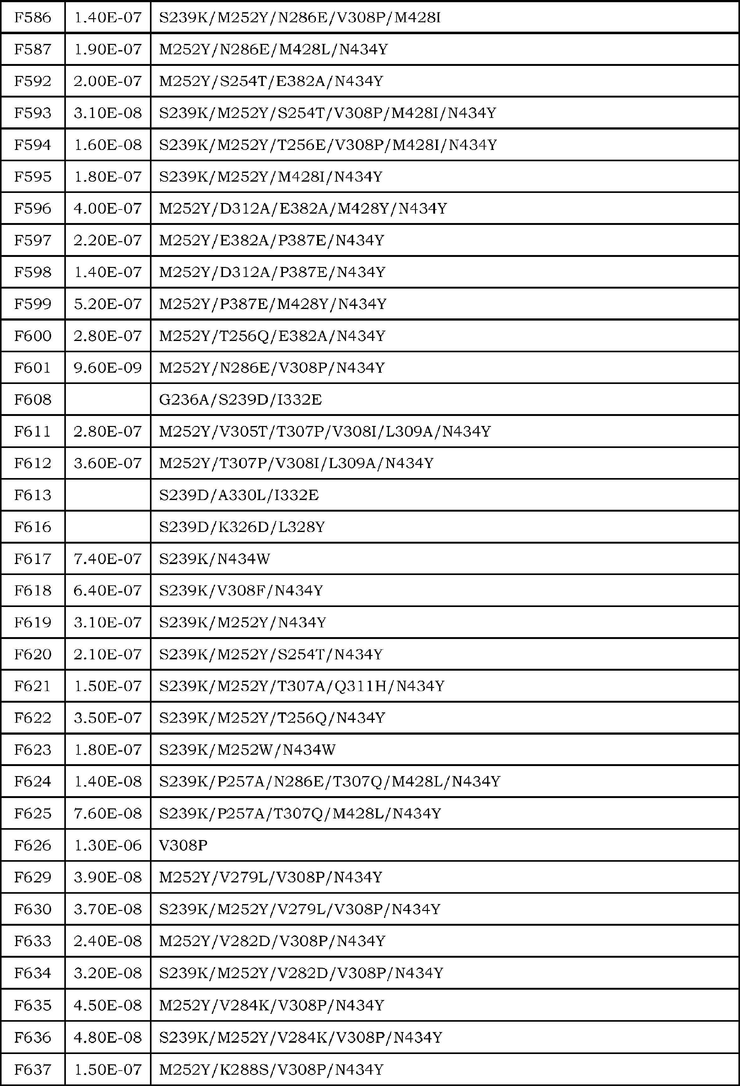


Fcγレセプター(FcγRとも記載される)とは、IgG1、IgG2、IgG3、IgG4モノクローナル抗体のFc領域に結合し得るレセプターをいい、実質的にFcγレセプター遺伝子にコードされるタンパク質のファミリーのいかなるメンバーをも意味する。ヒトでは、このファミリーには、アイソフォームFcγRIa、FcγRIbおよびFcγRIcを含むFcγRI(CD64);アイソフォームFcγRIIa(アロタイプH131およびR131を含む。即ち、FcγRIIa (H)およびFcγRIIa (R))、FcγRIIb(FcγRIIb-1およびFcγRIIb-2を含む)およびFcγRIIcを含むFcγRII(CD32);およびアイソフォームFcγRIIIa(アロタイプV158およびF158を含む。即ち、FcγRIIIa (V)およびFcγRIIIa (F))およびFcγRIIIb(アロタイプFcγRIIIb-NA1およびFcγRIIIb-NA2を含む)を含むFcγRIII(CD16)、並びにいかなる未発見のヒトFcγR類またはFcγRアイソフォームまたはアロタイプも含まれるが、これらに限定されるものではない。FcγRは、ヒト、マウス、ラット、ウサギおよびサルを含むが、これらに限定されるものではない、いかなる生物由来でもよい。マウスFcγR類には、FcγRI(CD64)、FcγRII(CD32)、FcγRIII(CD16)およびFcγRIII-2(FcγRIV、CD16-2)、並びにいかなる未発見のマウスFcγR類またはFcγRアイソフォームまたはアロタイプも含まれるが、これらに限定されない。こうしたFcγレセプターの好適な例としてはヒトFcγRI(CD64)、FcγRIIa(CD32)、FcγRIIb(CD32)、FcγRIIIa(CD16)及び/又はFcγRIIIb(CD16)が挙げられる。ヒトFcγRIのポリヌクレオチド配列及びアミノ酸配列はそれぞれ配列番号:15(NM_000566.3)及び16(NP_000557.1)に、ヒトFcγRIIa(アロタイプH131)のポリヌクレオチド配列及びアミノ酸配列はそれぞれ配列番号:17(BC020823.1)及び18(AAH20823.1)に(アロタイプR131は配列番号:18の166番目のアミノ酸がArgに置換されている配列である)、FcγRIIbのポリヌクレオチド配列及びアミノ酸配列はそれぞれ配列番号:19(BC146678.1)及び20(AAI46679.1)に、FcγRIIIaのポリヌクレオチド配列及びアミノ酸配列はそれぞれ配列番号:21(BC033678.1)及び22(AAH33678.1)に、及びFcγRIIIbのポリヌクレオチド配列及びアミノ酸配列は、それぞれ配列番号:23(BC128562.1)及び24(AAI28563.1)に記載されている(カッコ内はRefSeq登録番号を示す)。例えばアロタイプV158が用いられる場合にFcγRIIIaVと表記されているように、特記されないかぎり、アロタイプF158が用いられているが、本願で記載されるアイソフォームFcγRIIIaのアロタイプが特に限定して解釈されるものではない。 Fcγ receptor Fcγ receptor (also referred to as FcγR) refers to a receptor that can bind to the Fc region of IgG1, IgG2, IgG3, and IgG4 monoclonal antibodies, and virtually any member of the family of proteins encoded by the Fcγ receptor gene Also means. In humans, this family includes FcγRI (CD64), including isoforms FcγRIa, FcγRIb and FcγRIc; isoforms FcγRIIa (including allotypes H131 and R131, ie FcγRIIa (H) and FcγRIIa (R)), FcγRIIb (FcγRIIb− 1 and FcγRIIb-2) and FcγRIIc containing FcγRII (CD32); and isoforms FcγRIIIa (including allotypes V158 and F158, ie FcγRIIIa (V) and FcγRIIIa (F)) and FcγRIIIb (allotypes FcγRIIIb-NA1 and FcγR Including, but not limited to, FcγRIII (CD16), including -NA2, as well as any undiscovered human FcγRs or FcγR isoforms or allotypes. FcγR may be derived from any organism, including but not limited to humans, mice, rats, rabbits and monkeys. Mouse FcγRs include FcγRI (CD64), FcγRII (CD32), FcγRIII (CD16) and FcγRIII-2 (FcγRIV, CD16-2), as well as any undiscovered mouse FcγRs or FcγR isoforms or allotypes However, it is not limited to these. Suitable examples of such Fcγ receptors include human FcγRI (CD64), FcγRIIa (CD32), FcγRIIb (CD32), FcγRIIIa (CD16) and / or FcγRIIIb (CD16). The polynucleotide sequence and amino acid sequence of human FcγRI are SEQ ID NO: 15 (NM_000566.3) and 16 (NP_000557.1), respectively. The polynucleotide sequence and amino acid sequence of human FcγRIIa (allotype H131) are respectively SEQ ID NO: 17 (BC020823). .1) and 18 (AAH20823.1) (allotype R131 is a sequence in which the 166th amino acid of SEQ ID NO: 18 is substituted with Arg), the polynucleotide sequence and amino acid sequence of FcγRIIb are SEQ ID NO: 19 respectively. (BC146678.1) and 20 (AAI46679.1), the polynucleotide sequence and amino acid sequence of FcγRIIIa are SEQ ID NO: 21 (BC033678.1) and 22 (AAH33678.1), respectively, and the polynucleotide sequence and amino acid sequence of FcγRIIIb. The sequences are described in SEQ ID NOs: 23 (BC128562.1) and 24 (AAI28563.1), respectively (the RefSeq registration numbers are shown in parentheses). For example, when allotype V158 is used, allotype F158 is used unless otherwise specified, as indicated as FcγRIIIaV, but the allotype of isoform FcγRIIIa described in this application is not particularly limited. Absent.
前述されるように、本発明の抗原結合分子に含まれるFc領域として、Fcγレセプターに対する結合活性を有するFc領域が挙げられる。そのようなFc領域の非限定な一態様として、ヒトIgG1(配列番号:9)、IgG2(配列番号:10)、IgG3(配列番号:11)、またはIgG4(配列番号:12)で表されるFc領域が例示される。Fcγレセプターが、IgG1、IgG2、IgG3、IgG4モノクローナル抗体のFc領域に結合活性を有するか否かは、上記に記載されるFACSやELISAフォーマットのほか、ALPHAスクリーン(Amplified Luminescent Proximity Homogeneous Assay)や表面プラズモン共鳴(SPR)現象を利用したBIACORE法等によって確認され得る(Proc.Natl.Acad.Sci.USA (2006) 103 (11), 4005-4010)。 As avidity aforementioned Fc region for Fc [gamma] R, as the Fc region contained in the antigen-binding molecules of the present invention include Fc region with binding activity for Fcγ receptor. One non-limiting embodiment of such an Fc region is represented by human IgG1 (SEQ ID NO: 9), IgG2 (SEQ ID NO: 10), IgG3 (SEQ ID NO: 11), or IgG4 (SEQ ID NO: 12). The Fc region is exemplified. Whether the Fcγ receptor has binding activity to the Fc region of IgG1, IgG2, IgG3, or IgG4 monoclonal antibodies, in addition to the FACS and ELISA formats described above, ALPHA screen (Amplified Luminescent Proximity Homogeneous Assay) and surface plasmon It can be confirmed by the BIACORE method using the resonance (SPR) phenomenon (Proc. Natl. Acad. Sci. USA (2006) 103 (11), 4005-4010).
本発明に使用するために、特に好ましい改変としては、例えば、Fc領域のEUナンバリングで表される;
221位のアミノ酸がLysまたはTyrのいずれか、
222位のアミノ酸がPhe、Trp、GluまたはTyrのいずれか、
223位のアミノ酸がPhe、Trp、GluまたはLysのいずれか、
224位のアミノ酸がPhe、Trp、GluまたはTyrのいずれか、
225位のアミノ酸がGlu、LysまたはTrpのいずれか、
227位のアミノ酸がGlu、Gly、LysまたはTyrのいずれか、
228位のアミノ酸がGlu、Gly、LysまたはTyrのいずれか、
230位のアミノ酸がAla、Glu、GlyまたはTyrのいずれか、
231位のアミノ酸がGlu、Gly、Lys、ProまたはTyrのいずれか、
232位のアミノ酸がGlu、Gly、LysまたはTyrのいずれか、
233位のアミノ酸がAla、Asp、Phe、Gly、His、Ile、Lys、Leu、Met、Asn、Gln、Arg、Ser、Thr、Val、TrpまたはTyrのいずれか、
234位のアミノ酸がAla、Asp、Glu、Phe、Gly、His、Ile、Lys、Met、Asn、Pro、Gln、Arg、Ser、Thr、Val、TrpまたはTyrのいずれか、
235位のアミノ酸がAla、Asp、Glu、Phe、Gly、His、Ile、Lys、Met、Asn、Pro、Gln、Arg、Ser、Thr、Val、TrpまたはTyrのいずれか、
236位のアミノ酸がAla、Asp、Glu、Phe、His、Ile、Lys、Leu、Met、Asn、Pro、Gln、Arg、Ser、Thr、Val、TrpまたはTyrのいずれか、
237位のアミノ酸がAsp、Glu、Phe、His、Ile、Lys、Leu、Met、Asn、Pro、Gln、Arg、Ser、Thr、Val、TrpまたはTyrのいずれか、
238位のアミノ酸がAsp、Glu、Phe、Gly、His、Ile、Lys、Leu、Met、Asn、Gln、Arg、Ser、Thr、Val、TrpまたはTyrのいずれか、
239位のアミノ酸がAsp、Glu、Phe、Gly、His、Ile、Lys、Leu、Met、Asn、Pro、Gln、Arg、Thr、Val、TrpまたはTyrのいずれか、
240位のアミノ酸がAla、Ile、MetまたはThrのいずれか、
241位のアミノ酸がAsp、Glu、Leu、Arg、TrpまたはTyrのいずれか、
243位のアミノ酸がLeu、Glu、Leu、Gln、Arg、TrpまたはTyrのいずれか、
244位のアミノ酸がHis、
245位のアミノ酸がAla、
246位のアミノ酸がAsp、Glu、HisまたはTyrのいずれか、
247位のアミノ酸がAla、Phe、Gly、His、Ile、Leu、Met、Thr、ValまたはTyrのいずれか、
249位のアミノ酸がGlu、His、GlnまたはTyrのいずれか、
250位のアミノ酸がGluまたはGlnのいずれか、
251位のアミノ酸がPhe、
254位のアミノ酸がPhe、MetまたはTyrのいずれか、
255位のアミノ酸がGlu、LeuまたはTyrのいずれか、
256位のアミノ酸がAla、MetまたはProのいずれか、
258位のアミノ酸がAsp、Glu、His、SerまたはTyrのいずれか、
260位のアミノ酸がAsp、Glu、HisまたはTyrのいずれか、
262位のアミノ酸がAla、Glu、Phe、IleまたはThrのいずれか、
263位のアミノ酸がAla、Ile、MetまたはThrのいずれか、
264位のアミノ酸がAsp、Glu、Phe、Gly、His、Ile、Lys、Leu、Met、Asn、Pro、Gln、Arg、Ser、Thr、TrpまたはTyrのいずれか、
265位のアミノ酸がAla、Leu、Phe、Gly、His、Ile、Lys、Leu、Met、Asn、Pro、Gln、Arg、Ser、Thr、Val、TrpまたはTyrのいずれか、
266位のアミノ酸がAla、Ile、MetまたはThrのいずれか、
267位のアミノ酸がAsp、Glu、Phe、His、Ile、Lys、Leu、Met、Asn、Pro、Gln、Arg、Thr、Val、TrpまたはTyrのいずれか、
268位のアミノ酸がAsp、Glu、Phe、Gly、Ile、Lys、Leu、Met、Pro、Gln、Arg、Thr、ValまたはTrpのいずれか、
269位のアミノ酸がPhe、Gly、His、Ile、Lys、Leu、Met、Asn、Pro、Arg、Ser、Thr、Val、TrpまたはTyrのいずれか、
270位のアミノ酸がGlu、Phe、Gly、His、Ile、Leu、Met、Pro、Gln、Arg、Ser、Thr、TrpまたはTyrのいずれか、
271位のアミノ酸がAla、Asp、Glu、Phe、Gly、His、Ile、Lys、Leu、Met、Asn、Gln、Arg、Ser、Thr、Val、TrpまたはTyrのいずれか、
272位のアミノ酸がAsp、Phe、Gly、His、Ile、Lys、Leu、Met、Pro、Arg、Ser、Thr、Val、TrpまたはTyrのいずれか、
273位のアミノ酸がPheまたはIleのいずれか、
274位のアミノ酸がAsp、Glu、Phe、Gly、His、Ile、Leu、Met、Asn、Pro、Arg、Ser、Thr、Val、TrpまたはTyrのいずれか、
275位のアミノ酸がLeuまたはTrpのいずれか、
276位のアミノ酸が、Asp、Glu、Phe、Gly、His、Ile、Leu、Met、Pro、Arg、Ser、Thr、Val、TrpまたはTyrのいずれか、
278位のアミノ酸がAsp、Glu、Gly、His、Ile、Lys、Leu、Met、Asn、Pro、Gln、Arg、Ser、Thr、ValまたはTrpのいずれか、
279位のアミノ酸がAla、
280位のアミノ酸がAla、Gly、His、Lys、Leu、Pro、Gln、TrpまたはTyrのいずれか、
281位のアミノ酸がAsp、Lys、ProまたはTyrのいずれか、
282位のアミノ酸がGlu、Gly、Lys、ProまたはTyrのいずれか、
283位のアミノ酸がAla、Gly、His、Ile、Lys、Leu、Met、Pro、ArgまたはTyrのいずれか、
284位のアミノ酸がAsp、Glu、Leu、Asn、ThrまたはTyrのいずれか、
285位のアミノ酸がAsp、Glu、Lys、Gln、TrpまたはTyrのいずれか、
286位のアミノ酸がGlu、Gly、ProまたはTyrのいずれか、
288位のアミノ酸がAsn、Asp、GluまたはTyrのいずれか、
290位のアミノ酸がAsp、Gly、His、Leu、Asn、Ser、Thr、TrpまたはTyrのいずれか、
291位のアミノ酸がAsp、Glu、Gly、His、Ile、GlnまたはThrのいずれか、
292位のアミノ酸がAla、Asp、Glu、Pro、ThrまたはTyrのいずれか、
293位のアミノ酸がPhe、Gly、His、Ile、Leu、Met、Asn、Pro、Arg、Ser、Thr、Val、TrpまたはTyrのいずれか、
294位のアミノ酸がPhe、Gly、His、Ile、Lys、Leu、Met、Asn、Pro、Arg、Ser、Thr、Val、TrpまたはTyrのいずれか、
295位のアミノ酸がAsp、Glu、Phe、Gly、His、Ile、Lys、Met、Asn、Pro、Arg、Ser、Thr、Val、TrpまたはTyrのいずれか、
296位のアミノ酸がAla、Asp、Glu、Gly、His、Ile、Lys、Leu、Met、Asn、Gln、Arg、Ser、ThrまたはValのいずれか、
297位のアミノ酸がAsp、Glu、Phe、Gly、His、Ile、Lys、Leu、Met、Pro、Gln、Arg、Ser、Thr、Val、TrpまたはTyrのいずれか、
298位のアミノ酸がAla、Asp、Glu、Phe、His、Ile、Lys、Met、Asn、Gln、Arg、Thr、Val、TrpまたはTyrのいずれか、
299位のアミノ酸がAla、Asp、Glu、Phe、Gly、His、Ile、Lys、Leu、Met、Asn、Pro、Gln、Arg、Ser、Val、TrpまたはTyrのいずれか、
300位のアミノ酸がAla、Asp、Glu、Gly、His、Ile、Lys、Leu、Met、Asn、Pro、Gln、Arg、Ser、Thr、ValまたはTrpのいずれか、
301位のアミノ酸がAsp、Glu、HisまたはTyrのいずれか、
302位のアミノ酸がIle、
303位のアミノ酸がAsp、GlyまたはTyrのいずれか、
304位のアミノ酸がAsp、His、Leu、AsnまたはThrのいずれか、
305位のアミノ酸がGlu、Ile、ThrまたはTyrのいずれか、
311位のアミノ酸がAla、Asp、Asn、Thr、ValまたはTyrのいずれか、
313位のアミノ酸がPhe、
315位のアミノ酸がLeu、
317位のアミノ酸がGluまたはGln、
318位のアミノ酸がHis、Leu、Asn、Pro、Gln、Arg、Thr、ValまたはTyrのいずれか、
320位のアミノ酸がAsp、Phe、Gly、His、Ile、Leu、Asn、Pro、Ser、Thr、Val、TrpまたはTyrのいずれか、
322位のアミノ酸がAla、Asp、Phe、Gly、His、Ile、Pro、Ser、Thr、Val、TrpまたはTyrのいずれか、
323位のアミノ酸がIle、
324位のアミノ酸がAsp、Phe、Gly、His、Ile、Leu、Met、Pro、Arg、Thr、Val、TrpまたはTyrのいずれか、
325位のアミノ酸がAla、Asp、Glu、Phe、Gly、His、Ile、Lys、Leu、Met、Pro、Gln、Arg、Ser、Thr、Val、TrpまたはTyrのいずれか、
326位のアミノ酸がAla、Asp、Glu、Gly、Ile、Leu、Met、Asn、Pro、Gln、Ser、Thr、Val、TrpまたはTyrのいずれか、
327位のアミノ酸がAla、Asp、Glu、Phe、Gly、His、Ile、Lys、Leu、Met、Asn、Pro、Arg、Thr、Val、TrpまたはTyrのいずれか、
328位のアミノ酸がAla、Asp、Glu、Phe、Gly、His、Ile、Lys、Met、Asn、Pro、Gln、Arg、Ser、Thr、Val、TrpまたはTyrのいずれか、
329位のアミノ酸がAsp、Glu、Phe、Gly、His、Ile、Lys、Leu、Met、Asn、Gln、Arg、Ser、Thr、Val、TrpまたはTyrのいずれか、
330位のアミノ酸がCys、Glu、Phe、Gly、His、Ile、Lys、Leu、Met、Asn、Pro、Arg、Ser、Thr、Val、TrpまたはTyrのいずれか、
331位のアミノ酸がAsp、Phe、His、Ile、Leu、Met、Gln、Arg、Thr、Val、TrpまたはTyrのいずれか、
332位のアミノ酸がAla、Asp、Glu、Phe、Gly、His、Lys、Leu、Met、Asn、Pro、Gln、Arg、Ser、Thr、Val、TrpまたはTyrのいずれか、
333位のアミノ酸がAla、Asp、Glu、Phe、Gly、His、Ile、Leu、Met、Pro、Ser、Thr、ValまたはTyrのいずれか、
334位のアミノ酸がAla、Glu、Phe、Ile、Leu、ProまたはThrのいずれか、
335位のアミノ酸がAsp、Phe、Gly、His、Ile、Leu、Met、Asn、Pro、Arg、Ser、Val、TrpまたはTyrのいずれか、
336位のアミノ酸がGlu、LysまたはTyrのいずれか、
337位のアミノ酸がGlu、HisまたはAsnのいずれか、
339位のアミノ酸がAsp、Phe、Gly、Ile、Lys、Met、Asn、Gln、Arg、SerまたはThrのいずれか、
376位のアミノ酸がAlaまたはValのいずれか、
377位のアミノ酸がGlyまたはLysのいずれか、
378位のアミノ酸がAsp、
379位のアミノ酸がAsn、
380位のアミノ酸がAla、AsnまたはSerのいずれか、
382位のアミノ酸がAlaまたはIleのいずれか、
385位のアミノ酸がGlu、
392位のアミノ酸がThr、
396位のアミノ酸がLeu、
421位のアミノ酸がLys、
427位のアミノ酸がAsn、
428位のアミノ酸がPheまたはLeuのいずれか、
429位のアミノ酸がMet、
434位のアミノ酸がTrp、
436位のアミノ酸がIle、もしくは
440位のアミノ酸がGly、His、Ile、LeuまたはTyrのいずれか、
の群から選択される少なくとも一つ以上のアミノ酸の改変が挙げられる。また、改変されるアミノ酸の数は特に限定されず、一箇所のみのアミノ酸が改変され得るし、二箇所以上のアミノ酸が改変され得る。二箇所以上のアミノ酸の改変の組合せとしては、例えば表6(表6-1~表6-3)に記載されるような組合せが挙げられる。 Examples of amino acids that can be modified include, for example, EU positions, 221nd, 222th, 223rd, 224th, 225th, 227th, 228th, 230th, 231st, 232nd, 233th , 234, 235, 236, 237, 238, 239, 240, 241, 243, 244, 245, 246, 247, 249, 250, 251, 254 , 255, 256, 258, 260, 262, 263, 264, 265, 266, 267, 268, 269, 270, 271, 272, 273, 274, 275, 276, 278, 279, 280, 281, 282, 283, 284, 285, 286, 288, 290, 291, 292, 293 , 294, 295, 296, 297, 298, 299, 299, 300, 301, 302, 303, 304, 305, 311, 313, 315, 315, 317, 318 , 320, 322, 323, 324, 325, 326, 327, 328, 329, 329, 330, 331, 332, 333, 334, 335, 336, 337, 339, 376, 377, 378, 379 380 position, 382 position, 385 position, 392 position, 396 position, 421 position, 427 position, 428 position, 429 position, 434 position, 436 position, and 440 position. It is done. By modifying these amino acids, it is possible to obtain an Fc region (FcγR-binding modified Fc region) having a higher binding activity to the Fcγ receptor than the binding activity of the Fc region of natural human IgG to the Fcγ receptor.
Particularly preferred modifications for use in the present invention are represented, for example, by EU numbering of the Fc region;
221 of the amino acid is either Lys or Tyr,
222 of the amino acid is Phe, Trp, Glu or Tyr,
223 of the amino acid is Phe, Trp, either Glu or Lys,
224 of the amino acid is Phe, Trp, Glu or Tyr,
225 of the amino acid is Glu, one of the Lys or Trp,
227 of the amino acid is Glu, Gly, one of Lys or Tyr,
228 of the amino acid is Glu, Gly, one of Lys or Tyr,
230 of the amino acid is one of Ala, Glu, Gly or Tyr,
231 of the amino acid is Glu, Gly, Lys, Pro or Tyr,
232 of the amino acid is Glu, Gly, one of Lys or Tyr,
233 of the amino acid is Ala, Asp, Phe, Gly, His, Ile, Lys, Leu, Met, Asn, Gln, Arg, Ser, Thr, Val, any of Trp or Tyr,
234 of the amino acid is Ala, Asp, Glu, Phe, Gly, His, Ile, Lys, Met, Asn, Pro, Gln, Arg, Ser, Thr, Val, any of Trp or Tyr,
235 of the amino acid is Ala, Asp, Glu, Phe, Gly, His, Ile, Lys, Met, Asn, Pro, Gln, Arg, Ser, Thr, Val, any of Trp or Tyr,
236 of the amino acid is Ala, Asp, Glu, Phe, His, Ile, Lys, Leu, Met, Asn, Pro, Gln, Arg, Ser, Thr, Val, any of Trp or Tyr,
237 of the amino acid is Asp, Glu, Phe, His, Ile, Lys, Leu, Met, Asn, Pro, Gln, Arg, Ser, Thr, Val, any of Trp or Tyr,
238 of the amino acid is Asp, Glu, Phe, Gly, His, Ile, Lys, Leu, Met, Asn, Gln, Arg, Ser, Thr, Val, one of Trp or Tyr,
239th amino acid is Asp, Glu, Phe, Gly, His, Ile, Lys, Leu, Met, Asn, Pro, Gln, Arg, Thr, Val, one of Trp or Tyr,
The 240th amino acid is Ala, Ile, Met or Thr,
241 of the amino acid is Asp, Glu, Leu, Arg, one of Trp or Tyr,
243 of the amino acid is Leu, Glu, Leu, Gln, Arg, one of Trp or Tyr,
The amino acid at position 244 is His,
Ala for the amino acid at position 245
246 of the amino acid is Asp, Glu, His or Tyr,
247 of the amino acid is Ala, Phe, Gly, His, Ile, Leu, Met, Thr, one of Val or Tyr,
249 of the amino acid is Glu, His, Gln or Tyr,
The amino acid at position 250 is either Glu or Gln,
Phe is the amino acid at position 251;
254 of the amino acid is one of Phe, Met or Tyr,
The amino acid at position 255 is either Glu, Leu or Tyr,
256 of the amino acid is one of Ala, Met or Pro,
258 of the amino acid is Asp, Glu, His, one of Ser or Tyr,
260 of the amino acid is Asp, Glu, His or Tyr,
262 of the amino acid is Ala, Glu, Phe, Ile or Thr,
263 of the amino acid is Ala, Ile, Met or Thr,
264 of the amino acid is Asp, Glu, Phe, Gly, His, Ile, Lys, Leu, Met, Asn, Pro, Gln, Arg, Ser, Thr, one of Trp or Tyr,
265 of the amino acid is Ala, Leu, Phe, Gly, His, Ile, Lys, Leu, Met, Asn, Pro, Gln, Arg, Ser, Thr, Val, any of Trp or Tyr,
266 of the amino acid is one of Ala, Ile, Met or Thr,
267 of the amino acid is Asp, Glu, Phe, His, Ile, Lys, Leu, Met, Asn, Pro, Gln, Arg, Thr, Val, one of Trp or Tyr,
268 of the amino acid is Asp, Glu, Phe, Gly, Ile, Lys, Leu, Met, Pro, Gln, Arg, Thr, one of Val or Trp,
269 of the amino acid is Phe, Gly, His, Ile, Lys, Leu, Met, Asn, Pro, Arg, Ser, Thr, Val, one of Trp or Tyr,
270 amino acid is Glu, Phe, Gly, His, Ile, Leu, Met, Pro, Gln, Arg, Ser, Thr, one of Trp or Tyr,
271 of the amino acid is Ala, Asp, Glu, Phe, Gly, His, Ile, Lys, Leu, Met, Asn, Gln, Arg, Ser, Thr, Val, any of Trp or Tyr,
272 of the amino acid is Asp, Phe, Gly, His, Ile, Lys, Leu, Met, Pro, Arg, Ser, Thr, Val, one of Trp or Tyr,
273 of the amino acid is either Phe or Ile,
274 of the amino acid is Asp, Glu, Phe, Gly, His, Ile, Leu, Met, Asn, Pro, Arg, Ser, Thr, Val, one of Trp or Tyr,
275 of the amino acid is either Leu or Trp,
276 of the amino acid is Asp, Glu, Phe, Gly, His, Ile, Leu, Met, Pro, Arg, Ser, Thr, Val, one of Trp or Tyr,
278 of the amino acid is Asp, Glu, Gly, His, Ile, Lys, Leu, Met, Asn, Pro, Gln, Arg, Ser, Thr, one of Val or Trp,
Ala for the amino acid at position 279
280 of the amino acid is Ala, Gly, His, Lys, Leu, Pro, Gln, one of Trp or Tyr,
281 of the amino acid is Asp, Lys, Pro or Tyr,
282 of the amino acid is Glu, Gly, Lys, Pro or Tyr,
283 of the amino acid is Ala, Gly, His, Ile, Lys, Leu, Met, Pro, Arg or Tyr,
284 of the amino acid is Asp, Glu, Leu, Asn, one of Thr or Tyr,
285 of the amino acid is Asp, Glu, Lys, Gln, one of Trp or Tyr,
286 of the amino acid is Glu, Gly, Pro or Tyr,
288 of the amino acid is Asn, Asp, one of Glu or Tyr,
290 of the amino acid is Asp, Gly, His, Leu, Asn, Ser, Thr, one of Trp or Tyr,
291 of the amino acid is Asp, Glu, Gly, His, Ile, one of Gln or Thr,
292 of the amino acid is Ala, Asp, Glu, Pro, one of Thr or Tyr,
293 of the amino acid is Phe, Gly, His, Ile, Leu, Met, Asn, Pro, Arg, Ser, Thr, Val, one of Trp or Tyr,
294 of the amino acid is Phe, Gly, His, Ile, Lys, Leu, Met, Asn, Pro, Arg, Ser, Thr, Val, one of Trp or Tyr,
295 of the amino acid is Asp, Glu, Phe, Gly, His, Ile, Lys, Met, Asn, Pro, Arg, Ser, Thr, Val, one of Trp or Tyr,
296 of the amino acid is Ala, Asp, Glu, Gly, His, Ile, Lys, Leu, Met, Asn, Gln, Arg, Ser, one of Thr or Val,
297 of the amino acid is Asp, Glu, Phe, Gly, His, Ile, Lys, Leu, Met, Pro, Gln, Arg, Ser, Thr, Val, any of Trp or Tyr,
298 of the amino acid is Ala, Asp, Glu, Phe, His, Ile, Lys, Met, Asn, Gln, Arg, Thr, Val, one of Trp or Tyr,
299 of the amino acid is Ala, Asp, Glu, Phe, Gly, His, Ile, Lys, Leu, Met, Asn, Pro, Gln, Arg, Ser, Val, one of Trp or Tyr,
300th amino acid is Ala, Asp, Glu, Gly, His, Ile, Lys, Leu, Met, Asn, Pro, Gln, Arg, Ser, Thr, Val or any of Trp,
301 of the amino acid is Asp, Glu, His or Tyr,
Ile, the amino acid at position 302
The amino acid at position 303 is either Asp, Gly or Tyr,
304 of the amino acid is Asp, His, Leu, Asn or Thr,
305 of the amino acid is Glu, Ile, Thr or Tyr,
311 position amino acid is Ala, Asp, Asn, Thr, one of Val or Tyr,
Phe is the amino acid at position 313.
Leu, the amino acid at position 315
317 of the amino acid is Glu or Gln,
318 of the amino acid is His, Leu, Asn, Pro, Gln, Arg, Thr, one of Val or Tyr,
The amino acid at position 320 is Asp, Phe, Gly, His, Ile, Leu, Asn, Pro, Ser, Thr, Val, one of Trp or Tyr,
322 of the amino acid is Ala, Asp, Phe, Gly, His, Ile, Pro, Ser, Thr, Val, one of Trp or Tyr,
The amino acid at position 323 is Ile,
324 of the amino acid is Asp, Phe, Gly, His, Ile, Leu, Met, Pro, Arg, Thr, Val, one of Trp or Tyr,
325 of the amino acid is Ala, Asp, Glu, Phe, Gly, His, Ile, Lys, Leu, Met, Pro, Gln, Arg, Ser, Thr, Val, Trp or Tyr,
326 of the amino acid is Ala, Asp, Glu, Gly, Ile, Leu, Met, Asn, Pro, Gln, Ser, Thr, Val, one of Trp or Tyr,
327 of the amino acid is Ala, Asp, Glu, Phe, Gly, His, Ile, Lys, Leu, Met, Asn, Pro, Arg, Thr, Val, any of Trp or Tyr,
328 of the amino acid is Ala, Asp, Glu, Phe, Gly, His, Ile, Lys, Met, Asn, Pro, Gln, Arg, Ser, Thr, Val, any of Trp or Tyr,
329 of the amino acid is Asp, Glu, Phe, Gly, His, Ile, Lys, Leu, Met, Asn, Gln, Arg, Ser, Thr, Val, one of Trp or Tyr,
The amino acid at position 330 is Cys, Glu, Phe, Gly, His, Ile, Lys, Leu, Met, Asn, Pro, Arg, Ser, Thr, Val, one of Trp or Tyr,
331 of the amino acid is Asp, Phe, His, Ile, Leu, Met, Gln, Arg, Thr, Val, one of Trp or Tyr,
332 of the amino acid is Ala, Asp, Glu, Phe, Gly, His, Lys, Leu, Met, Asn, Pro, Gln, Arg, Ser, Thr, Val, any of Trp or Tyr,
333 of the amino acid is Ala, Asp, Glu, Phe, Gly, His, Ile, Leu, Met, Pro, Ser, Thr, one of Val or Tyr,
334 of the amino acid is Ala, Glu, Phe, Ile, Leu, Pro or Thr,
335 of the amino acid is Asp, Phe, Gly, His, Ile, Leu, Met, Asn, Pro, Arg, Ser, Val, one of Trp or Tyr,
336 of the amino acid is either Glu, Lys or Tyr,
337 of the amino acid is Glu, His or Asn,
339 of the amino acid is Asp, Phe, Gly, Ile, Lys, Met, Asn, Gln, Arg, one of Ser or Thr,
376 of the amino acid is either Ala or Val,
377 of the amino acid is either Gly or Lys,
Asp for the amino acid at position 378
Asn for the amino acid at position 379;
380 of the amino acid is Ala, Asn or Ser,
382 of the amino acid is either Ala or Ile,
385th amino acid is Glu,
392 of the amino acid is Thr,
Leu for the amino acid at position 396
Lys for the amino acid at position 421;
Asn for the amino acid at position 427;
428 of the amino acid is either Phe or Leu,
Met is the amino acid at position 429.
Trp for the amino acid of position 434;
436th amino acid is Ile, or
440 of the amino acid is Gly, His, Ile, Leu or Tyr,
Modification of at least one amino acid selected from the group of Further, the number of amino acids to be modified is not particularly limited, and only one amino acid can be modified, or two or more amino acids can be modified. Examples of combinations of amino acid modifications at two or more sites include combinations described in Table 6 (Tables 6-1 to 6-3).


本発明において好適に用いられる、Fc領域の例として、特定のFcγレセプターに対する結合活性がそのほかのFcγレセプターに対する結合活性よりも高い性質を有するFc領域(選択的なFcγレセプターに対する結合活性を有するFc領域)もまた好適に挙げられる。抗原結合分子として抗体が用いられる場合には、一分子の抗体は一分子のFcγレセプターとしか結合できないため、一分子の抗原結合分子は抑制型Fcγレセプターに結合した状態で他の活性型FcγRに結合することはできないし、活性型Fcγレセプターに結合した状態で他の活性型Fcγレセプターや抑制型Fcγレセプターに結合することはできない。 Fc region having selective Fcγ receptor binding activity As an example of an Fc region preferably used in the present invention, an Fc region having a property that binding activity to a specific Fcγ receptor is higher than binding activity to other Fcγ receptors ( Preferable examples also include an Fc region having a selective Fcγ receptor-binding activity. When an antibody is used as an antigen-binding molecule, one molecule of antibody can only bind to one molecule of Fcγ receptor, so one molecule of antigen-binding molecule binds to the inhibitory Fcγ receptor to another active FcγR. It cannot bind, and cannot bind to other active Fcγ receptors or inhibitory Fcγ receptors in the state of being bound to active Fcγ receptors.
本発明において、抗原結合分子は抗原結合ドメインおよびFc領域を含む分子を表す最も広義な意味として使用されており、具体的には、それらが抗原に対する結合活性を示す限り、様々な分子型が含まれる。例えば、抗原結合ドメインがFc領域と結合した分子の例として、抗体が挙げられる。抗体には、単一のモノクローナル抗体(アゴニストおよびアンタゴニスト抗体を含む)、ヒト抗体、ヒト化抗体、キメラ抗体等が含まれ得る。また抗体の断片として使用される場合としては、抗原結合ドメインおよび抗原結合断片(例えば、Fab、F(ab')2、scFvおよびFv)が好適に挙げられ得る。既存の安定なα/βバレルタンパク質構造等の立体構造が scaffold(土台)として用いられ、その一部分の構造のみが抗原結合ドメインの構築のためにライブラリ化されたスキャフォールド分子も、本発明の抗原結合分子に含まれ得る 。 Antigen-binding molecule In the present invention, an antigen-binding molecule is used in the broadest sense of a molecule containing an antigen-binding domain and an Fc region. The type is included. For example, an antibody is an example of a molecule in which an antigen-binding domain is bound to an Fc region. Antibodies can include single monoclonal antibodies (including agonist and antagonist antibodies), human antibodies, humanized antibodies, chimeric antibodies, and the like. Further, when used as an antibody fragment, an antigen-binding domain and an antigen-binding fragment (for example, Fab, F (ab ′) 2, scFv and Fv) can be preferably mentioned. A scaffold molecule in which a three-dimensional structure such as an existing stable α / β barrel protein structure is used as a scaffold and only a part of the structure is made into a library for the construction of an antigen-binding domain is also an antigen of the present invention. It can be included in the binding molecule.
Ser
Gly・Ser
Gly・Gly・Ser
Ser・Gly・Gly
Gly・Gly・Gly・Ser(配列番号:25)
Ser・Gly・Gly・Gly(配列番号:26)
Gly・Gly・Gly・Gly・Ser(配列番号:27)
Ser・Gly・Gly・Gly・Gly(配列番号:28)
Gly・Gly・Gly・Gly・Gly・Ser(配列番号:29)
Ser・Gly・Gly・Gly・Gly・Gly(配列番号:30)
Gly・Gly・Gly・Gly・Gly・Gly・Ser(配列番号:31)
Ser・Gly・Gly・Gly・Gly・Gly・Gly(配列番号:32)
(Gly・Gly・Gly・Gly・Ser(配列番号:27))n
(Ser・Gly・Gly・Gly・Gly(配列番号:28))n
[nは1以上の整数である]等が好適に挙げられる。但し、ペプチドリンカーの長さや配列は目的に応じて当業者が適宜選択することができる。 For example, for a peptide linker:
Ser
Gly ・ Ser
Gly ・ Gly ・ Ser
Ser ・ Gly ・ Gly
Gly, Gly, Gly, Ser (SEQ ID NO: 25)
Ser, Gly, Gly, Gly (SEQ ID NO: 26)
Gly, Gly, Gly, Gly, Ser (SEQ ID NO: 27)
Ser, Gly, Gly, Gly, Gly (SEQ ID NO: 28)
Gly, Gly, Gly, Gly, Gly, Ser (SEQ ID NO: 29)
Ser, Gly, Gly, Gly, Gly, Gly (SEQ ID NO: 30)
Gly, Gly, Gly, Gly, Gly, Gly, Ser (SEQ ID NO: 31)
Ser, Gly, Gly, Gly, Gly, Gly, Gly (SEQ ID NO: 32)
(Gly, Gly, Gly, Gly, Ser (SEQ ID NO: 27)) n
(Ser, Gly, Gly, Gly, Gly (SEQ ID NO: 28)) n
[N is an integer of 1 or more] and the like. However, the length and sequence of the peptide linker can be appropriately selected by those skilled in the art according to the purpose.
また、上記記載で例示されるリンカーのほか、例えばHisタグ、HAタグ、mycタグ、FLAGタグ等のペプチドタグを有するリンカーも適宜使用され得る。また、水素結合、ジスルフィド結合、共有結合、イオン性相互作用またはこれらの結合の組合せにより互いに結合する性質もまた好適に利用され得る。例えば、抗体のCH1とCL間の親和性が利用されたり、ヘテロFc領域の会合に際して前述の二重特異性抗体を起源とするFc領域が用いられたりする。さらに、ドメイン間に形成されるジスルフィド結合もまた好適に利用され得る。 When a plurality of linkers for linking each domain are used, the same type of linker can be used, or a heterogeneous linker can also be used.
In addition to the linkers exemplified in the above description, for example, a linker having a peptide tag such as a His tag, an HA tag, a myc tag, or a FLAG tag can be used as appropriate. Moreover, the property which couple | bonds together by a hydrogen bond, a disulfide bond, a covalent bond, an ionic interaction, or the combination of these bonds can also be utilized suitably. For example, the affinity between CH1 and CL of an antibody is used, or the Fc region originating from the above-mentioned bispecific antibody is used in association with a hetero Fc region. Furthermore, a disulfide bond formed between domains can also be suitably used.
本発明の抗原結合分子は、会合化していない抗原に対する結合活性と比較したときの、会合化した抗原に対する結合活性がより高いものを選択すれば、その消失効果をより高めることができる。そのような抗原結合分子は、例えば、後述の抗原結合分子の製造方法の記載に従って作製した抗原結合分子の中から、会合化した抗原に対する結合活性と会合していない抗原に対する結合活性の差がより大きな抗原結合分子を選択することによって得ることができる。
当該消失効果を高めるためには、会合化した抗原に特異的なエピトープに結合する抗原結合分子を選択することによっても達成することができる。そのようなエピトープに結合する抗原結合分子は、公知の方法を用いて得ることができる。例えば、会合化した抗原に結合する分子を取得後、その抗原結合分子の会合化した抗原への結合と会合化していない抗原を比較し、会合化した抗原への結合が高い抗原結合分子を選択することで得ることができる(国際公開2006/016644、EMBO J. (1994)13, 1166-75.、国際公開2009/008529等)。
また、当該消失効果を高めるためには、会合化した抗原と抗原結合分子の複合体のFcRnあるいはFcγレセプターに対する結合活性が、会合化していない抗原と抗原結合分子の複合体のFcRnあるいはFcγレセプターに対する結合活性より高いものを選択することによっても達成することができる。そのような抗原結合分子は、例えば、後述の抗原結合分子の製造方法の記載に従って得られた抗原結合分子に前述のFcRn又はFcγレセプターに対する結合が増強する効果をもたらす改変を導入し、当該改変が導入された抗原結合分子と会合化した抗原あるいは会合していない抗原とで複合体を形成させ、当該複合体のFcRnあるいはFcγレセプターに対する結合活性の差がより大きな抗原結合分子を選択することによって得ることができる。
本発明において、会合化した抗原が会合化していない抗原よりも優先して消失しているかどうかは、血漿中における会合化した抗原のクリアランスと会合化していない抗原のクリアランスを比較することによって、確認することができる。具体的には、被験抗原結合分子の非存在下での会合化した抗原のクリアランスに対する、当該抗原結合分子の存在下での会合化した抗原のクリアランス比(抗原結合分子存在下での会合化した抗原のクリアランス/抗原結合分子非存在下での会合化した抗原のクリアランス)が、会合化していない抗原のクリアランス比(抗原結合分子存在下での会合化していない抗原のクリアランス/抗原結合分子非存在下での会合化していない抗原のクリアランス)よりも高ければ、当該抗原結合分子は、会合化した抗原を会合化していない抗原よりも優先して消失していると判断することができる。本発明においては当該会合化した抗原のクリアランス比は、会合化していない抗原のクリアランス比の1.5倍(会合化した抗原のクリアランス比/会合化していない抗原のクリアランス比)以上であることが好ましい。 Antigen-binding molecule in which the associated antigen disappears preferentially over the unassociated antigen in plasma The antigen-binding molecule of the present invention has an associated antigen as compared to the binding activity against the unassociated antigen. If a higher binding activity is selected, the disappearance effect can be further enhanced. Such an antigen-binding molecule has, for example, a difference between the binding activity to an associated antigen and the binding activity to an unassociated antigen among antigen-binding molecules prepared according to the description of the method for producing an antigen-binding molecule described below. It can be obtained by selecting a large antigen binding molecule.
Increasing the elimination effect can also be achieved by selecting an antigen-binding molecule that binds to an epitope specific for the associated antigen. Antigen-binding molecules that bind to such epitopes can be obtained using known methods. For example, after obtaining a molecule that binds to an associated antigen, compare the binding of that antigen-binding molecule to the associated antigen and an unassociated antigen, and select an antigen-binding molecule that has high binding to the associated antigen. (International Publication 2006/016644, EMBO J. (1994) 13, 1166-75., International Publication 2009/008529, etc.).
In order to enhance the disappearance effect, the binding activity of the complex of the associated antigen and antigen-binding molecule to FcRn or Fcγ receptor is less than that of the complex of the unassociated antigen and antigen-binding molecule to FcRn or Fcγ receptor. It can also be achieved by selecting one having a higher binding activity. Such an antigen-binding molecule, for example, introduces a modification that brings about the effect of enhancing the binding to the aforementioned FcRn or Fcγ receptor to the antigen-binding molecule obtained according to the description of the method for producing an antigen-binding molecule described later, It is obtained by forming a complex with the introduced antigen-binding molecule and an associated or non-associated antigen, and selecting an antigen-binding molecule with a greater difference in binding activity of the complex to FcRn or Fcγ receptor. be able to.
In the present invention, whether or not the associated antigen disappears in preference to the unassociated antigen is confirmed by comparing the clearance of the associated antigen and the clearance of the unassociated antigen in plasma. can do. Specifically, the clearance ratio of the associated antigen in the presence of the antigen-binding molecule to the clearance of the associated antigen in the absence of the test antigen-binding molecule (associated in the presence of the antigen-binding molecule) Antigen clearance / clearance of associated antigen in the absence of antigen binding molecule) is the clearance ratio of unassociated antigen (clearance of unassociated antigen in the presence of antigen binding molecule / antigen binding molecule absent) If it is higher than the clearance of the unassociated antigen below, it can be determined that the antigen-binding molecule has disappeared preferentially over the associated antigen. In the present invention, the clearance ratio of the associated antigen is preferably 1.5 times the clearance ratio of the unassociated antigen (clearance ratio of the associated antigen / clearance ratio of the unassociated antigen) or more.
本発明において、「会合化した抗原を血漿中から消失させる」とは、抗原結合分子が生体内に投与された、あるいは、抗原結合分子の生体内への分泌が生じた際に、血漿中に存在する抗原を血漿中から消失させる能力のことをいう。従って、本発明において、「抗原結合分子の血漿中における会合化した抗原の消失が優先される」とは、抗原結合分子を投与した際に、会合化していない抗原と比較して、血漿中における会合化した抗原の消失が促進されていればよい。抗原結合分子の血漿中における会合化した抗原の消失が優先されているか否かは、例えば、上述のクリアランス比を測定することにより判断することが可能である。会合化した抗原に結合可能な抗原結合分子の、pH酸性域における抗原に対する結合活性をpH中性域における抗原に対する結合活性より低下させる(または、低カルシウムイオン濃度における抗原に対する結合活性を高カルシウムイオン濃度における抗原に対する結合活性より低下させる)等によりイオン濃度によって抗原に対する結合活性が変化した抗原結合分子について上述のクリアランス比を測定し、会合化してない抗原に対するクリアランス比との違い(クリアランス比(会合化した抗原)/クリアランス比(会合化していない抗原))が大きくなれば優先性が向上したと判断することができる。 In the present invention, “dissociating the associated antigen from the plasma” means that the antigen-binding molecule is administered into the living body or when the antigen-binding molecule is secreted into the living body. This refers to the ability of an antigen present in plasma to disappear from plasma. Therefore, in the present invention, “the disappearance of the associated antigen in the plasma of the antigen-binding molecule is given priority” means that the administration of the antigen-binding molecule in the plasma compared to the unassociated antigen. It is only necessary that the disappearance of the associated antigen is promoted. Whether or not the disappearance of the associated antigen in the plasma of the antigen-binding molecule is prioritized can be determined, for example, by measuring the clearance ratio described above. Reduces the binding activity of antigen-binding molecules that can bind to the associated antigen to the antigen in the acidic pH range from the binding activity to the antigen in the neutral pH range (or lowers the binding activity to the antigen at low calcium ion concentration. The above clearance ratio is measured for antigen-binding molecules whose binding activity to the antigen has been changed by the ion concentration, such as by reducing the binding activity to the antigen at the concentration (clearance ratio (association) It is possible to determine that the priority has been improved if the ratio of the conjugated antigen) / clearance ratio (unassociated antigen)) increases.
本発明において「薬物動態の向上」、「薬物動態の改善」、および「優れた薬物動態」は、「血漿中(血中)滞留性の向上」、「血漿中(血中)滞留性の改善」、「優れた血漿中(血中)滞留性」、「血漿中(血中)滞留性を長くする」と言い換えることが可能であり、これらの語句は同じ意味で使用される。 Improvement of pharmacokinetics In the present invention, “ improvement of pharmacokinetics”, “improvement of pharmacokinetics”, and “excellent pharmacokinetics” are “improved retention in plasma (blood)”, “in plasma (blood)” It can be rephrased as “improved retention”, “excellent plasma (blood) retention”, “increase plasma (blood) retention”, and these terms are used interchangeably. .
A値=各時点での抗原のモル濃度
B値=各時点での抗原結合分子のモル濃度
C値=各時点での抗原結合分子のモル濃度あたりの抗原のモル濃度(抗原/抗原結合分子モル比)
C=A/B。
C値がより小さい場合は、抗原結合分子あたりの抗原消失効率がより高いことを示し、C値がより大きい場合は、抗原結合分子あたりの抗原消失効率がより低いことを示す。 The antigen / antigen binding molecule molar ratio can be calculated as shown below:
A value = molar concentration of antigen at each time point
B value = molar concentration of antigen-binding molecule at each time point
C value = molar concentration of antigen per molar concentration of antigen-binding molecule at each time point (antigen / antigen-binding molecule molar ratio)
C = A / B.
A smaller C value indicates a higher antigen disappearance efficiency per antigen-binding molecule, and a larger C value indicates a lower antigen disappearance efficiency per antigen-binding molecule.
本発明によって提供される、血漿中から当該会合した抗原を消失させるための方法における抗原結合分子の使用の非限定な一態様として、対象から単離された血漿を本発明の抗原結合分子と接触せしめ形成させた、当該抗原結合分子、および、会合した抗原を含む免疫複合体を、FcRnおよび/またはFcγレセプターを発現する細胞に接触させることを含む、血漿中から当該会合した抗原を消失させるための、いわゆるex vivoの方法における当該抗原結合分子の使用も例示される。 Ex vivo method for eliminating the associated antigen from plasma As a non-limiting embodiment of the use of an antigen binding molecule in the method for eliminating the associated antigen from plasma provided by the present invention, Contacting a cell expressing FcRn and / or Fcγ receptor with the immune complex containing the antigen-binding molecule and the associated antigen formed by contacting plasma isolated from a subject with the antigen-binding molecule of the present invention. Also exemplified is the use of the antigen-binding molecule in a so-called ex vivo method to eliminate the associated antigen from plasma, including
本発明の非限定な一態様では、前記のように選択された条件によって結合活性が変化する抗原結合ドメインをコードするポリヌクレオチドが単離された後に、当該ポリヌクレオチドが適切な発現ベクターに挿入される。例えば、抗原結合ドメインが抗体の可変領域である場合には、当該可変領域をコードするcDNAが得られた後に、当該cDNAの両末端に挿入された制限酵素サイトを認識する制限酵素によって該cDNAが消化される。好ましい制限酵素は、抗原結合分子の遺伝子を構成する塩基配列に出現する頻度が低い塩基配列を認識して消化する。更に1コピーの消化断片をベクターに正しい方向で挿入するためには、付着末端を与える制限酵素の挿入が好ましい。上記のように消化された抗原結合分子の可変領域をコードするcDNAを適当な発現ベクターに挿入することによって、本発明の抗原結合分子の発現ベクターが取得され得る。このとき、抗体定常領域(C領域)をコードする遺伝子と、前記可変領域をコードする遺伝子とがインフレームで融合され得る。 Method for producing antigen-binding molecule comprising antigen-binding domain whose binding activity to Fc region and antigen is ion concentration-dependent In one non-limiting aspect of the present invention, an antigen whose binding activity varies depending on the conditions selected as described above After the polynucleotide encoding the binding domain is isolated, the polynucleotide is inserted into an appropriate expression vector. For example, when the antigen-binding domain is an antibody variable region, the cDNA is encoded by a restriction enzyme that recognizes restriction enzyme sites inserted at both ends of the cDNA after cDNA encoding the variable region is obtained. Digested. A preferred restriction enzyme recognizes and digests a base sequence that appears infrequently in the base sequence constituting the antigen-binding molecule gene. Furthermore, in order to insert one copy of the digested fragment into the vector in the correct direction, it is preferable to insert a restriction enzyme that gives a sticky end. By inserting the cDNA encoding the variable region of the antigen-binding molecule digested as described above into an appropriate expression vector, the expression vector of the antigen-binding molecule of the present invention can be obtained. At this time, the gene encoding the antibody constant region (C region) and the gene encoding the variable region can be fused in frame.
237位のアミノ酸がMet、
248位のアミノ酸がIle、
250位のアミノ酸がAla、Phe、Ile、Met、Gln、Ser、Val、Trp、またはTyrのいずれか、
252位のアミノ酸がPhe、Trp、またはTyrのいずれか、
254位のアミノ酸がThr、
255位のアミノ酸がGlu、
256位のアミノ酸がAsp、Asn、Glu、またはGlnのいずれか、
257位のアミノ酸がAla、Gly、Ile、Leu、Met、Asn、Ser、Thr、またはValのいずれか、
258位のアミノ酸がHis、
265位のアミノ酸がAla、
286位のアミノ酸がAlaまたはGluのいずれか、
289位のアミノ酸がHis、
297位のアミノ酸がAla、
303位のアミノ酸がAla、
305位のアミノ酸がAla、
307位のアミノ酸がAla、Asp、Phe、Gly、His、Ile、Lys、Leu、Met、Asn、Pro、Gln、Arg、Ser、Val、Trp、またはTyrのいずれか、
308位のアミノ酸がAla、Phe、Ile、Leu、Met、Pro、Gln、またはThrのいずれか、
309位のアミノ酸がAla、Asp、Glu、Pro、またはArgのいずれか、
311位のアミノ酸がAla、His、またはIleのいずれか、
312位のアミノ酸がAlaまたはHisのいずれか、
314位のアミノ酸がLysまたはArgのいずれか、
315位のアミノ酸がAla、AspまたはHisのいずれか、
317位のアミノ酸がAla、
332位のアミノ酸がVal、
334位のアミノ酸がLeu、
360位のアミノ酸がHis、
376位のアミノ酸がAla、
380位のアミノ酸がAla、
382位のアミノ酸がAla、
384位のアミノ酸がAla、
385位のアミノ酸がAspまたはHisのいずれか、
386位のアミノ酸がPro、
387位のアミノ酸がGlu、
389位のアミノ酸がAlaまたはSerのいずれか、
424位のアミノ酸がAla、
428位のアミノ酸がAla、Asp、Phe、Gly、His、Ile、Lys、Leu、Asn、Pro、Gln、Ser、Thr、Val、Trp、またはTyrのいずれか、
433位のアミノ酸がLys、
434位のアミノ酸がAla、Phe、His、Ser、Trp、またはTyrのいずれか、もしくは
436位のアミノ酸がHis 、Ile、Leu、Phe、Thr、またはVal、
の群から選択される少なくともひとつ以上のアミノ酸の改変が挙げられる。また、改変されるアミノ酸の数は特に限定されず、一箇所のみのアミノ酸が改変され得るし、二箇所以上のアミノ酸が改変され得る。二箇所以上のアミノ酸の改変の組合せとしては、例えば表5-1~5-32に記載されるような組合せが挙げられる。 A particularly preferable modification is represented by, for example, EU numbering of the Fc region.
Met is the amino acid at position 237.
Ile, the amino acid at position 248
250 of the amino acid is Ala, Phe, Ile, Met, Gln, Ser, Val, Trp, or Tyr,
252 of the amino acid is Phe, Trp, or Tyr,
Thr, the amino acid at position 254
Glu for the 255th amino acid
The 256th amino acid is Asp, Asn, Glu, or Gln,
257 of the amino acid is one of Ala, Gly, Ile, Leu, Met, Asn, Ser, Thr, or Val,
The amino acid at position 258 is His,
Ala for the amino acid at position 265
286 of the amino acid is either Ala or Glu,
The amino acid at position 289 is His,
297 of the amino acid is Ala,
Ala at position 303
Ala for the amino acid at position 305
307 of the amino acid is Ala, Asp, Phe, Gly, His, Ile, Lys, Leu, Met, Asn, Pro, Gln, Arg, Ser, Val, Trp, or Tyr,
308 of the amino acid is Ala, Phe, Ile, Leu, Met, Pro, Gln, or Thr,
309 of the amino acid is Ala, Asp, Glu, Pro, or Arg,
The amino acid at position 311 is Ala, His, or Ile,
The amino acid at position 312 is either Ala or His,
314 of the amino acid is either Lys or Arg,
315 of the amino acid is one of Ala, Asp or His,
317 of the amino acid is Ala,
332 of the amino acid is Val,
Leu for the amino acid at position 334
The amino acid at position 360 is His,
Ala for the amino acid at position 376;
380th amino acid is Ala,
382 of the amino acid is Ala,
Ala for the amino acid at position 384
385 of the amino acid is either Asp or His,
386 of the amino acid is Pro,
Glu for the amino acid at position 387;
389 of the amino acid is either Ala or Ser,
Ala for the amino acid of position 424;
428 of the amino acid is Ala, Asp, Phe, Gly, His, Ile, Lys, Leu, Asn, Pro, Gln, Ser, Thr, Val, Trp, or Tyr,
Lys for the amino acid at position 433
434 of the amino acid is Ala, Phe, His, Ser, Trp, or Tyr, or
436 of the amino acid is His, Ile, Leu, Phe, Thr, or Val,
Modification of at least one amino acid selected from the group consisting of: Further, the number of amino acids to be modified is not particularly limited, and only one amino acid can be modified, or two or more amino acids can be modified. Examples of combinations of amino acid modifications at two or more sites include combinations described in Tables 5-1 to 5-32.
221位のアミノ酸がLysまたはTyrのいずれか、
222位のアミノ酸がPhe、Trp、GluまたはTyrのいずれか、
223位のアミノ酸がPhe、Trp、GluまたはLysのいずれか、
224位のアミノ酸がPhe、Trp、GluまたはTyrのいずれか、
225位のアミノ酸がGlu、LysまたはTrpのいずれか、
227位のアミノ酸がGlu、Gly、LysまたはTyrのいずれか、
228位のアミノ酸がGlu、Gly、LysまたはTyrのいずれか、
230位のアミノ酸がAla、Glu、GlyまたはTyrのいずれか、
231位のアミノ酸がGlu、Gly、Lys、ProまたはTyrのいずれか、
232位のアミノ酸がGlu、Gly、LysまたはTyrのいずれか、
233位のアミノ酸がAla、Asp、Phe、Gly、His、Ile、Lys、Leu、Met、Asn、Gln、Arg、Ser、Thr、Val、TrpまたはTyrのいずれか、
234位のアミノ酸がAla、Asp、Glu、Phe、Gly、His、Ile、Lys、Met、Asn、Pro、Gln、Arg、Ser、Thr、Val、TrpまたはTyrのいずれか、
235位のアミノ酸がAla、Asp、Glu、Phe、Gly、His、Ile、Lys、Met、Asn、Pro、Gln、Arg、Ser、Thr、Val、TrpまたはTyrのいずれか、
236位のアミノ酸がAla、Asp、Glu、Phe、His、Ile、Lys、Leu、Met、Asn、Pro、Gln、Arg、Ser、Thr、Val、TrpまたはTyrのいずれか、
237位のアミノ酸がAsp、Glu、Phe、His、Ile、Lys、Leu、Met、Asn、Pro、Gln、Arg、Ser、Thr、Val、TrpまたはTyrのいずれか、
238位のアミノ酸がAsp、Glu、Phe、Gly、His、Ile、Lys、Leu、Met、Asn、Gln、Arg、Ser、Thr、Val、TrpまたはTyrのいずれか、
239位のアミノ酸がAsp、Glu、Phe、Gly、His、Ile、Lys、Leu、Met、Asn、Pro、Gln、Arg、Thr、Val、TrpまたはTyrのいずれか、
240位のアミノ酸がAla、Ile、MetまたはThrのいずれか、
241位のアミノ酸がAsp、Glu、Leu、Arg、TrpまたはTyrのいずれか、
243位のアミノ酸がLeu、Glu、Leu、Gln、Arg、TrpまたはTyrのいずれか、
244位のアミノ酸がHis、
245位のアミノ酸がAla、
246位のアミノ酸がAsp、Glu、HisまたはTyrのいずれか、
247位のアミノ酸がAla、Phe、Gly、His、Ile、Leu、Met、Thr、ValまたはTyrのいずれか、
249位のアミノ酸がGlu、His、GlnまたはTyrのいずれか、
250位のアミノ酸がGluまたはGlnのいずれか、
251位のアミノ酸がPhe、
254位のアミノ酸がPhe、MetまたはTyrのいずれか、
255位のアミノ酸がGlu、LeuまたはTyrのいずれか、
256位のアミノ酸がAla、MetまたはProのいずれか、
258位のアミノ酸がAsp、Glu、His、SerまたはTyrのいずれか、
260位のアミノ酸がAsp、Glu、HisまたはTyrのいずれか、
262位のアミノ酸がAla、Glu、Phe、IleまたはThrのいずれか、
263位のアミノ酸がAla、Ile、MetまたはThrのいずれか、
264位のアミノ酸がAsp、Glu、Phe、Gly、His、Ile、Lys、Leu、Met、Asn、Pro、Gln、Arg、Ser、Thr、TrpまたはTyrのいずれか、
265位のアミノ酸がAla、Leu、Phe、Gly、His、Ile、Lys、Leu、Met、Asn、Pro、Gln、Arg、Ser、Thr、Val、TrpまたはTyrのいずれか、
266位のアミノ酸がAla、Ile、MetまたはThrのいずれか、
267位のアミノ酸がAsp、Glu、Phe、His、Ile、Lys、Leu、Met、Asn、Pro、Gln、Arg、Thr、Val、TrpまたはTyrのいずれか、
268位のアミノ酸がAsp、Glu、Phe、Gly、Ile、Lys、Leu、Met、Pro、Gln、Arg、Thr、ValまたはTrpのいずれか、
269位のアミノ酸がPhe、Gly、His、Ile、Lys、Leu、Met、Asn、Pro、Arg、Ser、Thr、Val、TrpまたはTyrのいずれか、
270位のアミノ酸がGlu、Phe、Gly、His、Ile、Leu、Met、Pro、Gln、Arg、Ser、Thr、TrpまたはTyrのいずれか、
271位のアミノ酸がAla、Asp、Glu、Phe、Gly、His、Ile、Lys、Leu、Met、Asn、Gln、Arg、Ser、Thr、Val、TrpまたはTyrのいずれか、
272位のアミノ酸がAsp、Phe、Gly、His、Ile、Lys、Leu、Met、Pro、Arg、Ser、Thr、Val、TrpまたはTyrのいずれか、
273位のアミノ酸がPheまたはIleのいずれか、
274位のアミノ酸がAsp、Glu、Phe、Gly、His、Ile、Leu、Met、Asn、Pro、Arg、Ser、Thr、Val、TrpまたはTyrのいずれか、
275位のアミノ酸がLeuまたはTrpのいずれか、
276位のアミノ酸が、Asp、Glu、Phe、Gly、His、Ile、Leu、Met、Pro、Arg、Ser、Thr、Val、TrpまたはTyrのいずれか、
278位のアミノ酸がAsp、Glu、Gly、His、Ile、Lys、Leu、Met、Asn、Pro、Gln、Arg、Ser、Thr、ValまたはTrpのいずれか、
279位のアミノ酸がAla、
280位のアミノ酸がAla、Gly、His、Lys、Leu、Pro、Gln、TrpまたはTyrのいずれか、
281位のアミノ酸がAsp、Lys、ProまたはTyrのいずれか、
282位のアミノ酸がGlu、Gly、Lys、ProまたはTyrのいずれか、
283位のアミノ酸がAla、Gly、His、Ile、Lys、Leu、Met、Pro、ArgまたはTyrのいずれか、
284位のアミノ酸がAsp、Glu、Leu、Asn、ThrまたはTyrのいずれか、
285位のアミノ酸がAsp、Glu、Lys、Gln、TrpまたはTyrのいずれか、
286位のアミノ酸がGlu、Gly、ProまたはTyrのいずれか、
288位のアミノ酸がAsn、Asp、GluまたはTyrのいずれか、
290位のアミノ酸がAsp、Gly、His、Leu、Asn、Ser、Thr、TrpまたはTyrのいずれか、
291位のアミノ酸がAsp、Glu、Gly、His、Ile、GlnまたはThrのいずれか、
292位のアミノ酸がAla、Asp、Glu、Pro、ThrまたはTyrのいずれか、
293位のアミノ酸がPhe、Gly、His、Ile、Leu、Met、Asn、Pro、Arg、Ser、Thr、Val、TrpまたはTyrのいずれか、
294位のアミノ酸がPhe、Gly、His、Ile、Lys、Leu、Met、Asn、Pro、Arg、Ser、Thr、Val、TrpまたはTyrのいずれか、
295位のアミノ酸がAsp、Glu、Phe、Gly、His、Ile、Lys、Met、Asn、Pro、Arg、Ser、Thr、Val、TrpまたはTyrのいずれか、
296位のアミノ酸がAla、Asp、Glu、Gly、His、Ile、Lys、Leu、Met、Asn、Gln、Arg、Ser、ThrまたはValのいずれか、
297位のアミノ酸がAsp、Glu、Phe、Gly、His、Ile、Lys、Leu、Met、Pro、Gln、Arg、Ser、Thr、Val、TrpまたはTyrのいずれか、
298位のアミノ酸がAla、Asp、Glu、Phe、His、Ile、Lys、Met、Asn、Gln、Arg、Thr、Val、TrpまたはTyrのいずれか、
299位のアミノ酸がAla、Asp、Glu、Phe、Gly、His、Ile、Lys、Leu、Met、Asn、Pro、Gln、Arg、Ser、Val、TrpまたはTyrのいずれか、
300位のアミノ酸がAla、Asp、Glu、Gly、His、Ile、Lys、Leu、Met、Asn、Pro、Gln、Arg、Ser、Thr、ValまたはTrpのいずれか、
301位のアミノ酸がAsp、Glu、HisまたはTyrのいずれか、
302位のアミノ酸がIle、
303位のアミノ酸がAsp、GlyまたはTyrのいずれか、
304位のアミノ酸がAsp、His、Leu、AsnまたはThrのいずれか、
305位のアミノ酸がGlu、Ile、ThrまたはTyrのいずれか、
311位のアミノ酸がAla、Asp、Asn、Thr、ValまたはTyrのいずれか、
313位のアミノ酸がPhe、
315位のアミノ酸がLeu、
317位のアミノ酸がGluまたはGln、
318位のアミノ酸がHis、Leu、Asn、Pro、Gln、Arg、Thr、ValまたはTyrのいずれか、
320位のアミノ酸がAsp、Phe、Gly、His、Ile、Leu、Asn、Pro、Ser、Thr、Val、TrpまたはTyrのいずれか、
322位のアミノ酸がAla、Asp、Phe、Gly、His、Ile、Pro、Ser、Thr、Val、TrpまたはTyrのいずれか、
323位のアミノ酸がIle、
324位のアミノ酸がAsp、Phe、Gly、His、Ile、Leu、Met、Pro、Arg、Thr、Val、TrpまたはTyrのいずれか、
325位のアミノ酸がAla、Asp、Glu、Phe、Gly、His、Ile、Lys、Leu、Met、Pro、Gln、Arg、Ser、Thr、Val、TrpまたはTyrのいずれか、
326位のアミノ酸がAla、Asp、Glu、Gly、Ile、Leu、Met、Asn、Pro、Gln、Ser、Thr、Val、TrpまたはTyrのいずれか、
327位のアミノ酸がAla、Asp、Glu、Phe、Gly、His、Ile、Lys、Leu、Met、Asn、Pro、Arg、Thr、Val、TrpまたはTyrのいずれか、
328位のアミノ酸がAla、Asp、Glu、Phe、Gly、His、Ile、Lys、Met、Asn、Pro、Gln、Arg、Ser、Thr、Val、TrpまたはTyrのいずれか、
329位のアミノ酸がAsp、Glu、Phe、Gly、His、Ile、Lys、Leu、Met、Asn、Gln、Arg、Ser、Thr、Val、TrpまたはTyrのいずれか、
330位のアミノ酸がCys、Glu、Phe、Gly、His、Ile、Lys、Leu、Met、Asn、Pro、Arg、Ser、Thr、Val、TrpまたはTyrのいずれか、
331位のアミノ酸がAsp、Phe、His、Ile、Leu、Met、Gln、Arg、Thr、Val、TrpまたはTyrのいずれか、
332位のアミノ酸がAla、Asp、Glu、Phe、Gly、His、Lys、Leu、Met、Asn、Pro、Gln、Arg、Ser、Thr、Val、TrpまたはTyrのいずれか、
333位のアミノ酸がAla、Asp、Glu、Phe、Gly、His、Ile、Leu、Met、Pro、Ser、Thr、ValまたはTyrのいずれか、
334位のアミノ酸がAla、Glu、Phe、Ile、Leu、ProまたはThrのいずれか、
335位のアミノ酸がAsp、Phe、Gly、His、Ile、Leu、Met、Asn、Pro、Arg、Ser、Val、TrpまたはTyrのいずれか、
336位のアミノ酸がGlu、LysまたはTyrのいずれか、
337位のアミノ酸がGlu、HisまたはAsnのいずれか、
339位のアミノ酸がAsp、Phe、Gly、Ile、Lys、Met、Asn、Gln、Arg、SerまたはThrのいずれか、
376位のアミノ酸がAlaまたはValのいずれか、
377位のアミノ酸がGlyまたはLysのいずれか、
378位のアミノ酸がAsp、
379位のアミノ酸がAsn、
380位のアミノ酸がAla、AsnまたはSerのいずれか、
382位のアミノ酸がAlaまたはIleのいずれか、
385位のアミノ酸がGlu、
392位のアミノ酸がThr、
396位のアミノ酸がLeu、
421位のアミノ酸がLys、
427位のアミノ酸がAsn、
428位のアミノ酸がPheまたはLeuのいずれか、
429位のアミノ酸がMet、
434位のアミノ酸がTrp、
436位のアミノ酸がIle、もしくは
440位のアミノ酸がGly、His、Ile、LeuまたはTyrのいずれか、
の群から選択される少なくともひとつ以上のアミノ酸の改変が挙げられる。また、改変されるアミノ酸の数は特に限定されず、一箇所のみのアミノ酸が改変され得るし、二箇所以上のアミノ酸が改変され得る。二箇所以上のアミノ酸の改変の組合せとしては、例えば表6(表6-1~表6-3)に記載されるような組合せが挙げられる。 Particularly preferred modifications for use in the present invention are represented, for example, by EU numbering of the Fc region;
221 of the amino acid is either Lys or Tyr,
222 of the amino acid is Phe, Trp, Glu or Tyr,
223 of the amino acid is Phe, Trp, either Glu or Lys,
224 of the amino acid is Phe, Trp, Glu or Tyr,
225 of the amino acid is Glu, one of the Lys or Trp,
227 of the amino acid is Glu, Gly, one of Lys or Tyr,
228 of the amino acid is Glu, Gly, one of Lys or Tyr,
230 of the amino acid is one of Ala, Glu, Gly or Tyr,
231 of the amino acid is Glu, Gly, Lys, Pro or Tyr,
232 of the amino acid is Glu, Gly, one of Lys or Tyr,
233 of the amino acid is Ala, Asp, Phe, Gly, His, Ile, Lys, Leu, Met, Asn, Gln, Arg, Ser, Thr, Val, any of Trp or Tyr,
234 of the amino acid is Ala, Asp, Glu, Phe, Gly, His, Ile, Lys, Met, Asn, Pro, Gln, Arg, Ser, Thr, Val, any of Trp or Tyr,
235 of the amino acid is Ala, Asp, Glu, Phe, Gly, His, Ile, Lys, Met, Asn, Pro, Gln, Arg, Ser, Thr, Val, any of Trp or Tyr,
236 of the amino acid is Ala, Asp, Glu, Phe, His, Ile, Lys, Leu, Met, Asn, Pro, Gln, Arg, Ser, Thr, Val, any of Trp or Tyr,
237 of the amino acid is Asp, Glu, Phe, His, Ile, Lys, Leu, Met, Asn, Pro, Gln, Arg, Ser, Thr, Val, any of Trp or Tyr,
238 of the amino acid is Asp, Glu, Phe, Gly, His, Ile, Lys, Leu, Met, Asn, Gln, Arg, Ser, Thr, Val, one of Trp or Tyr,
239th amino acid is Asp, Glu, Phe, Gly, His, Ile, Lys, Leu, Met, Asn, Pro, Gln, Arg, Thr, Val, one of Trp or Tyr,
The 240th amino acid is Ala, Ile, Met or Thr,
241 of the amino acid is Asp, Glu, Leu, Arg, one of Trp or Tyr,
243 of the amino acid is Leu, Glu, Leu, Gln, Arg, one of Trp or Tyr,
The amino acid at position 244 is His,
Ala for the amino acid at position 245
246 of the amino acid is Asp, Glu, His or Tyr,
247 of the amino acid is Ala, Phe, Gly, His, Ile, Leu, Met, Thr, one of Val or Tyr,
249 of the amino acid is Glu, His, Gln or Tyr,
The amino acid at position 250 is either Glu or Gln,
Phe is the amino acid at position 251;
254 of the amino acid is one of Phe, Met or Tyr,
The amino acid at position 255 is either Glu, Leu or Tyr,
256 of the amino acid is one of Ala, Met or Pro,
258 of the amino acid is Asp, Glu, His, one of Ser or Tyr,
260 of the amino acid is Asp, Glu, His or Tyr,
262 of the amino acid is Ala, Glu, Phe, Ile or Thr,
263 of the amino acid is Ala, Ile, Met or Thr,
264 of the amino acid is Asp, Glu, Phe, Gly, His, Ile, Lys, Leu, Met, Asn, Pro, Gln, Arg, Ser, Thr, one of Trp or Tyr,
265 of the amino acid is Ala, Leu, Phe, Gly, His, Ile, Lys, Leu, Met, Asn, Pro, Gln, Arg, Ser, Thr, Val, any of Trp or Tyr,
266 of the amino acid is one of Ala, Ile, Met or Thr,
267 of the amino acid is Asp, Glu, Phe, His, Ile, Lys, Leu, Met, Asn, Pro, Gln, Arg, Thr, Val, one of Trp or Tyr,
268 of the amino acid is Asp, Glu, Phe, Gly, Ile, Lys, Leu, Met, Pro, Gln, Arg, Thr, one of Val or Trp,
269 of the amino acid is Phe, Gly, His, Ile, Lys, Leu, Met, Asn, Pro, Arg, Ser, Thr, Val, one of Trp or Tyr,
270 amino acid is Glu, Phe, Gly, His, Ile, Leu, Met, Pro, Gln, Arg, Ser, Thr, one of Trp or Tyr,
271 of the amino acid is Ala, Asp, Glu, Phe, Gly, His, Ile, Lys, Leu, Met, Asn, Gln, Arg, Ser, Thr, Val, any of Trp or Tyr,
272 of the amino acid is Asp, Phe, Gly, His, Ile, Lys, Leu, Met, Pro, Arg, Ser, Thr, Val, one of Trp or Tyr,
273 of the amino acid is either Phe or Ile,
274 of the amino acid is Asp, Glu, Phe, Gly, His, Ile, Leu, Met, Asn, Pro, Arg, Ser, Thr, Val, one of Trp or Tyr,
275 of the amino acid is either Leu or Trp,
276 of the amino acid is Asp, Glu, Phe, Gly, His, Ile, Leu, Met, Pro, Arg, Ser, Thr, Val, one of Trp or Tyr,
278 of the amino acid is Asp, Glu, Gly, His, Ile, Lys, Leu, Met, Asn, Pro, Gln, Arg, Ser, Thr, one of Val or Trp,
Ala for the amino acid at position 279
280 of the amino acid is Ala, Gly, His, Lys, Leu, Pro, Gln, one of Trp or Tyr,
281 of the amino acid is Asp, Lys, Pro or Tyr,
282 of the amino acid is Glu, Gly, Lys, Pro or Tyr,
283 of the amino acid is Ala, Gly, His, Ile, Lys, Leu, Met, Pro, Arg or Tyr,
284 of the amino acid is Asp, Glu, Leu, Asn, one of Thr or Tyr,
285 of the amino acid is Asp, Glu, Lys, Gln, one of Trp or Tyr,
286 of the amino acid is Glu, Gly, Pro or Tyr,
288 of the amino acid is Asn, Asp, one of Glu or Tyr,
290 of the amino acid is Asp, Gly, His, Leu, Asn, Ser, Thr, one of Trp or Tyr,
291 of the amino acid is Asp, Glu, Gly, His, Ile, one of Gln or Thr,
292 of the amino acid is Ala, Asp, Glu, Pro, one of Thr or Tyr,
293 of the amino acid is Phe, Gly, His, Ile, Leu, Met, Asn, Pro, Arg, Ser, Thr, Val, one of Trp or Tyr,
294 of the amino acid is Phe, Gly, His, Ile, Lys, Leu, Met, Asn, Pro, Arg, Ser, Thr, Val, one of Trp or Tyr,
295 of the amino acid is Asp, Glu, Phe, Gly, His, Ile, Lys, Met, Asn, Pro, Arg, Ser, Thr, Val, one of Trp or Tyr,
296 of the amino acid is Ala, Asp, Glu, Gly, His, Ile, Lys, Leu, Met, Asn, Gln, Arg, Ser, one of Thr or Val,
297 of the amino acid is Asp, Glu, Phe, Gly, His, Ile, Lys, Leu, Met, Pro, Gln, Arg, Ser, Thr, Val, any of Trp or Tyr,
298 of the amino acid is Ala, Asp, Glu, Phe, His, Ile, Lys, Met, Asn, Gln, Arg, Thr, Val, one of Trp or Tyr,
299 of the amino acid is Ala, Asp, Glu, Phe, Gly, His, Ile, Lys, Leu, Met, Asn, Pro, Gln, Arg, Ser, Val, one of Trp or Tyr,
300th amino acid is Ala, Asp, Glu, Gly, His, Ile, Lys, Leu, Met, Asn, Pro, Gln, Arg, Ser, Thr, Val or any of Trp,
301 of the amino acid is Asp, Glu, His or Tyr,
Ile, the amino acid at position 302
The amino acid at position 303 is either Asp, Gly or Tyr,
304 of the amino acid is Asp, His, Leu, Asn or Thr,
305 of the amino acid is Glu, Ile, Thr or Tyr,
311 position amino acid is Ala, Asp, Asn, Thr, one of Val or Tyr,
Phe is the amino acid at position 313.
Leu, the amino acid at position 315
317 of the amino acid is Glu or Gln,
318 of the amino acid is His, Leu, Asn, Pro, Gln, Arg, Thr, one of Val or Tyr,
The amino acid at position 320 is Asp, Phe, Gly, His, Ile, Leu, Asn, Pro, Ser, Thr, Val, one of Trp or Tyr,
322 of the amino acid is Ala, Asp, Phe, Gly, His, Ile, Pro, Ser, Thr, Val, one of Trp or Tyr,
The amino acid at position 323 is Ile,
324 of the amino acid is Asp, Phe, Gly, His, Ile, Leu, Met, Pro, Arg, Thr, Val, one of Trp or Tyr,
325 of the amino acid is Ala, Asp, Glu, Phe, Gly, His, Ile, Lys, Leu, Met, Pro, Gln, Arg, Ser, Thr, Val, Trp or Tyr,
326 of the amino acid is Ala, Asp, Glu, Gly, Ile, Leu, Met, Asn, Pro, Gln, Ser, Thr, Val, one of Trp or Tyr,
327 of the amino acid is Ala, Asp, Glu, Phe, Gly, His, Ile, Lys, Leu, Met, Asn, Pro, Arg, Thr, Val, any of Trp or Tyr,
328 of the amino acid is Ala, Asp, Glu, Phe, Gly, His, Ile, Lys, Met, Asn, Pro, Gln, Arg, Ser, Thr, Val, any of Trp or Tyr,
329 of the amino acid is Asp, Glu, Phe, Gly, His, Ile, Lys, Leu, Met, Asn, Gln, Arg, Ser, Thr, Val, one of Trp or Tyr,
The amino acid at position 330 is Cys, Glu, Phe, Gly, His, Ile, Lys, Leu, Met, Asn, Pro, Arg, Ser, Thr, Val, one of Trp or Tyr,
331 of the amino acid is Asp, Phe, His, Ile, Leu, Met, Gln, Arg, Thr, Val, one of Trp or Tyr,
332 of the amino acid is Ala, Asp, Glu, Phe, Gly, His, Lys, Leu, Met, Asn, Pro, Gln, Arg, Ser, Thr, Val, any of Trp or Tyr,
333 of the amino acid is Ala, Asp, Glu, Phe, Gly, His, Ile, Leu, Met, Pro, Ser, Thr, one of Val or Tyr,
334 of the amino acid is Ala, Glu, Phe, Ile, Leu, Pro or Thr,
335 of the amino acid is Asp, Phe, Gly, His, Ile, Leu, Met, Asn, Pro, Arg, Ser, Val, one of Trp or Tyr,
336 of the amino acid is either Glu, Lys or Tyr,
337 of the amino acid is Glu, His or Asn,
339 of the amino acid is Asp, Phe, Gly, Ile, Lys, Met, Asn, Gln, Arg, one of Ser or Thr,
376 of the amino acid is either Ala or Val,
377 of the amino acid is either Gly or Lys,
Asp for the amino acid at position 378
Asn for the amino acid at position 379;
380 of the amino acid is Ala, Asn or Ser,
382 of the amino acid is either Ala or Ile,
385th amino acid is Glu,
392 of the amino acid is Thr,
Leu for the amino acid at position 396
Lys for the amino acid at position 421;
Asn for the amino acid at position 427;
428 of the amino acid is either Phe or Leu,
Met is the amino acid at position 429.
Trp for the amino acid of position 434;
436th amino acid is Ile, or
440 of the amino acid is Gly, His, Ile, Leu or Tyr,
Modification of at least one amino acid selected from the group consisting of: Further, the number of amino acids to be modified is not particularly limited, and only one amino acid can be modified, or two or more amino acids can be modified. Examples of combinations of amino acid modifications at two or more sites include combinations described in Table 6 (Tables 6-1 to 6-3).
本発明は、本発明の抗原結合分子、本発明の改変方法により作製された抗原結合分子、または本発明の製造方法により製造された抗原結合分子を含む医薬組成物に関する。本発明の抗原結合分子または本発明の製造方法により製造された抗原結合分子はその投与により通常の抗原結合分子と比較して血漿中の抗原濃度を低下させる作用が高い上に、投与された生体による免疫応答や当該生体中の薬物動態等が改変されていることから医薬組成物として有用である。本発明の医薬組成物には医薬的に許容される担体が含まれ得る。
本発明において医薬組成物とは、通常、疾患の治療もしくは予防、あるいは検査・診断のための薬剤をいう。 Pharmaceutical Composition The present invention relates to a pharmaceutical composition comprising the antigen-binding molecule of the present invention, the antigen-binding molecule produced by the modification method of the present invention, or the antigen-binding molecule produced by the production method of the present invention. The antigen-binding molecule of the present invention or the antigen-binding molecule produced by the production method of the present invention has a higher effect of lowering the antigen concentration in plasma than that of a normal antigen-binding molecule by its administration, and the administered biological body. It is useful as a pharmaceutical composition because the immune response due to the above and the pharmacokinetics in the living body are modified. The pharmaceutical composition of the present invention may include a pharmaceutically acceptable carrier.
In the present invention, the pharmaceutical composition usually refers to a drug for treatment or prevention of a disease, or examination / diagnosis.
本発明が提供する一つの態様である、会合化した抗原に結合し、該会合化した抗原を血漿中から消失させる機能を有する抗原結合分子は、以下の工程を含む抗原結合分子のスクリーニングによって取得され得る。
(a) 細胞内イオン濃度条件下における抗原結合分子の会合化した抗原に対する抗原結合活性が、細胞外イオン濃度条件下における抗原結合活性より低い抗原結合分子を選択する工程。 Screening Method and Manufacturing Method An antigen-binding molecule that binds to an associated antigen and has a function of eliminating the associated antigen from plasma, which is one embodiment provided by the present invention, comprises the following steps: It can be obtained by molecular screening.
(a) A step of selecting an antigen-binding molecule having an antigen-binding activity with respect to an antigen associated with the antigen-binding molecule under intracellular ion concentration conditions lower than the antigen-binding activity under extracellular ion concentration conditions.
(b):
(i) 細胞外イオン濃度条件下における抗原結合分子の会合化した抗原に対する結合活性が、会合化していない抗原に対する結合活性よりも高くなる抗原結合分子を選択する工程、
及び/又は、
(ii) 細胞外イオン濃度条件下における会合化した抗原と抗原結合分子の複合体のFcRnあるいはFcγレセプターに対する結合活性が、会合化していない抗原と抗原結合分子の複合体のFcRnあるいはFcγレセプターに対する結合活性より高くなる抗原結合分子を選択する工程。 The screening method of the present invention further includes those comprising the following steps, and by performing these steps, the antigen binding has a function of causing the associated antigen to disappear more preferentially than the unassociated antigen. A molecule can be obtained.
(b):
(i) a step of selecting an antigen-binding molecule in which the binding activity of the antigen-binding molecule to the associated antigen under extracellular ion concentration conditions is higher than the binding activity to the non-associated antigen;
And / or
(ii) The binding activity of the complex of the associated antigen and antigen-binding molecule to the FcRn or Fcγ receptor under extracellular ion concentration conditions is the binding of the complex of the unassociated antigen and antigen-binding molecule to the FcRn or Fcγ receptor. Selecting an antigen-binding molecule that is higher than the activity.
(c) 前記工程(a)、又は、前記工程(a)及び(b)で選択された抗原結合分子をコードする遺伝子を含むベクターを含む宿主細胞を培養する工程、
(d) 前記工程(c)で得られた培養液から抗原結合分子を単離する工程。
これらの工程は、上述の抗体の項で記載されたような公知の抗体作製方法を用いて行うことができる。 An antigen-binding molecule that has a function of binding to an associated antigen and eliminating the associated antigen from plasma, which is one embodiment provided by the present invention, is the step (a) or the step (a ) And step (b), and can be obtained by a method for producing an antigen-binding molecule comprising the following steps.
(c) culturing a host cell containing a vector containing a gene encoding the antigen-binding molecule selected in the step (a) or the steps (a) and (b),
(d) A step of isolating the antigen-binding molecule from the culture solution obtained in the step (c).
These steps can be performed using a known antibody production method as described in the above-mentioned antibody section.


(1-1)MRA-hIgA、GC-hIgA-FLAG、GC-hIgA-MYCの調製
ヒトIgAとして、MRA-hIgA(重鎖配列番号:33、軽鎖配列番号:36)GC-hIgA-FLAG(重鎖配列番号:34、軽鎖配列番号:37)およびGC-hIgA- MYC(重鎖配列番号:35、軽鎖配列番号:37)を以下のように調製した。 [Example 1] Preparation of antibody binding to human IgA in a calcium-dependent manner
(1-1) Preparation of MRA-hIgA, GC-hIgA-FLAG, GC-hIgA-MYC As human IgA, MRA-hIgA (heavy chain SEQ ID NO: 33, light chain SEQ ID NO: 36) GC-hIgA-FLAG ( Heavy chain SEQ ID NO: 34, light chain SEQ ID NO: 37) and GC-hIgA-MYC (heavy chain SEQ ID NO: 35, light chain SEQ ID NO: 37) were prepared as follows.
ヒトIgAの組換え体であるMRA-hIgA(以下MRA-hIgA)は以下のように調製された。MRA-hIgA(重鎖配列番号:33、軽鎖配列番号:36)をコードする遺伝子断片が、動物細胞発現用ベクターに組み込まれた。構築されたプラスミドベクターは、FreeStyle293 (Invitrogen)に293Fectin (Invitrogen)を用いて、EBNA1を発現する遺伝子と共に導入された。その後、遺伝子導入された細胞を37℃、CO2 8%で培養して、MRA-IgAタンパク質を培養上清中に分泌させた。当業者公知の方法によってイオン交換クロマトグラフィーおよびゲルろ過クロマトグラフィーを用いて精製した。 Preparation of MRA-hIgA MRA-hIgA (hereinafter referred to as MRA-hIgA), which is a recombinant human IgA, was prepared as follows. A gene fragment encoding MRA-hIgA (heavy chain SEQ ID NO: 33, light chain SEQ ID NO: 36) was incorporated into an animal cell expression vector. The constructed plasmid vector was introduced into FreeStyle293 (Invitrogen) together with a gene expressing EBNA1 using 293Fectin (Invitrogen). Thereafter, the transfected cells were cultured at 37 ° C. and 8% CO 2 to secrete MRA-IgA protein into the culture supernatant. Purification using ion exchange chromatography and gel filtration chromatography by methods known to those skilled in the art.
ヒトIgAの組換え体であるGC-hIgA-FLAG(以下GC-hIgA-FLAG)は、以下のように調製された。GC-hIgA-FLAG(重鎖配列番号:34、軽鎖配列番号:37)をコードする遺伝子断片が、動物細胞発現用ベクターに組み込まれた。構築されたプラスミドベクターは、FreeStyle293 (Invitrogen)に293Fectin (Invitrogen)を用いて、EBNA1を発現する遺伝子と共に導入された。その後、遺伝子導入された細胞を37℃、CO2 8%で6日間培養して、GC-hIgAタンパク質を培養上清中に分泌させた。 Preparation of GC-hIgA-FLAG GC-hIgA-FLAG (hereinafter referred to as GC-hIgA-FLAG), which is a recombinant human IgA, was prepared as follows. A gene fragment encoding GC-hIgA-FLAG (heavy chain SEQ ID NO: 34, light chain SEQ ID NO: 37) was incorporated into an animal cell expression vector. The constructed plasmid vector was introduced into FreeStyle293 (Invitrogen) together with a gene expressing EBNA1 using 293Fectin (Invitrogen). Thereafter, the transfected cells were cultured for 6 days at 37 ° C. and 8% CO 2 to secrete GC-hIgA protein into the culture supernatant.
ヒトIgAの組換え体であるGC-hIgA-MYC(以下GC-hIgA-MYC)は、以下のように調製された。GC-hIgA- MYC(重鎖配列番号:35、軽鎖配列番号:37)をコードする遺伝子断片が、動物細胞発現用ベクターに組み込まれた。構築されたプラスミドベクターは、FreeStyle293 (Invitrogen)に293Fectin (Invitrogen)を用いて、EBNA1を発現する遺伝子と共に導入された。その後、遺伝子導入された細胞を37℃、CO2 8%で6日間培養して、GC-hIgAタンパク質を培養上清中に分泌させた。 Preparation of GC-hIgA-MYC GC-hIgA-MYC (hereinafter referred to as GC-hIgA-MYC), which is a recombinant human IgA, was prepared as follows. A gene fragment encoding GC-hIgA-MYC (heavy chain SEQ ID NO: 35, light chain SEQ ID NO: 37) was incorporated into an animal cell expression vector. The constructed plasmid vector was introduced into FreeStyle293 (Invitrogen) together with a gene expressing EBNA1 using 293Fectin (Invitrogen). Thereafter, the transfected cells were cultured for 6 days at 37 ° C. and 8% CO 2 to secrete GC-hIgA protein into the culture supernatant.
国際公開WO 2009/125825に記載されているH54/L28-IgG1はヒト化抗IL-6レセプター抗体であり、Fv4-IgG1は、H54/L28-IgG1に対して可溶型ヒトIL-6レセプターへpH依存的に結合する特性(中性条件下において結合し、酸性条件下において解離する)を付与したヒト化抗IL-6レセプター抗体である。国際公開WO 2009/125825に記載されているマウスのin vivo試験において、H54/L28-IgG1と抗原である可溶型ヒトIL-6レセプターの混合物を投与した群と比較して、Fv4-IgG1と抗原である可溶型ヒトIL-6レセプターの混合物を投与した群において、可溶型ヒトIL-6レセプターの消失を大幅に加速できることが示された。 (1-2) Calcium-dependent binding antibody H54 / L28-IgG1 described in International Publication WO 2009/125825 is a humanized anti-IL-6 receptor antibody, and Fv4-IgG1 is H54 / L28-IgG1. On the other hand, it is a humanized anti-IL-6 receptor antibody imparted with a pH-dependent binding property to a soluble human IL-6 receptor (binding under neutral conditions and dissociating under acidic conditions). In an in vivo test of mice described in International Publication WO 2009/125825, Fv4-IgG1 and Fv4-IgG1 were compared with the group administered with a mixture of H54 / L28-IgG1 and soluble human IL-6 receptor as an antigen. It was shown that the disappearance of soluble human IL-6 receptor can be greatly accelerated in the group administered with a mixture of soluble human IL-6 receptor as an antigen.
GA2-IgG1(重鎖配列番号:38、軽鎖配列番号:39)は新規に取得されたヒトIgAに結合する抗体である。GA2-IgG1(重鎖配列番号:38、軽鎖配列番号:39)をコードするDNA配列が動物細胞発現用プラスミドに当業者公知の方法で組み込まれた。抗体の発現は以下の方法を用いて行った。ヒト胎児腎細胞由来FreeStyle 293-F株(Invitrogen)をFreeStyle 293 Expression Medium培地(Invitrogen)へ懸濁し、1.33 x 10⁶個 /mLの細胞密度で6well plateの各ウェルへ3 mLずつ蒔きこみ、リポフェクション法により調製したプラスミドを細胞へ導入した。CO2インキュベーター(37℃、8%CO2, 90 rpm)で5日間培養を行い、得られた培養上清から、rProtein A SepharoseTM Fast Flow(Amersham Biosciences)を用いて当業者公知の方法で抗体を精製した。精製抗体濃度は、分光光度計を用いて280 nmでの吸光度を測定した。得られた値からPACE法により算出された吸光係数を用いて抗体濃度を算出した(Protein Science 1995 ; 4 : 2411-2423)。 (1-3) Expression and purification of antibodies that bind to human IgA GA2-IgG1 (heavy chain SEQ ID NO: 38, light chain SEQ ID NO: 39) is a newly obtained antibody that binds to human IgA. A DNA sequence encoding GA2-IgG1 (heavy chain SEQ ID NO: 38, light chain SEQ ID NO: 39) was incorporated into an animal cell expression plasmid by a method known to those skilled in the art. Antibody expression was carried out using the following method. Suspend human fetal kidney cell-derived FreeStyle 293-F strain (Invitrogen) in FreeStyle 293 Expression Medium medium (Invitrogen), inoculate 3 mL into each well of a 6- well plate at a cell density of 1.33 x 10 6 cells / mL, and use lipofection. The plasmid prepared by the method was introduced into the cells. Cultivate in a CO 2 incubator (37 ° C, 8% CO 2 , 90 rpm) for 5 days. From the resulting culture supernatant, use rProtein A Sepharose ™ Fast Flow (Amersham Biosciences) to prepare antibodies using methods known to those skilled in the art. Was purified. The purified antibody concentration was determined by measuring the absorbance at 280 nm using a spectrophotometer. The antibody concentration was calculated from the obtained value using the extinction coefficient calculated by the PACE method (Protein Science 1995; 4: 2411-2423).
取得された抗体を、Biacore T200(GE Healthcare)を用いてヒトIgA結合活性(解離定数KD (M))に関して評価した。ランニングバッファーとして3μMもしくは1.2 mM CaCl2を含有する0.05% tween20, 20 mmol/l ACES, 150 mmol/l NaCl(pH7.4およびpH5.8)、0.1μMもしくは10 mM CaCl2を含有する0.05% tween20, 20 mmol/l ACES, 150 mmol/l NaCl、pH8.0を用いて測定を行った。 (1-4) Evaluation of calcium-dependent binding ability of the obtained antibody to human IgA The obtained antibody was evaluated for human IgA binding activity (dissociation constant K D (M)) using Biacore T200 (GE Healthcare). . 0.05% tween20 containing 3 μM or 1.2 mM CaCl 2 as running buffer, 20 mmol / l ACES, 150 mmol / l NaCl (pH 7.4 and pH 5.8), 0.05% tween20 containing 0.1 μM or 10 mM CaCl 2 , 20 mmol / l ACES, 150 mmol / l NaCl, pH 8.0.

(2-1)会合化したhIgAの調製
架橋剤SPDP (N-Succinimidyl 3-(2-pyridyldithio)propionate、Thermo scientific)を用いて会合化したhIgAを調製した。実施例1で調製したGC-hIgA-MYCをSPDPで修飾し、その後何も処理しない画分と還元条件で処理した画分を混合することにより、hIgA同士を架橋させた。架橋反応後、ゲルろ過クロマトグラフィーにて高分子成分を分画し、会合化したhIgAとした。会合化したhIgAのゲルろ過クロマトグラフィー分析の結果を図3に示した。分子量マーカーの溶出位置から算出した会合したhIgAの見かけの分子量は、780 kDaであった。溶出ピークがブロードであることから、様々な大きさの会合体を形成していると考えられる。対象として、SPDPで処理していないGC-hIgA-FLAGを会合化していないhIgAとして用いた。 (Example 2)
(2-1) Preparation of Associated hIgA Associated hIgA was prepared using the cross-linking agent SPDP (N-Succinimidyl 3- (2-pyridyldithio) propionate, Thermo scientific). The GC-hIgA-MYC prepared in Example 1 was modified with SPDP, and then the hIgA was cross-linked by mixing the fraction not treated with the fraction treated under reducing conditions. After the crosslinking reaction, the polymer component was fractionated by gel filtration chromatography to obtain associated hIgA. The results of gel filtration chromatography analysis of the associated hIgA are shown in FIG. The apparent molecular weight of the associated hIgA calculated from the elution position of the molecular weight marker was 780 kDa. Since the elution peak is broad, it is considered that aggregates of various sizes are formed. As a subject, GC-hIgA-FLAG that was not treated with SPDP was used as unassociated hIgA.
取得された抗体を、Biacore T200(GE Healthcare)を用いて、会合化していないhIgA及び会合化したhIgAへの結合に関して評価した。ランニングバッファーとして3μMもしくは1.2 mM CaCl2を含有する0.05% tween20, 20 mmol/l ACES, 150 mmol/l NaCl(pH7.4およびpH5.8)、を用いて測定を行った。 (2-2) Evaluation of pH / Ca-dependent binding ability of the obtained antibody to the associated hIgA Using the Biacore T200 (GE Healthcare), the obtained antibody was not associated with the hIgA and the association. The binding to conjugated hIgA was evaluated. The measurement was performed using 0.05% tween 20, 20 mmol / l ACES, 150 mmol / l NaCl (pH 7.4 and pH 5.8) containing 3 μM or 1.2 mM CaCl 2 as a running buffer.
(3-1)ノーマルマウスを用いたin vivo試験
ノーマルマウス(C57BL/6J mouse、Charles River Japan)に会合化していないhIgA(実施例1にて作製)及び会合化したhIgA(実施例2にて作製)を単独投与、もしくは1日前に抗hIgA抗体を投与した後に会合化していないhIgA及び会合したhIgAを投与した際の抗hIgA抗体及びhIgAの体内動態を評価した。抗hIgA抗体、会合化していないhIgA溶液(300 ug/mL)及び会合化したhIgA溶液(300 ug/mL)は尾静脈に10 mL/kgで投与された。抗hIgA抗体としては、上述のGA2-IgG1を使用した。 [Example 3] Evaluation of the effect of human IgA-binding antibody on the plasma retention of associated and non-associated hIgA using normal mice
(3-1) In vivo test using normal mice hIgA (produced in Example 1) not associated with normal mice (C57BL / 6J mouse, Charles River Japan) and hIgA associated (in Example 2) Preparation) was administered alone, or the pharmacokinetics of anti-hIgA antibody and hIgA when hIgA that was not associated and hIgA that was associated after administration of anti-hIgA antibody one day before was evaluated. Anti-hIgA antibody, unassociated hIgA solution (300 ug / mL) and associated hIgA solution (300 ug / mL) were administered to the tail vein at 10 mL / kg. The above-mentioned GA2-IgG1 was used as an anti-hIgA antibody.
マウス血漿中の抗ヒトIgA抗体濃度はELISA法にて測定した。まずAnti-Human IgG(γ-chain specific) F(ab')2 Fragment of Antibody (SIGMA) をNunc-Immuno Plate, MaxiSoup(Nalge nunc International)に分注し、4℃で1晩静置しAnti-Human IgG固相化プレートを作成した。血漿中濃度として2、1、0.5、0.25、0.125、0.0625、0.03125 μg/mLの検量線試料および100倍以上希釈したマウス血漿測定試料を調製し、Anti-Human IgG固相化プレートに分注して室温で1時間インキュベーションした。その後Goat Anti-Human IgG (γ chain specific) Biotin (BIOT) Conjugate(Southern Biotechnology Associats Inc.)を室温で1時間反応させ、さらにStreptavidin-PolyHRP80(Stereospecific Detection Technologies)を室温で1時間反応させた。次にTMB One Component HRP Microwell Substrate(BioFX Laboratories)を基質として用い発色反応を行い、1N-Sulfuric acid(Showa Chemical)で反応停止後、マイクロプレートリーダーにて450 nmの吸光度を測定した。マウス血漿中濃度は検量線の吸光度から解析ソフトウェアSOFTmax PRO(Molecular Devices)を用いて算出した。この方法で測定した静脈内投与後のノーマルマウスにおけるGA2-IgG1の血漿中抗体濃度推移を図5に示した。 (3-2) Measurement of anti-human IgA antibody concentration in normal mouse plasma by ELISA method Anti-human IgA antibody concentration in mouse plasma was measured by ELISA method. First, Anti-Human IgG (γ-chain specific) F (ab ') 2 Fragment of Antibody (SIGMA) is dispensed into Nunc-Immuno Plate, MaxiSoup (Nalge nunc International), and left at 4 ° C overnight to allow anti- Human IgG solid phase plate was prepared. Prepare a calibration curve sample with a plasma concentration of 2, 1, 0.5, 0.25, 0.125, 0.0625, 0.03125 μg / mL and a mouse plasma measurement sample diluted 100 times or more, and dispense it to an Anti-Human IgG solid-phase plate. And incubated at room temperature for 1 hour. Thereafter, Goat Anti-Human IgG (γ chain specific) Biotin (BIOT) Conjugate (Southern Biotechnology Associats Inc.) was reacted at room temperature for 1 hour, and Streptavidin-PolyHRP80 (Stereospecific Detection Technologies) was reacted at room temperature for 1 hour. Next, a color reaction was performed using TMB One Component HRP Microwell Substrate (BioFX Laboratories) as a substrate, the reaction was stopped with 1N-Sulfuric acid (Showa Chemical), and the absorbance at 450 nm was measured with a microplate reader. The mouse plasma concentration was calculated from the absorbance of the calibration curve using analysis software SOFTmax PRO (Molecular Devices). FIG. 5 shows the change in plasma antibody concentration of GA2-IgG1 in normal mice after intravenous administration measured by this method.
マウスの血漿中の会合化していないヒトIgA(単量体ヒトIgA)濃度はELISA法にて測定した。まずMouse anti-FLAG Antibody(SIGMA)をNunc-Immuno Plate, MaxiSoup(Nalge nunc International)に分注し、4℃で1晩静置しAnti-FLAG固相化プレートを作成した。血漿中濃度として20、10、5、2.5、1.25、0.625、0.3125μg/mLのヒトIgA検量線試料および100倍以上希釈したマウス血漿測定試料を調製し、Anti-FLAG固相化プレートに分注して室温で1晩インキュベーションした。その後500 ng/mLのGPC3(R&D Systems)を100μL加え、室温で1時間反応させ、さらに1 ug/mLのAnti-GPC3 Antibody Biotinylatedを100μL加え、室温で1時間反応させた。次にStreptavidin-PolyHRP80(Stereospecific Detection Technologies)を室温で1時間反応させ、TMB One Component HRP Microwell Substrate(BioFX Laboratories)を基質として用い発色反応を行い、1N-Sulfuric acid(Showa Chemical)で反応停止後、マイクロプレートリーダーにて450 nmの吸光度を測定した。マウス血漿中濃度は検量線の吸光度から解析ソフトウェアSOFTmax PRO(Molecular Devices)を用いて算出した。 (3-3) Measurement of plasma human IgA concentration by ELISA method The concentration of unassociated human IgA (monomer human IgA) in the plasma of mice was measured by ELISA method. First, Mouse anti-FLAG Antibody (SIGMA) was dispensed into Nunc-Immuno Plate and MaxiSoup (Nalgennunc International), and allowed to stand at 4 ° C. overnight to prepare an Anti-FLAG immobilized plate. Prepare human IgA calibration curve samples with a plasma concentration of 20, 10, 5, 2.5, 1.25, 0.625, 0.3125 μg / mL and mouse plasma measurement samples diluted 100-fold or more, and dispense them to an Anti-FLAG solid-phase plate And incubated overnight at room temperature. Thereafter, 100 μL of 500 ng / mL GPC3 (R & D Systems) was added and reacted at room temperature for 1 hour, and further 100 μL of 1 ug / mL Anti-GPC3 Antibody Biotinylated was added and reacted at room temperature for 1 hour. Next, Streptavidin-PolyHRP80 (Stereospecific Detection Technologies) was allowed to react at room temperature for 1 hour, color reaction was performed using TMB One Component HRP Microwell Substrate (BioFX Laboratories) as a substrate, and the reaction was stopped with 1N-Sulfuric acid (Showa Chemical). Absorbance at 450 nm was measured with a microplate reader. The mouse plasma concentration was calculated from the absorbance of the calibration curve using analysis software SOFTmax PRO (Molecular Devices).

結果として、単独投与時の会合化していないヒトIgAの消失と比較すると、GA2-IgG1との同時投与時の会合化していないヒトIgAの消失がわずかに早くなるだけであったのに対して、会合化したヒトIgAにおいては、単独投与時の消失と比較して、GA2-IgG1との同時投与時の消失が大幅に加速していた。会合化していないヒトIgAの単独投与時と抗体同時投与時のクリアランスの比が1.1~1.6倍なのに対し、会合化したヒトIgAの単独投与時と抗体同時投与時のクリアランスの比は1.8~16.2倍となり、会合化していないヒトIgAよりも会合化したヒトIgAの方が抗体同時投与時の消失の加速度が大きいことが示された。 As discussed results, when compared with the loss of human IgA unassociated of combination therapy, whereas the loss of human IgA unassociated of time of simultaneous administration with GA2-IgG1 was only slightly faster In the associated human IgA, disappearance at the time of co-administration with GA2-IgG1 was greatly accelerated compared to disappearance at the time of single administration. The ratio of clearance between unadministered human IgA alone and antibody co-administration is 1.1 to 1.6 times, whereas the ratio of clearance between associated human IgA alone and antibody co-administration is 1.8 to 16.2 times Thus, it was shown that associated human IgA has a higher acceleration of disappearance upon simultaneous antibody administration than unaggregated human IgA.
会合化していないヒトIgAの消失よりも会合化したヒトIgAの消失の方が大幅に加速した理由としては以下のメカニズムが考えられる。 As for the pharmacokinetics of GA2-IgG1, the disappearance after 1 day after administration was gradual at the time of co-administration of unassociated human IgA and associated human IgA.
The following mechanism can be considered as the reason why the disappearance of the associated human IgA is significantly accelerated rather than the disappearance of the unassociated human IgA.
このようにGA2-IgG1は、会合化していないヒトIgAよりも優位に会合化したヒトIgAを血漿中から除去する作用を持つことが示された。 That is, GA2-IgG1 binds to human IgA that is associated more strongly than multiparticulate human IgA to form a huge immune complex, and various Fc receptors via multiple Fc. It is thought that the acceleration of disappearance of the associated human IgA was achieved by the two multivalent binding effects of binding strongly and multivalently.
Thus, GA2-IgG1 was shown to have an action of removing the associated human IgA from the plasma more preferentially than the unassociated human IgA.
Claims (34)
- 会合化した抗原に結合し、イオン濃度の条件によって抗原に対する結合活性が変化する抗原結合ドメイン及びFc領域を含む、抗原結合分子。 An antigen-binding molecule comprising an antigen-binding domain and an Fc region that bind to an associated antigen and whose binding activity to the antigen varies depending on ion concentration conditions.
- 会合化した抗原に対する結合活性が、会合化していない抗原に対する結合活性より高い、請求項1に記載の抗原結合分子。 2. The antigen-binding molecule according to claim 1, wherein the binding activity to an associated antigen is higher than the binding activity to an unassociated antigen.
- 会合化した抗原と抗原結合分子の複合体のFcRnあるいはFcγレセプターに対する結合活性が、会合化していない抗原と抗原結合分子の複合体のFcRnあるいはFcγレセプターに対する結合活性より高い、請求項1または2に記載の抗原結合分子。 The binding activity of the complex of the associated antigen and antigen-binding molecule to FcRn or Fcγ receptor is higher than the binding activity of the complex of the unassociated antigen and antigen-binding molecule to FcRn or Fcγ receptor. The antigen-binding molecule described.
- 血漿中において、会合化した抗原が会合化していない抗原よりも優先して消失する、請求項1から3のいずれかに記載の抗原結合分子。 The antigen-binding molecule according to any one of claims 1 to 3, wherein the associated antigen disappears in plasma in preference to the unassociated antigen.
- 血漿中において、抗原結合分子の非存在下での会合化した抗原のクリアランスに対する当該抗原結合分子の存在下での会合化した抗原のクリアランス比が、会合化していない抗原の同クリアランス比の1.5倍以上である、請求項1から4のいずれかに記載の抗原結合分子。 In plasma, the clearance ratio of the associated antigen in the presence of the antigen-binding molecule to the clearance of the associated antigen in the absence of the antigen-binding molecule is 1.5 times the clearance ratio of the unassociated antigen. The antigen-binding molecule according to any one of claims 1 to 4, which is as described above.
- 前記抗原結合ドメインが、カルシウムイオン濃度の条件によって抗原に対する結合活性が変化する、請求項1から5のいずれかに記載の抗原結合分子。 The antigen-binding molecule according to any one of claims 1 to 5, wherein the antigen-binding domain has an antigen-binding activity that varies depending on a calcium ion concentration condition.
- 前記抗原結合ドメインが、低カルシウムイオン濃度の条件下での抗原に対する結合活性が高カルシウムイオン濃度の条件下での抗原に対する結合活性よりも低い抗原結合ドメインである、請求項6に記載の抗原結合分子。 The antigen-binding domain according to claim 6, wherein the antigen-binding domain is an antigen-binding domain whose binding activity to an antigen under conditions of low calcium ion concentration is lower than binding activity to an antigen under conditions of high calcium ion concentration. molecule.
- 前記抗原結合ドメインが、pHの条件によって抗原に対する結合活性が変化する抗原結合ドメインである、請求項1から7のいずれかに記載の抗原結合分子。 The antigen-binding molecule according to any one of claims 1 to 7, wherein the antigen-binding domain is an antigen-binding domain whose binding activity to an antigen varies depending on pH conditions.
- 前記抗原結合ドメインが、pH酸性域における抗原に対する結合活性がpH中性域の条件における抗原に対する結合活性よりも低い抗原結合ドメインである、請求項8に記載の抗原結合分子。 The antigen-binding molecule according to claim 8, wherein the antigen-binding domain is an antigen-binding domain whose binding activity to an antigen in an acidic pH range is lower than the binding activity to an antigen in a neutral pH range.
- 前記抗原が、血漿中において会合化する抗原である、請求項1~9のいずれかに記載の抗原結合分子。 The antigen-binding molecule according to any one of claims 1 to 9, wherein the antigen is an antigen that associates in plasma.
- 前記抗原が、ハンチンチン、ataxin-1、ataxin-2、Caチャンネルα1A、ataxin-7、TATA binding protein、MDJ、DRPLA、アンドロゲンレセプター、α1-アンチトリプシン、α1-アンチキモトリプシン、ニューロセルピン、C1インヒビター、アンチトロンビンIII、Aβ、L-ch、トランスサイレチン、SAA、β2M、H-ch、シスタチンC、αシヌクレイン、amylin、ヘモグロビン、クリスタリン、IgA、Tauタンパク、TAR DNA-binding protein 43kDa (TDP-43)、Superoxide dismutase (SOD1)、FUS(Fused in Sarcoma gene)、Prion、PHOX2B、ARX、poly-adenylate binding protein nuclear 1(PABPN1)、ジスフェルリン、デスミン、またはGFAP、ケラチン5/14のいずれかである請求項10に記載の抗原結合分子。 The antigen is huntingtin, ataxin-1, ataxin-2, Ca channel α1A, ataxin-7, TATA binding protein, MDJ, DRPLA, androgen receptor, α1-antitrypsin, α1-antichymotrypsin, neuroserpin, C1 inhibitor, Antithrombin III, Aβ, L-ch, transthyretin, SAA, β2M, H-ch, cystatin C, α-synuclein, amylin, hemoglobin, crystallin, IgA, Tau protein, TAR DNA-binding protein 43kDa (TDP-43) , Superoxide dismutase (SOD1), FUS (Fused in Sarcoma gene), Prion, PHOX2B, ARX, poly-adenylate binding protein nuclear 1 (PABPN1), dysferlin, desmin, or GFAP, keratin 5/14 10. The antigen-binding molecule according to 10.
- 前記Fc領域が、それぞれ配列番号:9、10、11、または12のいずれかで表されるFc領域である、請求項1から11のいずれかに記載の抗原結合分子。 The antigen-binding molecule according to any one of claims 1 to 11, wherein the Fc region is an Fc region represented by any of SEQ ID NOs: 9, 10, 11, or 12, respectively.
- 前記Fc領域のpH酸性域の条件下でのFcRnに対する結合活性が、配列番号:9、10、11、または12のいずれかで表されるFc領域のFcRnに対する結合活性より増強されているFc領域である、請求項1から11のいずれかに記載の抗原結合分子。 Fc region whose FcRn binding activity under the acidic pH condition of the Fc region is enhanced compared to the FcRn binding activity of the Fc region represented by any of SEQ ID NOs: 9, 10, 11, or 12. The antigen-binding molecule according to any one of claims 1 to 11, wherein
- 前記Fc領域が、配列番号:9、10、11、または12のいずれかで表されるFc領域のアミノ酸配列のうち、EUナンバリングで表される238位、244位、245位、249位、250位、251位、252位、253位、254位、255位、256位、257位、258位、260位、262位、265位、270位、272位、279位、283位、285位、286位、288位、293位、303位、305位、307位、308位、309位、311位、312位、314位、316位、317位、318位、332位、339位、340位、341位、343位、356位、360位、362位、375位、376位、377位、378位、380位、382位、385位、386位、387位、388位、389位、400位、413位、415位、423位、424位、427位、428位、430位、431位、433位、434位、435位、436位、438位、439位、440位、442位または447位の群から選択される少なくとも一つ以上のアミノ酸が置換されているFc領域である請求項13に記載の抗原結合分子。 Of the amino acid sequence of the Fc region represented by any of SEQ ID NOs: 9, 10, 11, or 12, the Fc region is represented by EU numbering at positions 238, 244, 245, 249, 250 251nd, 252nd, 253rd, 254th, 255th, 256th, 256th, 257th, 258th, 260th, 262th, 265th, 270th, 272th, 279th, 283th, 285th, 286, 288, 293, 303, 305, 307, 308, 309, 311, 312, 314, 316, 317, 318, 332, 332, 339, 340 , 341, 343, 356, 360, 362, 375, 376, 377, 378, 380, 382, 385, 386, 387, 388, 388, 389, 400 413, 415, 423, 424, 427, 428, 430, 431, 433, 434, 435, 436, 438, 439, 440, 442 or The antigen-binding molecule according to claim 13, which is an Fc region substituted with at least one amino acid selected from the group at position 447.
- 前記Fc領域が、配列番号:9、10、11、または12のいずれかで表されるFc領域のアミノ酸配列のうち、EUナンバリングで表される;
238位のアミノ酸がLeu、
244位のアミノ酸がLeu、
245位のアミノ酸がArg、
249位のアミノ酸がPro、
250位のアミノ酸がGlnまたはGluのいずれか、もしくは
251位のアミノ酸がArg、Asp、Glu、またはLeuのいずれか、
252位のアミノ酸がPhe、Ser、Thr、またはTyrのいずれか、
254位のアミノ酸がSerまたはThrのいずれか、
255位のアミノ酸がArg、Gly、Ile、またはLeuのいずれか、
256位のアミノ酸がAla、Arg、Asn、Asp、Gln、Glu、Pro、またはThrのいずれか、
257位のアミノ酸がAla、Ile、Met、Asn、Ser、またはValのいずれか、
258位のアミノ酸がAsp、
260位のアミノ酸がSer、
262位のアミノ酸がLeu、
270位のアミノ酸がLys、
272位のアミノ酸がLeu、またはArgのいずれか、
279位のアミノ酸がAla、Asp、Gly、His、Met、Asn、Gln、Arg、Ser、Thr、Trp、またはTyrのいずれか、
283位のアミノ酸がAla、Asp、Phe、Gly、His、Ile、Lys、Leu、Asn、Pro、Gln、Arg、Ser、Thr、Trp、またはTyrのいずれか、
285位のアミノ酸がAsn、
286位のアミノ酸がPhe、
288位のアミノ酸がAsn、またはProのいずれか、
293位のアミノ酸がVal、
307位のアミノ酸がAla、Glu、Gln、またはMetのいずれか、
311位のアミノ酸がAla、Glu、Ile、Lys、Leu、Met、Ser 、Val、またはTrpのいずれか、
309位のアミノ酸がPro、
312位のアミノ酸がAla、Asp、またはProのいずれか、
314位のアミノ酸がAlaまたはLeuのいずれか、
316位のアミノ酸がLys、
317位のアミノ酸がPro、
318位のアミノ酸がAsn、またはThrのいずれか、
332位のアミノ酸がPhe、His、Lys、Leu、Met、Arg、Ser、またはTrpのいずれか、
339位のアミノ酸がAsn、Thr、またはTrpのいずれか、
341位のアミノ酸がPro、
343位のアミノ酸がGlu、His、Lys、Gln、Arg、Thr、またはTyrのいずれか、
375位のアミノ酸がArg、
376位のアミノ酸がGly、Ile、Met、Pro、Thr、またはValのいずれか、
377位のアミノ酸がLys、
378位のアミノ酸がAsp、Asn、またはValのいずれか、
380位のアミノ酸がAla、Asn、Ser、またはThrのいずれか
382位のアミノ酸がPhe、His、Ile、Lys、Leu、Met、Asn、Gln、Arg、Ser、Thr、Val、Trp、またはTyrのいずれか、
385位のアミノ酸がAla、Arg、Asp、Gly、His、Lys、Ser、またはThrのいずれか、
386位のアミノ酸がArg、Asp、Ile、Lys、Met、Pro、Ser、またはThrのいずれか、
387位のアミノ酸がAla、Arg、His、Pro、Ser、またはThrのいずれか、
389位のアミノ酸がAsn、Pro、またはSerのいずれか、
423位のアミノ酸がAsn、
427位のアミノ酸がAsn、
428位のアミノ酸がLeu、Met、Phe、Ser、またはThrのいずれか
430位のアミノ酸がAla、Phe、Gly、His、Ile、Lys、Leu、Met、Asn、Gln、Arg、Ser、Thr、Val、またはTyrのいずれか、
431位のアミノ酸がHis、またはAsnのいずれか、
433位のアミノ酸がArg、Gln、His、Ile、Lys、Pro、またはSerのいずれか、
434位のアミノ酸がAla、Gly、His、Phe、Ser、Trp、またはTyrのいずれか、
436位のアミノ酸がArg、Asn、His、Ile、Leu、Lys、Met、またはThrのいずれか、
438位のアミノ酸がLys、Leu、Thr、またはTrpのいずれか、
440位のアミノ酸がLys、もしくは、
442位のアミノ酸がLys、308位のアミノ酸がIle、Pro、またはThrのいずれか、
の群から選択される少なくとも一つ以上のアミノ酸である、請求項14に記載の抗原結合分子。 The Fc region is represented by EU numbering among the amino acid sequences of the Fc region represented by any of SEQ ID NOs: 9, 10, 11, or 12;
Leu for amino acid at position 238
Leu for the amino acid at position 244
Arg for the amino acid of position 245;
249 of the amino acid is Pro,
The amino acid at position 250 is either Gln or Glu, or
251 of the amino acid is Arg, Asp, Glu, or Leu,
252 of the amino acid is Phe, Ser, Thr, or Tyr,
254 of the amino acid is either Ser or Thr,
The 255th amino acid is Arg, Gly, Ile, or Leu,
256 of the amino acid is Ala, Arg, Asn, Asp, Gln, Glu, Pro, or Thr,
257 of the amino acid is Ala, Ile, Met, Asn, Ser, or Val,
Asp for the amino acid at position 258
The amino acid at position 260 is Ser,
Leu for the amino acid at position 262
Lys for amino acid at position 270
272 of the amino acid is either Leu or Arg,
279 of the amino acid is Ala, Asp, Gly, His, Met, Asn, Gln, Arg, Ser, Thr, Trp, or Tyr,
283 of the amino acid is Ala, Asp, Phe, Gly, His, Ile, Lys, Leu, Asn, Pro, Gln, Arg, Ser, Thr, Trp, or Tyr,
Asn for the amino acid at position 285;
Phe is the amino acid at position 286.
288 of the amino acid is either Asn or Pro,
293 of the amino acid is Val,
307 of the amino acid is one of Ala, Glu, Gln, or Met,
The amino acid at position 311 is Ala, Glu, Ile, Lys, Leu, Met, Ser, Val, or Trp,
309 amino acid is Pro,
The amino acid at position 312 is either Ala, Asp, or Pro,
314 of the amino acid is either Ala or Leu,
Lys for the amino acid at position 316
317 amino acid is Pro,
318 of the amino acid is either Asn or Thr,
332 of the amino acid is Phe, His, Lys, Leu, Met, Arg, Ser, or Trp,
339 of the amino acid is either Asn, Thr, or Trp,
The amino acid at position 341 is Pro,
343 of the amino acid is Glu, His, Lys, Gln, Arg, Thr, or Tyr,
Arg for the amino acid at position 375;
376 of the amino acid is Gly, Ile, Met, Pro, Thr, or Val,
Lys for the amino acid of position 377;
378 of the amino acid is one of Asp, Asn, or Val,
The amino acid at position 380 is either Ala, Asn, Ser, or Thr
382 of the amino acid is Phe, His, Ile, Lys, Leu, Met, Asn, Gln, Arg, Ser, Thr, Val, Trp, or Tyr,
385 of the amino acid is Ala, Arg, Asp, Gly, His, Lys, Ser, or Thr,
386 of the amino acid is Arg, Asp, Ile, Lys, Met, Pro, Ser, or Thr,
387 of the amino acid is Ala, Arg, His, Pro, Ser, or Thr,
389 of the amino acid is either Asn, Pro, or Ser,
Asn for the amino acid at position 423;
Asn for the amino acid at position 427;
428th amino acid is Leu, Met, Phe, Ser, or Thr
430 of the amino acid is Ala, Phe, Gly, His, Ile, Lys, Leu, Met, Asn, Gln, Arg, Ser, Thr, Val, or Tyr,
431 amino acid is either His or Asn,
433 of the amino acid is Arg, Gln, His, Ile, Lys, Pro, or Ser,
434 of the amino acid is Ala, Gly, His, Phe, Ser, Trp, or Tyr,
436 of the amino acid is Arg, Asn, His, Ile, Leu, Lys, Met, or Thr,
438 of the amino acid is one of Lys, Leu, Thr, or Trp,
440th amino acid is Lys, or
The amino acid at position 442 is Lys, the amino acid at position 308 is either Ile, Pro, or Thr,
The antigen-binding molecule according to claim 14, which is at least one amino acid selected from the group consisting of: - 前記Fc領域のpH中性域の条件下でのFcRnに対する結合活性が、配列番号:9、10、11、または12のいずれかで表されるFc領域のFcRnに対する結合活性より増強されているFc領域である、請求項1から11のいずれかに記載の抗原結合分子。 FcRc binding activity under Fc region neutral pH conditions is higher than FcRn binding activity of Fc region represented by any of SEQ ID NOs: 9, 10, 11, or 12. The antigen-binding molecule according to any one of claims 1 to 11, which is a region.
- 前記Fc領域が、配列番号:9、10、11、または12のいずれかで表されるFc領域のアミノ酸配列のうち、EUナンバリングで表される237位、248位、250位、252位、254位、255位、256位、257位、258位、265位、286位、289位、297位、298位、303位、305位、307位、308位、309位、311位、312位、314位、315位、317位、332位、334位、360位、376位、380位、382位、384位、385位、386位、387位、389位、424位、428位、433位、434位または436位の群から選択される少なくとも一つ以上のアミノ酸が置換されているFc領域である請求項16に記載の抗原結合分子。 Among the amino acid sequences of the Fc region represented by any of SEQ ID NOs: 9, 10, 11, or 12, the Fc region is represented by EU numbering at positions 237, 248, 250, 252 and 254. , 255, 256, 257, 258, 265, 286, 289, 297, 298, 303, 305, 307, 308, 309, 311, 312, 314, 315, 317, 332, 334, 360, 360, 376, 380, 382, 384, 385, 386, 387, 389, 424, 428, 433 The antigen-binding molecule according to claim 16, which is an Fc region in which at least one amino acid selected from the group of positions 434 and 436 is substituted.
- 前記Fc領域が、配列番号:9、10、11、または12のいずれかで表されるFc領域のアミノ酸配列のうち、EUナンバリングで表される;
237位のアミノ酸がMet、
248位のアミノ酸がIle、
250位のアミノ酸がAla、Phe、Ile、Met、Gln、Ser、Val、Trp、またはTyrのいずれか、
252位のアミノ酸がPhe、Trp、またはTyrのいずれか、
254位のアミノ酸がThr、
255位のアミノ酸がGlu、
256位のアミノ酸がAsp、Asn、Glu、またはGlnのいずれか、
257位のアミノ酸がAla、Gly、Ile、Leu、Met、Asn、Ser、Thr、またはValのいずれか、
258位のアミノ酸がHis、
265位のアミノ酸がAla、
286位のアミノ酸がAlaまたはGluのいずれか、
289位のアミノ酸がHis、
297位のアミノ酸がAla、
303位のアミノ酸がAla、
305位のアミノ酸がAla、
307位のアミノ酸がAla、Asp、Phe、Gly、His、Ile、Lys、Leu、Met、Asn、Pro、Gln、Arg、Ser、Val、Trp、またはTyrのいずれか、
308位のアミノ酸がAla、Phe、Ile、Leu、Met、Pro、Gln、またはThrのいずれか、
309位のアミノ酸がAla、Asp、Glu、Pro、またはArgのいずれか、
311位のアミノ酸がAla、His、またはIleのいずれか、
312位のアミノ酸がAlaまたはHisのいずれか、
314位のアミノ酸がLysまたはArgのいずれか、
315位のアミノ酸がAla、AspまたはHisのいずれか、
317位のアミノ酸がAla、
332位のアミノ酸がVal、
334位のアミノ酸がLeu、
360位のアミノ酸がHis、
376位のアミノ酸がAla、
380位のアミノ酸がAla、
382位のアミノ酸がAla、
384位のアミノ酸がAla、
385位のアミノ酸がAspまたはHisのいずれか、
386位のアミノ酸がPro、
387位のアミノ酸がGlu、
389位のアミノ酸がAlaまたはSerのいずれか、
424位のアミノ酸がAla、
428位のアミノ酸がAla、Asp、Phe、Gly、His、Ile、Lys、Leu、Asn、Pro、Gln、Ser、Thr、Val、Trp、またはTyrのいずれか、
433位のアミノ酸がLys、
434位のアミノ酸がAla、Phe、His、Ser、Trp、またはTyrのいずれか、もしくは
436位のアミノ酸がHis 、Ile、Leu、Phe、Thr、またはVal、
の群から選択される少なくとも一つ以上のアミノ酸である、請求項17に記載の抗原結合分子。 The Fc region is represented by EU numbering among the amino acid sequences of the Fc region represented by any of SEQ ID NOs: 9, 10, 11, or 12;
Met is the amino acid at position 237.
Ile, the amino acid at position 248
250 of the amino acid is Ala, Phe, Ile, Met, Gln, Ser, Val, Trp, or Tyr,
252 of the amino acid is Phe, Trp, or Tyr,
Thr, the amino acid at position 254
Glu for the 255th amino acid
The 256th amino acid is Asp, Asn, Glu, or Gln,
257 of the amino acid is one of Ala, Gly, Ile, Leu, Met, Asn, Ser, Thr, or Val,
The amino acid at position 258 is His,
Ala for the amino acid at position 265
286 of the amino acid is either Ala or Glu,
The amino acid at position 289 is His,
297 of the amino acid is Ala,
Ala at position 303
Ala for the amino acid at position 305
307 of the amino acid is Ala, Asp, Phe, Gly, His, Ile, Lys, Leu, Met, Asn, Pro, Gln, Arg, Ser, Val, Trp, or Tyr,
308 of the amino acid is Ala, Phe, Ile, Leu, Met, Pro, Gln, or Thr,
309 of the amino acid is Ala, Asp, Glu, Pro, or Arg,
The amino acid at position 311 is Ala, His, or Ile,
The amino acid at position 312 is either Ala or His,
314 of the amino acid is either Lys or Arg,
315 of the amino acid is one of Ala, Asp or His,
317 of the amino acid is Ala,
332 of the amino acid is Val,
Leu for the amino acid at position 334
The amino acid at position 360 is His,
Ala for the amino acid at position 376;
380th amino acid is Ala,
382 of the amino acid is Ala,
Ala for the amino acid at position 384
385 of the amino acid is either Asp or His,
386 of the amino acid is Pro,
Glu for the amino acid at position 387;
389 of the amino acid is either Ala or Ser,
Ala for the amino acid of position 424;
428 of the amino acid is Ala, Asp, Phe, Gly, His, Ile, Lys, Leu, Asn, Pro, Gln, Ser, Thr, Val, Trp, or Tyr,
Lys for the amino acid at position 433
434 of the amino acid is Ala, Phe, His, Ser, Trp, or Tyr, or
436 of the amino acid is His, Ile, Leu, Phe, Thr, or Val,
The antigen-binding molecule according to claim 17, which is at least one amino acid selected from the group consisting of: - 前記Fc領域が、天然型ヒトIgGのFc領域のFcγレセプターに対する結合活性よりもFcγレセプターに対する結合活性が高いFc領域を含む請求項1から15のいずれかに記載の抗原結合分子。 The antigen-binding molecule according to any one of claims 1 to 15, wherein the Fc region includes an Fc region having a higher binding activity to the Fcγ receptor than that of the native human IgG Fc region.
- 前記Fc領域のアミノ酸配列のうち、EUナンバリングで表される221位、222位、223位、224位、225位、227位、228位、230位、231位、232位、233位、234位、235位、236位、237位、238位、239位、240位、241位、243位、244位、245位、246位、247位、249位、250位、251位、254位、255位、256位、258位、260位、262位、263位、264位、265位、266位、267位、268位、269位、270位、271位、272位、273位、274位、275位、276位、278位、279位、280位、281位、282位、283位、284位、285位、286位、288位、290位、291位、292位、293位、294位、295位、296位、297位、298位、299位、300位、301位、302位、303位、304位、305位、311位、313位、315位、317位、318位、320位、322位、323位、324位、325位、326位、327位、328位、329位、330位、331位、332位、333位、334位、335位、336位、337位、339位、376位、377位、378位、379位、380位、382位、385位、392位、396位、421位、427位、428位、429位、434位、436位または440位の群から選択される少なくとも一つ以上のアミノ酸が天然型ヒトIgGのFc領域のアミノ酸と異なるアミノ酸を含む請求項19に記載の抗原結合分子。 Among the amino acid sequences of the Fc region, positions 221, 222, 223, 224, 225, 225, 227, 228, 230, 231, 232, 233, 233, 234 represented by EU numbering , 235, 236, 237, 238, 239, 240, 241, 243, 244, 245, 246, 247, 249, 250, 251, 254, 255 , 256, 258, 260, 262, 263, 264, 265, 266, 267, 268, 269, 270, 271, 272, 273, 274, 275, 276, 278, 279, 280, 281, 282, 283, 284, 285, 286, 288, 290, 291, 292, 292, 293, 294 , 295, 296, 297, 298, 299, 300, 301, 302, 303, 304, 305, 311, 313, 315, 317, 318, 320 Rank, 322, 323, 324, 325, 326, 327, 328, 329, 329, 330, 331, 332, 333, 334, 335, 336, 336, 337, 339, 376, 377, 378, 379, 380, 382 , 385, 392, 396, 421, 427, 428, 429, 434, 436 or 440, at least one amino acid selected from the group of natural human IgG Fc region The antigen-binding molecule according to claim 19, comprising an amino acid different from the amino acid of.
- 前記Fc領域のアミノ酸配列のうち、EUナンバリングで表される;
221位のアミノ酸がLysまたはTyrのいずれか、
222位のアミノ酸がPhe、Trp、GluまたはTyrのいずれか、
223位のアミノ酸がPhe、Trp、GluまたはLysのいずれか、
224位のアミノ酸がPhe、Trp、GluまたはTyrのいずれか、
225位のアミノ酸がGlu、LysまたはTrpのいずれか、
227位のアミノ酸がGlu、Gly、LysまたはTyrのいずれか、
228位のアミノ酸がGlu、Gly、LysまたはTyrのいずれか、
230位のアミノ酸がAla、Glu、GlyまたはTyrのいずれか、
231位のアミノ酸がGlu、Gly、Lys、ProまたはTyrのいずれか、
232位のアミノ酸がGlu、Gly、LysまたはTyrのいずれか、
233位のアミノ酸がAla、Asp、Phe、Gly、His、Ile、Lys、Leu、Met、Asn、Gln、Arg、Ser、Thr、Val、TrpまたはTyrのいずれか、
234位のアミノ酸がAla、Asp、Glu、Phe、Gly、His、Ile、Lys、Met、Asn、Pro、Gln、Arg、Ser、Thr、Val、TrpまたはTyrのいずれか、
235位のアミノ酸がAla、Asp、Glu、Phe、Gly、His、Ile、Lys、Met、Asn、Pro、Gln、Arg、Ser、Thr、Val、TrpまたはTyrのいずれか、
236位のアミノ酸がAla、Asp、Glu、Phe、His、Ile、Lys、Leu、Met、Asn、Pro、Gln、Arg、Ser、Thr、Val、TrpまたはTyrのいずれか、
237位のアミノ酸がAsp、Glu、Phe、His、Ile、Lys、Leu、Met、Asn、Pro、Gln、Arg、Ser、Thr、Val、TrpまたはTyrのいずれか、
238位のアミノ酸がAsp、Glu、Phe、Gly、His、Ile、Lys、Leu、Met、Asn、Gln、Arg、Ser、Thr、Val、TrpまたはTyrのいずれか、
239位のアミノ酸がAsp、Glu、Phe、Gly、His、Ile、Lys、Leu、Met、Asn、Pro、Gln、Arg、Thr、Val、TrpまたはTyrのいずれか、
240位のアミノ酸がAla、Ile、MetまたはThrのいずれか、
241位のアミノ酸がAsp、Glu、Leu、Arg、TrpまたはTyrのいずれか、
243位のアミノ酸がLeu、Glu、Leu、Gln、Arg、TrpまたはTyrのいずれか、
244位のアミノ酸がHis、
245位のアミノ酸がAla、
246位のアミノ酸がAsp、Glu、HisまたはTyrのいずれか、
247位のアミノ酸がAla、Phe、Gly、His、Ile、Leu、Met、Thr、ValまたはTyrのいずれか、
249位のアミノ酸がGlu、His、GlnまたはTyrのいずれか、
250位のアミノ酸がGluまたはGlnのいずれか、
251位のアミノ酸がPhe、
254位のアミノ酸がPhe、MetまたはTyrのいずれか、
255位のアミノ酸がGlu、LeuまたはTyrのいずれか、
256位のアミノ酸がAla、MetまたはProのいずれか、
258位のアミノ酸がAsp、Glu、His、SerまたはTyrのいずれか、
260位のアミノ酸がAsp、Glu、HisまたはTyrのいずれか、
262位のアミノ酸がAla、Glu、Phe、IleまたはThrのいずれか、
263位のアミノ酸がAla、Ile、MetまたはThrのいずれか、
264位のアミノ酸がAsp、Glu、Phe、Gly、His、Ile、Lys、Leu、Met、Asn、Pro、Gln、Arg、Ser、Thr、TrpまたはTyrのいずれか、
265位のアミノ酸がAla、Leu、Phe、Gly、His、Ile、Lys、Leu、Met、Asn、Pro、Gln、Arg、Ser、Thr、Val、TrpまたはTyrのいずれか、
266位のアミノ酸がAla、Ile、MetまたはThrのいずれか、
267位のアミノ酸がAsp、Glu、Phe、His、Ile、Lys、Leu、Met、Asn、Pro、Gln、Arg、Thr、Val、TrpまたはTyrのいずれか、
268位のアミノ酸がAsp、Glu、Phe、Gly、Ile、Lys、Leu、Met、Pro、Gln、Arg、Thr、ValまたはTrpのいずれか、
269位のアミノ酸がPhe、Gly、His、Ile、Lys、Leu、Met、Asn、Pro、Arg、Ser、Thr、Val、TrpまたはTyrのいずれか、
270位のアミノ酸がGlu、Phe、Gly、His、Ile、Leu、Met、Pro、Gln、Arg、Ser、Thr、TrpまたはTyrのいずれか、
271位のアミノ酸がAla、Asp、Glu、Phe、Gly、His、Ile、Lys、Leu、Met、Asn、Gln、Arg、Ser、Thr、Val、TrpまたはTyrのいずれか、
272位のアミノ酸がAsp、Phe、Gly、His、Ile、Lys、Leu、Met、Pro、Arg、Ser、Thr、Val、TrpまたはTyrのいずれか、
273位のアミノ酸がPheまたはIleのいずれか、
274位のアミノ酸がAsp、Glu、Phe、Gly、His、Ile、Leu、Met、Asn、Pro、Arg、Ser、Thr、Val、TrpまたはTyrのいずれか、
275位のアミノ酸がLeuまたはTrpのいずれか、
276位のアミノ酸が、Asp、Glu、Phe、Gly、His、Ile、Leu、Met、Pro、Arg、Ser、Thr、Val、TrpまたはTyrのいずれか、
278位のアミノ酸がAsp、Glu、Gly、His、Ile、Lys、Leu、Met、Asn、Pro、Gln、Arg、Ser、Thr、ValまたはTrpのいずれか、
279位のアミノ酸がAla、
280位のアミノ酸がAla、Gly、His、Lys、Leu、Pro、Gln、TrpまたはTyrのいずれか、
281位のアミノ酸がAsp、Lys、ProまたはTyrのいずれか、
282位のアミノ酸がGlu、Gly、Lys、ProまたはTyrのいずれか、
283位のアミノ酸がAla、Gly、His、Ile、Lys、Leu、Met、Pro、ArgまたはTyrのいずれか、
284位のアミノ酸がAsp、Glu、Leu、Asn、ThrまたはTyrのいずれか、
285位のアミノ酸がAsp、Glu、Lys、Gln、TrpまたはTyrのいずれか、
286位のアミノ酸がGlu、Gly、ProまたはTyrのいずれか、
288位のアミノ酸がAsn、Asp、GluまたはTyrのいずれか、
290位のアミノ酸がAsp、Gly、His、Leu、Asn、Ser、Thr、TrpまたはTyrのいずれか、
291位のアミノ酸がAsp、Glu、Gly、His、Ile、GlnまたはThrのいずれか、
292位のアミノ酸がAla、Asp、Glu、Pro、ThrまたはTyrのいずれか、
293位のアミノ酸がPhe、Gly、His、Ile、Leu、Met、Asn、Pro、Arg、Ser、Thr、Val、TrpまたはTyrのいずれか、
294位のアミノ酸がPhe、Gly、His、Ile、Lys、Leu、Met、Asn、Pro、Arg、Ser、Thr、Val、TrpまたはTyrのいずれか、
295位のアミノ酸がAsp、Glu、Phe、Gly、His、Ile、Lys、Met、Asn、Pro、Arg、Ser、Thr、Val、TrpまたはTyrのいずれか、
296位のアミノ酸がAla、Asp、Glu、Gly、His、Ile、Lys、Leu、Met、Asn、Gln、Arg、Ser、ThrまたはValのいずれか、
297位のアミノ酸がAsp、Glu、Phe、Gly、His、Ile、Lys、Leu、Met、Pro、Gln、Arg、Ser、Thr、Val、TrpまたはTyrのいずれか、
298位のアミノ酸がAla、Asp、Glu、Phe、His、Ile、Lys、Met、Asn、Gln、Arg、Thr、Val、TrpまたはTyrのいずれか、
299位のアミノ酸がAla、Asp、Glu、Phe、Gly、His、Ile、Lys、Leu、Met、Asn、Pro、Gln、Arg、Ser、Val、TrpまたはTyrのいずれか、
300位のアミノ酸がAla、Asp、Glu、Gly、His、Ile、Lys、Leu、Met、Asn、Pro、Gln、Arg、Ser、Thr、ValまたはTrpのいずれか、
301位のアミノ酸がAsp、Glu、HisまたはTyrのいずれか、
302位のアミノ酸がIle、
303位のアミノ酸がAsp、GlyまたはTyrのいずれか、
304位のアミノ酸がAsp、His、Leu、AsnまたはThrのいずれか、
305位のアミノ酸がGlu、Ile、ThrまたはTyrのいずれか、
311位のアミノ酸がAla、Asp、Asn、Thr、ValまたはTyrのいずれか、
313位のアミノ酸がPhe、
315位のアミノ酸がLeu、
317位のアミノ酸がGluまたはGln、
318位のアミノ酸がHis、Leu、Asn、Pro、Gln、Arg、Thr、ValまたはTyrのいずれか、
320位のアミノ酸がAsp、Phe、Gly、His、Ile、Leu、Asn、Pro、Ser、Thr、Val、TrpまたはTyrのいずれか、
322位のアミノ酸がAla、Asp、Phe、Gly、His、Ile、Pro、Ser、Thr、Val、TrpまたはTyrのいずれか、
323位のアミノ酸がIle、
324位のアミノ酸がAsp、Phe、Gly、His、Ile、Leu、Met、Pro、Arg、Thr、Val、TrpまたはTyrのいずれか、
325位のアミノ酸がAla、Asp、Glu、Phe、Gly、His、Ile、Lys、Leu、Met、Pro、Gln、Arg、Ser、Thr、Val、TrpまたはTyrのいずれか、
326位のアミノ酸がAla、Asp、Glu、Gly、Ile、Leu、Met、Asn、Pro、Gln、Ser、Thr、Val、TrpまたはTyrのいずれか、
327位のアミノ酸がAla、Asp、Glu、Phe、Gly、His、Ile、Lys、Leu、Met、Asn、Pro、Arg、Thr、Val、TrpまたはTyrのいずれか、
328位のアミノ酸がAla、Asp、Glu、Phe、Gly、His、Ile、Lys、Met、Asn、Pro、Gln、Arg、Ser、Thr、Val、TrpまたはTyrのいずれか、
329位のアミノ酸がAsp、Glu、Phe、Gly、His、Ile、Lys、Leu、Met、Asn、Gln、Arg、Ser、Thr、Val、TrpまたはTyrのいずれか、
330位のアミノ酸がCys、Glu、Phe、Gly、His、Ile、Lys、Leu、Met、Asn、Pro、Arg、Ser、Thr、Val、TrpまたはTyrのいずれか、
331位のアミノ酸がAsp、Phe、His、Ile、Leu、Met、Gln、Arg、Thr、Val、TrpまたはTyrのいずれか、
332位のアミノ酸がAla、Asp、Glu、Phe、Gly、His、Lys、Leu、Met、Asn、Pro、Gln、Arg、Ser、Thr、Val、TrpまたはTyrのいずれか、
333位のアミノ酸がAla、Asp、Glu、Phe、Gly、His、Ile、Leu、Met、Pro、Ser、Thr、ValまたはTyrのいずれか、
334位のアミノ酸がAla、Glu、Phe、Ile、Leu、ProまたはThrのいずれか、
335位のアミノ酸がAsp、Phe、Gly、His、Ile、Leu、Met、Asn、Pro、Arg、Ser、Val、TrpまたはTyrのいずれか、
336位のアミノ酸がGlu、LysまたはTyrのいずれか、
337位のアミノ酸がGlu、HisまたはAsnのいずれか、
339位のアミノ酸がAsp、Phe、Gly、Ile、Lys、Met、Asn、Gln、Arg、SerまたはThrのいずれか、
376位のアミノ酸がAlaまたはValのいずれか、
377位のアミノ酸がGlyまたはLysのいずれか、
378位のアミノ酸がAsp、
379位のアミノ酸がAsn、
380位のアミノ酸がAla、AsnまたはSerのいずれか、
382位のアミノ酸がAlaまたはIleのいずれか、
385位のアミノ酸がGlu、
392位のアミノ酸がThr、
396位のアミノ酸がLeu、
421位のアミノ酸がLys、
427位のアミノ酸がAsn、
428位のアミノ酸がPheまたはLeuのいずれか、
429位のアミノ酸がMet、
434位のアミノ酸がTrp、
436位のアミノ酸がIle、もしくは
440位のアミノ酸がGly、His、Ile、LeuまたはTyrのいずれか、
の群から選択される少なくとも一つ以上のアミノ酸を含む請求項20に記載の抗原結合分子。 Of the amino acid sequence of the Fc region, represented by EU numbering;
221 of the amino acid is either Lys or Tyr,
222 of the amino acid is Phe, Trp, Glu or Tyr,
223 of the amino acid is Phe, Trp, either Glu or Lys,
224 of the amino acid is Phe, Trp, Glu or Tyr,
225 of the amino acid is Glu, one of the Lys or Trp,
227 of the amino acid is Glu, Gly, one of Lys or Tyr,
228 of the amino acid is Glu, Gly, one of Lys or Tyr,
230 of the amino acid is one of Ala, Glu, Gly or Tyr,
231 of the amino acid is Glu, Gly, Lys, Pro or Tyr,
232 of the amino acid is Glu, Gly, one of Lys or Tyr,
233 of the amino acid is Ala, Asp, Phe, Gly, His, Ile, Lys, Leu, Met, Asn, Gln, Arg, Ser, Thr, Val, any of Trp or Tyr,
234 of the amino acid is Ala, Asp, Glu, Phe, Gly, His, Ile, Lys, Met, Asn, Pro, Gln, Arg, Ser, Thr, Val, any of Trp or Tyr,
235 of the amino acid is Ala, Asp, Glu, Phe, Gly, His, Ile, Lys, Met, Asn, Pro, Gln, Arg, Ser, Thr, Val, any of Trp or Tyr,
236 of the amino acid is Ala, Asp, Glu, Phe, His, Ile, Lys, Leu, Met, Asn, Pro, Gln, Arg, Ser, Thr, Val, any of Trp or Tyr,
237 of the amino acid is Asp, Glu, Phe, His, Ile, Lys, Leu, Met, Asn, Pro, Gln, Arg, Ser, Thr, Val, any of Trp or Tyr,
238 of the amino acid is Asp, Glu, Phe, Gly, His, Ile, Lys, Leu, Met, Asn, Gln, Arg, Ser, Thr, Val, one of Trp or Tyr,
239th amino acid is Asp, Glu, Phe, Gly, His, Ile, Lys, Leu, Met, Asn, Pro, Gln, Arg, Thr, Val, one of Trp or Tyr,
The 240th amino acid is Ala, Ile, Met or Thr,
241 of the amino acid is Asp, Glu, Leu, Arg, one of Trp or Tyr,
243 of the amino acid is Leu, Glu, Leu, Gln, Arg, one of Trp or Tyr,
The amino acid at position 244 is His,
Ala for the amino acid at position 245
246 of the amino acid is Asp, Glu, His or Tyr,
247 of the amino acid is Ala, Phe, Gly, His, Ile, Leu, Met, Thr, one of Val or Tyr,
249 of the amino acid is Glu, His, Gln or Tyr,
The amino acid at position 250 is either Glu or Gln,
Phe is the amino acid at position 251;
254 of the amino acid is one of Phe, Met or Tyr,
The amino acid at position 255 is either Glu, Leu or Tyr,
256 of the amino acid is one of Ala, Met or Pro,
258 of the amino acid is Asp, Glu, His, one of Ser or Tyr,
260 of the amino acid is Asp, Glu, His or Tyr,
262 of the amino acid is Ala, Glu, Phe, Ile or Thr,
263 of the amino acid is Ala, Ile, Met or Thr,
264 of the amino acid is Asp, Glu, Phe, Gly, His, Ile, Lys, Leu, Met, Asn, Pro, Gln, Arg, Ser, Thr, one of Trp or Tyr,
265 of the amino acid is Ala, Leu, Phe, Gly, His, Ile, Lys, Leu, Met, Asn, Pro, Gln, Arg, Ser, Thr, Val, any of Trp or Tyr,
266 of the amino acid is one of Ala, Ile, Met or Thr,
267 of the amino acid is Asp, Glu, Phe, His, Ile, Lys, Leu, Met, Asn, Pro, Gln, Arg, Thr, Val, one of Trp or Tyr,
268 of the amino acid is Asp, Glu, Phe, Gly, Ile, Lys, Leu, Met, Pro, Gln, Arg, Thr, one of Val or Trp,
269 of the amino acid is Phe, Gly, His, Ile, Lys, Leu, Met, Asn, Pro, Arg, Ser, Thr, Val, one of Trp or Tyr,
270 amino acid is Glu, Phe, Gly, His, Ile, Leu, Met, Pro, Gln, Arg, Ser, Thr, one of Trp or Tyr,
271 of the amino acid is Ala, Asp, Glu, Phe, Gly, His, Ile, Lys, Leu, Met, Asn, Gln, Arg, Ser, Thr, Val, any of Trp or Tyr,
272 of the amino acid is Asp, Phe, Gly, His, Ile, Lys, Leu, Met, Pro, Arg, Ser, Thr, Val, one of Trp or Tyr,
273 of the amino acid is either Phe or Ile,
274 of the amino acid is Asp, Glu, Phe, Gly, His, Ile, Leu, Met, Asn, Pro, Arg, Ser, Thr, Val, one of Trp or Tyr,
275 of the amino acid is either Leu or Trp,
276 of the amino acid is Asp, Glu, Phe, Gly, His, Ile, Leu, Met, Pro, Arg, Ser, Thr, Val, one of Trp or Tyr,
278 of the amino acid is Asp, Glu, Gly, His, Ile, Lys, Leu, Met, Asn, Pro, Gln, Arg, Ser, Thr, one of Val or Trp,
Ala for the amino acid at position 279
280 of the amino acid is Ala, Gly, His, Lys, Leu, Pro, Gln, one of Trp or Tyr,
281 of the amino acid is Asp, Lys, Pro or Tyr,
282 of the amino acid is Glu, Gly, Lys, Pro or Tyr,
283 of the amino acid is Ala, Gly, His, Ile, Lys, Leu, Met, Pro, Arg or Tyr,
284 of the amino acid is Asp, Glu, Leu, Asn, one of Thr or Tyr,
285 of the amino acid is Asp, Glu, Lys, Gln, one of Trp or Tyr,
286 of the amino acid is Glu, Gly, Pro or Tyr,
288 of the amino acid is Asn, Asp, one of Glu or Tyr,
290 of the amino acid is Asp, Gly, His, Leu, Asn, Ser, Thr, one of Trp or Tyr,
291 of the amino acid is Asp, Glu, Gly, His, Ile, one of Gln or Thr,
292 of the amino acid is Ala, Asp, Glu, Pro, one of Thr or Tyr,
293 of the amino acid is Phe, Gly, His, Ile, Leu, Met, Asn, Pro, Arg, Ser, Thr, Val, one of Trp or Tyr,
294 of the amino acid is Phe, Gly, His, Ile, Lys, Leu, Met, Asn, Pro, Arg, Ser, Thr, Val, one of Trp or Tyr,
295 of the amino acid is Asp, Glu, Phe, Gly, His, Ile, Lys, Met, Asn, Pro, Arg, Ser, Thr, Val, one of Trp or Tyr,
296 of the amino acid is Ala, Asp, Glu, Gly, His, Ile, Lys, Leu, Met, Asn, Gln, Arg, Ser, one of Thr or Val,
297 of the amino acid is Asp, Glu, Phe, Gly, His, Ile, Lys, Leu, Met, Pro, Gln, Arg, Ser, Thr, Val, any of Trp or Tyr,
298 of the amino acid is Ala, Asp, Glu, Phe, His, Ile, Lys, Met, Asn, Gln, Arg, Thr, Val, one of Trp or Tyr,
299 of the amino acid is Ala, Asp, Glu, Phe, Gly, His, Ile, Lys, Leu, Met, Asn, Pro, Gln, Arg, Ser, Val, one of Trp or Tyr,
300th amino acid is Ala, Asp, Glu, Gly, His, Ile, Lys, Leu, Met, Asn, Pro, Gln, Arg, Ser, Thr, Val or any of Trp,
301 of the amino acid is Asp, Glu, His or Tyr,
Ile, the amino acid at position 302
The amino acid at position 303 is either Asp, Gly or Tyr,
304 of the amino acid is Asp, His, Leu, Asn or Thr,
305 of the amino acid is Glu, Ile, Thr or Tyr,
311 position amino acid is Ala, Asp, Asn, Thr, one of Val or Tyr,
Phe is the amino acid at position 313.
Leu, the amino acid at position 315
317 of the amino acid is Glu or Gln,
318 of the amino acid is His, Leu, Asn, Pro, Gln, Arg, Thr, one of Val or Tyr,
The amino acid at position 320 is Asp, Phe, Gly, His, Ile, Leu, Asn, Pro, Ser, Thr, Val, one of Trp or Tyr,
322 of the amino acid is Ala, Asp, Phe, Gly, His, Ile, Pro, Ser, Thr, Val, one of Trp or Tyr,
The amino acid at position 323 is Ile,
324 of the amino acid is Asp, Phe, Gly, His, Ile, Leu, Met, Pro, Arg, Thr, Val, one of Trp or Tyr,
325 of the amino acid is Ala, Asp, Glu, Phe, Gly, His, Ile, Lys, Leu, Met, Pro, Gln, Arg, Ser, Thr, Val, Trp or Tyr,
326 of the amino acid is Ala, Asp, Glu, Gly, Ile, Leu, Met, Asn, Pro, Gln, Ser, Thr, Val, one of Trp or Tyr,
327 of the amino acid is Ala, Asp, Glu, Phe, Gly, His, Ile, Lys, Leu, Met, Asn, Pro, Arg, Thr, Val, any of Trp or Tyr,
328 of the amino acid is Ala, Asp, Glu, Phe, Gly, His, Ile, Lys, Met, Asn, Pro, Gln, Arg, Ser, Thr, Val, any of Trp or Tyr,
329 of the amino acid is Asp, Glu, Phe, Gly, His, Ile, Lys, Leu, Met, Asn, Gln, Arg, Ser, Thr, Val, one of Trp or Tyr,
The amino acid at position 330 is Cys, Glu, Phe, Gly, His, Ile, Lys, Leu, Met, Asn, Pro, Arg, Ser, Thr, Val, one of Trp or Tyr,
331 of the amino acid is Asp, Phe, His, Ile, Leu, Met, Gln, Arg, Thr, Val, one of Trp or Tyr,
332 of the amino acid is Ala, Asp, Glu, Phe, Gly, His, Lys, Leu, Met, Asn, Pro, Gln, Arg, Ser, Thr, Val, any of Trp or Tyr,
333 of the amino acid is Ala, Asp, Glu, Phe, Gly, His, Ile, Leu, Met, Pro, Ser, Thr, one of Val or Tyr,
334 of the amino acid is Ala, Glu, Phe, Ile, Leu, Pro or Thr,
335 of the amino acid is Asp, Phe, Gly, His, Ile, Leu, Met, Asn, Pro, Arg, Ser, Val, one of Trp or Tyr,
336 of the amino acid is either Glu, Lys or Tyr,
337 of the amino acid is Glu, His or Asn,
339 of the amino acid is Asp, Phe, Gly, Ile, Lys, Met, Asn, Gln, Arg, one of Ser or Thr,
376 of the amino acid is either Ala or Val,
377 of the amino acid is either Gly or Lys,
Asp for the amino acid at position 378
Asn for the amino acid at position 379;
380 of the amino acid is Ala, Asn or Ser,
382 of the amino acid is either Ala or Ile,
385th amino acid is Glu,
392 of the amino acid is Thr,
Leu for the amino acid at position 396
Lys for the amino acid at position 421;
Asn for the amino acid at position 427;
428 of the amino acid is either Phe or Leu,
Met is the amino acid at position 429.
Trp for the amino acid of position 434;
436th amino acid is Ile, or
440 of the amino acid is Gly, His, Ile, Leu or Tyr,
21. The antigen-binding molecule according to claim 20, comprising at least one amino acid selected from the group consisting of: - 前記Fc領域が、活性型Fcγレセプターに対する結合活性よりも抑制型Fcγレセプターに対する結合活性が高いFc領域である、請求項1から18のいずれかに記載の抗原結合分子。 The antigen-binding molecule according to any one of claims 1 to 18, wherein the Fc region is an Fc region having higher binding activity to an inhibitory Fcγ receptor than binding activity to an active Fcγ receptor.
- 前記抑制型FcγレセプターがヒトFcγRIIbである、請求項22に記載の抗原結合分子。 The antigen-binding molecule according to claim 22, wherein the inhibitory Fcγ receptor is human FcγRIIb.
- 前記活性型FcγレセプターがヒトFcγRIa、ヒトFcγRIIa(R)、ヒトFcγRIIa(H)、ヒトFcγRIIIa(V)またはヒトFcγRIIIa(F)である、請求項21または22に記載の抗原結合分子。 The antigen-binding molecule according to claim 21 or 22, wherein the active Fcγ receptor is human FcγRIa, human FcγRIIa (R), human FcγRIIa (H), human FcγRIIIa (V) or human FcγRIIIa (F).
- 前記Fc領域のEUナンバリングで表される238位または328位のアミノ酸が天然型ヒトIgGのFc領域のアミノ酸と異なるアミノ酸を含む、請求項22から24のいずれかに記載の抗原結合分子。 25. The antigen-binding molecule according to any one of claims 22 to 24, wherein the amino acid at position 238 or 328 represented by EU numbering of the Fc region includes an amino acid different from the amino acid of the Fc region of natural human IgG.
- 前記Fc領域のEUナンバリングで表される238位のアミノ酸がAsp、または328位のアミノ酸がGluである、請求項25に記載の抗原結合分子。 The antigen-binding molecule according to claim 25, wherein the amino acid at position 238 represented by EU numbering in the Fc region is Asp, or the amino acid at position 328 is Glu.
- 前記Fc領域のアミノ酸配列のうち、EUナンバリングで表される;
233位のアミノ酸がAsp、
234位のアミノ酸がTrp、またはTyrのいずれか、
237位のアミノ酸がAla、Asp、Glu、Leu、Met、Phe、TrpまたはTyrのいずれか、
239位のアミノ酸がAsp、
267位のアミノ酸がAla、GlnまたはValのいずれか、
268位のアミノ酸がAsn、Asp、またはGluのいずれか、
271位のアミノ酸がGly、
326位のアミノ酸がAla、Asn、Asp、Gln、Glu、Leu、Met、SerまたはThrのいずれか、
330位のアミノ酸がArg、Lys、またはMetのいずれか、
323位のアミノ酸がIle、Leu、またはMetのいずれか、もしくは
296位のアミノ酸がAsp、
の群から選択される少なくとも一つ以上のアミノ酸である、請求項25または26に記載の抗原結合分子。 Of the amino acid sequence of the Fc region, represented by EU numbering;
Asp for the amino acid at position 233
234 of the amino acid is either Trp or Tyr,
237 of the amino acid is Ala, Asp, Glu, Leu, Met, Phe, one of Trp or Tyr,
Asp for the amino acid at position 239
267 of the amino acid is one of Ala, Gln or Val,
268 of the amino acid is either Asn, Asp, or Glu,
Gly is the 271st amino acid
326 of the amino acid is Ala, Asn, Asp, Gln, Glu, Leu, Met, one of Ser or Thr,
330 of the amino acid is Arg, Lys, or Met,
323 of the amino acid is Ile, Leu, or Met, or
Asp for the 296th amino acid
27. The antigen-binding molecule according to claim 25 or 26, wherein the antigen-binding molecule is at least one amino acid selected from the group consisting of: - 請求項1から27のいずれかに記載の抗原結合分子を有効成分として含む医薬組成物。 A pharmaceutical composition comprising the antigen-binding molecule according to any one of claims 1 to 27 as an active ingredient.
- 血漿中において、会合化した抗原を消失させるための、請求項1から27のいずれかに記載の抗原結合分子の使用。 Use of the antigen-binding molecule according to any one of claims 1 to 27 for eliminating the associated antigen in plasma.
- 前記会合化した抗原が、会合化していない抗原よりも優先して消失される、請求項29に記載の抗原結合分子の使用。 30. Use of an antigen binding molecule according to claim 29, wherein the associated antigen is preferentially lost over an unassociated antigen.
- 以下の工程(a)を含む、会合化した抗原に結合し、当該会合化した抗原を血漿中から消失させる機能を有する抗原結合分子のスクリーニング方法;
(a) 細胞内イオン濃度条件下における抗原結合分子の会合化した抗原に対する抗原結合活性が、細胞外イオン濃度条件下における抗原結合活性より低い抗原結合分子を選択する工程。 A method for screening an antigen-binding molecule having a function of binding to an associated antigen and eliminating the associated antigen from plasma, comprising the following step (a):
(a) A step of selecting an antigen-binding molecule having an antigen-binding activity with respect to an associated antigen of the antigen-binding molecule under intracellular ion concentration conditions lower than the antigen-binding activity under extracellular ion concentration conditions. - 更に、以下の工程を含む、請求項31に記載のスクリーニング方法;
(i) 細胞外イオン濃度条件下における抗原結合分子の会合化した抗原に対する結合活性が、会合化していない抗原に対する結合活性よりも高くなる抗原結合分子を選択する工程、
及び/又は、
(ii) 細胞外イオン濃度条件下における会合化した抗原と抗原結合分子の複合体のFcRnあるいはFcγレセプターに対する結合活性が、会合化していない抗原と抗原結合分子の複合体のFcRnあるいはFcγレセプターに対する結合活性より高くなる抗原結合分子を選択する工程。 Furthermore, the screening method of Claim 31 including the following processes;
(i) a step of selecting an antigen-binding molecule in which the binding activity of the antigen-binding molecule to the associated antigen under extracellular ion concentration conditions is higher than the binding activity to the non-associated antigen;
And / or
(ii) The binding activity of the complex of the associated antigen and antigen-binding molecule to the FcRn or Fcγ receptor under extracellular ion concentration conditions is the binding of the complex of the unassociated antigen and antigen-binding molecule to the FcRn or Fcγ receptor. Selecting an antigen-binding molecule that is higher than the activity. - 以下の工程(a)~(c)を含む、会合化した抗原と結合し、当該会合化した抗原を血漿中から消失させる機能を有する抗原結合分子の製造方法;
(a) 細胞内イオン濃度条件下における抗原結合分子の会合化した抗原に対する抗原結合活性が、細胞外イオン濃度条件下における抗原結合活性より低い抗原結合分子を選択する工程、
(b) 前記工程(a)で選択された抗原結合分子をコードする遺伝子を含むベクターを含む宿主細胞を培養する工程、及び、
(c) 前記工程(b)で得られた培養液から抗原結合分子を単離する工程。 A method for producing an antigen-binding molecule having a function of binding to an associated antigen and eliminating the associated antigen from plasma, comprising the following steps (a) to (c);
(a) selecting an antigen-binding molecule that has an antigen-binding activity against an associated antigen of the antigen-binding molecule under intracellular ionic concentration conditions lower than the antigen-binding activity under extracellular ionic concentration conditions;
(b) culturing a host cell containing a vector containing a gene encoding the antigen-binding molecule selected in step (a), and
(c) A step of isolating an antigen-binding molecule from the culture solution obtained in the step (b). - 以下の工程(a)~(c)を含む、会合化した抗原と結合し、当該会合化した抗原を血漿中から消失させる機能を有する抗原結合分子の製造方法;
(a) 細胞内イオン濃度条件下における抗原結合分子の会合化した抗原に対する抗原結合活性が、細胞外イオン濃度条件下における抗原結合活性より低い抗原結合分子を選択する工程、
(b) (i) 細胞外イオン濃度条件下における抗原結合分子の会合化した抗原に対する結合活性が、会合化していない抗原に対する結合活性よりも高くなる抗原結合分子を選択する工程、及び/又は、(ii) 細胞外イオン濃度条件下における会合化した抗原と抗原結合分子の複合体のFcRnあるいはFcγレセプターに対する結合活性が、会合化していない抗原と抗原結合分子の複合体のFcRnあるいはFcγレセプターに対する結合活性より高くなる抗原結合分子を選択する工程、
(c) 前記工程(a)及び(b)で選択された抗原結合分子をコードする遺伝子を含むベクターを含む宿主細胞を培養する工程、および
(d) 前記工程(c)で得られた培養液から抗原結合分子を単離する工程。 A method for producing an antigen-binding molecule having a function of binding to an associated antigen and eliminating the associated antigen from plasma, comprising the following steps (a) to (c);
(a) selecting an antigen-binding molecule that has an antigen-binding activity against an associated antigen of the antigen-binding molecule under intracellular ionic concentration conditions lower than the antigen-binding activity under extracellular ionic concentration conditions;
(b) (i) selecting an antigen-binding molecule whose binding activity to an associated antigen of the antigen-binding molecule under extracellular ion concentration conditions is higher than the binding activity to an unassociated antigen, and / or (ii) The binding activity of the complex of the associated antigen and antigen-binding molecule to the FcRn or Fcγ receptor under extracellular ion concentration conditions is the binding of the complex of the unassociated antigen and antigen-binding molecule to the FcRn or Fcγ receptor. Selecting an antigen binding molecule that is higher than the activity,
(c) culturing a host cell containing a vector containing a gene encoding the antigen-binding molecule selected in steps (a) and (b), and
(d) A step of isolating the antigen-binding molecule from the culture solution obtained in the step (c).
Priority Applications (7)
| Application Number | Priority Date | Filing Date | Title |
|---|---|---|---|
| DK13796975.4T DK2857419T3 (en) | 2012-05-30 | 2013-05-30 | Antigen-binding molecule for the elimination of aggregated antigens |
| EP13796975.4A EP2857419B1 (en) | 2012-05-30 | 2013-05-30 | Antigen-binding molecule for eliminating aggregated antigens |
| EP21151149.8A EP3892638A1 (en) | 2012-05-30 | 2013-05-30 | Antigen-binding molecule for eliminating aggregated antigens |
| JP2014518714A JPWO2013180201A1 (en) | 2012-05-30 | 2013-05-30 | Antigen-binding molecules that eliminate associated antigens |
| ES13796975T ES2856272T3 (en) | 2012-05-30 | 2013-05-30 | Antigen-binding molecule to remove aggregated antigens |
| US14/404,051 US20150353630A1 (en) | 2012-05-30 | 2013-05-30 | Antigen-binding molecule for eliminating aggregated antigens |
| US17/144,342 US20210122812A1 (en) | 2012-05-30 | 2021-01-08 | Antigen-binding molecule for eliminating aggregated antigens |
Applications Claiming Priority (2)
| Application Number | Priority Date | Filing Date | Title |
|---|---|---|---|
| JP2012-123782 | 2012-05-30 | ||
| JP2012123782 | 2012-05-30 |
Related Child Applications (2)
| Application Number | Title | Priority Date | Filing Date |
|---|---|---|---|
| US14/404,051 A-371-Of-International US20150353630A1 (en) | 2012-05-30 | 2013-05-30 | Antigen-binding molecule for eliminating aggregated antigens |
| US17/144,342 Division US20210122812A1 (en) | 2012-05-30 | 2021-01-08 | Antigen-binding molecule for eliminating aggregated antigens |
Publications (1)
| Publication Number | Publication Date |
|---|---|
| WO2013180201A1 true WO2013180201A1 (en) | 2013-12-05 |
Family
ID=49673388
Family Applications (1)
| Application Number | Title | Priority Date | Filing Date |
|---|---|---|---|
| PCT/JP2013/064979 WO2013180201A1 (en) | 2012-05-30 | 2013-05-30 | Antigen-binding molecule for eliminating aggregated antigens |
Country Status (6)
| Country | Link |
|---|---|
| US (2) | US20150353630A1 (en) |
| EP (2) | EP3892638A1 (en) |
| JP (4) | JPWO2013180201A1 (en) |
| DK (1) | DK2857419T3 (en) |
| ES (1) | ES2856272T3 (en) |
| WO (1) | WO2013180201A1 (en) |
Cited By (64)
| Publication number | Priority date | Publication date | Assignee | Title |
|---|---|---|---|---|
| WO2016014984A1 (en) * | 2014-07-24 | 2016-01-28 | Xencor, Inc. | Rapid clearance of antigen complexes using novel antibodies |
| US9493578B2 (en) | 2009-09-02 | 2016-11-15 | Xencor, Inc. | Compositions and methods for simultaneous bivalent and monovalent co-engagement of antigens |
| US9605084B2 (en) | 2013-03-15 | 2017-03-28 | Xencor, Inc. | Heterodimeric proteins |
| US9605061B2 (en) | 2010-07-29 | 2017-03-28 | Xencor, Inc. | Antibodies with modified isoelectric points |
| US9650446B2 (en) | 2013-01-14 | 2017-05-16 | Xencor, Inc. | Heterodimeric proteins |
| JP2017513813A (en) * | 2014-03-04 | 2017-06-01 | カイマブ・リミテッド | Antibodies, uses, and methods |
| US9701759B2 (en) | 2013-01-14 | 2017-07-11 | Xencor, Inc. | Heterodimeric proteins |
| US9738722B2 (en) | 2013-01-15 | 2017-08-22 | Xencor, Inc. | Rapid clearance of antigen complexes using novel antibodies |
| US9822186B2 (en) | 2014-03-28 | 2017-11-21 | Xencor, Inc. | Bispecific antibodies that bind to CD38 and CD3 |
| US9850320B2 (en) | 2014-11-26 | 2017-12-26 | Xencor, Inc. | Heterodimeric antibodies to CD3 X CD20 |
| US9856327B2 (en) | 2014-11-26 | 2018-01-02 | Xencor, Inc. | Heterodimeric antibodies to CD3 X CD123 |
| US10106624B2 (en) | 2013-03-15 | 2018-10-23 | Xencor, Inc. | Heterodimeric proteins |
| US10131710B2 (en) | 2013-01-14 | 2018-11-20 | Xencor, Inc. | Optimized antibody variable regions |
| US10227411B2 (en) | 2015-03-05 | 2019-03-12 | Xencor, Inc. | Modulation of T cells with bispecific antibodies and FC fusions |
| US10227410B2 (en) | 2015-12-07 | 2019-03-12 | Xencor, Inc. | Heterodimeric antibodies that bind CD3 and PSMA |
| US10253100B2 (en) | 2011-09-30 | 2019-04-09 | Chugai Seiyaku Kabushiki Kaisha | Therapeutic antigen-binding molecule with a FcRn-binding domain that promotes antigen clearance |
| US10316088B2 (en) | 2016-06-28 | 2019-06-11 | Xencor, Inc. | Heterodimeric antibodies that bind somatostatin receptor 2 |
| US10428155B2 (en) | 2014-12-22 | 2019-10-01 | Xencor, Inc. | Trispecific antibodies |
| US10487155B2 (en) | 2013-01-14 | 2019-11-26 | Xencor, Inc. | Heterodimeric proteins |
| US10501543B2 (en) | 2016-10-14 | 2019-12-10 | Xencor, Inc. | IL15/IL15Rα heterodimeric Fc-fusion proteins |
| US10519242B2 (en) | 2013-03-15 | 2019-12-31 | Xencor, Inc. | Targeting regulatory T cells with heterodimeric proteins |
| US10526417B2 (en) | 2014-11-26 | 2020-01-07 | Xencor, Inc. | Heterodimeric antibodies that bind CD3 and CD38 |
| US10544187B2 (en) | 2013-03-15 | 2020-01-28 | Xencor, Inc. | Targeting regulatory T cells with heterodimeric proteins |
| WO2020032230A1 (en) | 2018-08-10 | 2020-02-13 | 中外製薬株式会社 | Anti-cd137 antigen-binding molecule and utilization thereof |
| US10604561B2 (en) | 2016-09-16 | 2020-03-31 | Chugai Seiyaku Kabushiki Kaisha | Anti-dengue virus antibodies, polypeptides containing variant Fc regions, and methods of use |
| US10618965B2 (en) | 2011-02-25 | 2020-04-14 | Chugai Seiyaku Kabushiki Kaisha | Method for altering plasma retention and immunogenicity of antigen-binding molecule |
| US10787518B2 (en) | 2016-06-14 | 2020-09-29 | Xencor, Inc. | Bispecific checkpoint inhibitor antibodies |
| US10793632B2 (en) | 2016-08-30 | 2020-10-06 | Xencor, Inc. | Bispecific immunomodulatory antibodies that bind costimulatory and checkpoint receptors |
| WO2020209318A1 (en) | 2019-04-10 | 2020-10-15 | 中外製薬株式会社 | Method for purifying fc region-modified antibody |
| US10851178B2 (en) | 2011-10-10 | 2020-12-01 | Xencor, Inc. | Heterodimeric human IgG1 polypeptides with isoelectric point modifications |
| US10858417B2 (en) | 2013-03-15 | 2020-12-08 | Xencor, Inc. | Heterodimeric proteins |
| US10919953B2 (en) | 2012-08-24 | 2021-02-16 | Chugai Seiyaku Kabushiki Kaisha | FcgammaRIIB-specific Fc region variant |
| US10968276B2 (en) | 2013-03-12 | 2021-04-06 | Xencor, Inc. | Optimized anti-CD3 variable regions |
| US10981992B2 (en) | 2017-11-08 | 2021-04-20 | Xencor, Inc. | Bispecific immunomodulatory antibodies that bind costimulatory and checkpoint receptors |
| US10982006B2 (en) | 2018-04-04 | 2021-04-20 | Xencor, Inc. | Heterodimeric antibodies that bind fibroblast activation protein |
| WO2021122733A1 (en) | 2019-12-18 | 2021-06-24 | F. Hoffmann-La Roche Ag | Bispecific anti-ccl2 antibodies |
| WO2021131021A1 (en) | 2019-12-27 | 2021-07-01 | 中外製薬株式会社 | Anti-ctla-4 antibody and use thereof |
| US11053316B2 (en) | 2013-01-14 | 2021-07-06 | Xencor, Inc. | Optimized antibody variable regions |
| US11053308B2 (en) | 2016-08-05 | 2021-07-06 | Chugai Seiyaku Kabushiki Kaisha | Method for treating IL-8-related diseases |
| US11084863B2 (en) | 2017-06-30 | 2021-08-10 | Xencor, Inc. | Targeted heterodimeric Fc fusion proteins containing IL-15 IL-15alpha and antigen binding domains |
| WO2021162020A1 (en) | 2020-02-12 | 2021-08-19 | 中外製薬株式会社 | Anti-cd137 antigen-binding molecule for use in cancer treatment |
| US11180548B2 (en) | 2015-02-05 | 2021-11-23 | Chugai Seiyaku Kabushiki Kaisha | Methods of neutralizing IL-8 biological activity |
| US11261242B2 (en) | 2017-12-15 | 2022-03-01 | UCB Biopharma SRL | Anti-alpha-synuclein antibodies |
| WO2022045276A1 (en) | 2020-08-28 | 2022-03-03 | 中外製薬株式会社 | Heterodimer fc polypeptide |
| US11267868B2 (en) | 2013-04-02 | 2022-03-08 | Chugai Seiyaku Kabushiki Kaisha | Fc region variant |
| US11292831B2 (en) | 2017-12-15 | 2022-04-05 | UCB Biopharma SRL | Anti-alpha synuclein antibodies |
| US11312770B2 (en) | 2017-11-08 | 2022-04-26 | Xencor, Inc. | Bispecific and monospecific antibodies using novel anti-PD-1 sequences |
| US11319355B2 (en) | 2017-12-19 | 2022-05-03 | Xencor, Inc. | Engineered IL-2 Fc fusion proteins |
| US11332533B2 (en) | 2007-09-26 | 2022-05-17 | Chugai Seiyaku Kabushiki Kaisha | Modified antibody constant region |
| US11358999B2 (en) | 2018-10-03 | 2022-06-14 | Xencor, Inc. | IL-12 heterodimeric Fc-fusion proteins |
| US11472890B2 (en) | 2019-03-01 | 2022-10-18 | Xencor, Inc. | Heterodimeric antibodies that bind ENPP3 and CD3 |
| US11505595B2 (en) | 2018-04-18 | 2022-11-22 | Xencor, Inc. | TIM-3 targeted heterodimeric fusion proteins containing IL-15/IL-15RA Fc-fusion proteins and TIM-3 antigen binding domains |
| US11524991B2 (en) | 2018-04-18 | 2022-12-13 | Xencor, Inc. | PD-1 targeted heterodimeric fusion proteins containing IL-15/IL-15Ra Fc-fusion proteins and PD-1 antigen binding domains and uses thereof |
| WO2022263501A1 (en) | 2021-06-18 | 2022-12-22 | F. Hoffmann-La Roche Ag | Bispecific anti-ccl2 antibodies |
| WO2022270612A1 (en) | 2021-06-25 | 2022-12-29 | 中外製薬株式会社 | Use of anti-ctla-4 antibody |
| WO2022270611A1 (en) | 2021-06-25 | 2022-12-29 | 中外製薬株式会社 | Anti–ctla-4 antibody |
| US11591401B2 (en) | 2020-08-19 | 2023-02-28 | Xencor, Inc. | Anti-CD28 compositions |
| US11739144B2 (en) | 2021-03-09 | 2023-08-29 | Xencor, Inc. | Heterodimeric antibodies that bind CD3 and CLDN6 |
| US11779604B2 (en) | 2016-11-03 | 2023-10-10 | Kymab Limited | Antibodies, combinations comprising antibodies, biomarkers, uses and methods |
| US11827699B2 (en) | 2011-09-30 | 2023-11-28 | Chugai Seiyaku Kabushiki Kaisha | Methods for producing antibodies promoting disappearance of antigens having plurality of biological activities |
| US11858980B2 (en) | 2016-08-02 | 2024-01-02 | Visterra, Inc. | Engineered polypeptides and uses thereof |
| US11859012B2 (en) | 2021-03-10 | 2024-01-02 | Xencor, Inc. | Heterodimeric antibodies that bind CD3 and GPC3 |
| US11891432B2 (en) | 2018-03-15 | 2024-02-06 | Chugai Seiyaku Kabushiki Kaisha | Anti-dengue virus antibodies having cross-reactivity to Zika virus and methods of use |
| US11919956B2 (en) | 2020-05-14 | 2024-03-05 | Xencor, Inc. | Heterodimeric antibodies that bind prostate specific membrane antigen (PSMA) and CD3 |
Families Citing this family (25)
| Publication number | Priority date | Publication date | Assignee | Title |
|---|---|---|---|---|
| WO2007114319A1 (en) | 2006-03-31 | 2007-10-11 | Chugai Seiyaku Kabushiki Kaisha | Method for control of blood kinetics of antibody |
| EP2202245B1 (en) | 2007-09-26 | 2016-08-24 | Chugai Seiyaku Kabushiki Kaisha | Method of modifying isoelectric point of antibody via amino acid substitution in cdr |
| SI2275443T1 (en) | 2008-04-11 | 2016-03-31 | Chugai Seiyaku Kabushiki Kaisha | Antigen-binding molecule capable of binding to two or more antigen molecules repeatedly |
| TWI440469B (en) | 2008-09-26 | 2014-06-11 | Chugai Pharmaceutical Co Ltd | Improved antibody molecules |
| TWI654204B (en) | 2010-11-30 | 2019-03-21 | 中外製藥股份有限公司 | Antibody with calcium-dependent antigen binding ability |
| TWI687439B (en) | 2011-06-30 | 2020-03-11 | 中外製藥股份有限公司 | Heterodimerized polypeptide |
| CA2850322C (en) | 2011-09-30 | 2023-10-10 | Chugai Seiyaku Kabushiki Kaisha | Antigen-binding molecule inducing immune response to target antigen |
| CN104080909A (en) | 2011-11-30 | 2014-10-01 | 中外制药株式会社 | Drug containing carrier into cell for forming immune complex |
| RU2743463C2 (en) | 2012-05-30 | 2021-02-18 | Чугаи Сейяку Кабусики Кайся | Antigen-binding molecule specific to target tissue |
| JP6774164B2 (en) | 2012-08-24 | 2020-10-21 | 中外製薬株式会社 | Mouse FcγRII specific Fc antibody |
| US10766960B2 (en) | 2012-12-27 | 2020-09-08 | Chugai Seiyaku Kabushiki Kaisha | Heterodimerized polypeptide |
| KR101860280B1 (en) | 2014-12-19 | 2018-05-21 | 추가이 세이야쿠 가부시키가이샤 | Anti-myostatin antibodies, polypeptides containing variant fc regions, and methods of use |
| US9765135B2 (en) | 2014-12-19 | 2017-09-19 | Chugai Seiyaku Kabushiki Kaisha | Anti-C5 antibodies |
| US10464999B2 (en) | 2015-01-28 | 2019-11-05 | Prothena Biosciences Limited | Anti-transthyretin antibodies |
| US10633433B2 (en) | 2015-01-28 | 2020-04-28 | Prothena Biosciences Limited | Anti-transthyretin antibodies |
| US9879080B2 (en) | 2015-01-28 | 2018-01-30 | Prothena Biosciences Limited | Anti-transthyretin antibodies |
| RU2730590C2 (en) | 2015-02-27 | 2020-08-24 | Чугаи Сейяку Кабусики Кайся | Composition for treating diseases associated with il-6 |
| EP3394098A4 (en) | 2015-12-25 | 2019-11-13 | Chugai Seiyaku Kabushiki Kaisha | Anti-myostatin antibodies and methods of use |
| EP3574010A4 (en) | 2017-01-30 | 2020-12-16 | Chugai Seiyaku Kabushiki Kaisha | Anti-sclerostin antibodies and methods of use |
| CA3052837A1 (en) | 2017-02-28 | 2018-09-07 | Seattle Genetics, Inc. | Cysteine mutated antibodies for conjugation |
| EP3620531A4 (en) | 2017-05-02 | 2021-03-17 | National Center of Neurology and Psychiatry | Method for predicting and evaluating therapeutic effect in diseases related to il-6 and neutrophils |
| US11149094B2 (en) * | 2017-06-05 | 2021-10-19 | Janssen Biotech, Inc. | Engineered multispecific antibodies and other multimeric proteins with asymmetrical CH2-CH3 region mutations |
| AU2018314257A1 (en) * | 2017-08-11 | 2020-02-13 | Research Development Foundation | Engineered antibody Fc variants for enhanced serum half life |
| JP7357609B2 (en) | 2017-10-06 | 2023-10-06 | ノボ ノルディスク エー/エス | anti-transthyretin antibody |
| JOP20200132A1 (en) | 2017-11-29 | 2022-10-30 | Prothena Biosciences Ltd [Ie/Ie] | Lyophilized formulation of a monoclonal antibody against transthyretin |
Citations (50)
| Publication number | Priority date | Publication date | Assignee | Title |
|---|---|---|---|---|
| EP0239400A2 (en) | 1986-03-27 | 1987-09-30 | Medical Research Council | Recombinant antibodies and methods for their production |
| JPH02163085A (en) * | 1989-11-08 | 1990-06-22 | Teijin Ltd | Isolation of human protein c |
| WO1992001047A1 (en) | 1990-07-10 | 1992-01-23 | Cambridge Antibody Technology Limited | Methods for producing members of specific binding pairs |
| WO1992003918A1 (en) | 1990-08-29 | 1992-03-19 | Genpharm International, Inc. | Transgenic non-human animals capable of producing heterologous antibodies |
| WO1992020791A1 (en) | 1990-07-10 | 1992-11-26 | Cambridge Antibody Technology Limited | Methods for producing members of specific binding pairs |
| WO1993006213A1 (en) | 1991-09-23 | 1993-04-01 | Medical Research Council | Production of chimeric antibodies - a combinatorial approach |
| WO1993011236A1 (en) | 1991-12-02 | 1993-06-10 | Medical Research Council | Production of anti-self antibodies from antibody segment repertoires and displayed on phage |
| WO1993012227A1 (en) | 1991-12-17 | 1993-06-24 | Genpharm International, Inc. | Transgenic non-human animals capable of producing heterologous antibodies |
| WO1993019172A1 (en) | 1992-03-24 | 1993-09-30 | Cambridge Antibody Technology Limited | Methods for producing members of specific binding pairs |
| WO1994002602A1 (en) | 1992-07-24 | 1994-02-03 | Cell Genesys, Inc. | Generation of xenogeneic antibodies |
| WO1994011523A2 (en) | 1992-11-13 | 1994-05-26 | Idec Pharmaceuticals Corporation | Fully impaired consensus kozac sequences for mammalian expression |
| WO1994025585A1 (en) | 1993-04-26 | 1994-11-10 | Genpharm International, Inc. | Transgenic non-human animals capable of producing heterologous antibodies |
| WO1995001438A1 (en) | 1993-06-30 | 1995-01-12 | Medical Research Council | Sbp members with a chemical moiety covalently bound within the binding site; production and selection thereof |
| WO1995001937A1 (en) | 1993-07-09 | 1995-01-19 | Association Gradient | Method for processing combustion residues and plant using same |
| WO1995015388A1 (en) | 1993-12-03 | 1995-06-08 | Medical Research Council | Recombinant binding proteins and peptides |
| WO1996002576A1 (en) | 1994-07-13 | 1996-02-01 | Chugai Seiyaku Kabushiki Kaisha | Reconstituted human antibody against human interleukin-8 |
| WO1996034096A1 (en) | 1995-04-28 | 1996-10-31 | Abgenix, Inc. | Human antibodies derived from immunized xenomice |
| WO1996033735A1 (en) | 1995-04-27 | 1996-10-31 | Abgenix, Inc. | Human antibodies derived from immunized xenomice |
| WO1997034631A1 (en) | 1996-03-18 | 1997-09-25 | Board Of Regents, The University Of Texas System | Immunoglobin-like domains with increased half lives |
| WO2000042072A2 (en) | 1999-01-15 | 2000-07-20 | Genentech, Inc. | Polypeptide variants with altered effector function |
| WO2002020565A2 (en) | 2000-09-08 | 2002-03-14 | Universität Zürich | Collections of repeat proteins comprising repeat modules |
| WO2002032925A2 (en) | 2000-10-16 | 2002-04-25 | Phylos, Inc. | Protein scaffolds for antibody mimics and other binding proteins |
| WO2002060919A2 (en) | 2000-12-12 | 2002-08-08 | Medimmune, Inc. | Molecules with extended half-lives, compositions and uses thereof |
| WO2003029462A1 (en) | 2001-09-27 | 2003-04-10 | Pieris Proteolab Ag | Muteins of human neutrophil gelatinase-associated lipocalin and related proteins |
| WO2004029207A2 (en) | 2002-09-27 | 2004-04-08 | Xencor Inc. | Optimized fc variants and methods for their generation |
| WO2004044011A2 (en) | 2002-11-06 | 2004-05-27 | Avidia Research Institute | Combinatorial libraries of monomer domains |
| WO2004092219A2 (en) | 2003-04-10 | 2004-10-28 | Protein Design Labs, Inc | Alteration of fcrn binding affinities or serum half-lives of antibodies by mutagenesis |
| WO2004099249A2 (en) | 2003-05-02 | 2004-11-18 | Xencor, Inc. | Optimized fc variants and methods for their generation |
| WO2005040229A2 (en) | 2003-10-24 | 2005-05-06 | Avidia, Inc. | Ldl receptor class a and egf domain monomers and multimers |
| WO2005070963A1 (en) | 2004-01-12 | 2005-08-04 | Applied Molecular Evolution, Inc | Fc region variants |
| WO2006016644A1 (en) | 2004-08-11 | 2006-02-16 | Mitsubishi Chemical Corporation | Antibody and utilization of the same |
| WO2006020114A2 (en) | 2004-08-04 | 2006-02-23 | Applied Molecular Evolution, Inc. | Variant fc regions |
| WO2006023403A2 (en) | 2004-08-16 | 2006-03-02 | Medimmune, Inc. | Eph receptor fc variants with enhanced antibody dependent cell-mediated cytotoxicity activity |
| WO2006031370A2 (en) | 2004-08-19 | 2006-03-23 | Genentech, Inc. | Polypeptide variants with altered effector function |
| WO2006105338A2 (en) | 2005-03-31 | 2006-10-05 | Xencor, Inc. | Fc VARIANTS WITH OPTIMIZED PROPERTIES |
| WO2006116260A2 (en) | 2005-04-26 | 2006-11-02 | Medimmune, Inc. | Modulation of antibody effector function by hinge domain engineering |
| WO2007021841A2 (en) | 2005-08-10 | 2007-02-22 | Macrogenics, Inc. | Identification and engineering of antibodies with variant fc regions and methods of using same |
| WO2007024249A2 (en) | 2004-11-10 | 2007-03-01 | Macrogenics, Inc. | Engineering fc antibody regions to confer effector function |
| WO2007041635A2 (en) | 2005-10-03 | 2007-04-12 | Xencor, Inc. | Fc variants with optimized fc receptor binding properties |
| WO2008016854A2 (en) | 2006-08-02 | 2008-02-07 | The Uab Research Foundation | Methods and compositions related to soluble monoclonal variable lymphocyte receptors of defined antigen specificity |
| WO2008081008A1 (en) | 2007-01-05 | 2008-07-10 | University Of Zurich | Method of providing disease-specific binding molecules and targets |
| WO2008092117A2 (en) | 2007-01-25 | 2008-07-31 | Xencor, Inc. | Immunoglobulins with modifications in the fcr binding region |
| WO2009008529A1 (en) | 2007-07-06 | 2009-01-15 | Tokyo Metropolitan Organization For Medical Research | Antibody binding specifically to tdp-43 aggregate |
| US20090035836A1 (en) | 2004-03-30 | 2009-02-05 | California Institute Of Technology | Modulating ph-sensitive binding using non-natural amino acids |
| US20090136485A1 (en) | 2007-05-30 | 2009-05-28 | Xencor, Inc. | Methods and compositions for inhibiting CD32B expressing cells |
| WO2009086320A1 (en) | 2007-12-26 | 2009-07-09 | Xencor, Inc | Fc variants with altered binding to fcrn |
| WO2009125825A1 (en) | 2008-04-11 | 2009-10-15 | 中外製薬株式会社 | Antigen-binding molecule capable of binding to two or more antigen molecules repeatedly |
| WO2010045193A1 (en) | 2008-10-14 | 2010-04-22 | Genentech, Inc. | Immunoglobulin variants and uses thereof |
| WO2010058860A1 (en) * | 2008-11-18 | 2010-05-27 | 株式会社シノテスト | Measurement method and measurement reagent for c-reactive protein in sample |
| WO2011122011A2 (en) | 2010-03-30 | 2011-10-06 | Chugai Seiyaku Kabushiki Kaisha | Antibodies with modified affinity to fcrn that promote antigen clearance |
Family Cites Families (10)
| Publication number | Priority date | Publication date | Assignee | Title |
|---|---|---|---|---|
| US7183387B1 (en) * | 1999-01-15 | 2007-02-27 | Genentech, Inc. | Polypeptide variants with altered effector function |
| KR20070026802A (en) * | 2004-06-18 | 2007-03-08 | 노바티스 백신즈 앤드 다이아그노스틱스 인코포레이티드 | Methods and reagents for diagnosing hantavirus infection |
| US9200079B2 (en) * | 2004-11-12 | 2015-12-01 | Xencor, Inc. | Fc variants with altered binding to FcRn |
| WO2007114319A1 (en) * | 2006-03-31 | 2007-10-11 | Chugai Seiyaku Kabushiki Kaisha | Method for control of blood kinetics of antibody |
| JP5889181B2 (en) * | 2010-03-04 | 2016-03-22 | 中外製薬株式会社 | Antibody constant region variants |
| TWI654204B (en) * | 2010-11-30 | 2019-03-21 | 中外製藥股份有限公司 | Antibody with calcium-dependent antigen binding ability |
| RU2014117505A (en) * | 2011-09-30 | 2015-11-10 | Чугаи Сейяку Кабусики Кайся | ANTIGEN-BINDING MOLECULE FOR ACCELERATION OF REMOVAL OF ANTIGENS |
| TW201326209A (en) * | 2011-09-30 | 2013-07-01 | Chugai Pharmaceutical Co Ltd | Therapeutic antigen-binding molecule with a FcRn-binding domain that promotes antigen clearance |
| CN104080909A (en) * | 2011-11-30 | 2014-10-01 | 中外制药株式会社 | Drug containing carrier into cell for forming immune complex |
| AU2013223087B2 (en) * | 2012-02-24 | 2018-02-22 | Chugai Seiyaku Kabushiki Kaisha | Antigen-binding molecule for promoting disappearance of antigen via FcgammaRIIb |
-
2013
- 2013-05-30 DK DK13796975.4T patent/DK2857419T3/en active
- 2013-05-30 US US14/404,051 patent/US20150353630A1/en not_active Abandoned
- 2013-05-30 EP EP21151149.8A patent/EP3892638A1/en active Pending
- 2013-05-30 ES ES13796975T patent/ES2856272T3/en active Active
- 2013-05-30 EP EP13796975.4A patent/EP2857419B1/en active Active
- 2013-05-30 JP JP2014518714A patent/JPWO2013180201A1/en not_active Withdrawn
- 2013-05-30 WO PCT/JP2013/064979 patent/WO2013180201A1/en active Application Filing
-
2018
- 2018-04-27 JP JP2018087413A patent/JP7016769B2/en active Active
-
2020
- 2020-11-20 JP JP2020192994A patent/JP2021042225A/en active Pending
-
2021
- 2021-01-08 US US17/144,342 patent/US20210122812A1/en active Pending
-
2022
- 2022-07-28 JP JP2022120135A patent/JP2022141941A/en active Pending
Patent Citations (50)
| Publication number | Priority date | Publication date | Assignee | Title |
|---|---|---|---|---|
| EP0239400A2 (en) | 1986-03-27 | 1987-09-30 | Medical Research Council | Recombinant antibodies and methods for their production |
| JPH02163085A (en) * | 1989-11-08 | 1990-06-22 | Teijin Ltd | Isolation of human protein c |
| WO1992001047A1 (en) | 1990-07-10 | 1992-01-23 | Cambridge Antibody Technology Limited | Methods for producing members of specific binding pairs |
| WO1992020791A1 (en) | 1990-07-10 | 1992-11-26 | Cambridge Antibody Technology Limited | Methods for producing members of specific binding pairs |
| WO1992003918A1 (en) | 1990-08-29 | 1992-03-19 | Genpharm International, Inc. | Transgenic non-human animals capable of producing heterologous antibodies |
| WO1993006213A1 (en) | 1991-09-23 | 1993-04-01 | Medical Research Council | Production of chimeric antibodies - a combinatorial approach |
| WO1993011236A1 (en) | 1991-12-02 | 1993-06-10 | Medical Research Council | Production of anti-self antibodies from antibody segment repertoires and displayed on phage |
| WO1993012227A1 (en) | 1991-12-17 | 1993-06-24 | Genpharm International, Inc. | Transgenic non-human animals capable of producing heterologous antibodies |
| WO1993019172A1 (en) | 1992-03-24 | 1993-09-30 | Cambridge Antibody Technology Limited | Methods for producing members of specific binding pairs |
| WO1994002602A1 (en) | 1992-07-24 | 1994-02-03 | Cell Genesys, Inc. | Generation of xenogeneic antibodies |
| WO1994011523A2 (en) | 1992-11-13 | 1994-05-26 | Idec Pharmaceuticals Corporation | Fully impaired consensus kozac sequences for mammalian expression |
| WO1994025585A1 (en) | 1993-04-26 | 1994-11-10 | Genpharm International, Inc. | Transgenic non-human animals capable of producing heterologous antibodies |
| WO1995001438A1 (en) | 1993-06-30 | 1995-01-12 | Medical Research Council | Sbp members with a chemical moiety covalently bound within the binding site; production and selection thereof |
| WO1995001937A1 (en) | 1993-07-09 | 1995-01-19 | Association Gradient | Method for processing combustion residues and plant using same |
| WO1995015388A1 (en) | 1993-12-03 | 1995-06-08 | Medical Research Council | Recombinant binding proteins and peptides |
| WO1996002576A1 (en) | 1994-07-13 | 1996-02-01 | Chugai Seiyaku Kabushiki Kaisha | Reconstituted human antibody against human interleukin-8 |
| WO1996033735A1 (en) | 1995-04-27 | 1996-10-31 | Abgenix, Inc. | Human antibodies derived from immunized xenomice |
| WO1996034096A1 (en) | 1995-04-28 | 1996-10-31 | Abgenix, Inc. | Human antibodies derived from immunized xenomice |
| WO1997034631A1 (en) | 1996-03-18 | 1997-09-25 | Board Of Regents, The University Of Texas System | Immunoglobin-like domains with increased half lives |
| WO2000042072A2 (en) | 1999-01-15 | 2000-07-20 | Genentech, Inc. | Polypeptide variants with altered effector function |
| WO2002020565A2 (en) | 2000-09-08 | 2002-03-14 | Universität Zürich | Collections of repeat proteins comprising repeat modules |
| WO2002032925A2 (en) | 2000-10-16 | 2002-04-25 | Phylos, Inc. | Protein scaffolds for antibody mimics and other binding proteins |
| WO2002060919A2 (en) | 2000-12-12 | 2002-08-08 | Medimmune, Inc. | Molecules with extended half-lives, compositions and uses thereof |
| WO2003029462A1 (en) | 2001-09-27 | 2003-04-10 | Pieris Proteolab Ag | Muteins of human neutrophil gelatinase-associated lipocalin and related proteins |
| WO2004029207A2 (en) | 2002-09-27 | 2004-04-08 | Xencor Inc. | Optimized fc variants and methods for their generation |
| WO2004044011A2 (en) | 2002-11-06 | 2004-05-27 | Avidia Research Institute | Combinatorial libraries of monomer domains |
| WO2004092219A2 (en) | 2003-04-10 | 2004-10-28 | Protein Design Labs, Inc | Alteration of fcrn binding affinities or serum half-lives of antibodies by mutagenesis |
| WO2004099249A2 (en) | 2003-05-02 | 2004-11-18 | Xencor, Inc. | Optimized fc variants and methods for their generation |
| WO2005040229A2 (en) | 2003-10-24 | 2005-05-06 | Avidia, Inc. | Ldl receptor class a and egf domain monomers and multimers |
| WO2005070963A1 (en) | 2004-01-12 | 2005-08-04 | Applied Molecular Evolution, Inc | Fc region variants |
| US20090035836A1 (en) | 2004-03-30 | 2009-02-05 | California Institute Of Technology | Modulating ph-sensitive binding using non-natural amino acids |
| WO2006020114A2 (en) | 2004-08-04 | 2006-02-23 | Applied Molecular Evolution, Inc. | Variant fc regions |
| WO2006016644A1 (en) | 2004-08-11 | 2006-02-16 | Mitsubishi Chemical Corporation | Antibody and utilization of the same |
| WO2006023403A2 (en) | 2004-08-16 | 2006-03-02 | Medimmune, Inc. | Eph receptor fc variants with enhanced antibody dependent cell-mediated cytotoxicity activity |
| WO2006031370A2 (en) | 2004-08-19 | 2006-03-23 | Genentech, Inc. | Polypeptide variants with altered effector function |
| WO2007024249A2 (en) | 2004-11-10 | 2007-03-01 | Macrogenics, Inc. | Engineering fc antibody regions to confer effector function |
| WO2006105338A2 (en) | 2005-03-31 | 2006-10-05 | Xencor, Inc. | Fc VARIANTS WITH OPTIMIZED PROPERTIES |
| WO2006116260A2 (en) | 2005-04-26 | 2006-11-02 | Medimmune, Inc. | Modulation of antibody effector function by hinge domain engineering |
| WO2007021841A2 (en) | 2005-08-10 | 2007-02-22 | Macrogenics, Inc. | Identification and engineering of antibodies with variant fc regions and methods of using same |
| WO2007041635A2 (en) | 2005-10-03 | 2007-04-12 | Xencor, Inc. | Fc variants with optimized fc receptor binding properties |
| WO2008016854A2 (en) | 2006-08-02 | 2008-02-07 | The Uab Research Foundation | Methods and compositions related to soluble monoclonal variable lymphocyte receptors of defined antigen specificity |
| WO2008081008A1 (en) | 2007-01-05 | 2008-07-10 | University Of Zurich | Method of providing disease-specific binding molecules and targets |
| WO2008092117A2 (en) | 2007-01-25 | 2008-07-31 | Xencor, Inc. | Immunoglobulins with modifications in the fcr binding region |
| US20090136485A1 (en) | 2007-05-30 | 2009-05-28 | Xencor, Inc. | Methods and compositions for inhibiting CD32B expressing cells |
| WO2009008529A1 (en) | 2007-07-06 | 2009-01-15 | Tokyo Metropolitan Organization For Medical Research | Antibody binding specifically to tdp-43 aggregate |
| WO2009086320A1 (en) | 2007-12-26 | 2009-07-09 | Xencor, Inc | Fc variants with altered binding to fcrn |
| WO2009125825A1 (en) | 2008-04-11 | 2009-10-15 | 中外製薬株式会社 | Antigen-binding molecule capable of binding to two or more antigen molecules repeatedly |
| WO2010045193A1 (en) | 2008-10-14 | 2010-04-22 | Genentech, Inc. | Immunoglobulin variants and uses thereof |
| WO2010058860A1 (en) * | 2008-11-18 | 2010-05-27 | 株式会社シノテスト | Measurement method and measurement reagent for c-reactive protein in sample |
| WO2011122011A2 (en) | 2010-03-30 | 2011-10-06 | Chugai Seiyaku Kabushiki Kaisha | Antibodies with modified affinity to fcrn that promote antigen clearance |
Non-Patent Citations (86)
| Title |
|---|
| "Antibodies: A Laboratory Manual", 1988, COLD SPRING HARBOR LABORATORY, pages: 359 - 420 |
| "Epitope Mapping Protocols in Methods in Molecular Biology", vol. 66, 1996 |
| "Sequences of Proteins of Immunological Interest", 1987, NATIONAL INSTITUTE OF HEALTH |
| "Sequences of proteins of immunological interest", NIH PUBLICATION NO. 91-3242 |
| ANAL. BIOCHEM., vol. 162, no. 1, 1987, pages 156 - 159 |
| ANNU. REV. BIOPHYS. BIOMOL. STRUCT., vol. 35, 2006, pages 225 - 249 |
| BAIROCH; COX, FEBS LETT., vol. 269, 1990, pages 454 - 456 |
| BERNASCONI ET AL., SCIENCE, vol. 298, 2002, pages 2199 - 2202 |
| BIGGE ET AL., ANAL. BIOCHEM., vol. 230, no. 2, 1995, pages 229 - 238 |
| BIOCHEMISTRY, vol. 18, no. 24, 1979, pages 5294 - 5299 |
| BIOORG. MED. CHEM., vol. 11, no. 17, 2003, pages 3761 - 3768 |
| BRENSING-KUPPERS ET AL., GENE, vol. 191, 1997, pages 173 - 181 |
| C. EUR. J. IMMUNOL., vol. 6, no. 7, 1976, pages 511 - 519 |
| CANCER RES., vol. 53, 1993, pages 851 - 856 |
| CARDOSO ET AL., SCAND. J. IMMUNOL., vol. 51, 2000, pages 337 - 344 |
| CELL, vol. 8, no. 3, 1976, pages 405 - 415 |
| CHAUVAUX ET AL., BIOCHEM. J., vol. 265, 1990, pages 261 - 265 |
| CLAMERI ET AL., NATURE, vol. 391, 1998, pages 288 - 291 |
| CLIN PHARMACOL., vol. 48, no. 4, April 2008 (2008-04-01), pages 406 - 417 |
| CONNOLLY, J. APPL. CRYST., vol. 16, 1983, pages 548 - 558 |
| COX ET AL., NAT. GENETICS, vol. 7, 1994, pages 162 - 168 |
| CURR. OPIN. BIOTECHNOL., vol. 20, no. 6, 2009, pages 685 - 91 |
| CURR. OPIN. IMMUNOL., vol. 20, no. 4, 2008, pages 460 - 470 |
| CURRENT PROTOCOLS IN PROTEIN SCIENCE, May 2001 (2001-05-01) |
| CURRENT TOPICS IN MICROBIOLOGY AND IMMUNOLOGY, vol. 81, 1978, pages 1 - 7 |
| DAVIS, NEW BIOL., vol. 2, 1990, pages 410 - 419 |
| EBERT, K. M., BIO/TECHNOLOGY, vol. 12, no. 7, 1994, pages 699 - 702 |
| ECONOMOU ET AL., EMBO J., vol. 9, 1990, pages 349 - 354 |
| EMBO J., vol. 13, 1994, pages 1166 - 75 |
| GEJIMA ET AL., HUMAN ANTIBODIES, vol. 11, 2002, pages 121 - 129 |
| GHETIE, IMMUNOL. TODAY, vol. 18, no. 12, 1997, pages 592 - 598 |
| HAMILTON, ROBERT G.: "Molecular engineering: applications to the clinical laboratory.", CLINICAL CHEMISTRY, vol. 39, no. 9, 1993, pages 1988 - 1997, XP055132119 * |
| IGAWA T: "Antibody recycling by engineered pH-dependent antigen binding improves the duration of antigen neutralization", NAT BIOTECHNOL., vol. 28, 2010, pages 1203, XP009153598, DOI: doi:10.1038/nbt.1691 |
| INT IMMUNOL., vol. 13, no. 12, 2001, pages 1551 - 1559 |
| J. EXP. MED., vol. 148, no. 1, 1978, pages 313 - 323 |
| J. IMMUNOL. METHODS, vol. 35, no. 1-2, 1980, pages 1 - 21 |
| J. IMMUNOL., vol. 123, no. 4, 1979, pages 1548 - 1550 |
| J. MOL. MODEL., vol. 1, 1995, pages 46 - 53 |
| JACKSON LABORATORIES, METHODS MOL BIOL., vol. 602, 2010, pages 93 - 104 |
| JANICE M REICHERT; CLARK J ROSENSWEIG; LAURA B FADEN; MATTHEW C DEWITZ: "Monoclonal antibody successes in the clinic", NATURE BIOTECHNOLOGY, vol. 23, 2005, pages 1073 - 1078, XP002555770, DOI: doi:10.1038/nbt0905-1073 |
| JOURNAL OF IMMUNOLOGY, vol. 182, 2009, pages 7663 - 7671 |
| KABAT: "Sequences of Proteins of Immunological Interest", 1987, NATIONAL INSTITUTE OF HEALTH BETHESDA MD |
| KAWASAKI ET AL., EUR. J. IMMUNOL., vol. 31, 2001, pages 1017 - 1028 |
| KAWASAKI ET AL., GENOME RES., vol. 7, 1997, pages 250 - 261 |
| KAWASAKI; KRETSINGER, PROTEIN PROF., vol. 2, 1995, pages 305 - 490 |
| KOHLER; MILSTEIN ET AL., METHODS ENZYMOL., vol. 73, 1981, pages 3 - 46 |
| KUNKEL ET AL., PROC. NATL. ACAD. SCI. USA, vol. 82, 1985, pages 488 - 492 |
| LEE; RICHARDS, J. MOL. BIOL., vol. 55, 1971, pages 379 - 400 |
| LOW ET AL., J. MOL. BIOL., vol. 250, 1996, pages 359 - 368 |
| MARKS ET AL., BIO/TECHNOLOGY, vol. 10, 1992, pages 779 - 783 |
| MATSUDA ET AL., J. EXP. MED., vol. 188, 1998, pages 1973 - 1975 |
| METHODS MOL BIOL., vol. 602, 2010, pages 93 - 104 |
| MONCRIEF ET AL., J. MOL. EVOL., vol. 30, 1990, pages 522 - 562 |
| NATURE, vol. 276, no. 5685, 1978, pages 269 - 270 |
| NATURE, vol. 277, no. 5692, 1979, pages 131 - 133 |
| NUCLEIC ACIDS RES., vol. 17, no. 8, 1989, pages 2919 - 2932 |
| PACIOS, COMPUT. CHEM., vol. 18, no. 4, 1994, pages 377 - 386 |
| PATTEN ET AL., CURR. OPIN. BIOTECHNOL., vol. 8, 1997, pages 724 - 733 |
| PAVLOU AK; BELSEY MJ.: "The therapeutic antibodies market to 2008.", EUR J PHARM BIOPHARM., vol. 59, no. 3, April 2005 (2005-04-01), pages 389 - 96, XP025317626, DOI: doi:10.1016/j.ejpb.2004.11.007 |
| PHARM RES., vol. 23, no. 1, January 2006 (2006-01-01), pages 95 - 103 |
| PRICKETT, KATHRYN S. ET AL.: "A calcium- dependent antibody for identification and purification of recombinant proteins.", BIOTECHNIQUES, vol. 7, no. 6, 1989, pages 580 - 589, XP008174362 * |
| PROC. NATL. ACAD. SCI. U.S.A., vol. 100, no. 11, 2003, pages 6353 - 6357 |
| PROC. NATL. ACAD. SCI. USA, vol. 103, no. 11, 2006, pages 4005 - 4010 |
| PROC. NATL. ACAD. SCI. USA, vol. 85, no. 23, 1988, pages 8998 - 9002 |
| PROTEIN ENG. DES. SEL., vol. 23, no. 4, 2010, pages 195 - 202 |
| PROTEIN ENGINEERING, vol. 9, no. 3, 1996, pages 299 - 305 |
| PROTEIN SCIENCE, vol. 4, 1995, pages 2411 - 2423 |
| RAGHAVAN ET AL., IMMUNITY, vol. 1, 1994, pages 303 - 315 |
| RAJPAL A; BEYAZ N; HABER L; CAPPUCCILLI G; YEE H; BHATT RR; TAKEUCHI T; LERNER RA; CREA R.: "A general method for greatly improving the affinity of antibodies by using combinatorial libraries.", PROC. NATL. ACAD. SCI. U. S. A., vol. 102, no. 24, 2005, pages 8466 - 8471, XP002392777, DOI: doi:10.1073/pnas.0503543102 |
| RATHANASWAMI P; ROALSTAD S; ROSKOS L; SU QJ; LACKIE S; BABCOOK J.: "Demonstration of an in vivo generated sub-picomolar affinity fully human monoclonal antibody to interleukin-8.", BIOCHEM. BIOPHYS. RES. COMMUN., vol. 334, no. 4, 2005, pages 1004 - 1013, XP027459137, DOI: doi:10.1016/j.bbrc.2005.07.002 |
| SCHABLE; ZACHAU, BIOL. CHEM. HOPPE SEYLER, vol. 374, 1993, pages 1001 - 1022 |
| SCHAEFER ET AL., GENOMICS, vol. 25, 1995, pages 638 - 643 |
| SCHENK ET AL., J. CLIN. INVESTIGATION, vol. 108, no. 11, 2001, pages 1687 - 1695 |
| SCHULKE, NORBERT ET AL.: "The homodimer of prostate-specific membrane antigen is a functional target for cancer therapy.", PROCEEDINGS OF THE NATIONAL ACADEMY OF SCIENCES OF THE UNITED STATES OF AMERICA, vol. 100, no. 22, 2003, pages 12590 - 12595, XP002323660 * |
| SHIELDS, ROBERT L. ET AL.: "High resolution mapping of the binding site on human IgG1 for Fc gamma RI, Fc gamma RII, Fc gamma RIII, and FcRn and design of IgG1 variants with improved binding to the Fc gamma R.", THE JOURNAL OF BIOLOGICAL CHEMISTRY, vol. 276, no. 9, 2001, pages 6591 - 6604, XP002495886 * |
| SPRINGER ET AL., CELL, vol. 102, 2000, pages 275 - 277 |
| THOMPSON ET AL., J. MOL. BIOL., vol. 256, 1996, pages 77 - 88 |
| TOMLINSON ET AL., J. MOL. BIOL., vol. 227, 1992, pages 776 - 798 |
| VANDAMME ET AL., EUR. J. BIOCHEM., vol. 192, no. 3, 1990, pages 767 - 775 |
| WEITZHANDLER ET AL., J. PHARMA. SCIENCES, vol. 83, no. 12, 1994, pages 1670 - 1675 |
| WILLIAMS; WINTER, EUR. J. IMMUNOL., vol. 23, 1993, pages 1456 - 1461 |
| WU ET AL., J. EXP. MED., vol. 132, 1970, pages 211 - 250 |
| WURZBURG ET AL., STRUCTURE, vol. 14, no. 6, 2006, pages 1049 - 1058 |
| YANG ET AL., J. MOL. BIOL., vol. 254, 1995, pages 392 - 403 |
| YARMUSH, MARTIN L. ET AL.: "Immunoadsorption: strategies for antigen elution and production of reusable adsorbents.", BIOTECHNOLOGY PROGRESS, vol. 8, no. 3, 1992, pages 168 - 178, XP008143270 * |
| YEUNG, YIK ANDY ET AL.: "Engineering human IgG1 affinity to human neonatal Fc receptor: impact of affinity improvement on pharmacokinetics in primates", JOURNAL OF IMMUNOLOGY, vol. 182, no. 12, 2009, pages 7663 - 7671, XP002566420 * |
Cited By (101)
| Publication number | Priority date | Publication date | Assignee | Title |
|---|---|---|---|---|
| US11332533B2 (en) | 2007-09-26 | 2022-05-17 | Chugai Seiyaku Kabushiki Kaisha | Modified antibody constant region |
| US9493578B2 (en) | 2009-09-02 | 2016-11-15 | Xencor, Inc. | Compositions and methods for simultaneous bivalent and monovalent co-engagement of antigens |
| US9605061B2 (en) | 2010-07-29 | 2017-03-28 | Xencor, Inc. | Antibodies with modified isoelectric points |
| US11718678B2 (en) | 2011-02-25 | 2023-08-08 | Chugai Seiyaku Kabushiki Kaisha | Method for altering plasma retention and immunogenicity of antigen-binding molecule |
| US10618965B2 (en) | 2011-02-25 | 2020-04-14 | Chugai Seiyaku Kabushiki Kaisha | Method for altering plasma retention and immunogenicity of antigen-binding molecule |
| US10253100B2 (en) | 2011-09-30 | 2019-04-09 | Chugai Seiyaku Kabushiki Kaisha | Therapeutic antigen-binding molecule with a FcRn-binding domain that promotes antigen clearance |
| US11827699B2 (en) | 2011-09-30 | 2023-11-28 | Chugai Seiyaku Kabushiki Kaisha | Methods for producing antibodies promoting disappearance of antigens having plurality of biological activities |
| US10851178B2 (en) | 2011-10-10 | 2020-12-01 | Xencor, Inc. | Heterodimeric human IgG1 polypeptides with isoelectric point modifications |
| US10919953B2 (en) | 2012-08-24 | 2021-02-16 | Chugai Seiyaku Kabushiki Kaisha | FcgammaRIIB-specific Fc region variant |
| US11053316B2 (en) | 2013-01-14 | 2021-07-06 | Xencor, Inc. | Optimized antibody variable regions |
| US10738132B2 (en) | 2013-01-14 | 2020-08-11 | Xencor, Inc. | Heterodimeric proteins |
| US9701759B2 (en) | 2013-01-14 | 2017-07-11 | Xencor, Inc. | Heterodimeric proteins |
| US10131710B2 (en) | 2013-01-14 | 2018-11-20 | Xencor, Inc. | Optimized antibody variable regions |
| US10738133B2 (en) | 2013-01-14 | 2020-08-11 | Xencor, Inc. | Heterodimeric proteins |
| US9650446B2 (en) | 2013-01-14 | 2017-05-16 | Xencor, Inc. | Heterodimeric proteins |
| US11718667B2 (en) | 2013-01-14 | 2023-08-08 | Xencor, Inc. | Optimized antibody variable regions |
| US10487155B2 (en) | 2013-01-14 | 2019-11-26 | Xencor, Inc. | Heterodimeric proteins |
| US10472427B2 (en) | 2013-01-14 | 2019-11-12 | Xencor, Inc. | Heterodimeric proteins |
| US11634506B2 (en) | 2013-01-14 | 2023-04-25 | Xencor, Inc. | Heterodimeric proteins |
| US9738722B2 (en) | 2013-01-15 | 2017-08-22 | Xencor, Inc. | Rapid clearance of antigen complexes using novel antibodies |
| US10968276B2 (en) | 2013-03-12 | 2021-04-06 | Xencor, Inc. | Optimized anti-CD3 variable regions |
| US11814423B2 (en) | 2013-03-15 | 2023-11-14 | Xencor, Inc. | Heterodimeric proteins |
| US10287364B2 (en) | 2013-03-15 | 2019-05-14 | Xencor, Inc. | Heterodimeric proteins |
| US10519242B2 (en) | 2013-03-15 | 2019-12-31 | Xencor, Inc. | Targeting regulatory T cells with heterodimeric proteins |
| US11299554B2 (en) | 2013-03-15 | 2022-04-12 | Xencor, Inc. | Heterodimeric proteins |
| US10544187B2 (en) | 2013-03-15 | 2020-01-28 | Xencor, Inc. | Targeting regulatory T cells with heterodimeric proteins |
| US10858417B2 (en) | 2013-03-15 | 2020-12-08 | Xencor, Inc. | Heterodimeric proteins |
| US10106624B2 (en) | 2013-03-15 | 2018-10-23 | Xencor, Inc. | Heterodimeric proteins |
| US9605084B2 (en) | 2013-03-15 | 2017-03-28 | Xencor, Inc. | Heterodimeric proteins |
| US11267868B2 (en) | 2013-04-02 | 2022-03-08 | Chugai Seiyaku Kabushiki Kaisha | Fc region variant |
| US11753479B2 (en) | 2014-03-04 | 2023-09-12 | Kymab Limited | Nucleic acids encoding anti-OX40L antibodies |
| JP2017513813A (en) * | 2014-03-04 | 2017-06-01 | カイマブ・リミテッド | Antibodies, uses, and methods |
| JP7022181B2 (en) | 2014-03-04 | 2022-02-17 | カイマブ・リミテッド | Antibodies, uses, and methods |
| JP2022078042A (en) * | 2014-03-04 | 2022-05-24 | カイマブ・リミテッド | Antibody, use and method |
| JP7375062B2 (en) | 2014-03-04 | 2023-11-07 | カイマブ・リミテッド | Antibodies, uses, and methods |
| US11773175B2 (en) | 2014-03-04 | 2023-10-03 | Kymab Limited | Antibodies, uses and methods |
| JP2020202866A (en) * | 2014-03-04 | 2020-12-24 | カイマブ・リミテッド | Antibodies, uses and methods |
| US9822186B2 (en) | 2014-03-28 | 2017-11-21 | Xencor, Inc. | Bispecific antibodies that bind to CD38 and CD3 |
| US11840579B2 (en) | 2014-03-28 | 2023-12-12 | Xencor, Inc. | Bispecific antibodies that bind to CD38 and CD3 |
| US10858451B2 (en) | 2014-03-28 | 2020-12-08 | Xencor, Inc. | Bispecific antibodies that bind to CD38 and CD3 |
| WO2016014984A1 (en) * | 2014-07-24 | 2016-01-28 | Xencor, Inc. | Rapid clearance of antigen complexes using novel antibodies |
| US11111315B2 (en) | 2014-11-26 | 2021-09-07 | Xencor, Inc. | Heterodimeric antibodies that bind CD3 and tumor antigens |
| US11352442B2 (en) | 2014-11-26 | 2022-06-07 | Xencor, Inc. | Heterodimeric antibodies that bind CD3 and CD38 |
| US9850320B2 (en) | 2014-11-26 | 2017-12-26 | Xencor, Inc. | Heterodimeric antibodies to CD3 X CD20 |
| US10259887B2 (en) | 2014-11-26 | 2019-04-16 | Xencor, Inc. | Heterodimeric antibodies that bind CD3 and tumor antigens |
| US11859011B2 (en) | 2014-11-26 | 2024-01-02 | Xencor, Inc. | Heterodimeric antibodies that bind CD3 and tumor antigens |
| US10526417B2 (en) | 2014-11-26 | 2020-01-07 | Xencor, Inc. | Heterodimeric antibodies that bind CD3 and CD38 |
| US11945880B2 (en) | 2014-11-26 | 2024-04-02 | Xencor, Inc. | Heterodimeric antibodies that bind CD3 and tumor antigens |
| US11225528B2 (en) | 2014-11-26 | 2022-01-18 | Xencor, Inc. | Heterodimeric antibodies that bind CD3 and tumor antigens |
| US9856327B2 (en) | 2014-11-26 | 2018-01-02 | Xencor, Inc. | Heterodimeric antibodies to CD3 X CD123 |
| US11673972B2 (en) | 2014-11-26 | 2023-06-13 | Xencor, Inc. | Heterodimeric antibodies that bind CD3 and tumor antigens |
| US10913803B2 (en) | 2014-11-26 | 2021-02-09 | Xencor, Inc. | Heterodimeric antibodies that bind CD3 and tumor antigens |
| US10889653B2 (en) | 2014-11-26 | 2021-01-12 | Xencor, Inc. | Heterodimeric antibodies that bind CD3 and tumor antigens |
| US10428155B2 (en) | 2014-12-22 | 2019-10-01 | Xencor, Inc. | Trispecific antibodies |
| US11180548B2 (en) | 2015-02-05 | 2021-11-23 | Chugai Seiyaku Kabushiki Kaisha | Methods of neutralizing IL-8 biological activity |
| US10227411B2 (en) | 2015-03-05 | 2019-03-12 | Xencor, Inc. | Modulation of T cells with bispecific antibodies and FC fusions |
| US11091548B2 (en) | 2015-03-05 | 2021-08-17 | Xencor, Inc. | Modulation of T cells with bispecific antibodies and Fc fusions |
| US11623957B2 (en) | 2015-12-07 | 2023-04-11 | Xencor, Inc. | Heterodimeric antibodies that bind CD3 and PSMA |
| US10227410B2 (en) | 2015-12-07 | 2019-03-12 | Xencor, Inc. | Heterodimeric antibodies that bind CD3 and PSMA |
| US11236170B2 (en) | 2016-06-14 | 2022-02-01 | Xencor, Inc. | Bispecific checkpoint inhibitor antibodies |
| US11492407B2 (en) | 2016-06-14 | 2022-11-08 | Xencor, Inc. | Bispecific checkpoint inhibitor antibodies |
| US10787518B2 (en) | 2016-06-14 | 2020-09-29 | Xencor, Inc. | Bispecific checkpoint inhibitor antibodies |
| US11225521B2 (en) | 2016-06-28 | 2022-01-18 | Xencor, Inc. | Heterodimeric antibodies that bind somatostatin receptor 2 |
| US10316088B2 (en) | 2016-06-28 | 2019-06-11 | Xencor, Inc. | Heterodimeric antibodies that bind somatostatin receptor 2 |
| US11858980B2 (en) | 2016-08-02 | 2024-01-02 | Visterra, Inc. | Engineered polypeptides and uses thereof |
| US11053308B2 (en) | 2016-08-05 | 2021-07-06 | Chugai Seiyaku Kabushiki Kaisha | Method for treating IL-8-related diseases |
| US11780912B2 (en) | 2016-08-05 | 2023-10-10 | Chugai Seiyaku Kabushiki Kaisha | Composition for prophylaxis or treatment of IL-8 related diseases |
| US10793632B2 (en) | 2016-08-30 | 2020-10-06 | Xencor, Inc. | Bispecific immunomodulatory antibodies that bind costimulatory and checkpoint receptors |
| US10604561B2 (en) | 2016-09-16 | 2020-03-31 | Chugai Seiyaku Kabushiki Kaisha | Anti-dengue virus antibodies, polypeptides containing variant Fc regions, and methods of use |
| US10844113B2 (en) | 2016-09-16 | 2020-11-24 | Chugai Seiyaku Kabushiki Kaisha | Anti-dengue virus antibodies, polypeptides containing variant Fc regions, and methods of use |
| US11780908B2 (en) | 2016-09-16 | 2023-10-10 | Chugai Seiyaku Kabushiki Kaisha | Anti-dengue virus antibodies, polypeptides containing variant FC regions, and methods of use |
| US10501543B2 (en) | 2016-10-14 | 2019-12-10 | Xencor, Inc. | IL15/IL15Rα heterodimeric Fc-fusion proteins |
| US10550185B2 (en) | 2016-10-14 | 2020-02-04 | Xencor, Inc. | Bispecific heterodimeric fusion proteins containing IL-15-IL-15Rα Fc-fusion proteins and PD-1 antibody fragments |
| US11779604B2 (en) | 2016-11-03 | 2023-10-10 | Kymab Limited | Antibodies, combinations comprising antibodies, biomarkers, uses and methods |
| US11084863B2 (en) | 2017-06-30 | 2021-08-10 | Xencor, Inc. | Targeted heterodimeric Fc fusion proteins containing IL-15 IL-15alpha and antigen binding domains |
| US11312770B2 (en) | 2017-11-08 | 2022-04-26 | Xencor, Inc. | Bispecific and monospecific antibodies using novel anti-PD-1 sequences |
| US10981992B2 (en) | 2017-11-08 | 2021-04-20 | Xencor, Inc. | Bispecific immunomodulatory antibodies that bind costimulatory and checkpoint receptors |
| US11261242B2 (en) | 2017-12-15 | 2022-03-01 | UCB Biopharma SRL | Anti-alpha-synuclein antibodies |
| US11292831B2 (en) | 2017-12-15 | 2022-04-05 | UCB Biopharma SRL | Anti-alpha synuclein antibodies |
| US11319355B2 (en) | 2017-12-19 | 2022-05-03 | Xencor, Inc. | Engineered IL-2 Fc fusion proteins |
| US11891432B2 (en) | 2018-03-15 | 2024-02-06 | Chugai Seiyaku Kabushiki Kaisha | Anti-dengue virus antibodies having cross-reactivity to Zika virus and methods of use |
| US10982006B2 (en) | 2018-04-04 | 2021-04-20 | Xencor, Inc. | Heterodimeric antibodies that bind fibroblast activation protein |
| US11524991B2 (en) | 2018-04-18 | 2022-12-13 | Xencor, Inc. | PD-1 targeted heterodimeric fusion proteins containing IL-15/IL-15Ra Fc-fusion proteins and PD-1 antigen binding domains and uses thereof |
| US11505595B2 (en) | 2018-04-18 | 2022-11-22 | Xencor, Inc. | TIM-3 targeted heterodimeric fusion proteins containing IL-15/IL-15RA Fc-fusion proteins and TIM-3 antigen binding domains |
| WO2020032230A1 (en) | 2018-08-10 | 2020-02-13 | 中外製薬株式会社 | Anti-cd137 antigen-binding molecule and utilization thereof |
| US11358999B2 (en) | 2018-10-03 | 2022-06-14 | Xencor, Inc. | IL-12 heterodimeric Fc-fusion proteins |
| US11472890B2 (en) | 2019-03-01 | 2022-10-18 | Xencor, Inc. | Heterodimeric antibodies that bind ENPP3 and CD3 |
| WO2020209318A1 (en) | 2019-04-10 | 2020-10-15 | 中外製薬株式会社 | Method for purifying fc region-modified antibody |
| WO2021122733A1 (en) | 2019-12-18 | 2021-06-24 | F. Hoffmann-La Roche Ag | Bispecific anti-ccl2 antibodies |
| US11739142B2 (en) | 2019-12-18 | 2023-08-29 | Hoffmann-La Roche Inc. | Bispecific anti-CCL2 antibodies |
| WO2021131021A1 (en) | 2019-12-27 | 2021-07-01 | 中外製薬株式会社 | Anti-ctla-4 antibody and use thereof |
| WO2021162020A1 (en) | 2020-02-12 | 2021-08-19 | 中外製薬株式会社 | Anti-cd137 antigen-binding molecule for use in cancer treatment |
| US11919956B2 (en) | 2020-05-14 | 2024-03-05 | Xencor, Inc. | Heterodimeric antibodies that bind prostate specific membrane antigen (PSMA) and CD3 |
| US11591401B2 (en) | 2020-08-19 | 2023-02-28 | Xencor, Inc. | Anti-CD28 compositions |
| US11919958B2 (en) | 2020-08-19 | 2024-03-05 | Xencor, Inc. | Anti-CD28 compositions |
| WO2022045276A1 (en) | 2020-08-28 | 2022-03-03 | 中外製薬株式会社 | Heterodimer fc polypeptide |
| US11739144B2 (en) | 2021-03-09 | 2023-08-29 | Xencor, Inc. | Heterodimeric antibodies that bind CD3 and CLDN6 |
| US11859012B2 (en) | 2021-03-10 | 2024-01-02 | Xencor, Inc. | Heterodimeric antibodies that bind CD3 and GPC3 |
| WO2022263501A1 (en) | 2021-06-18 | 2022-12-22 | F. Hoffmann-La Roche Ag | Bispecific anti-ccl2 antibodies |
| WO2022270611A1 (en) | 2021-06-25 | 2022-12-29 | 中外製薬株式会社 | Anti–ctla-4 antibody |
| WO2022270612A1 (en) | 2021-06-25 | 2022-12-29 | 中外製薬株式会社 | Use of anti-ctla-4 antibody |
Also Published As
| Publication number | Publication date |
|---|---|
| JP7016769B2 (en) | 2022-02-07 |
| JP2021042225A (en) | 2021-03-18 |
| DK2857419T3 (en) | 2021-03-29 |
| JPWO2013180201A1 (en) | 2016-01-21 |
| ES2856272T3 (en) | 2021-09-27 |
| EP2857419A4 (en) | 2016-01-06 |
| US20210122812A1 (en) | 2021-04-29 |
| JP2022141941A (en) | 2022-09-29 |
| US20150353630A1 (en) | 2015-12-10 |
| EP2857419A1 (en) | 2015-04-08 |
| EP2857419B1 (en) | 2021-01-13 |
| EP3892638A1 (en) | 2021-10-13 |
| JP2018172384A (en) | 2018-11-08 |
Similar Documents
| Publication | Publication Date | Title |
|---|---|---|
| JP7016769B2 (en) | Antigen-binding molecule that eliminates associated antigens | |
| JP7149306B2 (en) | Antigen-binding molecule that promotes elimination of antigens with multiple physiological activities | |
| JP7288466B2 (en) | Method for modifying plasma retention and immunogenicity of antigen-binding molecule | |
| JP6826620B2 (en) | Methods of Modifying Plasma Retention and Immunogenicity of Antigen-Binding Molecules | |
| AU2012233313C1 (en) | Method for altering plasma retention and immunogenicity of antigen-binding molecule | |
| JP6266164B2 (en) | Antibodies that bind to IL-8 and uses thereof | |
| JP6124800B2 (en) | Medicament containing carrier (carrier) into cells forming immune complex | |
| WO2013125667A1 (en) | ANTIGEN-BINDING MOLECULE FOR PROMOTING DISAPPEARANCE OF ANTIGEN VIA FcγRIIB | |
| JP2018510842A (en) | Antibodies comprising ion concentration dependent antigen binding domains, Fc region variants, antibodies that bind to IL-8, and uses thereof | |
| JP6280300B2 (en) | Brain disease treatment | |
| JP2023106564A (en) | Methods of modifying plasma retentivity and immunogenicity of antigen-binding molecules | |
| RU2799423C1 (en) | Method of changing plasma retention and immunogenicity of antigen-binding molecules | |
| RU2812226C1 (en) | ANTIGEN-BINDING MOLECULE TO ACCELERATE ANTIGEN DISAPPEARANCE THROUGH FcγRIIB |
Legal Events
| Date | Code | Title | Description |
|---|---|---|---|
| 121 | Ep: the epo has been informed by wipo that ep was designated in this application |
Ref document number: 13796975 Country of ref document: EP Kind code of ref document: A1 |
|
| ENP | Entry into the national phase |
Ref document number: 2014518714 Country of ref document: JP Kind code of ref document: A |
|
| WWE | Wipo information: entry into national phase |
Ref document number: 14404051 Country of ref document: US |
|
| NENP | Non-entry into the national phase |
Ref country code: DE |
|
| WWE | Wipo information: entry into national phase |
Ref document number: 2013796975 Country of ref document: EP |