WO2014018625A1 - Anti-kit antibodies and uses thereof - Google Patents
Anti-kit antibodies and uses thereof Download PDFInfo
- Publication number
- WO2014018625A1 WO2014018625A1 PCT/US2013/051815 US2013051815W WO2014018625A1 WO 2014018625 A1 WO2014018625 A1 WO 2014018625A1 US 2013051815 W US2013051815 W US 2013051815W WO 2014018625 A1 WO2014018625 A1 WO 2014018625A1
- Authority
- WO
- WIPO (PCT)
- Prior art keywords
- amino acid
- seq
- kit
- antibody
- region
- Prior art date
Links
- 239000000427 antigen Substances 0.000 claims abstract description 281
- 108091007433 antigens Proteins 0.000 claims abstract description 281
- 102000036639 antigens Human genes 0.000 claims abstract description 281
- 101001008874 Homo sapiens Mast/stem cell growth factor receptor Kit Proteins 0.000 claims abstract description 264
- 102000055152 human KIT Human genes 0.000 claims abstract description 263
- 238000000034 method Methods 0.000 claims abstract description 144
- 108091033319 polynucleotide Proteins 0.000 claims abstract description 117
- 102000040430 polynucleotide Human genes 0.000 claims abstract description 117
- 239000002157 polynucleotide Substances 0.000 claims abstract description 117
- 230000000694 effects Effects 0.000 claims abstract description 75
- 230000026731 phosphorylation Effects 0.000 claims abstract description 72
- 238000006366 phosphorylation reaction Methods 0.000 claims abstract description 72
- 208000037265 diseases, disorders, signs and symptoms Diseases 0.000 claims abstract description 31
- 208000035475 disorder Diseases 0.000 claims abstract description 22
- 108091028043 Nucleic acid sequence Proteins 0.000 claims abstract description 12
- 239000013604 expression vector Substances 0.000 claims abstract description 10
- 239000008194 pharmaceutical composition Substances 0.000 claims abstract description 6
- 125000003275 alpha amino acid group Chemical group 0.000 claims description 334
- 150000001413 amino acids Chemical class 0.000 claims description 317
- 230000027455 binding Effects 0.000 claims description 293
- 239000012634 fragment Substances 0.000 claims description 242
- 210000004027 cell Anatomy 0.000 claims description 227
- 108090000765 processed proteins & peptides Proteins 0.000 claims description 187
- 241000282414 Homo sapiens Species 0.000 claims description 181
- 102000004196 processed proteins & peptides Human genes 0.000 claims description 176
- 229920001184 polypeptide Polymers 0.000 claims description 175
- 206010028980 Neoplasm Diseases 0.000 claims description 47
- 239000003814 drug Substances 0.000 claims description 46
- 230000002163 immunogen Effects 0.000 claims description 39
- 125000001931 aliphatic group Chemical group 0.000 claims description 38
- 201000011510 cancer Diseases 0.000 claims description 31
- OUYCCCASQSFEME-QMMMGPOBSA-N L-tyrosine Chemical compound OC(=O)[C@@H](N)CC1=CC=C(O)C=C1 OUYCCCASQSFEME-QMMMGPOBSA-N 0.000 claims description 29
- OUYCCCASQSFEME-UHFFFAOYSA-N tyrosine Natural products OC(=O)C(N)CC1=CC=C(O)C=C1 OUYCCCASQSFEME-UHFFFAOYSA-N 0.000 claims description 29
- 239000003795 chemical substances by application Substances 0.000 claims description 27
- FWMNVWWHGCHHJJ-SKKKGAJSSA-N 4-amino-1-[(2r)-6-amino-2-[[(2r)-2-[[(2r)-2-[[(2r)-2-amino-3-phenylpropanoyl]amino]-3-phenylpropanoyl]amino]-4-methylpentanoyl]amino]hexanoyl]piperidine-4-carboxylic acid Chemical compound C([C@H](C(=O)N[C@H](CC(C)C)C(=O)N[C@H](CCCCN)C(=O)N1CCC(N)(CC1)C(O)=O)NC(=O)[C@H](N)CC=1C=CC=CC=1)C1=CC=CC=C1 FWMNVWWHGCHHJJ-SKKKGAJSSA-N 0.000 claims description 26
- 230000002378 acidificating effect Effects 0.000 claims description 24
- 210000004978 chinese hamster ovary cell Anatomy 0.000 claims description 17
- 239000013598 vector Substances 0.000 claims description 17
- 230000014509 gene expression Effects 0.000 claims description 16
- 125000003118 aryl group Chemical group 0.000 claims description 14
- 102000004190 Enzymes Human genes 0.000 claims description 13
- 108090000790 Enzymes Proteins 0.000 claims description 13
- 230000006907 apoptotic process Effects 0.000 claims description 12
- 201000011243 gastrointestinal stromal tumor Diseases 0.000 claims description 11
- 230000002401 inhibitory effect Effects 0.000 claims description 10
- 206010051066 Gastrointestinal stromal tumour Diseases 0.000 claims description 9
- 230000001939 inductive effect Effects 0.000 claims description 9
- 230000002285 radioactive effect Effects 0.000 claims description 9
- 229940124597 therapeutic agent Drugs 0.000 claims description 9
- 229940121358 tyrosine kinase inhibitor Drugs 0.000 claims description 9
- 239000005483 tyrosine kinase inhibitor Substances 0.000 claims description 9
- 150000004917 tyrosine kinase inhibitor derivatives Chemical class 0.000 claims description 9
- 108010054477 Immunoglobulin Fab Fragments Proteins 0.000 claims description 8
- 102000001706 Immunoglobulin Fab Fragments Human genes 0.000 claims description 8
- 150000001408 amides Chemical class 0.000 claims description 8
- 238000004519 manufacturing process Methods 0.000 claims description 8
- 229910052731 fluorine Inorganic materials 0.000 claims description 7
- 229910052717 sulfur Inorganic materials 0.000 claims description 7
- 229940127089 cytotoxic agent Drugs 0.000 claims description 6
- 238000000684 flow cytometry Methods 0.000 claims description 6
- 208000032839 leukemia Diseases 0.000 claims description 6
- 210000000130 stem cell Anatomy 0.000 claims description 6
- 238000011282 treatment Methods 0.000 claims description 6
- 206010016654 Fibrosis Diseases 0.000 claims description 5
- 230000024245 cell differentiation Effects 0.000 claims description 5
- 230000002708 enhancing effect Effects 0.000 claims description 5
- 230000004761 fibrosis Effects 0.000 claims description 5
- 229960003685 imatinib mesylate Drugs 0.000 claims description 5
- YLMAHDNUQAMNNX-UHFFFAOYSA-N imatinib methanesulfonate Chemical group CS(O)(=O)=O.C1CN(C)CCN1CC1=CC=C(C(=O)NC=2C=C(NC=3N=C(C=CN=3)C=3C=NC=CC=3)C(C)=CC=2)C=C1 YLMAHDNUQAMNNX-UHFFFAOYSA-N 0.000 claims description 5
- 230000004968 inflammatory condition Effects 0.000 claims description 5
- 208000000587 small cell lung carcinoma Diseases 0.000 claims description 5
- 239000003053 toxin Substances 0.000 claims description 5
- 231100000765 toxin Toxicity 0.000 claims description 5
- 229910052727 yttrium Inorganic materials 0.000 claims description 5
- 208000032791 BCR-ABL1 positive chronic myelogenous leukemia Diseases 0.000 claims description 4
- 208000010833 Chronic myeloid leukaemia Diseases 0.000 claims description 4
- 208000033761 Myelogenous Chronic BCR-ABL Positive Leukemia Diseases 0.000 claims description 4
- 239000002246 antineoplastic agent Substances 0.000 claims description 4
- 239000003937 drug carrier Substances 0.000 claims description 4
- 210000001672 ovary Anatomy 0.000 claims description 4
- 108010066676 Abrin Proteins 0.000 claims description 3
- 102000009016 Cholera Toxin Human genes 0.000 claims description 3
- 108010049048 Cholera Toxin Proteins 0.000 claims description 3
- 241000699802 Cricetulus griseus Species 0.000 claims description 3
- 102000004127 Cytokines Human genes 0.000 claims description 3
- 108090000695 Cytokines Proteins 0.000 claims description 3
- 102000016607 Diphtheria Toxin Human genes 0.000 claims description 3
- 108010053187 Diphtheria Toxin Proteins 0.000 claims description 3
- 206010058467 Lung neoplasm malignant Diseases 0.000 claims description 3
- 101000762949 Pseudomonas aeruginosa (strain ATCC 15692 / DSM 22644 / CIP 104116 / JCM 14847 / LMG 12228 / 1C / PRS 101 / PAO1) Exotoxin A Proteins 0.000 claims description 3
- 108010039491 Ricin Proteins 0.000 claims description 3
- 206010041067 Small cell lung cancer Diseases 0.000 claims description 3
- 238000012258 culturing Methods 0.000 claims description 3
- 229940121372 histone deacetylase inhibitor Drugs 0.000 claims description 3
- 239000003276 histone deacetylase inhibitor Substances 0.000 claims description 3
- 201000005202 lung cancer Diseases 0.000 claims description 3
- 208000020816 lung neoplasm Diseases 0.000 claims description 3
- 238000002965 ELISA Methods 0.000 claims description 2
- 102000005962 receptors Human genes 0.000 abstract description 54
- 108020003175 receptors Proteins 0.000 abstract description 54
- 239000003446 ligand Substances 0.000 abstract description 19
- 201000010099 disease Diseases 0.000 abstract description 9
- 235000001014 amino acid Nutrition 0.000 description 327
- 229940024606 amino acid Drugs 0.000 description 298
- 108010047041 Complementarity Determining Regions Proteins 0.000 description 184
- NFGXHKASABOEEW-UHFFFAOYSA-N 1-methylethyl 11-methoxy-3,7,11-trimethyl-2,4-dodecadienoate Chemical compound COC(C)(C)CCCC(C)CC=CC(C)=CC(=O)OC(C)C NFGXHKASABOEEW-UHFFFAOYSA-N 0.000 description 143
- 102000043131 MHC class II family Human genes 0.000 description 67
- 108091054438 MHC class II family Proteins 0.000 description 67
- 238000003556 assay Methods 0.000 description 62
- 239000000562 conjugate Substances 0.000 description 57
- 125000000539 amino acid group Chemical group 0.000 description 56
- 102100020880 Kit ligand Human genes 0.000 description 54
- 108010039445 Stem Cell Factor Proteins 0.000 description 54
- 108090000623 proteins and genes Proteins 0.000 description 50
- 102000004169 proteins and genes Human genes 0.000 description 46
- 235000018102 proteins Nutrition 0.000 description 44
- -1 aliphatic amino acid Chemical class 0.000 description 42
- 210000004602 germ cell Anatomy 0.000 description 40
- 229940079593 drug Drugs 0.000 description 37
- 239000011230 binding agent Substances 0.000 description 36
- 230000001225 therapeutic effect Effects 0.000 description 33
- 230000001086 cytosolic effect Effects 0.000 description 31
- 230000013595 glycosylation Effects 0.000 description 30
- 238000006206 glycosylation reaction Methods 0.000 description 30
- 235000002374 tyrosine Nutrition 0.000 description 29
- 238000006471 dimerization reaction Methods 0.000 description 27
- 108060003951 Immunoglobulin Proteins 0.000 description 24
- 102000018358 immunoglobulin Human genes 0.000 description 24
- 230000004048 modification Effects 0.000 description 24
- 238000012986 modification Methods 0.000 description 24
- 230000010056 antibody-dependent cellular cytotoxicity Effects 0.000 description 23
- 230000000638 stimulation Effects 0.000 description 23
- 230000006870 function Effects 0.000 description 21
- 238000006467 substitution reaction Methods 0.000 description 21
- 150000007523 nucleic acids Chemical class 0.000 description 19
- 230000011664 signaling Effects 0.000 description 19
- 230000005764 inhibitory process Effects 0.000 description 18
- 230000009870 specific binding Effects 0.000 description 18
- 150000005693 branched-chain amino acids Chemical class 0.000 description 16
- 102000039446 nucleic acids Human genes 0.000 description 16
- 108020004707 nucleic acids Proteins 0.000 description 16
- 108020001507 fusion proteins Proteins 0.000 description 15
- 102000037865 fusion proteins Human genes 0.000 description 15
- 239000012636 effector Substances 0.000 description 14
- 238000003119 immunoblot Methods 0.000 description 14
- 230000000670 limiting effect Effects 0.000 description 14
- 230000035772 mutation Effects 0.000 description 14
- 125000001493 tyrosinyl group Chemical group [H]OC1=C([H])C([H])=C(C([H])=C1[H])C([H])([H])C([H])(N([H])[H])C(*)=O 0.000 description 14
- 102000009109 Fc receptors Human genes 0.000 description 13
- 108010087819 Fc receptors Proteins 0.000 description 13
- 239000003112 inhibitor Substances 0.000 description 13
- 239000007790 solid phase Substances 0.000 description 13
- 230000004083 survival effect Effects 0.000 description 13
- 108020004414 DNA Proteins 0.000 description 12
- DHMQDGOQFOQNFH-UHFFFAOYSA-N Glycine Chemical compound NCC(O)=O DHMQDGOQFOQNFH-UHFFFAOYSA-N 0.000 description 12
- 229940088598 enzyme Drugs 0.000 description 12
- 210000004881 tumor cell Anatomy 0.000 description 11
- 238000012286 ELISA Assay Methods 0.000 description 10
- 241001529936 Murinae Species 0.000 description 10
- 230000001413 cellular effect Effects 0.000 description 10
- 108091054455 MAP kinase family Proteins 0.000 description 9
- 102000043136 MAP kinase family Human genes 0.000 description 9
- 241000288906 Primates Species 0.000 description 9
- 229930195731 calicheamicin Natural products 0.000 description 9
- 230000015556 catabolic process Effects 0.000 description 9
- 230000004663 cell proliferation Effects 0.000 description 9
- 238000006731 degradation reaction Methods 0.000 description 9
- 238000012217 deletion Methods 0.000 description 9
- 230000037430 deletion Effects 0.000 description 9
- 210000004408 hybridoma Anatomy 0.000 description 9
- 238000000159 protein binding assay Methods 0.000 description 9
- 230000002829 reductive effect Effects 0.000 description 9
- DCXYFEDJOCDNAF-UHFFFAOYSA-N Asparagine Natural products OC(=O)C(N)CC(N)=O DCXYFEDJOCDNAF-UHFFFAOYSA-N 0.000 description 8
- SHZGCJCMOBCMKK-UHFFFAOYSA-N D-mannomethylose Natural products CC1OC(O)C(O)C(O)C1O SHZGCJCMOBCMKK-UHFFFAOYSA-N 0.000 description 8
- PNNNRSAQSRJVSB-SLPGGIOYSA-N Fucose Natural products C[C@H](O)[C@@H](O)[C@H](O)[C@H](O)C=O PNNNRSAQSRJVSB-SLPGGIOYSA-N 0.000 description 8
- SHZGCJCMOBCMKK-DHVFOXMCSA-N L-fucopyranose Chemical compound C[C@@H]1OC(O)[C@@H](O)[C@H](O)[C@@H]1O SHZGCJCMOBCMKK-DHVFOXMCSA-N 0.000 description 8
- AYFVYJQAPQTCCC-GBXIJSLDSA-N L-threonine Chemical compound C[C@@H](O)[C@H](N)C(O)=O AYFVYJQAPQTCCC-GBXIJSLDSA-N 0.000 description 8
- AYFVYJQAPQTCCC-UHFFFAOYSA-N Threonine Natural products CC(O)C(N)C(O)=O AYFVYJQAPQTCCC-UHFFFAOYSA-N 0.000 description 8
- 239000004473 Threonine Substances 0.000 description 8
- 235000009582 asparagine Nutrition 0.000 description 8
- 229960001230 asparagine Drugs 0.000 description 8
- HXCHCVDVKSCDHU-LULTVBGHSA-N calicheamicin Chemical compound C1[C@H](OC)[C@@H](NCC)CO[C@H]1O[C@H]1[C@H](O[C@@H]2C\3=C(NC(=O)OC)C(=O)C[C@](C/3=C/CSSSC)(O)C#C\C=C/C#C2)O[C@H](C)[C@@H](NO[C@@H]2O[C@H](C)[C@@H](SC(=O)C=3C(=C(OC)C(O[C@H]4[C@@H]([C@H](OC)[C@@H](O)[C@H](C)O4)O)=C(I)C=3C)OC)[C@@H](O)C2)[C@@H]1O HXCHCVDVKSCDHU-LULTVBGHSA-N 0.000 description 8
- 238000010494 dissociation reaction Methods 0.000 description 8
- 230000005593 dissociations Effects 0.000 description 8
- 230000001900 immune effect Effects 0.000 description 8
- 238000001727 in vivo Methods 0.000 description 8
- 238000003127 radioimmunoassay Methods 0.000 description 8
- 239000000523 sample Substances 0.000 description 8
- 210000002966 serum Anatomy 0.000 description 8
- 235000008521 threonine Nutrition 0.000 description 8
- 238000001262 western blot Methods 0.000 description 8
- 241000282693 Cercopithecidae Species 0.000 description 7
- DCXYFEDJOCDNAF-REOHCLBHSA-N L-asparagine Chemical compound OC(=O)[C@@H](N)CC(N)=O DCXYFEDJOCDNAF-REOHCLBHSA-N 0.000 description 7
- 241000699670 Mus sp. Species 0.000 description 7
- MTCFGRXMJLQNBG-UHFFFAOYSA-N Serine Natural products OCC(N)C(O)=O MTCFGRXMJLQNBG-UHFFFAOYSA-N 0.000 description 7
- 238000007792 addition Methods 0.000 description 7
- 150000001875 compounds Chemical class 0.000 description 7
- 230000000875 corresponding effect Effects 0.000 description 7
- 238000001514 detection method Methods 0.000 description 7
- 239000000539 dimer Substances 0.000 description 7
- 230000002209 hydrophobic effect Effects 0.000 description 7
- 230000001965 increasing effect Effects 0.000 description 7
- 239000002773 nucleotide Substances 0.000 description 7
- 125000003729 nucleotide group Chemical group 0.000 description 7
- 230000004044 response Effects 0.000 description 7
- 235000004400 serine Nutrition 0.000 description 7
- 230000007306 turnover Effects 0.000 description 7
- 239000004471 Glycine Substances 0.000 description 6
- 102000008394 Immunoglobulin Fragments Human genes 0.000 description 6
- 108010021625 Immunoglobulin Fragments Proteins 0.000 description 6
- XUJNEKJLAYXESH-REOHCLBHSA-N L-Cysteine Chemical compound SC[C@H](N)C(O)=O XUJNEKJLAYXESH-REOHCLBHSA-N 0.000 description 6
- QNAYBMKLOCPYGJ-REOHCLBHSA-N L-alanine Chemical compound C[C@H](N)C(O)=O QNAYBMKLOCPYGJ-REOHCLBHSA-N 0.000 description 6
- ZDXPYRJPNDTMRX-VKHMYHEASA-N L-glutamine Chemical compound OC(=O)[C@@H](N)CCC(N)=O ZDXPYRJPNDTMRX-VKHMYHEASA-N 0.000 description 6
- 241001465754 Metazoa Species 0.000 description 6
- 108091000080 Phosphotransferase Proteins 0.000 description 6
- 235000004279 alanine Nutrition 0.000 description 6
- 230000000903 blocking effect Effects 0.000 description 6
- 210000004369 blood Anatomy 0.000 description 6
- 239000008280 blood Substances 0.000 description 6
- 230000003833 cell viability Effects 0.000 description 6
- 239000002131 composite material Substances 0.000 description 6
- 235000018417 cysteine Nutrition 0.000 description 6
- XUJNEKJLAYXESH-UHFFFAOYSA-N cysteine Natural products SCC(N)C(O)=O XUJNEKJLAYXESH-UHFFFAOYSA-N 0.000 description 6
- 231100000599 cytotoxic agent Toxicity 0.000 description 6
- ZDXPYRJPNDTMRX-UHFFFAOYSA-N glutamine Natural products OC(=O)C(N)CCC(N)=O ZDXPYRJPNDTMRX-UHFFFAOYSA-N 0.000 description 6
- 235000004554 glutamine Nutrition 0.000 description 6
- 230000036541 health Effects 0.000 description 6
- 239000000463 material Substances 0.000 description 6
- 239000000178 monomer Substances 0.000 description 6
- 238000003566 phosphorylation assay Methods 0.000 description 6
- 102000020233 phosphotransferase Human genes 0.000 description 6
- 229940002612 prodrug Drugs 0.000 description 6
- 239000000651 prodrug Substances 0.000 description 6
- 241000282472 Canis lupus familiaris Species 0.000 description 5
- 108060001084 Luciferase Proteins 0.000 description 5
- 241000699666 Mus <mouse, genus> Species 0.000 description 5
- 108010014608 Proto-Oncogene Proteins c-kit Proteins 0.000 description 5
- 102100033810 RAC-alpha serine/threonine-protein kinase Human genes 0.000 description 5
- 210000001744 T-lymphocyte Anatomy 0.000 description 5
- 150000001720 carbohydrates Chemical class 0.000 description 5
- 235000014633 carbohydrates Nutrition 0.000 description 5
- 229910052799 carbon Inorganic materials 0.000 description 5
- 230000010261 cell growth Effects 0.000 description 5
- 230000021615 conjugation Effects 0.000 description 5
- 231100000433 cytotoxic Toxicity 0.000 description 5
- 230000001472 cytotoxic effect Effects 0.000 description 5
- 230000007423 decrease Effects 0.000 description 5
- 238000000099 in vitro assay Methods 0.000 description 5
- 230000003993 interaction Effects 0.000 description 5
- 230000037361 pathway Effects 0.000 description 5
- 238000000746 purification Methods 0.000 description 5
- UOWVMDUEMSNCAV-WYENRQIDSA-N rachelmycin Chemical compound C1([C@]23C[C@@H]2CN1C(=O)C=1NC=2C(OC)=C(O)C4=C(C=2C=1)CCN4C(=O)C1=CC=2C=4CCN(C=4C(O)=C(C=2N1)OC)C(N)=O)=CC(=O)C1=C3C(C)=CN1 UOWVMDUEMSNCAV-WYENRQIDSA-N 0.000 description 5
- YBJHBAHKTGYVGT-ZKWXMUAHSA-N (+)-Biotin Chemical compound N1C(=O)N[C@@H]2[C@H](CCCCC(=O)O)SC[C@@H]21 YBJHBAHKTGYVGT-ZKWXMUAHSA-N 0.000 description 4
- MFRNYXJJRJQHNW-DEMKXPNLSA-N (2s)-2-[[(2r,3r)-3-methoxy-3-[(2s)-1-[(3r,4s,5s)-3-methoxy-5-methyl-4-[methyl-[(2s)-3-methyl-2-[[(2s)-3-methyl-2-(methylamino)butanoyl]amino]butanoyl]amino]heptanoyl]pyrrolidin-2-yl]-2-methylpropanoyl]amino]-3-phenylpropanoic acid Chemical compound CN[C@@H](C(C)C)C(=O)N[C@@H](C(C)C)C(=O)N(C)[C@@H]([C@@H](C)CC)[C@H](OC)CC(=O)N1CCC[C@H]1[C@H](OC)[C@@H](C)C(=O)N[C@H](C(O)=O)CC1=CC=CC=C1 MFRNYXJJRJQHNW-DEMKXPNLSA-N 0.000 description 4
- WOWDZACBATWTAU-FEFUEGSOSA-N (2s)-2-[[(2s)-2-(dimethylamino)-3-methylbutanoyl]amino]-n-[(3r,4s,5s)-1-[(2s)-2-[(1r,2r)-3-[[(1s,2r)-1-hydroxy-1-phenylpropan-2-yl]amino]-1-methoxy-2-methyl-3-oxopropyl]pyrrolidin-1-yl]-3-methoxy-5-methyl-1-oxoheptan-4-yl]-n,3-dimethylbutanamide Chemical compound CC(C)[C@H](N(C)C)C(=O)N[C@@H](C(C)C)C(=O)N(C)[C@@H]([C@@H](C)CC)[C@H](OC)CC(=O)N1CCC[C@H]1[C@H](OC)[C@@H](C)C(=O)N[C@H](C)[C@@H](O)C1=CC=CC=C1 WOWDZACBATWTAU-FEFUEGSOSA-N 0.000 description 4
- 229920001817 Agar Polymers 0.000 description 4
- AOJJSUZBOXZQNB-TZSSRYMLSA-N Doxorubicin Chemical compound O([C@H]1C[C@@](O)(CC=2C(O)=C3C(=O)C=4C=CC=C(C=4C(=O)C3=C(O)C=21)OC)C(=O)CO)[C@H]1C[C@H](N)[C@H](O)[C@H](C)O1 AOJJSUZBOXZQNB-TZSSRYMLSA-N 0.000 description 4
- 108010043121 Green Fluorescent Proteins Proteins 0.000 description 4
- 102000004144 Green Fluorescent Proteins Human genes 0.000 description 4
- 239000005089 Luciferase Substances 0.000 description 4
- 102000003993 Phosphatidylinositol 3-kinases Human genes 0.000 description 4
- 108090000430 Phosphatidylinositol 3-kinases Proteins 0.000 description 4
- 108010029485 Protein Isoforms Proteins 0.000 description 4
- 102000001708 Protein Isoforms Human genes 0.000 description 4
- 241000283984 Rodentia Species 0.000 description 4
- 101150099493 STAT3 gene Proteins 0.000 description 4
- IEDXPSOJFSVCKU-HOKPPMCLSA-N [4-[[(2S)-5-(carbamoylamino)-2-[[(2S)-2-[6-(2,5-dioxopyrrolidin-1-yl)hexanoylamino]-3-methylbutanoyl]amino]pentanoyl]amino]phenyl]methyl N-[(2S)-1-[[(2S)-1-[[(3R,4S,5S)-1-[(2S)-2-[(1R,2R)-3-[[(1S,2R)-1-hydroxy-1-phenylpropan-2-yl]amino]-1-methoxy-2-methyl-3-oxopropyl]pyrrolidin-1-yl]-3-methoxy-5-methyl-1-oxoheptan-4-yl]-methylamino]-3-methyl-1-oxobutan-2-yl]amino]-3-methyl-1-oxobutan-2-yl]-N-methylcarbamate Chemical compound CC[C@H](C)[C@@H]([C@@H](CC(=O)N1CCC[C@H]1[C@H](OC)[C@@H](C)C(=O)N[C@H](C)[C@@H](O)c1ccccc1)OC)N(C)C(=O)[C@@H](NC(=O)[C@H](C(C)C)N(C)C(=O)OCc1ccc(NC(=O)[C@H](CCCNC(N)=O)NC(=O)[C@@H](NC(=O)CCCCCN2C(=O)CCC2=O)C(C)C)cc1)C(C)C IEDXPSOJFSVCKU-HOKPPMCLSA-N 0.000 description 4
- 239000008272 agar Substances 0.000 description 4
- 238000004458 analytical method Methods 0.000 description 4
- 230000004071 biological effect Effects 0.000 description 4
- DQLATGHUWYMOKM-UHFFFAOYSA-L cisplatin Chemical compound N[Pt](N)(Cl)Cl DQLATGHUWYMOKM-UHFFFAOYSA-L 0.000 description 4
- 230000009089 cytolysis Effects 0.000 description 4
- 230000003247 decreasing effect Effects 0.000 description 4
- 238000002866 fluorescence resonance energy transfer Methods 0.000 description 4
- 239000005090 green fluorescent protein Substances 0.000 description 4
- 230000005847 immunogenicity Effects 0.000 description 4
- 238000001114 immunoprecipitation Methods 0.000 description 4
- 238000012004 kinetic exclusion assay Methods 0.000 description 4
- 239000000203 mixture Substances 0.000 description 4
- 108010093470 monomethyl auristatin E Proteins 0.000 description 4
- 108010059074 monomethylauristatin F Proteins 0.000 description 4
- 238000010172 mouse model Methods 0.000 description 4
- 229920001542 oligosaccharide Polymers 0.000 description 4
- 150000002482 oligosaccharides Chemical class 0.000 description 4
- 238000011275 oncology therapy Methods 0.000 description 4
- 230000036961 partial effect Effects 0.000 description 4
- 150000003905 phosphatidylinositols Chemical class 0.000 description 4
- 125000002924 primary amino group Chemical group [H]N([H])* 0.000 description 4
- 230000033300 receptor internalization Effects 0.000 description 4
- 239000007787 solid Substances 0.000 description 4
- 102000009076 src-Family Kinases Human genes 0.000 description 4
- 108010087686 src-Family Kinases Proteins 0.000 description 4
- 239000000126 substance Substances 0.000 description 4
- 238000013414 tumor xenograft model Methods 0.000 description 4
- 238000012800 visualization Methods 0.000 description 4
- ZIIUUSVHCHPIQD-UHFFFAOYSA-N 2,4,6-trimethyl-N-[3-(trifluoromethyl)phenyl]benzenesulfonamide Chemical compound CC1=CC(C)=CC(C)=C1S(=O)(=O)NC1=CC=CC(C(F)(F)F)=C1 ZIIUUSVHCHPIQD-UHFFFAOYSA-N 0.000 description 3
- STQGQHZAVUOBTE-UHFFFAOYSA-N 7-Cyan-hept-2t-en-4,6-diinsaeure Natural products C1=2C(O)=C3C(=O)C=4C(OC)=CC=CC=4C(=O)C3=C(O)C=2CC(O)(C(C)=O)CC1OC1CC(N)C(O)C(C)O1 STQGQHZAVUOBTE-UHFFFAOYSA-N 0.000 description 3
- 206010069754 Acquired gene mutation Diseases 0.000 description 3
- 241000282465 Canis Species 0.000 description 3
- 241000283707 Capra Species 0.000 description 3
- 102000003952 Caspase 3 Human genes 0.000 description 3
- 108090000397 Caspase 3 Proteins 0.000 description 3
- 102000000844 Cell Surface Receptors Human genes 0.000 description 3
- 108010001857 Cell Surface Receptors Proteins 0.000 description 3
- 102100029375 Crk-like protein Human genes 0.000 description 3
- BWGNESOTFCXPMA-UHFFFAOYSA-N Dihydrogen disulfide Chemical compound SS BWGNESOTFCXPMA-UHFFFAOYSA-N 0.000 description 3
- 102000053333 GRB2 Adaptor Human genes 0.000 description 3
- 108700031836 GRB2 Adaptor Proteins 0.000 description 3
- 101000919315 Homo sapiens Crk-like protein Proteins 0.000 description 3
- 102100026120 IgG receptor FcRn large subunit p51 Human genes 0.000 description 3
- 101710177940 IgG receptor FcRn large subunit p51 Proteins 0.000 description 3
- 108700005091 Immunoglobulin Genes Proteins 0.000 description 3
- 101710135898 Myc proto-oncogene protein Proteins 0.000 description 3
- 102100038895 Myc proto-oncogene protein Human genes 0.000 description 3
- NWIBSHFKIJFRCO-WUDYKRTCSA-N Mytomycin Chemical compound C1N2C(C(C(C)=C(N)C3=O)=O)=C3[C@@H](COC(N)=O)[C@@]2(OC)[C@@H]2[C@H]1N2 NWIBSHFKIJFRCO-WUDYKRTCSA-N 0.000 description 3
- 241000283973 Oryctolagus cuniculus Species 0.000 description 3
- KDLHZDBZIXYQEI-UHFFFAOYSA-N Palladium Chemical compound [Pd] KDLHZDBZIXYQEI-UHFFFAOYSA-N 0.000 description 3
- 102000035195 Peptidases Human genes 0.000 description 3
- 108091005804 Peptidases Proteins 0.000 description 3
- 102000015439 Phospholipases Human genes 0.000 description 3
- 108010064785 Phospholipases Proteins 0.000 description 3
- 102000012338 Poly(ADP-ribose) Polymerases Human genes 0.000 description 3
- 108010061844 Poly(ADP-ribose) Polymerases Proteins 0.000 description 3
- 229920000776 Poly(Adenosine diphosphate-ribose) polymerase Polymers 0.000 description 3
- 239000004365 Protease Substances 0.000 description 3
- 241000700159 Rattus Species 0.000 description 3
- 102000014400 SH2 domains Human genes 0.000 description 3
- 108050003452 SH2 domains Proteins 0.000 description 3
- 101710150448 Transcriptional regulator Myc Proteins 0.000 description 3
- 102100033019 Tyrosine-protein phosphatase non-receptor type 11 Human genes 0.000 description 3
- 101710116241 Tyrosine-protein phosphatase non-receptor type 11 Proteins 0.000 description 3
- RJURFGZVJUQBHK-UHFFFAOYSA-N actinomycin D Chemical compound CC1OC(=O)C(C(C)C)N(C)C(=O)CN(C)C(=O)C2CCCN2C(=O)C(C(C)C)NC(=O)C1NC(=O)C1=C(N)C(=O)C(C)=C2OC(C(C)=CC=C3C(=O)NC4C(=O)NC(C(N5CCCC5C(=O)N(C)CC(=O)N(C)C(C(C)C)C(=O)OC4C)=O)C(C)C)=C3N=C21 RJURFGZVJUQBHK-UHFFFAOYSA-N 0.000 description 3
- 239000000611 antibody drug conjugate Substances 0.000 description 3
- 229940049595 antibody-drug conjugate Drugs 0.000 description 3
- 108010044540 auristatin Proteins 0.000 description 3
- 125000003178 carboxy group Chemical group [H]OC(*)=O 0.000 description 3
- 229940097217 cardiac glycoside Drugs 0.000 description 3
- 239000002368 cardiac glycoside Substances 0.000 description 3
- 238000001516 cell proliferation assay Methods 0.000 description 3
- 230000015271 coagulation Effects 0.000 description 3
- 238000005345 coagulation Methods 0.000 description 3
- 238000012875 competitive assay Methods 0.000 description 3
- 230000006957 competitive inhibition Effects 0.000 description 3
- 230000000295 complement effect Effects 0.000 description 3
- 230000001268 conjugating effect Effects 0.000 description 3
- 239000002254 cytotoxic agent Substances 0.000 description 3
- 239000002619 cytotoxin Substances 0.000 description 3
- STQGQHZAVUOBTE-VGBVRHCVSA-N daunorubicin Chemical compound O([C@H]1C[C@@](O)(CC=2C(O)=C3C(=O)C=4C=CC=C(C=4C(=O)C3=C(O)C=21)OC)C(C)=O)[C@H]1C[C@H](N)[C@H](O)[C@H](C)O1 STQGQHZAVUOBTE-VGBVRHCVSA-N 0.000 description 3
- 150000002019 disulfides Chemical class 0.000 description 3
- GNBHRKFJIUUOQI-UHFFFAOYSA-N fluorescein Chemical compound O1C(=O)C2=CC=CC=C2C21C1=CC=C(O)C=C1OC1=CC(O)=CC=C21 GNBHRKFJIUUOQI-UHFFFAOYSA-N 0.000 description 3
- 238000003018 immunoassay Methods 0.000 description 3
- 238000010348 incorporation Methods 0.000 description 3
- 230000006698 induction Effects 0.000 description 3
- 210000004962 mammalian cell Anatomy 0.000 description 3
- 229910021645 metal ion Inorganic materials 0.000 description 3
- 238000012544 monitoring process Methods 0.000 description 3
- LPMXVESGRSUGHW-HBYQJFLCSA-N ouabain Chemical group O[C@@H]1[C@H](O)[C@@H](O)[C@H](C)O[C@H]1O[C@@H]1C[C@@]2(O)CC[C@H]3[C@@]4(O)CC[C@H](C=5COC(=O)C=5)[C@@]4(C)C[C@@H](O)[C@@H]3[C@@]2(CO)[C@H](O)C1 LPMXVESGRSUGHW-HBYQJFLCSA-N 0.000 description 3
- 230000002018 overexpression Effects 0.000 description 3
- 102000016914 ras Proteins Human genes 0.000 description 3
- 238000003259 recombinant expression Methods 0.000 description 3
- 238000002415 sodium dodecyl sulfate polyacrylamide gel electrophoresis Methods 0.000 description 3
- 230000037439 somatic mutation Effects 0.000 description 3
- 229930002534 steroid glycoside Natural products 0.000 description 3
- 238000012360 testing method Methods 0.000 description 3
- 210000001519 tissue Anatomy 0.000 description 3
- 108700012359 toxins Proteins 0.000 description 3
- 238000012447 xenograft mouse model Methods 0.000 description 3
- MTCFGRXMJLQNBG-REOHCLBHSA-N (2S)-2-Amino-3-hydroxypropansäure Chemical compound OC[C@H](N)C(O)=O MTCFGRXMJLQNBG-REOHCLBHSA-N 0.000 description 2
- ZGGHKIMDNBDHJB-NRFPMOEYSA-M (3R,5S)-fluvastatin sodium Chemical compound [Na+].C12=CC=CC=C2N(C(C)C)C(\C=C\[C@@H](O)C[C@@H](O)CC([O-])=O)=C1C1=CC=C(F)C=C1 ZGGHKIMDNBDHJB-NRFPMOEYSA-M 0.000 description 2
- HBZBAMXERPYTFS-SECBINFHSA-N (4S)-2-(6,7-dihydro-5H-pyrrolo[3,2-f][1,3]benzothiazol-2-yl)-4,5-dihydro-1,3-thiazole-4-carboxylic acid Chemical compound OC(=O)[C@H]1CSC(=N1)c1nc2cc3CCNc3cc2s1 HBZBAMXERPYTFS-SECBINFHSA-N 0.000 description 2
- RTQWWZBSTRGEAV-PKHIMPSTSA-N 2-[[(2s)-2-[bis(carboxymethyl)amino]-3-[4-(methylcarbamoylamino)phenyl]propyl]-[2-[bis(carboxymethyl)amino]propyl]amino]acetic acid Chemical compound CNC(=O)NC1=CC=C(C[C@@H](CN(CC(C)N(CC(O)=O)CC(O)=O)CC(O)=O)N(CC(O)=O)CC(O)=O)C=C1 RTQWWZBSTRGEAV-PKHIMPSTSA-N 0.000 description 2
- MSLYUFAHRVRLKU-UHFFFAOYSA-N 7,10,15-trihydroxypentacyclo[10.7.1.02,11.03,8.016,20]icosa-1,3(8),4,6,10,12(20),13,15,18-nonaene-9,17-dione Chemical compound Oc1ccc2c3c(O)c(=O)c4c(O)cccc4c3c3ccc(=O)c1c23 MSLYUFAHRVRLKU-UHFFFAOYSA-N 0.000 description 2
- 101100288236 Arabidopsis thaliana KRP4 gene Proteins 0.000 description 2
- XUKUURHRXDUEBC-KAYWLYCHSA-N Atorvastatin Chemical compound C=1C=CC=CC=1C1=C(C=2C=CC(F)=CC=2)N(CC[C@@H](O)C[C@@H](O)CC(O)=O)C(C(C)C)=C1C(=O)NC1=CC=CC=C1 XUKUURHRXDUEBC-KAYWLYCHSA-N 0.000 description 2
- XUKUURHRXDUEBC-UHFFFAOYSA-N Atorvastatin Natural products C=1C=CC=CC=1C1=C(C=2C=CC(F)=CC=2)N(CCC(O)CC(O)CC(O)=O)C(C(C)C)=C1C(=O)NC1=CC=CC=C1 XUKUURHRXDUEBC-UHFFFAOYSA-N 0.000 description 2
- 101100433979 Bos taurus TNK2 gene Proteins 0.000 description 2
- 238000010599 BrdU assay Methods 0.000 description 2
- DLGOEMSEDOSKAD-UHFFFAOYSA-N Carmustine Chemical compound ClCCNC(=O)N(N=O)CCCl DLGOEMSEDOSKAD-UHFFFAOYSA-N 0.000 description 2
- 206010057248 Cell death Diseases 0.000 description 2
- 231100000023 Cell-mediated cytotoxicity Toxicity 0.000 description 2
- 206010057250 Cell-mediated cytotoxicity Diseases 0.000 description 2
- 108091035707 Consensus sequence Proteins 0.000 description 2
- 101710112752 Cytotoxin Proteins 0.000 description 2
- 108010092160 Dactinomycin Proteins 0.000 description 2
- YWHLKYXPLRWGSE-UHFFFAOYSA-N Dimethyl trisulfide Chemical compound CSSSC YWHLKYXPLRWGSE-UHFFFAOYSA-N 0.000 description 2
- 241000283074 Equus asinus Species 0.000 description 2
- 208000006168 Ewing Sarcoma Diseases 0.000 description 2
- 108010080865 Factor XII Proteins 0.000 description 2
- 102000000429 Factor XII Human genes 0.000 description 2
- 108091006020 Fc-tagged proteins Proteins 0.000 description 2
- 241000282326 Felis catus Species 0.000 description 2
- 102000004269 Granulocyte Colony-Stimulating Factor Human genes 0.000 description 2
- 108010017080 Granulocyte Colony-Stimulating Factor Proteins 0.000 description 2
- 108010017213 Granulocyte-Macrophage Colony-Stimulating Factor Proteins 0.000 description 2
- 102100039620 Granulocyte-macrophage colony-stimulating factor Human genes 0.000 description 2
- 229940121710 HMGCoA reductase inhibitor Drugs 0.000 description 2
- 101710154606 Hemagglutinin Proteins 0.000 description 2
- 108010000487 High-Molecular-Weight Kininogen Proteins 0.000 description 2
- 108010093488 His-His-His-His-His-His Proteins 0.000 description 2
- 108010074328 Interferon-gamma Proteins 0.000 description 2
- 102000008070 Interferon-gamma Human genes 0.000 description 2
- 102000000589 Interleukin-1 Human genes 0.000 description 2
- 108010002352 Interleukin-1 Proteins 0.000 description 2
- 102000013462 Interleukin-12 Human genes 0.000 description 2
- 108010065805 Interleukin-12 Proteins 0.000 description 2
- 102000003812 Interleukin-15 Human genes 0.000 description 2
- 108090000172 Interleukin-15 Proteins 0.000 description 2
- 102000000588 Interleukin-2 Human genes 0.000 description 2
- 108010002350 Interleukin-2 Proteins 0.000 description 2
- 102000013264 Interleukin-23 Human genes 0.000 description 2
- 108010065637 Interleukin-23 Proteins 0.000 description 2
- 102000000743 Interleukin-5 Human genes 0.000 description 2
- 108010002616 Interleukin-5 Proteins 0.000 description 2
- 102000004889 Interleukin-6 Human genes 0.000 description 2
- 108090001005 Interleukin-6 Proteins 0.000 description 2
- 102100021592 Interleukin-7 Human genes 0.000 description 2
- 108010002586 Interleukin-7 Proteins 0.000 description 2
- 102000000585 Interleukin-9 Human genes 0.000 description 2
- 108010002335 Interleukin-9 Proteins 0.000 description 2
- 102100035792 Kininogen-1 Human genes 0.000 description 2
- HNDVDQJCIGZPNO-YFKPBYRVSA-N L-histidine Chemical compound OC(=O)[C@@H](N)CC1=CN=CN1 HNDVDQJCIGZPNO-YFKPBYRVSA-N 0.000 description 2
- AGPKZVBTJJNPAG-WHFBIAKZSA-N L-isoleucine Chemical compound CC[C@H](C)[C@H](N)C(O)=O AGPKZVBTJJNPAG-WHFBIAKZSA-N 0.000 description 2
- COLNVLDHVKWLRT-QMMMGPOBSA-N L-phenylalanine Chemical compound OC(=O)[C@@H](N)CC1=CC=CC=C1 COLNVLDHVKWLRT-QMMMGPOBSA-N 0.000 description 2
- QIVBCDIJIAJPQS-VIFPVBQESA-N L-tryptophane Chemical compound C1=CC=C2C(C[C@H](N)C(O)=O)=CNC2=C1 QIVBCDIJIAJPQS-VIFPVBQESA-N 0.000 description 2
- KZSNJWFQEVHDMF-BYPYZUCNSA-N L-valine Chemical compound CC(C)[C@H](N)C(O)=O KZSNJWFQEVHDMF-BYPYZUCNSA-N 0.000 description 2
- GQYIWUVLTXOXAJ-UHFFFAOYSA-N Lomustine Chemical compound ClCCN(N=O)C(=O)NC1CCCCC1 GQYIWUVLTXOXAJ-UHFFFAOYSA-N 0.000 description 2
- 102100027754 Mast/stem cell growth factor receptor Kit Human genes 0.000 description 2
- 229930126263 Maytansine Natural products 0.000 description 2
- 241000699660 Mus musculus Species 0.000 description 2
- 206010029260 Neuroblastoma Diseases 0.000 description 2
- KKMPSGJPCCJYRV-UHFFFAOYSA-N Nitidine Chemical compound C1=C2C3=[N+](C)C=C4C=C(OC)C(OC)=CC4=C3C=CC2=CC2=C1OCO2 KKMPSGJPCCJYRV-UHFFFAOYSA-N 0.000 description 2
- 230000004989 O-glycosylation Effects 0.000 description 2
- 101710093908 Outer capsid protein VP4 Proteins 0.000 description 2
- 101710135467 Outer capsid protein sigma-1 Proteins 0.000 description 2
- 239000002033 PVDF binder Substances 0.000 description 2
- 241000282577 Pan troglodytes Species 0.000 description 2
- 108090000113 Plasma Kallikrein Proteins 0.000 description 2
- 239000002202 Polyethylene glycol Substances 0.000 description 2
- MYEJFUXQJGHEQK-ALRJYLEOSA-N Proscillaridin Chemical compound O[C@@H]1[C@H](O)[C@@H](O)[C@H](C)O[C@H]1O[C@@H]1C=C2CC[C@H]3[C@@]4(O)CC[C@H](C5=COC(=O)C=C5)[C@@]4(C)CC[C@@H]3[C@@]2(C)CC1 MYEJFUXQJGHEQK-ALRJYLEOSA-N 0.000 description 2
- 101710176177 Protein A56 Proteins 0.000 description 2
- 102000016971 Proto-Oncogene Proteins c-kit Human genes 0.000 description 2
- 241000282898 Sus scrofa Species 0.000 description 2
- 101150019400 TMK1 gene Proteins 0.000 description 2
- GLNADSQYFUSGOU-GPTZEZBUSA-J Trypan blue Chemical compound [Na+].[Na+].[Na+].[Na+].C1=C(S([O-])(=O)=O)C=C2C=C(S([O-])(=O)=O)C(/N=N/C3=CC=C(C=C3C)C=3C=C(C(=CC=3)\N=N\C=3C(=CC4=CC(=CC(N)=C4C=3O)S([O-])(=O)=O)S([O-])(=O)=O)C)=C(O)C2=C1N GLNADSQYFUSGOU-GPTZEZBUSA-J 0.000 description 2
- QIVBCDIJIAJPQS-UHFFFAOYSA-N Tryptophan Natural products C1=CC=C2C(CC(N)C(O)=O)=CNC2=C1 QIVBCDIJIAJPQS-UHFFFAOYSA-N 0.000 description 2
- VGQOVCHZGQWAOI-UHFFFAOYSA-N UNPD55612 Natural products N1C(O)C2CC(C=CC(N)=O)=CN2C(=O)C2=CC=C(C)C(O)=C12 VGQOVCHZGQWAOI-UHFFFAOYSA-N 0.000 description 2
- KZSNJWFQEVHDMF-UHFFFAOYSA-N Valine Natural products CC(C)C(N)C(O)=O KZSNJWFQEVHDMF-UHFFFAOYSA-N 0.000 description 2
- 101100385394 Zea mays ACK2 gene Proteins 0.000 description 2
- 239000002253 acid Substances 0.000 description 2
- 229940100198 alkylating agent Drugs 0.000 description 2
- 239000002168 alkylating agent Substances 0.000 description 2
- VGQOVCHZGQWAOI-HYUHUPJXSA-N anthramycin Chemical compound N1[C@@H](O)[C@@H]2CC(\C=C\C(N)=O)=CN2C(=O)C2=CC=C(C)C(O)=C12 VGQOVCHZGQWAOI-HYUHUPJXSA-N 0.000 description 2
- 238000003782 apoptosis assay Methods 0.000 description 2
- 229960005370 atorvastatin Drugs 0.000 description 2
- 108010009065 auristatin PYE Proteins 0.000 description 2
- QZPQTZZNNJUOLS-UHFFFAOYSA-N beta-lapachone Chemical compound C12=CC=CC=C2C(=O)C(=O)C2=C1OC(C)(C)CC2 QZPQTZZNNJUOLS-UHFFFAOYSA-N 0.000 description 2
- 230000008512 biological response Effects 0.000 description 2
- 230000015572 biosynthetic process Effects 0.000 description 2
- 229960002685 biotin Drugs 0.000 description 2
- 235000020958 biotin Nutrition 0.000 description 2
- 239000011616 biotin Substances 0.000 description 2
- 210000004899 c-terminal region Anatomy 0.000 description 2
- 230000005890 cell-mediated cytotoxicity Effects 0.000 description 2
- 230000008859 change Effects 0.000 description 2
- 239000002738 chelating agent Substances 0.000 description 2
- 239000012707 chemical precursor Substances 0.000 description 2
- 238000004587 chromatography analysis Methods 0.000 description 2
- 238000003776 cleavage reaction Methods 0.000 description 2
- 230000024203 complement activation Effects 0.000 description 2
- 230000003013 cytotoxicity Effects 0.000 description 2
- 231100000135 cytotoxicity Toxicity 0.000 description 2
- 229960000975 daunorubicin Drugs 0.000 description 2
- CFCUWKMKBJTWLW-UHFFFAOYSA-N deoliosyl-3C-alpha-L-digitoxosyl-MTM Natural products CC=1C(O)=C2C(O)=C3C(=O)C(OC4OC(C)C(O)C(OC5OC(C)C(O)C(OC6OC(C)C(O)C(C)(O)C6)C5)C4)C(C(OC)C(=O)C(O)C(C)O)CC3=CC2=CC=1OC(OC(C)C1O)CC1OC1CC(O)C(O)C(C)O1 CFCUWKMKBJTWLW-UHFFFAOYSA-N 0.000 description 2
- 238000011161 development Methods 0.000 description 2
- 230000018109 developmental process Effects 0.000 description 2
- 231100000673 dose–response relationship Toxicity 0.000 description 2
- 229960004679 doxorubicin Drugs 0.000 description 2
- 238000012377 drug delivery Methods 0.000 description 2
- 238000005516 engineering process Methods 0.000 description 2
- VJJPUSNTGOMMGY-MRVIYFEKSA-N etoposide Chemical compound COC1=C(O)C(OC)=CC([C@@H]2C3=CC=4OCOC=4C=C3[C@@H](O[C@H]3[C@@H]([C@@H](O)[C@@H]4O[C@H](C)OC[C@H]4O3)O)[C@@H]3[C@@H]2C(OC3)=O)=C1 VJJPUSNTGOMMGY-MRVIYFEKSA-N 0.000 description 2
- 229960005420 etoposide Drugs 0.000 description 2
- 238000002474 experimental method Methods 0.000 description 2
- 239000000284 extract Substances 0.000 description 2
- 210000002950 fibroblast Anatomy 0.000 description 2
- 125000000524 functional group Chemical group 0.000 description 2
- 238000001502 gel electrophoresis Methods 0.000 description 2
- 238000002523 gelfiltration Methods 0.000 description 2
- 238000010353 genetic engineering Methods 0.000 description 2
- RWSXRVCMGQZWBV-WDSKDSINSA-N glutathione Chemical compound OC(=O)[C@@H](N)CCC(=O)N[C@@H](CS)C(=O)NCC(O)=O RWSXRVCMGQZWBV-WDSKDSINSA-N 0.000 description 2
- 210000003958 hematopoietic stem cell Anatomy 0.000 description 2
- 210000003630 histaminocyte Anatomy 0.000 description 2
- HNDVDQJCIGZPNO-UHFFFAOYSA-N histidine Natural products OC(=O)C(N)CC1=CN=CN1 HNDVDQJCIGZPNO-UHFFFAOYSA-N 0.000 description 2
- 150000007857 hydrazones Chemical class 0.000 description 2
- 125000002887 hydroxy group Chemical group [H]O* 0.000 description 2
- 229960001001 ibritumomab tiuxetan Drugs 0.000 description 2
- 229940127121 immunoconjugate Drugs 0.000 description 2
- 229940072221 immunoglobulins Drugs 0.000 description 2
- 238000000338 in vitro Methods 0.000 description 2
- 230000003834 intracellular effect Effects 0.000 description 2
- AGPKZVBTJJNPAG-UHFFFAOYSA-N isoleucine Natural products CCC(C)C(N)C(O)=O AGPKZVBTJJNPAG-UHFFFAOYSA-N 0.000 description 2
- 229960000310 isoleucine Drugs 0.000 description 2
- 230000000155 isotopic effect Effects 0.000 description 2
- 239000003550 marker Substances 0.000 description 2
- WKPWGQKGSOKKOO-RSFHAFMBSA-N maytansine Chemical compound CO[C@@H]([C@@]1(O)C[C@](OC(=O)N1)([C@H]([C@@H]1O[C@@]1(C)[C@@H](OC(=O)[C@H](C)N(C)C(C)=O)CC(=O)N1C)C)[H])\C=C\C=C(C)\CC2=CC(OC)=C(Cl)C1=C2 WKPWGQKGSOKKOO-RSFHAFMBSA-N 0.000 description 2
- 230000001404 mediated effect Effects 0.000 description 2
- GLVAUDGFNGKCSF-UHFFFAOYSA-N mercaptopurine Chemical compound S=C1NC=NC2=C1NC=N2 GLVAUDGFNGKCSF-UHFFFAOYSA-N 0.000 description 2
- 229960005558 mertansine Drugs 0.000 description 2
- ANZJBCHSOXCCRQ-FKUXLPTCSA-N mertansine Chemical compound CO[C@@H]([C@@]1(O)C[C@H](OC(=O)N1)[C@@H](C)[C@@H]1O[C@@]1(C)[C@@H](OC(=O)[C@H](C)N(C)C(=O)CCS)CC(=O)N1C)\C=C\C=C(C)\CC2=CC(OC)=C(Cl)C1=C2 ANZJBCHSOXCCRQ-FKUXLPTCSA-N 0.000 description 2
- 108020004999 messenger RNA Proteins 0.000 description 2
- CFCUWKMKBJTWLW-BKHRDMLASA-N mithramycin Chemical compound O([C@@H]1C[C@@H](O[C@H](C)[C@H]1O)OC=1C=C2C=C3C[C@H]([C@@H](C(=O)C3=C(O)C2=C(O)C=1C)O[C@@H]1O[C@H](C)[C@@H](O)[C@H](O[C@@H]2O[C@H](C)[C@H](O)[C@H](O[C@@H]3O[C@H](C)[C@@H](O)[C@@](C)(O)C3)C2)C1)[C@H](OC)C(=O)[C@@H](O)[C@@H](C)O)[C@H]1C[C@@H](O)[C@H](O)[C@@H](C)O1 CFCUWKMKBJTWLW-BKHRDMLASA-N 0.000 description 2
- 229960004857 mitomycin Drugs 0.000 description 2
- 238000000329 molecular dynamics simulation Methods 0.000 description 2
- 238000002703 mutagenesis Methods 0.000 description 2
- 231100000350 mutagenesis Toxicity 0.000 description 2
- 238000007857 nested PCR Methods 0.000 description 2
- 238000011580 nude mouse model Methods 0.000 description 2
- 210000003819 peripheral blood mononuclear cell Anatomy 0.000 description 2
- COLNVLDHVKWLRT-UHFFFAOYSA-N phenylalanine Natural products OC(=O)C(N)CC1=CC=CC=C1 COLNVLDHVKWLRT-UHFFFAOYSA-N 0.000 description 2
- 229910052698 phosphorus Inorganic materials 0.000 description 2
- 229960003171 plicamycin Drugs 0.000 description 2
- 229920001223 polyethylene glycol Polymers 0.000 description 2
- 229920002981 polyvinylidene fluoride Polymers 0.000 description 2
- 230000029279 positive regulation of transcription, DNA-dependent Effects 0.000 description 2
- 238000002360 preparation method Methods 0.000 description 2
- AQHHHDLHHXJYJD-UHFFFAOYSA-N propranolol Chemical compound C1=CC=C2C(OCC(O)CNC(C)C)=CC=CC2=C1 AQHHHDLHHXJYJD-UHFFFAOYSA-N 0.000 description 2
- 229930190098 proscillaridin Natural products 0.000 description 2
- 229960003584 proscillaridin Drugs 0.000 description 2
- MYEJFUXQJGHEQK-UHFFFAOYSA-N proscillaridin A Natural products OC1C(O)C(O)C(C)OC1OC1C=C2CCC3C4(O)CCC(C5=COC(=O)C=C5)C4(C)CCC3C2(C)CC1 MYEJFUXQJGHEQK-UHFFFAOYSA-N 0.000 description 2
- 235000019419 proteases Nutrition 0.000 description 2
- RXWNCPJZOCPEPQ-NVWDDTSBSA-N puromycin Chemical compound C1=CC(OC)=CC=C1C[C@H](N)C(=O)N[C@H]1[C@@H](O)[C@H](N2C3=NC=NC(=C3N=C2)N(C)C)O[C@@H]1CO RXWNCPJZOCPEPQ-NVWDDTSBSA-N 0.000 description 2
- YUOCYTRGANSSRY-UHFFFAOYSA-N pyrrolo[2,3-i][1,2]benzodiazepine Chemical class C1=CN=NC2=C3C=CN=C3C=CC2=C1 YUOCYTRGANSSRY-UHFFFAOYSA-N 0.000 description 2
- 108091008598 receptor tyrosine kinases Proteins 0.000 description 2
- 102000027426 receptor tyrosine kinases Human genes 0.000 description 2
- 238000010188 recombinant method Methods 0.000 description 2
- 238000012552 review Methods 0.000 description 2
- CGFVUVWMYIHGHS-UHFFFAOYSA-N saintopin Chemical compound C1=C(O)C=C2C=C(C(=O)C=3C(=C(O)C=C(C=3)O)C3=O)C3=C(O)C2=C1O CGFVUVWMYIHGHS-UHFFFAOYSA-N 0.000 description 2
- 230000007017 scission Effects 0.000 description 2
- 238000002741 site-directed mutagenesis Methods 0.000 description 2
- 238000002198 surface plasmon resonance spectroscopy Methods 0.000 description 2
- 238000003786 synthesis reaction Methods 0.000 description 2
- 238000002560 therapeutic procedure Methods 0.000 description 2
- WYWHKKSPHMUBEB-UHFFFAOYSA-N tioguanine Chemical compound N1C(N)=NC(=S)C2=C1N=CN2 WYWHKKSPHMUBEB-UHFFFAOYSA-N 0.000 description 2
- 229960000303 topotecan Drugs 0.000 description 2
- UCFGDBYHRUNTLO-QHCPKHFHSA-N topotecan Chemical compound C1=C(O)C(CN(C)C)=C2C=C(CN3C4=CC5=C(C3=O)COC(=O)[C@]5(O)CC)C4=NC2=C1 UCFGDBYHRUNTLO-QHCPKHFHSA-N 0.000 description 2
- 229960005267 tositumomab Drugs 0.000 description 2
- 230000035897 transcription Effects 0.000 description 2
- 238000013518 transcription Methods 0.000 description 2
- 230000004614 tumor growth Effects 0.000 description 2
- 150000003668 tyrosines Chemical class 0.000 description 2
- 239000004474 valine Substances 0.000 description 2
- 230000000007 visual effect Effects 0.000 description 2
- OMRPLUKQNWNZAV-CONSDPRKSA-N (6as)-3-[3-[[(6as)-2-methoxy-8-(4-methoxyphenyl)-11-oxo-6a,7-dihydropyrrolo[2,1-c][1,4]benzodiazepin-3-yl]oxy]propoxy]-8-(4-aminophenyl)-2-methoxy-6a,7-dihydropyrrolo[2,1-c][1,4]benzodiazepin-11-one Chemical compound C1=CC(OC)=CC=C1C1=CN2C(=O)C3=CC(OC)=C(OCCCOC=4C(=CC=5C(=O)N6C=C(C[C@H]6C=NC=5C=4)C=4C=CC(N)=CC=4)OC)C=C3N=C[C@@H]2C1 OMRPLUKQNWNZAV-CONSDPRKSA-N 0.000 description 1
- RWZVMMQNDHPRQD-SFTDATJTSA-N (6as)-3-[3-[[(6as)-2-methoxy-8-methylidene-11-oxo-7,9-dihydro-6ah-pyrrolo[2,1-c][1,4]benzodiazepin-3-yl]oxy]propoxy]-2-methoxy-8-methylidene-7,9-dihydro-6ah-pyrrolo[2,1-c][1,4]benzodiazepin-11-one Chemical compound N1=C[C@@H]2CC(=C)CN2C(=O)C(C=C2OC)=C1C=C2OCCCOC1=CC(N=C[C@H]2N(CC(=C)C2)C2=O)=C2C=C1OC RWZVMMQNDHPRQD-SFTDATJTSA-N 0.000 description 1
- FDKXTQMXEQVLRF-ZHACJKMWSA-N (E)-dacarbazine Chemical compound CN(C)\N=N\c1[nH]cnc1C(N)=O FDKXTQMXEQVLRF-ZHACJKMWSA-N 0.000 description 1
- IAKHMKGGTNLKSZ-INIZCTEOSA-N (S)-colchicine Chemical compound C1([C@@H](NC(C)=O)CC2)=CC(=O)C(OC)=CC=C1C1=C2C=C(OC)C(OC)=C1OC IAKHMKGGTNLKSZ-INIZCTEOSA-N 0.000 description 1
- 108091032973 (ribonucleotides)n+m Proteins 0.000 description 1
- PDYBUYVOPAJLKP-UHFFFAOYSA-M 2,3,10,11-tetramethoxy-8-methylisoquinolino[2,1-b]isoquinolin-7-ium;chloride Chemical compound [Cl-].C1=C(OC)C(OC)=CC2=CC3=C(C=C(C(OC)=C4)OC)C4=CC=[N+]3C(C)=C21 PDYBUYVOPAJLKP-UHFFFAOYSA-M 0.000 description 1
- VVEPUJCLNRDIEQ-UHFFFAOYSA-N 3,8,9-trimethoxy-5-methylbenzo[c]phenanthridin-5-ium-2-ol;chloride Chemical compound [Cl-].C1=C(OC)C(OC)=CC2=C[N+](C)=C3C(C=C(C(=C4)O)OC)=C4C=CC3=C21 VVEPUJCLNRDIEQ-UHFFFAOYSA-N 0.000 description 1
- WOVKYSAHUYNSMH-RRKCRQDMSA-N 5-bromodeoxyuridine Chemical compound C1[C@H](O)[C@@H](CO)O[C@H]1N1C(=O)NC(=O)C(Br)=C1 WOVKYSAHUYNSMH-RRKCRQDMSA-N 0.000 description 1
- CJIJXIFQYOPWTF-UHFFFAOYSA-N 7-hydroxycoumarin Natural products O1C(=O)C=CC2=CC(O)=CC=C21 CJIJXIFQYOPWTF-UHFFFAOYSA-N 0.000 description 1
- ZCYVEMRRCGMTRW-UHFFFAOYSA-N 7553-56-2 Chemical compound [I] ZCYVEMRRCGMTRW-UHFFFAOYSA-N 0.000 description 1
- FUXVKZWTXQUGMW-FQEVSTJZSA-N 9-Aminocamptothecin Chemical compound C1=CC(N)=C2C=C(CN3C4=CC5=C(C3=O)COC(=O)[C@]5(O)CC)C4=NC2=C1 FUXVKZWTXQUGMW-FQEVSTJZSA-N 0.000 description 1
- MNFPZBOQEWMBOK-UHFFFAOYSA-N AS-I-145 Chemical compound C1=CC=CC2=C(CCCl)C(NC(=O)C3=CC=4C=C(C(=C(OC)C=4N3)OC)OC)=CC(N)=C21 MNFPZBOQEWMBOK-UHFFFAOYSA-N 0.000 description 1
- 102000012440 Acetylcholinesterase Human genes 0.000 description 1
- 108010022752 Acetylcholinesterase Proteins 0.000 description 1
- 108010000239 Aequorin Proteins 0.000 description 1
- ZGCSNRKSJLVANE-UHFFFAOYSA-N Aglycone-Rebeccamycin Natural products N1C2=C3NC4=C(Cl)C=CC=C4C3=C(C(=O)NC3=O)C3=C2C2=C1C(Cl)=CC=C2 ZGCSNRKSJLVANE-UHFFFAOYSA-N 0.000 description 1
- 230000007730 Akt signaling Effects 0.000 description 1
- OGSPWJRAVKPPFI-UHFFFAOYSA-N Alendronic Acid Chemical compound NCCCC(O)(P(O)(O)=O)P(O)(O)=O OGSPWJRAVKPPFI-UHFFFAOYSA-N 0.000 description 1
- 102000002260 Alkaline Phosphatase Human genes 0.000 description 1
- 108020004774 Alkaline Phosphatase Proteins 0.000 description 1
- 102000004400 Aminopeptidases Human genes 0.000 description 1
- 108090000915 Aminopeptidases Proteins 0.000 description 1
- 102000012936 Angiostatins Human genes 0.000 description 1
- 108010079709 Angiostatins Proteins 0.000 description 1
- 108020000948 Antisense Oligonucleotides Proteins 0.000 description 1
- 239000004475 Arginine Substances 0.000 description 1
- 108090001008 Avidin Proteins 0.000 description 1
- OLCWFLWEHWLBTO-HSZRJFAPSA-N BMS-214662 Chemical compound C=1C=CSC=1S(=O)(=O)N([C@@H](C1)CC=2C=CC=CC=2)CC2=CC(C#N)=CC=C2N1CC1=CN=CN1 OLCWFLWEHWLBTO-HSZRJFAPSA-N 0.000 description 1
- 102100024775 Beta-1,4-mannosyl-glycoprotein 4-beta-N-acetylglucosaminyltransferase Human genes 0.000 description 1
- 102100026189 Beta-galactosidase Human genes 0.000 description 1
- 229940122361 Bisphosphonate Drugs 0.000 description 1
- 108010006654 Bleomycin Proteins 0.000 description 1
- 241000283690 Bos taurus Species 0.000 description 1
- COVZYZSDYWQREU-UHFFFAOYSA-N Busulfan Chemical compound CS(=O)(=O)OCCCCOS(C)(=O)=O COVZYZSDYWQREU-UHFFFAOYSA-N 0.000 description 1
- 125000001433 C-terminal amino-acid group Chemical group 0.000 description 1
- 101150012716 CDK1 gene Proteins 0.000 description 1
- 101100268645 Caenorhabditis elegans abl-1 gene Proteins 0.000 description 1
- OYPRJOBELJOOCE-UHFFFAOYSA-N Calcium Chemical compound [Ca] OYPRJOBELJOOCE-UHFFFAOYSA-N 0.000 description 1
- 102100033620 Calponin-1 Human genes 0.000 description 1
- 241000282836 Camelus dromedarius Species 0.000 description 1
- KLWPJMFMVPTNCC-UHFFFAOYSA-N Camptothecin Natural products CCC1(O)C(=O)OCC2=C1C=C3C4Nc5ccccc5C=C4CN3C2=O KLWPJMFMVPTNCC-UHFFFAOYSA-N 0.000 description 1
- OKTJSMMVPCPJKN-UHFFFAOYSA-N Carbon Chemical compound [C] OKTJSMMVPCPJKN-UHFFFAOYSA-N 0.000 description 1
- 102000005600 Cathepsins Human genes 0.000 description 1
- 108010084457 Cathepsins Proteins 0.000 description 1
- PTOAARAWEBMLNO-KVQBGUIXSA-N Cladribine Chemical compound C1=NC=2C(N)=NC(Cl)=NC=2N1[C@H]1C[C@H](O)[C@@H](CO)O1 PTOAARAWEBMLNO-KVQBGUIXSA-N 0.000 description 1
- 102100031162 Collagen alpha-1(XVIII) chain Human genes 0.000 description 1
- 241000699800 Cricetinae Species 0.000 description 1
- 102000002554 Cyclin A Human genes 0.000 description 1
- 108010068192 Cyclin A Proteins 0.000 description 1
- UHDGCWIWMRVCDJ-CCXZUQQUSA-N Cytarabine Chemical compound O=C1N=C(N)C=CN1[C@H]1[C@@H](O)[C@H](O)[C@@H](CO)O1 UHDGCWIWMRVCDJ-CCXZUQQUSA-N 0.000 description 1
- IGXWBGJHJZYPQS-SSDOTTSWSA-N D-Luciferin Chemical compound OC(=O)[C@H]1CSC(C=2SC3=CC=C(O)C=C3N=2)=N1 IGXWBGJHJZYPQS-SSDOTTSWSA-N 0.000 description 1
- 108010076525 DNA Repair Enzymes Proteins 0.000 description 1
- 230000004544 DNA amplification Effects 0.000 description 1
- 102100033195 DNA ligase 4 Human genes 0.000 description 1
- 239000012626 DNA minor groove binder Substances 0.000 description 1
- XPDXVDYUQZHFPV-UHFFFAOYSA-N Dansyl Chloride Chemical compound C1=CC=C2C(N(C)C)=CC=CC2=C1S(Cl)(=O)=O XPDXVDYUQZHFPV-UHFFFAOYSA-N 0.000 description 1
- WEAHRLBPCANXCN-UHFFFAOYSA-N Daunomycin Natural products CCC1(O)CC(OC2CC(N)C(O)C(C)O2)c3cc4C(=O)c5c(OC)cccc5C(=O)c4c(O)c3C1 WEAHRLBPCANXCN-UHFFFAOYSA-N 0.000 description 1
- CYCGRDQQIOGCKX-UHFFFAOYSA-N Dehydro-luciferin Natural products OC(=O)C1=CSC(C=2SC3=CC(O)=CC=C3N=2)=N1 CYCGRDQQIOGCKX-UHFFFAOYSA-N 0.000 description 1
- 231100000491 EC50 Toxicity 0.000 description 1
- 101100059559 Emericella nidulans (strain FGSC A4 / ATCC 38163 / CBS 112.46 / NRRL 194 / M139) nimX gene Proteins 0.000 description 1
- MBYXEBXZARTUSS-QLWBXOBMSA-N Emetamine Natural products O(C)c1c(OC)cc2c(c(C[C@@H]3[C@H](CC)CN4[C@H](c5c(cc(OC)c(OC)c5)CC4)C3)ncc2)c1 MBYXEBXZARTUSS-QLWBXOBMSA-N 0.000 description 1
- 108010079505 Endostatins Proteins 0.000 description 1
- 208000031637 Erythroblastic Acute Leukemia Diseases 0.000 description 1
- 208000036566 Erythroleukaemia Diseases 0.000 description 1
- 241000588724 Escherichia coli Species 0.000 description 1
- DBVJJBKOTRCVKF-UHFFFAOYSA-N Etidronic acid Chemical compound OP(=O)(O)C(O)(C)P(O)(O)=O DBVJJBKOTRCVKF-UHFFFAOYSA-N 0.000 description 1
- 108010014172 Factor V Proteins 0.000 description 1
- 102100027286 Fanconi anemia group C protein Human genes 0.000 description 1
- BWGVNKXGVNDBDI-UHFFFAOYSA-N Fibrin monomer Chemical group CNC(=O)CNC(=O)CN BWGVNKXGVNDBDI-UHFFFAOYSA-N 0.000 description 1
- BJGNCJDXODQBOB-UHFFFAOYSA-N Fivefly Luciferin Natural products OC(=O)C1CSC(C=2SC3=CC(O)=CC=C3N=2)=N1 BJGNCJDXODQBOB-UHFFFAOYSA-N 0.000 description 1
- PXGOKWXKJXAPGV-UHFFFAOYSA-N Fluorine Chemical compound FF PXGOKWXKJXAPGV-UHFFFAOYSA-N 0.000 description 1
- GHASVSINZRGABV-UHFFFAOYSA-N Fluorouracil Chemical compound FC1=CNC(=O)NC1=O GHASVSINZRGABV-UHFFFAOYSA-N 0.000 description 1
- GYHNNYVSQQEPJS-UHFFFAOYSA-N Gallium Chemical compound [Ga] GYHNNYVSQQEPJS-UHFFFAOYSA-N 0.000 description 1
- WHUUTDBJXJRKMK-UHFFFAOYSA-N Glutamic acid Natural products OC(=O)C(N)CCC(O)=O WHUUTDBJXJRKMK-UHFFFAOYSA-N 0.000 description 1
- 108010024636 Glutathione Proteins 0.000 description 1
- 241000282575 Gorilla Species 0.000 description 1
- 108010026389 Gramicidin Proteins 0.000 description 1
- 108010051696 Growth Hormone Proteins 0.000 description 1
- 241001272567 Hominoidea Species 0.000 description 1
- 241000282412 Homo Species 0.000 description 1
- 101000690301 Homo sapiens Aldo-keto reductase family 1 member C4 Proteins 0.000 description 1
- 101000945318 Homo sapiens Calponin-1 Proteins 0.000 description 1
- 101000840258 Homo sapiens Immunoglobulin J chain Proteins 0.000 description 1
- 101001116548 Homo sapiens Protein CBFA2T1 Proteins 0.000 description 1
- 101000573199 Homo sapiens Protein PML Proteins 0.000 description 1
- 101000652736 Homo sapiens Transgelin Proteins 0.000 description 1
- 108010001336 Horseradish Peroxidase Proteins 0.000 description 1
- LCWXJXMHJVIJFK-UHFFFAOYSA-N Hydroxylysine Natural products NCC(O)CC(N)CC(O)=O LCWXJXMHJVIJFK-UHFFFAOYSA-N 0.000 description 1
- MPBVHIBUJCELCL-UHFFFAOYSA-N Ibandronate Chemical compound CCCCCN(C)CCC(O)(P(O)(O)=O)P(O)(O)=O MPBVHIBUJCELCL-UHFFFAOYSA-N 0.000 description 1
- 102100029571 Immunoglobulin J chain Human genes 0.000 description 1
- 108010067060 Immunoglobulin Variable Region Proteins 0.000 description 1
- 102000017727 Immunoglobulin Variable Region Human genes 0.000 description 1
- 102000003814 Interleukin-10 Human genes 0.000 description 1
- 108090000174 Interleukin-10 Proteins 0.000 description 1
- ONIBWKKTOPOVIA-BYPYZUCNSA-N L-Proline Chemical compound OC(=O)[C@@H]1CCCN1 ONIBWKKTOPOVIA-BYPYZUCNSA-N 0.000 description 1
- ODKSFYDXXFIFQN-BYPYZUCNSA-P L-argininium(2+) Chemical compound NC(=[NH2+])NCCC[C@H]([NH3+])C(O)=O ODKSFYDXXFIFQN-BYPYZUCNSA-P 0.000 description 1
- CKLJMWTZIZZHCS-REOHCLBHSA-N L-aspartic acid Chemical compound OC(=O)[C@@H](N)CC(O)=O CKLJMWTZIZZHCS-REOHCLBHSA-N 0.000 description 1
- WHUUTDBJXJRKMK-VKHMYHEASA-N L-glutamic acid Chemical compound OC(=O)[C@@H](N)CCC(O)=O WHUUTDBJXJRKMK-VKHMYHEASA-N 0.000 description 1
- ROHFNLRQFUQHCH-YFKPBYRVSA-N L-leucine Chemical compound CC(C)C[C@H](N)C(O)=O ROHFNLRQFUQHCH-YFKPBYRVSA-N 0.000 description 1
- KDXKERNSBIXSRK-YFKPBYRVSA-N L-lysine Chemical compound NCCCC[C@H](N)C(O)=O KDXKERNSBIXSRK-YFKPBYRVSA-N 0.000 description 1
- FFEARJCKVFRZRR-BYPYZUCNSA-N L-methionine Chemical compound CSCC[C@H](N)C(O)=O FFEARJCKVFRZRR-BYPYZUCNSA-N 0.000 description 1
- FBOZXECLQNJBKD-ZDUSSCGKSA-N L-methotrexate Chemical compound C=1N=C2N=C(N)N=C(N)C2=NC=1CN(C)C1=CC=C(C(=O)N[C@@H](CCC(O)=O)C(O)=O)C=C1 FBOZXECLQNJBKD-ZDUSSCGKSA-N 0.000 description 1
- ROHFNLRQFUQHCH-UHFFFAOYSA-N Leucine Natural products CC(C)CC(N)C(O)=O ROHFNLRQFUQHCH-UHFFFAOYSA-N 0.000 description 1
- NNJVILVZKWQKPM-UHFFFAOYSA-N Lidocaine Chemical compound CCN(CC)CC(=O)NC1=C(C)C=CC=C1C NNJVILVZKWQKPM-UHFFFAOYSA-N 0.000 description 1
- DDWFXDSYGUXRAY-UHFFFAOYSA-N Luciferin Natural products CCc1c(C)c(CC2NC(=O)C(=C2C=C)C)[nH]c1Cc3[nH]c4C(=C5/NC(CC(=O)O)C(C)C5CC(=O)O)CC(=O)c4c3C DDWFXDSYGUXRAY-UHFFFAOYSA-N 0.000 description 1
- KDXKERNSBIXSRK-UHFFFAOYSA-N Lysine Natural products NCCCCC(N)C(O)=O KDXKERNSBIXSRK-UHFFFAOYSA-N 0.000 description 1
- 239000004472 Lysine Substances 0.000 description 1
- 241000282553 Macaca Species 0.000 description 1
- 241000282567 Macaca fascicularis Species 0.000 description 1
- 241001502883 Marcia Species 0.000 description 1
- 101710087603 Mast/stem cell growth factor receptor Kit Proteins 0.000 description 1
- VFKZTMPDYBFSTM-KVTDHHQDSA-N Mitobronitol Chemical compound BrC[C@@H](O)[C@@H](O)[C@H](O)[C@H](O)CBr VFKZTMPDYBFSTM-KVTDHHQDSA-N 0.000 description 1
- 229930192392 Mitomycin Natural products 0.000 description 1
- ZOKXTWBITQBERF-AKLPVKDBSA-N Molybdenum Mo-99 Chemical compound [99Mo] ZOKXTWBITQBERF-AKLPVKDBSA-N 0.000 description 1
- PCZOHLXUXFIOCF-UHFFFAOYSA-N Monacolin X Natural products C12C(OC(=O)C(C)CC)CC(C)C=C2C=CC(C)C1CCC1CC(O)CC(=O)O1 PCZOHLXUXFIOCF-UHFFFAOYSA-N 0.000 description 1
- OVRNDRQMDRJTHS-RTRLPJTCSA-N N-acetyl-D-glucosamine Chemical group CC(=O)N[C@H]1C(O)O[C@H](CO)[C@@H](O)[C@@H]1O OVRNDRQMDRJTHS-RTRLPJTCSA-N 0.000 description 1
- 230000004988 N-glycosylation Effects 0.000 description 1
- 125000000729 N-terminal amino-acid group Chemical group 0.000 description 1
- 206010061309 Neoplasm progression Diseases 0.000 description 1
- 108010025020 Nerve Growth Factor Proteins 0.000 description 1
- 102000015336 Nerve Growth Factor Human genes 0.000 description 1
- 239000000020 Nitrocellulose Substances 0.000 description 1
- 239000004677 Nylon Substances 0.000 description 1
- 108091034117 Oligonucleotide Proteins 0.000 description 1
- 108700020796 Oncogene Proteins 0.000 description 1
- 229930012538 Paclitaxel Natural products 0.000 description 1
- 241000282576 Pan paniscus Species 0.000 description 1
- 108090000526 Papain Proteins 0.000 description 1
- 102000057297 Pepsin A Human genes 0.000 description 1
- 108090000284 Pepsin A Proteins 0.000 description 1
- 229940049937 Pgp inhibitor Drugs 0.000 description 1
- 108010004729 Phycoerythrin Proteins 0.000 description 1
- 102000010780 Platelet-Derived Growth Factor Human genes 0.000 description 1
- 108010038512 Platelet-Derived Growth Factor Proteins 0.000 description 1
- 241000276498 Pollachius virens Species 0.000 description 1
- 239000004743 Polypropylene Substances 0.000 description 1
- 239000004793 Polystyrene Substances 0.000 description 1
- TUZYXOIXSAXUGO-UHFFFAOYSA-N Pravastatin Natural products C1=CC(C)C(CCC(O)CC(O)CC(O)=O)C2C(OC(=O)C(C)CC)CC(O)C=C21 TUZYXOIXSAXUGO-UHFFFAOYSA-N 0.000 description 1
- ONIBWKKTOPOVIA-UHFFFAOYSA-N Proline Natural products OC(=O)C1CCCN1 ONIBWKKTOPOVIA-UHFFFAOYSA-N 0.000 description 1
- 102000001253 Protein Kinase Human genes 0.000 description 1
- 108010076504 Protein Sorting Signals Proteins 0.000 description 1
- 102000004022 Protein-Tyrosine Kinases Human genes 0.000 description 1
- 108090000412 Protein-Tyrosine Kinases Proteins 0.000 description 1
- 108010094028 Prothrombin Proteins 0.000 description 1
- 102100027378 Prothrombin Human genes 0.000 description 1
- 102000052575 Proto-Oncogene Human genes 0.000 description 1
- 108700020978 Proto-Oncogene Proteins 0.000 description 1
- 241000442474 Pulsatilla vulgaris Species 0.000 description 1
- QEHOIJJIZXRMAN-UHFFFAOYSA-N Rebeccamycin Natural products OC1C(O)C(OC)C(CO)OC1N1C2=C3NC4=C(Cl)C=CC=C4C3=C3C(=O)NC(=O)C3=C2C2=CC=CC(Cl)=C21 QEHOIJJIZXRMAN-UHFFFAOYSA-N 0.000 description 1
- 102000006382 Ribonucleases Human genes 0.000 description 1
- 108010083644 Ribonucleases Proteins 0.000 description 1
- IIDJRNMFWXDHID-UHFFFAOYSA-N Risedronic acid Chemical compound OP(=O)(O)C(P(O)(O)=O)(O)CC1=CC=CN=C1 IIDJRNMFWXDHID-UHFFFAOYSA-N 0.000 description 1
- AUVVAXYIELKVAI-UHFFFAOYSA-N SJ000285215 Natural products N1CCC2=CC(OC)=C(OC)C=C2C1CC1CC2C3=CC(OC)=C(OC)C=C3CCN2CC1CC AUVVAXYIELKVAI-UHFFFAOYSA-N 0.000 description 1
- RYMZZMVNJRMUDD-UHFFFAOYSA-N SJ000286063 Natural products C12C(OC(=O)C(C)(C)CC)CC(C)C=C2C=CC(C)C1CCC1CC(O)CC(=O)O1 RYMZZMVNJRMUDD-UHFFFAOYSA-N 0.000 description 1
- 108010003723 Single-Domain Antibodies Proteins 0.000 description 1
- 102100038803 Somatotropin Human genes 0.000 description 1
- 108010090804 Streptavidin Proteins 0.000 description 1
- ZSJLQEPLLKMAKR-UHFFFAOYSA-N Streptozotocin Natural products O=NN(C)C(=O)NC1C(O)OC(CO)C(O)C1O ZSJLQEPLLKMAKR-UHFFFAOYSA-N 0.000 description 1
- NINIDFKCEFEMDL-UHFFFAOYSA-N Sulfur Chemical compound [S] NINIDFKCEFEMDL-UHFFFAOYSA-N 0.000 description 1
- 230000006044 T cell activation Effects 0.000 description 1
- DOLKUORRHHIIJM-UHFFFAOYSA-N TAN-1518 A Natural products COC1(C)C2CC3C(OC(=O)c4ccccc4O)C(=C(C(=O)N)C(=O)C3(O)C(=C2C(=O)c5c(O)c(ccc15)C6CC(O)C(OC(=O)C(=C/C)C)C(C)O6)O)O DOLKUORRHHIIJM-UHFFFAOYSA-N 0.000 description 1
- HVFAJVYSVMYBQY-UHFFFAOYSA-N TAN-1518 B Natural products CCC(=CC)C(=O)OC1C(C)OC(CC1O)c2ccc3c(C(=O)C4=C(O)C5(OC)C(CC4C3(C)OC)C(OC(=O)c6ccccc6O)C(=C(C(=O)N)C5=O)O)c2O HVFAJVYSVMYBQY-UHFFFAOYSA-N 0.000 description 1
- 229940123237 Taxane Drugs 0.000 description 1
- 108010000499 Thromboplastin Proteins 0.000 description 1
- 102000002262 Thromboplastin Human genes 0.000 description 1
- IQFYYKKMVGJFEH-XLPZGREQSA-N Thymidine Chemical compound O=C1NC(=O)C(C)=CN1[C@@H]1O[C@H](CO)[C@@H](O)C1 IQFYYKKMVGJFEH-XLPZGREQSA-N 0.000 description 1
- DKJJVAGXPKPDRL-UHFFFAOYSA-N Tiludronic acid Chemical compound OP(O)(=O)C(P(O)(O)=O)SC1=CC=C(Cl)C=C1 DKJJVAGXPKPDRL-UHFFFAOYSA-N 0.000 description 1
- 102000003978 Tissue Plasminogen Activator Human genes 0.000 description 1
- 108090000373 Tissue Plasminogen Activator Proteins 0.000 description 1
- 101000980463 Treponema pallidum (strain Nichols) Chaperonin GroEL Proteins 0.000 description 1
- YZCKVEUIGOORGS-NJFSPNSNSA-N Tritium Chemical compound [3H] YZCKVEUIGOORGS-NJFSPNSNSA-N 0.000 description 1
- 108060008682 Tumor Necrosis Factor Proteins 0.000 description 1
- 102000000852 Tumor Necrosis Factor-alpha Human genes 0.000 description 1
- 102100031988 Tumor necrosis factor ligand superfamily member 6 Human genes 0.000 description 1
- 108050002568 Tumor necrosis factor ligand superfamily member 6 Proteins 0.000 description 1
- GBOGMAARMMDZGR-UHFFFAOYSA-N UNPD149280 Natural products N1C(=O)C23OC(=O)C=CC(O)CCCC(C)CC=CC3C(O)C(=C)C(C)C2C1CC1=CC=CC=C1 GBOGMAARMMDZGR-UHFFFAOYSA-N 0.000 description 1
- 102000005789 Vascular Endothelial Growth Factors Human genes 0.000 description 1
- 108010019530 Vascular Endothelial Growth Factors Proteins 0.000 description 1
- JXLYSJRDGCGARV-WWYNWVTFSA-N Vinblastine Natural products O=C(O[C@H]1[C@](O)(C(=O)OC)[C@@H]2N(C)c3c(cc(c(OC)c3)[C@]3(C(=O)OC)c4[nH]c5c(c4CCN4C[C@](O)(CC)C[C@H](C3)C4)cccc5)[C@@]32[C@H]2[C@@]1(CC)C=CCN2CC3)C JXLYSJRDGCGARV-WWYNWVTFSA-N 0.000 description 1
- 229930003448 Vitamin K Natural products 0.000 description 1
- FHNFHKCVQCLJFQ-NJFSPNSNSA-N Xenon-133 Chemical compound [133Xe] FHNFHKCVQCLJFQ-NJFSPNSNSA-N 0.000 description 1
- 101100273808 Xenopus laevis cdk1-b gene Proteins 0.000 description 1
- JLCPHMBAVCMARE-UHFFFAOYSA-N [3-[[3-[[3-[[3-[[3-[[3-[[3-[[3-[[3-[[3-[[3-[[5-(2-amino-6-oxo-1H-purin-9-yl)-3-[[3-[[3-[[3-[[3-[[3-[[5-(2-amino-6-oxo-1H-purin-9-yl)-3-[[5-(2-amino-6-oxo-1H-purin-9-yl)-3-hydroxyoxolan-2-yl]methoxy-hydroxyphosphoryl]oxyoxolan-2-yl]methoxy-hydroxyphosphoryl]oxy-5-(5-methyl-2,4-dioxopyrimidin-1-yl)oxolan-2-yl]methoxy-hydroxyphosphoryl]oxy-5-(6-aminopurin-9-yl)oxolan-2-yl]methoxy-hydroxyphosphoryl]oxy-5-(6-aminopurin-9-yl)oxolan-2-yl]methoxy-hydroxyphosphoryl]oxy-5-(6-aminopurin-9-yl)oxolan-2-yl]methoxy-hydroxyphosphoryl]oxy-5-(6-aminopurin-9-yl)oxolan-2-yl]methoxy-hydroxyphosphoryl]oxyoxolan-2-yl]methoxy-hydroxyphosphoryl]oxy-5-(5-methyl-2,4-dioxopyrimidin-1-yl)oxolan-2-yl]methoxy-hydroxyphosphoryl]oxy-5-(4-amino-2-oxopyrimidin-1-yl)oxolan-2-yl]methoxy-hydroxyphosphoryl]oxy-5-(5-methyl-2,4-dioxopyrimidin-1-yl)oxolan-2-yl]methoxy-hydroxyphosphoryl]oxy-5-(5-methyl-2,4-dioxopyrimidin-1-yl)oxolan-2-yl]methoxy-hydroxyphosphoryl]oxy-5-(6-aminopurin-9-yl)oxolan-2-yl]methoxy-hydroxyphosphoryl]oxy-5-(6-aminopurin-9-yl)oxolan-2-yl]methoxy-hydroxyphosphoryl]oxy-5-(4-amino-2-oxopyrimidin-1-yl)oxolan-2-yl]methoxy-hydroxyphosphoryl]oxy-5-(4-amino-2-oxopyrimidin-1-yl)oxolan-2-yl]methoxy-hydroxyphosphoryl]oxy-5-(4-amino-2-oxopyrimidin-1-yl)oxolan-2-yl]methoxy-hydroxyphosphoryl]oxy-5-(6-aminopurin-9-yl)oxolan-2-yl]methoxy-hydroxyphosphoryl]oxy-5-(4-amino-2-oxopyrimidin-1-yl)oxolan-2-yl]methyl [5-(6-aminopurin-9-yl)-2-(hydroxymethyl)oxolan-3-yl] hydrogen phosphate Polymers Cc1cn(C2CC(OP(O)(=O)OCC3OC(CC3OP(O)(=O)OCC3OC(CC3O)n3cnc4c3nc(N)[nH]c4=O)n3cnc4c3nc(N)[nH]c4=O)C(COP(O)(=O)OC3CC(OC3COP(O)(=O)OC3CC(OC3COP(O)(=O)OC3CC(OC3COP(O)(=O)OC3CC(OC3COP(O)(=O)OC3CC(OC3COP(O)(=O)OC3CC(OC3COP(O)(=O)OC3CC(OC3COP(O)(=O)OC3CC(OC3COP(O)(=O)OC3CC(OC3COP(O)(=O)OC3CC(OC3COP(O)(=O)OC3CC(OC3COP(O)(=O)OC3CC(OC3COP(O)(=O)OC3CC(OC3COP(O)(=O)OC3CC(OC3COP(O)(=O)OC3CC(OC3COP(O)(=O)OC3CC(OC3COP(O)(=O)OC3CC(OC3CO)n3cnc4c(N)ncnc34)n3ccc(N)nc3=O)n3cnc4c(N)ncnc34)n3ccc(N)nc3=O)n3ccc(N)nc3=O)n3ccc(N)nc3=O)n3cnc4c(N)ncnc34)n3cnc4c(N)ncnc34)n3cc(C)c(=O)[nH]c3=O)n3cc(C)c(=O)[nH]c3=O)n3ccc(N)nc3=O)n3cc(C)c(=O)[nH]c3=O)n3cnc4c3nc(N)[nH]c4=O)n3cnc4c(N)ncnc34)n3cnc4c(N)ncnc34)n3cnc4c(N)ncnc34)n3cnc4c(N)ncnc34)O2)c(=O)[nH]c1=O JLCPHMBAVCMARE-UHFFFAOYSA-N 0.000 description 1
- SNWJINGOFNDVIF-GIDUJCDVSA-N [3-carbamoyl-4,4a,6,7-tetrahydroxy-8-[4-hydroxy-6-methyl-5-[(E)-2-methylbut-2-enoyl]oxyoxan-2-yl]-11-methoxy-11-methyl-2,5-dioxo-1,11a,12,12a-tetrahydrotetracen-1-yl] 2-hydroxybenzoate Chemical compound O=C1C(C(N)=O)=C(O)C2(O)C(=O)C3=C(O)C4=C(O)C(C5OC(C)C(OC(=O)C(\C)=C\C)C(O)C5)=CC=C4C(OC)(C)C3CC2C1OC(=O)C1=CC=CC=C1O SNWJINGOFNDVIF-GIDUJCDVSA-N 0.000 description 1
- 230000002159 abnormal effect Effects 0.000 description 1
- 230000021736 acetylation Effects 0.000 description 1
- 238000006640 acetylation reaction Methods 0.000 description 1
- 229940022698 acetylcholinesterase Drugs 0.000 description 1
- 150000007513 acids Chemical class 0.000 description 1
- 229930183665 actinomycin Natural products 0.000 description 1
- RJURFGZVJUQBHK-IIXSONLDSA-N actinomycin D Chemical compound C[C@H]1OC(=O)[C@H](C(C)C)N(C)C(=O)CN(C)C(=O)[C@@H]2CCCN2C(=O)[C@@H](C(C)C)NC(=O)[C@H]1NC(=O)C1=C(N)C(=O)C(C)=C2OC(C(C)=CC=C3C(=O)N[C@@H]4C(=O)N[C@@H](C(N5CCC[C@H]5C(=O)N(C)CC(=O)N(C)[C@@H](C(C)C)C(=O)O[C@@H]4C)=O)C(C)C)=C3N=C21 RJURFGZVJUQBHK-IIXSONLDSA-N 0.000 description 1
- 230000004913 activation Effects 0.000 description 1
- 208000021841 acute erythroid leukemia Diseases 0.000 description 1
- 230000006978 adaptation Effects 0.000 description 1
- 239000002487 adenosine deaminase inhibitor Substances 0.000 description 1
- 229940062527 alendronate Drugs 0.000 description 1
- 230000009435 amidation Effects 0.000 description 1
- 238000007112 amidation reaction Methods 0.000 description 1
- 239000003098 androgen Substances 0.000 description 1
- 229940030486 androgens Drugs 0.000 description 1
- 239000004037 angiogenesis inhibitor Substances 0.000 description 1
- 238000010171 animal model Methods 0.000 description 1
- MWPLVEDNUUSJAV-UHFFFAOYSA-N anthracene Chemical compound C1=CC=CC2=CC3=CC=CC=C3C=C21 MWPLVEDNUUSJAV-UHFFFAOYSA-N 0.000 description 1
- 229940045799 anthracyclines and related substance Drugs 0.000 description 1
- 239000003242 anti bacterial agent Substances 0.000 description 1
- 230000000340 anti-metabolite Effects 0.000 description 1
- 229940088710 antibiotic agent Drugs 0.000 description 1
- 229940100197 antimetabolite Drugs 0.000 description 1
- 239000002256 antimetabolite Substances 0.000 description 1
- 229940041181 antineoplastic drug Drugs 0.000 description 1
- 239000000074 antisense oligonucleotide Substances 0.000 description 1
- 238000012230 antisense oligonucleotides Methods 0.000 description 1
- 230000001640 apoptogenic effect Effects 0.000 description 1
- 238000013459 approach Methods 0.000 description 1
- ODKSFYDXXFIFQN-UHFFFAOYSA-N arginine Natural products OC(=O)C(N)CCCNC(N)=N ODKSFYDXXFIFQN-UHFFFAOYSA-N 0.000 description 1
- 206010003246 arthritis Diseases 0.000 description 1
- 235000003704 aspartic acid Nutrition 0.000 description 1
- FZCSTZYAHCUGEM-UHFFFAOYSA-N aspergillomarasmine B Natural products OC(=O)CNC(C(O)=O)CNC(C(O)=O)CC(O)=O FZCSTZYAHCUGEM-UHFFFAOYSA-N 0.000 description 1
- 238000003149 assay kit Methods 0.000 description 1
- 108010008739 auristatin PHE Proteins 0.000 description 1
- 230000001580 bacterial effect Effects 0.000 description 1
- 230000008901 benefit Effects 0.000 description 1
- 108010087667 beta-1,4-mannosyl-glycoprotein beta-1,4-N-acetylglucosaminyltransferase Proteins 0.000 description 1
- 108010005774 beta-Galactosidase Proteins 0.000 description 1
- OQFSQFPPLPISGP-UHFFFAOYSA-N beta-carboxyaspartic acid Natural products OC(=O)C(N)C(C(O)=O)C(O)=O OQFSQFPPLPISGP-UHFFFAOYSA-N 0.000 description 1
- 150000004663 bisphosphonates Chemical class 0.000 description 1
- 229960001561 bleomycin Drugs 0.000 description 1
- OYVAGSVQBOHSSS-UAPAGMARSA-O bleomycin A2 Chemical compound N([C@H](C(=O)N[C@H](C)[C@@H](O)[C@H](C)C(=O)N[C@@H]([C@H](O)C)C(=O)NCCC=1SC=C(N=1)C=1SC=C(N=1)C(=O)NCCC[S+](C)C)[C@@H](O[C@H]1[C@H]([C@@H](O)[C@H](O)[C@H](CO)O1)O[C@@H]1[C@H]([C@@H](OC(N)=O)[C@H](O)[C@@H](CO)O1)O)C=1N=CNC=1)C(=O)C1=NC([C@H](CC(N)=O)NC[C@H](N)C(N)=O)=NC(N)=C1C OYVAGSVQBOHSSS-UAPAGMARSA-O 0.000 description 1
- 230000023555 blood coagulation Effects 0.000 description 1
- RSIHSRDYCUFFLA-DYKIIFRCSA-N boldenone Chemical compound O=C1C=C[C@]2(C)[C@H]3CC[C@](C)([C@H](CC4)O)[C@@H]4[C@@H]3CCC2=C1 RSIHSRDYCUFFLA-DYKIIFRCSA-N 0.000 description 1
- 210000004958 brain cell Anatomy 0.000 description 1
- MJQUEDHRCUIRLF-TVIXENOKSA-N bryostatin 1 Chemical compound C([C@@H]1CC(/[C@@H]([C@@](C(C)(C)/C=C/2)(O)O1)OC(=O)/C=C/C=C/CCC)=C\C(=O)OC)[C@H]([C@@H](C)O)OC(=O)C[C@H](O)C[C@@H](O1)C[C@H](OC(C)=O)C(C)(C)[C@]1(O)C[C@@H]1C\C(=C\C(=O)OC)C[C@H]\2O1 MJQUEDHRCUIRLF-TVIXENOKSA-N 0.000 description 1
- 229960005539 bryostatin 1 Drugs 0.000 description 1
- 229960002092 busulfan Drugs 0.000 description 1
- 239000011575 calcium Substances 0.000 description 1
- 229910052791 calcium Inorganic materials 0.000 description 1
- 229940127093 camptothecin Drugs 0.000 description 1
- VSJKWCGYPAHWDS-FQEVSTJZSA-N camptothecin Chemical compound C1=CC=C2C=C(CN3C4=CC5=C(C3=O)COC(=O)[C@]5(O)CC)C4=NC2=C1 VSJKWCGYPAHWDS-FQEVSTJZSA-N 0.000 description 1
- 231100000504 carcinogenesis Toxicity 0.000 description 1
- 229960005243 carmustine Drugs 0.000 description 1
- 239000000969 carrier Substances 0.000 description 1
- 238000012219 cassette mutagenesis Methods 0.000 description 1
- 238000000423 cell based assay Methods 0.000 description 1
- 238000004113 cell culture Methods 0.000 description 1
- 230000022131 cell cycle Effects 0.000 description 1
- 230000030833 cell death Effects 0.000 description 1
- 210000000170 cell membrane Anatomy 0.000 description 1
- 238000003570 cell viability assay Methods 0.000 description 1
- 238000012054 celltiter-glo Methods 0.000 description 1
- 239000001913 cellulose Substances 0.000 description 1
- 229920002678 cellulose Polymers 0.000 description 1
- 229960005110 cerivastatin Drugs 0.000 description 1
- SEERZIQQUAZTOL-ANMDKAQQSA-N cerivastatin Chemical compound COCC1=C(C(C)C)N=C(C(C)C)C(\C=C\[C@@H](O)C[C@@H](O)CC(O)=O)=C1C1=CC=C(F)C=C1 SEERZIQQUAZTOL-ANMDKAQQSA-N 0.000 description 1
- HZCWPKGYTCJSEB-UHFFFAOYSA-N chembl118841 Chemical compound C12=CC(OC)=CC=C2NC2=C([N+]([O-])=O)C=CC3=C2C1=NN3CCCN(C)C HZCWPKGYTCJSEB-UHFFFAOYSA-N 0.000 description 1
- SMNPLAKEGAEPJD-UHFFFAOYSA-N chembl34922 Chemical compound Cl.Cl.Cl.C1CN(C)CCN1C1=CC=C(NC(=N2)C=3C=C4N=C(NC4=CC=3)C=3C=CC(O)=CC=3)C2=C1 SMNPLAKEGAEPJD-UHFFFAOYSA-N 0.000 description 1
- NDAYQJDHGXTBJL-MWWSRJDJSA-N chembl557217 Chemical compound C1=CC=C2C(C[C@H](NC(=O)[C@@H](CC(C)C)NC(=O)[C@H](CC=3C4=CC=CC=C4NC=3)NC(=O)[C@@H](CC(C)C)NC(=O)[C@H](CC=3C4=CC=CC=C4NC=3)NC(=O)[C@@H](CC(C)C)NC(=O)[C@H](CC=3C4=CC=CC=C4NC=3)NC(=O)[C@@H](C(C)C)NC(=O)[C@H](C(C)C)NC(=O)[C@@H](C(C)C)NC(=O)[C@H](C)NC(=O)[C@H](NC(=O)CNC(=O)[C@@H](NC=O)C(C)C)CC(C)C)C(=O)NCCO)=CNC2=C1 NDAYQJDHGXTBJL-MWWSRJDJSA-N 0.000 description 1
- 238000007385 chemical modification Methods 0.000 description 1
- 239000003153 chemical reaction reagent Substances 0.000 description 1
- 239000003638 chemical reducing agent Substances 0.000 description 1
- JCKYGMPEJWAADB-UHFFFAOYSA-N chlorambucil Chemical compound OC(=O)CCCC1=CC=C(N(CCCl)CCCl)C=C1 JCKYGMPEJWAADB-UHFFFAOYSA-N 0.000 description 1
- 229960004630 chlorambucil Drugs 0.000 description 1
- 229960004316 cisplatin Drugs 0.000 description 1
- ACSIXWWBWUQEHA-UHFFFAOYSA-N clodronic acid Chemical compound OP(O)(=O)C(Cl)(Cl)P(O)(O)=O ACSIXWWBWUQEHA-UHFFFAOYSA-N 0.000 description 1
- 229960002286 clodronic acid Drugs 0.000 description 1
- 238000010367 cloning Methods 0.000 description 1
- 230000004186 co-expression Effects 0.000 description 1
- 230000005757 colony formation Effects 0.000 description 1
- 230000004154 complement system Effects 0.000 description 1
- 230000004540 complement-dependent cytotoxicity Effects 0.000 description 1
- 239000002299 complementary DNA Substances 0.000 description 1
- 230000006552 constitutive activation Effects 0.000 description 1
- 239000013068 control sample Substances 0.000 description 1
- 239000000599 controlled substance Substances 0.000 description 1
- 230000002596 correlated effect Effects 0.000 description 1
- 230000008878 coupling Effects 0.000 description 1
- 238000010168 coupling process Methods 0.000 description 1
- 238000005859 coupling reaction Methods 0.000 description 1
- 229960000684 cytarabine Drugs 0.000 description 1
- GBOGMAARMMDZGR-TYHYBEHESA-N cytochalasin B Chemical compound C([C@H]1[C@@H]2[C@@H](C([C@@H](O)[C@@H]3/C=C/C[C@H](C)CCC[C@@H](O)/C=C/C(=O)O[C@@]23C(=O)N1)=C)C)C1=CC=CC=C1 GBOGMAARMMDZGR-TYHYBEHESA-N 0.000 description 1
- GBOGMAARMMDZGR-JREHFAHYSA-N cytochalasin B Natural products C[C@H]1CCC[C@@H](O)C=CC(=O)O[C@@]23[C@H](C=CC1)[C@H](O)C(=C)[C@@H](C)[C@@H]2[C@H](Cc4ccccc4)NC3=O GBOGMAARMMDZGR-JREHFAHYSA-N 0.000 description 1
- 231100000409 cytocidal Toxicity 0.000 description 1
- 230000000445 cytocidal effect Effects 0.000 description 1
- 239000000824 cytostatic agent Substances 0.000 description 1
- 230000001085 cytostatic effect Effects 0.000 description 1
- 229960000640 dactinomycin Drugs 0.000 description 1
- 230000034994 death Effects 0.000 description 1
- RSIHSRDYCUFFLA-UHFFFAOYSA-N dehydrotestosterone Natural products O=C1C=CC2(C)C3CCC(C)(C(CC4)O)C4C3CCC2=C1 RSIHSRDYCUFFLA-UHFFFAOYSA-N 0.000 description 1
- YSMODUONRAFBET-UHFFFAOYSA-N delta-DL-hydroxylysine Natural products NCC(O)CCC(N)C(O)=O YSMODUONRAFBET-UHFFFAOYSA-N 0.000 description 1
- 230000001419 dependent effect Effects 0.000 description 1
- 238000001212 derivatisation Methods 0.000 description 1
- 230000001627 detrimental effect Effects 0.000 description 1
- 238000003745 diagnosis Methods 0.000 description 1
- 238000002405 diagnostic procedure Methods 0.000 description 1
- MXCPYJZDGPQDRA-UHFFFAOYSA-N dialuminum;2-acetyloxybenzoic acid;oxygen(2-) Chemical compound [O-2].[O-2].[O-2].[Al+3].[Al+3].CC(=O)OC1=CC=CC=C1C(O)=O MXCPYJZDGPQDRA-UHFFFAOYSA-N 0.000 description 1
- 125000000118 dimethyl group Chemical group [H]C([H])([H])* 0.000 description 1
- 108010083141 dipeptidyl carboxypeptidase Proteins 0.000 description 1
- 125000002228 disulfide group Chemical group 0.000 description 1
- VSJKWCGYPAHWDS-UHFFFAOYSA-N dl-camptothecin Natural products C1=CC=C2C=C(CN3C4=CC5=C(C3=O)COC(=O)C5(O)CC)C4=NC2=C1 VSJKWCGYPAHWDS-UHFFFAOYSA-N 0.000 description 1
- 239000003534 dna topoisomerase inhibitor Substances 0.000 description 1
- 230000007783 downstream signaling Effects 0.000 description 1
- 229960005501 duocarmycin Drugs 0.000 description 1
- VQNATVDKACXKTF-XELLLNAOSA-N duocarmycin Chemical class COC1=C(OC)C(OC)=C2NC(C(=O)N3C4=CC(=O)C5=C([C@@]64C[C@@H]6C3)C=C(N5)C(=O)OC)=CC2=C1 VQNATVDKACXKTF-XELLLNAOSA-N 0.000 description 1
- 229930184221 duocarmycin Natural products 0.000 description 1
- 239000000975 dye Substances 0.000 description 1
- 230000008030 elimination Effects 0.000 description 1
- 238000003379 elimination reaction Methods 0.000 description 1
- 210000001671 embryonic stem cell Anatomy 0.000 description 1
- AUVVAXYIELKVAI-CKBKHPSWSA-N emetine Chemical compound N1CCC2=CC(OC)=C(OC)C=C2[C@H]1C[C@H]1C[C@H]2C3=CC(OC)=C(OC)C=C3CCN2C[C@@H]1CC AUVVAXYIELKVAI-CKBKHPSWSA-N 0.000 description 1
- 229960002694 emetine Drugs 0.000 description 1
- AUVVAXYIELKVAI-UWBTVBNJSA-N emetine Natural products N1CCC2=CC(OC)=C(OC)C=C2[C@H]1C[C@H]1C[C@H]2C3=CC(OC)=C(OC)C=C3CCN2C[C@H]1CC AUVVAXYIELKVAI-UWBTVBNJSA-N 0.000 description 1
- 230000007613 environmental effect Effects 0.000 description 1
- 239000002532 enzyme inhibitor Substances 0.000 description 1
- FPJQGFLUORYYPE-UHFFFAOYSA-N epiberberine Chemical compound C1=C2C=C(C3=C(C=C(C(=C3)OC)OC)CC3)[N+]3=CC2=C2OCOC2=C1 FPJQGFLUORYYPE-UHFFFAOYSA-N 0.000 description 1
- 150000002148 esters Chemical group 0.000 description 1
- 229940011871 estrogen Drugs 0.000 description 1
- 239000000262 estrogen Substances 0.000 description 1
- 229960005542 ethidium bromide Drugs 0.000 description 1
- ZMMJGEGLRURXTF-UHFFFAOYSA-N ethidium bromide Chemical compound [Br-].C12=CC(N)=CC=C2C2=CC=C(N)C=C2[N+](CC)=C1C1=CC=CC=C1 ZMMJGEGLRURXTF-UHFFFAOYSA-N 0.000 description 1
- 229940009626 etidronate Drugs 0.000 description 1
- ZVYVPGLRVWUPMP-FYSMJZIKSA-N exatecan Chemical compound C1C[C@H](N)C2=C(CN3C4=CC5=C(C3=O)COC(=O)[C@]5(O)CC)C4=NC3=CC(F)=C(C)C1=C32 ZVYVPGLRVWUPMP-FYSMJZIKSA-N 0.000 description 1
- 229950009429 exatecan Drugs 0.000 description 1
- 230000007717 exclusion Effects 0.000 description 1
- 108010073651 fibrinmonomer Proteins 0.000 description 1
- GIUYCYHIANZCFB-FJFJXFQQSA-N fludarabine phosphate Chemical compound C1=NC=2C(N)=NC(F)=NC=2N1[C@@H]1O[C@H](COP(O)(O)=O)[C@@H](O)[C@@H]1O GIUYCYHIANZCFB-FJFJXFQQSA-N 0.000 description 1
- 229960005304 fludarabine phosphate Drugs 0.000 description 1
- 239000007850 fluorescent dye Substances 0.000 description 1
- 239000011737 fluorine Substances 0.000 description 1
- 229960002949 fluorouracil Drugs 0.000 description 1
- 229960003765 fluvastatin Drugs 0.000 description 1
- 230000033581 fucosylation Effects 0.000 description 1
- 229910052733 gallium Inorganic materials 0.000 description 1
- 230000006543 gametophyte development Effects 0.000 description 1
- 229940044627 gamma-interferon Drugs 0.000 description 1
- 239000000499 gel Substances 0.000 description 1
- 238000009650 gentamicin protection assay Methods 0.000 description 1
- 239000011521 glass Substances 0.000 description 1
- 239000003862 glucocorticoid Substances 0.000 description 1
- 235000013922 glutamic acid Nutrition 0.000 description 1
- 239000004220 glutamic acid Substances 0.000 description 1
- 229960003180 glutathione Drugs 0.000 description 1
- 125000003827 glycol group Chemical group 0.000 description 1
- 230000012010 growth Effects 0.000 description 1
- 239000003102 growth factor Substances 0.000 description 1
- 239000000122 growth hormone Substances 0.000 description 1
- 239000001963 growth medium Substances 0.000 description 1
- 239000003481 heat shock protein 90 inhibitor Substances 0.000 description 1
- 239000000185 hemagglutinin Substances 0.000 description 1
- 230000011132 hemopoiesis Effects 0.000 description 1
- 239000005556 hormone Substances 0.000 description 1
- 229940088597 hormone Drugs 0.000 description 1
- 102000054896 human PML Human genes 0.000 description 1
- 102000054751 human RUNX1T1 Human genes 0.000 description 1
- 230000007062 hydrolysis Effects 0.000 description 1
- 238000006460 hydrolysis reaction Methods 0.000 description 1
- QJHBJHUKURJDLG-UHFFFAOYSA-N hydroxy-L-lysine Natural products NCCCCC(NO)C(O)=O QJHBJHUKURJDLG-UHFFFAOYSA-N 0.000 description 1
- 239000002471 hydroxymethylglutaryl coenzyme A reductase inhibitor Substances 0.000 description 1
- 229940015872 ibandronate Drugs 0.000 description 1
- KTUFNOKKBVMGRW-UHFFFAOYSA-N imatinib Chemical compound C1CN(C)CCN1CC1=CC=C(C(=O)NC=2C=C(NC=3N=C(C=CN=3)C=3C=NC=CC=3)C(C)=CC=2)C=C1 KTUFNOKKBVMGRW-UHFFFAOYSA-N 0.000 description 1
- 210000002865 immune cell Anatomy 0.000 description 1
- 239000012642 immune effector Substances 0.000 description 1
- 238000003364 immunohistochemistry Methods 0.000 description 1
- 229940121354 immunomodulator Drugs 0.000 description 1
- 230000001976 improved effect Effects 0.000 description 1
- 238000000126 in silico method Methods 0.000 description 1
- 229910052738 indium Inorganic materials 0.000 description 1
- APFVFJFRJDLVQX-UHFFFAOYSA-N indium atom Chemical compound [In] APFVFJFRJDLVQX-UHFFFAOYSA-N 0.000 description 1
- 206010022000 influenza Diseases 0.000 description 1
- 239000007924 injection Substances 0.000 description 1
- 238000002347 injection Methods 0.000 description 1
- 238000003780 insertion Methods 0.000 description 1
- 230000037431 insertion Effects 0.000 description 1
- 230000010354 integration Effects 0.000 description 1
- 229940079322 interferon Drugs 0.000 description 1
- 229960003130 interferon gamma Drugs 0.000 description 1
- 229940076144 interleukin-10 Drugs 0.000 description 1
- 229940117681 interleukin-12 Drugs 0.000 description 1
- 229940124829 interleukin-23 Drugs 0.000 description 1
- 229940100602 interleukin-5 Drugs 0.000 description 1
- 229940100601 interleukin-6 Drugs 0.000 description 1
- 229940100994 interleukin-7 Drugs 0.000 description 1
- 229940118526 interleukin-9 Drugs 0.000 description 1
- 210000004495 interstitial cells of cajal Anatomy 0.000 description 1
- 239000011630 iodine Substances 0.000 description 1
- 229910052740 iodine Inorganic materials 0.000 description 1
- 150000002500 ions Chemical class 0.000 description 1
- 229960004768 irinotecan Drugs 0.000 description 1
- UWKQSNNFCGGAFS-XIFFEERXSA-N irinotecan Chemical compound C1=C2C(CC)=C3CN(C(C4=C([C@@](C(=O)OC4)(O)CC)C=4)=O)C=4C3=NC2=CC=C1OC(=O)N(CC1)CCC1N1CCCCC1 UWKQSNNFCGGAFS-XIFFEERXSA-N 0.000 description 1
- QRWUHWVLCGLLOK-UHFFFAOYSA-N ist 622 Chemical compound C=1C2=C(C=3C4=5)OC(=O)C4=C(C)C=CC=5OC(=O)C=3C(OC(=O)CCOCC)=C2C=CC=1OC1OC(C)C2OC(C=3C=CC=CC=3)OC2C1OC1OC(C)C(O)C(OC)C1O QRWUHWVLCGLLOK-UHFFFAOYSA-N 0.000 description 1
- 230000002147 killing effect Effects 0.000 description 1
- 229940043355 kinase inhibitor Drugs 0.000 description 1
- 238000002372 labelling Methods 0.000 description 1
- 229940095570 lescol Drugs 0.000 description 1
- 229960004194 lidocaine Drugs 0.000 description 1
- 229960002247 lomustine Drugs 0.000 description 1
- 229960004844 lovastatin Drugs 0.000 description 1
- PCZOHLXUXFIOCF-BXMDZJJMSA-N lovastatin Chemical compound C([C@H]1[C@@H](C)C=CC2=C[C@H](C)C[C@@H]([C@H]12)OC(=O)[C@@H](C)CC)C[C@@H]1C[C@@H](O)CC(=O)O1 PCZOHLXUXFIOCF-BXMDZJJMSA-N 0.000 description 1
- QLJODMDSTUBWDW-UHFFFAOYSA-N lovastatin hydroxy acid Natural products C1=CC(C)C(CCC(O)CC(O)CC(O)=O)C2C(OC(=O)C(C)CC)CC(C)C=C21 QLJODMDSTUBWDW-UHFFFAOYSA-N 0.000 description 1
- HWYHZTIRURJOHG-UHFFFAOYSA-N luminol Chemical compound O=C1NNC(=O)C2=C1C(N)=CC=C2 HWYHZTIRURJOHG-UHFFFAOYSA-N 0.000 description 1
- 239000006166 lysate Substances 0.000 description 1
- 230000002934 lysing effect Effects 0.000 description 1
- 210000002540 macrophage Anatomy 0.000 description 1
- 230000007246 mechanism Effects 0.000 description 1
- 229960004961 mechlorethamine Drugs 0.000 description 1
- HAWPXGHAZFHHAD-UHFFFAOYSA-N mechlorethamine Chemical compound ClCCN(C)CCCl HAWPXGHAZFHHAD-UHFFFAOYSA-N 0.000 description 1
- 239000002609 medium Substances 0.000 description 1
- 210000003866 melanoblast Anatomy 0.000 description 1
- 210000002752 melanocyte Anatomy 0.000 description 1
- 230000003061 melanogenesis Effects 0.000 description 1
- SGDBTWWWUNNDEQ-LBPRGKRZSA-N melphalan Chemical compound OC(=O)[C@@H](N)CC1=CC=C(N(CCCl)CCCl)C=C1 SGDBTWWWUNNDEQ-LBPRGKRZSA-N 0.000 description 1
- 229960001924 melphalan Drugs 0.000 description 1
- 239000012528 membrane Substances 0.000 description 1
- 210000004379 membrane Anatomy 0.000 description 1
- 229960001428 mercaptopurine Drugs 0.000 description 1
- 229910052751 metal Inorganic materials 0.000 description 1
- 239000002184 metal Substances 0.000 description 1
- 150000002739 metals Chemical class 0.000 description 1
- MYWUZJCMWCOHBA-VIFPVBQESA-N methamphetamine Chemical compound CN[C@@H](C)CC1=CC=CC=C1 MYWUZJCMWCOHBA-VIFPVBQESA-N 0.000 description 1
- 229930182817 methionine Natural products 0.000 description 1
- 229960000485 methotrexate Drugs 0.000 description 1
- 238000000386 microscopy Methods 0.000 description 1
- 229960005485 mitobronitol Drugs 0.000 description 1
- 239000003226 mitogen Substances 0.000 description 1
- 229960001156 mitoxantrone Drugs 0.000 description 1
- KKZJGLLVHKMTCM-UHFFFAOYSA-N mitoxantrone Chemical compound O=C1C2=C(O)C=CC(O)=C2C(=O)C2=C1C(NCCNCCO)=CC=C2NCCNCCO KKZJGLLVHKMTCM-UHFFFAOYSA-N 0.000 description 1
- 239000003607 modifier Substances 0.000 description 1
- 230000004660 morphological change Effects 0.000 description 1
- 208000025113 myeloid leukemia Diseases 0.000 description 1
- ZTLGJPIZUOVDMT-UHFFFAOYSA-N n,n-dichlorotriazin-4-amine Chemical compound ClN(Cl)C1=CC=NN=N1 ZTLGJPIZUOVDMT-UHFFFAOYSA-N 0.000 description 1
- 230000009826 neoplastic cell growth Effects 0.000 description 1
- PUUSSSIBPPTKTP-UHFFFAOYSA-N neridronic acid Chemical compound NCCCCCC(O)(P(O)(O)=O)P(O)(O)=O PUUSSSIBPPTKTP-UHFFFAOYSA-N 0.000 description 1
- 229950010733 neridronic acid Drugs 0.000 description 1
- 229940053128 nerve growth factor Drugs 0.000 description 1
- PGSADBUBUOPOJS-UHFFFAOYSA-N neutral red Chemical compound Cl.C1=C(C)C(N)=CC2=NC3=CC(N(C)C)=CC=C3N=C21 PGSADBUBUOPOJS-UHFFFAOYSA-N 0.000 description 1
- 210000000440 neutrophil Anatomy 0.000 description 1
- 229920001220 nitrocellulos Polymers 0.000 description 1
- 229910052757 nitrogen Inorganic materials 0.000 description 1
- 229920001778 nylon Polymers 0.000 description 1
- 210000000287 oocyte Anatomy 0.000 description 1
- 239000007800 oxidant agent Substances 0.000 description 1
- 229960001592 paclitaxel Drugs 0.000 description 1
- 229910052763 palladium Inorganic materials 0.000 description 1
- WRUUGTRCQOWXEG-UHFFFAOYSA-N pamidronate Chemical compound NCCC(O)(P(O)(O)=O)P(O)(O)=O WRUUGTRCQOWXEG-UHFFFAOYSA-N 0.000 description 1
- 229940046231 pamidronate Drugs 0.000 description 1
- 229940055729 papain Drugs 0.000 description 1
- 235000019834 papain Nutrition 0.000 description 1
- 230000005298 paramagnetic effect Effects 0.000 description 1
- 230000007170 pathology Effects 0.000 description 1
- 230000006320 pegylation Effects 0.000 description 1
- 229940111202 pepsin Drugs 0.000 description 1
- 150000003904 phospholipids Chemical class 0.000 description 1
- 239000003757 phosphotransferase inhibitor Substances 0.000 description 1
- DCWXELXMIBXGTH-UHFFFAOYSA-N phosphotyrosine Chemical compound OC(=O)C(N)CC1=CC=C(OP(O)(O)=O)C=C1 DCWXELXMIBXGTH-UHFFFAOYSA-N 0.000 description 1
- SHUZOJHMOBOZST-UHFFFAOYSA-N phylloquinone Natural products CC(C)CCCCC(C)CCC(C)CCCC(=CCC1=C(C)C(=O)c2ccccc2C1=O)C SHUZOJHMOBOZST-UHFFFAOYSA-N 0.000 description 1
- 238000000053 physical method Methods 0.000 description 1
- 229940096701 plain lipid modifying drug hmg coa reductase inhibitors Drugs 0.000 description 1
- 229940012957 plasmin Drugs 0.000 description 1
- 229920002401 polyacrylamide Polymers 0.000 description 1
- 238000003752 polymerase chain reaction Methods 0.000 description 1
- 102000054765 polymorphisms of proteins Human genes 0.000 description 1
- 229920001155 polypropylene Polymers 0.000 description 1
- 229920002223 polystyrene Polymers 0.000 description 1
- 239000004800 polyvinyl chloride Substances 0.000 description 1
- 229920000915 polyvinyl chloride Polymers 0.000 description 1
- 238000002600 positron emission tomography Methods 0.000 description 1
- 229960002965 pravastatin Drugs 0.000 description 1
- TUZYXOIXSAXUGO-PZAWKZKUSA-N pravastatin Chemical compound C1=C[C@H](C)[C@H](CC[C@@H](O)C[C@@H](O)CC(O)=O)[C@H]2[C@@H](OC(=O)[C@@H](C)CC)C[C@H](O)C=C21 TUZYXOIXSAXUGO-PZAWKZKUSA-N 0.000 description 1
- 229960004919 procaine Drugs 0.000 description 1
- MFDFERRIHVXMIY-UHFFFAOYSA-N procaine Chemical compound CCN(CC)CCOC(=O)C1=CC=C(N)C=C1 MFDFERRIHVXMIY-UHFFFAOYSA-N 0.000 description 1
- 238000012545 processing Methods 0.000 description 1
- 239000000047 product Substances 0.000 description 1
- 239000000583 progesterone congener Substances 0.000 description 1
- 230000005522 programmed cell death Effects 0.000 description 1
- 230000035755 proliferation Effects 0.000 description 1
- 230000000069 prophylactic effect Effects 0.000 description 1
- 229960003712 propranolol Drugs 0.000 description 1
- 235000019833 protease Nutrition 0.000 description 1
- 230000001681 protective effect Effects 0.000 description 1
- 239000003528 protein farnesyltransferase inhibitor Substances 0.000 description 1
- 239000003531 protein hydrolysate Substances 0.000 description 1
- 108060006633 protein kinase Proteins 0.000 description 1
- 230000006337 proteolytic cleavage Effects 0.000 description 1
- 229940039716 prothrombin Drugs 0.000 description 1
- 229950010131 puromycin Drugs 0.000 description 1
- 239000012857 radioactive material Substances 0.000 description 1
- 238000002708 random mutagenesis Methods 0.000 description 1
- 229960005567 rebeccamycin Drugs 0.000 description 1
- INSACQSBHKIWNS-QZQSLCQPSA-N rebeccamycin Chemical compound O[C@@H]1[C@@H](O)[C@H](OC)[C@@H](CO)O[C@H]1N1C2=C3N=C4[C](Cl)C=CC=C4C3=C3C(=O)NC(=O)C3=C2C2=CC=CC(Cl)=C21 INSACQSBHKIWNS-QZQSLCQPSA-N 0.000 description 1
- 230000006798 recombination Effects 0.000 description 1
- 238000005215 recombination Methods 0.000 description 1
- 230000009467 reduction Effects 0.000 description 1
- 239000013074 reference sample Substances 0.000 description 1
- 238000000611 regression analysis Methods 0.000 description 1
- 230000022532 regulation of transcription, DNA-dependent Effects 0.000 description 1
- 238000010839 reverse transcription Methods 0.000 description 1
- PYWVYCXTNDRMGF-UHFFFAOYSA-N rhodamine B Chemical compound [Cl-].C=12C=CC(=[N+](CC)CC)C=C2OC2=CC(N(CC)CC)=CC=C2C=1C1=CC=CC=C1C(O)=O PYWVYCXTNDRMGF-UHFFFAOYSA-N 0.000 description 1
- 229940089617 risedronate Drugs 0.000 description 1
- 229960000672 rosuvastatin Drugs 0.000 description 1
- BPRHUIZQVSMCRT-VEUZHWNKSA-N rosuvastatin Chemical compound CC(C)C1=NC(N(C)S(C)(=O)=O)=NC(C=2C=CC(F)=CC=2)=C1\C=C\[C@@H](O)C[C@@H](O)CC(O)=O BPRHUIZQVSMCRT-VEUZHWNKSA-N 0.000 description 1
- VHXNKPBCCMUMSW-FQEVSTJZSA-N rubitecan Chemical compound C1=CC([N+]([O-])=O)=C2C=C(CN3C4=CC5=C(C3=O)COC(=O)[C@]5(O)CC)C4=NC2=C1 VHXNKPBCCMUMSW-FQEVSTJZSA-N 0.000 description 1
- 229950009213 rubitecan Drugs 0.000 description 1
- 150000003839 salts Chemical class 0.000 description 1
- 238000012216 screening Methods 0.000 description 1
- 230000019491 signal transduction Effects 0.000 description 1
- 230000007781 signaling event Effects 0.000 description 1
- 229960002855 simvastatin Drugs 0.000 description 1
- RYMZZMVNJRMUDD-HGQWONQESA-N simvastatin Chemical compound C([C@H]1[C@@H](C)C=CC2=C[C@H](C)C[C@@H]([C@H]12)OC(=O)C(C)(C)CC)C[C@@H]1C[C@@H](O)CC(=O)O1 RYMZZMVNJRMUDD-HGQWONQESA-N 0.000 description 1
- 239000012453 solvate Substances 0.000 description 1
- 230000000392 somatic effect Effects 0.000 description 1
- 125000006850 spacer group Chemical group 0.000 description 1
- 230000000087 stabilizing effect Effects 0.000 description 1
- 238000010186 staining Methods 0.000 description 1
- 229960001052 streptozocin Drugs 0.000 description 1
- ZSJLQEPLLKMAKR-GKHCUFPYSA-N streptozocin Chemical compound O=NN(C)C(=O)N[C@H]1[C@@H](O)O[C@H](CO)[C@@H](O)[C@@H]1O ZSJLQEPLLKMAKR-GKHCUFPYSA-N 0.000 description 1
- 239000000758 substrate Substances 0.000 description 1
- 239000011593 sulfur Substances 0.000 description 1
- 229940037128 systemic glucocorticoids Drugs 0.000 description 1
- 230000008685 targeting Effects 0.000 description 1
- RCINICONZNJXQF-MZXODVADSA-N taxol Chemical compound O([C@@H]1[C@@]2(C[C@@H](C(C)=C(C2(C)C)[C@H](C([C@]2(C)[C@@H](O)C[C@H]3OC[C@]3([C@H]21)OC(C)=O)=O)OC(=O)C)OC(=O)[C@H](O)[C@@H](NC(=O)C=1C=CC=CC=1)C=1C=CC=CC=1)O)C(=O)C1=CC=CC=C1 RCINICONZNJXQF-MZXODVADSA-N 0.000 description 1
- 229910052713 technetium Inorganic materials 0.000 description 1
- GKLVYJBZJHMRIY-UHFFFAOYSA-N technetium atom Chemical compound [Tc] GKLVYJBZJHMRIY-UHFFFAOYSA-N 0.000 description 1
- NRUKOCRGYNPUPR-QBPJDGROSA-N teniposide Chemical compound COC1=C(O)C(OC)=CC([C@@H]2C3=CC=4OCOC=4C=C3[C@@H](O[C@H]3[C@@H]([C@@H](O)[C@@H]4O[C@@H](OC[C@H]4O3)C=3SC=CC=3)O)[C@@H]3[C@@H]2C(OC3)=O)=C1 NRUKOCRGYNPUPR-QBPJDGROSA-N 0.000 description 1
- 229960001278 teniposide Drugs 0.000 description 1
- 238000012956 testing procedure Methods 0.000 description 1
- 229960002372 tetracaine Drugs 0.000 description 1
- GKCBAIGFKIBETG-UHFFFAOYSA-N tetracaine Chemical compound CCCCNC1=CC=C(C(=O)OCCN(C)C)C=C1 GKCBAIGFKIBETG-UHFFFAOYSA-N 0.000 description 1
- 229910052716 thallium Inorganic materials 0.000 description 1
- BKVIYDNLLOSFOA-UHFFFAOYSA-N thallium Chemical compound [Tl] BKVIYDNLLOSFOA-UHFFFAOYSA-N 0.000 description 1
- 150000003573 thiols Chemical class 0.000 description 1
- 229940019375 tiludronate Drugs 0.000 description 1
- 229960003087 tioguanine Drugs 0.000 description 1
- 229960000187 tissue plasminogen activator Drugs 0.000 description 1
- 229940044693 topoisomerase inhibitor Drugs 0.000 description 1
- 231100000331 toxic Toxicity 0.000 description 1
- 230000002588 toxic effect Effects 0.000 description 1
- 230000009466 transformation Effects 0.000 description 1
- 230000009261 transgenic effect Effects 0.000 description 1
- 238000013519 translation Methods 0.000 description 1
- 230000014616 translation Effects 0.000 description 1
- 238000002054 transplantation Methods 0.000 description 1
- 150000003852 triazoles Chemical class 0.000 description 1
- 229910052722 tritium Inorganic materials 0.000 description 1
- 229960001814 trypan blue Drugs 0.000 description 1
- 231100000164 trypan blue assay Toxicity 0.000 description 1
- 230000005751 tumor progression Effects 0.000 description 1
- 238000010798 ubiquitination Methods 0.000 description 1
- 230000034512 ubiquitination Effects 0.000 description 1
- KKGVHKUKFAVMNN-UHFFFAOYSA-N uce-1022 Chemical compound C1=C(O)C=C2C=C(C(=O)C=3C(=C(O)C=C(C=3)O)C3=O)C3=C(O)C2=C1OS(O)(=O)=O KKGVHKUKFAVMNN-UHFFFAOYSA-N 0.000 description 1
- YNAKLZFMOFNLRE-UHFFFAOYSA-N uce-6 Chemical compound O=C1C2=CC(O)=C(C)C(O)=C2C(=O)C2=C1C=C1C=C(O)C=C(CC(=O)CC(O)C)C1=C2O YNAKLZFMOFNLRE-UHFFFAOYSA-N 0.000 description 1
- ORHBXUUXSCNDEV-UHFFFAOYSA-N umbelliferone Chemical compound C1=CC(=O)OC2=CC(O)=CC=C21 ORHBXUUXSCNDEV-UHFFFAOYSA-N 0.000 description 1
- HFTAFOQKODTIJY-UHFFFAOYSA-N umbelliferone Natural products Cc1cc2C=CC(=O)Oc2cc1OCC=CC(C)(C)O HFTAFOQKODTIJY-UHFFFAOYSA-N 0.000 description 1
- 229960003048 vinblastine Drugs 0.000 description 1
- JXLYSJRDGCGARV-XQKSVPLYSA-N vincaleukoblastine Chemical compound C([C@@H](C[C@]1(C(=O)OC)C=2C(=CC3=C([C@]45[C@H]([C@@]([C@H](OC(C)=O)[C@]6(CC)C=CCN([C@H]56)CC4)(O)C(=O)OC)N3C)C=2)OC)C[C@@](C2)(O)CC)N2CCC2=C1NC1=CC=CC=C21 JXLYSJRDGCGARV-XQKSVPLYSA-N 0.000 description 1
- 229960004528 vincristine Drugs 0.000 description 1
- OGWKCGZFUXNPDA-XQKSVPLYSA-N vincristine Chemical compound C([N@]1C[C@@H](C[C@]2(C(=O)OC)C=3C(=CC4=C([C@]56[C@H]([C@@]([C@H](OC(C)=O)[C@]7(CC)C=CCN([C@H]67)CC5)(O)C(=O)OC)N4C=O)C=3)OC)C[C@@](C1)(O)CC)CC1=C2NC2=CC=CC=C12 OGWKCGZFUXNPDA-XQKSVPLYSA-N 0.000 description 1
- OGWKCGZFUXNPDA-UHFFFAOYSA-N vincristine Natural products C1C(CC)(O)CC(CC2(C(=O)OC)C=3C(=CC4=C(C56C(C(C(OC(C)=O)C7(CC)C=CCN(C67)CC5)(O)C(=O)OC)N4C=O)C=3)OC)CN1CCC1=C2NC2=CC=CC=C12 OGWKCGZFUXNPDA-UHFFFAOYSA-N 0.000 description 1
- 235000019168 vitamin K Nutrition 0.000 description 1
- 239000011712 vitamin K Substances 0.000 description 1
- 150000003721 vitamin K derivatives Chemical class 0.000 description 1
- 229940046010 vitamin k Drugs 0.000 description 1
- 239000011800 void material Substances 0.000 description 1
- JFCFGYGEYRIEBE-YVLHJLIDSA-N wob38vs2ni Chemical compound CO[C@@H]([C@@]1(O)C[C@H](OC(=O)N1)[C@@H](C)[C@@H]1O[C@@]1(C)[C@@H](OC(=O)[C@H](C)N(C)C(=O)CCC(C)(C)S)CC(=O)N1C)\C=C\C=C(C)\CC2=CC(OC)=C(Cl)C1=C2 JFCFGYGEYRIEBE-YVLHJLIDSA-N 0.000 description 1
Classifications
-
- C—CHEMISTRY; METALLURGY
- C07—ORGANIC CHEMISTRY
- C07K—PEPTIDES
- C07K16/00—Immunoglobulins [IGs], e.g. monoclonal or polyclonal antibodies
- C07K16/40—Immunoglobulins [IGs], e.g. monoclonal or polyclonal antibodies against enzymes
-
- A—HUMAN NECESSITIES
- A61—MEDICAL OR VETERINARY SCIENCE; HYGIENE
- A61K—PREPARATIONS FOR MEDICAL, DENTAL OR TOILETRY PURPOSES
- A61K31/00—Medicinal preparations containing organic active ingredients
- A61K31/33—Heterocyclic compounds
- A61K31/395—Heterocyclic compounds having nitrogen as a ring hetero atom, e.g. guanethidine or rifamycins
- A61K31/40—Heterocyclic compounds having nitrogen as a ring hetero atom, e.g. guanethidine or rifamycins having five-membered rings with one nitrogen as the only ring hetero atom, e.g. sulpiride, succinimide, tolmetin, buflomedil
- A61K31/403—Heterocyclic compounds having nitrogen as a ring hetero atom, e.g. guanethidine or rifamycins having five-membered rings with one nitrogen as the only ring hetero atom, e.g. sulpiride, succinimide, tolmetin, buflomedil condensed with carbocyclic rings, e.g. carbazole
- A61K31/404—Indoles, e.g. pindolol
- A61K31/4045—Indole-alkylamines; Amides thereof, e.g. serotonin, melatonin
-
- A—HUMAN NECESSITIES
- A61—MEDICAL OR VETERINARY SCIENCE; HYGIENE
- A61K—PREPARATIONS FOR MEDICAL, DENTAL OR TOILETRY PURPOSES
- A61K31/00—Medicinal preparations containing organic active ingredients
- A61K31/33—Heterocyclic compounds
- A61K31/395—Heterocyclic compounds having nitrogen as a ring hetero atom, e.g. guanethidine or rifamycins
- A61K31/495—Heterocyclic compounds having nitrogen as a ring hetero atom, e.g. guanethidine or rifamycins having six-membered rings with two or more nitrogen atoms as the only ring heteroatoms, e.g. piperazine or tetrazines
- A61K31/505—Pyrimidines; Hydrogenated pyrimidines, e.g. trimethoprim
- A61K31/506—Pyrimidines; Hydrogenated pyrimidines, e.g. trimethoprim not condensed and containing further heterocyclic rings
-
- A—HUMAN NECESSITIES
- A61—MEDICAL OR VETERINARY SCIENCE; HYGIENE
- A61K—PREPARATIONS FOR MEDICAL, DENTAL OR TOILETRY PURPOSES
- A61K38/00—Medicinal preparations containing peptides
- A61K38/16—Peptides having more than 20 amino acids; Gastrins; Somatostatins; Melanotropins; Derivatives thereof
- A61K38/17—Peptides having more than 20 amino acids; Gastrins; Somatostatins; Melanotropins; Derivatives thereof from animals; from humans
- A61K38/19—Cytokines; Lymphokines; Interferons
-
- A—HUMAN NECESSITIES
- A61—MEDICAL OR VETERINARY SCIENCE; HYGIENE
- A61K—PREPARATIONS FOR MEDICAL, DENTAL OR TOILETRY PURPOSES
- A61K39/00—Medicinal preparations containing antigens or antibodies
- A61K39/395—Antibodies; Immunoglobulins; Immune serum, e.g. antilymphocytic serum
- A61K39/39533—Antibodies; Immunoglobulins; Immune serum, e.g. antilymphocytic serum against materials from animals
- A61K39/3955—Antibodies; Immunoglobulins; Immune serum, e.g. antilymphocytic serum against materials from animals against proteinaceous materials, e.g. enzymes, hormones, lymphokines
-
- A—HUMAN NECESSITIES
- A61—MEDICAL OR VETERINARY SCIENCE; HYGIENE
- A61K—PREPARATIONS FOR MEDICAL, DENTAL OR TOILETRY PURPOSES
- A61K39/00—Medicinal preparations containing antigens or antibodies
- A61K39/395—Antibodies; Immunoglobulins; Immune serum, e.g. antilymphocytic serum
- A61K39/39533—Antibodies; Immunoglobulins; Immune serum, e.g. antilymphocytic serum against materials from animals
- A61K39/39558—Antibodies; Immunoglobulins; Immune serum, e.g. antilymphocytic serum against materials from animals against tumor tissues, cells, antigens
-
- A—HUMAN NECESSITIES
- A61—MEDICAL OR VETERINARY SCIENCE; HYGIENE
- A61K—PREPARATIONS FOR MEDICAL, DENTAL OR TOILETRY PURPOSES
- A61K45/00—Medicinal preparations containing active ingredients not provided for in groups A61K31/00 - A61K41/00
- A61K45/06—Mixtures of active ingredients without chemical characterisation, e.g. antiphlogistics and cardiaca
-
- A—HUMAN NECESSITIES
- A61—MEDICAL OR VETERINARY SCIENCE; HYGIENE
- A61K—PREPARATIONS FOR MEDICAL, DENTAL OR TOILETRY PURPOSES
- A61K47/00—Medicinal preparations characterised by the non-active ingredients used, e.g. carriers or inert additives; Targeting or modifying agents chemically bound to the active ingredient
- A61K47/50—Medicinal preparations characterised by the non-active ingredients used, e.g. carriers or inert additives; Targeting or modifying agents chemically bound to the active ingredient the non-active ingredient being chemically bound to the active ingredient, e.g. polymer-drug conjugates
- A61K47/51—Medicinal preparations characterised by the non-active ingredients used, e.g. carriers or inert additives; Targeting or modifying agents chemically bound to the active ingredient the non-active ingredient being chemically bound to the active ingredient, e.g. polymer-drug conjugates the non-active ingredient being a modifying agent
- A61K47/68—Medicinal preparations characterised by the non-active ingredients used, e.g. carriers or inert additives; Targeting or modifying agents chemically bound to the active ingredient the non-active ingredient being chemically bound to the active ingredient, e.g. polymer-drug conjugates the non-active ingredient being a modifying agent the modifying agent being an antibody, an immunoglobulin or a fragment thereof, e.g. an Fc-fragment
- A61K47/6835—Medicinal preparations characterised by the non-active ingredients used, e.g. carriers or inert additives; Targeting or modifying agents chemically bound to the active ingredient the non-active ingredient being chemically bound to the active ingredient, e.g. polymer-drug conjugates the non-active ingredient being a modifying agent the modifying agent being an antibody, an immunoglobulin or a fragment thereof, e.g. an Fc-fragment the modifying agent being an antibody or an immunoglobulin bearing at least one antigen-binding site
- A61K47/6851—Medicinal preparations characterised by the non-active ingredients used, e.g. carriers or inert additives; Targeting or modifying agents chemically bound to the active ingredient the non-active ingredient being chemically bound to the active ingredient, e.g. polymer-drug conjugates the non-active ingredient being a modifying agent the modifying agent being an antibody, an immunoglobulin or a fragment thereof, e.g. an Fc-fragment the modifying agent being an antibody or an immunoglobulin bearing at least one antigen-binding site the antibody targeting a determinant of a tumour cell
-
- A—HUMAN NECESSITIES
- A61—MEDICAL OR VETERINARY SCIENCE; HYGIENE
- A61P—SPECIFIC THERAPEUTIC ACTIVITY OF CHEMICAL COMPOUNDS OR MEDICINAL PREPARATIONS
- A61P11/00—Drugs for disorders of the respiratory system
- A61P11/06—Antiasthmatics
-
- A—HUMAN NECESSITIES
- A61—MEDICAL OR VETERINARY SCIENCE; HYGIENE
- A61P—SPECIFIC THERAPEUTIC ACTIVITY OF CHEMICAL COMPOUNDS OR MEDICINAL PREPARATIONS
- A61P19/00—Drugs for skeletal disorders
- A61P19/04—Drugs for skeletal disorders for non-specific disorders of the connective tissue
-
- A—HUMAN NECESSITIES
- A61—MEDICAL OR VETERINARY SCIENCE; HYGIENE
- A61P—SPECIFIC THERAPEUTIC ACTIVITY OF CHEMICAL COMPOUNDS OR MEDICINAL PREPARATIONS
- A61P29/00—Non-central analgesic, antipyretic or antiinflammatory agents, e.g. antirheumatic agents; Non-steroidal antiinflammatory drugs [NSAID]
-
- A—HUMAN NECESSITIES
- A61—MEDICAL OR VETERINARY SCIENCE; HYGIENE
- A61P—SPECIFIC THERAPEUTIC ACTIVITY OF CHEMICAL COMPOUNDS OR MEDICINAL PREPARATIONS
- A61P35/00—Antineoplastic agents
-
- A—HUMAN NECESSITIES
- A61—MEDICAL OR VETERINARY SCIENCE; HYGIENE
- A61P—SPECIFIC THERAPEUTIC ACTIVITY OF CHEMICAL COMPOUNDS OR MEDICINAL PREPARATIONS
- A61P35/00—Antineoplastic agents
- A61P35/02—Antineoplastic agents specific for leukemia
-
- A—HUMAN NECESSITIES
- A61—MEDICAL OR VETERINARY SCIENCE; HYGIENE
- A61P—SPECIFIC THERAPEUTIC ACTIVITY OF CHEMICAL COMPOUNDS OR MEDICINAL PREPARATIONS
- A61P43/00—Drugs for specific purposes, not provided for in groups A61P1/00-A61P41/00
-
- C—CHEMISTRY; METALLURGY
- C07—ORGANIC CHEMISTRY
- C07K—PEPTIDES
- C07K16/00—Immunoglobulins [IGs], e.g. monoclonal or polyclonal antibodies
- C07K16/18—Immunoglobulins [IGs], e.g. monoclonal or polyclonal antibodies against material from animals or humans
- C07K16/28—Immunoglobulins [IGs], e.g. monoclonal or polyclonal antibodies against material from animals or humans against receptors, cell surface antigens or cell surface determinants
- C07K16/2803—Immunoglobulins [IGs], e.g. monoclonal or polyclonal antibodies against material from animals or humans against receptors, cell surface antigens or cell surface determinants against the immunoglobulin superfamily
-
- C—CHEMISTRY; METALLURGY
- C07—ORGANIC CHEMISTRY
- C07K—PEPTIDES
- C07K16/00—Immunoglobulins [IGs], e.g. monoclonal or polyclonal antibodies
- C07K16/18—Immunoglobulins [IGs], e.g. monoclonal or polyclonal antibodies against material from animals or humans
- C07K16/32—Immunoglobulins [IGs], e.g. monoclonal or polyclonal antibodies against material from animals or humans against translation products of oncogenes
-
- C—CHEMISTRY; METALLURGY
- C12—BIOCHEMISTRY; BEER; SPIRITS; WINE; VINEGAR; MICROBIOLOGY; ENZYMOLOGY; MUTATION OR GENETIC ENGINEERING
- C12Y—ENZYMES
- C12Y207/00—Transferases transferring phosphorus-containing groups (2.7)
- C12Y207/10—Protein-tyrosine kinases (2.7.10)
- C12Y207/10001—Receptor protein-tyrosine kinase (2.7.10.1)
-
- G—PHYSICS
- G01—MEASURING; TESTING
- G01N—INVESTIGATING OR ANALYSING MATERIALS BY DETERMINING THEIR CHEMICAL OR PHYSICAL PROPERTIES
- G01N33/00—Investigating or analysing materials by specific methods not covered by groups G01N1/00 - G01N31/00
- G01N33/48—Biological material, e.g. blood, urine; Haemocytometers
- G01N33/50—Chemical analysis of biological material, e.g. blood, urine; Testing involving biospecific ligand binding methods; Immunological testing
- G01N33/53—Immunoassay; Biospecific binding assay; Materials therefor
- G01N33/566—Immunoassay; Biospecific binding assay; Materials therefor using specific carrier or receptor proteins as ligand binding reagents where possible specific carrier or receptor proteins are classified with their target compounds
-
- G—PHYSICS
- G01—MEASURING; TESTING
- G01N—INVESTIGATING OR ANALYSING MATERIALS BY DETERMINING THEIR CHEMICAL OR PHYSICAL PROPERTIES
- G01N33/00—Investigating or analysing materials by specific methods not covered by groups G01N1/00 - G01N31/00
- G01N33/48—Biological material, e.g. blood, urine; Haemocytometers
- G01N33/50—Chemical analysis of biological material, e.g. blood, urine; Testing involving biospecific ligand binding methods; Immunological testing
- G01N33/53—Immunoassay; Biospecific binding assay; Materials therefor
- G01N33/574—Immunoassay; Biospecific binding assay; Materials therefor for cancer
- G01N33/5748—Immunoassay; Biospecific binding assay; Materials therefor for cancer involving oncogenic proteins
-
- A—HUMAN NECESSITIES
- A61—MEDICAL OR VETERINARY SCIENCE; HYGIENE
- A61K—PREPARATIONS FOR MEDICAL, DENTAL OR TOILETRY PURPOSES
- A61K39/00—Medicinal preparations containing antigens or antibodies
- A61K2039/505—Medicinal preparations containing antigens or antibodies comprising antibodies
-
- C—CHEMISTRY; METALLURGY
- C07—ORGANIC CHEMISTRY
- C07K—PEPTIDES
- C07K2317/00—Immunoglobulins specific features
- C07K2317/10—Immunoglobulins specific features characterized by their source of isolation or production
- C07K2317/14—Specific host cells or culture conditions, e.g. components, pH or temperature
-
- C—CHEMISTRY; METALLURGY
- C07—ORGANIC CHEMISTRY
- C07K—PEPTIDES
- C07K2317/00—Immunoglobulins specific features
- C07K2317/20—Immunoglobulins specific features characterized by taxonomic origin
- C07K2317/21—Immunoglobulins specific features characterized by taxonomic origin from primates, e.g. man
-
- C—CHEMISTRY; METALLURGY
- C07—ORGANIC CHEMISTRY
- C07K—PEPTIDES
- C07K2317/00—Immunoglobulins specific features
- C07K2317/20—Immunoglobulins specific features characterized by taxonomic origin
- C07K2317/24—Immunoglobulins specific features characterized by taxonomic origin containing regions, domains or residues from different species, e.g. chimeric, humanized or veneered
-
- C—CHEMISTRY; METALLURGY
- C07—ORGANIC CHEMISTRY
- C07K—PEPTIDES
- C07K2317/00—Immunoglobulins specific features
- C07K2317/30—Immunoglobulins specific features characterized by aspects of specificity or valency
- C07K2317/31—Immunoglobulins specific features characterized by aspects of specificity or valency multispecific
-
- C—CHEMISTRY; METALLURGY
- C07—ORGANIC CHEMISTRY
- C07K—PEPTIDES
- C07K2317/00—Immunoglobulins specific features
- C07K2317/50—Immunoglobulins specific features characterized by immunoglobulin fragments
- C07K2317/55—Fab or Fab'
-
- C—CHEMISTRY; METALLURGY
- C07—ORGANIC CHEMISTRY
- C07K—PEPTIDES
- C07K2317/00—Immunoglobulins specific features
- C07K2317/50—Immunoglobulins specific features characterized by immunoglobulin fragments
- C07K2317/56—Immunoglobulins specific features characterized by immunoglobulin fragments variable (Fv) region, i.e. VH and/or VL
- C07K2317/565—Complementarity determining region [CDR]
-
- C—CHEMISTRY; METALLURGY
- C07—ORGANIC CHEMISTRY
- C07K—PEPTIDES
- C07K2317/00—Immunoglobulins specific features
- C07K2317/50—Immunoglobulins specific features characterized by immunoglobulin fragments
- C07K2317/56—Immunoglobulins specific features characterized by immunoglobulin fragments variable (Fv) region, i.e. VH and/or VL
- C07K2317/567—Framework region [FR]
-
- C—CHEMISTRY; METALLURGY
- C07—ORGANIC CHEMISTRY
- C07K—PEPTIDES
- C07K2317/00—Immunoglobulins specific features
- C07K2317/70—Immunoglobulins specific features characterized by effect upon binding to a cell or to an antigen
- C07K2317/76—Antagonist effect on antigen, e.g. neutralization or inhibition of binding
-
- C—CHEMISTRY; METALLURGY
- C07—ORGANIC CHEMISTRY
- C07K—PEPTIDES
- C07K2317/00—Immunoglobulins specific features
- C07K2317/70—Immunoglobulins specific features characterized by effect upon binding to a cell or to an antigen
- C07K2317/77—Internalization into the cell
-
- C—CHEMISTRY; METALLURGY
- C07—ORGANIC CHEMISTRY
- C07K—PEPTIDES
- C07K2317/00—Immunoglobulins specific features
- C07K2317/90—Immunoglobulins specific features characterized by (pharmaco)kinetic aspects or by stability of the immunoglobulin
- C07K2317/92—Affinity (KD), association rate (Ka), dissociation rate (Kd) or EC50 value
-
- G—PHYSICS
- G01—MEASURING; TESTING
- G01N—INVESTIGATING OR ANALYSING MATERIALS BY DETERMINING THEIR CHEMICAL OR PHYSICAL PROPERTIES
- G01N2333/00—Assays involving biological materials from specific organisms or of a specific nature
- G01N2333/435—Assays involving biological materials from specific organisms or of a specific nature from animals; from humans
- G01N2333/705—Assays involving receptors, cell surface antigens or cell surface determinants
- G01N2333/70596—Molecules with a "CD"-designation not provided for elsewhere in G01N2333/705
-
- G—PHYSICS
- G01—MEASURING; TESTING
- G01N—INVESTIGATING OR ANALYSING MATERIALS BY DETERMINING THEIR CHEMICAL OR PHYSICAL PROPERTIES
- G01N2333/00—Assays involving biological materials from specific organisms or of a specific nature
- G01N2333/90—Enzymes; Proenzymes
- G01N2333/91—Transferases (2.)
- G01N2333/912—Transferases (2.) transferring phosphorus containing groups, e.g. kinases (2.7)
- G01N2333/91205—Phosphotransferases in general
Definitions
- kits and pharmaceutical compositions comprising antibodies that immunospecifically bind to a KIT antigen, uses and methods for treating or managing a KIT-associated disorder, and diagnostic methods.
- KIT (or c-Kit) is a type III receptor tyrosine kinase encoded by the c-kit gene.
- KIT comprises five extracellular immunoglobulin (Ig)-like domains, a single transmembrane region, an inhibitory cytoplasmic juxtamembrane domain, and a split cytoplasmic kinase domain separated by a kinase insert segment (see, e.g., Yarden et al, Nature, 1986, 323:226-232; Ullrich and Schlessinger, Cell, 1990, 61 :203-212; Clifford et al, J. Biol. Chem., 2003, 278:31461-31464).
- the human c-kit gene encoding the KIT receptor has been cloned as described by Yarden et al, EMBO J., 1987, 6:3341-3351.
- KIT is also known as CD117 or stem cell factor receptor
- SCFR stem cell factor
- SCF stem cell factor
- Kit Ligand Ste cell factor
- SCF ligand binding to the first three extracellular Ig-like domains of KIT induces receptor dimerization, and thereby activates intrinsic tyrosine kinase activity through the phosphorylation of specific tyrosine residues in the juxtamembrane and kinase domains (see, e.g. , Weiss and Schlessinger, Cell, 1998, 94:277-280; Clifford et al, J. Biol. Chem., 2003, 278:31461- i 31464).
- Members of the Stat, Src, ERK, and AKT signaling pathways have been shown to be downstream signal transducers of KIT signaling.
- the fourth (D4) and fifth (D5) extracellular Ig-like domains of KIT are believed to mediate receptor dimerization (see, e.g., International Patent Application Publication No. WO 2008/153926; Yuzawa et al, Cell, 2007, 130:323-334).
- KIT has been detected in various cell types, such as mast cells, stem cells, brain cells, melanoblasts, ovary cells, and cancer cells ⁇ e.g., leukemia cells).
- studies of loss- of-function KIT mutations indicate that KIT is important for the normal growth of hematopoietic progenitor cells, mast cells, melanocytes, primordial germ cells, and the interstitial cells of Cajal (see, e.g., Besmer, P., Curr. Opin. Cell Biol, 1991, 3:939-946; Lyman et al, Blood, 1998, 91 : 1101-1134; Ashman, L. K., Int. J. Biochem.
- KIT plays an important role in hematopoiesis, melanogenesis, and gametogenesis (see Ueda et al, Blood, 2002, 99:3342-3349).
- Abnormal KIT activity has been implicated in connection with a number of cancers.
- gain-of-function KIT mutations resulting in SCF-independent, constitutive activation of KIT are found in certain cancer cells and are associated with certain cancers such as leukemia ⁇ e.g., chronic myelogenous leukemia) and gastrointestinal stromal tumors ⁇ see, e.g., Mol et al, J. Biol. Chem., 2003, 278:31461-31464).
- D4 domain 4
- KIT extracellular domain of KIT
- an antibody, or an antigen binding fragment thereof, which immunospecifically binds to a D4 of human KIT comprising:
- VL light chain variable region
- VH heavy chain variable region
- the VL and VH of an antibody provided herein or an antigen-binding fragment thereof are non-immunogenic in a human.
- the antibody can be expressed in Chinese hamster ovary (CHO) cells at a titer of at least 0.45 ⁇ g/mL.
- the antibody can be expressed in Chinese hamster ovary (CHO) cells at a titer of at least 0.3 ⁇ g/mL, at least 0.6 ⁇ g/mL, at least 0.75 ⁇ g/mL, or at least 1 ⁇ g/mL.
- antibody or an antigen-binding fragment thereof, or a conjugate thereof, which immunospecifically binds to a D4 of human KIT, comprising:
- VL light chain variable region
- ⁇ is an amino acid with an aromatic or aliphatic hydroxyl side chain
- ⁇ 2 is an amino acid with an aromatic or aliphatic hydroxyl side chain
- ⁇ 3 is an amino acid with an aliphatic hydroxyl side chain
- ⁇ 4 is an amino acid with an aliphatic hydroxyl side chain or is P
- X KS is an amino acid with a charged or acidic side chain
- ⁇ 6 is an amino acid with an aromatic side chain
- VH heavy chain variable region
- antibody or a fragment thereof or a conjugate thereof, which immunospecifically binds to a D4 of human KIT, comprising:
- VL comprising a VL CDR1, VL CDR2, and VL CDR3 having the amino acid
- X m is an amino acid with an aliphatic side chain
- X R2 is an amino acid with an aliphatic side chain
- X R3 is an amino acid with a polar or basic side chain
- X H4 is an amino acid with an aliphatic side chain
- X HS is an amino acid with an aliphatic side chain
- X R6 is an amino acid with an acidic side chain
- X H7 is an amino acid with an acidic or amide derivative side chain
- X H8 is an amino acid with an aliphatic hydroxyl side chain.
- X K1 is the amino acid F or S
- ⁇ 2 is the amino acid A or S
- X K3 is the amino acid T or S
- ⁇ 4 is the amino acid S or P
- X KS is the amino acid D or T
- X K 6 is the amino acid F or Y.
- ⁇ is the amino acid S
- ⁇ 2 is the amino acid A
- ⁇ 3 is the amino acid T
- ⁇ 4 is the amino acid P
- X KS is the amino acid D
- ⁇ 6 is the amino acid F.
- ⁇ is the amino acid F
- ⁇ 2 is the amino acid A
- ⁇ 3 is the amino acid T
- ⁇ 4 is the amino acid S
- X K S is the amino acid D
- ⁇ 6 is the amino acid F.
- ⁇ is the amino acid F or S
- ⁇ 2 is the amino acid A
- X K3 is the amino acid T ⁇ 4 is the amino acid S or P, X K5 is the amino acid D and ⁇ 6 is the amino acid F.
- ⁇ is the amino acid S
- ⁇ 2 is the amino acid A
- ⁇ 3 is the amino acid T
- ⁇ 4 is the amino acid P
- X K S is the amino acid D
- ⁇ 6 is the amino acid F.
- X K1 is the amino acid S
- ⁇ 2 is the amino acid S
- ⁇ 3 is the amino acid S
- ⁇ 4 is the amino acid P
- X KS is the amino acid T
- ⁇ 6 is the amino acid Y.
- X RI is the amino acid L or V
- X H2 is the amino acid L or V
- X H3 is the amino acid K or R X R4 is the amino acid V or A
- X HS is the amino acid L or I
- X R6 is the amino acid E or D
- X H7 is the amino acid Q or E
- X HS is the amino acid S or T.
- X H1 is the amino acid V
- X R2 is the amino acid L or V
- X H3 is the amino acid R or Q
- X R4 is the amino acid A
- X HS is the amino acid L or I
- X R6 is the amino acid D
- X H7 is the amino acid Q or E
- X HS is the amino acid T.
- X RI is the amino acid V
- X H2 is the amino acid L
- X R3 is the amino acid
- X R4 is the amino acid A
- X H5 is the amino acid L
- X H6 is the amino acid D
- X H7 is the amino acid Q
- X HS is the amino acid T.
- X RI is the amino acid V
- X H2 is the amino acid V
- X H3 is the amino acid R
- X H4 is the amino acid A
- X HS is the amino acid I
- X H6 is the amino acid D
- X R7 is the amino acid E
- X HS is the amino acid T.
- X RI is the amino acid L
- X R2 is the amino acid L
- X R3 is the amino acid K
- X R4 is the amino acid A
- X H5 is the amino acid L
- X H6 is the amino acid E
- X H7 is the amino acid Q
- X H S is the amino acid S.
- X RI is the amino acid V
- X H2 is the amino acid L
- X R3 is the amino acid K
- X H4 is the amino acid A
- X HS is the amino acid L
- X R6 is the amino acid E
- X R7 is the amino acid Q
- X HS is the amino acid T.
- X RI is the amino acid V
- X H2 is the amino acid V
- X H3 is the amino acid
- R X H4 is the amino acid V
- X H S is the amino acid I
- X H6 is the amino acid D
- X R7 is the amino acid E
- X HS is the amino acid T.
- X K i to ⁇ 6 is an amino acid set forth in Table 6A
- X HI to X HS is an amino acid set forth in Table 6B.
- antibody or an antigen-binding fragment thereof, or a conjugate thereof, which immunospecifically binds to a D4 of human KIT, comprising:
- VL comprising an amino acid sequence that is: at least 90% identical to SEQ ID NO: 7, at least 88% identical to SEQ ID NO : 8, at least 87% identical to SEQ ID NO : 9, or at least 84% identical to SEQ ID NO: 10; and ii) a VH comprising an amino acid sequence that is: at least 93% identical to SEQ ID NO: 2, at least 92% identical to SEQ ID NO : 3, at least 90% identical to SEQ ID NO : 4, at least 87% identical to SEQ ID NO: 5, or at least 86% identical to SEQ ID NO: 6.
- antibody or an antigen-binding fragment thereof, or a conjugate thereof, which immunospecifically binds to a D4 region of human KIT, comprising:
- VL light chain variable region
- ⁇ is an amino acid with an aromatic or aliphatic hydroxyl side chain
- ⁇ 2 is an amino acid with an aromatic or aliphatic hydroxyl side chain
- ⁇ 3 is an amino acid with an aliphatic hydroxyl side chain
- ⁇ 4 is an amino acid with an aliphatic hydroxyl side chain or is P
- X KS is an amino acid with a charged or acidic side chain
- ⁇ is an amino acid with an aromatic side chain
- ii) a VH comprising the amino acid sequence:
- X m is an amino acid with an aliphatic side chain
- X H2 is an amino acid with an aliphatic side chain
- X H3 is an amino acid with a polar or basic side chain
- X R4 is an amino acid with an aliphatic side chain
- X H s is an amino acid with an aliphatic side chain
- X H6 is an amino acid with an acidic side chain
- X R7 is an amino acid with an acidic or amide derivative side chain
- X HS is an amino acid with an aliphatic hydroxyl side chain.
- X K1 to ⁇ 6 is an amino acid set forth in Table 6A
- X RI to X HS is an amino acid set forth in Table 6B.
- an antibody described herein specifically binds to
- an antibody described herein specifically binds to a recombinant D4/D5 region of human KIT with an EC50 of about 600 pM or less, or about 250 pM to about 600 pM, as determined by flow cytometry.
- an antibody described herein specifically binds to a recombinant D4/D5 region of human KIT with an EC50 of about 600 pM or less, or about 250 pM to about 600 pM, as determined by flow cytometry.
- an antibody described herein inhibits tyrosine phosphorylation of KIT with an IC50 of about 600 pM or less as determined by ELISA.
- an antibody described herein can be expressed in CHO cells with a titer of at least 1.0 ⁇ g/mL, or at least 1.1 ⁇ g/mL, or at least 1.2 ⁇ g/mL.
- an antibody described herein further comprises a human light chain constant region and a human heavy chain constant region.
- the human light chain constant region of an antibody described herein is a human kappa light chain constant region.
- the human heavy chain constant region of an antibody described herein is a human gamma heavy chain constant region.
- an antibody described herein is a human IgGl or IgG4 antibody.
- an antibody described herein is an antigen-binding fragment or a Fab fragment.
- an antibody described herein is an antigen-binding fragment or a Fab fragment.
- an antibody described herein is a bispecific antibody.
- an antibody described herein is internalized by a cell.
- conjugate comprising an antibody described herein, or a KIT -binding fragment thereof, linked to an agent.
- the agent is a toxin.
- the toxin is abrin, ricin A,
- the conjugate is internalized by a cell.
- a pharmaceutical composition comprising a conjugate described herein and a pharmaceutically acceptable carrier.
- composition comprising an antibody described herein and a pharmaceutically acceptable carrier.
- a polynucleotide comprising nucleotide sequences encoding a VH chain region, a VL chain region, or both a VL chain region and a VH chain region, of an antibody described herein.
- a polynucleotide (e.g., isolated polynucleotide) provided herein comprises SEQ ID NO: 22, 23, 24, 25, or 26 encoding a VH.
- a polynucleotide (e.g. , isolated polynucleotide) provided herein comprises SEQ ID NO: 27, 28, 29, or 30 encoding a VL.
- a polynucleotide (e.g., isolated polynucleotide) or a population of polynucleotides (e.g. , population of isolated polynucleotides) provided herein comprises SEQ ID NO: 22, 23, 24, 25, or 26 encoding a VH, and SEQ ID NO: 27, 28, 29, or 30 encoding a VL.
- a polynucleotide e.g., isolated polynucleotide
- a population of polynucleotides e.g., population of isolated polynucleotides
- SEQ ID NO: 22 encoding a VH
- SEQ ID NO: 27 encoding a VL
- a polynucleotide e.g., isolated polynucleotide or a population of polynucleotides (e.g., population of isolated polynucleotides) provided herein comprises SEQ ID NO: 22 encoding a VH, and SEQ ID NO: 28 encoding a VL.
- a polynucleotide e.g., isolated polynucleotide or a population of polynucleotides (e.g., population of isolated polynucleotides) provided herein comprises SEQ ID NO: 22 encoding a VH, and SEQ ID NO: 29 encoding a VL.
- a polynucleotide e.g., isolated polynucleotide
- a population of polynucleotides e.g., population of isolated polynucleotides
- SEQ ID NO: 22 encoding a VH
- SEQ ID NO: 30 encoding a VL
- a polynucleotide e.g., isolated polynucleotide
- a population of polynucleotides e.g., population of isolated polynucleotides
- SEQ ID NO: 23 encoding a VH
- SEQ ID NO: 27 encoding a VL
- a polynucleotide e.g., isolated polynucleotide
- a population of polynucleotides e.g., population of isolated polynucleotides
- SEQ ID NO: 23 encoding a VH
- SEQ ID NO: 28 encoding a VL
- a polynucleotide e.g., isolated polynucleotide
- a population of polynucleotides e.g., population of isolated polynucleotides
- SEQ ID NO: 23 encoding a VH
- SEQ ID NO: 29 encoding a VL
- a polynucleotide e.g., isolated polynucleotide
- a population of polynucleotides e.g., population of isolated polynucleotides
- SEQ ID NO: 23 encoding a VH
- SEQ ID NO: 30 encoding a VL
- a polynucleotide e.g., isolated polynucleotide
- a population of polynucleotides e.g., population of isolated polynucleotides
- SEQ ID NO: 24 encoding a VH
- SEQ ID NO: 27 encoding a VL
- a polynucleotide e.g., isolated polynucleotide
- a population of polynucleotides e.g., population of isolated polynucleotides
- SEQ ID NO: 24 encoding a VH
- SEQ ID NO: 28 encoding a VL
- a polynucleotide e.g., isolated polynucleotide
- a population of polynucleotides e.g., population of isolated polynucleotides
- SEQ ID NO: 24 encoding a VH
- SEQ ID NO: 29 encoding a VL
- a polynucleotide e.g., isolated polynucleotide or a population of polynucleotides (e.g., population of isolated polynucleotides) provided herein comprises SEQ ID NO: 24 encoding a VH, and SEQ ID NO: 30 encoding a VL.
- a polynucleotide e.g., isolated polynucleotide or a population of polynucleotides (e.g., population of isolated polynucleotides) provided herein comprises SEQ ID NO: 25 encoding a VH, and SEQ ID NO: 27 encoding a VL.
- a polynucleotide e.g., isolated polynucleotide
- a population of polynucleotides e.g., population of isolated polynucleotides
- SEQ ID NO: 25 encoding a VH
- SEQ ID NO: 28 encoding a VL
- a polynucleotide e.g., isolated polynucleotide
- a population of polynucleotides e.g., population of isolated polynucleotides
- SEQ ID NO: 25 encoding a VH
- SEQ ID NO: 29 encoding a VL
- a polynucleotide e.g., isolated polynucleotide
- a population of polynucleotides e.g., population of isolated polynucleotides
- SEQ ID NO: 25 encoding a VH
- SEQ ID NO: 30 encoding a VL
- a polynucleotide e.g., isolated polynucleotide
- a population of polynucleotides e.g., population of isolated polynucleotides
- SEQ ID NO: 26 encoding a VH
- SEQ ID NO: 27 encoding a VL
- a polynucleotide e.g., isolated polynucleotide
- a population of polynucleotides e.g., population of isolated polynucleotides
- SEQ ID NO: 26 encoding a VH
- SEQ ID NO: 28 encoding a VL
- a polynucleotide e.g., isolated polynucleotide
- a population of polynucleotides e.g., population of isolated polynucleotides
- SEQ ID NO: 26 encoding a VH
- SEQ ID NO: 29 encoding a VL
- a polynucleotide e.g., isolated polynucleotide
- a population of polynucleotides e.g., population of isolated polynucleotides
- SEQ ID NO: 26 encoding a VH
- SEQ ID NO: 30 encoding a VL
- a vector comprising a polynucleotide described herein for expressing an anti-KIT antibody or a fragment thereof.
- a vector provided herein is a mammalian expression vector.
- a host cell comprising a vector provided herein or one or more polynucleotides provided herein for expressing an anti-KIT antibody or a fragment thereof.
- a cell producing an antibody described herein is provided herein.
- a cell provided herein comprises one or more polynucleotides described herein, wherein the cell can express an antibody which specifically binds to a D4 of human KIT.
- the cell comprises a vector described herein.
- kits comprising an antibody (or antigen- binding fragment thereof or conjugate thereof) described herein.
- a kit comprises a conjugate described herein.
- a method for treating or managing a KIT- associated disorder comprising administering to a subject in need thereof a therapeutically effective amount of an antibody described herein or an antigen-binding fragment thereof or a conjugate thereof.
- a method for treating or managing a KIT- associated disorder comprising administering to a subject in need thereof a therapeutically effective amount of a conjugate described herein.
- the KIT-associated disorder is cancer, an
- the cancer is leukemia, chronic myelogenous leukemia, lung cancer, small cell lung cancer, or gastrointestinal stromal tumors.
- the cancer is refractory to treatment by a tyrosine kinase inhibitor.
- the tyrosine kinase inhibitor is imatinib mesylate or SU1 1248.
- a method provided herein further comprises
- the second agent is a chemotherapeutic agent, tyrosine kinase inhibitor, a histone deacetylase inhibitor, an antibody, or a cytokine.
- the tyrosine kinase inhibitor is imatinib mesylate or SU1 1248.
- KIT-associated disorder comprising contacting cells or a sample obtained from the subject with an antibody described herein (or an antigen-binding fragment thereof or a conjugate thereof) and detecting the expression level of KIT in the cells or the sample.
- an antibody described herein or an antigen-binding fragment thereof or a conjugate thereof
- detection of the binding of an antibody described herein to a KIT antigen present in the cell or sample can be correlated to the expression level of KIT in the cell or sample.
- the antibody is conjugated to a detectable molecule.
- the detectable molecule is an enzyme, a fluorescent molecule, a luminescent molecule, or a radioactive molecule.
- a method for inhibiting KIT activity in a cell expressing KIT comprising contacting the cell with an effective amount of an antibody described herein (or an antigen-binding fragment thereof or a conjugate thereof).
- a method for inducing or enhancing apoptosis in a cell expressing KIT comprising contacting the cell with an effective amount an antibody described herein (or an antigen-binding fragment thereof or a conjugate thereof).
- a method for inducing cell differentiation comprising contacting a cell expressing KIT with an effective amount of an antibody described herein (or an antigen-binding fragment thereof or a conjugate thereof).
- the cell is a stem cell.
- provided herein is a method of making an antibody which immunospecifically binds to a D4 region of human KIT comprising culturing a cell or host cell described herein.
- a method of making an antibody which immunospecifically binds to a D4 region of human KIT comprising expressing the antibody using a cell or host cell described herein.
- the cell is an isolated cell.
- the method further comprises the step of purifying the antibody obtained from the cell or host cell.
- an antibody or antigen-binding fragment thereof which immunospecifically binds to a D4 region of human KIT, wherein said antibody or antigen- binding fragment thereof comprises:
- VL light chain variable region
- VH heavy chain variable region
- an antibody or antigen-binding fragment thereof wherein said antibody or antigen-binding fragment thereof comprises:
- VL comprising a VL FR1, VL FR2, VL FR3, AND VL FR4 selected from the group set forth in Tables 20-23;
- the antibody or antigen-binding fragment described herein comprises an Fc region with an amino acid modification.
- the antibody or antigen-binding fragment described herein comprises an Fc region which is an IgGl isotype or an IgG4 isotype.
- the antibody or antigen-binding fragment described herein is a humanized antibody.
- the antibody or antigen-binding fragment thereof described herein is a bispecific antibody.
- an antibody or antigen-binding fragment thereof which is conjugated to another agent.
- composition comprising an antibody or antigen- binding fragment thereof described herein.
- a polynucleotide comprising nucleotide sequences encoding a VH chain region, a VL chain region, or both a VL chain region and a VH chain region, of an antibody or antigen-binding fragment thereof described herein (e.g., antibody or antigen binding fragment thereof comprising sequences set forth in Tables 10-15).
- a vector comprising the polynucleotide described herein.
- the vector is a mammalian expression vector.
- a host cell comprising a vector of or one or more polynucleotides described herein.
- a cell producing an antibody or antigen-binding fragment thereof described herein (e.g., antibody or antigen binding fragment thereof comprising sequences set forth in Tables 10-15).
- kits comprising an antibody or antigen- binding fragment thereof described herein (e.g., antibody or antigen binding fragment thereof comprising sequences set forth in Tables 10-15).
- a method for treating or managing a KIT- associated disorder comprising administering to a subject in need thereof a therapeutically effective amount of an antibody or antigen-binding fragment thereof described herein (e.g., antibody or antigen binding fragment thereof comprising sequences set forth in Tables 10-15).
- the KIT-associated disorder is cancer, an inflammatory condition, or fibrosis.
- the cancer is leukemia, chronic myelogenous leukemia, lung cancer, small cell lung cancer, or gastrointestinal stromal tumors.
- the method for treating or managing a KIT-associated disorder described herein further comprises administering a second agent.
- the second agent is a chemotherapeutic agent, tyrosine kinase inhibitor, a histone deacetylase inhibitor, an antibody, a cytokine, an HSP90 inhibitor, a PGP inhibitor, or a proteosome inhibitor.
- a method for diagnosing a subject with a KIT- associated disorder comprising contacting cells or a sample obtained from the subject with an antibody or antigen-binding fragment thereof described herein (e.g., antibody or antigen binding fragment thereof comprising sequences set forth in Tables 10-15) and detecting the expression level of KIT in the cells or the sample.
- the antibody is conjugated to a detectable molecule.
- a method for inhibiting KIT activity in a cell expressing KIT comprising contacting the cell with an effective amount of an antibody or antigen-binding fragment thereof described herein (e.g., antibody or antigen binding fragment thereof comprising sequences set forth in Tables 10-15).
- a method for inducing or enhancing apoptosis in a cell expressing KIT comprising contacting the cell with an effective amount of an antibody or antigen-binding fragment thereof described herein (e.g., antibody or antigen binding fragment thereof comprising sequences set forth in Tables 10-15).
- a method of making an antibody which immunospecifically binds to a D4 region of human KIT comprising culturing, and/or expressing the antibody using, a cell described herein.
- Fig. 1 depicts the amino acid sequence of full length human KIT (SEQ ID NO: 1)
- the first through fifth extracellular Ig-like domains i.e., Dl, D2, D3, D4, and D5 are indicated; " ⁇ ” depicts the amino-terminal residue of each domain and " ⁇ ” depicts the carboxyl-terminal residue of each domain.
- the Dl domain is depicted at P34 to Rl 12
- the D2 domain is depicted at Dl 13 to P206
- the D3 domain is depicted at A207 to D309
- the D4 domain is depicted at K310 to N410 (SEQ ID NO: 15)
- the hinge region between D4 and D5 is located at V409 to N410
- the D5 domain is depicted at T411 to K509.
- the D1/D2 hinge region is located at Dl 13 to LI 17; the D2/D3 hinge region is located at P206 to A210; and the D3/D4 hinge region is located at D309 to G311.
- the D4/D5 region comprises K310 to K509.
- the transmembrane domain comprises residues F525 to Q545, and the kinase domain comprises residues K589 to S933.
- Fig. 2 depicts the amino acid sequence of a recombinant KIT D4/D5.
- Human KIT amino acids V308 to H515 (SEQ ID NO: 73) are depicted in bold.
- the polypeptide depicted (SEQ ID NO: 14) contains (i) the first 33 amino acids (i.e., Ml to E33) of the amino terminus of human KIT (including the signal peptide, underlined, not bold), (ii) the D4/D5 region of human KIT (bold), and (iii) a 5xHis tag (italics) at the carboxyl terminus.
- Fig. 3 A depicts the amino acid sequence (SEQ ID NO: 2) of the HI VH domain, and a DNA (SEQ ID NO:22) encoding the amino acid sequence.
- the framework regions (FR1, FR2, FR3, and FR4), and CDRs (CDRl, CDR2, and CDR3) are indicated. Both Kabat numbering and numerical numbering of the amino acid residues are indicated.
- Fig. 3B depicts the amino acid sequence (SEQ ID NO: 3) of the H2 VH domain and a DNA (SEQ ID NO:23) encoding the amino acid sequence.
- the framework regions (FR1, FR2, FR3, and FR4), and CDRs (CDRl, CDR2, and CDR3) are indicated. Both Kabat numbering and numerical numbering of the amino acid residues are indicated.
- Fig. 3C depicts the amino acid sequence (SEQ ID NO: 4) of the H3 VH domain and a DNA (SEQ ID NO:24) encoding the amino acid sequence.
- the framework regions (FR1, FR2, FR3, and FR4), and CDRs (CDRl, CDR2, and CDR3) are indicated. Both Kabat numbering and numerical numbering of the amino acid residues are indicated.
- Fig. 3D depicts the amino acid sequence (SEQ ID NO: 5) of the H4 VH domain and a DNA (SEQ ID NO:25) encoding the amino acid sequence.
- the framework regions (FR1, FR2, FR3, and FR4), and CDRs (CDRl, CDR2, and CDR3) are indicated. Both Kabat numbering and numerical numbering of the amino acid residues are indicated.
- Fig. 3E depicts the amino acid sequence (SEQ ID NO: 6) of the H5 VH domain and a DNA (SEQ ID NO:26) encoding the amino acid sequence.
- the framework regions (FR1, FR2, FR3, and FR4), and CDRs (CDRl, CDR2, and CDR3) are indicated. Both Kabat numbering and numerical numbering of the amino acid residues are indicated.
- Fig. 3F depicts the amino acid sequence (SEQ ID NO: 7) of the LI VL domain and a DNA (SEQ ID NO:27) encoding the amino acid sequence.
- the framework regions (FR1, FR2, FR3, and FR4), and CDRs (CDRl, CDR2, and CDR3) are indicated. Both Kabat numbering and numerical numbering of the amino acid residues are indicated.
- Fig. 3G depicts the amino acid sequence (SEQ ID NO: 8) of the K2 VL domain and a DNA (SEQ ID NO:28) encoding the amino acid sequence.
- the framework regions (FR1, FR2, FR3, and FR4), and CDRs (CDRl, CDR2, and CDR3) are indicated. Both Kabat numbering and numerical numbering of the amino acid residues are indicated.
- Fig. 3H depicts the amino acid sequence (SEQ ID NO: 9) of the L3 VL domain and a DNA (SEQ ID NO:29) encoding the amino acid sequence.
- the framework regions (FR1, FR2, FR3, and FR4), and CDRs (CDRl, CDR2, and CDR3) are indicated. Both Kabat numbering and numerical numbering of the amino acid residues are indicated.
- Fig. 31 depicts the amino acid sequence (SEQ ID NO: 10) of the L4 VL domain and a DNA (SEQ ID NO:30) encoding the amino acid sequence.
- the framework regions (FR1, FR2, FR3, and FR4), and CDRs (CDRl, CDR2, and CDR3) are indicated. Both Kabat numbering and numerical numbering of the amino acid residues are indicated.
- Fig. 4A depicts the consensus sequence of a VH domain.
- X HI - HS indicate amino acids which can be any amino acid.
- Fig. 4B depicts the consensus sequence of a VL domain.
- ⁇ - ⁇ 6 indicate amino acids which can be any amino acid.
- FIG. 5 depicts the binding activity of antibodies Huml7, Hum8, Hum4, and
- HumlO as well as a chimera of antibody 37M ("chimera"), to a recombinant polypeptide of the D4/D5 region of human KIT as determined by solid phase ELISA.
- the EC50 value for each antibody is indicated.
- Fig. 6 depicts a graph of the results of binding assays performed by flow cytomery with CHO cells recombinantly expressing wild-type human KIT to characterize the KIT binding activity of antibodies Huml7, Hum8, Hum4, and HumlO, in comparison to a chimera of antibody 37M ("chimera"). The EC50 value for each antibody is indicated.
- Fig. 7 depicts a graph of the results of KIT phosphorylation inhibition assays performed by ELISA with CHO cells recombinantly expressing wild-type KIT to characterize the phosphorylation blocking activity of antibodies Huml7, Hum8, Hum4, and HumlO, in comparison to a chimera of antibody 37M ("chimera").
- the IC50 values for each antibody are indicated. 5.
- antibodies, and antigen-binding fragments thereof that immunospecifically bind to a KIT polypeptide (e.g., a KIT polypeptide containing a human KIT D4 domain), and conjugates thereof.
- isolated nucleic acids polynucleotides
- vectors e.g., expression vectors
- cells e.g., host cells
- a reference sample e.g., a control sample
- methods and uses for inhibiting KIT activity in a cell expressing KIT comprising contacting the cell with an effective amount of an antibody or an antigen-binding fragment thereof.
- methods for inducing or enhancing cell differentiation or apoptosis in a cell expressing KIT comprising contacting the cell with an effective amount of an antibody or antibodies described herein.
- D4/D5 region or “D4/D5 domain” refer to a region within a KIT polypeptide spanning the fourth Ig-like extracellular (“D4") domain, the fifth Ig-like extracellular (“D5") domain, and the hinge region in between the D4 and D5 domains ("D4-D5 hinge region"), of KIT, in the following order from the amino terminus to the carboxyl terminus: D4, D4-D5 hinge region, and D5.
- amino acids V308 to H515 of Figure 1 and the polypeptide depicted at Figure 2 herein are considered examples of a D4/D5 region or domain.
- KIT KIT receptor or KIT polypeptide
- KIT polypeptide refers to any form of full-length KIT including, but not limited to, native KIT, an isoform of KIT, an interspecies KIT homolog, or a KIT variant, e.g., naturally occurring (for example, allelic or splice variant, or mutant, e.g., somatic mutant) or artificially constructed variant (for example, a recombinant or chemically modified variant).
- KIT is a type III receptor tyrosine kinase encoded by the c-kit gene (see, e.g., Yarden et al, Nature, 1986, 323:226-232; Ullrich and Schlessinger, Cell, 1990, 61 :203-212; Clifford et al, J. Biol. Chem., 2003, 278:31461-31464; Yarden et al, EMBO J., 1987, 6:3341-3351; Mol et al, J. Biol. Chem., 2003, 278:31461-31464).
- GenBankTM accession number NM 000222 provides an exemplary human KIT nucleic acid sequence.
- GenBankTM accession numbers NP 001087241, PI 0721, and AAC50969 provide exemplary human KIT amino acid sequences.
- GenBankTM accession number AAH75716 provides an exemplary murine KIT amino acid sequence.
- Native KIT comprises five extracellular
- immunoglobulin (Ig)-like domains (Dl, D2, D3, D4, D5), a single transmembrane region, an inhibitory cytoplasmic juxtamembrane domain, and a split cytoplasmic kinase domain separated by a kinase insert segment (see, e.g., Yarden et al, Nature, 1986, 323:226-232; Ullrich and Schlessinger, Cell, 1990, 61 :203-212; Clifford et al, J. Biol. Chem., 2003, 278:31461-31464).
- An exemplary amino acid sequence of the D4/D5 region of human KIT is provided in Figure 1, at amino acid residues V308 to H515.
- KIT is human KIT.
- KIT can exist as a monomer, dimer, multimer, native form, or denatured form.
- fragment refers to a peptide or polypeptide that comprises a less than full length amino acid sequence. Such a fragment can arise, for example, from a truncation at the amino terminus, a truncation at the carboxy terminus, and/or an internal deletion of a residue(s) from the amino acid sequence.
- KIT fragments or antibody fragments ⁇ e.g. , antibody fragments that immunospecifically bind to a KIT polypeptide) include polypeptides comprising an amino acid sequence of at least 5 contiguous amino acid residues, at least 10 contiguous amino acid residues, at least 15 contiguous amino acid residues, at least 20 contiguous amino acid residues, at least 25 contiguous amino acid residues, at least 40 contiguous amino acid residues, at least 50 contiguous amino acid residues, at least 60 contiguous amino residues, at least 70 contiguous amino acid residues, at least 80 contiguous amino acid residues, at least 90 contiguous amino acid residues, at least contiguous 100 amino acid residues, at least 125 contiguous amino acid residues, at least 150 contiguous amino acid residues, at least 175 contiguous amino acid residues, at least 200 contiguous amino acid residue
- an antibody that immunospecifically binds to a KIT polypeptide an antibody that immunospecifically binds to a KIT polypeptide
- a fragment of a KIT polypeptide or an antibody e.g. , an antibody that immunospecifically binds to a KIT polypeptide retains at least 1, at least 2, or at least 3 functions of the polypeptide or antibody.
- the term "host cell” refers to a particular cell that comprises an exogenous nucleic acid molecule, e.g., a cell that has been transfected or transformed with a nucleic acid molecule, and the progeny or potential progeny of such a parent cell. Progeny of such a cell may not be identical to the parent cell due to mutations or environmental influences that can occur in succeeding generations or integration of the nucleic acid molecule into the host cell genome.
- antibody and “immunoglobulin” and “Ig” are terms of art and can be used interchangeably herein and refer to a molecule with an antigen binding site that immunospecifically binds an antigen.
- an "antigen" is a moiety or molecule that contains an epitope, and, as such, also is specifically bound by antibody.
- the antigen, to which an antibody described herein binds is KIT (e.g., human KIT), or a fragment thereof, for example, an extracellular domain of KIT (e.g., human KIT) or a D4 region of KIT (e.g., human KIT).
- an epitope is a term in the art and refers to a localized region of an antigen to which an antibody can specifically bind.
- a region or a polypeptide contributing to an epitope can be contiguous amino acids of the polypeptide or an epitope can come together from two or more non-contiguous regions of the polypeptide.
- antigen binding fragment refers to a portion of an antibody molecule which comprises the amino acid residues that interact with an antigen and confer on the antibody molecule its specificity for the antigen (e.g., the complementarity determining regions (CDR)).
- the antigen binding region can be derived from any animal species, such as rodents (e.g., mouse, rat or hamster) and humans.
- the CDRs of an antibody molecule can be determined by any method well known to one of skill in the art. In particular, the CDRs can be determined according to the Kabat numbering system (see Kabat et al. (1991) Sequences of Proteins of Immunological Interest. (U.S.
- the CDRs of an antibody can be determined according to (i) the Chothia numbering scheme, which will be referred to herein as the "Chothia CDRs" (see, e.g., Chothia and Lesk, 1987, J. Mol. Biol, 196:901-917; Al-Lazikani et al, 1997, J. Mol. Biol, 273:927-948; and U.S. Patent No.
- discontinuous epitope refers to one comprised of at least two amino acids which are not consecutive amino acids in a single protein chain.
- a conformational epitope can be comprised of two or more amino acids which are separated by a stretch of intervening amino acids but which are close enough to be recognized by an antibody (e.g. , an anti-KIT antibody) described herein as a single epitope.
- amino acids which are separated by intervening amino acids on a single protein chain, or amino acids which exist on separate protein chains can be brought into proximity due to the conformational shape of a protein structure or complex to become a conformational epitope which can be bound by an anti-KIT antibody described herein.
- an anti-KIT antibody described herein may or may not be dependent on the secondary, tertiary, or quaternary structure of the KIT receptor.
- an anti-KIT antibody described herein binds to a group of amino acids regardless of whether they are folded in a natural three dimensional protein structure.
- an anti-KIT antibody described herein does not recognize the individual amino acid residues making up the epitope, and require a particular conformation (bend, twist, turn or fold) in order to recognize and bind the epitope.
- the term “constant region” or “constant domain” refers to an antibody portion, e.g., a carboxyl terminal portion of a light and/or heavy chain which is not directly involved in binding of an antibody to antigen but which exhibits various effector functions, such as interaction with the Fc receptor.
- the terms refer to a portion of an immunoglobulin molecule having a generally more conserved amino acid sequence relative to an immunoglobulin variable domain.
- the term "heavy chain” when used in reference to an antibody refers to any distinct types, e.g., alpha (a), delta ( ⁇ ), epsilon ( ⁇ ), gamma ( ⁇ ) and mu ( ⁇ ), based on the amino acid sequence of the constant domain, which give rise to IgA, IgD, IgE, IgG and IgM classes of antibodies, respectively, including subclasses of IgG, e.g., IgGi, IgG 2 , IgG 3 and IgG 4 .
- the heavy chain is a human heavy chain.
- the terms “immunospecifically binds,” “immunospecifically recognizes,” “specifically binds,” and “specifically recognizes” are analogous terms in the context of antibodies and refer to molecules that bind to an antigen ⁇ e.g. , epitope or immune complex) as such binding is understood by one skilled in the art.
- a molecule that specifically binds to an antigen may bind to other peptides or polypeptides, generally with lower affinity as determined by, e.g., immunoassays, BiacoreTM, KinExA 3000 instrument (Sapidyne Instruments, Boise, ID), or other assays known in the art.
- molecules that are analogous terms in the context of antibodies and refer to molecules that bind to an antigen ⁇ e.g. , epitope or immune complex) as such binding is understood by one skilled in the art.
- a molecule that specifically binds to an antigen may bind to other peptides or polypeptides, generally with
- immunospecifically bind to an antigen bind to the antigen with a K a that is at least 2 logs, 2.5 logs, 3 logs, 4 logs or greater than the K a when the molecules bind to another antigen.
- molecules that immunospecifically bind to an antigen do not cross react with other proteins.
- molecules that immunospecifically bind to an antigen do not cross react with other non-KIT proteins.
- an “isolated” or “purified” antibody is substantially free of cellular material or other contaminating proteins from the cell or tissue source from which the antibody is derived, or substantially free of chemical precursors or other chemicals when chemically synthesized.
- Kabat numbering and like terms are recognized in the art and refer to a system of numbering amino acid residues in the heavy and light chain variable regions of an antibody, or an antigen binding portion thereof (Kabat et al. (1971) Ann. NY Acad. Sci. 190:382- 391 and, Kabat et al. (1991) Sequences of Proteins of Immunological Interest, Fifth Edition, U.S. Department of Health and Human Services, NIH Publication No. 91-3242).
- CDRs within an antibody heavy chain molecule are typically present at amino acid positions 31 to 35 (“CDR1"), amino acid positions 50 to 65 (“CDR2”), and amino acid positions 95 to 102 (“CDR3").
- CDRs within an antibody light chain molecule are typically present at amino acid positions 24 to 34 (CDR1), amino acid positions 50 to 56 (CDR2), and amino acid positions 89 to 97 (CDR3).
- the term "light chain” when used in reference to an antibody refers to any distinct types, e.g., kappa ( ⁇ ) of lambda ( ⁇ ) based on the amino acid sequence of the constant domains. Light chain amino acid sequences are well known in the art. In specific embodiments, the light chain is a human light chain.
- the term “monoclonal antibody” refers to an antibody obtained from a population of homogenous or substantially homogeneous antibodies, and each monoclonal antibody will typically recognize a single epitope on the antigen.
- the term “monoclonal” is not limited to any particular method for making the antibody. Generally, a population of monoclonal antibodies can be generated by cells, a population of cells, or a cell line.
- a “monoclonal antibody,” as used herein, is an antibody produced by a single hybridoma or other cell ⁇ e.g.
- a KIT epitope ⁇ e.g., an epitope of a D4 of human KIT
- ELISA antigen-binding or competitive binding assay known in the art or in the Examples provided herein.
- Monoclonal antibodies described herein can, for example, be made by the hybridoma method as described in Kohler et ah; Nature, 256:495 (1975) or can be isolated from phage libraries using the techniques as described herein, for example.
- polyclonal antibodies refers to an antibody population that includes a variety of different antibodies directed to the same and to different epitopes within an antigen or antigens. Methods for producing polyclonal antibodies are known in the art (See, e.g., see, for example, Chapter 11 in: Short Protocols in Molecular Biology, (2002) 5th Ed., Ausubel et ah, eds., John Wiley and Sons, New York).
- recombinant human antibody includes human antibodies that are isolated, prepared, expressed, or created by recombinant means, such as antibodies expressed using a recombinant expression vector transfected into a host cell, antibodies isolated from a recombinant, combinatorial human antibody library, antibodies isolated from an animal ⁇ e.g., a mouse, rabbit, goat, or cow) that is transgenic and/or transchromosomal for human immunoglobulin genes (see e.g., Taylor, L. D. et al. (1992) Nucl. Acids Res.
- Such recombinant human antibodies can have variable and constant regions derived from human germline immunoglobulin sequences.
- the amino acid sequences of such recombinant human antibodies have been modified such thus the amino acid sequences of the VH and/or VL regions of the
- recombinant antibodies are sequences that, while derived from and related to human germline VH and VL sequences, do not naturally exist within the human antibody germline repertoire in vivo.
- a recombinant human antibody can be obtained by assembling several human sequence fragments into a composite human sequence of a recombinant human antibody.
- variable region refers to a portion of an antibody, generally, a portion of a light or heavy chain, typically about the amino-terminal 110 to 120 amino acids in the mature heavy chain and about 90 to 100 amino acids in the mature light chain, which differ extensively in sequence among antibodies and are used in the binding and specificity of a particular antibody for its particular antigen.
- the variability in sequence is concentrated in those regions called complementarity determining regions (CDRs) while the more highly conserved regions in the variable domain are called framework regions (FR).
- CDRs complementarity determining regions
- FR framework regions
- numbering of amino acid positions of antibodies described herein is according to the EU Index, as in Kabat et al. (1991) Sequences of Proteins of Immunological Interest, Fifth Edition, U.S. Department of Health and Human Services, NIH Publication No. 91- 3242 ("Kabat et al ").
- the CDRs of an antibody can be determined according to (i) the Chothia numbering scheme, which will be referred to herein as the "Chothia CDRs" (see, e.g., Chothia and Lesk, 1987, J. Mol. Biol, 196:901-917; Al-Lazikani et al, 1997, J. Mol.
- variable region is a human variable region.
- variable region comprises rodent or murine CDRs and human framework regions (FRs).
- FRs human framework regions
- variable region is a primate (e.g., non- human primate) variable region.
- variable region comprises rodent or murine CDRs and primate (e.g., non-human primate) framework regions (FRs).
- primate e.g., non-human primate
- FRs framework regions
- antibodies including antigen-binding fragments thereof, such as humanized antibodies, that immunospecifically bind to a D4 of human KIT and a D4/D5 region of KIT, e.g., human KIT.
- Amino acid residues V308 to H515 (SEQ ID NO: 73) of Figures 1 and 2 represent an exemplary D4/D5 region of human KIT, and amino acids K310 to N410 (SEQ ID NO: 15), as depicted in Figures 1 and 2, represent an exemplary D4 of human KIT.
- an antibody described herein (or an antigen-binding fragment thereof) immunospecifically binds to a D5 domain of KIT, e.g., human KIT, with lower affinity than to a D4 domain of KIT, e.g., human KIT.
- an antibody described herein (or an antigen-binding fragment thereof) immunospecifically binds to a D4 domain of KIT, e.g., human KIT, with higher affinity than to a D5 domain of KIT, e.g., human KIT; for example, the higher affinity is at least 10 fold, 20 fold, 50 fold, 100 fold, 500 fold, or 1000 fold as determined by methods known in the art, e.g., ELISA or Biacore assays.
- an antibody described herein (or an antigen-binding fragment thereof) immunospecifically binds to a D4 or D4/D5 region of KIT, e.g., human KIT, and has higher affinity for a KIT antigen consisting essentially of a D4 domain only than a KIT antigen consisting essentially of a D5 domain only.
- an antibody described herein (or an antigen-binding fragment thereof) immunospecifically binds to a D4 or D4/D5 region of KIT, e.g., human KIT, and has at least 1 fold, 2 fold, 3 fold, 4 fold, 5 fold, or 10 fold higher affinity for a KIT antigen consisting essentially of a D4 domain only than a KIT antigen consisting essentially of a D5 domain only.
- an antibody described herein (or an antigen-binding fragment thereof) immunospecifically binds to a D4 or D4/D5 region of KIT, e.g., human KIT, and has higher binding affinity (e.g., approximately a 2 fold to 3 fold higher affinity) for a KIT antigen consisting essentially of a D4 domain only or a D4/D5 region only, than a KIT antigen consisting essentially of a D5 domain only.
- a D4 or D4/D5 region of KIT e.g., human KIT
- an antibody described herein immunospecifically binds to a KIT antigen comprising or consisting essentially of the amino acid sequence of SEQ ID NO: 15.
- an antibody described herein (or an antigen-binding fragment thereof) immunospecifically binds to a D4 domain of KIT, e.g., human KIT.
- an antibody described herein immunospecifically binds to a KIT antigen comprising or consisting essentially of a D4 of human KIT.
- antibodies or antigen-binding fragments thereof which immunospecifically bind to a KIT polypeptide (e.g., a D4 region of KIT, for example, human KIT, e.g., SEQ ID NO: 15 [a human D4 sequence]) and comprise an amino acid sequence as described herein.
- a KIT polypeptide e.g., a D4 region of KIT, for example, human KIT, e.g., SEQ ID NO: 15 [a human D4 sequence]
- antibodies e.g., human or humanized antibodies
- antigen-binding fragments thereof comprising:
- VH CDRs of a VH domain comprising the amino acid sequence of SEQ ID NO: 31 (QVQLKQSGAELVRPGASVKLSCKASGYTFTDYYINWVKQRPGQGLEWIARIYPG SGNTYYNEKFKGKATLTAEKSSSTAYMQLSSLTSEDSAVYFCARGVYYFDYWGQ GTTLTVSS) or SEQ ID NO: 69
- VL CDRs of a VL domain comprising the amino acid sequence of SEQ ID NO: 32 (DI VMTQ S QKFM ST S VGDRV S
- an antibody e.g., a human or humanized antibody described herein, which immunospecifically binds to a KIT polypeptide (e.g. , the D4 region of human KIT), comprises the VH CDRs (SEQ ID NOs: 16-18) and VL CDRs (SEQ ID NOs: 19-21) described in Table 1.
- an antibody e.g., a human or humanized antibody described herein, which immunospecifically binds to a KIT polypeptide (e.g. , the D4 region of human KIT)
- an antibody e.g., a human or humanized antibody described herein, which immunospecifically binds to a KIT polypeptide (e.g., the D4 region of human KIT), comprises the VH CDRs and VL CDRs described in Table 3 (AbM CDRs or Contact CDRs).
- VH domains e.g., HI, H2, H3, H4 and H5 comprising SEQ ID NOs: 2-6, respectively
- VL domains e.g., LI, L2, L3, and L4 comprising SEQ ID NOs: 7-10, respectively
- antibodies comprising such VH and VL domains, as set forth, for example, in Table 4 (i.e., antibodies Huml- Hum20).
- these antibodies comprise VH CDRsl-3 and VL CDRs 1-3 comprising SEQ ID NOs: 16-18 and 19-21, respectively.
- an antibody described herein, or an antigen-binding fragment thereof comprises a variable light (VL) chain region comprising an amino acid sequence described herein, for example, any one of SEQ ID NOs: 7-10 (e.g., see Figures 3F-3I) or SEQ ID NO: 12.
- VL variable light
- an antibody described herein, or an antigen-binding fragment thereof comprises a variable heavy (VH) chain region comprising an amino acid sequence described herein, for example any one of SEQ ID NOs: 2-6 (e.g., see Figures 3A-3E) or SEQ ID NO: 11.
- VH variable heavy chain region comprising an amino acid sequence described herein, for example any one of SEQ ID NOs: 2-6 (e.g., see Figures 3A-3E) or SEQ ID NO: 11.
- KIT polypeptide e.g., the D4 region of human KIT
- KIT polypeptide comprises (i) the VH domain HI (SEQ ID NO: 2), H2 (SEQ ID NO: 3), H3 (SEQ ID NO: 4), H4 (SEQ ID NO: 5), or H5 (SEQ ID NO: 6) and/or (ii) the VL domain LI (SEQ ID NO: 7), L2 (SEQ ID NO: 8), L3 (SEQ ID NO: 9), or L4 (SEQ ID NO: 10).
- an antibody described herein, or an antigen-binding fragment thereof can immunospecifically bind to a KIT polypeptide (e.g., the D4 region of human KIT) and comprise a VH domain and/or a VL domain of any one of antibodies Huml-Hum20 (see Table 4).
- a KIT polypeptide e.g., the D4 region of human KIT
- an antibody described herein, or an antigen-binding fragment thereof comprises a VH domain and/or a VL domain of any one of antibodies Hum4, Hum8, HumlO, or Huml7.
- an antibody which immunospecifically binds to a
- KIT polypeptide e.g., the D4 region of human KIT
- KIT polypeptide comprises HI (SEQ ID NO: 2) and LI (SEQ ID NO: 7).
- an antibody, which immunospecifically binds to a KIT polypeptide comprises HI (SEQ ID NO: 2) and L2 (SEQ ID NO: 8).
- an antibody, which immunospecifically binds to a KIT polypeptide comprises HI (SEQ ID NO: 2) and L3 (SEQ ID NO: 9).
- an antibody, which immunospecifically binds to a KIT polypeptide comprises HI (SEQ ID NO: 2) and L4 (SEQ ID NO: 10).
- an antibody, which immunospecifically binds to a KIT polypeptide comprises H2 (SEQ ID NO: 3) and LI (SEQ ID NO: 7).
- an antibody, which immunospecifically binds to a KIT polypeptide comprises H2 (SEQ ID NO: 3) and L2 (SEQ ID NO: 8).
- an antibody, which immunospecifically binds to a KIT polypeptide comprises H2 (SEQ ID NO: 3) and L3 (SEQ ID NO: 9).
- an antibody, which immunospecifically binds to a KIT polypeptide comprises H2 (SEQ ID NO: 3) and L4 (SEQ ID NO: 10).
- an antibody, which immunospecifically binds to a KIT polypeptide comprises H3 (SEQ ID NO: 4) and LI (SEQ ID NO: 7).
- an antibody, which immunospecifically binds to a KIT polypeptide comprises H3 (SEQ ID NO: 4) and L2 (SEQ ID NO: 8).
- an antibody, which immunospecifically binds to a KIT polypeptide comprises H3 (SEQ ID NO: 4) and L3 (SEQ ID NO: 9).
- an antibody, which immunospecifically binds to a KIT polypeptide comprises H3 (SEQ ID NO: 4) and L4 (SEQ ID NO: 10).
- an antibody, which immunospecifically binds to a KIT polypeptide comprises H4 (SEQ ID NO: 5) and LI (SEQ ID NO: 7).
- an antibody, which immunospecifically binds to a KIT polypeptide comprises H4 (SEQ ID NO: 5) and L2 (SEQ ID NO: 8).
- an antibody, which immunospecifically binds to a KIT polypeptide comprises H4 (SEQ ID NO: 5) and L3 (SEQ ID NO: 9).
- an antibody, which immunospecifically binds to a KIT polypeptide comprises H4 (SEQ ID NO: 5) and L4 (SEQ ID NO: 10).
- an antibody, which immunospecifically binds to a KIT polypeptide comprises H5 (SEQ ID NO: 6) and LI (SEQ ID NO: 7).
- an antibody, which immunospecifically binds to a KIT polypeptide comprises H5 (SEQ ID NO: 6) and L2 (SEQ ID NO: 8).
- an antibody, which immunospecifically binds to a KIT polypeptide comprises H5 (SEQ ID NO: 6) and L3 (SEQ ID NO: 9).
- an antibody, which immunospecifically binds to a KIT polypeptide comprises H5 (SEQ ID NO: 6) and L4 (SEQ ID NO: 10).
- an antibody, or an antigen-binding fragment thereof is non- immunogenic in a human.
- a non-immunogenic amino acid sequence is devoid of epitopes identified to be binders to human MHC class II, e.g., epitopes that are non- human germline binders to human MHC class II.
- Such epitope databases can be used alone or in combination with in vitro assays described in the art, e.g., MHC class II binding assays and T cell activation or proliferation assays. Alternatively, such in vitro assays can be used independently of such epitope databases.
- Methods for determining immunogenicity of an agent, such as an antibody, or for removing or reducing immunogenicity of an agent, such as an antibody have been described in the art, see, e.g., Altschul et ah, Nucleic Acids Res., 1997, 25:3389-3402; Baker et ah, Curr. Opin. Drug Discov. Devel, 2007, 10:219; Hill et al, Arthritis Res.
- an antibody described herein which immunospecifically binds to a D4 region of human KIT comprises a VH domain and a VL domain that are not immunogenic, as determined by the T Cell Epitope DatabaseTM (TCEDTM).
- an antibody described herein immunospecifically binds to a D4 region of human KIT, and comprises a VH domain and a VL domain that are not immunogenic, as determined by an in vitro assay described in the art, see, e.g., Wang et al, 2008, PLoS Coomputational Biology, 2008, 4(4):el000048; and Arnold et al, 2002, J. Immunol, 169:739-749.
- an antibody e.g., human or humanized antibody
- an antigen-binding fragment thereof which immunospecifically binds to a KIT polypeptide (e.g., the D4 region of human KIT), comprising a VH domain that has at least 93% sequence identity to HI (SEQ ID NO: 2).
- an antibody, which immunospecifically binds to a KIT polypeptide comprises a VH domain that has at least 94%, or at least 95%, or at least 96%, or at least 97%, or at least 98%, or at least 99% sequence identity to HI (SEQ ID NO: 2).
- the VH domain is non-immunogenic, for example as determined by the absence of epitopes that binds to MHC class II, e.g., non-human germline binders to MHC class II.
- such antibody or antigen-binding fragment thereof comprises a VH domain comprising VH CDRl, VH CDR2, and VH CDR3 comprising the amino acid sequences of SEQ ID NOs: 16-18, respectively.
- an antibody or an antigen-binding fragment thereof which immunospecifically binds to a KIT polypeptide (e.g., the D4 region of human KIT), comprising a VH domain that has at least 92% sequence identity to H2 (SEQ ID NO: 3).
- an antibody, which immunospecifically binds to a KIT polypeptide (e.g., the D4 region of human KIT), or antigen-binding fragment thereof comprises a VH domain that has at least 93%, at least 94%, or at least 95%, or at least 96%, or at least 97%, or at least 98%, or at least 99% sequence identity to H2 (SEQ ID NO: 3).
- the VH domain is non-immunogenic, for example as determined by the absence of epitopes that bind to MHC class II, e.g., non-human germline binders to MHC class II.
- such antibody, or antigen-binding fragment thereof comprises a VH domain comprising VH CDRl, VH CDR2, and VH CDR3 comprising the amino acid sequences of SEQ ID NOs: 16-18, respectively.
- an antibody or an antigen-binding fragment thereof which immunospecifically binds to a KIT polypeptide (e.g., the D4 region of human KIT), comprising a VH domain that has at least 90% sequence identity to H3 (SEQ ID NO: 4).
- a KIT polypeptide e.g., the D4 region of human KIT
- a VH domain that has at least 90% sequence identity to H3 (SEQ ID NO: 4).
- an antibody or an antigen-binding fragment thereof which immunospecifically binds to a KIT polypeptide (e.g., the D4 region of human KIT), comprising a VH domain that has at least 90% sequence identity to H3 (SEQ ID NO: 4).
- a KIT polypeptide e.g., the D4 region of human KIT
- a KIT polypeptide comprises a VH domain that has at least 92%, at least 93%, at least 94%, or at least 95%, or at least 96%, or at least 97%, or at least 98%, or at least 99% sequence identity to H3 (SEQ ID NO: 4).
- the VH domain is non-immunogenic, for example as determined by the absence of epitopes that binds to MHC class II, e.g., non-human germline binders to MHC class II.
- such antibody or antigen-binding fragment thereof comprises a VH domain comprising VH CDRl , VH CDR2, and VH CDR3 comprising the amino acid sequences of SEQ ID NOs: 16-18, respectively.
- an antibody or an antigen-binding fragment thereof which immunospecifically binds to a KIT polypeptide (e.g., the D4 region of human KIT), comprising a VH domain that has at least 87% sequence identity to H4 (SEQ ID NO: 5).
- a KIT polypeptide e.g., the D4 region of human KIT
- an antibody or an antigen-binding fragment thereof which immunospecifically binds to a KIT polypeptide (e.g., the D4 region of human KIT), comprising a VH domain that has at least 87% sequence identity to H4 (SEQ ID NO: 5).
- a KIT polypeptide e.g., the D4 region of human KIT
- a KIT polypeptide comprises a VH domain that has at least 92%, at least 93%, at least 94%, or at least 95%, or at least 96%, or at least 97%, or at least 98%, or at least 99% sequence identity to H4 (SEQ ID NO: 5).
- the VH domain is non-immunogenic, for example as determined by the absence of epitopes that binds to MHC class II, e.g., non-human germline binders to MHC class II.
- such antibody or antigen-binding fragment thereof comprises a VH domain comprising VH CDRl , VH CDR2, and VH CDR3 comprising the amino acid sequences of SEQ ID NOs: 16-18, respectively.
- an antibody or an antigen-binding fragment thereof which immunospecifically binds to a KIT polypeptide (e.g., the D4 region of human KIT), comprising a VH domain that has at least 86% sequence identity to H5 (SEQ ID NO: 6).
- a KIT polypeptide e.g., the D4 region of human KIT
- VH domain that has at least 86% sequence identity to H5 (SEQ ID NO: 6).
- an antibody or an antigen-binding fragment thereof which immunospecifically binds to a KIT polypeptide (e.g., the D4 region of human KIT), comprising a VH domain that has at least 86% sequence identity to H5 (SEQ ID NO: 6).
- a KIT polypeptide e.g., the D4 region of human KIT
- a KIT polypeptide comprises a VH domain that has at least 92%, at least 93%, at least 94%, or at least 95%, or at least 96%, or at least 97%, or at least 98%, or at least 99% sequence identity to H5 (SEQ ID NO: 6).
- the VH domain is non-immunogenic, for example as determined by the absence of epitopes that binds to MHC class II, e.g., non-human germline binders to MHC class II.
- such antibody or antigen-binding fragment thereof comprises a VH domain comprising VH CDRl, VH CDR2, and VH CDR3 comprising the amino acid sequences of SEQ ID NOs: 16-18, respectively.
- an antibody or an antigen-binding fragment thereof which immunospecifically binds to a KIT polypeptide (e.g., the D4 region of human KIT), comprising a VL domain that has at least 90% sequence identity to LI (SEQ ID NO: 7).
- a KIT polypeptide e.g., the D4 region of human KIT
- a VL domain that has at least 90% sequence identity to LI (SEQ ID NO: 7).
- an antibody or an antigen-binding fragment thereof which immunospecifically binds to a KIT polypeptide (e.g., the D4 region of human KIT), comprising a VL domain that has at least 90% sequence identity to LI (SEQ ID NO: 7).
- a KIT polypeptide e.g., the D4 region of human KIT
- a KIT polypeptide comprises a VL domain that has at least 92%, at least 93%, at least 94%, or at least 95%, or at least 96%, or at least 97%, or at least 98%, or at least 99% sequence identity to LI (SEQ ID NO: 7).
- the VL domain is non-immunogenic, for example as determined by the absence of epitopes that binds to MHC class II, e.g., non-human germline binders to MHC class II.
- such antibody or antigen-binding fragment thereof comprises a VL domain comprising VL CDRl, VL CDR2, and VL CDR3 comprising the amino acid sequences of SEQ ID NO: 19-21, respectively.
- an antibody or an antigen-binding fragment thereof which immunospecifically binds to a KIT polypeptide (e.g., the D4 region of human KIT), comprising a VL domain that has at least 88% sequence identity to L2 (SEQ ID NO: 8).
- a KIT polypeptide e.g., the D4 region of human KIT
- an antibody, which immunospecifically binds to a KIT polypeptide comprises a VL domain that has at least 92%, at least 93%, at least 94%, or at least 95%, or at least 96%, or at least 97%, or at least 98%, or at least 99% sequence identity to L2 (SEQ ID NO: 8).
- the VL domain is non-immunogenic, for example as determined by the absence of epitopes that binds to MHC class II, e.g., non-human germline binders to MHC class II.
- such antibody or antigen-binding fragment thereof comprises a VL domain comprising VL CDRl, VL CDR2, and VL CDR3 comprising the amino acid sequences of SEQ ID NO: 19-21, respectively.
- an antibody or an antigen-binding fragment thereof which immunospecifically binds to a KIT polypeptide (e.g., the D4 region of human KIT), comprising a VL domain that has at least 87% sequence identity to L3 (SEQ ID NO: 9).
- a KIT polypeptide e.g., the D4 region of human KIT
- VL domain that has at least 87% sequence identity to L3 (SEQ ID NO: 9).
- an antibody or an antigen-binding fragment thereof which immunospecifically binds to a KIT polypeptide (e.g., the D4 region of human KIT), comprising a VL domain that has at least 87% sequence identity to L3 (SEQ ID NO: 9).
- a KIT polypeptide e.g., the D4 region of human KIT
- a KIT polypeptide comprises a VL domain that has at least 92%, at least 93%, at least 94%, or at least 95%, or at least 96%, or at least 97%, or at least 98%, or at least 99% sequence identity to L3 (SEQ ID NO: 9).
- the VL domain is non-immunogenic, for example as determined by the absence of epitopes that binds to MHC class II, e.g., non-human germline binders to MHC class II.
- such antibody or antigen-binding fragment thereof comprises a VL domain comprising VL CDRl , VL CDR2, and VL CDR3 comprising the amino acid sequences of SEQ ID NO: 19-21 , respectively.
- an antibody or an antigen-binding fragment thereof which immunospecifically binds to a KIT polypeptide (e.g., the D4 region of human KIT), comprising a VL domain that has at least 84% sequence identity to L4 (SEQ ID NO: 10).
- a KIT polypeptide e.g., the D4 region of human KIT
- VL domain e.g., the VL domain that has at least 84% sequence identity to L4 (SEQ ID NO: 10).
- an antibody or an antigen-binding fragment thereof which immunospecifically binds to a KIT polypeptide (e.g., the D4 region of human KIT), comprising a VL domain that has at least 84% sequence identity to L4 (SEQ ID NO: 10).
- a KIT polypeptide e.g., the D4 region of human KIT
- a KIT polypeptide comprises a VL domain that has at least 92%, at least 93%, at least 94%, or at least 95%, or at least 96%, or at least 97%, or at least 98%, or at least 99% sequence identity to L4 (SEQ ID NO: 10).
- the VL domain is non-immunogenic, for example as determined by the absence of epitopes that binds to MHC class II, e.g., non-human germline binders to MHC class II.
- such antibody or antigen-binding fragment thereof comprises a VL domain comprising VL CDRl , VL CDR2, and VL CDR3 comprising the amino acid sequences of SEQ ID NO: 19-21 , respectively.
- an antibody e.g., human or humanized antibody
- an antigen-binding fragment thereof which immunospecifically binds to a KIT polypeptide (e.g., the D4 region of human KIT), comprising (i) a VH domain comprising an amino acid sequence that has at least 93% or at least 95% sequence identity to HI (SEQ ID NO: 2); and (ii) a VL domain comprising an amino acid sequence that has at least 90% or at least 92% sequence identity to LI (SEQ ID NO: 7).
- the VL and VH domains are non-immunogenic, for example as determined by the absence of epitopes that binds to MHC class II, e.g., non-human germline binders to MHC class II.
- such antibody or antigen-binding fragment thereof comprises VL CDRs 1-3 comprising the amino acid sequences of SEQ ID NOs: 19-21 , respectively, and VH CDRs 1-3 comprising the amino acid sequences of SEQ ID NOs: 16-18, respectively.
- an antibody e.g., human or humanized antibody
- an antigen-binding fragment thereof which immunospecifically binds to a KIT polypeptide (e.g., the D4 region of human KIT), comprising (i) a VH domain comprising an amino acid sequence that has at least 92% or at least 94% sequence identity to H2 (SEQ ID NO:
- VL domain comprising an amino acid sequence that has at least 90% or at least 92% sequence identity to LI (SEQ ID NO: 7).
- the VL and VH domains are non-immunogenic, for example as determined by the absence of epitopes that binds to MHC class II, e.g., non-human germline binders to MHC class II.
- such antibody or antigen-binding fragment thereof comprises VL CDRs 1-3 comprising the amino acid sequences of SEQ ID NOs: 19-21 , respectively, and VH CDRs 1-3 comprising the amino acid sequences of SEQ ID NOs: 16-18, respectively.
- an antibody e.g., human or humanized antibody
- an antigen-binding fragment thereof which immunospecifically binds to a KIT polypeptide (e.g., the D4 region of human KIT), comprising (i) a VH domain comprising an amino acid sequence that has at least 90% or at least 92% sequence identity to H3 (SEQ ID NO:
- VL domain comprising an amino acid sequence that has at least 90% or at least 92% sequence identity to LI (SEQ ID NO: 7).
- the VL and VH domains are non-immunogenic, for example as determined by the absence of epitopes that binds to MHC class II, e.g., non-human germline binders to MHC class II.
- such antibody or antigen-binding fragment thereof comprises VL CDRs 1-3 comprising the amino acid sequences of SEQ ID NOs: 19-21 , respectively, and VH CDRs 1-3 comprising the amino acid sequences of SEQ ID NOs: 16-18, respectively.
- an antibody e.g., human or humanized antibody
- an antigen-binding fragment thereof which immunospecifically binds to a KIT polypeptide (e.g., the D4 region of human KIT), comprising (i) a VH domain comprising an amino acid sequence that has at least 87% or at least 90% sequence identity to H4 (SEQ ID NO:
- VL domain comprising an amino acid sequence that has at least 90% or at least 92% sequence identity to LI (SEQ ID NO: 7).
- the VL and VH domains are non-immunogenic, for example as determined by the absence of epitopes that binds to MHC class II, e.g., non-human germline binders to MHC class II.
- such antibody or antigen-binding fragment thereof comprises VL CDRs 1-3 comprising the amino acid sequences of SEQ ID NOs: 19-21 , respectively, and VH CDRs 1-3 comprising the amino acid sequences of SEQ ID NOs: 16-18, respectively.
- an antibody e.g., human or humanized antibody
- an antigen-binding fragment thereof which immunospecifically binds to a KIT polypeptide (e.g., the D4 region of human KIT), comprising (i) a VH domain comprising an amino acid sequence that has at least 86% or at least 88% sequence identity to H5 (SEQ ID NO: 6); and (ii) a VL domain comprising an amino acid sequence that has at least 90% or at least 92% sequence identity to LI (SEQ ID NO: 7).
- the VL and VH domains are non-immunogenic, for example as determined by the absence of epitopes that binds to MHC class II, e.g., non-human germline binders to MHC class II.
- such antibody or antigen-binding fragment thereof comprises VL CDRs 1-3 comprising the amino acid sequences of SEQ ID NOs: 19-21 , respectively, and VH CDRs 1-3 comprising the amino acid sequences of SEQ ID NOs: 16-18, respectively.
- an antibody e.g., human or humanized antibody
- an antigen-binding fragment thereof which immunospecifically binds to a KIT polypeptide (e.g., the D4 region of human KIT), comprising (i) a VH domain comprising an amino acid sequence that has at least 93% or at least 95% sequence identity to HI (SEQ ID NO: 2); and (ii) a VL domain comprising an amino acid sequence that has at least 88% or at least 90% sequence identity to L2 (SEQ ID NO: 8).
- the VL and VH domains are non-immunogenic, for example as determined by the absence of epitopes that binds to MHC class II, e.g., non-human germline binders to MHC class II.
- such antibody or antigen-binding fragment thereof comprises VL CDRs 1-3 comprising the amino acid sequences of SEQ ID NOs: 19-21 , respectively, and VH CDRs 1-3 comprising the amino acid sequences of SEQ ID NOs: 16-18, respectively.
- an antibody e.g., human or humanized antibody
- an antigen-binding fragment thereof which immunospecifically binds to a KIT polypeptide (e.g., the D4 region of human KIT), comprising (i) a VH domain comprising an amino acid sequence that has at least 92% or at least 94% sequence identity to H2 (SEQ ID NO:
- VL domain comprising an amino acid sequence that has at least 88% or at least 90% sequence identity to L2 (SEQ ID NO: 8).
- the VL and VH domains are non-immunogenic, for example as determined by the absence of epitopes that binds to MHC class II, e.g., non-human germline binders to MHC class II.
- such antibody or antigen-binding fragment thereof comprises VL CDRs 1-3 comprising the amino acid sequences of SEQ ID NOs: 19-21, respectively, and VH CDRs 1-3 comprising the amino acid sequences of SEQ ID NOs: 16-18, respectively.
- an antibody e.g., human or humanized antibody
- an antigen-binding fragment thereof which immunospecifically binds to a KIT polypeptide (e.g., the D4 region of human KIT), comprising (i) a VH domain comprising an amino acid sequence that has at least 90% or at least 92% sequence identity to H3 (SEQ ID NO:
- VL domain comprising an amino acid sequence that has at least 88% or at least 90% sequence identity to L2 (SEQ ID NO: 8).
- the VL and VH domains are non-immunogenic, for example as determined by the absence of epitopes that binds to MHC class II, e.g., non-human germline binders to MHC class II.
- such antibody or antigen-binding fragment thereof comprises VL CDRs 1-3 comprising the amino acid sequences of SEQ ID NOs: 19-21, respectively, and VH CDRs 1-3 comprising the amino acid sequences of SEQ ID NOs: 16-18, respectively.
- an antibody e.g., human or humanized antibody
- an antigen-binding fragment thereof which immunospecifically binds to a KIT polypeptide (e.g., the D4 region of human KIT), comprising (i) a VH domain comprising an amino acid sequence that has at least 87% or at least 90% sequence identity to H4 (SEQ ID NO:
- VL domain comprising an amino acid sequence that has at least 88% or at least 90% sequence identity to L2 (SEQ ID NO: 8).
- the VL and VH domains are non-immunogenic, for example as determined by the absence of epitopes that binds to MHC class II, e.g., non-human germline binders to MHC class II.
- such antibody or antigen-binding fragment thereof comprises VL CDRs 1-3 comprising the amino acid sequences of SEQ ID NOs: 19-21, respectively, and VH CDRs 1-3 comprising the amino acid sequences of SEQ ID NOs: 16-18, respectively.
- an antibody e.g., human or humanized antibody
- an antigen-binding fragment thereof which immunospecifically binds to a KIT polypeptide (e.g., the D4 region of human KIT), comprising (i) a VH domain comprising an amino acid sequence that has at least 86% or at least 88% sequence identity to H5 (SEQ ID NO: 6); and (ii) a VL domain comprising an amino acid sequence that has at least 88% or at least 90% sequence identity to L2 (SEQ ID NO: 8).
- the VL and VH domains are non-immunogenic, for example as determined by the absence of epitopes that binds to MHC class II, e.g., non-human germline binders to MHC class II.
- such antibody or antigen-binding fragment thereof comprises VL CDRs 1-3 comprising the amino acid sequences of SEQ ID NOs: 19-21 , respectively, and VH CDRs 1-3 comprising the amino acid sequences of SEQ ID NOs: 16-18, respectively.
- an antibody e.g., human or humanized antibody
- an antigen-binding fragment thereof which immunospecifically binds to a KIT polypeptide (e.g., the D4 region of human KIT), comprising (i) a VH domain comprising an amino acid sequence that has at least 93% or at least 95% sequence identity to HI (SEQ ID NO:
- VL domain comprising an amino acid sequence that has at least 87% or at least 90% sequence identity to L3 (SEQ ID NO: 9).
- the VL and VH domains are non-immunogenic, for example as determined by the absence of epitopes that binds to MHC class II, e.g., non-human germline binders to MHC class II.
- such antibody or antigen-binding fragment thereof comprises VL CDRs 1-3 comprising the amino acid sequences of SEQ ID NOs: 19-21 , respectively, and VH CDRs 1-3 comprising the amino acid sequences of SEQ ID NOs: 16-18, respectively.
- an antibody e.g., human or humanized antibody
- an antigen-binding fragment thereof which immunospecifically binds to a KIT polypeptide (e.g., the D4 region of human KIT), comprising (i) a VH domain comprising an amino acid sequence that has at least 92% or at least 94% sequence identity to H2 (SEQ ID NO:
- VL domain comprising an amino acid sequence that has at least 87% or at least 90% sequence identity to L3 (SEQ ID NO: 9).
- the VL and VH domains are non-immunogenic, for example as determined by the absence of epitopes that binds to MHC class II, e.g., non-human germline binders to MHC class II.
- such antibody or antigen-binding fragment thereof comprises VL CDRs 1-3 comprising the amino acid sequences of SEQ ID NOs: 19-21 , respectively, and VH CDRs 1-3 comprising the amino acid sequences of SEQ ID NOs: 16-18, respectively.
- an antibody e.g., human or humanized antibody
- an antigen-binding fragment thereof which immunospecifically binds to a KIT polypeptide (e.g., the D4 region of human KIT), comprising (i) a VH domain comprising an amino acid sequence that has at least 90% or at least 92% sequence identity to H3 (SEQ ID NO:
- VL domain comprising an amino acid sequence that has at least 87% or at least 90% sequence identity to L3 (SEQ ID NO: 9).
- the VL and VH domains are non-immunogenic, for example as determined by the absence of epitopes that binds to MHC class II, e.g., non-human germline binders to MHC class II.
- such antibody or antigen-binding fragment thereof comprises VL CDRs 1-3 comprising the amino acid sequences of SEQ ID NOs: 19-21 , respectively, and VH CDRs 1-3 comprising the amino acid sequences of SEQ ID NOs: 16-18, respectively.
- an antibody e.g., human or humanized antibody
- an antigen-binding fragment thereof which immunospecifically binds to a KIT polypeptide (e.g., the D4 region of human KIT), comprising (i) a VH domain comprising an amino acid sequence that has at least 87% or at least 90% sequence identity to H4 (SEQ ID NO:
- VL domain comprising an amino acid sequence that has at least 87% or at least 90% sequence identity to L3 (SEQ ID NO: 9).
- the VL and VH domains are non-immunogenic, for example as determined by the absence of epitopes that binds to MHC class II, e.g., non-human germline binders to MHC class II.
- such antibody or antigen-binding fragment thereof comprises VL CDRs 1-3 comprising the amino acid sequences of SEQ ID NOs: 19-21 , respectively, and VH CDRs 1-3 comprising the amino acid sequences of SEQ ID NOs: 16-18, respectively.
- an antibody e.g., human or humanized antibody
- an antigen-binding fragment thereof which immunospecifically binds to a KIT polypeptide (e.g., the D4 region of human KIT), comprising (i) a VH domain comprising an amino acid sequence that has at least 86% or at least 88% sequence identity to H5 (SEQ ID NO:
- VL domain comprising an amino acid sequence that has at least 87% or at least 90% sequence identity to L3 (SEQ ID NO: 9).
- the VL and VH domains are non-immunogenic, for example as determined by the absence of epitopes that binds to MHC class II, e.g., non-human germline binders to MHC class II.
- such antibody or antigen-binding fragment thereof comprises VL CDRs 1-3 comprising the amino acid sequences of SEQ ID NOs: 19-21 , respectively, and VH CDRs 1-3 comprising the amino acid sequences of SEQ ID NOs: 16-18, respectively.
- an antibody e.g., human or humanized antibody
- an antigen-binding fragment thereof which immunospecifically binds to a KIT polypeptide (e.g., the D4 region of human KIT), comprising (i) a VH domain comprising an amino acid sequence that has at least 93% or at least 95% sequence identity to HI (SEQ ID NO:
- VL domain comprising an amino acid sequence that has at least 84% or at least 86% sequence identity to L4 (SEQ ID NO: 10).
- the VL and VH domains are non-immunogenic, for example as determined by the absence of epitopes that binds to MHC class II, e.g., non-human germline binders to MHC class II.
- such antibody or antigen-binding fragment thereof comprises VL CDRs 1-3 comprising the amino acid sequences of SEQ ID NOs: 19-21 , respectively, and VH CDRs 1-3 comprising the amino acid sequences of SEQ ID NOs: 16-18, respectively.
- an antibody e.g., human or humanized antibody
- an antigen-binding fragment thereof which immunospecifically binds to a KIT polypeptide (e.g., the D4 region of human KIT), comprising (i) a VH domain comprising an amino acid sequence that has at least 92% or at least 94% sequence identity to H2 (SEQ ID NO:
- VL domain comprising an amino acid sequence that has at least 84% or at least 86% sequence identity to L4 (SEQ ID NO: 10).
- the VL and VH domains are non-immunogenic, for example as determined by the absence of epitopes that binds to MHC class II, e.g., non-human germline binders to MHC class II.
- such antibody or antigen-binding fragment thereof comprises VL CDRs 1-3 comprising the amino acid sequences of SEQ ID NOs: 19-21 , respectively, and VH CDRs 1-3 comprising the amino acid sequences of SEQ ID NOs: 16-18, respectively.
- an antibody e.g., human or humanized antibody
- an antigen-binding fragment thereof which immunospecifically binds to a KIT polypeptide (e.g., the D4 region of human KIT), comprising (i) a VH domain comprising an amino acid sequence that has at least 90% or at least 92% sequence identity to H3 (SEQ ID NO:
- VL domain comprising an amino acid sequence that has at least 84% or at least 86% sequence identity to L4 (SEQ ID NO: 10).
- the VL and VH domains are non-immunogenic, for example as determined by the absence of epitopes that binds to MHC class II, e.g., non-human germline binders to MHC class II.
- such antibody or antigen-binding fragment thereof comprises VL CDRs 1-3 comprising the amino acid sequences of SEQ ID NOs: 19-21, respectively, and VH CDRs 1-3 comprising the amino acid sequences of SEQ ID NOs: 16-18, respectively.
- an antibody e.g., human or humanized antibody
- an antigen-binding fragment thereof which immunospecifically binds to a KIT polypeptide (e.g., the D4 region of human KIT), comprising (i) a VH domain comprising an amino acid sequence that has at least 87% or at least 90% sequence identity to H4 (SEQ ID NO:
- VL domain comprising an amino acid sequence that has at least 84% or at least 86% sequence identity to L4 (SEQ ID NO: 10).
- the VL and VH domains are non-immunogenic, for example as determined by the absence of epitopes that binds to MHC class II, e.g., non-human germline binders to MHC class II.
- such antibody or antigen-binding fragment thereof comprises VL CDRs 1-3 comprising the amino acid sequences of SEQ ID NOs: 19-21, respectively, and VH CDRs 1-3 comprising the amino acid sequences of SEQ ID NOs: 16-18, respectively.
- an antibody e.g., human or humanized antibody
- an antigen-binding fragment thereof which immunospecifically binds to a KIT polypeptide (e.g., the D4 region of human KIT), comprising (i) a VH domain comprising an amino acid sequence that has at least 86% or at least 88% sequence identity to H5 (SEQ ID NO:
- VL domain comprising an amino acid sequence that has at least 84% or at least 86% sequence identity to L4 (SEQ ID NO: 10).
- the VL and VH domains are non-immunogenic, for example as determined by the absence of epitopes that binds to MHC class II, e.g., non-human germline binders to MHC class II.
- such antibody or antigen-binding fragment thereof comprises VL CDRs 1-3 comprising the amino acid sequences of SEQ ID NOs: 19-21, respectively, and VH CDRs 1-3 comprising the amino acid sequences of SEQ ID NOs: 16-18, respectively.
- the sequences are aligned for optimal comparison purposes (e.g., gaps can be introduced in the sequence of a first amino acid or nucleic acid sequence for optimal alignment with a second amino acid or nucleic acid sequence).
- the amino acid residues or nucleotides at corresponding amino acid positions or nucleotide positions are then compared. When a position in the first sequence is occupied by the same amino acid residue or nucleotide as the corresponding position in the second sequence, then the molecules are identical at that position.
- the two sequences are the same length.
- the percent identity is determined over the entire length of an amino acid sequence or nucleotide sequence.
- the determination of percent identity between two sequences can also be accomplished using a mathematical algorithm.
- a preferred, non-limiting example of a mathematical algorithm utilized for the comparison of two sequences is the algorithm of Karlin and Altschul, 1990, Proc. Natl. Acad. Sci. U.S.A. 87:2264 2268, modified as in Karlin and Altschul, 1993, Proc. Natl. Acad. Sci. U.S.A. 90:5873 5877.
- Such an algorithm is incorporated into the NBLAST and XBLAST programs of Altschul et al., 1990, J. Mol. Biol. 215:403.
- Gapped BLAST can be utilized as described in Altschul et al, 1997, Nucleic Acids Res. 25:3389 3402.
- PSI BLAST can be used to perform an iterated search which detects distant relationships between molecules (Id.).
- the default parameters of the respective programs e.g., of XBLAST and NBLAST
- the default parameters of the respective programs e.g., of XBLAST and NBLAST
- the default parameters of the respective programs e.g., of XBLAST and NBL
- NCBI Biotechnology Information
- Another preferred, non limiting example of a mathematical algorithm utilized for the comparison of sequences is the algorithm of Myers and Miller, 1988, CABIOS 4: 11 17.
- Such an algorithm is incorporated in the ALIGN program (version 2.0) which is part of the GCG sequence alignment software package.
- ALIGN program version 2.0
- a PAM120 weight residue table When utilizing the ALIGN program for comparing amino acid sequences, a PAM120 weight residue table, a gap length penalty of 12, and a gap penalty of 4 can be used.
- the percent identity between two sequences can be determined using techniques similar to those described above, with or without allowing gaps. In calculating percent identity, typically only exact matches are counted.
- an antibody or an antigen-binding fragment thereof which immunospecifically binds to a KIT polypeptide (e.g., the D4 region of human KIT), comprising: (i) a VH domain comprising VH CDRl , VH CDR2, and VH CDR3 comprising the amino acid sequences of SEQ ID NOs: 16-18, respectively, and one, two, three or four framework regions of HI , H2, H3, H4 or H5 (see Table 5 A); and/or (ii) a VL domain comprising VL CDRl , VL CDR2, and VL CDR3 comprising the amino acid sequences of SEQ ID NO: 19-21 , respectively, and one, two, three or four framework regions of LI , L2, L3, or L4 (see Table 5B).
- a KIT polypeptide e.g., the D4 region of human KIT
- a VH domain comprising VH CDRl , VH CDR2, and VH CDR3 comprising the amino acid
- an antibody described herein which immunospecifically binds to a KIT polypeptide (e.g., the D4 region of human KIT), comprises a VH domain comprising VH CDRl , VH CDR2, and VH CDR3 comprising the amino acid sequences of SEQ ID NOs: 16-18, respectively, and framework region FR1 of HI , H2, H3, H4 or H5.
- a KIT polypeptide e.g., the D4 region of human KIT
- VH domain comprising VH CDRl , VH CDR2, and VH CDR3 comprising the amino acid sequences of SEQ ID NOs: 16-18, respectively, and framework region FR1 of HI , H2, H3, H4 or H5.
- an antibody described herein which immunospecifically binds to a KIT polypeptide (e.g., the D4 region of human KIT), comprises a VH domain comprising VH CDRl , VH CDR2, and VH CDR3 comprising the amino acid sequences of SEQ ID NOs: 16-18, respectively, and framework region FR2 of HI , H2, H3, H4 or H5.
- a KIT polypeptide e.g., the D4 region of human KIT
- VH domain comprising VH CDRl , VH CDR2, and VH CDR3 comprising the amino acid sequences of SEQ ID NOs: 16-18, respectively, and framework region FR2 of HI , H2, H3, H4 or H5.
- an antibody described herein which immunospecifically binds to a KIT polypeptide (e.g., the D4 region of human KIT), comprises a VH domain comprising VH CDRl , VH CDR2, and VH CDR3 comprising the amino acid sequences of SEQ ID NOs: 16-18, respectively, and framework region FR3 of HI , H2, H3, H4 or H5.
- a KIT polypeptide e.g., the D4 region of human KIT
- VH domain comprising VH CDRl , VH CDR2, and VH CDR3 comprising the amino acid sequences of SEQ ID NOs: 16-18, respectively, and framework region FR3 of HI , H2, H3, H4 or H5.
- an antibody described herein which immunospecifically binds to a KIT polypeptide (e.g., the D4 region of human KIT), comprises a VH domain comprising VH CDRl , VH CDR2, and VH CDR3 comprising the amino acid sequences of SEQ ID NOs: 16-18, respectively, and framework region FR4 of HI , H2, H3, H4 or H5.
- a KIT polypeptide e.g., the D4 region of human KIT
- VH domain comprising VH CDRl , VH CDR2, and VH CDR3 comprising the amino acid sequences of SEQ ID NOs: 16-18, respectively, and framework region FR4 of HI , H2, H3, H4 or H5.
- an antibody described herein which immunospecifically binds to a KIT polypeptide (e.g., the D4 region of human KIT), comprises a VH domain comprising VH CDRl , VH CDR2, and VH CDR3 comprising the amino acid sequences of SEQ ID NOs: 16-18, respectively, and framework regions FR1 and FR2 of HI , H2, H3, H4 or H5.
- a KIT polypeptide e.g., the D4 region of human KIT
- VH domain comprising VH CDRl , VH CDR2, and VH CDR3 comprising the amino acid sequences of SEQ ID NOs: 16-18, respectively, and framework regions FR1 and FR2 of HI , H2, H3, H4 or H5.
- an antibody described herein which immunospecifically binds to a KIT polypeptide (e.g., the D4 region of human KIT), comprises a VH domain comprising VH CDRl , VH CDR2, and VH CDR3 comprising the amino acid sequences of SEQ ID NOs: 16-18, respectively, and framework regions FR1 , FR2, and FR3 of HI , H2, H3, H4 or H5.
- an antibody described herein which immunospecifically binds to a KIT polypeptide (e.g., the D4 region of human KIT), comprises a VH domain comprising VH CDRl , VH CDR2, and VH CDR3 comprising the amino acid sequences of SEQ ID NOs: 16-18, respectively, and framework regions FR1 , FR2, FR3, and FR4 of HI , H2, H3, H4 or H5.
- an antibody described herein which immunospecifically binds to a KIT polypeptide (e.g., the D4 region of human KIT), comprises a VH domain comprising VH CDRl , VH CDR2, and VH CDR3 comprising the amino acid sequences of SEQ ID NOs: 16-18, respectively, and framework regions FR1 and FR3 of HI , H2, H3, H4 or H5.
- a KIT polypeptide e.g., the D4 region of human KIT
- VH domain comprising VH CDRl , VH CDR2, and VH CDR3 comprising the amino acid sequences of SEQ ID NOs: 16-18, respectively, and framework regions FR1 and FR3 of HI , H2, H3, H4 or H5.
- an antibody described herein which immunospecifically binds to a KIT polypeptide (e.g., the D4 region of human KIT), comprises a VH domain comprising VH CDRl , VH CDR2, and VH CDR3 comprising the amino acid sequences of SEQ ID NOs: 16-18, respectively, and framework regions FR1 , FR3, and FR4 of HI , H2, H3, H4 or H5.
- a KIT polypeptide e.g., the D4 region of human KIT
- VH domain comprising VH CDRl , VH CDR2, and VH CDR3 comprising the amino acid sequences of SEQ ID NOs: 16-18, respectively, and framework regions FR1 , FR3, and FR4 of HI , H2, H3, H4 or H5.
- an antibody described herein which immunospecifically binds to a KIT polypeptide (e.g., the D4 region of human KIT), comprises a VH domain comprising VH CDRl , VH CDR2, and VH CDR3 comprising the amino acid sequences of SEQ ID NOs: 16-18, respectively, and framework regions FR1 and FR4 of HI , H2, H3, H4 or H5.
- a KIT polypeptide e.g., the D4 region of human KIT
- VH domain comprising VH CDRl , VH CDR2, and VH CDR3 comprising the amino acid sequences of SEQ ID NOs: 16-18, respectively, and framework regions FR1 and FR4 of HI , H2, H3, H4 or H5.
- an antibody described herein which immunospecifically binds to a KIT polypeptide (e.g., the D4 region of human KIT), comprises a VH domain comprising VH CDRl , VH CDR2, and VH CDR3 comprising the amino acid sequences of SEQ ID NOs: 16-18, respectively, and framework regions FR1 , FR2, and FR4 of HI , H2, H3, H4 or H5.
- a KIT polypeptide e.g., the D4 region of human KIT
- VH domain comprising VH CDRl , VH CDR2, and VH CDR3 comprising the amino acid sequences of SEQ ID NOs: 16-18, respectively, and framework regions FR1 , FR2, and FR4 of HI , H2, H3, H4 or H5.
- a human or humanized antibody described herein which immunospecifically binds to a KIT polypeptide (e.g., the D4 region of human KIT), comprises a VH domain comprising VH CDRl , VH CDR2, and VH CDR3 comprising the amino acid sequences of SEQ ID NOs: 16-18, respectively, and framework regions FR2 and FR3 of HI , H2, H3, H4 or H5.
- an antibody described herein which immunospecifically binds to a KIT polypeptide (e.g., the D4 region of human KIT), comprises a VH domain comprising VH CDRl , VH CDR2, and VH CDR3 comprising the amino acid sequences of SEQ ID NOs: 16-18, respectively, and framework regions FR2, FR3, and FR4 of HI , H2, H3, H4 or H5.
- a KIT polypeptide e.g., the D4 region of human KIT
- VH domain comprising VH CDRl , VH CDR2, and VH CDR3 comprising the amino acid sequences of SEQ ID NOs: 16-18, respectively, and framework regions FR2, FR3, and FR4 of HI , H2, H3, H4 or H5.
- an antibody described herein which immunospecifically binds to a KIT polypeptide (e.g., the D4 region of human KIT), comprises a VH domain comprising VH CDRl , VH CDR2, and VH CDR3 comprising the amino acid sequences of SEQ ID NOs: 16-18, respectively, and framework regions FR3 and FR4 of HI , H2, H3, H4 or H5.
- a KIT polypeptide e.g., the D4 region of human KIT
- VH domain comprising VH CDRl , VH CDR2, and VH CDR3 comprising the amino acid sequences of SEQ ID NOs: 16-18, respectively, and framework regions FR3 and FR4 of HI , H2, H3, H4 or H5.
- an antibody described herein which immunospecifically binds to a KIT polypeptide (e.g., the D4 region of human KIT), comprises a VL domain comprising VL CDRl , VL CDR2, and VL CDR3 comprising the amino acid sequences of SEQ ID NOs: 19-21 , respectively, and framework region FR1 of LI , L2, L3, or L4.
- a KIT polypeptide e.g., the D4 region of human KIT
- an antibody described herein, which immunospecifically binds to a KIT polypeptide comprises a VL domain comprising VL CDRl , VL CDR2, and VL CDR3 comprising the amino acid sequences of SEQ ID NOs: 19-21 , respectively, and framework region FR1 of LI , L2, L3, or L4.
- an antibody described herein, which immunospecifically binds to a KIT polypeptide e.g.
- the D4 region of human KIT comprises a VL domain comprising VL CDRl , VL CDR2, and VL CDR3 comprising the amino acid sequences of SEQ ID NOs: 19-21 , respectively, and framework region FR2 of LI , L2, L3, or L4.
- an antibody described herein which
- KIT polypeptide e.g., the D4 region of human KIT
- a KIT polypeptide comprises a VL domain comprising VL CDRl , VL CDR2, and VL CDR3 comprising the amino acid sequences of SEQ ID NOs: 19-21 , respectively, and framework region FR3 of LI , L2, L3, or L4.
- an antibody described herein which immunospecifically binds to a KIT polypeptide (e.g., the D4 region of human KIT), comprises a VL domain comprising VL CDRl , VL CDR2, and VL CDR3 comprising the amino acid sequences of SEQ ID NOs: 19-21 , respectively, and framework region FR4 of LI , L2, L3, or L4.
- a KIT polypeptide e.g., the D4 region of human KIT
- VL domain comprising VL CDRl , VL CDR2, and VL CDR3 comprising the amino acid sequences of SEQ ID NOs: 19-21 , respectively, and framework region FR4 of LI , L2, L3, or L4.
- an antibody described herein which immunospecifically binds to a KIT polypeptide (e.g., the D4 region of human KIT), comprises a VL domain comprising VL CDRl , VL CDR2, and VL CDR3 comprising the amino acid sequences of SEQ ID NOs: 19-21 , respectively, and framework regions FR1 and FR2 of LI , L2, L3, or L4.
- a KIT polypeptide e.g., the D4 region of human KIT
- VL domain comprising VL CDRl , VL CDR2, and VL CDR3 comprising the amino acid sequences of SEQ ID NOs: 19-21 , respectively, and framework regions FR1 and FR2 of LI , L2, L3, or L4.
- an antibody described herein which immunospecifically binds to a KIT polypeptide (e.g., the D4 region of human KIT), comprises a VL domain comprising VL CDRl , VL CDR2, and VL CDR3 comprising the amino acid sequences of SEQ ID NOs: 19-21 , respectively, and framework regions FR1 , FR2, and FR3 of LI , L2, L3, or L4.
- a KIT polypeptide e.g., the D4 region of human KIT
- a KIT polypeptide e.g., the D4 region of human KIT
- a KIT polypeptide comprises a VL domain comprising VL CDRl , VL CDR2, and VL CDR3 comprising the amino acid sequences of SEQ ID NOs: 19-21 , respectively, and framework regions FR1 , FR2, and FR3 of LI , L2, L3, or L4.
- the D4 region of human KIT comprises a VL domain comprising VL CDRl , VL CDR2, and VL CDR3 comprising the amino acid sequences of SEQ ID NOs: 19-21 , respectively, and framework regions FR1 , FR2, FR3, and FR4 of LI , L2, L3, or L4.
- an antibody described herein which immunospecifically binds to a KIT polypeptide (e.g., the D4 region of human KIT), comprises a VL domain comprising VL CDRl , VL CDR2, and VL CDR3 comprising the amino acid sequences of SEQ ID NOs: 19-21 , respectively, and framework regions FR1 and FR3 of LI , L2, L3, or L4.
- a KIT polypeptide e.g., the D4 region of human KIT
- VL domain comprising VL CDRl , VL CDR2, and VL CDR3 comprising the amino acid sequences of SEQ ID NOs: 19-21 , respectively, and framework regions FR1 and FR3 of LI , L2, L3, or L4.
- an antibody described herein which immunospecifically binds to a KIT polypeptide (e.g., the D4 region of human KIT), comprises a VL domain comprising VL CDRl , VL CDR2, and VL CDR3 comprising the amino acid sequences of SEQ ID NOs: 19-21 , respectively, and framework regions FR1 , FR3, and FR4 of LI , L2, L3, or L4.
- a KIT polypeptide e.g., the D4 region of human KIT
- VL domain comprising VL CDRl , VL CDR2, and VL CDR3 comprising the amino acid sequences of SEQ ID NOs: 19-21 , respectively, and framework regions FR1 , FR3, and FR4 of LI , L2, L3, or L4.
- an antibody described herein which immunospecifically binds to a KIT polypeptide (e.g., the D4 region of human KIT), comprises a VL domain comprising VL CDRl , VL CDR2, and VL CDR3 comprising the amino acid sequences of SEQ ID NOs: 19-21 , respectively, and framework regions FR1 and FR4 of LI , L2, L3, or L4.
- a KIT polypeptide e.g., the D4 region of human KIT
- VL domain comprising VL CDRl , VL CDR2, and VL CDR3 comprising the amino acid sequences of SEQ ID NOs: 19-21 , respectively, and framework regions FR1 and FR4 of LI , L2, L3, or L4.
- an antibody described herein which immunospecifically binds to a KIT polypeptide (e.g., the D4 region of human KIT), comprises a VL domain comprising VL CDRl , VL CDR2, and VL CDR3 comprising the amino acid sequences of SEQ ID NOs: 19-21 , respectively, and framework regions FR1 , FR2, and FR4 of LI , L2, L3, or L4.
- a KIT polypeptide e.g., the D4 region of human KIT
- VL domain comprising VL CDRl , VL CDR2, and VL CDR3 comprising the amino acid sequences of SEQ ID NOs: 19-21 , respectively, and framework regions FR1 , FR2, and FR4 of LI , L2, L3, or L4.
- an antibody described herein which immunospecifically binds to a KIT polypeptide (e.g., the D4 region of human KIT), comprises a VL domain comprising VL CDRl , VL CDR2, and VL CDR3 comprising the amino acid sequences of SEQ ID NOs: 19-21 , respectively, and framework regions FR2 and FR3 of LI , L2, L3, or L4.
- a KIT polypeptide e.g., the D4 region of human KIT
- VL domain comprising VL CDRl , VL CDR2, and VL CDR3 comprising the amino acid sequences of SEQ ID NOs: 19-21 , respectively, and framework regions FR2 and FR3 of LI , L2, L3, or L4.
- an antibody described herein which immunospecifically binds to a KIT polypeptide (e.g., the D4 region of human KIT), comprises a VL domain comprising VL CDRl , VL CDR2, and VL CDR3 comprising the amino acid sequences of SEQ ID NOs: 19-21 , respectively, and framework regions FR2, FR3, and FR4 of LI , L2, L3, or L4.
- a KIT polypeptide e.g., the D4 region of human KIT
- VL domain comprising VL CDRl , VL CDR2, and VL CDR3 comprising the amino acid sequences of SEQ ID NOs: 19-21 , respectively, and framework regions FR2, FR3, and FR4 of LI , L2, L3, or L4.
- an antibody described herein which immunospecifically binds to a KIT polypeptide (e.g., the D4 region of human KIT), comprises a VL domain comprising VL CDRl , VL CDR2, and VL CDR3 comprising the amino acid sequences of SEQ ID NOs: 19-21 , respectively, and framework regions FR3 and FR4 of LI , L2, L3, or L4.
- a KIT polypeptide e.g., the D4 region of human KIT
- VL domain comprising VL CDRl , VL CDR2, and VL CDR3 comprising the amino acid sequences of SEQ ID NOs: 19-21 , respectively, and framework regions FR3 and FR4 of LI , L2, L3, or L4.
- an antibody or an antigen-binding fragment thereof which immunospecifically binds to a KIT polypeptide (e.g., the D4 region of human KIT), comprising: (i) a VH domain comprising VH CDRl , VH CDR2, and VH CDR3 comprising the amino acid sequences of SEQ ID NOs: 16-18, respectively, and framework regions FR1-FR4 of any one of VH domains HH257-HH281 (see Table 5C); and (ii) a VL domain comprising VL CDRl , VL CDR2, and VL CDR3 comprising the amino acid sequences of SEQ ID NO: 19-21 , respectively, and FR1-FR4 of any one of VL domains LL65-LL76 (see Table 5D).
- a KIT polypeptide e.g., the D4 region of human KIT
- a VH domain comprising VH CDRl , VH CDR2, and VH CDR3 comprising the amino acid sequence
- an antibody or an antigen-binding fragment thereof which immunospecifically binds to a KIT polypeptide (e.g., the D4 region of human KIT), comprising: (i) a VH domain comprising VH CDRl , VH CDR2, and VH CDR3 comprising a combination of amino acid sequences set forth in Table 2 or 3, and framework regions FR1-FR4 of any one of VH domains H1-H5 (Table 5A) and HH257-HH281 (see Table 5C); and (ii) a VL domain comprising VL CDRl , VL CDR2, and VL CDR3 comprising a combination of amino acid sequences set forth in either Table 2 (set 1 or set 2) or 3 (AbM or Contact CDRs), respectively, and FR1-FR4 of any one of VL domains L1-L4 (Table 5B) and LL65-LL76 (see Table 5D).
- a KIT polypeptide e.g., the D4 region of
- an antibody or an antigen-binding fragment thereof which immunospecifically binds to a KIT polypeptide (e.g., the D4 region of human KIT), comprising: (i) a VH domain comprising VH CDRl , VH CDR2, and VH CDR3 comprising a combination of amino acid sequences set forth in Table 2 or 3, and corresponding framework regions FR1-FR4 comprising sequences flanking the VH CDRs for example, as depicted in any one of Figures 3A-3I; and (ii) a VL domain comprising VL CDRl , VL CDR2, and VL CDR3 comprising a combination of amino acid sequences set forth in in either Table 2 (set 1 or set 2) or 3 (AbM or Contact CDRs), respectively, and corresponding framework regions FR1- FR4 comprising sequences flanking the VL CDRs, for example as depicted in any one of Figures
- a KIT polypeptide e.g., the D4 region of human
- Table 5C Framework region sequences of VH domains HH257 to HH281
- Table 5D Framework region sequences of VL domains LL65 to LL76
- an antibody e.g., human or humanized antibody
- an antigen-binding fragment thereof which immunospecifically binds to a KIT polypeptide (e.g., the D4 region of human KIT), comprising: (i) a VH domain comprising the amino acid sequence:
- VH and/or VL domain is non-immunogenic, for example as determined by the absence of epitopes that binds to MHC class II, e.g., non-human germline binders to MHC class II.
- an antibody which immunospecifically binds to a KIT polypeptide (e.g., the D4 region of human KIT), comprising: (i) a VH domain comprising the amino acid sequence:
- DIVMTQSPSXKILSASVGDRVTITCKASQNVRTNVAWYQQKPGKAPKXKILIYSASYRYSG VPDRFXK3GSGSGTDFTLTISSLQXK4EDFAXK 5 YXK6CQQYNSYPRTFGGGTKVEIK (SEQ ID NO: 12), wherein ⁇ at Kabat position 10, ⁇ 2 at Kabat position 46, X at Kabat position 63, XK4 at Kabat position 80, XKS at Kabat position 85, and ⁇ 6 at Kabat position 87 are selected from the combination of amino acids set forth in Table 6A.
- the VH and/or VL domain is non-immunogenic, for example as determined by the absence of epitopes that binds to MHC class II, e.g., non-human germline binders to MHC class II.
- X at Kabat position 1 1 is an amino acid with an aliphatic side chain (e.g. , hydrophobic side chain, or nonpolar side chain branched-chain amino acid (BCAA)), such as L or V.
- Xm at Kabat position 20 is an amino acid with an aliphatic side chain (e.g., hydrophobic side chain, or nonpolar side chain branched-chain amino acid (BCAA)), such as L or V.
- X at Kabat position 38 is an amino acid with a polar side chain (e.g., hydrophilic side chain, basic side chain, or charged side chain, e.g., positively charged side chain or negatively charged side chain), such as K or R.
- a polar side chain e.g., hydrophilic side chain, basic side chain, or charged side chain, e.g., positively charged side chain or negatively charged side chain
- XR 4 at Kabat position 67 is an amino acid with an aliphatic side chain (e.g., hydrophobic side chain, or nonpolar side chain branched-chain amino acid (BCAA)), such as V or A.
- XHS at Kabat position 69 is an amino acid with an aliphatic side chain (e.g., hydrophobic side chain, or nonpolar side chain branched-chain amino acid (BCAA)), such as L or I.
- XR6 at Kabat position 72 is an amino acid with an acidic side chain, such as E or D.
- X at Kabat position 81 is an amino acid with an acidic side chain or its amide derivative, such as Q (uncharged/amide derivative of E) or E.
- X H 8 at Kabat position 87 is an amino acid with an aliphatic hydroxyl group or a hydrophilic side chain, such as S or T.
- X at Kabat position 1 1 is an aliphatic amino acid, such as a branched-chain amino acid (BCAA), for example V;
- XH 2 at Kabat position 20 is an aliphatic amino acid, such as a branched-chain amino acid (BCAA), for example L;
- X H3 at Kabat position 38 is an amino acid with a polar side chain, such as R;
- XH 4 at Kabat position 67 is an amino acid with an aliphatic side chain, such as A;
- X H5 at Kabat position 69 is an amino acid with an aliphatic side chain, such as L;
- XR6 at Kabat position 72 is an amino acid with a polar side chain, such as D;
- XH7 at Kabat position 81 is an amino acid with an an amide derivative of an acidic amino acid, such as Q; and
- XHS at Kabat position 87 is an amino acid with an aliphatic hydroxyl side chain, such as T.
- XRI at Kabat position 1 1 is an aliphatic amino acid, such as a branched-chain amino acid (BCAA), for example V
- XH 2 at Kabat position 20 is an aliphatic amino acid, such as a branched-chain amino acid (BCAA), for example V
- XH 3 at Kabat position 38 is an amino acid with a polar side chain, such as R
- XR 4 at Kabat position 67 is an amino acid with an aliphatic side chain, such as A
- XHS at Kabat position 69 is an amino acid with an aliphatic side chain, such as I
- XH 6 at Kabat position 72 is an amino acid with a polar side chain, such as D
- XH7 at Kabat position 81 is an acidic amino acid, such as E
- XHS at Kabat position 87 is an amino acid with an aliphatic hydroxyl side chain, such as T.
- ⁇ at Kabat position 10 is an aromatic amino acid such as F or an amino acid with an aliphatic hydroxyl side chain such as S.
- XK2 at Kabat position 46 is an amino acid with an aliphatic side chain (e.g., hydrophobic amino acid) such as A or an amino acid with an aliphatic hydroxyl side chain such as S.
- X K3 at Kabat position 63 is an amino acid with an aliphatic hydroxyl side chain such as T or S.
- XK 4 at Kabat position 80 is an amino acid with an aliphatic hydroxyl side chain such as S or an aromatic amino acid such as P.
- XKS at Kabat position 85 is an acidic amino acid such as D or an amino acid with an aliphatic hydroxyl side chain such as T.
- X K6 at Kabat position 87 is an aromatic amino acid such as F or Y.
- ⁇ at Kabat position 10 is an amino acid with an aliphatic hydroxyl side chain such as S; ⁇ 2 at Kabat position 46 is an amino acid with an aliphatic side chain (e.g., hydrophobic amino acid) such as A; X K3 at Kabat position 63 is an amino acid with an aliphatic hydroxyl side chain such as T; XK 4 at Kabat position 80 is an aromatic amino acid such as P; XKS at Kabat position 85 is an acidic amino acid such as D; and XK6 at Kabat position 87 is an aromatic amino acid such as F.
- S an amino acid with an aliphatic hydroxyl side chain
- ⁇ 2 at Kabat position 46 is an amino acid with an aliphatic side chain (e.g., hydrophobic amino acid) such as A
- X K3 at Kabat position 63 is an amino acid with an aliphatic hydroxyl side chain such as T
- XK 4 at Kabat position 80 is an aromatic amino acid such as P
- ⁇ at Kabat position 10 is an aromatic amino acid such as F
- XK2 at Kabat position 46 is an amino acid with an aliphatic side chain (e.g., hydrophobic amino acid) such as A
- ⁇ 3 at Kabat position 63 is an amino acid with an aliphatic hydroxyl side chain such as T
- ⁇ 4 at Kabat position 80 is an aliphatic hydroxyl side chain such as S
- XKS at Kabat position 85 is an acidic amino acid such as D
- ⁇ 6 at Kabat position 87 is an aromatic amino acid such as F.
- an antibody described herein or a antigen- binding fragment thereof, which immunospecifically binds to a KIT polypeptide comprises: (i) a VH domain comprising the amino acid sequence:
- DIVMTQSPSXKILSASVGDRVTITCKASQNVRTNVAWYQQKPGKAPKXKILIYSASYRYSG VPDRFXK3GSGSGTDFTLTISSLQXK4EDFAXK 5 YXK6CQQYNSYPRTFGGGTKVEIK (SEQ ID NO: 12), wherein ⁇ at Kabat position 10 is an amino acid with an aliphatic hydroxyl side chain such as S, XK2 at Kabat position 46 is an amino acid with an aliphatic side chain such as A, X K3 at Kabat position 63 is an amino acid with an aliphatic hydroxyl side chain such as T, ⁇ 4 at Kabat position 80 is an aromatic amino acid such as P, XKS at Kabat position 85 is an acidic amino acid such as D, and ⁇ 6 at Kabat position 87 is an aromatic amino acid such as F.
- an antibody described herein or a antigen- binding fragment thereof, which immunospecifically binds to a KIT polypeptide comprises: (i) a VH domain comprising the amino acid sequence:
- ⁇ at Kabat position 10 is an aromatic amino acid such as F
- ⁇ 2 at Kabat position 46 is an amino acid with an aliphatic side chain such as A
- ⁇ 3 at Kabat position 63 is an amino acid with an aliphatic hydroxyl side chain such as T
- XK 4 at Kabat position 80 is an amino acid with an aliphatic hydroxyl side chain such as S
- XKS at Kabat position 85 is an acidic amino acid such as D
- ⁇ 6 at Kabat position 87 is an aromatic amino acid such as F.
- an antibody described herein or an antigen-binding fragment thereof which immunospecifically binds to a KIT polypeptide (e.g., the D4 region of human KIT), comprises (i) a VH domain comprising VH CDR1 , VH CDR2, and VH CDR3 comprising the amino acid sequences of SEQ ID NOs: 16-18, respectively; and (ii) a VL domain comprising SEQ ID NO: 7, 8, 9, or 10.
- an antibody described herein or an antigen-binding fragment thereof, which immunospecifically binds to a KIT polypeptide comprises (i) a VH domain comprising VH CDR1 , VH CDR2, and VH CDR3 comprising the amino acid sequences of SEQ ID NOs: 16-18, respectively; and (ii) a VL domain comprising the amino acid sequence
- DIVMTQSPSXKILSASVGDRVTITCKASQNVRTNVAWYQQKPGKAPKXKILIYSASYRYSG VPDRFXK3GSGSGTDFTLTISSLQXK4EDFAXK 5 YXK6CQQYNSYPRTFGGGTKVEIK (SEQ ID NO: 12), wherein ⁇ at Kabat position 10, ⁇ 2 at Kabat position 46, X at Kabat position 63, XK4 at Kabat position 80, XKS at Kabat position 85, and ⁇ 6 at Kabat position 87 are selected from the combination of amino acids set forth in Table 6A.
- an antibody described herein or an antigen-binding fragment thereof, which immunospecifically binds to a KIT polypeptide comprises (i) a VH domain comprising the amino acid sequence of SEQ ID NO: 2, 3, 4, or 5; and (ii) a VL domain comprising VL CDRl , VL CDR2, and VL CDR3 comprising the amino acid sequences of SEQ ID NOs: 19-21 , respectively.
- an antibody described herein or an antigen-binding fragment thereof, which immunospecifically binds to a KIT polypeptide comprises (i) a VH domain comprising the amino acid sequence:
- the position (i.e. , boundary) of a VL chain region described herein relative to the constant region may change by one, two, three, or four amino acid positions so long as immunospecific binding to KIT (e.g. , the D4 region of human KIT) is maintained (e.g. , substantially maintained, for example, at least 50%, at least 60%, at least 70%, at least 80%), at least 90%>, at least 95%).
- the position (i.e. , boundary) of a VH chain region described herein relative to the constant region may change by one, two, three, or four amino acid positions so long as immunospecific binding to KIT (e.g. , the D4 region of human KIT) is maintained (e.g. , substantially maintained, for example, at least 50%, at least 60%, at least 70%, at least 80%, at least 90%, at least 95%).
- VH CDRs and/or VL
- VH CDRs and VL CDRs are arranged in a spatial orientation that confers specific binding to a D4 region of human KIT.
- a moiety comprising VH CDRs comprising the amino acid sequences of SEQ ID NOs: 16-18 and VL CDRs comprising the amino acid sequences of SEQ ID NOs: 19-21 , VH CDRs comprising the amino acid sequences of SEQ ID NOs: 56, 62 and 63 and VL CDRs comprising the amino acid sequences of SEQ ID NOs: 59-61 , VH CDRs comprising the amino acid sequences of SEQ ID NOs: 70-72 and VL CDRs comprising the amino acid sequences of SEQ ID NOs: 66-68, wherein the VH CDRs and VL CDRs are arranged in a spatial orientation that confers specific binding to a D4 region of human KIT.
- the moiety is an antibody or an antigen-binding fragment thereof.
- the moiety is a protein, such as a fusion protein comprising an Fc region.
- a moiety comprising VH CDRs selected from Tables 13-15 and/or VL CDRs selected from Tables 10-12, wherein the VH CDRs and VL CDRs are arranged in a spatial orientation that confers specific binding to a D4 region of human KIT.
- the moiety is an antibody or an antigen-binding fragment thereof.
- the moiety is a protein, such as a fusion protein comprising an Fc region.
- a moiety such as an antibody or an antigen- binding fragment thereof, comprising VH CDRs 1-3 and VL CDRs 1-3 selected from the ones presented in Tables 10-15, wherein the VH CDRs and VL CDRs are arranged in a spatial orientation that confers specific binding to a D4 region of human KIT.
- a moiety described herein comprises linkers, such as peptide linkers, that link the VH CDRs 1-3 and/or VL CDRs 1-3 in the spatial orientation that confers specific binding to a D4 region of human KIT.
- a moiety described herein comprises linkers, such as peptide linkers, that links the VH CDRs and/or VL CDRs in a spatial orientation that confers specific binding to a D4 region of human KIT.
- an antibody or an antigen-binding fragment thereof comprising VL CDRs 1-3 and VH CDRs 1-3 selected from the ones presented in Tables 10-15, wherein the antibody or antigen-binding fragment thereof immunospecifically binds a D4 region of KIT, such as human KIT.
- the "X" amino acid of a CDR in any one of Tables 10-15 is a non-natural amino acid that maintains specific binding affinity to a D4 region of human KIT.
- the "X" amino acid of a CDR in any one of Tables 10-15 is a conservative substitution of the corresponding amino acid of the CDRs having the amino acid sequences of SEQ ID NOs: 16-21 , wherein specific binding affinity to a D4 region of human KIT is maintained.
- the "X" amino acid of a CDR in any one of Tables 10-15 is amino acid A, G, T, Y, C, or S.
- specific binding affinity to a D4 region of human KIT is maintained.
- an antibody described herein or an antigen-binding fragment thereof comprises VH CDRs and/or VL CDRs selected from those presented in Tables 10-15.
- a CDR such as any one of VL CDRs 1-3 and VH
- CDRs 1-3 depicted in Tables 10-15 comprises one or more (e.g., two, three, four, or five) "X" amino acids, wherein each "X" amino acid can be any amino acid which can maintain specific binding of the antibody or fragment thereof to a D4 region of human KIT.
- Table 10 VL CDR1
- an antibody or an antigen-binding fragment thereof comprising VL FRs 1-4 selected from the ones presented in Tables 20-23 and/or VH FRs 1-4 selected from the ones presented in Tables 16-19, wherein the antibody or antigen-binding fragment thereof immunospecifically binds a D4 region of KIT, such as human KIT.
- the "X" amino acid of an FR in any one of Tables 16-23 is any naturally occurring amino acid that maintains specific binding affinity to a D4 region of human KIT.
- the "X" amino acid of a CDR in any one of Tables 16-23 is a non-natural amino acid that maintains specific binding affinity to a D4 region of human KIT.
- the "X" amino acid of a CDR in any one of Tables 16-23 is a conservative substitution of the corresponding amino acid of the CDRs having the amino acid sequences of SEQ ID NOs: 16-21 , wherein specific binding affinity to a D4 region of human KIT is maintained.
- the "X" amino acid of a CDR in any one of Tables 16-23 is amino acid A, G, T, Y, C, or S. In certain aspects of these
- specific binding affinity to a D4 region of human KIT is maintained.
- an antibody described herein or an antigen-binding fragment thereof comprises VH CDRs and/or VL CDRs selected from those presented in Tables 16-23.
- an FR such as any one of VL FRs 1-4 and VH FRs 1-
- each "X" amino acid can be any amino acid which can maintain specific binding of the antibody or fragment thereof to a D4 region of human KIT.
- an antibody described herein comprises an Fc region that comprises one or more amino acid deletions, additions and/or modifications.
- an "Fc region,” as used herein, includes polypeptides comprising a constant region of an antibody excluding the first constant region immunoglobulin domain, and thus refers to the last two constant region immunoglobulin domains of IgA, IgD, and IgG, the last three constant region immunoglobulin domains of IgE and IgM, and the flexible hinge N-terminal to these domains.
- an Fc region may include the J chain.
- an Fc region may comprise immunoglobulin domains Cgamma2 and Cgamma3 (Cy2 and Cy3) and the hinge between Cgammal (Cyl) and Cgamma2 (Cy2).
- an Fc region generally comprises residues C226 or P230 to its carboxyl-terminus, wherein the numbering is according to the EU index as in Kabat et al. (1991, NIH Publication 91-3242, National Technical Information Service, Springfield, VA).
- the "EU index as set forth in Kabat” refers to the residue numbering of the human IgGl EU antibody as described in Kabat et al. supra.
- an Fc region comprises a non-naturally occurring Fc region.
- one or more polymorphisms are present one or more Fc positions, including but not limited to Kabat 270, 272, 312, 315, 356, and 358.
- an antibody as described herein, which specifically binds to a D4 region of KIT, or an antigen-binding fragment thereof, comprising an Fc region which has altered binding properties for an Fc ligand (e.g., an Fc receptor, such as Clq) relative to a comparable antibody (e.g. , one having the same amino acid sequence except having a wild type Fc region).
- an Fc ligand e.g., an Fc receptor, such as Clq
- a comparable antibody e.g. , one having the same amino acid sequence except having a wild type Fc region.
- the affinities and binding properties of an Fc region for its ligand may be determined by a variety of in vitro assay methods (biochemical or immunological based assays) known in the art for determining Fc-FcyR interactions, i.e., specific binding of an Fc region to an FcyR including but not limited to, equilibrium methods ⁇ e.g., enzyme-linked immunoabsorbent assay (ELISA), or radioimmunoassay (RIA)), or kinetics ⁇ e.g., BIACORE® analysis), and other methods such as indirect binding assays, competitive inhibition assays, fluorescence resonance energy transfer (FRET), gel electrophoresis and chromatography ⁇ e.g., gel filtration).
- ELISA enzyme-linked immunoabsorbent assay
- RIA radioimmunoassay
- kinetics e.g., BIACORE® analysis
- indirect binding assays e.g., competitive inhibition assays, fluorescence resonance energy
- an Fc region of an antibody described herein has enhanced binding to one or more Fc ligand relative to a comparable molecule.
- an Fc region of an antibody described herein has enhanced binding to an Fc receptor.
- an Fc region of an antibody described herein has enhanced binding to the Fc receptor FcyRIIIA.
- an Fc region of an antibody described herein has enhanced binding to the Fc receptor FcRn.
- an Fc region of an antibody described herein has enhanced binding to Clq relative to a comparable molecule.
- the serum half-life of proteins comprising Fc regions may be increased by increasing the binding affinity of the Fc region for FcRn.
- an Fc region of an antibody described herein has enhanced serum half-life relative to comparable molecule.
- ADCC antibody-dependent cell-mediated cytotoxicity
- FcRs Fc receptors
- NK Natural Killer
- neutrophils neutrophils
- macrophages a form of cytotoxicity in which secreted Ig bound onto Fc receptors (FcRs) present on certain cytotoxic cells ⁇ e.g. , Natural Killer (NK) cells, neutrophils, and macrophages) enables these cytotoxic cells to bind specifically to an antigen-bearing target cell and subsequently kill the target cell with cytotoxins.
- NK Natural Killer
- IgG antibodies directed to the surface of target cells "arm" the cytotoxic cells, for such killing. Lysis of the target cell involves direct cell-to-cell contact, and does not involve complement.
- ADCC activity the cell-mediated cytotoxicity resulting from the activity of an Fc fusion protein is also referred to herein as ADCC activity.
- any particular protein, e.g., antibody, comprising an Fc region to mediate lysis of the target cell by ADCC can be assayed.
- a target cell-binding antibody comprising the Fc region is added to target cells in combination with immune effector cells, which may be activated by the antigen-antibody complexes resulting in cytolysis of the target cell. Cytolysis is generally detected by the release of label (e.g. radioactive substrates, fluorescent dyes or natural intracellular proteins) from the lysed cells.
- label e.g. radioactive substrates, fluorescent dyes or natural intracellular proteins
- useful effector cells for such assays include peripheral blood mononuclear cells (PBMC) and Natural Killer (NK) cells.
- PBMC peripheral blood mononuclear cells
- NK Natural Killer
- ADCC activity of a protein comprising an Fc region may be assessed in vivo, e.g., in a animal model such as that disclosed in Clynes et al, 1998, Proc. Natl. Acad. Sci. USA 95:652-656.
- a protein e.g., an antibody described herein, comprising an Fc region has enhanced ADCC activity relative to a comparable protein, e.g. , antibody.
- an antibody described herein comprising an Fc region has enhanced binding to the Fc receptor FcyRIIIA and has enhanced ADCC activity relative to a comparable antibody.
- an antibody described herein comprising an Fc region has both enhanced ADCC activity and enhanced serum half-life relative to a comparable antibody.
- Complement dependent cytotoxicity and “CDC” refer to the lysing of a target cell in the presence of complement.
- the complement activation pathway is initiated by the binding of the first component of the complement system (Clq) to a molecule, an antibody for example, complexed with a cognate antigen.
- a CDC assay as described in Gazzano-Santoro et al, 1996, J. Immunol. Methods, 202: 163, may be performed, for example.
- an antibody described herein comprising an Fc region has enhanced CDC activity relative to a comparable antibody.
- an antibody described herein comprising an Fc region has both enhanced CDC activity and enhanced serum half life relative to a comparable antibody.
- an antibody described herein comprises an Fc region that comprises an amino acid modification (e.g., substitution, deletion or addition, or a non-naturally occurring amino acid residue) at one or more positions (e.g., at one, two, three, or four positions) selected from the group consisting of 234, 235, 236, 239, 240, 241 , 243, 244, 245, 247, 252, 254, 256, 262, 263, 264, 265, 266, 267, 269, 296, 297, 298, 299, 313, 325, 326, 327, 328, 329, 330, 332, 333, and 334 as numbered by the EU index as set forth in Kabat.
- the Fc region may comprise an amino acid modification (e.g., substitution, deletion or addition) or a non- naturally occurring amino acid residue at additional and/or alternative positions known to one skilled in the art (see, e.g., U.S. Patents 5,624,821 ; 6,277,375; 6,737,056; PCT Patent Publications WO 01/58957;WO 04/016750; WO 04/029207; WO 04/035752 and WO 05/040217, each of which is incorporated herein in its entirety, but particularly for the disclosure of such
- one or more functions of an Fc region is maintained (e.g., substantially maintained, for example, at least 50%, at least 60%, at least 70%>, at least 80%>, at least 90%, at least 95%) by the Fc region comprising an amino acid modification (e.g., substitution, deletion or addition) or a non-naturally occurring amino acid residue at one or more positions.
- Fc functions can include effector function, such as CDC or ADCC, or binding affinity to an Fc receptor.
- the Fc region, comprising an amino acid modification e.g.
- the Fc region comprising an amino acid modification (e.g. , substitution, deletion or addition) or a non-naturally occurring amino acid residue at one or more positions, exhibits decreased Fc activity, e.g., decreased stability/half-life, or decreased effector function, such as ADCC or CDC.
- an antibody described herein comprises an Fc region, wherein the Fc region comprises at least one (e.g., one, two, three, or four) amino acid
- modification e.g. , substitution, deletion or addition
- at least one non-naturally occurring amino acid residue e.g., one, two, three, or four selected from the group consisting of 234D, 234E, 234N, 234Q, 234T, 234H, 234Y, 2341, 234V, 234F, 235A, 235D, 235R, 235W, 235P, 235S, 235N, 235Q, 235T, 235H, 235Y, 2351, 235V, 235F, 236E, 239D, 239E, 239N, 239Q, 239F, 239T, 239H, 239Y, 2401, 240A, 240T, 240M, 241W, 241 L, 241Y, 241E, 241 R.
- the Fc region may comprise additional and/or alternative non-naturally occurring amino acid residues known to one skilled in the art (see, e.g., U.S. Patents 5,624,821; 6,277,375; 6,737,056; PCT Patent Publications WO 01/58957; WO 04/016750; WO 04/029207; WO 04/035752 and WO 05/040217).
- an antibody comprising an Fc region, wherein the Fc region comprises at least a non-naturally occurring amino acid at one or more positions selected from the group consisting of 239, 330 and 332, as numbered by the EU index as set forth in Kabat.
- an antibody comprising an Fc region wherein the Fc region comprises at least one non-naturally occurring amino acid selected from the group consisting of 239D, 330L and 332E, as numbered by the EU index as set forth in Kabat.
- the Fc region may further comprise additional non-naturally occurring amino acid at one or more positions selected from the group consisting of 252, 254, and 256, as numbered by the EU index as set forth in Kabat.
- an antibody comprising an Fc region, wherein the Fc region comprises at least one non-naturally occurring amino acid selected from the group consisting of 239D, 330L and 332E, as numbered by the EU index as set forth in Kabat and at least one non-naturally occurring amino acid at one or more positions are selected from the group consisting of 252Y, 254T and 256E, as numbered by the EU index as set forth in Kabat.
- an Fc region comprising such sequence exhibits one or more Fc activity, for example, binding affinity to an Fc receptor or effector function, such as ADCC or CDC.
- an Fc region comprising such sequence exhibits reduced Fc activity, for example, reduced binding affinity to an Fc receptor or reduced effector function, such as ADCC or CDC.
- an Fc region comprising such sequence exhibits enhanced Fc activity, for example, enhanced half-life, enhanced binding affinity to an Fc receptor, or enhanced effector function, such as ADCC or CDC.
- an antibody described herein comprises one or more
- an antibody described herein comprises one or more (e.g., one, two, three, or four) modifications to an IgGl Fc region, such as a human IgGl Fc region.
- an antibody described herein comprises one or more (e.g., one, two, three, or four) modifications to an IgG2 Fc region, such as a human IgG2 Fc region.
- an antibody described herein comprises one or more (e.g., one, two, three, or four) modifications to an IgG4 Fc region, such as a human IgG4 Fc region.
- an antibody described herein comprises an Fc region, wherein the
- Fc region comprises one or more (e.g., one, two, three, or four) glycoform modifications (e.g., removal or substitution of one or more glycoforms), such as engineered glycoforms, i.e., a carbohydrate composition that is covalently attached to a molecule comprising an Fc region.
- glycoform modifications e.g., removal or substitution of one or more glycoforms
- engineered glycoforms i.e., a carbohydrate composition that is covalently attached to a molecule comprising an Fc region.
- Engineered glycoforms may be useful for a variety of purposes, including but not limited to enhancing or reducing effector function.
- Engineered glycoforms may be generated by any method known to one skilled in the art, for example by using engineered or variant expression strains, by co-expression with one or more enzymes, for example DI N-acetylglucosaminyltransferase III (GnTIl 1), by expressing a molecule comprising an Fc region in various organisms or cell lines from various organisms, or by modifying carbohydrate(s) after the molecule comprising Fc region has been expressed.
- Methods for generating engineered glycoforms are known in the art, and include but are not limited to those described in Umana et al, 1999, Nat.
- glycosylation of an Fc region can be modified to increase or decrease effector function.
- an Fc region of an antibody described herein comprises modified glycosylation of amino acid residues.
- the modified glycosylation of amino acid residues results in lowered effector function, such as ADCC or CDC.
- the modified glycosylation of the amino acid residues results in increased effector function.
- the amino acid residues results in increased effector function.
- glycosylation patterns of the antibodies provided herein are modified to enhance ADCC and CDC effector function (see, for example, Shields et al, (2002) JBC. 277:26733; Shinkawa et al, (2003) JBC. 278:3466 and Okazaki et al, (2004) J. Mol. Biol, 336: 1239).
- such modified glycosylation is different than glycosylation of an Fc region found naturally in vivo, or such modified glycosylation is a non-naturally occurring glycosylation of an Fc region.
- modified glycosylation of an Fc region can be achieved by modifying an amino acid or by expressing the protein antibody in a cell that has been engineered to contain a different glycosylation machinery than its parental cell that has not been modified.
- a cell can be engineered to not express a certain glycosylation enzyme or to express a certain glycosylation enzyme that is not present in the parental cell.
- amino acid substitutions and/or deletions can be generated by mutagenesis methods, including, but not limited to, site-directed mutagenesis (Kunkel, Proc. Natl. Acad. Sci. USA 82:488-492 (1985) ), PCR mutagenesis (Higuchi, in “PCR Protocols: A Guide to Methods and Applications", Academic Press, San Diego, pp. 177-183 (1990)), and cassette mutagenesis (Wells et ah, Gene 34:315-323 (1985)).
- Site-directed mutagenesis can be performed by the overlap- extension PCR method (Higuchi, in “PCR Technology: Principles and Applications for DNA Amplification", Stockton Press, New York, pp. 61-70 (1989)).
- the technique of overlap-extension PCR can be used to introduce any desired mutation(s) into a target sequence (the starting DNA).
- Other methods useful for the generation of Fc region modifications are known in the art (see, e.g., U.S. Patent Nos.5, 624, 821; 5,885,573; 5,677,425; 6,165,745; 6,277,375; 5,869,046; 6,121,022; 5,624,821; 5,648,260; 6,528,624; 6,194,551;
- the Fc region has reduced fucosylation.
- the Fc region is afucosylated (see for examples, U.S. Patent Application Publication No. 2005/0226867).
- an antibody described herein is a non-fucosylated antibody, for example an Fc region of the antibody does not contain sugar chains with a fucose, e.g., a fucose bound to N-acetylglucosamines (see, e.g., U.S. Pat. Nos. 7,214,775; 7,682,610; and 7,741,442).
- a fucose e.g., a fucose bound to N-acetylglucosamines
- Methods for making non-fucosylated antibodies are known in the art, see, e.g., U.S. Pat. No. 7,708,992.
- non-fucosylated antibodies can be generated using engineered host cells, see, e.g., U.S. Pat. No.
- an antibody described herein comprises a fucose content of less than 100%, for example, less than 65%, relative to the fucose content of a reference antibody (see, e.g., U.S. Pat. Nos. 7,931,895 and 7,846,434).
- an antibody described herein is characterized by a fucose content wherein at least about 60% of the N-linked oligosaccharides, e.g. N-linked oligosaccharides in the CH2-derived domains, contain no fucose.
- an antibody described herein which specifically binds to a D4 region of human KIT, comprises a fucose content of less than 65% and exhibits increased ADCC.
- an antibody described herein comprises an Fc region with altered FcyR binding activity displaying reduced binding to an FcyR and comprises an amino acid modification at any one or more (e.g., one, two, three, or four) of amino acid positions 238, 239, 248, 249, 252, 254, 265, 268, 269, 270, 272, 278, 289, 292, 293, 294, 295, 296, 298, 301 , 303, 322, 324, 327, 329, 333, 335, 338, 340, 373, 376, 382, 388, 389, 414, 416, 419, 434, 435, 437, 438 or 439 of the Fc region, wherein the numbering of the residues in the Fc region is that of the EU index as in Kabat (see, e.g., U.S.
- an antibody described herein comprising an Fc region displays reduced binding to an FcyRI and comprise an amino acid modification at any one or more (one, two, three, or four) of amino acid positions 238, 265, 269, 270, 327 or 329 of the Fc region, wherein the numbering of the residues in the Fc region is that of the EU index as in Kabat.
- an antibody described herein comprising an Fc region displays reduced binding to an FcyRII and comprises an amino acid modification at any one or more (e.g., one, two, three, or four) of amino acid positions 238, 265, 269, 270, 292, 294, 295, 298, 303, 324, 327, 329, 333, 335, 338, 373, 376, 414, 416, 419, 435, 438 or 439 of the Fc region, wherein the numbering of the residues in the Fc region is that of the EU index as in Kabat.
- an antibody described herein comprising an Fc region displays reduced binding to an FCYRIII and comprises an amino acid modification at one or more (e.g., one, two, three, or four) of amino acid positions 238, 239, 248, 249, 252, 254, 265, 268, 269, 270, 272, 278, 289, 293, 294, 295, 296, 301 , 303, 322, 327, 329, 338, 340, 373, 376, 382, 388, 389, 416, 434, 435 or 437 of the Fc region, wherein the numbering of the residues in the Fc region is that of the EU index as in Kabat.
- the serum half-life of a protein is increased by increasing the binding affinity of an Fc region for FcRn.
- an antibody described herein or an antigen-binding fragment thereof specifically binds to a KIT polypeptide (e.g., the D4 region of human KIT) with an EC 50 (half maximal effective concentration) value of about 50 nM or less as determined by ELISA.
- a KIT polypeptide e.g., the D4 region of human KIT
- EC 50 half maximal effective concentration
- an antibody described herein or an antigen-binding fragment thereof specifically binds to a KIT polypeptide (e.g., the D4 region of human KIT) with an EC 50 value of about 150 pM or less as determined by FACs with CHO-WT-KIT cells (CHO cells engineered to recombinantly express wild-type human KIT).
- a KIT polypeptide e.g., the D4 region of human KIT
- FACs with CHO-WT-KIT cells CHO cells engineered to recombinantly express wild-type human KIT.
- an antibody described herein or an antigen-binding fragment thereof, which specifically binds to a KIT polypeptide is capable of blocking KIT phosphorylation with IC 50 (50% inhibition concentration) value of about 600 pM or less.
- an antibody described herein or an antigen-binding fragment thereof, which specifically binds to a KIT polypeptide e.g., the D4 region of human KIT
- a KIT polypeptide e.g., the D4 region of human KIT
- an antibody described herein or an antigen-binding fragment thereof, which specifically binds to a KIT polypeptide be recombinently expressed in CHO cells at an average titer of at least 0.5 ⁇ g/mL.
- an antibody described herein or an antigen-binding fragment thereof, which specifically binds to a KIT polypeptide e.g., the D4 region of human KIT
- an antibody described herein or an antigen-binding fragment thereof, which specifically binds to a KIT polypeptide comprises a VH domain and a VL domain that are non-immunogenic, for example, the VH domain and VL domain do not contain T cell epitopes.
- an antibody described herein does not bind the extracellular ligand binding site of KIT, e.g., the SCF binding site of KIT.
- an antibody described herein (or an antigen-binding fragment thereof) does not inhibit ligand binding to KIT, e.g., does not inhibit KIT ligand (e.g., SCF) binding to KIT.
- antibodies e.g., human or humanized antibodies
- inhibitory antibodies that is, antibodies that inhibit (e.g., partially inhibit) KIT activity, i.e., one or more KIT activities.
- partial inhibition of a KIT activity results in, for example, about 25% to about 65 %> or 75 %> inhibition.
- partial inhibition of a KIT activity results in, for example, about 35% to about 85% or 95% inhibition.
- KIT activities include KIT dimerization, KIT phosphorylation (e.g., tyrosine phosphorylation), signaling downstream of KIT (e.g., Stat, AKT, MAPK, or Ras signaling), induction or enhancement of gene transcription (e.g., c-Myc), induction or enhancement of cell proliferation or cell survival.
- KIT phosphorylation e.g., tyrosine phosphorylation
- signaling downstream of KIT e.g., Stat, AKT, MAPK, or Ras signaling
- induction or enhancement of gene transcription e.g., c-Myc
- an antibody described herein inhibits KIT phosphorylation (e.g., ligand-induced phosphorylation).
- an antibody described herein inhibits KIT tyrosine phosphorylation in the KIT cytoplasmic domain.
- an antibody described herein inhibits cell proliferation.
- an antibody described herein inhibits cell survival.
- an antibody described herein induces apoptosis.
- an antibody described herein induces cell differentiation, e.g., cell differentiation in a cell expressing KIT, e.g., human KIT.
- an antibody described herein inhibits KIT activity but does not inhibit KIT dimerization.
- an antibody described herein inhibits KIT activity and does not inhibit ligand binding to KIT, e.g., does not inhibit KIT ligand (e.g., SCF) binding to KIT, but does inhibit KIT dimerization.
- an antibody described herein inhibits a KIT activity, such as ligand-induced tyrosine phosphorylation of a KIT cytoplasmic domain, by about 25% to about 65% or 75%, as determined by a cell-based phosphorylation assay well known in the art, for example, the cell-based phosphorylation assay described herein.
- a KIT activity such as ligand-induced tyrosine phosphorylation of a KIT cytoplasmic domain
- an antibody described herein inhibits a KIT activity, such as ligand-induced tyrosine phosphorylation of a KIT cytoplasmic domain, by about 35% to about 85% or 95%, as determined by a cell-based phosphorylation assay well known in the art, for example, the cell-based phosphorylation assay described herein.
- a KIT activity such as ligand-induced tyrosine phosphorylation of a KIT cytoplasmic domain
- an antibody described herein, or an antigen-binding fragment thereof or a conjugate thereof inhibits a KIT activity, such as ligand-induced tyrosine phosphorylation of a KIT cytoplasmic domain, with a 50% inhibition concentration (IC 50 ) of less than about 600 pM, or less than about 500 pM, or less than about 250 pM, as determined by a cell- based phosphorylation assay well known in the art, for example, the cell-based phosphorylation assay described herein. In a specific embodiment, the IC 50 is less than about 550 pM or 200 pM.
- the IC 50 is in the range of about 50 pM to about 225 pM, or in the range of 100 pM to about 600 pM. In a specific embodiment, the IC 50 is in the range of about 50 pM to about 550 pM, or about 50 pM to about 600 pM, or about 150 pM to about 550 pM.
- an antibody described herein, or an antigen-binding fragment thereof or a conjugate thereof (i) immunospecifically binds to a KIT polypeptide comprising the D4 region of human KIT, (ii) inhibits KIT phosphorylation (e.g., tyrosine phosphorylation), and (iii) does not affect KIT ligand (e.g., SCF) binding to KIT.
- KIT phosphorylation e.g., tyrosine phosphorylation
- KIT ligand e.g., SCF
- such an antibody does not inhibit KIT dimerization.
- such an antibody can be recombinently expressed by CHO cells at an average titer of at least 0.5 ⁇ g/mL, for example at least 1.0 ⁇ g/mL.
- such an antibody comprises a VH domain and a VL domain that are non-immunogenic, for example, the VH domain and VL domain do not contain T cell epitopes.
- an antibody described herein, or an antigen- binding fragment thereof or a conjugate thereof immunospecifically binds to a monomeric form of KIT (e.g., human KIT).
- an antibody described herein does not immunospecifically bind to a monomeric form of KIT (e.g., human KIT).
- an antibody described herein, or an antigen-binding fragment thereof or a conjugate thereof immunospecifically binds to a dimeric form of KIT (e.g., human KIT).
- a dimeric form of KIT e.g., human KIT.
- an antibody described herein, or an antigen-binding fragment thereof or a conjugate thereof does not bind to a monomeric form of KIT and specifically binds to a dimeric form of KIT or multimeric form of KIT.
- an antibody has higher affinity for a KIT monomer than a KIT dimer.
- an antibody has higher affinity for a KIT monomer than a KIT multimer.
- an anti-KIT antibody described herein (or an antigen- binding fragment thereof or a conjugate thereof) specifically binds to a native isoform or native variant of KIT (that is a naturally occurring isoform or variant of KIT in an animal (e.g., monkey, mouse, goat, donkey, dog, cat, rabbit, pig, rat, human, frog, or bird) that can be isolated from an animal, preferably a human).
- an antibody described herein binds to a native isoform or native variant of KIT (that is a naturally occurring isoform or variant of KIT in an animal (e.g., monkey, mouse, goat, donkey, dog, cat, rabbit, pig, rat, human, frog, or bird) that can be isolated from an animal, preferably a human).
- an antibody described herein e.g., monkey, mouse, goat, donkey, dog, cat, rabbit, pig, rat, human, frog, or bird
- an anti- KIT antibody described herein specifically binds to human KIT or a fragment thereof and does not specifically bind to a non- human KIT (e.g., monkey, mouse, goat, donkey, dog, cat, rabbit, pig, rat, or bird) or a fragment thereof.
- a non- human KIT e.g., monkey, mouse, goat, donkey, dog, cat, rabbit, pig, rat, or bird
- an anti-KIT antibody described herein (or an antigen-binding fragment thereof or a conjugate thereof) specifically binds to human KIT or a fragment thereof and does not specifically bind to murine KIT.
- an anti-KIT antibody described herein specifically binds to human KIT or a fragment thereof (e.g., a D4 region of human KIT) and to canine (dog) and non-human primate (e.g., monkey) KIT.
- an anti-KIT antibody described herein specifically binds to human KIT or a fragment thereof (e.g., a D4 region of human KIT) and to canine (dog) KIT.
- an anti-KIT antibody described herein specifically binds to human KIT or a fragment thereof (e.g., a D4 region of human KIT) and to non-human primate (e.g., monkey) KIT.
- an anti-KIT antibody described herein specifically binds to human KIT or a fragment thereof (e.g., a D4 region of human KIT) and to canine (dog) and non-human primate (e.g. , monkey) KIT, but does not specifically bind to murine KIT or a fragment thereof (e.g. , a D4 region of murine KIT).
- an antibody described herein or antigen-binding fragment thereof binds to an extracellular domain of human KIT comprising a mutation, for example a somatic mutation associated with cancer (e.g., GIST), such as a mutation in exon 9 of human KIT wherein the Ala and Tyr residues at positions 502 and 503 are duplicated.
- an antibody described herein or antigen-binding fragment thereof binds to an extracellular domain of wild-type human KIT and an extracellular domain of human KIT comprising a mutation, for example a somatic mutation associated with cancer (e.g.
- GIST such as a mutation in exon 9 of human KIT wherein the Ala and Tyr residues at positions 502 and 503 are duplicated (see, e.g., Marcia et ah, (2000) Am. J. Pathol. 156(3):791-795; and Debiec-Rychter et al., (2004) European Journal of Cancer. 40:689-695, which are both incorporated herein by reference in their entireties, describing KIT mutations).
- an antibody described herein or antigen-binding fragment thereof binds to an extracellular domain of human KIT which is glycosylated.
- an antibody described herein or antigen-binding fragment thereof binds to two different glycosylated forms of an extracellular domain of human KIT. For example, two forms of human KIT with different molecular weights, indicating different glycosylation patterns, have been observed by immunoblotting.
- an antibody described herein may specifically bind to both of these forms of human KIT which have different glycosylation patterns, e.g., one form is more glycosylated than the other.
- an antibody described herein or antigen-binding fragment thereof binds to an extracellular domain of human KIT which is not glycosylated.
- an antibody described herein does not
- an anti-KIT antibody described herein is not an antibody selected from the group consisting of: SR-1 antibody (see U.S. Patent Application Publication No. US 2007/0253951 Al; International Patent Application Publication No. WO 2007/127317); anti- KIT antibody obtained from hybridoma cell lines DSM ACC 2007, DSM ACC 2008, or DSM ACC 2009, which have been deposited at the Deutsche Sammlung von Mikroorganismen und Zellkulturen GmbH, DSM, Mascheroder Weg 1 b, D-38124 Braumschweig, Germany (see U.S. Patent No. 5,545,533; International Patent Application Publication No.
- WO 92/ 021766 antibody produced by hybridoma cell line DSM ACC 2247 (or A3C6E2; Deposit No. DSM ACC 2247, at the Deutsche Sammlung von Mikroorganismen und Zellkulturen GmbH, DSM, Mascheroder Weg 1 b, D-38124 Braumschweig, Germany) (see U.S. Patent No. 5,808,002); and anti-KIT antibodies designated K27, K44, K45, K49, K57, K69, and K94 (see, e.g., Blechman et al, Stem Cells, 1993, 11 : 12-21; Blechman et al, Cell, 1995, 80: 103-113; Lev et al, Mol. Cell.
- an anti-KIT antibody described herein does not comprise a CDR of an antibody selected from such group.
- an anti-KIT antibody described herein does not comprise one or more ⁇ e.g., two, three, four, five, or six) CDRs ⁇ e.g., 3 VL CDRs and/or 3 VH CDRs) of an antibody selected from such group.
- an antibody described herein is not competitively blocked ⁇ e.g., competitively blocked in a dose-dependent manner) by one of those antibodies, for example, as determined by competition binding assays ⁇ e.g., ELISAs).
- an anti-KIT antibody described herein is not antibody Abl or Ab21, which is described in U.S. Provisional Application No. 61/426,387, filed December 22, 2010.
- an anti-KIT antibody described herein is not an antibody selected from the group consisting of: Abl, Ab2, Ab3, Ab4, Ab5, Ab6, Ab7, Ab8, Ab9, AblO, Abl 1, Abl2, Abl3, Abl4, Abl5, Abl6, Abl7, Abl8, Abl9, Ab20, and Ab21, as described in U.S. Provisional Application No. 61/426,387, filed December 22, 2010 and Ab24-Abl92 as described in PCT International Patent Application No. PCT/US201 1/29980 filed March 25, 201 1.
- an anti-KIT antibody described herein does not comprise a CDR, or one or more CDRs (e.g. , 3 VL CDRs and/or 3VH CDRs), of an antibody selected from the group consisting of: Abl , Ab2, Ab3, Ab4, Ab5, Ab6, Ab7, Ab8, Ab9, AblO, AM I , Abl2, Abl3, Abl4, Abl5, Abl6, Abl7, Abl 8, Abl9, Ab20, and Ab21 , as described in U.S. Provisional Application No. 61/426,387, filed December 22, 2010, and Ab24-Abl92 as described in PCT International Patent Application No. PCT/US201 1/29980 filed March 25, 201 1.
- an anti-KIT antibody described herein does not comprise a CDR, or one or more CDRs (e.g., 3 VL CDRs and/or 3VH CDRs), VL chain region, or VH chain region of an antibody selected from the antibodies (e.g., antibodies Abl-Ab21 and Ab24-Abl92) described in U.S. Provisional Application No.
- an anti-KIT antibody described herein is not antibody Abl or Ab21 , or an antibody comprising CDRs (e.g., one, two, three, four, five, or six CDRs) of antibody Abl or Ab21 , as described in U.S. Provisional
- an antibody described herein is not antibody 37M or 37C as described in PCT International Patent Application No. PCT/US2012/022471 filed January 25, 2012.
- an antibody described herein does not comprise a VL domain comprising SEQ ID NO: 32 or a VH domain comprising SEQ ID NO: 31.
- an antibody described herein or antigen-binding fragment thereof, which immunospecifically bind to a KIT polypeptide does not comprise one or more (e.g., two, three, four, five, or six) CDRs (e.g., VL CDRl , VL CDR2, VL CDR3, VH CDRl , VH CDR2, and VH CDR3) of an antibody described in US Patent Application Publication NO. US 2008/0287309, for example antibody 36C 1 , 84H7, 63C10, or 65A12.
- an antibody described herein is not a human or humanized version of an antibody produced by the hybridoma (BA7.3C.9) having the American Type Culture Collection (ATCC) Accession number HB10716, as described for example in U.S. Patent No. 5,919,91 1 or U.S. Patent No. 5,489,516.
- ATC American Type Culture Collection
- an antibody described herein does not comprise the CDRs (e.g., VL CDRl , VL CDR2, VL CDR3, VH CDRl , VH CDR2, and/or VH CDR3) of the antibody produced by the hybridoma (BA7.3C.9) having the American Type Culture Collection (ATCC) Accession number HB10716, as described for example in U.S. Patent No. 5,919,911 or U.S. Patent No. 5,489,516.
- an antibody described herein does not comprise the CDRs of the SR-1 antibody described for example in U.S. Patent No. 5,919,911 or U.S. Patent No. 5,489,516 or U.S. Patent Application Publication No.
- an antibody described herein is not an antibody of the antibody produced by the hybridoma (BA7.3C.9) having the American Type Culture Collection (ATCC) Accession number HB10716, as described for example in U.S. Patent No. 5,919,911 or U.S. Patent No. 5,489,516.
- an antibody described herein is not the humanized antibodies of the SR-1 antibody as described in U.S. Patent Application Publication No. US 2007/0253951 Al .
- an antibody described herein does not comprise one or more amino acid sequences selected from the group consisting of: SEQ ID NO: 2, SEQ ID NO: 4, SEQ ID NO: 6, SEQ ID NO: 8, and SEQ ID NO: 10 referenced in U.S. Patent Application Publication No. US 2007/0253951 Al .
- an antibody described herein does not comprise the amino acid sequences of SEQ ID NOs: 2 and 4 or of SEQ ID NOs: 2 and 6 referenced in U.S. Patent Application Publication No.
- an antibody described herein does not comprise one or more amino acid sequence that is at least 90% identical to the amino acid sequence selected from the group consisting of: SEQ ID NO: 2, SEQ ID NO: 4, SEQ ID NO: 6, SEQ ID NO: 8, and SEQ ID NO: 10 referenced in U.S. Patent Application Publication No. US 2007/0253951 Al .
- an antibody described herein does not comprise one or more CDRs described in U.S. Patent
- amino acids 44 to 58 of SEQ ID NO: 8 (VL CDRl of antibody SR-1; RASESVDIYGNSFMH), amino acids 74 to 80 of SEQ ID NO: 8 (VL CDR2 of antibody SR-1; LASNLES), amino acids 111 to 121 of SEQ ID NO: 8 (VL CDR3 of antibody SR-1; QQNNEDPYT), amino acids 50 to 54 of SEQ ID NO: 10 (VH CDRl of antibody SR-1; SYNMH), amino acids 69 to 85 of SEQ ID NO: 10 (VH CDR2 of antibody SR-1; VIYSGNGDTSYNQKFKG), and/or amino acids 118 to 125 of SEQ ID NO: 10 (VH CDR3 of antibody SR-1; RDTRFGN), where SEQ ID NOs: 8 and 10 are those referenced in U.S.
- Patent Application Publication No. US 2007/0253951 Al see, e.g., If [0032] or If [0023]).
- an antibody described herein does not comprise one or more CDRs described in U.S. Patent Application Publication No.
- an antibody described herein is not an antibody of antibody SR-1 as described in U.S. Patent Application Publication No. US 2007/0253951 Al .
- an antibody described herein is not an antibody selected from the group consisting of: antibody Anti-SlOO, ACK2, and ACK4 as described in U.S. Patent No. 6,989,248 or U.S. Patent No. 7,449,309.
- an antibody described herein is not a human or humanized version of an antibody selected from such group.
- an antibody described herein is not an antibody comprising one or more CDRs (e.g., 3 VL CDRs and/or 3 VH CDRs) of an antibody selected from the group consisting of: antibody Anti-SlOO, ACK2, and ACK4 as described in U.S. Patent No. 6,989,248 or U.S. Patent No.
- competition binding assays can be used to assist in identifying a target epitope of an antibody or to determine whether an antibody is competitively blocked, e.g., in a dose dependent manner, by another antibody for example, an antibody that binds essentially the same epitope, or overlapping epitopes, as a reference antibody, when the two antibodies recognize identical or sterically overlapping epitopes in competition binding assays such as competition ELISA assays, which can be configured in all number of different formats, using either labeled antigen or labeled antibody.
- such an assay involves the use of purified antigen (e.g., KIT, such as extracellular domain of KIT or a D4 region of KIT) bound to a solid surface or cells bearing either of these, an unlabeled test immunoglobulin and a labeled reference immunoglobulin.
- KIT purified antigen
- Competitive inhibition can be measured by determining the amount of label bound to the solid surface or cells in the presence of the test immunoglobulin. Usually the test immunoglobulin is present in excess.
- a competing antibody when a competing antibody is present in excess, it will inhibit specific binding of a reference antibody to a common antigen by at least 50-55%, 55-60%, 60-65%), 65-70%) 70-75%> or more.
- the antigen is immobilized on a 96-well plate. The ability of unlabeled antibodies to block the binding of labeled antibodies to the antigen is then measured using radioactive or enzyme labels. For further details see, for example, Wagener et al, J. Immunol, 1983, 130:2308-2315; Wagener et al, J. Immunol.
- an antibody described herein which immunospecifically binds to a D4 region of a KIT polypeptide ⁇ e.g., human KIT polypeptide may be described by its VL chain region ⁇ e.g., any one of SEQ ID NOs: 7-10) or VH chain region ⁇ e.g., any one of SEQ ID NOs: 2-6), or by its 3 VL CDRs or its 3 VH CDRs. See, for example, Rader et al, 1998, Proc. Natl. Acad. Sci. USA, 95: 8910-8915, which is incorporated herein by reference in its entirety, which describes the humanization of the mouse anti-avP3 antibody by identifying a
- an antibody which immunospecifically binds to a D4 region of a KIT polypeptide ⁇ e.g., human KIT polypeptide), comprising a VL domain comprising the amino acid sequence of SEQ ID NO: 7 or 8.
- an antibody which immunospecifically binds to a D4 region of a KIT polypeptide (e.g., human KIT polypeptide), comprising a VH domain comprising the amino acid sequence of SEQ ID NO: 4 or 5.
- the CDRs of an antibody described herein is determined according to the method of Chothia and Lesk, 1987, J. Mol. Biol, 196:901-917, which will be referred to herein as the "Chothia CDRs" (see also, e.g., U.S. Patent No. 7,709,226).
- Chothia CDRs within an antibody heavy chain molecule are typically present at amino acid positions 26 to 32 ("CDR1"), amino acid positions 53 to 55 (“CDR2”), and amino acid positions 96 to 101 (“CDR3").
- Chothia CDRs within an antibody light chain molecule are typically present at amino acid positions 26 to 33 (CDR1), amino acid positions 50 to 52 (CDR2), and amino acid positions 91 to 96 (CDR3).
- the position of a CDR along the VH and/or VL region of an antibody described herein may vary by one, two, three or four amino acid positions so long as immunospecific binding to KIT (e.g., the D4 region of human KIT) is maintained (e.g., substantially maintained, for example, at least 50%, at least 60%, at least 70%, at least 80%, at least 90%, at least 95%).
- KIT e.g., the D4 region of human KIT
- the position defining a CDR of an antibody described herein may vary by shifting the N-terminal and/or C-terminal boundary of the CDR by one, two, three, or four, amino acids, relative to the CDR position depicted in Figures 3A- 31, so long as immunospecific binding to KIT (e.g., the D4 region) is maintained (e.g.,
- substantially maintained for example, at least 50%, at least 60%, at least 70%, at least 80%, at least 90%, at least 95%).
- an antibody comprising an antibody light chain and heavy chain, e.g., a separate light chain and heavy chain.
- the light chain of an antibody described herein is a kappa light chain.
- the light chain of an antibody described herein is a lambda light chain.
- the light chain of an antibody described herein is a human kappa light chain or a human lambda light chain.
- an antibody described herein which immunospecifically binds to a KIT polypeptide (e.g., a KIT polypeptide comprising a D4 region of KIT, for example human KIT (e.g., SEQ ID NO: 15)) comprises a light chain wherein the amino acid sequence of the VL chain region comprises any amino acid sequence described herein (e.g., SEQ ID NO: 7, 8, 9, or 10), and wherein the constant region of the light chain comprises the amino acid sequence of a human kappa light chain constant region.
- Non- limiting examples of human light chain constant region sequences have been described in the art, e.g., see U.S. Patent No. 5,693,780 and Kabat et al. (1991) Sequences of Proteins of Immunological Interest, Fifth Edition, U.S. Department of Health and Human
- the heavy chain of an antibody described herein can be an alpha (a), delta ( ⁇ ), epsilon ( ⁇ ), gamma ( ⁇ ) or mu ( ⁇ ) heavy chain.
- the heavy chain of an antibody described can comprise a human alpha (a), delta ( ⁇ ), epsilon ( ⁇ ), gamma ( ⁇ ) or mu ( ⁇ ) heavy chain.
- an antibody described herein which immunospecifically binds to a KIT polypeptide ⁇ e.g., a KIT polypeptide comprising a KIT polypeptide comprising a D4 region of KIT, for example human KIT ⁇ e.g., SEQ ID NO: 15)), comprises a heavy chain wherein the amino acid sequence of the VH chain region can comprise any amino acid sequence described herein ⁇ e.g. , any of SEQ ID NOs: 2-6), and wherein the constant region of the heavy chain comprises the amino acid sequence of a human gamma ( ⁇ ) heavy chain constant region.
- Non-limiting examples of human heavy chain constant region sequences have been described in the art, e.g., see U.S. Patent No. 5,693,780 and Kabat et al. (1991) Sequences of Proteins of Immunological Interest, Fifth Edition, U.S. Department of Health and Human Services, NIH Publication No. 91-3242.
- a KIT polypeptide ⁇ e.g., a D4 region of KIT, for example human KIT
- a KIT polypeptide comprises a VL chain region and a VH chain region comprising any amino acid sequences described herein, and wherein the constant regions comprise the amino acid sequences of the constant regions of an IgG, IgE, IgM, IgD, IgA or IgY immunoglobulin molecule, or a human IgG, IgE, IgM, IgD, IgA or IgY immunoglobulin molecule.
- an antibody described herein which immunospecifically binds to a KIT polypeptide ⁇ e.g., a D4 region of KIT, for example human KIT) comprises a VL chain region and a VH chain region comprising any amino acid sequences described herein, and wherein the constant regions comprise the amino acid sequences of the constant regions of an IgG, IgE, IgM, IgD, IgA or IgY
- the constant regions comprise the amino acid sequences of the constant regions of a human IgG, IgE, IgM, IgD, IgA or IgY immunoglobulin molecule, any class (e.g., IgGl, IgG2, IgG3, IgG4, IgAl and IgA2), or any subclass (e.g., IgG2a and IgG2b) of immunoglobulin molecule.
- an antibody described herein which immunospecifically binds to a KIT polypeptide (e.g., a D4 region of KIT, for example human KIT), comprises a VL chain region and a VH chain region comprising any amino acid sequences described herein (e.g., any one of SEQ ID NOs: 2-6 and/or any one of SEQ ID NOs: 7-10), and wherein the constant regions comprise the amino acid sequences of the constant regions of a human IgGl or human IgG4.
- a KIT polypeptide e.g., a D4 region of KIT, for example human KIT
- a VL chain region and a VH chain region comprising any amino acid sequences described herein (e.g., any one of SEQ ID NOs: 2-6 and/or any one of SEQ ID NOs: 7-10), and wherein the constant regions comprise the amino acid sequences of the constant regions of a human IgGl or human IgG4.
- an antibody described herein which immunospecifically binds to a KIT polypeptide (e.g., a D4 region of KIT, for example human KIT) comprises a VL chain region and a VH chain region comprising any amino acid sequences described herein, and wherein the constant regions comprise the amino acid sequences of the constant region of a human IgGl .
- an antibody described herein which immunospecifically bind to a KIT polypeptide, e.g., a human KIT polypeptide, for example, a D4 region of KIT (e.g., human KIT, for example SEQ ID NO: 15), comprises framework regions (e.g., framework regions of the VL domain and/or VH domain) that are human framework regions or derived from human framework regions.
- framework regions e.g., framework regions of the VL domain and/or VH domain
- Non- limiting examples of human framework regions are described in the art, e.g., see Kabat et al. (1991) Sequences of Proteins of Immunological Interest, Fifth Edition, U.S. Department of Health and Human Services, NIH Publication No. 91-3242).
- an antibody described herein comprises framework regions (e.g., framework regions of the VL domain and/or VH domain) that are primate (e.g., non-human primate) framework regions or derived from primate (e.g., non-human primate) framework regions.
- primate e.g., non-human primate
- non-human primate e.g., non-human primate
- an antibody described herein comprises framework regions
- primate e.g., non- human primate, for example, ape such as Old World ape
- primate e.g., non-human primate
- VL chain region does not comprise non-human primate (e.g. , ape such as Old World ape) framework regions or is derived from non-human primate (e.g. , ape such as Old World ape) framework regions.
- the VH chain region does not comprise non- human primate (e.g. , ape such as Old World ape) framework regions or is derived from non- human primate (e.g., ape such as Old World ape) framework regions.
- Non-limiting examples of non-human primate framework regions include those from Old World apes, e.g., Pan troglodytes, Pan paniscus or Gorilla gorilla; chimpanzee Pan troglodytes; Old World monkey such as Old World monkey from the genus Macaca; and cynomolgus monkey Macaca cynomolgus.
- Non-limiting examples of non-human primate framework sequences are described in U.S. Patent Application Publication No. US 2005/0208625.
- antibodies which immunospecifically binds to a KIT polypeptide (e.g., a D4 region of KIT, for example human KIT), comprising one or more amino acid residue substitutions, e.g., in the VL chain region or VH chain region, for example, the CDRs or FRs.
- a KIT polypeptide e.g., a D4 region of KIT, for example human KIT
- amino acid residue substitutions e.g., in the VL chain region or VH chain region, for example, the CDRs or FRs.
- none of the amino acid residue substitutions are located within the CDRs.
- all of the amino acid substitutions are in the FRs (see, e.g., Tables 5A-6B).
- an amino acid substitution is a conservative amino acid substitution.
- a “conservative amino acid substitution” is one in which the amino acid residue is replaced with an amino acid residue having a side chain with a similar charge. Families of amino acid residues having side chains with similar charges have been defined in the art.
- amino acids with basic side chains e.g., lysine, arginine, histidine
- acidic side chains e.g., aspartic acid, glutamic acid
- uncharged polar side chains e.g., glycine, asparagine, glutamine, serine, threonine, tyrosine, cysteine
- nonpolar side chains e.g., alanine, valine, leucine, isoleucine, proline, phenylalanine, methionine, tryptophan
- beta-branched side chains e.g., threonine, valine, isoleucine
- aromatic side chains e.g., tyrosine, phenylalanine, tryptophan, histidine
- the glycosylation of antibodies described herein is modified.
- an aglycoslated antibody can be made (i.e., the antibody lacks
- glycosylation or an antibody comprising a mutation or substitution at one or more glycosylation sites to eliminate glycosylation at the one or more glycosylation sites can be made.
- Glycosylation can be altered to, for example, increase the affinity of the antibody for a target antigen (e.g., human KIT, for example, a D4 region of human KIT).
- target antigen e.g., human KIT, for example, a D4 region of human KIT.
- Such carbohydrate modifications can be accomplished by, for example, altering one or more sites of glycosylation within the antibody sequence.
- one or more amino acid substitutions can be made that result in elimination of one or more variable region (e.g., VL and/or VH CDRs or VL and/or VH FRs) glycosylation sites to thereby eliminate glycosylation at that site.
- variable region e.g., VL and/or VH CDRs or VL and/or VH FRs
- Such aglycosylation can increase the affinity of the antibody for antigen (e.g., human KIT, for example, a D4 region of human KIT).
- antigen e.g., human KIT, for example, a D4 region of human KIT.
- Glycosylation can occur via N-linked (or asparagine-linked) glycosylation or O- linked glycosylation.
- N-linked glycosylation involves carbohydrate modification at the side-chain N3 ⁇ 4 group of an asparagine amino acid in a polypeptide.
- O-linked glycosylation involves carbohydrate modification at the hydroxyl group on the side chain of a serine, threonine, or hydroxylysine amino acid.
- an asparagine (N) residue within a VH e.g., SEQ ID NO: 1
- VL region e.g., SEQ ID NO: 7, 8, 9, or 10
- S serine
- another amino acid e.g., alanine, glycine, glutamine, threonine, tyrosine, cysteine
- an asparagine (N) residue within a VH CDR e.g., VH CDRl, VH CDR2, and/or VH CDR3 comprising the sequences of SEQ ID NOs: 16-18, respectively
- a VL CDR e.g., VL CDRl, VL CDR2, and/or VL CDR3 comprising the sequences of SEQ ID NOs: 19-21, respectively
- S serine
- another amino acid e.g., alanine, glycine, glutamine, threonine, tyrosine, cysteine
- an asparagine (N) residue within a VH FR e.g., VH FR1, VH FR2, VH FR3 and/or VH FR4 as set forth in Tables 5 A, 5C, and 6B
- a VL FR e.g., VL FR1, VL FR2, VL FR3, and/or VL FR4 as set forth in Table 5B, 5D, and 6A
- S serine
- another amino acid e.g. , alanine, glycine, glutamine, threonine, tyrosine, cysteine
- aglycosylated antibodies can be produced in bacterial cells which lack the necessary glycosylation machinery.
- Cells with altered glycosylation machinery have been described in the art and can be used as host cells in which to express recombinant antibodies described herein to thereby produce an antibody with altered
- one or more modifications can be made to the Fc region of an antibody described here, generally, to alter one or more functional properties of the antibody, such as serum half-life, complement fixation, Fc receptor binding, and/or antibody-dependent cellular cytotoxicity. These modifications are described, for example, in International Patent Application Publication No. WO 2008/153926 A2.
- an asparagine (N) residue within the constant region of a heavy chain and/or the constant region of a light region of an antibody described herein is substituted with a serine (S) or another amino acid (e.g., alanine, glycine, glutamine, threonine, tyrosine, cysteine).
- S serine
- another amino acid e.g., alanine, glycine, glutamine, threonine, tyrosine, cysteine
- an asparagine (N) residue within a heavy chain and/or a light region of an antibody described herein is substituted with a serine (S) or another amino acid (e.g., alanine, glycine, glutamine, threonine, tyrosine, cysteine).
- S serine
- another amino acid e.g., alanine, glycine, glutamine, threonine, tyrosine, cysteine
- antibodies that immunospecifically bind to KIT antigen and that can modulate KIT activity.
- an antibody provided herein is an antibody that immunospecifically bind to KIT antigen and that can modulate KIT activity.
- KIT activity can relate to any activity of KIT known or described in the art, e.g., KIT receptor dimerization, KIT receptor phosphorylation (tyrosine phosphorylation), signaling downstream of the KIT receptor (e.g., AKT, MAPK/ERK, Ras, Statl, Stat3, or Stat5 signaling), KIT ligand (e.g., SCF) induced transcriptional regulation (e.g., SCF-induced transcriptional activation of c-Myc), induction or enhancement of cell proliferation, or cell survival.
- KIT activity or KIT function are used interchangeably herein.
- KIT activity is induced by KIT ligand (e.g., SCF) binding to KIT receptor.
- KIT activity can be induced or enhanced by gain-of- function mutations which can result, for example, in dimerization and constitutively active KIT signaling (see, e.g., Mol et ah, J. Biol. Chem., 2003, 278:31461-31464; Hirota et ah, J. Pathology, 2001, 193:505-510).
- gain-of- function can allow for KIT receptor dimerization and KIT signaling to occur in the absence of KIT ligand (e.g., SCF) binding to KIT receptor.
- an increase in KIT activity or signaling can occur, in the absence of KIT ligand (e.g., SCF) binding KIT receptor, due to high (or overexpression) expression of KIT receptors.
- High or overexpression of KIT in a cell refers to an expression level which is at least about 10%, 20%, 30%, 40%, 50%, 60%, 70%, 80%, 90%, or 100% more than the expression level of a reference cell known to have normal KIT expression or KIT activity or more than the average expression level of KIT in a population of cells or samples known to have normal KIT expression or KIT activity.
- Expression levels of KIT can be assessed by methods described herein or known to one of skill in the art (e.g., Western blotting or immunohistochemistry).
- KIT activity that is higher than normal KIT activity can lead to cellular transformation, neoplasia, and tumorogenesis.
- KIT activity that is higher than normal KIT activity can lead to other KIT-associated disorders or diseases.
- an anti-KIT antibody described herein does not block or inhibit binding of KIT ligand (e.g., SCF) to KIT receptor.
- an anti-KIT antibody described herein only negligibly (e.g., less than about 2% or 3%) inhibits or reduces binding of KIT ligand (e.g., SCF) to KIT receptor.
- an anti-KIT antibody described herein does not induce or enhance dissociation of KIT ligand (e.g., SCF) from the KIT receptor.
- an anti-KIT antibody described herein only negligibly (e.g. , less than about 2% or 3%) induces or enhances dissociation of KIT ligand (e.g., SCF) from the KIT receptor.
- an anti-KIT antibody described herein does not block or inhibit KIT receptor dimerization. In certain embodiments, an anti-KIT antibody described herein only negligibly (e.g., less than about 2% or 3% or within a standard of error or deviation) inhibits or reduces KIT receptor dimerization. In certain embodiments, an anti-KIT antibody described herein does not induce or enhance KIT receptor dimer dissociation. In certain embodiments, an anti-KIT antibody described herein only negligibly (e.g., less than about 2% or 3% or within a standard of error or deviation) induces or enhances KIT receptor dimer dissociation.
- an anti-KIT antibody described herein can specifically bind to a KIT receptor dimer and do not block or inhibit KIT receptor dimerization. In a particular embodiment, an anti-KIT antibody described herein can specifically bind to a KIT receptor monomer and do not block or inhibit KIT receptor dimerization.
- an antibody described herein can block or inhibit (e.g., partially inhibit) dimerization of KIT.
- KIT receptor dimerization is induced when KIT ligand binds to KIT.
- antibodies described herein specifically bind to KIT and block or inhibit (e.g., partially inhibit) dimerization of KIT receptors by at least about 5%, 10%, 15%, 20%, 25%, 30%, 35%, 40%, 45%, 50%, 55%, 60%, 65%, 70%, 75%, 80%, 85%, 90%, 95%, 98%, or 99% as assessed by methods described herein or known to one of skill in the art, e.g., immunoprecipitation assay, relative to dimerization of KIT receptors in the presence of KIT ligand stimulation without any antibody or with an unrelated antibody (e.g., an antibody that does not immunospecifically bind to KIT).
- antibodies described herein specifically bind to KIT and partially inhibit
- dimerization of KIT receptors by about 25% to 75%.
- Blocking or inhibition (e.g., partial inhibition) of dimerization of KIT receptors by antibodies described herein can be assessed in the presences of KIT ligand stimulation.
- cells expressing KIT are contacted with KIT ligand in the presence or absence of anti-KIT antibodies described herein, and the level of KIT receptor dimerization is determined.
- KIT ligand induced KIT receptor dimerization in the absence of anti-KIT antibody is at least about 1 fold, 1.2 fold, 1.3 fold, 1.4 fold, 1.5 fold, 2 fold, 2.5 fold, 3 fold, 3.5 fold, 4 fold, 4.5 fold, 5 fold, 6 fold, 7 fold, 8 fold, 9 fold, 10 fold, 15 fold, 20 fold, 30 fold, 40 fold, 50 fold, 60 fold, 70 fold, 80 fold, 90 fold, or 100 fold higher than KIT receptor dimerization in the presence of anti-KIT antibody as assessed by methods described herein or known to one of skill in the art (e.g., immunoprecipitation assays). Tyrosine phosphorylation of one or more residues in the cytoplasmic domain of KIT can be an indicator of KIT receptor dimerization.
- an antibody described herein can inhibit (e.g., partially inhibit) KIT activity by at least about 10%, 15%, 20%, 25%, 30%, 35%, 40%, 45%, 50%, 55%, 60%, 65%, 70%, 75%, 80%, 85%, 90%, 95%, 98%, or 99% as assessed by methods described herein and/or known to one of skill in the art, relative to KIT activity in the presence of KIT ligand stimulation without any antibody or with an unrelated antibody (e.g., an antibody that does not immunospecifically bind to KIT).
- an antibody described herein can inhibit (e.g., partially inhibit) KIT activity by at least about 10%, 15%, 20%, 25%, 30%, 35%, 40%, 45%, 50%, 55%, 60%, 65%, 70%, 75%, 80%, 85%, 90%, 95%, 98%, or 99% as assessed by methods described herein and/or known to one of skill in the art, relative to KIT activity in the presence of KIT ligand stimulation without any antibody or with an unrelated antibody
- an antibody described herein can inhibit (e.g., partially inhibit) KIT activity by at least about 25% to about 65% as assessed by methods described herein and/or known to one of skill in the art, relative to KIT activity in the presence of KIT ligand stimulation without any antibody or with an unrelated antibody (e.g., an antibody that does not immunospecifically bind to KIT).
- KIT activity can include KIT receptor phosphorylation, KIT receptor signaling, KIT ligand (e.g., SCF) mediated cell proliferation, KIT ligand (e.g., SCF) mediated cell survival, and transcriptional activation of a KIT target gene (e.g., c-Myc).
- an antibody described herein can block (e.g., partially block) or inhibit (e.g., partially inhibit) phosphorylation of KIT, specifically tyrosine phosphorylation of one or more residues in the cytoplasmic domain of KIT.
- KIT receptor dimerization and/or phosphorylation is induced when KIT ligand binds to KIT.
- KIT receptor dimerization and/or phosphorylation can occur independently of KIT ligand binding to KIT receptor.
- KIT receptor dimerization and/or phosphorylation can occur due to gain-of- function mutations or overexpression of KIT.
- KIT that can be phosphorylated include 544, 546, 552, 567, 569, 577, 608, 645, 671, 674, 702, 719, 728, 745, 772, 821, 844, 853, 868, 878, 898, and 934 (see Ueda et ah, Blood, 2002, 99:3342-3349).
- the corresponding tyrosine residues in the cytoplasmic domain of human KIT can be readily determined.
- Non- limiting examples of tyrosine residues in the cytoplasmic domain of human KIT ⁇ e.g., GenBank Accession No.
- PI 0721 that can be phosphorylated, e.g., upon ligand stimulation, include residues 568, 570, 703, 721, 730, 747, 823, 900, and 936.
- an anti-KIT antibody described herein can inhibit receptor phosphorylation at tyrosine residue 719 of murine KIT.
- an anti-KIT antibody described herein can inhibit receptor phosphorylation at tyrosine residue 703 or 721 of human KIT.
- antibodies described herein specifically bind to KIT and block or inhibit tyrosine phosphorylation in the cytoplasmic domain of KIT by at least about 10%, 15%, 20%>, 25%, 30%, 35%, 40%, 45%, 50%, 55%, 60%, 65%, 70%, 75%, 80%, 85%, 90%, 95%, 98%, or 99% as assessed by methods described herein or known to one of skill in the art, e.g., ELISA assay as described in section 6 or immunob lotting assay, relative to phosphorylation in the presence of KIT ligand stimulation without any antibody or with an unrelated antibody ⁇ e.g., an antibody that does not immunospecifically bind to KIT).
- antibodies described herein specifically bind to KIT and block or inhibit tyrosine phosphorylation in the cytoplasmic domain of KIT by at least about 25%, optionally to about 65%> or 75%, as assessed by methods described herein or known to one of skill in the art, e.g., ELISA assay as described in section 6 or immunoblotting assay. In certain embodiments, antibodies described herein specifically bind to KIT and block or inhibit tyrosine phosphorylation of the cytoplasmic domain of KIT by at least about 25%) to about 80% as assessed by methods described herein or known to one of skill in the art, e.g., ELISA assay as described in section 6 or immunob lotting assay.
- antibodies described herein specifically bind to KIT and block or inhibit tyrosine phosphorylation of the cytoplasmic domain of KIT with an IC 50 of less than about 600 pM, or less than about 550 pM, or less than about 500 pM, or less than about 400 pM or less than about 300 pM as assessed by methods described herein (e.g. , phosphorylation inhibition assay with CHO cells expressing wild-type KIT as described in Section 6 below) or known to one of skill in the art.
- antibodies described herein specifically bind to KIT and block or inhibit tyrosine phosphorylation of the cytoplasmic domain of KIT with an IC 50 of less than about 600 pM.
- antibodies described herein specifically bind to KIT and block or inhibit tyrosine phosphorylation of the cytoplasmic domain of KIT with an IC 50 of less than about 550 pM. In specific embodiments, antibodies described herein specifically bind to KIT and block or inhibit tyrosine phosphorylation of the cytoplasmic domain of KIT with an IC 50 in the range of about 100 pM to about 500 pM, about 25 pM to about 550 pM, or about 40 pM to about 600 pM, or about 50 pM to about 350 pM.
- an IC 50 for inhibition of tyrosine phosphorylation can be determined by assaying lysates from cells, e.g., CHO cells, recombinantly expressing KIT, in ELISA which detects tyrosine phosphorylation, for example, as described in Section 6 below.
- cells e.g., CHO cells, recombinantly expressing KIT
- ELISA which detects tyrosine phosphorylation
- antibodies described herein specifically bind to KIT and reduce tyrosine
- phosphorylation of the cytoplasmic domain of KIT by at least about 10%, 15%, 20%>, 25%, 30%, 35%, 40%, 45%, 50%, 55%, 60%, 65%, 70%, 75%, 80%, 85%, 90%, 95%, 98%, or 99% as assessed by methods described herein or known to one of skill in the art, e.g., ELISA assay as described in section 6 or immunob lotting assay, relative to phosphorylation in the presence of KIT ligand stimulation without any antibody or with an unrelated antibody (e.g., an antibody that does not immunospecifically bind to KIT).
- an unrelated antibody e.g., an antibody that does not immunospecifically bind to KIT.
- antibodies described herein specifically bind to KIT and reduce tyrosine phosphorylation of the cytoplasmic domain of KIT by at least about 25% or 35%, optionally to about 75% as assessed by methods described herein or known to one of skill in the art, e.g., ELISA assay as described in section 6 or immunob lotting assay, relative to phosphorylation in the presence of KIT ligand stimulation without any antibody or with an unrelated antibody (e.g., an antibody that does not immunospecifically bind to KIT).
- antibodies described herein specifically bind to KIT and block or inhibit phosphorylation of one or more tyrosine residues in the cytoplasmic domain of KIT by at least about 10%, 15%, 20%, 25%, 30%, 35%, 40%, 45%, 50%, 55%, 60%, 65%, 70%, 75%, 80%, 85%, 90%, 95%, 98%, or 99% as assessed by methods described herein or known to one of skill in the art, e.g., immunob lotting assay, relative to phosphorylation in the presence of KIT ligand stimulation without any antibody or with an unrelated antibody (e.g., an antibody that does not immunospecifically bind to KIT).
- blocking or inhibition (e.g., partial inhibition) of phosphorylation of one or more tyrosine residues of the cytoplasmic domain of KIT by antibodies described herein can be assessed upon KIT ligand stimulation.
- cells expressing KIT are contacted with KIT ligand in the presence or absence of anti-KIT antibodies described herein, and the level of phosphorylation of one or more tyrosine residues in the cytoplasmic domain of KIT can be determined.
- KIT ligand induced phosphorylation of one or more tyrosine residues of the cytoplasmic domain of KIT the absence of anti-KIT antibody is at least about 1 fold, 1.2 fold, 1.3 fold, 1.4 fold, 1.5 fold, 2 fold, 2.5 fold, 3 fold, 3.5 fold, 4 fold, 4.5 fold, 5 fold, 6 fold, 7 fold, 8 fold, 9 fold, 10 fold, 15 fold, 20 fold, 30 fold, 40 fold, 50 fold, 60 fold, 70 fold, 80 fold, 90 fold, or 100 fold higher than KIT ligand induced phosphorylation of one or more tyrosine residues of the cytoplasmic domain of KIT in the presence of anti-KIT antibody, as assessed by methods described herein or known to one of skill in the art (e.g., immunob lotting assays), relative to phosphorylation in the presence of KIT ligand stimulation without any antibody or with an unrelated antibody (e.g., an antibody that does not immunospecifically bind to KIT).
- antibodies described herein specifically bind to KIT and induce or enhance KIT receptor internalization by at least about 5%, 10%, 15%, 20%, 25%, 30%, 35%, 40%, 45%, 50%, 55%, 60%, 65%, 70%, 75%, 80%, 85%, 90%, 95%, 98%, or 99% as assessed by methods described herein or known to one of skill in the art, relative to internalization in the presence of an unrelated antibody (e.g., an antibody that does not immunospecifically bind to KIT).
- an unrelated antibody e.g., an antibody that does not immunospecifically bind to KIT.
- antibodies described herein specifically bind to KIT and induce or enhance KIT receptor internalization by at least about 25% or 35%, optionally to about 75%, as assessed by methods described herein or known to one of skill in the art, relative to internalization in the presence of an unrelated antibody (e.g., an antibody that does not immunospecifically bind to KIT).
- antibodies described herein specifically bind to KIT and induce or enhance KIT receptor internalization by at least about 1 fold, 1.2 fold, 1.3 fold, 1.4 fold, 1.5 fold, 2 fold, 2.5 fold, 3 fold, 3.5 fold, 4 fold, 4.5 fold, 5 fold, 6 fold, 7 fold, 8 fold, 9 fold, 10 fold, 15 fold, 20 fold, 30 fold, 40 fold, 50 fold, 60 fold, 70 fold, 80 fold, 90 fold, or 100 fold as assessed by methods described herein or known to one of skill in the art, relative to internalization in the presence of an unrelated antibody (e.g., an antibody that does not immunospecifically bind to KIT).
- an unrelated antibody e.g., an antibody that does not immunospecifically bind to KIT
- Techniques for the quantitation or visualization of cell surface receptors include a variety of fluorescent and radioactive techniques. For example, one method involves incubating the cells with a radiolabeled anti-receptor antibody. Alternatively, the natural ligand of the receptor can be conjugated to a fluorescent molecule or radioactive-label and incubated with the cells. Additional receptor internalization assays are well known in the art and are described in, for example, Jimenez et al., Biochemical Pharmacology, 1999, 57: 1125-1131; Bernhagen et al., Nature
- antibodies described herein specifically bind to KIT and induce or enhance KIT receptor turnover by at least about 5%, 10%, 15%, 20%, 25%, 30%, 35%, 40%, 45%, 50%, 55%, 60%, 65%, 70%, 75%, 80%, 85%, 90%, 95%, 98%, or 99% as assessed by methods described herein or known to one of skill in the art (e.g., pulse-chase assay), relative to turnover in the presence of an unrelated antibody (e.g., an antibody that does not
- antibodies described herein specifically bind to KIT and induce or enhance KIT receptor turnover by at least about 25% or 35%, optionally to about 75%, as assessed by methods described herein or known to one of skill in the art (e.g., pulse-chase assay), relative to turnover in the presence of an unrelated antibody (e.g., an antibody that does not
- antibodies described herein specifically bind to KIT and induce or enhance KIT receptor turnover by at least about 1 fold, 1.2 fold, 1.3 fold, 1.4 fold, 1.5 fold, 2 fold, 2.5 fold, 3 fold, 3.5 fold, 4 fold, 4.5 fold, 5 fold, 6 fold, 7 fold, 8 fold, 9 fold, 10 fold, 15 fold, 20 fold, 30 fold, 40 fold, 50 fold, 60 fold, 70 fold, 80 fold, 90 fold, or 100 fold as assessed by methods described herein or known to one of skill in the art (e.g., pulse-chase assay), relative to turnover in the presence of an unrelated antibody (e.g., an antibody that does not immunospecifically bind to KIT).
- an unrelated antibody e.g., an antibody that does not immunospecifically bind to KIT.
- cells expressing KIT can be pulse-labeled using 35 S- EXPRESS Protein Labeling mix (NEG772, NEN Life Science Products), washed and chased with unlabeled medium for a period of time before protein lysates from the labeled cells are
- antibodies described herein specifically bind to KIT and induce or enhance KIT receptor degradation by at least about 5%, 10%, 15%, 20%, 25%, 30%, 35%, 40%, 45%, 50%, 55%, 60%, 65%, 70%, 75%, 80%, 85%, 90%, 95%, 98%, or 99% as assessed by methods described herein or known to one of skill in the art (e.g., pulse-chase assays), relative to degradation in the presence of an unrelated antibody (e.g., an antibody that does not immunospecifically bind to KIT).
- an unrelated antibody e.g., an antibody that does not immunospecifically bind to KIT.
- antibodies described herein specifically bind to KIT and induce or enhance KIT receptor degradation by at least about 25% or 35%), optionally to about 75%, as assessed by methods described herein or known to one of skill in the art (e.g., pulse-chase assays), relative to degradation in the presence of an unrelated antibody (e.g., an antibody that does not immunospecifically bind to KIT).
- an unrelated antibody e.g., an antibody that does not immunospecifically bind to KIT.
- antibodies described herein specifically bind to KIT and induce or enhance KIT receptor degradation by at least about 1 fold, 1.2 fold, 1.3 fold, 1.4 fold, 1.5 fold, 2 fold, 2.5 fold, 3 fold, 3.5 fold, 4 fold, 4.5 fold, 5 fold, 6 fold, 7 fold, 8 fold, 9 fold, 10 fold, 15 fold, 20 fold, 30 fold, 40 fold, 50 fold, 60 fold, 70 fold, 80 fold, 90 fold, or 100 fold as assessed by methods described herein or known to one of skill in the art (e.g., pulse-chase assays), relative to degradation in the presence of an unrelated antibody (e.g., an antibody that does not immunospecifically bind to KIT).
- an unrelated antibody e.g., an antibody that does not immunospecifically bind to KIT
- KIT ligand e.g., SCF binding to its receptor KIT stimulates several distinct signaling pathways, including for example members of Src family kinases, phosphatidylinositol (PI) 3-kinases, and Ras mitogen-activated protein kinase (MAPK) (see Munugalavadla et ah, Mol. Cell. Biol, 2005, 25:6747-6759).
- Phosphorylated tyrosines in the cytoplasmic domain of KIT can provide for binding sites for SH2 domain-containing proteins, which include, but are not limited to, proteins of the p21Ras-mitogen activated protein kinase (MAPK) pathway, the p85 subunit of PI 3-kinase, phospholipase C-gammai, the Grb2 adaptor protein, the Src family kinases (SFKs), Cbl, CRKL, p62Dok-l, SHPl, and SHP2 (see Ueda et ah, Blood, 2002, 99:3342-3349).
- MAPK p21Ras-mitogen activated protein kinase
- SFKs Src family kinases
- Cbl Cbl
- CRKL CRKL
- p62Dok-l p62Dok-l
- SHPl SHP2
- anti-KIT antibodies described herein which act as inhibitors of KIT activity can inhibit signaling of a member of the Src family kinases, PI 3- kinases, or Ras-MAPK.
- anti-KIT antibodies described herein which act as inhibitors of KIT activity can inhibit binding (or inhibit interaction), to the cytoplasmic domain of KIT, of one or more SH2 domain-containing proteins, such as proteins of the p21 Ras- MAPK pathway, the p85 subunit of PI 3-kinase, phospholipase C-gammal, the Grb2 adaptor protein, a member of the SFK, Cbl, CRKL, p62Dok-l, SHPl, and SHP2.
- anti-KIT antibodies described herein which act as inhibitors of KIT activity can inhibit activation by KIT of one or more SH2 domain-containing proteins, such as proteins of the p21 Ras-MAPK pathway, the p85 subunit of PI 3-kinase, phospholipase C-gammal, the Grb2 adaptor protein, a member of the SFK, Cbl, CRKL, p62Dok-l, SHPl, and SHP2.
- SH2 domain-containing proteins such as proteins of the p21 Ras-MAPK pathway, the p85 subunit of PI 3-kinase, phospholipase C-gammal, the Grb2 adaptor protein, a member of the SFK, Cbl, CRKL, p62Dok-l, SHPl, and SHP2.
- anti-KIT antibodies described herein which act as inhibitors of KIT activity can inhibit downstream signaling such as phosphorylation of MAPK, phosphorylation of AKT, or phosphorylation of Statl, Stat3, or Stat5.
- downstream signaling such as phosphorylation of MAPK, phosphorylation of AKT, or phosphorylation of Statl, Stat3, or Stat5.
- an anti-KIT antibody described herein can inhibit or reduce phosphorylation of MAPK (e.g., KIT ligand (e.g., SCF) induced phosphorylation of MAPK) by at least about 5%, 10%, 15%, 20%, 25%, 30%, 35%, 40%, 45%, 50%, 55%, 60%, 65%, 70%, 75%, 80%, 85%, 90%, 95%), 98%o, or 99% as assessed by methods described herein or known to one of skill in the art, e.g., Western blot or ELISA assay as described in section 6 or immunob lotting assay, relative to phosphorylation in the presence of KIT ligand stimulation without any antibody or with an unrelated antibody (e.g., an antibody that does not immunospecifically bind to KIT).
- MAPK e.g., KIT ligand (e.g., SCF) induced phosphorylation of MAPK
- MAPK e.g., KIT ligand (e.g., S
- an anti-KIT antibody described herein can inhibit or reduce phosphorylation of AKT (e.g., KIT ligand (e.g., SCF) induced phosphorylation of AKT) by at least about 5%, 10%, 15%, 20%, 25%, 30%, 35%, 40%, 45%, 50%, 55%, 60%, 65%, 70%, 75%, 80%, 85%, 90%, 95%, 98%o, or 99% as assessed by methods described herein or known to one of skill in the art, e.g., Western blot or ELISA assay as described in section 6 or immunoblotting assay, relative to phosphorylation in the presence of KIT ligand stimulation without any antibody or with an unrelated antibody (e.g., an antibody that does not immunospecifically bind to KIT).
- AKT e.g., KIT ligand (e.g., SCF) induced phosphorylation of AKT
- an anti-KIT antibody described herein can inhibit or reduce phosphorylation of Stat3 (e.g., KIT ligand (e.g., SCF) induced phosphorylation of Stat3) by at least about 5%, 10%>, 15%, 20%, 25%, 30%, 35%, 40%, 45%, 50%, 55%, 60%, 65%, 70%, 75%, 80%, 85%, 90%, 95%, 98%o, or 99%) as assessed by methods described herein or known to one of skill in the art, e.g., Western blot or ELISA assay as described in section 6 or immunoblotting assay, relative to phosphorylation in the presence of KIT ligand stimulation without any antibody or with an unrelated antibody (e.g., an antibody that does not immunospecifically bind to KIT).
- KIT ligand e.g., SCF
- an anti-KIT antibody described herein can inhibit or reduce phosphorylation of Statl or Stat5 (e.g., KIT ligand (e.g., SCF) induced phosphorylation) by at least about 5%, 10%>, 15%, 20%, 25%, 30%, 35%, 40%, 45%, 50%, 55%, 60%, 65%, 70%, 75%, 80%, 85%, 90%, 95%, 98%o, or 99%) as assessed by methods described herein or known to one of skill in the art, e.g., Western blot or ELISA assay as described in section 6 or immunoblotting assay, relative to phosphorylation in the presence of KIT ligand stimulation without any antibody or with an unrelated antibody (e.g., an antibody that does not immunospecifically bind to KIT).
- KIT ligand e.g., SCF
- an anti-KIT antibody described herein which can act as an inhibitor of KIT activity or activity can inhibit cellular proliferation of cells (e.g., cancer cells such as TF-1 cells) that express KIT and that respond to KIT signaling (e.g.,. cells that proliferate in response to KIT ligand stimulation and KIT signaling).
- cells e.g., cancer cells such as TF-1 cells
- KIT signaling e.g., cells that proliferate in response to KIT ligand stimulation and KIT signaling.
- Cell proliferation assays are described in the art and can be readily carried out by one of skill in the art. For example, cell proliferation can be assayed by measuring Bromodeoxyuridine (BrdU) incorporation (see, e.g., Hoshino et al., 1986, Int. J. Cancer 38, 369; Campana et al, 1988, J. Immunol. Meth.
- protein can be quantitated by known immunodiagnostic methods such as ELISA, Western blotting or immunoprecipitation using antibodies, including commercially available antibodies.
- mRNA can be quantitated using methods that are well known and routine in the art, for example, using northern analysis, RNase protection, or polymerase chain reaction in connection with reverse transcription.
- antibodies described herein specifically bind to KIT and inhibit cell proliferation by at least about 5%, 10%, 15%, 20%, 25%, 30%, 35%, 40%, 45%, 50%, 55%, 60%, 65%, 70%, 75%, 80%, 85%, 90%, 95%, 98%, or 99% as assessed by methods described herein or known to one of skill in the art (e.g., BrdU incorporation assay).
- antibodies described herein specifically bind to KIT and inhibit cell proliferation by at least about 1 fold, 1.2 fold, 1.3 fold, 1.4 fold, 1.5 fold, 2 fold, 2.5 fold, 3 fold, 3.5 fold, 4 fold, 4.5 fold, 5 fold, 6 fold, 7 fold, 8 fold, 9 fold, 10 fold, 15 fold, 20 fold, 30 fold, 40 fold, 50 fold, 60 fold, 70 fold, 80 fold, 90 fold, or 100 fold as assessed by methods described herein or known to one of skill in the art (e.g., BrdU incorporation assay).
- an anti-KIT antibody described herein which can act as an inhibitor of KIT activity, can reduce or inhibit survival of cells that express KIT and that respond to KIT signaling (e.g.,. cells that proliferate in response to KIT ligand stimulation and KIT signaling).
- Cell survival assays are described in the art and can be readily carried out by one of skill in the art. For example, cell viability can be assessed by using trypan-blue staining or other cell death or viability markers known in the art. In a specific embodiment, the level of cellular ATP is measured to determined cell viability.
- cell viability is measured in three-day and seven-day periods using an assay standard in the art, such as the CellTiter-Glo Assay Kit (Promega) which measures levels of intracellular ATP. A reduction in cellular ATP is indicative of a cytotoxic effect.
- cell viability can be measured in the neutral red uptake assay.
- visual observation for morphological changes can include enlargement, granularity, cells with ragged edges, a filmy appearance, rounding, detachment from the surface of the well, or other changes.
- T 50% toxic
- PVH partially toxic-very heavy-80%
- PH partially toxic-heavy-60%
- P partially toxic-40%
- Ps partially toxic-slight-20%)
- 0 no toxicity-0%
- a 50% cell inhibitory (cytotoxic) concentration (IC 50 ) is determined by regression analysis of these data.
- antibodies described herein specifically bind to KIT and inhibit (e.g, partially inhibit) cell (e.g., cancer cell) survival by at least about 5%, 10%, 15%, 20%, 25%, 30%, 35%, 40%, 45%, 50%, 55%, 60%, 65%, 70%, 75%, 80%, 85%, 90%, 95%, 98%, or 99% as assessed by methods described herein or known to one of skill in the art (e.g. , trypan blue exclusion assay).
- antibodies described herein specifically bind to KIT and inhibit cell (e.g., cancer cell) survival by at least about 1 fold, 1.2 fold, 1.3 fold, 1.4 fold, 1.5 fold, 2 fold, 2.5 fold, 3 fold, 3.5 fold, 4 fold, 4.5 fold, 5 fold, 6 fold, 7 fold, 8 fold, 9 fold, 10 fold, 15 fold, 20 fold, 30 fold, 40 fold, 50 fold, 60 fold, 70 fold, 80 fold, 90 fold, or 100 fold as assessed by methods described herein or known to one of skill in the art (e.g., trypan blue assay).
- an anti-KIT antibody described herein which can act as an inhibitor of KIT activity, is capable of inducing apoptosis (i.e., programmed cell death) of cells (e.g., cancer cells, such as M07E cells) that express KIT and that respond to KIT signaling (e.g.,. cells that proliferate in response to KIT ligand stimulation and KIT signaling).
- apoptosis i.e., programmed cell death
- cells e.g., cancer cells, such as M07E cells
- KIT signaling e.g.,. cells that proliferate in response to KIT ligand stimulation and KIT signaling.
- flow cytometry can be used to detect activated caspase 3, an apoptosis-mediating enzyme, in cells undergoing apoptosis, or Western blotting can be used to detect cleavage of poly(ADP-ribose) polymerase (PARP) (see, e.g., Smolich et al, Blood, 2001, 97: 1413-1421). Cleavage of PARP is an indicator of apoptosis.
- PARP poly(ADP-ribose) polymerase
- antibodies described herein specifically bind to KIT and induce or enhance apoptosis by at least about 5%, 10%>, 15%, 20%>, 25%, 30%, 35%, 40%, 45%, 50%, 55%, 60%, 65%, 70%, 75%, 80%, 85%, 90%, 95%, 98%, or 99% as assessed by methods described herein or known to one of skill in the art (e.g., flow cytometry to detect activated caspase 3).
- antibodies described herein specifically bind to KIT and induce or enhance apoptosis by at least about 1 fold, 1.2 fold, 1.3 fold, 1.4 fold, 1.5 fold, 2 fold, 2.5 fold, 3 fold, 3.5 fold, 4 fold, 4.5 fold, 5 fold, 6 fold, 7 fold, 8 fold, 9 fold, 10 fold, 15 fold, 20 fold, 30 fold, 40 fold, 50 fold, 60 fold, 70 fold, 80 fold, 90 fold, or 100 fold as assessed by methods described herein or known to one of skill in the art (e.g., flow cytometry to detect activated caspase 3).
- an anti-KIT antibody described herein which can act as an inhibitor of KIT activity, is capable of inhibiting or decreasing anchorage independent cell growth (e.g., colony formation) by cells (e.g., H526 cells or CHO cells expressing exogenous KIT) that express KIT and that respond to KIT signaling (e.g.,. cells that proliferate in response to KIT ligand stimulation and KIT signaling), as measured by methods commonly known in the art, e.g., soft agar assay.
- anchorage independent cell growth e.g., colony formation
- cells e.g., H526 cells or CHO cells expressing exogenous KIT
- KIT signaling e.g., cells that proliferate in response to KIT ligand stimulation and KIT signaling
- antibodies described herein specifically bind to KIT and inhibit or decrease anchorage independent cell growth by at least about 5%, 10%, 15%, 20%, 25%, 30%, 35%, 40%, 45%, 50%, 55%, 60%, 65%, 70%, 75%, 80%, 85%, 90%, 95%, 98%, or 99% as assessed by methods described herein or known to one of skill in the art (e.g., soft agar assay).
- antibodies described herein specifically bind to KIT and inhibit or decrease anchorage independent cell growth by at least about 25% or 35%, optionally to about 75%, as assessed by methods described herein or known to one of skill in the art (e.g., soft agar assay).
- antibodies described herein specifically bind to KIT and inhibit or decrease anchorage independent cell growth by at least about 1 fold, 1.2 fold, 1.3 fold, 1.4 fold, 1.5 fold, 2 fold, 2.5 fold, 3 fold, 3.5 fold, 4 fold, 4.5 fold, 5 fold, 6 fold, 7 fold, 8 fold, 9 fold, 10 fold, 15 fold, 20 fold, 30 fold, 40 fold, 50 fold, 60 fold, 70 fold, 80 fold, 90 fold, or 100 fold as assessed by methods described herein or known to one of skill in the art (e.g., soft agar assay).
- Cells and cell lines which are appropriate for use in the assays described herein relating to KIT activity are readily available (e.g., ATCC) or can be readily identified using methods known in the art.
- cells and/or cell lines that express KIT endogenously or that possess KIT signaling or activity are known to one of skill in the art.
- cells or cell lines that are appropriate for use in the assays described herein can express KIT, either endogenously or recombinantly.
- cells or cell lines for use in cell proliferation assays can express KIT, endogenously or recombinantly, and proliferate or increase proliferation in response to KIT ligand (e.g., SCF) stimulation.
- KIT ligand e.g., SCF
- Cells or cell lines for use in cell viability assays can express KIT, endogenously or recombinantly, and exert changes in cell viability in response to KIT ligand (e.g., SCF) stimulation.
- Cells or cell lines for use in apoptosis assays can express KIT, endogenously or recombinantly, and exert changes in apoptosis in response to KIT ligand (e.g., SCF) stimulation.
- Non- limiting examples of cells that can be used in the methods and assays described herein include primary cells, cancer cells, transformed cells, stem cells, mast cells, primordial germ cells, oocytes, spermatocytes, embryonic stem cells, hematopoietic cells, erythroleukemia cells (e.g., F36P and TF-1 cell lines), human myeloid leukemia cell lines, such as M07E cells; gastrointestinal stromal tumor cell lines such as ST-882, GIST-T1 , GIST48,
- GIST48B, GIST430, and GIST882 neuroblastoma cell lines such as SK-N-SH, SK-SY5Y, H- EP1 , SK-N-BE(2), SK-N-BE(ZkM17), SK-N-BE(2)C, LA-N-1, or LA-N-l-5s; Ewing's sarcoma cell lines such as TC71 , TC32, RD-ES, 5838, A4573, EWS-925, NCI-EWS-94, and NCI-EWS-95; and small cell lung carcinoma cell lines such as H526, ECC12, TMK1 , MKN7, GCIY, and HGC27.
- neuroblastoma cell lines such as SK-N-SH, SK-SY5Y, H- EP1 , SK-N-BE(2), SK-N-BE(ZkM17), SK-N-BE(2)C, LA-N-1, or LA-N-l-5s
- cells and cell lines that express KIT can routinely be generated recombinantly.
- Non-limiting examples of cells that can be engineered to express KIT recombinantly include COS cells, HEK 293 cells, CHO cells, fibroblasts (e.g., human fibroblasts) such as NIH3T3 cells, and MEFS.
- cells for use in the methods described herein are CHO cells, for example CHO cells from the CHO GS SystemTM (Lonza).
- these engineered cells exogenously expressing full-length human KIT (e.g., SEQ ID NO: 1).
- an anti-KIT antibody described herein which can act as an inhibitor of KIT activity, is capable of inhibiting tumor growth or inducing tumor regression in mouse model studies.
- tumor cell lines can be introduced into nude mice, and the mice can be administered with anti-KIT antibodies described herein one or more times, and tumor progression of the injected tumor cells can be monitored over a period of weeks and/or months.
- administration of anti-KIT antibodies to the nude mice can occur prior to introduction of the tumor cell lines.
- Any appropriate tumor cell line e.g., tumor cell line expressing KIT
- Non-limiting examples of tumor cell lines for use in these xenograft mouse models include megakaryoblastic leukemia cell lines such as M07e; gastrointestinal stromal tumor cell lines such as ST-882, GIST-T1 , GIST430, GIST48, GIST48B and GIST882; human erythroleukemic cell lines such as HEL and TF-1 ; human promyelocytic leukemia cell line, HL60; neuroblastoma cell lines such as SK-N-SH, SK-SY5Y, H-EP1 , SK-N-BE(2), SK-N-BE(ZkM17), SK-N-BE(2)C, LA-N-1, or LA-N-1 -5s; Ewing's sarcoma cell lines such as TC71 , TC32, RD-ES, 5838, A4573, EWS-925, NCI-EWS-94, and NCI- EWS-95; and small cell lung carcinoma cell lines such as H
- a tumor cell line for use in a xenograft mouse model is the GIST882, GIST430, GIST48, GIST48B, HEL, HL60, H526, DMS153, or DMS79 cell line.
- suitable cell lines for use in xenograft tumor models can be generated by recombinantly expressing KIT in cell.
- antibodies described herein specifically bind to KIT and inhibit tumor grow or induce tumor regression in a mouse model by at least about 5%, 10%, 15%, 20%, 25%, 30%, 35%, 40%, 45%, 50%, 55%, 60%, 65%, 70%, 75%, 80%, 85%, 90%, 95%, 98%, or 99% as assessed by methods described herein or known to one of skill in the art.
- antibodies described herein specifically bind to KIT and inhibit tumor grow or induce tumor regression in a mouse model by at least about 25% or 35%, optionally to about 75%, as assessed by methods described herein or known to one of skill in the art.
- antibodies described herein specifically bind to KIT and inhibit tumor grow or induce tumor regression in a mouse model by at least about 1 fold, 1.2 fold, 1.3 fold, 1.4 fold, 1.5 fold, 2 fold, 2.5 fold, 3 fold, 3.5 fold, 4 fold, 4.5 fold, 5 fold, 6 fold, 7 fold, 8 fold, 9 fold, 10 fold, 15 fold, 20 fold, 30 fold, 40 fold, 50 fold, 60 fold, 70 fold, 80 fold, 90 fold, or 100 fold as assessed by methods described herein or known to one of skill in the art. Determining tumor growth inhibition or tumor regression can be assessed by monitoring tumor size over a period of time, such as by physical measurement of palpable tumors, or other visual detection methods.
- tumor cell lines can be generated to recombinantly express a visualization agent, such as green fluorescent protein (GFP) or luciferase, then in vivo visualization of GFP can be carried out by microscopy, and in vivo visualization of luciferase can be carried out by administering luciferase substrate to the xenograft mice and detecting luminescent due to the luciferase enzyme processing the luciferase substrate. The degree or level of detection of GFP or luciferase correlates to the size of the tumor in the xenograft mice.
- a visualization agent such as green fluorescent protein (GFP) or luciferase
- anti-KIT antibodies described herein bind specifically to KIT antigen and can increase survival of animals in tumor xenograft models.
- antibodies described herein (or an antigen-binding fragment thereof, or a conjugate thereof) specifically bind to KIT and increase survival of mice in tumor xenograft models by at least about 5%, 10%, 15%, 20%, 25%, 30%, 35%, 40%, 45%, 50%, 55%, 60%, 65%, 70%, 75%, 80%, 85%, 90%), 95%), 98%), or 99% as assessed by methods described herein or known to one of skill in the art.
- antibodies described herein specifically bind to KIT and increase survival of mice in tumor xenograft models by at least about 25% or 35%, optionally to about 75%, as assessed by methods described herein or known to one of skill in the art.
- antibodies described herein specifically bind to KIT and increase survival of mice in tumor xenograft models by at least about 1 fold, 1 .2 fold, 1 .3 fold, 1 .4 fold, 1 .5 fold, 2 fold, 2.5 fold, 3 fold, 3.5 fold, 4 fold, 4.5 fold, 5 fold, 6 fold, 7 fold, 8 fold, 9 fold, 10 fold, 15 fold, 20 fold, 30 fold, 40 fold, 50 fold, 60 fold, 70 fold, 80 fold, 90 fold, or 100 fold as assessed by methods described herein or known to one of skill in the art. Survival can be determined by plotting a survival curve of number of surviving mice against time (e.g., days or weeks) after tumor cell line injection.
- a KIT polypeptide e.g., a human KIT polypeptide, e.g., a D4 region of KIT, for example, human KIT, with a particular affinity.
- Affinity of an antibody described herein for an epitope is a term well understood in the art and refers to the extent, or strength, of binding of an antibody to an epitope. Affinity can be measured and/or expressed in a number of ways known in the art, including, but not limited to, equilibrium dissociation constant (K D or Ka), apparent equilibrium dissociation constant (KD' or Ka'), and IC 50 (amount needed to effect 50% inhibition in a competition assay). It is understood that, for purposes described herein, an affinity is an average affinity for a given population of antibodies which bind to an epitope.
- K D ' described herein in terms of milligram (mg) Ig per mL or mg/mL indicate mg Ig per mL of serum, although plasma can be used.
- antibody affinity can be measured before and/or during treatment, and the values obtained can be used by a clinician in assessing whether a human patient is an appropriate candidate for treatment.
- antibodies or antigen-binding fragments thereof, or conjugates thereof that have a high binding affinity (e.g., antibodies having a KD of less than 250 nM, 100 nM, 50 nM, 10 nM, 1 nM, 500 pM, 200 pM, 100 pM, or 50 pM) for a KIT antigen, preferably a human KIT antigen, in particular the D4/D5 region of a human KIT.
- a KIT antigen preferably a human KIT antigen, in particular the D4/D5 region of a human KIT.
- an antibody described herein (or an antigen-binding fragment thereof, or a conjugate thereof) immunospecifically binds to a KIT antigen (e.g., a D4/D5 region of KIT, for example human KIT), and has a dissociation constant (K ) of less than
- an antibody described herein (or an antigen-binding fragment thereof, or a conjugate thereof) immunospecifically binds to a KIT antigen (e.g. , a D4/D5 region of KIT, for example human KIT), and has a K Q in the range of from
- an antibody described herein or an antigen-binding fragment thereof, or a conjugate thereof
- an antibody described herein or an antigen-binding fragment thereof, or a conjugate thereof
- KIT antigen e.g. , a D4 region of KIT, for example human KIT
- K Q a KIT antigen
- a BiacoreTM assay assay using KinExA 3000 instrument
- an anti-KIT antibody (or an antigen-binding fragment thereof, or a conjugate thereof) immunospecifically binds to a KIT antigen (e.g., a D4 region of KIT, for example human KIT), and has a concentration at 50% binding to antigen of less than 3000 pM (3 nM), less than 2500 pM (2.5 nM), less than 2000 pM, less than 1500 pM, less than 1000 pM, less than 750 pM, less than 500 pM, less than 250 pM, less than 200 pM, less than 150 pM, less than 100 pM, less than 75 pM as assessed using an assay described herein or known to one of skill in the art (e.g. , solid phase ELISA as described in section 6).
- a KIT antigen e.g., a D4 region of KIT, for example human KIT
- concentration at 50% binding to antigen of less than 3000 pM (3 nM), less than 2
- an antibody described herein (or an antigen-binding fragment thereof, or a conjugate thereof) immunospecifically binds to a KIT antigen (e.g. , a D4 region of KIT, for example human KIT), and has a concentration at 50% binding to antigen in the range of from 25 to 500,000 pM (500 nM), 25 to 250,000 pM (250 nM), 25 to 100,000 pM (100 nM), 25 to 75,000 pM (75 nM), 25 to 50,000 pM (50 nM), 25 to 40,000 pM (40 nM), 25 to 30,000 pM (30 nM), 25 to 20,000 pM (20 nM), 25 to 10,000 pM (10 nM), 25 to 1 ,000 pM (1 nM), 25 to 500 pM, 25 to 250 pM, 25 to 100 pM, or 25 to 50 pM as assessed using methods described herein or known to one of skill in the art (e.g., solid phase
- KIT antigen e.g., a D4 region of KIT, for example human KIT
- concentration at 50% binding to antigen of about 1 nM to about 25 nM, or any value in between, as assessed using methods described herein or known to one of skill in the art (e.g. , solid phase ELISA as described in section 6).
- an antibody described herein (or an antigen-binding fragment thereof, or a conjugate thereof) immunospecifically binds to a KIT antigen (e.g., a D4 region of KIT, for example human KIT), and has a concentration at 50% binding to antigen of about 50 pM to about 500 pM, or any value in between, as assessed using methods described herein or known to one of skill in the art (e.g., solid phase ELISA as described in section 6).
- a KIT antigen e.g., a D4 region of KIT, for example human KIT
- concentration at 50% binding to antigen of about 50 pM to about 500 pM, or any value in between, as assessed using methods described herein or known to one of skill in the art (e.g., solid phase ELISA as described in section 6).
- an antibody described herein (or an antigen-binding fragment thereof, or a conjugate thereof) immunospecifically binds to KIT antigen (e.g., a D4 region of KIT, for example human KIT), and has a concentration at 50% binding of about 0.5 nM, 0.25 nM, 0.1 nM, 1 nM, 1.5 nM, 2 nM, 2.5 nM, 3 nM, 3.5 nM, 4 nM, 4.5 nM, 5 nM, 5.5 nM, 6 nM, 6.5 nM, 7 nM, 8 nM, 9 nM, 10 nM, 11 nM, 12 nM, 13 nM, 14 nM, 15 nM, 16 nM, 17 nM, 18 nM, 19 nM, 20 nM, 30 nM, 40 nM, 50 nM, 60 nM, 70 nM, 80 nM, 90
- antibodies described herein immunospecifically bind to KIT antigen (e.g., a D4 region of KIT, for example human KIT), and have a concentration at 50% binding from about 100 pM to about 10 nM, as assessed using methods described herein or known to one of skill in the art (e.g., ELISA, assay using KinExA 3000 instrument, or BiacoreTM assay).
- KIT antigen e.g., a D4 region of KIT, for example human KIT
- concentration at 50% binding from about 100 pM to about 10 nM, as assessed using methods described herein or known to one of skill in the art (e.g., ELISA, assay using KinExA 3000 instrument, or BiacoreTM assay).
- biochemical or immunological based assays known in the art such as equilibrium methods (e.g., enzyme-linked immunoabsorbent assay (ELISA), or radioimmunoassay (RIA)), or kinetics (e.g., BiacoreTM analysis), and other methods such as indirect binding assays, competitive inhibition assays, fluorescence resonance energy transfer (FRET), immunoprecipitation, gel electrophoresis and chromatography (e.g., gel filtration).
- ELISA enzyme-linked immunoabsorbent assay
- RIA radioimmunoassay
- BiacoreTM analysis kinetics
- indirect binding assays e.g., competitive inhibition assays, fluorescence resonance energy transfer (FRET), immunoprecipitation, gel electrophoresis and chromatography (e.g., gel filtration).
- FRET fluorescence resonance energy transfer
- chromatography e.g., gel filtration
- the affinity of an antibody described herein for a KIT antigen can be characterized indirectly using cell-based assays.
- a KIT antigen e.g., human KIT, for example a D4 region of KIT ⁇ e.g., human KIT
- cells expressing KIT on their cell membrane surface can be contacted with anti-KIT antibodies, and cellular activities downstream of KIT can be determined using assays known in the art.
- phosphorylation of the cytoplasmic domain of KIT can be determined by immunob lotting (or Western blotting) following contacting the cells with an anti-KIT antibody; cellular extracts are obtained and processed for
- immunob lotting ⁇ e.g., subjecting the cellular extracts to sodium dodecyl sulfate polyacrylamide gel electrophoresis (SDS-PAGE) and transferring the proteins separated on the gel to a membrane ⁇ e.g., nitrocellulose or polyvinylidene fluoride (PVDF)) with an antibody that specifically binds to a phosphorylated tyrosine in the cytoplasmic domain of KIT, but does not bind an
- an anti-KIT antibody described herein specifically binds to a KIT antigen, e.g., human KIT, for example a D4 region of KIT ⁇ e.g., human KIT), and induces or enhances dimerization and phosphorylation of KIT, in the presence or absence of the KIT ligand SCF.
- a KIT antigen e.g., human KIT
- an anti-KIT antibody described herein can inhibit or decrease KIT ligand, e.g., SCF, binding to KIT (i.e., an anti-KIT antibody can compete with a KIT ligand, e.g., SCF, for binding to KIT).
- cells can be contacted with an anti-KIT antibody and a KIT ligand, and the degree of inhibition of KIT phosphorylation can be determined as an indication of the degree of the anti-KIT antibody competing with the KIT ligand for binding to KIT.
- Antibodies include, but are not limited to, monoclonal antibodies, recombinantly produced antibodies, multispecific antibodies (including bi-specific antibodies), human antibodies, humanized antibodies, chimeric antibodies, synthetic antibodies, tetrameric antibodies comprising two heavy chain and two light chain molecule, an antibody light chain monomer, an antibody heavy chain monomer, an antibody light chain dimer, an antibody heavy chain dimer, an antibody light chain- antibody heavy chain pair, intrabodies, heteroconjugate antibodies, single domain antibodies, monovalent antibodies, single chain antibodies or single-chain Fvs (scFv) (e.g., including monospecific, bispecific, etc.), camelized antibodies, affybodies, Fab fragments, F(ab') fragments, disulfide-linked Fvs (sdFv), anti-idiotypic (anti-Id) antibodies (including, e.g., anti- anti-Id antibodies), and epitope-binding fragments of any of the above.
- scFv single-chain Fvs
- antibodies described herein refer to polyclonal antibody populations.
- Antibodies can be of any type (e.g., IgG, IgE, IgM, IgD, IgA or IgY), any class, (e.g., Igd, IgG 2 , IgG 3 , IgG 4 , IgAi or IgA 2 ), or any subclass (e.g., IgG2a or IgG2b) of immunoglobulin molecule.
- antibodies described herein are IgG antibodies, or a class (e.g., human IgGi or IgG 4 ) or subclass thereof.
- a monoclonal antibody is an antibody produced by a single hybridoma or other cell, wherein the antibody immunospecifically binds to a D4 region of human KIT epitope as determined, e.g., by ELISA or other antigen-binding or competitive binding assay known in the art or in the Examples provided herein.
- the term "monoclonal" is not limited to any particular method for making the antibody.
- an antibody provided herein is a Fab fragment that immunospecifically binds to a KIT polypeptide, such as the D4 region of KIT.
- antibodies described herein are monoclonal antibodies or isolated monoclonal antibodies.
- an antibody described herein is a humanized monoclonal antibody.
- an antibody described herein is a recombinant antibody, for example, a recombinant human antibody, recombinant humanized antibody or a recombinant monoclonal antibody.
- an antibody described herein contains non-human amino acid sequences, e.g. , non-human CDRs or non-human (e.g. , non-human primate) framework residues.
- recombinant antibodies can be isolated, prepared, expressed, or created by recombinant means, such as antibodies expressed using a recombinant expression vector transfected into a host cell, antibodies isolated from a recombinant, combinatorial antibody library, or antibodies prepared, expressed, created or isolated by any other means that involves creation, e.g., via synthesis, genetic engineering of DNA sequences that encode human immunoglobulin sequences, or splicing of sequences that encode human immunoglobulins, e.g., human immunoglobulin gene sequences, to other such sequences.
- the amino acid sequences of such recombinant antibodies have been modified such thus the amino acid sequences of such antibodies, e.g., VH and/or VL regions, are sequences that do not naturally exist within an organism's antibody germline repertoire in vivo, for example a murine or human germline repertoire.
- a recombinant antibody can be obtained by assembling several sequence fragments that naturally exist in an organism ⁇ e.g., primate, such as human) into a composite sequence of a recombinant antibody, wherein the composite sequence does not naturally exist within an organism ⁇ e.g. , primate such as human).
- Antibodies provided herein include immunoglobulin molecules of any type ⁇ e.g.,
- an antibody provided herein is an IgG antibody ⁇ e.g., human IgG antibody), or a class ⁇ e.g., human IgGl or IgG4) or subclass thereof.
- an antibody described herein is an IgGl ⁇ e.g., human IgGl (isotype a, z, or f)) or IgG4 antibody.
- an antibody described herein is a whole or entire antibody, e.g. , a whole or entire humanized, human, or composite human antibody.
- Antibodies provided herein can include antibody fragments that retain the ability to specifically bind to an antigen, e.g., KIT epitope ⁇ e.g., a KIT epitope within a KIT polypeptide containing a D4 region of human KIT).
- KIT epitope e.g., a KIT epitope within a KIT polypeptide containing a D4 region of human KIT.
- fragments include Fab fragments (an antibody fragment that contains the antigen-binding domain and comprises a light chain and part of a heavy chain ⁇ i.e., the VH and CHI domains of a heavy chain) bridged by a disulfide bond); Fab' (an antibody fragment containing a single antigen-binding domain comprising an Fab and an additional portion of the heavy chain through the hinge region); F(ab') 2 (two Fab' molecules joined by interchain disulfide bonds in the hinge regions of the heavy chains; the Fab' molecules can be directed toward the same or different epitopes); a bispecific Fab (a Fab molecule having two antigen binding domains, each of which can be directed to a different epitope); a single chain Fab chain comprising a variable region, also known as a sFv (the variable, antigen-binding determinative region of a single light and heavy chain of an antibody linked together by a chain of 10-25 amino acids); a disulfide-
- Antibodies provided herein can also include one or more CDR sequences of an antibody.
- the CDR sequences can be linked together on a scaffold when two or more CDR sequences are present.
- an antibody comprises a single-chain Fv ("scFv").
- scFvs are antibody fragments comprising the VH and VL domains of an antibody, wherein these domains are present in a single polypeptide chain.
- the scFv polypeptide further comprises a polypeptide linker between the VH and VL domains which enables the scFv to form the desired structure for antigen binding.
- Fv molecules can be able to penetrate tissues because of their small size.
- a whole antibody can be enzymatically cleaved by pepsin to produce a F(ab') 2 fragment, or can be enzymatically cleaved by papain to produce two Fab fragments.
- antibodies described herein are human, composite human, or humanized monoclonal antibodies.
- an antibody described herein is an engineered antibody, for example, antibody produced by recombinant methods.
- an antibody described herein is a humanized antibody comprising one or more non- human (e.g., rodent or murine) CDRs and one or more human framework regions (FR), and optionally human heavy chain constant region and/or light chain constant region.
- an antibody described herein comprises one or more primate (or non-human primate) framework regions.
- an antibody described herein does not comprise non-human primate framework regions.
- Antibodies provided herein can include antibodies comprising chemical modifications, for example, antibodies which have been chemically modified, e.g., by covalent attachment of any type of molecule to the antibody.
- an anti-KIT antibody can be glycosylated, acetylated, pegylated, phosphorylated, or amidated, can be derivitized via protective/blocking groups, or can further comprise a cellular ligand and or other protein or peptide, etc.
- an antibody provided herein can be chemically modified, e.g., by glycosylation, acetylation, pegylation, phosphorylation, amidation, derivatization by known protecting/blocking groups, proteolytic cleavage, linkage to a cellular ligand or other protein, etc.
- an anti-KIT antibody described herein can contain one or more non-classical amino acids.
- an anti-KIT antibody which has been modified in a manner suitable for large scale manufacturing, e.g. , the manufacturing platform of Lonza (Basel, Switzerland).
- the BI-HEX ® technology platform can be used to adapt the anti-KIT antibodies described herein for suitable large scale manufacturing in recombinant mammalian cell expression systems.
- Such adaptation can involve cloning polynucleotide sequences encoding the necessary domains of an anti-KIT antibody, such as one or more CDRs or FRs, into a suitable expression vector which also contains polynucleotide sequences encoding suitable constant regions, so that an entire antibody is produced.
- the polynucleotide sequences provided by the expression vectors are nucleotide sequences which can be optimized to maximize antibody yield and stability for cell culture manufacturing conditions and purification processes.
- antibodies ⁇ e.g., human or humanized antibodies), or antigen-binding fragments thereof, conjugated or recombinantly fused to a diagnostic, detectable or therapeutic agent or any other molecule.
- recombinantly fused antibodies can be useful, e.g., for monitoring or prognosing the onset, development, progression and/or severity of a KIT-associated disorder or disease, for example, as part of a clinical testing procedure, such as determining the efficacy of a particular therapy.
- the conjugated or recombinantly fused antibodies can be useful, e.g., for treating or managing a KIT- associated disorder ⁇ e.g., cancer), or for treating or managing effects of a KIT-associated disorder ⁇ e.g., cancer).
- Antibodies described herein can also be conjugated to a molecule ⁇ e.g.,
- a conjugate comprising an agent (e.g., therapeutic agent) linked to an antibody described herein (or an antigen-binding fragment thereof), which antibody immunospecifically binds to a D4 region of human KIT (e.g., SEQ ID NO: 15).
- a conjugated antibody specifically binds a D4 region of KIT (e.g., human KIT), and comprises an antibody comprising the CDRs set forth in Table 1 , or Table 2, or Table 3.
- a conjugated antibody specifically binds a D4 region of KIT (e.g., human KIT) and comprises a VL chain region comprising VL CDRl , VL CDR2, and VL CDR3 having the amino acid sequence of SEQ ID NO: 19, 20, and 21 , respectively, and/or a VH chain region comprising VH CDRl , VH CDR2, and VH CDR3 having the amino acid sequence of SEQ ID NO: 16, 17, and 18, respectively.
- a conjugated antibody specifically binds a D4 region of KIT (e.g., human KIT), and comprises any one of antibodies Huml-Hum20.
- a conjugated antibody provided herein specifically binds a D4 region of KIT (e.g., human KIT), and comprises an antibody comprising a VL and/or VH comprising CDRs selected from Table 1 , or Table 2, or Table 3, and FRs selected from Tables 5A-5D.
- a conjugated antibody provided herein specifically binds a D4 region of KIT (e.g., human KIT), and comprises an antibody comprising a VL comprising SEQ ID NO: 12 and/or VH comprising SEQ ID NO: 1 1.
- an antibody that is conjugated is one that binds a D4 region of human KIT with an affinity, for example, an EC50 of about 200 pM or less.
- an antibody that is conjugated is one that inhibits a biological activity of KIT.
- a conjugate comprises an antibody described herein and a molecule (e.g., therapeutic or drug moiety), wherein the antibody is linked directly to the molecule, or by way of one or more linkers.
- an antibody is covalently conjugated to a molecule.
- an antibody is noncovalently conjugated to a molecule.
- an antibody described herein binds to wild-type human KIT.
- an antibody described herein, e.g., antibody conjugated to an agent binds to an extracellular domain of human KIT comprising a mutation, for example a somatic mutation associated with cancer (e.g., GIST), such as a mutation in exon 9 of human KIT wherein the Ala and Tyr residues at positions 502 and 503 are duplicated.
- a somatic mutation associated with cancer e.g., GIST
- Such diagnosis and detection can be accomplished, for example, by coupling the antibody to detectable molecules or substances including, but not limited to, various enzymes, such as, but not limited to, horseradish peroxidase, alkaline phosphatase, beta-galactosidase, or acetylcholinesterase; prosthetic groups, such as, but not limited to, streptavidin/biotin and avidin/biotin; fluorescent materials, such as, but not limited to, umbelliferone, fluorescein, fluorescein isothiocynate, rhodamine, dichlorotriazinylamine fluorescein, dansyl chloride or phycoerythrin; luminescent materials, such as, but not limited to, luminol; bioluminescent materials, such as but not limited to, luciferase, luciferin, and aequorin; radioactive materials, such as, but not limited to, iodine ( 131 I
- the antibody can be conjugated or recombinantly fused to a therapeutic moiety, such as a cytotoxin, e.g., a cytostatic or cytocidal agent, a therapeutic agent or a radioactive metal ion, e.g., alpha-emitters.
- a cytotoxin or cytotoxic agent includes any agent that is detrimental to cells.
- Therapeutic moieties include, but are not limited to, auristatin or a derivative thereof, e.g., monomethyl auristatin E (MMAE), monomethyl auristatin F (MMAF), auristatin PYE, and auristatin E (AE) (see, e.g., U.S. Pat. No. 7,662,387 and U.S. Pat. Application Publication Nos. 2008/0300192 and 2008/0025989, each of which is incorporated herein by reference); a microtubule-disrupting agent, e.g., maytansine or a derivative thereof, e.g., maytansinoid DM1 (see, e.g., U.S. Pat.
- auristatin or a derivative thereof e.g., monomethyl auristatin E (MMAE), monomethyl auristatin F (MMAF), auristatin PYE, and auristatin E (AE)
- a prodrug e.g., a prodrug of a CC-1065 (rachelmycin) analogue
- antimetabolites ⁇ e.g., methotrexate, 6-mercaptopurine, 6-thioguanine, cytarabine, 5- fluorouracil decarbazine
- alkylating agents ⁇ e.g., mechlorethamine, thioepa chlorambucil, melphalan, carmustine (BCNU) and lomustine (CCNU), cyclothosphamide, busulfan,
- anthracyclines ⁇ e.g., daunorubicin (formerly daunomycin) and doxorubicin
- antibiotics ⁇ e.g., d actinomycin (formerly actinomycin), bleomycin, mit
- hormones e.g., glucocorticoids, progestins, androgens, and estrogens
- DNA-repair enzyme inhibitors e.g., etoposide or topotecan
- kinase inhibitors e.g., compound ST1571, imatinib mesylate (Kantarjian et al, Clin Cancer Res.
- cytotoxic agents e.g., paclitaxel, cytochalasin B, gramicidin D, ethidium bromide, emetine, mitomycin, etoposide, tenoposide, vincristine, vinblastine, colchicin, doxorubicin, daunorubicin, dihydroxy anthracin dione, mitoxantrone, mithramycin, actinomycin D, 1-dehydrotestosterone, glucorticoids, procaine, tetracaine, lidocaine, propranolol, and puromycin and analogs or homologs thereof and those compounds disclosed in U.S. Patent Nos.
- topoisomerase inhibitors e.g., camptothecin; irinotecan; SN-38; topotecan; 9- aminocamptothecin; GG-211 (GI 147211); DX-8951f; IST-622; rubitecan; pyrazoloacridine; XR- 5000; saintopin; UCE6; UCE1022; TAN-1518A; TAN 1518B; KT6006; KT6528; ED-110; NB- 506; ED-110; NB-506; and rebeccamycin); bulgarein; DNA minor groove binders such as Hoescht dye 33342 and Hoechst dye 33258; nitidine;
- Patent Nos. 6,277,832, 5,998,596, 5,885,834, 5,734,033, and 5,618,709 each of which is incorporated herein by reference with respect to such oligonucleotides); adenosine deaminase inhibitors (e.g., Fludarabine phosphate and 2- Chlorodeoxyadenosine); ibritumomab tiuxetan (Zevalin®); tositumomab (Bexxar®)) and pharmaceutically acceptable salts, solvates, clathrates, and prodrugs thereof.
- adenosine deaminase inhibitors e.g., Fludarabine phosphate and 2- Chlorodeoxyadenosine
- ibritumomab tiuxetan Zevalin®
- tositumomab Bexxar®
- an antibody that is conjugated to such therapeutic/drug moiety is one that binds a D4 region of human KIT with an affinity of less than about 200 pM. In another embodiment, an antibody that is conjugated to such therapeutic/drug moiety is one that inhibits a biological activity of KIT.
- an antibody that is conjugated to such therapeutic/drug moiety is one that comprises the CDRs set forth in Table 1 (e.g., VL CDR1, VL CDR2, and VL CDR3 having the amino acid sequence of SEQ ID NO: 19, 20, and 21, respectively, and/or a VH chain region comprising VH CDR1, VH CDR2, and VH CDR3 having the amino acid sequence of SEQ ID NO: 16, 17, and 18, respectively), or Table 2, or Table 3.
- Table 1 e.g., VL CDR1, VL CDR2, and VL CDR3 having the amino acid sequence of SEQ ID NO: 19, 20, and 21, respectively, and/or a VH chain region comprising VH CDR1, VH CDR2, and VH CDR3 having the amino acid sequence of SEQ ID NO: 16, 17, and 18, respectively
- an antibody that is conjugated to such therapeutic/drug moiety is one that comprises a VL comprising SEQ ID NO: 7, 8, 9, 10, or 12 or a sequence set forth in Tables 5B and 5D, and/or VH comprising SEQ ID NO: 2, 3, 4, 5, 6, or 11 or a sequence set forth in Tables 5A and 5C.
- a therapeutic moiety or drug moiety is an antitubulin drug, such as an auristatin or a derivative thereof.
- auristatins include monomethyl auristatin E (MMAE), monomethyl auristatin F (MMAF), auristatin PYE, and auristatin E (AE) (see, e.g., U.S. Pat. No. 7,662,387 and U.S. Pat. Application Publication Nos. 2008/0300192 and 2008/0025989, each of which is incorporated herein by reference).
- a therapeutic moiety or drug moiety is a microtubule-disrupting agent such as maytansine or a derivative thereof, e.g., maytansinoid DM1 or DM4 (see, e.g., U.S. Pat. Nos. 7,851,432, 7,575,748, and 5,416,064, each of which is incorporated herein by reference).
- a therapeutic moiety or drug moiety is a prodrug, e.g., a prodrug of a CC- 1065 (rachelmycin) analogue (see, e.g., U.S. Patent Application Publication No. 2008/0279868, and PCT International Patent Application Publication Nos.
- an antibody that is conjugated to such therapeutic/drug moiety is one that binds a D4 region of human KIT with an affinity of less than about 200 pM. In another embodiment, an antibody that is conjugated to such therapeutic/drug moiety is one that inhibits a biological activity of KIT.
- an antibody e.g., human or humanized antibody
- an antibody that is conjugated to such therapeutic/drug moiety is one that comprises the CDRs set forth in Table 1 (e.g., VL CDR1 , VL CDR2, and VL CDR3 having the amino acid sequence of SEQ ID NO: 19, 20, and 21 ,
- an antibody e.g., human or humanized antibody
- an antibody that is conjugated to such therapeutic/drug moiety is one that comprises a VL comprising SEQ ID NO: 7, 8, 9, 10, or 12 or a sequence set forth in Tables 5B and 5D, and/or VH comprising SEQ ID NO: 2, 3, 4, 5, 6, or 1 1 or a sequence set forth in Tables 5 A and 5C.
- the antibody and therapeutic/drug agent are conjugated by way of one or more linkers. In another specific embodiment, the antibody and therapeutic/drug agent are conjugated directly.
- non-limiting examples of therapeutic moieties or drug moieties for conjugation to an antibody described herein include calicheamicins (e.g., LL-E33288 complex, for example, gamma-calicheamicin, see, e.g., U.S. Patent No.
- an antibody that is conjugated to such therapeutic/drug moiety is one that comprises the CDRs set forth in Table 1 (e.g., VL CDR1 , VL CDR2, and VL CDR3 having the amino acid sequence of SEQ ID NO: 20, 21 , and 22, respectively, and/or a VH chain region comprising VH CDR1 , VH CDR2, and VH CDR3 having the amino acid sequence of SEQ ID NO: 23, 24, and 25, respectively).
- an antibody that is conjugated to such therapeutic/drug moiety is one that comprises a VL comprising SEQ ID NO: 7, 8, 9, 10, or 12 or a sequence set forth in Tables 5B and 5D, and/or VH comprising SEQ ID NO: 2, 3, 4, 5, 6, or 1 1 or a sequence set forth in Tables 5 A and 5C.
- the antibody and therapeutic agent are conjugated by way of one or more linkers. In another specific embodiment, the antibody and therapeutic agent are conjugated directly.
- calicheamicins suitable for conjugation to an antibody described herein are disclosed, for example, in U.S. Patent Nos. 4,671 ,958; 5,053,394; 5,037,651 ; 5,079,233; and 5, 108,912; and PCT International Patent Application Publication Nos. WO 201 1/021 146, WO 2008/150261 , WO 2006/031653, WO 2005/089809, WO 2005/089807, and WO 2005/089808; each of which is incorporated herein by reference for such calicheamcin disclosure.
- these compounds may contain a methyltrisulfide that reacts with appropriate thiols to form disulfides, and at the same time introduces a functional group such as a hydrazide or other functional group that may be useful for conjugating calicheamicin to an antibody described herein.
- a functional group such as a hydrazide or other functional group that may be useful for conjugating calicheamicin to an antibody described herein.
- stabilizing the disulfide bond that is present in calicheamicin conjugates by adding dimethyl substituents may yield an improved antibody/drug conjugate.
- the calicheamicin derivative is N- acetyl gamma calicheamicin dimethyl hydrazide, or NAc-gamma DMH (CL-184,538), as one of the optimized derivatives for conjugation.
- Disulfide analogs of calicheamicin which can be conjugated to an antibody described herein are described, for example, in U.S. Patent Nos.
- a moiety e.g., calicheamicin or a derivative thereof
- a linker e.g., a moiety (e.g. , calicheamicin or a derivative thereof) is hydrolyzed from the antibody-drug conjugate at the linker.
- a moiety e.g. , calicheamicin or a derivative thereof
- non-limiting examples of therapeutic moieties or drug moieties for conjugation to an antibody described herein include pyrrolobenzodiazepines (PBDs) and derivatives thereof, for example, PBD dimers (e.g., SJG-136 or SG2000), C2-unsaturated PBD dimers, pyrrolobenzodiazepine dimers bearing C2 aryl substitutions (e.g., SG2285), PBD dimer pro-drug that is activated by hydrolysis (e.g., SG2285), and polypyrrole-PBD (e.g., SG2274) (see, e.g., PCT International Patent Application Publication Nos.
- PBD dimers e.g., SJG-136 or SG2000
- C2-unsaturated PBD dimers pyrrolobenzodiazepine dimers bearing C2 aryl substitutions
- PBD dimer pro-drug that is activated by hydrolysis e.g., SG2285
- an antibody that is conjugated to such therapeutic/drug moiety is one that comprises the CDRs set forth in Table 1 (e.g., VL CDR1, VL CDR2, and VL CDR3 having the amino acid sequence of SEQ ID NO: 19, 20, and 21, respectively, and/or a VH chain region comprising VH CDR1, VH CDR2, and VH CDR3 having the amino acid sequence of SEQ ID NO: 16, 17, and 18, respectively), or Table 2, or Table 3.
- an antibody that is conjugated to such therapeutic/drug moiety is one that comprises a VL comprising SEQ ID NO: 7, 8, 9, 10, or 12 or a sequence set forth in Tables 5B and 5D, and/or VH comprising SEQ ID NO: 2, 3, 4, 5, 6, or 11 or a sequence set forth in Tables 5A and 5C.
- the antibody and therapeutic agent is conjugated by way of one or more linkers.
- an antibody described herein can be conjugated or recombinantly fused to a therapeutic moiety or drug moiety that modifies a given biological response.
- Therapeutic moieties or drug moieties are not to be construed as limited to classical chemical therapeutic agents.
- the drug moiety can be a protein, peptide, or polypeptide possessing a desired biological activity.
- Such proteins can include, for example, a toxin such as abrin, ricin A, pseudomonas exotoxin, cholera toxin, or diphtheria toxin; a protein such as tumor necrosis factor, ⁇ -interferon, a-interferon, nerve growth factor, platelet derived growth factor, tissue plasminogen activator, an apoptotic agent, e.g., TNF- ⁇ , TNF- ⁇ , AIM I (see, International Publication No. WO 97/33899), AIM II (see, International Publication No. WO 97/34911), Fas Ligand (Takahashi et al, 1994, J. Immunol, 6: 1567-1574), and VEGF (see, International Publication No. WO
- an anti-angiogenic agent e.g., angiostatin, endostatin or a component of the coagulation pathway ⁇ e.g., tissue factor
- a biological response modifier such as, for example, a lymphokine ⁇ e.g., interferon gamma, interleukin-1 ("IL-1"), interleukin-2 (“IL-2”), interleukin-5 (“IL-5"), interleukin-6 (“IL-6”), interleukin-7 (“IL-7”), interleukin 9 (“IL-9”), interleukin-10 (“IL- 10"), interleukin- 12 (“IL-12”), interleukin- 15 (“IL-15”), interleukin-23 (“IL-23”), granulocyte macrophage colony stimulating factor (“GM-CSF”), and granulocyte colony stimulating factor (“G-CSF” )), or a growth factor ⁇ e.g., growth hormone (“GH”)), or a coagulation agent ⁇ e.g., calcium
- fusion proteins comprising an antigen-binding fragment of an antibody described herein (e.g., a Fab fragment, Fd fragment, Fv fragment, F(ab) 2 fragment, a VH domain, a VH CDR, a VL domain or a VL CDR) and a heterologous protein, polypeptide, or peptide.
- the heterologous protein, polypeptide, or peptide that the antibody is fused to is useful for targeting the antibody to a particular cell type, such as a cell that expresses KIT.
- a particular cell type such as a cell that expresses KIT.
- an antibody that immunospecifically binds to a cell surface receptor expressed by a particular cell type e.g., an immune cell
- the heterologous protein or polypeptide (or fragment thereof) binds to a second target (e.g., a target other than KIT) (see, e.g., PCT International Patent Application Publication No. WO 2009/088805 and U.S. Patent Application Publication No. US 2009/0148905).
- a conjugated or fusion protein comprising any antibody described herein, or an antigen-binding fragment thereof, and a heterologous polypeptide (e.g., a polypeptide other than KIT).
- a conjugated or fusion protein described herein comprises an anti-KIT antibody described herein, and a heterologous polypeptide.
- a conjugated or fusion protein provided herein comprises an antigen-binding fragment of an anti-KIT antibody described herein, and a heterologous polypeptide.
- a conjugated or fusion protein described herein comprises a VH domain having the amino acid sequence of any one of the VH domains of an anti-KIT antibody described herein, and/or a VL domain having the amino acid sequence of any one of the VL domains of an anti-KIT antibody described herein, and a heterologous polypeptide.
- a conjugated or fusion protein described herein comprises one or more VH CDRs having the amino acid sequence of any one of SEQ ID NO: 16, 17, and 18 (see, e.g., Table 1), or the amino acid sequence of any one of the CDRs set forth in Table 2 or 3, and a heterologous polypeptide.
- a conjugated or fusion protein comprises one or more VL CDRs having the amino acid sequence of any one of the VL CDRs of an anti-KIT antibody described herein (e.g., VL CDRs in Table 1, SEQ ID NOs: 19, 20, and 21, or VL CDRs of Table 2 or Table 3), and a heterologous polypeptide.
- a conjugated or fusion protein described herein comprises at least one VH domain and at least one VL domain of an anti-KIT antibody described herein, and a heterologous polypeptide.
- a conjugated or fusion protein described herein comprises at least one VH domain and at least a VL domain comprising SEQ ID NO: 7, 8, 9, 10, or 12 or a sequence set forth in Tables 5B and 5D, and/or VH domain comprising SEQ ID NO: 2, 3, 4, 5, 6, or 11 or a sequence set forth in Tables 5A and 5C, and a heterologous polypeptide.
- a conjugated or fusion protein described herein comprises at least one VH CDR and at least one VL CDR of an anti-KIT antibody described herein (e.g., VL CDRs and VH CDRs in Table 1 or Table 2 or Table 3), and a heterologous polypeptide.
- an antibody described herein can be conjugated to therapeutic moieties such as a radioactive metal ion, such as alpha-emitters such as 213 Bi or macrocyclic chelators useful for conjugating radiometal ions, including but not limited to, 1 1 3 J 1Tn, 1 1 3 J 1 1 LU, 1 1 3 J 1 1 Y, 1 1 3 J 1 1 Ho, 131 Sm, to polypeptides.
- the macrocyclic chelator is 1,4,7,10- tetraazacyclododecane-N,N',N",N" '-tetraacetic acid (DOT A) which can be attached to the antibody via a linker molecule.
- linker molecules are commonly known in the art and described in Denardo et al., 1998, Clin Cancer Res. 4(10):2483-90; Peterson et al, 1999,
- an antibody described herein, or an antigen-binding fragment thereof is conjugated to one or more molecules ⁇ e.g., therapeutic or drug moiety) directly or indirectly via one or more linker molecules.
- a linker is an enzyme-cleavable linker or a disulfide linker.
- the cleavable linker is cleavable via an enzyme such an aminopeptidase, an aminoesterase, a dipeptidyl carboxy peptidase, or a protease of the blood clotting cascade.
- a linker comprises 1, 2, 3, 4, 5, 6, 7, 8, 9, 10, 11, 12, 13, 14, 15, or 20 amino acid residues.
- a linker consists of 1 to 10 amino acid residues, 1 to 15 amino acid residues, 5 to 20 amino acid residues, 10 to 25 amino acid residues, 10 to 30 amino acid residues, or 10 to 50 amino acid residues.
- a moiety is conjugated to an antibody by one or more linkers.
- a moiety is hydrolyzed from the antibody-drug conjugate at the linker.
- a moiety is hydrolyzed from the antibody conjugate at the linker between about a pH of 3.0 and pH 4.0 for about 1-24 hours, and at a temperature from about 20 to 50°C, preferably 37 °C.
- a linker is stable in the blood stream but releases the conjugated moiety once it is inside the targeted cells.
- a moiety is conjugated to an antibody described herein via one or more triazole-containing linkers (see, e.g., International Patent Application Publication No. WO 2007/018431, which is incorporated herein by reference).
- Non- limiting examples of linkers and spacers for incorporation into antibody-drug conjugates described herein are disclosed in PCT International Patent
- antibodies described herein can be fused to marker sequences, such as a peptide to facilitate purification.
- the marker amino acid sequence is a hexa-histidine peptide, such as the tag provided in a pQE vector (QIAGEN, Inc.), among others, many of which are commercially available.
- hexa-histidine provides for convenient purification of the fusion protein.
- peptide tags useful for purification include, but are not limited to, the hemagglutinin ("HA") tag, which corresponds to an epitope derived from the influenza hemagglutinin protein (Wilson et al, 1984, Cell 37:767), and the "FLAG" tag.
- HA hemagglutinin
- FLAG FLAG
- Fusion proteins can be generated, for example, through the techniques of gene- shuffling, motif-shuffling, exon-shuffling, and/or codon-shuffling (collectively referred to as "DNA shuffling").
- DNA shuffling can be employed to alter the activities of antibodies described herein ⁇ e.g., antibodies with higher affinities and lower dissociation rates). See, generally, U.S. Patent Nos. 5,605,793, 5,811,238, 5,830,721, 5,834,252, and 5,837,458; Patten et al, 1997, Curr. Opinion Biotechnol. 8:724-33; Harayama, 1998, Trends Biotechnol.
- Antibodies, or the encoded antibodies can be altered by being subjected to random mutagenesis by error-prone PCR, random nucleotide insertion or other methods prior to recombination.
- a polynucleotide encoding an antibody described herein can be recombined with one or more components, motifs, sections, parts, domains, fragments, etc. of one or more heterologous molecules.
- An antibody described herein can also be conjugated to a second antibody to form an antibody heteroconjugate as described in U.S. Patent No. 4,676,980, which is incorporated herein by reference.
- the therapeutic moiety or drug conjugated or recombinantly fused to an antibody described herein that immunospecifically binds to a KIT antigen can be chosen to achieve the desired prophylactic or therapeutic effect(s), e.g., reducing tumor size or burden, reducing cancer cell growth or proliferation, or inducing death of cancer cells.
- the antibody is a modified antibody.
- a clinician or other medical personnel should consider the following when deciding on which therapeutic moiety or drug to conjugate or recombinantly fuse to an antibody described herein: the nature of the disease, the severity of the disease, and the condition of the subject.
- Antibodies described herein can also be attached to solid supports, which are particularly useful for immunoassays or purification of the target antigen.
- solid supports include, but are not limited to, glass, cellulose, polyacrylamide, nylon, polystyrene, polyvinyl chloride or polypropylene.
- an antibody described herein or an antigen-binding fragment thereof is an extracellular drug conjugate (ECD) comprising an antibody linked to a drug, optionally by a linker (see, e.g., PCT International Patent Application Publication
- the drug can act outside of the cell, and thus internalization of the conjugate is not required. After an ECD binds a target cell, the drug sends a signal into the cell.
- the linker of the ECD is a non-cleavable linker.
- non-cleavable linkers include linkers that contain polyethylene glycol chains or polyethylene chains that are not acid or base sensitive (such as hydrazone containing linkers), are not sensitive to reducing or oxidizing agents (such as those containing disulfide linkages), and are not sensitive to enzymes that may be found in cells or circulatory system.
- Specific examples of non-cleavable linkers include SMCC linker (US Patent Application 20090202536).
- cleavable linkers include linkers that contain non-hindered glutathione sensitive disulfides, esters, peptide sequences sensitive to the peptidases such as cathepsin or plasmin, pH sensitive hydrazones (see Bioconjugate Chem., 2010, 21 (1), pp 5-13) and non-hindered disulfide linker SPP (US Patent Application 20090202536).
- an ECD comprises a drug or agent that is a cardiac glycoside, for example, proscillaridin or a sugar-enhanced proscillaridin.
- the agent is composed from a cardiac glycoside which is void a sugar.
- the cardiac glycoside is a compound identified in PCT Pub. No. WO 2010/017480 (PCT/US2009/053159).
- polynucleotides comprising a nucleotide sequence encoding an antibody (e.g. , human or humanized antibody) described herein or a fragment thereof (e.g. , a variable light chain region and/or variable heavy chain region) that immunospecifically binds to a KIT antigen, and vectors, e.g. , vectors comprising such
- polynucleotides for recombinant expression in host cells e.g., E. coli and mammalian cells.
- host cells e.g., E. coli and mammalian cells.
- polynucleotides encoding KIT antigens e.g., SEQ ID NO: 14 or 15 for generating anti-KIT antibodies described herein.
- an "isolated" polynucleotide or nucleic acid molecule is one which is separated from other nucleic acid molecules which are present in the natural source (e.g., in a human) of the nucleic acid molecule.
- an "isolated" nucleic acid molecule such as a cDNA molecule, can be substantially free of other cellular material, or culture medium when produced by recombinant techniques, or substantially free of chemical precursors or other chemicals when chemically synthesized.
- the language "substantially free” includes preparations of polynucleotide or nucleic acid molecule having less than about 15%, 10%, 5%, 2%>, 1%), 0.5%), or 0.1%) (in particular less than about 10%>) of other material, e.g., cellular material, culture medium, other nucleic acid molecules, chemical precursors and/or other chemicals.
- a nucleic acid molecule(s) encoding an antibody described herein is isolated or purified.
- polynucleotides comprising nucleotide sequences encoding antibodies (e.g. , a humanized antibody) or antigen-binding fragments thereof, which immunospecifically bind to a KIT polypeptide (e.g., the D4 region of KIT, for example, human KIT) and comprises an amino acid sequence as described herein, as well as antibodies which compete with such antibodies for binding to a KIT polypeptide (e.g. , in a dose-dependent manner), or which binds to the same epitope as that of such antibodies.
- polynucleotides comprising a nucleotide sequence encoding the light chain or heavy chain of an antibody described herein.
- the polynucleotides can comprise nucleotide sequences encoding a light chain comprising the VL FRs and CDRs of antibodies described herein (see, e.g., Tables 1 and 5B).
- the polynucleotides can comprise nucleotide sequences encoding a heavy chain comprising the VH FRs and CDRs of antibodies described herein (see, e.g., Tables 1 and 5 A).
- a nucleotide sequence encoding the light chain or heavy chain of an antibody described herein can comprise nucleotide sequences encoding a light chain comprising the VL FRs and CDRs of antibodies described herein (see, e.g., Tables 1 and 5B).
- the polynucleotides can comprise nucleotide sequences encoding a heavy chain comprising the
- polynucleotide described herein encodes a VL chain region comprising the amino acid sequence of SEQ ID NO: 7, 8, 9 or 10.
- a polynucleotide described herein encodes a VH chain region comprising the amino acid sequence of any one of SEQ ID NOs: 2-6.
- a polynucleotide described herein encodes a VL chain region, wherein the polynucleotide comprises the nucleic acid sequence of SEQ ID NO: 27, 28, 29, or 30.
- a polynucleotide described herein encodes a VH chain region, wherein the polynucleotide comprises the nucleic acid sequence of SEQ ID NO: 22, 23, 24, 25, or 26.
- a polynucleotide encodes an antibody described herein, wherein the polynucleotide comprises the nucleic acid sequence of SEQ ID NO: 28 encoding a L2 VL chain region and the nucleic acid sequence of SEQ ID NO: 24 encoding a H3 VH chain region.
- one or more polynucleotides comprise the nucleic acid sequence of SEQ ID NO: 28 encoding a VL chain region and the nucleic acid sequence of SEQ ID NO: 24 encoding a VH chain region.
- a polynucleotide encodes an antibody described herein, wherein the polynucleotide comprises the nucleic acid sequence of SEQ ID NO: 27 encoding a LI VL chain region and the nucleic acid sequence of SEQ ID NO: 25 encoding a H4 VH chain region.
- one or more polynucleotides comprise the nucleic acid sequence of SEQ ID NO: 27 encoding a VL chain region and the nucleic acid sequence of SEQ ID NO: 25 encoding a VH chain region.
- a polynucleotide described herein encodes a VL chain region, wherein the polynucleotide comprises a nucleic acid sequence that is at least 80%, at least 85%, at least 90%, at least 95%, or at least 98%) identical to the nucleic acid sequence of SEQ ID NO: 27, 28, 29, or 30.
- a polynucleotide described herein encodes a VH chain region, wherein the polynucleotide comprises a nucleic acid sequence that is at least 80%>, at least 85%, at least 90%>, at least 95%, or at least 98%> identical to the nucleic acid sequence of SEQ ID NO: 22, 23, 24, 25, or 26.
- polynucleotides comprising a nucleotide sequence encoding an anti-KIT antibody comprising a VL chain region, e.g., containing FR1-CDR1-FR2-CDR2-FR3-CDR3-FR4, comprising an amino acid sequences described herein (e.g., see Tables 1, 5A-5B, and 6A-6B).
- a nucleotide sequence encoding an anti-KIT antibody comprising a VL chain region, e.g., containing FR1-CDR1-FR2-CDR2-FR3-CDR3-FR4, comprising an amino acid sequences described herein (e.g., see Tables 1, 5A-5B, and 6A-6B).
- polynucleotides comprising a nucleotide sequence encoding an anti-KIT antibody comprising a VH chain region, e.g., containing FR1-CDR1-FR2-CDR2-FR3-CDR3-FR4, comprising an amino acid sequence described herein (e.g., see Tables 1, 5A-5B, and 6A-6B).
- a polynucleotide described herein comprises a nucleotide sequence encoding an antibody provided herein comprising a variable light (VL) chain region comprising an amino acid described herein (e.g., see Figures 3A-3I), wherein the antibody immunospecifically binds to a KIT polypeptide, e.g., a human KIT polypeptide, for example, a D4 region of KIT (e.g., human KIT), for example SEQ ID NO: 15.
- VL variable light chain region comprising an amino acid described herein
- a polynucleotide described herein comprises a nucleotide sequence encoding an antibody provided herein comprising a variable heavy (VH) chain region comprising an amino acid sequence described herein (e.g., see Figures 3A-3I), wherein the antibody immunospecifically binds to a KIT polypeptide, e.g., a human KIT polypeptide, for example, a D4 region of KIT (e.g., human KIT), for example SEQ ID NO: 15.
- VH variable heavy chain region comprising an amino acid sequence described herein
- a polynucleotide comprises a nucleotide sequence encoding an antibody (e.g., human or humanized antibody) described herein comprising a VL chain region comprising one or more VL FRs having the amino acid sequence described herein (e.g., see Tables 5B and 5D), wherein the antibody immunospecifically binds to a KIT polypeptide, e.g., a human KIT polypeptide, for example, a D4 region of KIT (e.g., human KIT), for example SEQ ID NO: 15.
- an antibody e.g., human or humanized antibody
- VL chain region comprising one or more VL FRs having the amino acid sequence described herein (e.g., see Tables 5B and 5D)
- the antibody immunospecifically binds to a KIT polypeptide e.g., a human KIT polypeptide, for example, a D4 region of KIT (e.g., human KIT), for example SEQ
- a polynucleotide comprises a nucleotide sequence encoding an antibody described herein comprising a VH chain region comprising one or more VH FRs having the amino acid sequence described herein (e.g., see Tables 5 A and 5C), wherein the antibody
- KIT polypeptide e.g., a human KIT polypeptide, for example, a D4 region of KIT (e.g., human KIT), for example SEQ ID NO: 15.
- a polynucleotide provided herein comprises a nucleotide sequence encoding an antibody (e.g., human or humanized antibody) described herein comprising: framework regions (e.g. , framework regions of the VL domain and VH domain) that are human framework regions, wherein the antibody immunospecifically binds to a KIT polypeptide, e.g., a human KIT polypeptide, for example, a D4 region of KIT (e.g., human KIT, for example SEQ ID NO: 15).
- a KIT polypeptide e.g., a human KIT polypeptide, for example, a D4 region of KIT (e.g., human KIT, for example SEQ ID NO: 15).
- a polynucleotide comprising a nucleotide sequence encoding an antibody comprising a light chain and a heavy chain, e.g. , a separate light chain and heavy chain.
- a polynucleotide provided herein comprises a nucleotide sequence encoding a kappa light chain.
- a polynucleotide provided herein comprises a nucleotide sequence encoding a lambda light chain.
- a polynucleotide provided herein comprises a nucleotide sequence encoding an antibody described herein comprising a human kappa light chain or a human lambda light chain.
- a polynucleotide provided herein comprises a nucleotide sequence encoding an antibody described herein, which immunospecifically binds to a KIT polypeptide (e.g.
- a KIT polypeptide comprising a D4 region of KIT, for example human KIT (e.g., SEQ ID NO: 15)
- the antibody comprises a light chain
- the amino acid sequence of the VL chain region can comprise any amino acid sequence described herein (e.g., SEQ ID NO: 7, 8, 9, or 10 or 12)
- the constant region of the light chain comprises the amino acid sequence of a human kappa light chain constant region.
- the light chain comprises the amino acid sequence of SEQ ID NO: 12.
- a polynucleotide provided herein comprises a nucleotide sequence encoding an antibody described herein, which immunospecifically binds to a KIT polypeptide (e.g., a KIT polypeptide comprising a KIT polypeptide comprising a D4 region of KIT, for example human KIT (e.g., SEQ ID NO: 15)), and comprises a light chain, wherein the amino acid sequence of the VL chain region can comprises any amino acid sequence described herein (e.g., SEQ ID NO: 7, 8, 9, or 10 or 12), and wherein the constant region of the light chain comprises the amino acid sequence of a human lambda light chain constant region.
- human constant region sequences can be those described in U.S. Patent No. 5,693,780.
- a polynucleotide provided herein comprises a nucleotide sequence encoding an antibody described herein, which immunospecifically binds to a KIT polypeptide (e.g., a KIT polypeptide comprising a KIT polypeptide comprising a D4 region of KIT, for example human KIT (e.g., SEQ ID NO: 15)), wherein the antibody comprises a heavy chain, wherein the amino acid sequence of the VH chain region can comprise any amino acid sequence described herein (e.g., SEQ ID NO: 2, 3, 4, 5, or 6 or 11), and wherein the constant region of the heavy chain comprises the amino acid sequence of a human gamma ( ⁇ ) heavy chain constant region.
- a KIT polypeptide e.g., a KIT polypeptide comprising a KIT polypeptide comprising a D4 region of KIT, for example human KIT (e.g., SEQ ID NO: 15)
- the antibody comprises a heavy chain
- a polynucleotide provided herein comprises a nucleotide sequence encoding an antibody described herein (or an antigen-binding fragment thereof), which immunospecifically binds to a KIT polypeptide (e.g., a D4 region of KIT, for example human KIT), wherein the antibody comprises a VL chain region and a VH chain region comprising any amino acid sequences described herein, and wherein the constant regions comprise the amino acid sequences of the constant regions of a human IgGl (e.g., isotype a, z, or f) or human IgG4.
- a KIT polypeptide e.g., a D4 region of KIT, for example human KIT
- the constant regions comprise the amino acid sequences of the constant regions of a human IgGl (e.g., isotype a, z, or f) or human IgG4.
- polynucleotides comprising a nucleotide sequence encoding an anti-KIT antibody, or an antigen-binding fragment or domain thereof, designated herein, see, e.g., Tables 1-6B and Figures 3A-3I, for example antibody Huml- Hum20.
- polynucleotides encoding an anti-KIT antibody or a fragment thereof that are optimized, e.g., by codon/RNA optimization, replacement with heterologous signal sequences, and elimination of mRNA instability elements.
- Methods to generate optimized nucleic acids encoding an anti-KIT antibody or a fragment thereof (e.g. , light chain, heavy chain, VH domain, or VL domain) for recombinant expression by introducing codon changes and/or eliminating inhibitory regions in the mR A can be carried out by adapting the optimization methods described in, e.g., U.S. Patent Nos. 5,965,726; 6,174,666; 6,291,664;
- potential splice sites and instability elements ⁇ e.g. , A/T or A/U rich elements) within the R A can be mutated without altering the amino acids encoded by the nucleic acid sequences to increase stability of the RNA for recombinant expression.
- the alterations utilize the degeneracy of the genetic code, e.g. , using an alternative codon for an identical amino acid.
- it can be desirable to alter one or more codons to encode a conservative mutation, e.g. , a similar amino acid with similar chemical structure and properties and/or function as the original amino acid.
- Such methods can increase expression of an anti-KIT antibody or fragment thereof by at least 1 fold, 2 fold, 3 fold, 4 fold, 5 fold, 10 fold, 20 fold, 30 fold, 40 fold, 50 fold, 60 fold, 70 fold, 80 fold, 90 fold, or 100 fold or more relative to the expression of an anti-KIT antibody encoded by polynucleotides that have not been optimized.
- KIT antibody described herein or a fragment thereof ⁇ e.g., VL domain and/or VH domain can hybridize to an antisense ⁇ e.g., complementary) polynucleotide of an unoptimized polynucleotide sequence encoding an anti-KIT antibody described herein or a fragment thereof ⁇ e.g., VL domain and/or VH domain).
- an optimized nucleotide sequence encoding an anti-KIT antibody described herein or a fragment hybridizes under high stringency conditions to antisense polynucleotide of an unoptimized polynucleotide sequence encoding an anti-KIT antibody described herein or a fragment thereof.
- an optimized nucleotide sequence encoding an anti-KIT antibody described herein or a fragment thereof hybridizes under high stringency, intermediate or lower stringency hybridization conditions to an antisense polynucleotide of an unoptimized nucleotide sequence encoding an anti-KIT antibody described herein or a fragment thereof.
- Information regarding hybridization conditions have been described, see, e.g., U.S. Patent Application Publication No. US 2005/0048549 ⁇ e.g., paragraphs 72-73), which is incorporated herein by reference.
- an optimized polynucleotide sequence encoding a VL region of an antibody described herein is at least 75%, at least 80%, at least 85%, at least 90%, at least 95%, at least 98% identical to the nucleotide sequence of SEQ ID NO: 27, 28, 29, or 30.
- an optimized polynucleotide sequence encoding a VH region of an antibody described herein is at least 75%, at least 80%, at least 85%, at least 90%, at least 95%, at least 98% identical to the nucleotide sequence of SEQ ID NO: 22, 23, 24, 25, or 26.
- the polynucleotides can be obtained, and the nucleotide sequence of the polynucleotides determined, by any method known in the art.
- Nucleotide sequences encoding antibodies described herein, e.g., antibodies described in Tables 1-6B and Figures 3A-3I, and modified versions of these antibodies can be determined using methods well known in the art, i.e., nucleotide codons known to encode particular amino acids are assembled in such a way to generate a nucleic acid that encodes the antibody.
- Such a polynucleotide encoding the antibody can be assembled from chemically synthesized oligonucleotides ⁇ e.g., as described in Kutmeier et ah, 1994, BioTechniques 17:242), which, briefly, involves the synthesis of overlapping oligonucleotides containing portions of the sequence encoding the antibody, annealing and ligating of those oligonucleotides, and then amplification of the ligated oligonucleotides by PCR.
- a polynucleotide encoding an antibody described herein can be generated from nucleic acid from a suitable source ⁇ e.g., a hybridoma) using methods well known in the art ⁇ e.g., PCR and other molecular cloning methods). For example, PCR amplification using synthetic primers hybridizable to the 3 ' and 5 ' ends of a known sequence can be performed using genomic DNA obtained from hybridoma cells producing the antibody of interest. Such PCR amplification methods can be used to obtain nucleic acids comprising the sequence encoding the light chain and/or heavy chain of an antibody.
- Such PCR amplification methods can be used to obtain nucleic acids comprising the sequence encoding the variable light chain region and/or the variable heavy chain region of an antibody.
- the amplified nucleic acids can be cloned into vectors for expression in host cells and for further cloning, for example, to generate chimeric and humanized antibodies.
- a nucleic acid encoding the immunoglobulin can be chemically synthesized or obtained from a suitable source ⁇ e.g., an antibody cDNA library or a cDNA library generated from, or nucleic acid, preferably poly A+ RNA, isolated from, any tissue or cells expressing the antibody, such as hybridoma cells selected to express an antibody described herein) by PCR amplification using synthetic primers hybridizable to the 3' and 5' ends of the sequence or by cloning using an oligonucleotide probe specific for the particular gene sequence to identify, e.g., a cDNA clone from a cDNA library that encodes the antibody.
- a suitable source e.g., an antibody cDNA library or a cDNA library generated from, or nucleic acid, preferably poly A+ RNA, isolated from, any tissue or cells expressing the antibody, such as hybridoma cells selected to express an antibody described herein
- Amplified nucleic acids generated by PCR can then be cloned into replicable cloning vectors using any method well known in the art.
- DNA encoding anti-KIT antibodies described herein can be readily isolated and sequenced using conventional procedures (e.g., by using oligonucleotide probes that are capable of binding specifically to genes encoding the heavy and light chains of the anti-KIT antibodies).
- Hybridoma cells can serve as a source of such DNA. Once isolated, the DNA can be placed into expression vectors, which are then transfected into host cells such as E.
- coli cells simian COS cells, Chinese hamster ovary (CHO) cells (e.g., CHO cells from the CHO GS SystemTM (Lonza)), or myeloma cells that do not otherwise produce immunoglobulin protein, to obtain the synthesis of anti-KIT antibodies in the recombinant host cells.
- CHO Chinese hamster ovary
- PCR primers including VH or VL nucleotide sequences, a restriction site, and a flanking sequence to protect the restriction site can be used to amplify the VH or VL sequences in scFv clones.
- the PCR amplified VH domains can be cloned into vectors expressing a heavy chain constant region, e.g. , the human gamma 4 constant region, and the PCR amplified VL domains can be cloned into vectors expressing a light chain constant region, e.g., human kappa or lambda constant regions.
- the vectors for expressing the VH or VL domains comprise an EF-l promoter, a secretion signal, a cloning site for the variable domain, constant domains, and a selection marker such as neomycin.
- the VH and VL domains can also be cloned into one vector expressing the necessary constant regions.
- the heavy chain conversion vectors and light chain conversion vectors are then co-transfected into cell lines to generate stable or transient cell lines that express full-length antibodies, e.g., IgG, using techniques known to those of skill in the art.
- the DNA also can be modified, for example, by substituting the coding sequence for human heavy and light chain constant domains in place of the murine sequences, or by co valently joining to the immunoglobulin coding sequence all or part of the coding sequence for a non-immunoglobulin polypeptide.
- polynucleotides that hybridize under high stringency, intermediate or lower stringency hybridization conditions to polynucleotides that encode an antibody described herein hybridize under high stringency, intermediate or lower stringency hybridization conditions to polynucleotides encoding a VH chain region (e.g., SEQ ID NO: 2, 3, 4, 5, or 6) and/or VL chain region (e.g., SEQ ID NO: 7, 8, 9, or 10) provided herein.
- VH chain region e.g., SEQ ID NO: 2, 3, 4, 5, or 6
- VL chain region e.g., SEQ ID NO: 7, 8, 9, or 10
- polynucleotides described herein hybridize under high stringency or intermediate stringency hybridization conditions to polynucleotides which are complements to polynucleotides encoding a VH chain region (e.g. , SEQ ID NO: 3 or 5) and/or VL chain region (e.g. , a SEQ ID NO: 2) provided herein.
- VH chain region e.g. , SEQ ID NO: 3 or 5
- VL chain region e.g. , a SEQ ID NO: 2
- polynucleotides described herein hybridize under high stringency, intermediate or lower stringency hybridization conditions to polynucleotides which are complements to a polynucleotide comprising SEQ ID NO: 27, 28, 29, or 30 encoding a VL domain.
- polynucleotides described herein hybridize under high stringency or intermediate stringency hybridization conditions to polynucleotides which are complements to a polynucleotide comprising SEQ ID NO: 22, 23, 24, 25, or 26 encoding a VH domain.
- Hybridization conditions have been described in the art and are known to one of skill in the art.
- hybridization under stringent conditions can involve hybridization to filter-bound DNA in 6x sodium chloride/sodium citrate (SSC) at about 45° C followed by one or more washes in 0.2xSSC/0.1% SDS at about 50-65° C;
- hybridization under highly stringent conditions can involve hybridization to filter-bound nucleic acid in 6xSSC at about 45° C followed by one or more washes in O. lxSSC/0.2% SDS at about 68° C.
- Hybridization under other stringent hybridization conditions are known to those of skill in the art and have been described, see, for example, Ausubel, F.M. et ah , eds., 1989, Current Protocols in Molecular Biology, Vol. I, Green Publishing Associates, Inc. and John Wiley & Sons, Inc., New York at pages 6.3.1-6.3.6 and 2.10.3.
- host cells recombinantly expressing the antibodies described herein (or an antigen-binding fragment thereof) and related expression vectors.
- vectors e.g. , expression vectors
- host cells comprising such vectors for recombinantly expressing anti-KIT antibodies described herein (e.g. , human or humanized antibody).
- Recombinant expression of an antibody described herein e.g., a full-length antibody, heavy and/or light chain of an antibody, or a single chain antibody described herein
- an antibody described herein e.g., a full-length antibody, heavy and/or light chain of an antibody, or a single chain antibody described herein
- an expression vector containing a polynucleotide that encodes the antibody Once a polynucleotide encoding an antibody molecule, heavy and/or light chain of an antibody, or fragment thereof (preferably, but not necessarily, containing the heavy and/or light chain variable domain) described herein has been obtained, the vector for the production of the antibody molecule can be produced by recombinant DNA technology using techniques well-known in the art.
- a protein by expressing a polynucleotide containing an antibody encoding nucleotide sequence are described herein. Methods which are well known to those skilled in the art can be used to construct expression vectors containing antibody coding sequences and appropriate transcriptional and translational control signals. These methods include, for example, in vitro recombinant DNA techniques, synthetic techniques, and in vivo genetic recombination. Also provided are replicable vectors comprising a nucleotide sequence encoding an antibody molecule described herein, a heavy or light chain of an antibody, a heavy or light chain variable domain of an antibody or a fragment thereof, or a heavy or light chain CDR, operably linked to a promoter.
- Such vectors can, for example, include the nucleotide sequence encoding the constant region of the antibody molecule (see, e.g., International Publication Nos. WO 86/05807 and WO 89/01036; and U.S. Patent No. 5,122,464) and the variable domain of the antibody can be cloned into such a vector for expression of the entire heavy, the entire light chain, or both the entire heavy and light chains.
- An expression vector can be transferred to a cell (e.g., host cell) by conventional techniques and the resulting cells can then be cultured by conventional techniques to produce an antibody described herein or a fragment thereof.
- a cell e.g., host cell
- host cells containing a polynucleotide encoding an antibody described herein or fragments thereof, or a heavy or light chain thereof, or fragment thereof, or a single chain antibody described herein, operably linked to a promoter for expression of such sequences in the host cell.
- vectors encoding both the heavy and light chains, individually can be co-expressed in the host cell for expression of the entire immunoglobulin molecule, as detailed below.
- a host cell contains a vector comprising a polynucleotide encoding both the heavy chain and light chain of an antibody described herein, or a fragment thereof.
- a host cell contains two different vectors, a first vector comprising a polynucleotide encoding a heavy chain of an antibody described herein, or a fragment thereof, and a second vector comprising a polynucleotide encoding a light chain of an antibody described herein, or a fragment thereof.
- a first host cell comprises a first vector comprising a polynucleotide encoding a heavy chain of an antibody described herein, or a fragment thereof
- a second host cell comprises a second vector comprising a
- polynucleotide encoding a light chain of an antibody described herein.
- host-expression vector systems can be utilized to express antibody molecules described herein (see, e.g., U.S. Patent No. 5,807,715).
- host-expression systems represent vehicles by which the coding sequences of interest can be produced and subsequently purified, but also represent cells which can, when transformed or transfected with the appropriate nucleotide coding sequences, express an antibody molecule described herein in situ.
- These include but are not limited to microorganisms such as bacteria ⁇ e.g., E. coli and B.
- subtilis transformed with recombinant bacteriophage DNA, plasmid DNA or cosmid DNA expression vectors containing antibody coding sequences
- yeast ⁇ e.g., Saccharomyces Pichia
- insect cell systems infected with recombinant virus expression vectors ⁇ e.g., baculovirus) containing antibody coding sequences
- plant cell systems e.g., green algae such as Chlamydomonas reinhardtii
- plant cell systems e.g., green algae such as Chlamydomonas reinhardtii
- tobacco mosaic virus, TMV or transformed with recombinant plasmid expression vectors ⁇ e.g., Ti plasmid) containing antibody coding sequences; or mammalian cell systems ⁇ e.g., COS, CHO, BHK, MDCK, HEK 293, NS0, PER.C6, VERO, CRL7030, HsS78Bst, HeLa, and NIH 3T3 cells) harboring recombinant expression constructs containing promoters derived from the genome of mammalian cells ⁇ e.g., metallothionein promoter) or from mammalian viruses ⁇ e.g., the adenovirus late promoter; the vaccinia virus 7.5K promoter).
- promoters derived from the genome of mammalian cells
- mammalian viruses ⁇ e.g., the adenovirus late promoter; the vaccinia virus 7.5K promoter.
- cells for expressing antibodies described herein ⁇ e.g., Huml-Hum20) or an antigen-binding fragment thereof are CHO cells, for example CHO cells from the CHO GS SystemTM (Lonza).
- a mammalian expression vector is pOptiVECTM or pcDNA3.3.
- bacterial cells such as Escherichia coli, and more preferably, eukaryotic cells, especially for the expression of whole recombinant antibody molecule, are used for the expression of a recombinant antibody molecule.
- mammalian cells such as Chinese hamster ovary (CHO) cells
- CHO Chinese hamster ovary
- a vector such as the major intermediate early gene promoter element from human cytomegalovirus
- antibodies described herein are produced by CHO cells or NSO cells.
- the expression of nucleotide sequences encoding antibodies described herein which immunospecifically bind to a KIT antigen is regulated by a constitutive promoter, inducible promoter or tissue specific promoter.
- a number of expression vectors can be advantageously selected depending upon the use intended for the antibody molecule being expressed.
- vectors which direct the expression of high levels of fusion protein products that are readily purified can be desirable.
- Such vectors include, but are not limited to, the E. coli expression vector pUR278 (Ruther et al, 1983, EMBO 12: 1791), in which the antibody coding sequence can be ligated individually into the vector in frame with the lac Z coding region so that a fusion protein is produced; pIN vectors (Inouye & Inouye, 1985, Nucleic Acids Res.
- pGEX vectors can also be used to express foreign polypeptides as fusion proteins with glutathione 5-transferase (GST).
- GST glutathione 5-transferase
- fusion proteins are soluble and can easily be purified from lysed cells by adsorption and binding to matrix glutathione agarose beads followed by elution in the presence of free glutathione.
- the pGEX vectors are designed to include thrombin or factor Xa protease cleavage sites so that the cloned target gene product can be released from the GST moiety.
- AcNPV Autographa californica nuclear polyhedrosis virus
- the virus grows in Spodoptera frugiperda cells.
- the antibody coding sequence can be cloned individually into non-essential regions (for example the polyhedrin gene) of the virus and placed under control of an AcNPV promoter (for example the polyhedrin promoter).
- a number of viral-based expression systems can be utilized.
- the antibody coding sequence of interest can be ligated to an adenovirus transcription/translation control complex, e.g., the late promoter and tripartite leader sequence. This chimeric gene can then be inserted in the adenovirus genome by in vitro or in vivo recombination.
- Insertion in a non-essential region of the viral genome ⁇ e.g., region El or E3) will result in a recombinant virus that is viable and capable of expressing the antibody molecule in infected hosts ⁇ e.g., see Logan & Shenk, 1984, Proc. Natl. Acad. Sci. USA 8 1 :355-359).
- Specific initiation signals can also be required for efficient translation of inserted antibody coding sequences. These signals include the ATG initiation codon and adjacent sequences. Furthermore, the initiation codon must be in phase with the reading frame of the desired coding sequence to ensure translation of the entire insert.
- These exogenous translational control signals and initiation codons can be of a variety of origins, both natural and synthetic. The efficiency of expression can be enhanced by the inclusion of appropriate transcription enhancer elements, transcription terminators, etc. (see, e.g., Bittner et ah, 1987, Methods in Enzymol. 153:51-544).
- a host cell strain can be chosen which modulates the expression of the inserted sequences, or modifies and processes the gene product in the specific fashion desired. Such modifications ⁇ e.g., glycosylation) and processing ⁇ e.g., cleavage) of protein products can be important for the function of the protein.
- Different host cells have characteristic and specific mechanisms for the post-translational processing and modification of proteins and gene products. Appropriate cell lines or host systems can be chosen to ensure the correct modification and processing of the foreign protein expressed.
- eukaryotic host cells which possess the cellular machinery for proper processing of the primary transcript, glycosylation, and
- Such mammalian host cells include but are not limited to CHO, VERO, BHK, Hela, COS, MDCK, HEK 293, NIH 3T3, W138, BT483, Hs578T, HTB2, BT20 and T47D, NSO (a murine myeloma cell line that does not endogenously produce any immunoglobulin chains), CRL7030 and HsS78Bst cells.
- humanized monoclonal anti-KIT antibodies described herein are produced in mammalian cells, such as CHO cells.
- cell lines which stably express the antibody molecule can be engineered.
- host cells can be transformed with DNA controlled by appropriate expression control elements ⁇ e.g., promoter, enhancer, sequences, transcription terminators, polyadenylation sites, etc.), and a selectable marker.
- appropriate expression control elements e.g., promoter, enhancer, sequences, transcription terminators, polyadenylation sites, etc.
- engineered cells can be allowed to grow for 1-2 days in an enriched media, and then are switched to a selective media.
- the selectable marker in the recombinant plasmid confers resistance to the selection and allows cells to stably integrate the plasmid into their chromosomes and grow to form foci which in turn can be cloned and expanded into cell lines.
- This method can advantageously be used to engineer cell lines which express the antibody molecule.
- Such engineered cell lines can be particularly useful in screening and evaluation of compositions that interact directly or indirectly with the antibody molecule.
- a number of selection systems can be used, including but not limited to, the herpes simplex virus thymidine kinase (Wigler et al., 1977, Cell 1 1 :223), hypoxanthineguanine phosphoribosyltransferase (Szybalska & Szybalski, 1992, Proc. Natl. Acad. Sci. USA 48:202), and adenine phosphoribosyltransferase (Lowy et al, 1980, Cell 22:8-17) genes can be employed in tk-, hgprt- or aprt-cells, respectively.
- antimetabolite resistance can be used as the basis of selection for the following genes: dhfr, which confers resistance to methotrexate (Wigler et al,
- the expression levels of an antibody molecule can be increased by vector amplification (for a review, see Bebbington and Hentschel, The use of vectors based on gene amplification for the expression of cloned genes in mammalian cells in DNA cloning, Vol. 3 (Academic Press, New York, 1987)).
- vector amplification for a review, see Bebbington and Hentschel, The use of vectors based on gene amplification for the expression of cloned genes in mammalian cells in DNA cloning, Vol. 3 (Academic Press, New York, 1987)).
- a marker in the vector system expressing antibody is amplifiable
- increase in the level of inhibitor present in culture of host cell will increase the number of copies of the marker gene. Since the amplified region is associated with the antibody gene, production of the antibody will also increase (Crouse et ah, 1983, Mol. Cell. Biol. 3:257).
- the host cell can be co-transfected with two or more expression vectors described herein, the first vector encoding a heavy chain derived polypeptide and the second vector encoding a light chain derived polypeptide.
- the two vectors can contain identical selectable markers which enable equal expression of heavy and light chain polypeptides.
- the host cells can be co- transfected with different amounts of the two or more expression vectors.
- host cells can be transfected with any one of the following ratios of a first expression vector and a second expression vector: 1 : 1, 1 :2, 1 :3, 1 :4, 1 :5, 1 :6, 1 :7, 1 :8, 1 :9, 1 : 10, 1 : 12, 1 : 15, 1 :20, 1 :25, 1 :30, 1 :35, 1 :40, 1 :45, or 1 :50.
- a single vector can be used which encodes, and is capable of expressing, both heavy and light chain polypeptides.
- the light chain should be placed before the heavy chain to avoid an excess of toxic free heavy chain (Proudfoot, 1986, Nature 322:52; and Kohler, 1980, Proc. Natl. Acad. Sci. USA 77:2197-2199).
- the coding sequences for the heavy and light chains can comprise cDNA or genomic DNA.
- the expression vector can be monocistronic or multicistronic.
- a multicistronic nucleic acid construct can encode 2, 3, 4, 5, 6, 7, 8, 9, 10 or more, or in the range of 2-5, 5-10 or 10-20 genes/nucleotide sequences.
- a bicistronic nucleic acid construct can comprise in the following order a promoter, a first gene ⁇ e.g., heavy chain of an antibody described herein), and a second gene and ⁇ e.g., light chain of an antibody described herein).
- the transcription of both genes can be driven by the promoter, whereas the translation of the mRNA from the first gene can be by a cap-dependent scanning mechanism and the translation of the mRNA from the second gene can be by a cap-independent mechanism, e.g., by an IRES.
- an antibody molecule described herein has been produced by recombinant expression, it can be purified by any method known in the art for purification of an
- immunoglobulin molecule for example, by chromatography ⁇ e.g., ion exchange, affinity, particularly by affinity for the specific antigen after Protein A, and sizing column
- an antibody described herein is isolated or purified.
- an isolated antibody is one that is substantially free of other antibodies with different antigenic specificities than the isolated antibody.
- a preparation of an antibody described herein is substantially free of cellular material and/or chemical precursors.
- substantially free of cellular material includes preparations of an antibody in which the antibody is separated from cellular components of the cells from which it is isolated or recombinantly produced.
- an antibody that is substantially free of cellular material includes preparations of antibody having less than about 30%, 20%>, 10%>, 5%, 2%), 1%), 0.5%), or 0.1%) (by dry weight) of heterologous protein (also referred to herein as a "contaminating protein") and/or variants of an antibody, for example, different post-translational modified forms of an antibody or other different versions of an antibody (e.g. , antibody
- the antibody When the antibody is recombinantly produced, it is also generally substantially free of culture medium, i.e., culture medium represents less than about 20%>, 10%>, 2%, 1%, 0.5%, or 0.1% of the volume of the protein preparation.
- culture medium represents less than about 20%>, 10%>, 2%, 1%, 0.5%, or 0.1% of the volume of the protein preparation.
- the antibody When the antibody is produced by chemical synthesis, it is generally substantially free of chemical precursors or other chemicals, i.e., it is separated from chemical precursors or other chemicals which are involved in the synthesis of the protein. Accordingly, such preparations of the antibody have less than about 30%, 20%, 10%, or 5% (by dry weight) of chemical precursors or compounds other than the antibody of interest.
- antibodies described herein are isolated or purified.
- Antibodies ⁇ e.g., human or humanized antibodies) described herein (or an antigen- binding fragment thereof) that immunospecifically bind to a KIT antigen can be produced by any method known in the art for the synthesis of antibodies, for example, by chemical synthesis or by recombinant expression techniques.
- the methods described herein employs, unless otherwise indicated, conventional techniques in molecular biology, microbiology, genetic analysis, recombinant DNA, organic chemistry, biochemistry, PCR, oligonucleotide synthesis and modification, nucleic acid hybridization, and related fields within the skill of the art. These techniques are described in the references cited herein and are fully explained in the literature.
- humanized antibodies can be produced using a variety of techniques known in the art, including but not limited to, CDR-grafting (European Patent No. EP 239,400; International publication No. WO 91/09967; and U.S. Patent Nos. 5,225,539, 5,530,101, and 5,585,089), veneering or resurfacing (European Patent Nos. EP 592,106 and EP 519,596; Padlan, 1991, Molecular Immunology 28(4/5):489-498; Studnicka et al, 1994, Protein Engineering 7(6):805-814; and Roguska et al, 1994, PNAS 91 :969-973), chain shuffling (U.S. Patent No.
- a humanized antibody is capable of binding to a predetermined antigen and which comprises a framework region having substantially the amino acid sequence of a human immunoglobulin and CDRs having substantially the amino acid sequence of a non- human immunoglobulin ⁇ e.g., a murine immunoglobulin).
- a humanized antibody also comprises at least a portion of an immunoglobulin constant region (Fc), typically that of a human immunoglobulin.
- the antibody also can include the CHI, hinge, CH2, CH3, and CH4 regions of the heavy chain.
- a humanized antibody can be selected from any class of immunoglobulins, including IgM, IgG, IgD, IgA and IgE, and any isotype, including IgGi, IgG 2 , IgG 3 and IgG 4 .
- Monoclonal antibodies can be prepared using a wide variety of techniques known in the art including the use of hybridoma, recombinant, and phage display technologies, or a combination thereof.
- monoclonal antibodies can be produced using hybridoma techniques including those known in the art and taught, for example, in Harlow et al., Antibodies: A Laboratory Manual, (Cold Spring Harbor Laboratory Press, 2nd ed. 1988); Hammerling et al., in: Monoclonal Antibodies and T-Cell Hybridomas 563 681 (Elsevier, N.Y., 1981).
- the term "monoclonal antibody” as used herein is not limited to antibodies produced through hybridoma technology.
- monoclonal antibodies can be produced by recombinant techonology, e.g., recombinant monoclonal antibodies expressed by a host cell, such as a mammalian host cell.
- Methods for producing and screening for specific antibodies using hybridoma technology are routine and well known in the art.
- a mouse or other appropriate host animal such as a sheep, goat, rabbit, rat, hamster or macaque monkey, is immunized to elicit lymphocytes that produce or are capable of producing antibodies that will specifically bind to the protein (e.g., D4 region of human KIT) used for immunization.
- lymphocytes may be immunized in vitro.
- Lymphocytes then are fused with myeloma cells using a suitable fusing agent, such as polyethylene glycol, to form a hybridoma cell (Goding, Monoclonal Antibodies: Principles and Practice, pp. 59-103 (Academic Press, 1986)). Additionally, a RIMMS (repetitive immunization multiple sites) technique can be used to immunize an animal (Kilptrack et al., 1997 Hybridoma 16:381-9, which is incorporated herein by reference).
- a suitable fusing agent such as polyethylene glycol
- Non-limiting examples of myeloma cell lines include murine myeloma lines, such as those derived from MOPC-21 and MPC-11 mouse tumors available from the Salk Institute Cell Distribution Center, San Diego, CA, USA, and SP-2 or X63-Ag8.653 cells available from the American Type Culture Collection, Rockville, MD, USA.
- Human myeloma and mouse-human heteromyeloma cell lines also have been described for the production of human monoclonal antibodies (Kozbor, J. Immunol., 133:3001 (1984); Brodeur et al., Monoclonal Antibody
Abstract
Description


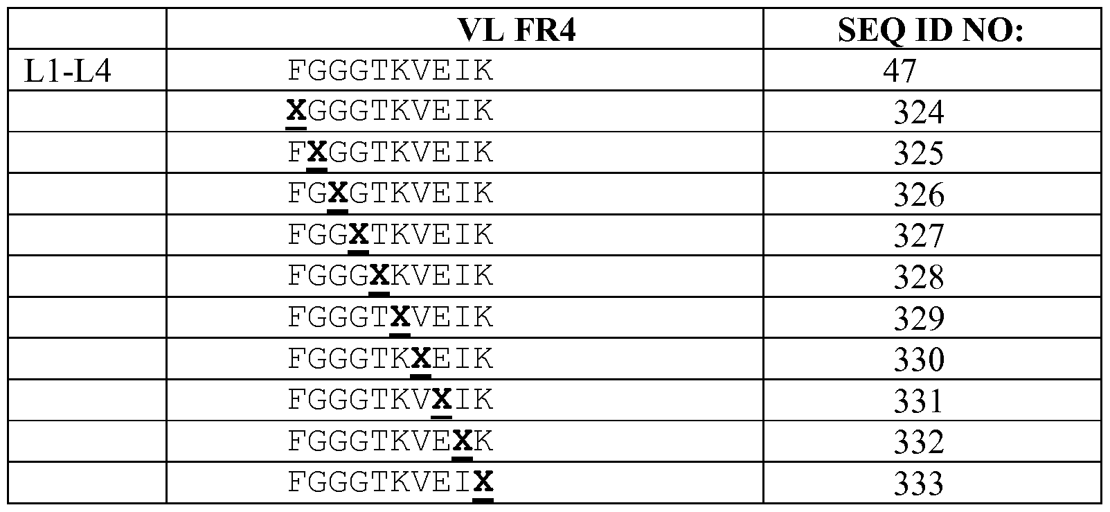


Claims
Priority Applications (22)
| Application Number | Priority Date | Filing Date | Title |
|---|---|---|---|
| CA2880007A CA2880007C (en) | 2012-07-25 | 2013-07-24 | Anti-kit antibodies and uses thereof |
| EP18159478.9A EP3381943B1 (en) | 2012-07-25 | 2013-07-24 | Anti-kit antibodies and uses thereof |
| MX2015000999A MX360620B (en) | 2012-07-25 | 2013-07-24 | Anti-kit antibodies and uses thereof. |
| BR112015001459-3A BR112015001459B1 (en) | 2012-07-25 | 2013-07-24 | ISOLATED ANTIBODY OR FRAGMENT THEREOF, CONJUGATE, USES THEREOF, PHARMACEUTICAL COMPOSITION, POLYNUCLEOTIDE, VECTOR, HOST CELL, ISOLATED CELL, KIT, IN VITRO METHOD TO INHIBIT KIT ACTIVITY, METHOD TO PRODUCE AN ANTIBODY |
| CN202310639870.2A CN116574185A (en) | 2012-07-25 | 2013-07-24 | anti-KIT antibodies and uses thereof |
| SG11201500489YA SG11201500489YA (en) | 2012-07-25 | 2013-07-24 | Anti-kit antibodies and uses thereof |
| EP22161986.9A EP4063391A1 (en) | 2012-07-25 | 2013-07-24 | Anti-kit antibodies and uses thereof |
| KR1020157004699A KR102268351B1 (en) | 2012-07-25 | 2013-07-24 | Anti-kit antibodies and uses thereof |
| CN201380048992.9A CN104812775B (en) | 2012-07-25 | 2013-07-24 | Anti- KIT antibody and application thereof |
| AU2013295848A AU2013295848B2 (en) | 2012-07-25 | 2013-07-24 | Anti-KIT antibodies and uses thereof |
| JP2015524415A JP6307075B2 (en) | 2012-07-25 | 2013-07-24 | Anti-KIT antibody and use thereof |
| EP13745296.7A EP2877493B1 (en) | 2012-07-25 | 2013-07-24 | Anti-kit antibodies and uses thereof |
| DK13745296.7T DK2877493T3 (en) | 2012-07-25 | 2013-07-24 | ANTI-KIT ANTIBODIES AND APPLICATIONS THEREOF |
| CN201910350594.1A CN110256559B (en) | 2012-07-25 | 2013-07-24 | anti-KIT antibodies and uses thereof |
| ES13745296.7T ES2673847T3 (en) | 2012-07-25 | 2013-07-24 | Anti KIT antibodies and uses thereof |
| RU2015106147A RU2681730C2 (en) | 2012-07-25 | 2013-07-24 | Anti-kit antibodies and uses thereof |
| NZ630363A NZ630363A (en) | 2012-07-25 | 2013-07-24 | Anti-kit antibodies and uses thereof |
| PH12015500113A PH12015500113A1 (en) | 2012-07-25 | 2015-01-20 | Anti-kit antibodies and uses thereof |
| IL236874A IL236874B (en) | 2012-07-25 | 2015-01-22 | Anti-kit antibodies and uses thereof |
| ZA2015/00457A ZA201500457B (en) | 2012-07-25 | 2015-01-22 | Anti-kit antibodies and uses thereof |
| CR20150023A CR20150023A (en) | 2012-07-25 | 2015-01-22 | ANTI KIT ANTIBODIES AND USES OF THE SAME |
| HK15111765.3A HK1211038A1 (en) | 2012-07-25 | 2015-11-30 | Anti-kit antibodies and uses thereof kit |
Applications Claiming Priority (4)
| Application Number | Priority Date | Filing Date | Title |
|---|---|---|---|
| US201261675762P | 2012-07-25 | 2012-07-25 | |
| US201261675751P | 2012-07-25 | 2012-07-25 | |
| US61/675,762 | 2012-07-25 | ||
| US61/675,751 | 2012-07-25 |
Publications (1)
| Publication Number | Publication Date |
|---|---|
| WO2014018625A1 true WO2014018625A1 (en) | 2014-01-30 |
Family
ID=48916252
Family Applications (1)
| Application Number | Title | Priority Date | Filing Date |
|---|---|---|---|
| PCT/US2013/051815 WO2014018625A1 (en) | 2012-07-25 | 2013-07-24 | Anti-kit antibodies and uses thereof |
Country Status (20)
| Country | Link |
|---|---|
| US (5) | US9334332B2 (en) |
| EP (3) | EP2877493B1 (en) |
| JP (4) | JP6307075B2 (en) |
| KR (1) | KR102268351B1 (en) |
| CN (3) | CN104812775B (en) |
| AU (1) | AU2013295848B2 (en) |
| BR (1) | BR112015001459B1 (en) |
| CA (1) | CA2880007C (en) |
| DK (2) | DK2877493T3 (en) |
| ES (1) | ES2673847T3 (en) |
| HK (1) | HK1211038A1 (en) |
| IL (1) | IL236874B (en) |
| MX (1) | MX360620B (en) |
| NZ (2) | NZ630363A (en) |
| PH (1) | PH12015500113A1 (en) |
| RU (1) | RU2681730C2 (en) |
| SG (1) | SG11201500489YA (en) |
| TW (1) | TWI596113B (en) |
| WO (1) | WO2014018625A1 (en) |
| ZA (1) | ZA201500457B (en) |
Cited By (11)
| Publication number | Priority date | Publication date | Assignee | Title |
|---|---|---|---|---|
| WO2015050959A1 (en) * | 2013-10-01 | 2015-04-09 | Yale University | Anti-kit antibodies and methods of use thereof |
| US9334332B2 (en) | 2012-07-25 | 2016-05-10 | Kolltan Pharmaceuticals, Inc. | Anti-kit antibodies |
| WO2016154588A1 (en) | 2015-03-25 | 2016-09-29 | Children's Hospital Medical Center | Use of kit inhibitors to condition subjects for a hematopoietic stem cell (hsc) transplantation |
| US9540443B2 (en) | 2011-01-26 | 2017-01-10 | Kolltan Pharmaceuticals, Inc. | Anti-kit antibodies |
| CN106659782A (en) * | 2014-05-23 | 2017-05-10 | 科尔坦制药公司 | Treatment of eosinophil or mast cell related disorders |
| WO2019155067A1 (en) | 2018-02-09 | 2019-08-15 | Ultrahuman Five Limited | C-kit antibodies |
| WO2020112687A2 (en) | 2018-11-26 | 2020-06-04 | Forty Seven, Inc. | Humanized antibodies against c-kit |
| US11357864B2 (en) | 2016-12-21 | 2022-06-14 | Novartis Ag | Antibody drug conjugates for ablating hematopoietic stem cells |
| US11712026B2 (en) | 2017-10-18 | 2023-08-01 | The Jackson Laboratory | Murine-MHC-deficient HLA-transgenic nod-mouse models for T1D therapy development |
| US11778994B2 (en) | 2017-05-12 | 2023-10-10 | The Jackson Laboratory | NSG mice lacking MHC class I and class II |
| EP3958909A4 (en) * | 2019-04-24 | 2024-01-10 | Magenta Therapeutics Inc | Anti-cd117 antibody-drug conjugates and uses thereof |
Families Citing this family (13)
| Publication number | Priority date | Publication date | Assignee | Title |
|---|---|---|---|---|
| EP3514178A1 (en) * | 2013-03-15 | 2019-07-24 | Novartis AG | Antibody drug conjugates |
| MA45408A (en) | 2016-06-17 | 2019-04-24 | Magenta Therapeutics Inc | COMPOSITIONS AND METHODS FOR DEPLOYING CD117 + CELLS |
| AU2017292184A1 (en) * | 2016-07-08 | 2019-02-07 | Staten Biotechnology B.V. | Anti-Apoc3 antibodies and methods of use thereof |
| CA3049501A1 (en) | 2017-01-20 | 2018-07-26 | Magenta Therapeutics, Inc. | Compositions and methods for the depletion of cd137+ cells |
| JP2020525432A (en) * | 2017-06-22 | 2020-08-27 | ディベロップメント センター フォー バイオテクノロジーDevelopment Center For Biotechnology | Asymmetric heterodimer FC-SCFV fusion anti-GLOBOH and anti-CD3 bispecific antibody and its use in cancer therapy |
| KR20200040407A (en) * | 2018-10-10 | 2020-04-20 | 주식회사 노벨티노빌리티 | Novel anti-c-kit antibody |
| USD966445S1 (en) * | 2019-02-13 | 2022-10-11 | Global Sports Innovation LTD | Sports training device |
| JP2022530443A (en) * | 2019-04-24 | 2022-06-29 | マジェンタ セラピューティクス インコーポレイテッド | Anti-CD117 antibody drug conjugate and its use |
| AU2020336440A1 (en) * | 2019-08-30 | 2022-03-24 | Qilu Puget Sound Biotherapeutics Corporation | Anti-CD20 antibodies, anti-CD37 antibodies, and mixtures thereof |
| KR20210063782A (en) * | 2019-11-25 | 2021-06-02 | 주식회사 노벨티노빌리티 | Antibodies Against c-kit and Uses Thereof |
| US11297054B1 (en) * | 2020-10-06 | 2022-04-05 | International Business Machines Corporation | Authentication system(s) with multiple authentication modes using one-time passwords of increased security |
| KR20230150275A (en) * | 2021-01-22 | 2023-10-30 | 셀덱스 쎄라퓨틱스, 인크. | Anti-KIT antibodies and uses thereof |
| CA3233871A1 (en) * | 2021-10-07 | 2023-04-13 | Sang Gyu Park | Treatment of mast cell related disorders |
Citations (222)
| Publication number | Priority date | Publication date | Assignee | Title |
|---|---|---|---|---|
| WO1986005807A1 (en) | 1985-04-01 | 1986-10-09 | Celltech Limited | Transformed myeloma cell-line and a process for the expression of a gene coding for a eukaryotic polypeptide employing same |
| US4671958A (en) | 1982-03-09 | 1987-06-09 | Cytogen Corporation | Antibody conjugates for the delivery of compounds to target sites |
| US4676980A (en) | 1985-09-23 | 1987-06-30 | The United States Of America As Represented By The Secretary Of The Department Of Health And Human Services | Target specific cross-linked heteroantibodies |
| EP0239400A2 (en) | 1986-03-27 | 1987-09-30 | Medical Research Council | Recombinant antibodies and methods for their production |
| WO1989001036A1 (en) | 1987-07-23 | 1989-02-09 | Celltech Limited | Recombinant dna expression vectors |
| EP0307434A1 (en) | 1987-03-18 | 1989-03-22 | Medical Res Council | Altered antibodies. |
| EP0367166A1 (en) | 1988-10-31 | 1990-05-09 | Takeda Chemical Industries, Ltd. | Modified interleukin-2 and production thereof |
| EP0394827A1 (en) | 1989-04-26 | 1990-10-31 | F. Hoffmann-La Roche Ag | Chimaeric CD4-immunoglobulin polypeptides |
| US4970198A (en) | 1985-10-17 | 1990-11-13 | American Cyanamid Company | Antitumor antibiotics (LL-E33288 complex) |
| WO1991006570A1 (en) | 1989-10-25 | 1991-05-16 | The University Of Melbourne | HYBRID Fc RECEPTOR MOLECULES |
| WO1991009967A1 (en) | 1989-12-21 | 1991-07-11 | Celltech Limited | Humanised antibodies |
| US5037651A (en) | 1987-01-30 | 1991-08-06 | American Cyanamid Company | Dihydro derivatives of LL-E33288 antibiotics |
| US5053394A (en) | 1988-09-21 | 1991-10-01 | American Cyanamid Company | Targeted forms of methyltrithio antitumor agents |
| US5079233A (en) | 1987-01-30 | 1992-01-07 | American Cyanamid Company | N-acyl derivatives of the LL-E33288 antitumor antibiotics, composition and methods for using the same |
| US5108912A (en) | 1987-01-30 | 1992-04-28 | American Cyanamid Company | Antitumor antibiotics (LL-E33288 complex) |
| US5112946A (en) | 1989-07-06 | 1992-05-12 | Repligen Corporation | Modified pf4 compositions and methods of use |
| US5122464A (en) | 1986-01-23 | 1992-06-16 | Celltech Limited, A British Company | Method for dominant selection in eucaryotic cells |
| WO1992017505A1 (en) * | 1991-04-05 | 1992-10-15 | Board Of Regents Of The University Of Washington | Monoclonal antibodies to stem cell factor receptors |
| WO1992021766A1 (en) | 1991-05-25 | 1992-12-10 | Boehringer Mannheim Gmbh | Monoclonal antibodies against c-kit |
| EP0519596A1 (en) | 1991-05-17 | 1992-12-23 | Merck & Co. Inc. | A method for reducing the immunogenicity of antibody variable domains |
| EP0548867A2 (en) | 1991-12-23 | 1993-06-30 | Yeda Research And Development Co. Ltd. | Soluble stem cell factor (SFC)-receptor |
| US5225539A (en) | 1986-03-27 | 1993-07-06 | Medical Research Council | Recombinant altered antibodies and methods of making altered antibodies |
| WO1993017105A1 (en) | 1992-02-19 | 1993-09-02 | Scotgen Limited | Altered antibodies, products and processes relating thereto |
| WO1994004678A1 (en) | 1992-08-21 | 1994-03-03 | Casterman Cecile | Immunoglobulins devoid of light chains |
| EP0592106A1 (en) | 1992-09-09 | 1994-04-13 | Immunogen Inc | Resurfacing of rodent antibodies |
| US5336603A (en) | 1987-10-02 | 1994-08-09 | Genentech, Inc. | CD4 adheson variants |
| US5349053A (en) | 1990-06-01 | 1994-09-20 | Protein Design Labs, Inc. | Chimeric ligand/immunoglobulin molecules and their uses |
| US5359046A (en) | 1990-12-14 | 1994-10-25 | Cell Genesys, Inc. | Chimeric chains for receptor-associated signal transduction pathways |
| WO1994025591A1 (en) | 1993-04-29 | 1994-11-10 | Unilever N.V. | PRODUCTION OF ANTIBODIES OR (FUNCTIONALIZED) FRAGMENTS THEREOF DERIVED FROM HEAVY CHAIN IMMUNOGLOBULINS OF $i(CAMELIDAE) |
| WO1994029351A2 (en) | 1993-06-16 | 1994-12-22 | Celltech Limited | Antibodies |
| US5416064A (en) | 1989-10-25 | 1995-05-16 | Immunogen, Inc. | Cytotoxic agents comprising maytansinoids and their therapeutic use |
| US5441050A (en) | 1992-12-18 | 1995-08-15 | Neoprobe Corporation | Radiation responsive surgical instrument |
| US5447851A (en) | 1992-04-02 | 1995-09-05 | Board Of Regents, The University Of Texas System | DNA encoding a chimeric polypeptide comprising the extracellular domain of TNF receptor fused to IgG, vectors, and host cells |
| WO1996004388A1 (en) | 1994-07-29 | 1996-02-15 | Smithkline Beecham Plc | Novel compounds |
| US5530101A (en) | 1988-12-28 | 1996-06-25 | Protein Design Labs, Inc. | Humanized immunoglobulins |
| WO1996022024A1 (en) | 1995-01-17 | 1996-07-25 | Brigham And Women's Hospital, Inc. | Receptor specific transepithelial transport of immunogens |
| US5545533A (en) | 1991-05-25 | 1996-08-13 | Boehringer Mannheim Gmbh | Monoclonal antibodies against c-kit and method of detecting a malignancy using c-kit specific antibodies |
| US5565332A (en) | 1991-09-23 | 1996-10-15 | Medical Research Council | Production of chimeric antibodies - a combinatorial approach |
| US5587459A (en) | 1994-08-19 | 1996-12-24 | Regents Of The University Of Minnesota | Immunoconjugates comprising tyrosine kinase inhibitors |
| US5606040A (en) | 1987-10-30 | 1997-02-25 | American Cyanamid Company | Antitumor and antibacterial substituted disulfide derivatives prepared from compounds possessing a methyl-trithio group |
| US5605793A (en) | 1994-02-17 | 1997-02-25 | Affymax Technologies N.V. | Methods for in vitro recombination |
| US5618709A (en) | 1994-01-14 | 1997-04-08 | University Of Pennsylvania | Antisense oligonucleotides specific for STK-1 and method for inhibiting expression of the STK-1 protein |
| US5622929A (en) | 1992-01-23 | 1997-04-22 | Bristol-Myers Squibb Company | Thioether conjugates |
| US5648239A (en) | 1996-06-21 | 1997-07-15 | Incyte Pharmaceuticals, Inc. | Human camp-dependent protein kinase inhibitor homolog |
| EP0787743A2 (en) * | 1996-01-10 | 1997-08-06 | Eberhard-Karls-Universität Tübingen Universitätsklinikum | A3C6E2, a monoclonal antibody to the human stem cell factor (SCF)-receptor |
| WO1997033899A1 (en) | 1996-03-14 | 1997-09-18 | Human Genome Sciences, Inc. | Apoptosis inducing molecule i |
| WO1997034911A1 (en) | 1996-03-22 | 1997-09-25 | Human Genome Sciences, Inc. | Apoptosis inducing molecule ii |
| WO1997034631A1 (en) | 1996-03-18 | 1997-09-25 | Board Of Regents, The University Of Texas System | Immunoglobin-like domains with increased half lives |
| US5677425A (en) | 1987-09-04 | 1997-10-14 | Celltech Therapeutics Limited | Recombinant antibody |
| US5693780A (en) | 1991-07-25 | 1997-12-02 | Idec Pharmaceuticals Corporation | Recombinant antibodies for human therapy |
| US5709874A (en) | 1993-04-14 | 1998-01-20 | Emory University | Device for local drug delivery and methods for using the same |
| US5714350A (en) | 1992-03-09 | 1998-02-03 | Protein Design Labs, Inc. | Increasing antibody affinity by altering glycosylation in the immunoglobulin variable region |
| US5723125A (en) | 1995-12-28 | 1998-03-03 | Tanox Biosystems, Inc. | Hybrid with interferon-alpha and an immunoglobulin Fc linked through a non-immunogenic peptide |
| US5728868A (en) | 1993-07-15 | 1998-03-17 | Cancer Research Campaign Technology Limited | Prodrugs of protein tyrosine kinase inhibitors |
| US5734033A (en) | 1988-12-22 | 1998-03-31 | The Trustees Of The University Of Pennsylvania | Antisense oligonucleotides inhibiting human bcl-2 gene expression |
| US5759542A (en) | 1994-08-05 | 1998-06-02 | New England Deaconess Hospital Corporation | Compositions and methods for the delivery of drugs by platelets for the treatment of cardiovascular and other diseases |
| US5766886A (en) | 1991-12-13 | 1998-06-16 | Xoma Corporation | Modified antibody variable domains |
| US5783181A (en) | 1994-07-29 | 1998-07-21 | Smithkline Beecham Corporation | Therapeutic uses of fusion proteins between mutant IL 4/IL13 antagonists and immunoglobulins |
| US5807715A (en) | 1984-08-27 | 1998-09-15 | The Board Of Trustees Of The Leland Stanford Junior University | Methods and transformed mammalian lymphocyte cells for producing functional antigen-binding protein including chimeric immunoglobulin |
| US5834252A (en) | 1995-04-18 | 1998-11-10 | Glaxo Group Limited | End-complementary polymerase reaction |
| US5837458A (en) | 1994-02-17 | 1998-11-17 | Maxygen, Inc. | Methods and compositions for cellular and metabolic engineering |
| US5840745A (en) | 1995-01-26 | 1998-11-24 | Pharmacia S. P. A. | Hydrosoluble 3-arylidene-2-oxindole derivatives as tyrosine kinase inhibitors |
| US5840674A (en) | 1990-11-01 | 1998-11-24 | Oregon Health Sciences University | Covalent microparticle-drug conjugates for biological targeting |
| US5844095A (en) | 1991-06-27 | 1998-12-01 | Bristol-Myers Squibb Company | CTLA4 Ig fusion proteins |
| US5863904A (en) | 1995-09-26 | 1999-01-26 | The University Of Michigan | Methods for treating cancers and restenosis with P21 |
| WO1999004813A1 (en) | 1997-07-24 | 1999-02-04 | Brigham & Women's Hospital, Inc. | Receptor specific transepithelial transport of therapeutics |
| US5869046A (en) | 1995-04-14 | 1999-02-09 | Genentech, Inc. | Altered polypeptides with increased half-life |
| US5885573A (en) | 1993-06-01 | 1999-03-23 | Arch Development Corporation | Methods and materials for modulation of the immunosuppressive activity and toxicity of monoclonal antibodies |
| US5885834A (en) | 1996-09-30 | 1999-03-23 | Epstein; Paul M. | Antisense oligodeoxynucleotide against phosphodiesterase |
| US5900252A (en) | 1990-04-17 | 1999-05-04 | Eurand International S.P.A. | Method for targeted and controlled release of drugs in the intestinal tract and more particularly in the colon |
| WO1999023105A1 (en) | 1997-11-03 | 1999-05-14 | Human Genome Sciences, Inc. | Vegi, an inhibitor of angiogenesis and tumor growth |
| US5911995A (en) | 1994-08-19 | 1999-06-15 | Regents Of The University Of Minnesota | EGF-genistein conjugates for the treatment of cancer |
| US5925376A (en) | 1994-01-10 | 1999-07-20 | Heng; Madalene C. Y. | Method for treating psoriasis using selected phosphorylase kinase inhibitor and additional compounds |
| US5958769A (en) | 1996-01-18 | 1999-09-28 | Fred Hutchinson Cancer Research Center | Compositions and methods for mediating cell cycle progression |
| US5965726A (en) | 1992-03-27 | 1999-10-12 | The United States Of America As Represented By The Department Of Health And Human Services | Method of eliminating inhibitory/ instability regions of mRNA |
| US5972366A (en) | 1994-11-28 | 1999-10-26 | The Unites States Of America As Represented By The Secretary Of The Army | Drug releasing surgical implant or dressing material |
| WO1999054342A1 (en) | 1998-04-20 | 1999-10-28 | Pablo Umana | Glycosylation engineering of antibodies for improving antibody-dependent cellular cytotoxicity |
| US5985877A (en) | 1997-08-15 | 1999-11-16 | Cephalon, Inc. | Combination of tyrosine kinase inhibitor and chemical castration to treat prostate cancer |
| US5985307A (en) | 1993-04-14 | 1999-11-16 | Emory University | Device and method for non-occlusive localized drug delivery |
| WO1999058572A1 (en) | 1998-05-08 | 1999-11-18 | Cambridge University Technical Services Limited | Binding molecules derived from immunoglobulins which do not trigger complement mediated lysis |
| US5998596A (en) | 1995-04-04 | 1999-12-07 | The United States Of America As Represented By The Department Of Health And Human Services | Inhibition of protein kinase activity by aptameric action of oligonucleotides |
| US6004534A (en) | 1993-07-23 | 1999-12-21 | Massachusetts Institute Of Technology | Targeted polymerized liposomes for improved drug delivery |
| US6005079A (en) | 1992-08-21 | 1999-12-21 | Vrije Universiteit Brussels | Immunoglobulins devoid of light chains |
| US6034053A (en) | 1998-07-13 | 2000-03-07 | Wayne Hughes Institute | EGF-isoflavone conjugates for the prevention of restenosis |
| WO2000012507A2 (en) | 1998-08-27 | 2000-03-09 | Spirogen Limited | Pyrrolobenzodiazepines |
| US6040305A (en) | 1996-09-13 | 2000-03-21 | Schering Corporation | Compounds useful for inhibition of farnesyl protein transferase |
| US6039975A (en) | 1995-10-17 | 2000-03-21 | Hoffman-La Roche Inc. | Colon targeted delivery system |
| US6048736A (en) | 1998-04-29 | 2000-04-11 | Kosak; Kenneth M. | Cyclodextrin polymers for carrying and releasing drugs |
| US6051582A (en) | 1997-06-17 | 2000-04-18 | Schering Corporation | Compounds useful for inhibition of farnesyl protein transferase |
| US6051574A (en) | 1996-04-03 | 2000-04-18 | Merck & Co., Inc. | Inhibitors of farnesyl-protein transferase |
| US6054466A (en) | 1997-12-04 | 2000-04-25 | Merck & Co., Inc. | Inhibitors of farnesyl-protein transferase |
| US6060082A (en) | 1997-04-18 | 2000-05-09 | Massachusetts Institute Of Technology | Polymerized liposomes targeted to M cells and useful for oral or mucosal drug delivery |
| US6063930A (en) | 1996-04-03 | 2000-05-16 | Merck & Co., Inc. | Substituted imidazole compounds useful as farnesyl-protein transferase inhibitors |
| US6066738A (en) | 1996-01-30 | 2000-05-23 | Merck & Co., Inc. | Inhibitors of farnesyl-protein transferase |
| US6071495A (en) | 1989-12-22 | 2000-06-06 | Imarx Pharmaceutical Corp. | Targeted gas and gaseous precursor-filled liposomes |
| US6071935A (en) | 1996-06-27 | 2000-06-06 | Pfizer Inc. | Derivatives of 2-(2-oxo-ethylidene)-imidazolidin-4-one and their use as farnesyl protein transferase inhibitors |
| US6077853A (en) | 1996-12-30 | 2000-06-20 | Merck & Co., Inc. | Inhibitors of farnesyl-protein transferase |
| US6080870A (en) | 1996-04-03 | 2000-06-27 | Merck & Co., Inc. | Biaryl substituted imidazole compounds useful as farnesyl-protein transferase inhibitors |
| US6087367A (en) | 1991-10-04 | 2000-07-11 | Sloan-Kettering Institute For Cancer Research | Potent inducers of terminal differentiation and methods of use thereof |
| US6090948A (en) | 1996-01-30 | 2000-07-18 | Merck & Co., Inc. | Inhibitors of farnesyl-protein transferase |
| WO2000042072A2 (en) | 1999-01-15 | 2000-07-20 | Genentech, Inc. | Polypeptide variants with altered effector function |
| US6093737A (en) | 1996-12-30 | 2000-07-25 | Merck & Co., Inc. | Inhibitors of farnesyl-protein transferase |
| US6103723A (en) | 1997-10-17 | 2000-08-15 | Merck & Co., Inc. | Inhibitors of farnesyl-protein transferase |
| US6120751A (en) | 1997-03-21 | 2000-09-19 | Imarx Pharmaceutical Corp. | Charged lipids and uses for the same |
| US6121022A (en) | 1995-04-14 | 2000-09-19 | Genentech, Inc. | Altered polypeptides with increased half-life |
| US6124465A (en) | 1997-11-25 | 2000-09-26 | Rhone-Poulenc S.A. | Farnesyl transferase inhibitors, their preparation, the pharmaceutical compositions which contain them and their use in the preparation of medicaments |
| US6124295A (en) | 1996-09-13 | 2000-09-26 | Schering Corporation | Compounds useful for inhibition of farnesyl protein transferase |
| US6127366A (en) | 1995-11-22 | 2000-10-03 | Merck & Co., Inc. | Inhibitors of farnesyl-protein transferase |
| US6133303A (en) | 1997-02-11 | 2000-10-17 | Warner-Lambert Company | Bicyclic inhibitors of protein farnesyl transferase |
| US6131570A (en) | 1998-06-30 | 2000-10-17 | Aradigm Corporation | Temperature controlling device for aerosol drug delivery |
| WO2000061739A1 (en) | 1999-04-09 | 2000-10-19 | Kyowa Hakko Kogyo Co., Ltd. | Method for controlling the activity of immunologically functional molecule |
| US6139865A (en) | 1996-10-01 | 2000-10-31 | Eurand America, Inc. | Taste-masked microcapsule compositions and methods of manufacture |
| US6143766A (en) | 1999-04-16 | 2000-11-07 | Warner-Lambert Company | Benzopyranone and quinolone inhibitors of ras farnesyl transferase |
| US6159984A (en) | 1997-06-17 | 2000-12-12 | Schering Corporation | Farnesyl protein transferase inhibitors |
| US6165745A (en) | 1992-04-24 | 2000-12-26 | Board Of Regents, The University Of Texas System | Recombinant production of immunoglobulin-like domains in prokaryotic cells |
| US6169096B1 (en) | 1995-12-08 | 2001-01-02 | Janssen Pharmacaeutic N.V. | Farnesyl protein transferase inhibiting (imidazol-5-yl)methyl-2-quinolinone derivatives |
| US6187786B1 (en) | 1997-03-10 | 2001-02-13 | Janssen Pharmaceutica N.V. | Farnesyl transferase inhibiting 1,8-annelated quinolinone derivatives substituted with N- or C-linked imidazoles |
| US6194551B1 (en) | 1998-04-02 | 2001-02-27 | Genentech, Inc. | Polypeptide variants |
| US6211193B1 (en) | 1997-06-17 | 2001-04-03 | Schering Corporation | Compounds useful for inhibition of farnesyl protein transferase |
| US6218406B1 (en) | 1996-12-30 | 2001-04-17 | Aventis Pharma S.A. | Farnesyl transferase inhibitors, their preparation, the pharmaceutical compositions which contain them and their use in the preparation of medicaments |
| US6218410B1 (en) | 1996-08-12 | 2001-04-17 | Yoshitomi Pharmaceutical Industries, Ltd. | Medicines comprising Rho kinase inhibitor |
| WO2001029226A2 (en) | 1999-10-21 | 2001-04-26 | Leadd B.V. | Apoptosis inducing proteinaceous substance |
| US6225322B1 (en) | 1997-06-17 | 2001-05-01 | Schering Corporation | Compounds useful for inhibition of farnesyl protein transferase |
| US6228865B1 (en) | 1997-06-17 | 2001-05-08 | Schering Corporation | Compounds useful for inhibition of farnesyl protein transferase |
| US6228856B1 (en) | 1996-09-13 | 2001-05-08 | Schering Corporation | Compounds useful for inhibition of farnesyl protein transferase |
| US6232338B1 (en) | 1995-08-04 | 2001-05-15 | Zeneca Limited | 4-Mercaptopyrrolidine derivatives as farnesyl transferase inhibitors |
| US6239140B1 (en) | 1997-06-17 | 2001-05-29 | Schering Corporation | Compounds useful for inhibition of farnesyl protein transferase |
| US6242196B1 (en) | 1997-12-11 | 2001-06-05 | Dana-Farber Cancer Institute | Methods and pharmaceutical compositions for inhibiting tumor cell growth |
| US6245759B1 (en) | 1999-03-11 | 2001-06-12 | Merck & Co., Inc. | Tyrosine kinase inhibitors |
| US6248756B1 (en) | 1996-04-03 | 2001-06-19 | Merck & Co., Inc. | Inhibitors of farnesyl-protein transferase |
| WO2001044301A1 (en) | 1999-11-29 | 2001-06-21 | Unilever Plc | Immobilized single domain antigen-binding molecules |
| US6253872B1 (en) | 1996-05-29 | 2001-07-03 | Gmundner Fertigteile Gesellschaft M.B.H & Co., Kg | Track soundproofing arrangement |
| US6268363B1 (en) | 1997-11-28 | 2001-07-31 | Lg Chemical Ltd. | Imidazole derivatives having an inhibitory activity for farnesyl transferase and process for preparation thereof |
| US6271242B1 (en) | 1992-02-10 | 2001-08-07 | Bristol-Myers Squibb Co. | Method for treating cancer using a tyrosine protein kinase inhibitor |
| US6271359B1 (en) | 1999-04-14 | 2001-08-07 | Musc Foundation For Research Development | Tissue-specific and pathogen-specific toxic agents and ribozymes |
| US6274552B1 (en) | 1993-03-18 | 2001-08-14 | Cytimmune Sciences, Inc. | Composition and method for delivery of biologically-active factors |
| WO2001058957A2 (en) | 2000-02-11 | 2001-08-16 | Lexigen Pharmaceuticals Corp. | Enhancing the circulating half-life of antibody-based fusion proteins |
| US6277832B1 (en) | 1995-06-01 | 2001-08-21 | Tadamitsu Kishimoto | Growth inhibitor for leukemia cells comprising antisense oligonucleotide derivative to wilms tumor gene (wt1) |
| US6277375B1 (en) | 1997-03-03 | 2001-08-21 | Board Of Regents, The University Of Texas System | Immunoglobulin-like domains with increased half-lives |
| US6300501B1 (en) | 1996-05-22 | 2001-10-09 | Warner-Lambert Company | Histidine-(N-benzyl glycinamide) inhibitors of protein farnesyl transferase |
| US6316652B1 (en) | 1995-06-06 | 2001-11-13 | Kosta Steliou | Drug mitochondrial targeting agents |
| US6335156B1 (en) | 1997-12-18 | 2002-01-01 | The Johns Hopkins University School Of Medicine | 14-3-3σ arrests the cell cycle |
| US6342765B1 (en) | 1997-10-22 | 2002-01-29 | Astrazeneca Uk Limited | Imidazole derivatives and their use as farnesyl protein transferase inhibitors |
| US6342487B1 (en) | 1998-12-21 | 2002-01-29 | Aventis Pharma Rorer S.A. | Compositions containing at least one farnesyl transferase inhibitor and at least one topoisomerase inhibitor and compositions containing at least one farnesyl transferase inhibitor and at least one taxoid |
| US6362188B1 (en) | 1998-12-18 | 2002-03-26 | Schering Corporation | Farnesyl protein transferase inhibitors |
| US6369034B1 (en) | 1998-04-27 | 2002-04-09 | Warner-Lambert Company | Functionalized alkyl and alenyl side chain derivatives of glycinamides as farnesyl transferase inhibitors |
| US6372747B1 (en) | 1998-12-18 | 2002-04-16 | Schering Corporation | Farnesyl protein transferase inhibitors |
| WO2002031140A1 (en) | 2000-10-06 | 2002-04-18 | Kyowa Hakko Kogyo Co., Ltd. | Cells producing antibody compositions |
| WO2002030954A1 (en) | 2000-10-06 | 2002-04-18 | Kyowa Hakko Kogyo Co., Ltd. | Method of purifying antibody |
| US6383790B1 (en) | 1999-01-11 | 2002-05-07 | Princeton University | High affinity protein kinase inhibitors |
| US6399633B1 (en) | 1999-02-01 | 2002-06-04 | Aventis Pharmaceuticals Inc. | Use of 4-H-1-benzopryan-4-one derivatives as inhibitors of smooth muscle cell proliferation |
| US6403581B1 (en) | 2000-01-19 | 2002-06-11 | American Cyanamid Company | Method of inhibition of farnesyl-protein transferase using substituted benz (cd) indol-2-imine and-amine derivatives |
| US6407213B1 (en) | 1991-06-14 | 2002-06-18 | Genentech, Inc. | Method for making humanized antibodies |
| US6410539B1 (en) | 1997-10-22 | 2002-06-25 | Astrazenca Uk Limited | Imidazole derivatives and their use as farnesyl protein transferase inhibitors |
| US6414145B1 (en) | 1997-01-29 | 2002-07-02 | Zeneca Limited | Imidazolyl compounds as inhibitors of farnesyl-protein tranferase |
| WO2002060919A2 (en) | 2000-12-12 | 2002-08-08 | Medimmune, Inc. | Molecules with extended half-lives, compositions and uses thereof |
| US6432959B1 (en) | 1998-12-23 | 2002-08-13 | Schering Corporation | Inhibitors of farnesyl-protein transferase |
| US6436960B1 (en) | 1998-02-02 | 2002-08-20 | Lg Chemical Ltd. | Farnesyl transferase inhibitors having a piperidine structure and process for preparation thereof |
| US6451812B1 (en) | 1998-07-06 | 2002-09-17 | Janssen Pharmaceutica N.V. | Farnesyl protein transferase inhibitors for treating arthropathies |
| US6458935B1 (en) | 1999-06-23 | 2002-10-01 | Merck & Co., Inc. | Radiolabeled farnesyl-protein transferase inhibitors |
| WO2002083180A1 (en) | 2001-03-23 | 2002-10-24 | Syntarga B.V. | Elongated and multiple spacers in activatible prodrugs |
| US6528624B1 (en) | 1998-04-02 | 2003-03-04 | Genentech, Inc. | Polypeptide variants |
| WO2003035835A2 (en) | 2001-10-25 | 2003-05-01 | Genentech, Inc. | Glycoprotein compositions |
| US20030115614A1 (en) | 2000-10-06 | 2003-06-19 | Yutaka Kanda | Antibody composition-producing cell |
| US20040002587A1 (en) | 2002-02-20 | 2004-01-01 | Watkins Jeffry D. | Fc region variants |
| WO2004016750A2 (en) | 2002-08-14 | 2004-02-26 | Macrogenics, Inc. | FcϜRIIB-SPECIFIC ANTIBODIES AND METHODS OF USE THEREOF |
| WO2004029207A2 (en) | 2002-09-27 | 2004-04-08 | Xencor Inc. | Optimized fc variants and methods for their generation |
| USRE38506E1 (en) | 1991-10-04 | 2004-04-20 | Sloan-Kettering Institute For Cancer Research | Potent inducers of terminal differentiation and methods of use thereof |
| WO2004035752A2 (en) | 2002-10-15 | 2004-04-29 | Protein Design Labs, Inc. | ALTERATION OF FcRn BINDING AFFINITIES OR SERUM HALF-LIVES OF ANTIBODIES BY MUTAGENESIS |
| US6737056B1 (en) | 1999-01-15 | 2004-05-18 | Genentech, Inc. | Polypeptide variants with altered effector function |
| WO2004043493A1 (en) | 2002-11-14 | 2004-05-27 | Syntarga B.V. | Prodrugs built as multiple self-elimination-release spacers |
| WO2004063351A2 (en) | 2003-01-09 | 2004-07-29 | Macrogenics, Inc. | IDENTIFICATION AND ENGINEERING OF ANTIBODIES WITH VARIANT Fc REGIONS AND METHODS OF USING SAME |
| WO2004099249A2 (en) | 2003-05-02 | 2004-11-18 | Xencor, Inc. | Optimized fc variants and methods for their generation |
| US20050042664A1 (en) | 2003-08-22 | 2005-02-24 | Medimmune, Inc. | Humanization of antibodies |
| US20050048549A1 (en) | 2003-01-21 | 2005-03-03 | Liangxian Cao | Methods and agents for screening for compounds capable of modulating gene expression |
| WO2005040217A2 (en) | 2003-10-17 | 2005-05-06 | Cambridge University Technical Services Limited | Antibodies having a mutated amino acid residue at position 268 (ch2 region) in constant regions |
| WO2005040170A2 (en) | 2003-10-22 | 2005-05-06 | Government Of The United States Of America, Represented By The Secretary, Department Of Health And Human Services | Pyrrolobenzodiazepine derivatives, compositions comprising the same and methods related thereto |
| WO2005085251A1 (en) | 2004-03-01 | 2005-09-15 | Spirogen Limited | 11-hydroxy-5h-pyrrolo[2,1-c][1,4]benzodiazepin-5-one derivatives as key intermediates for the preparation of c2 substituted pyrrolobenzodiazepines |
| US20050208625A1 (en) | 1998-04-28 | 2005-09-22 | Taylor Alexander H | Monoclonal antibodies with reduced immunogenicity |
| WO2005089808A2 (en) | 2004-03-15 | 2005-09-29 | Wyeth | Antibody calicheamicin conjugates |
| US20050226867A1 (en) | 2003-10-08 | 2005-10-13 | Kyowa Hakko Kogyo Co., Ltd. | IL-5R-specific antibody composition |
| WO2005110423A2 (en) | 2004-05-13 | 2005-11-24 | Spirogen Limited | Pyrrolobenzodiazepine therapeutic agents useful in the treatment of leukaemias |
| US6989248B2 (en) | 1999-05-06 | 2006-01-24 | The Trustees Of Columbia University In The City Of New York | Methods of use of compounds which inhibit the stem cell signaling pathway |
| WO2006031653A2 (en) | 2004-09-10 | 2006-03-23 | Wyeth | Humanized anti-5t4 antibodies and anti-5t4 antibody / calicheamicin conjugates |
| WO2007018431A2 (en) | 2005-08-05 | 2007-02-15 | Syntarga B.V. | Triazole-containing releasable linkers and conjugates comprising the same |
| US7183387B1 (en) | 1999-01-15 | 2007-02-27 | Genentech, Inc. | Polypeptide variants with altered effector function |
| WO2007039752A1 (en) | 2005-10-05 | 2007-04-12 | Spirogen Limited | Alkyl 4- [4- (5-oxo-2, 3, 5, 11a-tetrahyd0-5h-pyrr0l0 [2, 1-c] [1, 4] benzodiazepine-8-yloxy) -butyrylamino]-1h-pyrrole-2-carboxylate derivatives and related compounds for the treatment of a proliferative disease |
| WO2007089149A2 (en) | 2006-02-02 | 2007-08-09 | Syntarga B.V. | Water-soluble cc-1065 analogs and their conjugates |
| US20070253951A1 (en) | 2006-04-24 | 2007-11-01 | Gordon Ng | Humanized c-Kit antibody |
| US20080025989A1 (en) | 2003-02-20 | 2008-01-31 | Seattle Genetics, Inc. | Anti-cd70 antibody-drug conjugates and their use for the treatment of cancer and immune disorders |
| WO2008042146A1 (en) | 2006-09-28 | 2008-04-10 | Merck & Co., Inc. | Amine base salts of saha and polymorphs thereof |
| US20080279868A1 (en) | 2005-09-26 | 2008-11-13 | Medarex, Inc. | Antibody-Drug Conjugates and Methods of Use |
| US20080287309A1 (en) | 2004-07-10 | 2008-11-20 | Alexion Pharmaceuticals, Inc. | Methods for Discovering Antibodies Specific to Cancer Cells and Antibodies Discovered Thereby |
| US7456219B2 (en) | 2002-03-04 | 2008-11-25 | Merck Hdac Research, Llc | Polymorphs of suberoylanilide hydroxamic acid |
| US20080300192A1 (en) | 2004-11-12 | 2008-12-04 | Seattle Genetics, Inc. | Auristatins Having an Aminobenzoic Acid Unit at the N Terminus |
| WO2008150261A1 (en) | 2007-06-04 | 2008-12-11 | Wyeth | Detection and quantitation of calicheamicin |
| WO2008153926A2 (en) | 2007-06-05 | 2008-12-18 | Yale University | Inhibitors of receptor tyrosine kinases and methods of use thereof |
| WO2009017394A1 (en) | 2007-08-01 | 2009-02-05 | Syntarga B.V. | Substituted cc-1065 analogs and their conjugates |
| US20090148905A1 (en) | 2007-11-30 | 2009-06-11 | Claire Ashman | Antigen-binding constructs |
| WO2009088805A2 (en) | 2008-01-03 | 2009-07-16 | The Scripps Research Institute | Antibody targeting through a modular recognition domain |
| US20090202536A1 (en) | 2004-06-01 | 2009-08-13 | Genentech, Inc. | Antibody-drug conjugates and methods |
| US7575748B1 (en) | 2000-03-16 | 2009-08-18 | Genentech, Inc. | Methods of treatment using anti-ErbB antibody-maytansinoid conjugates |
| US7612062B2 (en) | 2005-04-21 | 2009-11-03 | Spirogen Limited | Pyrrolobenzodiazepines |
| WO2010017480A1 (en) | 2008-08-07 | 2010-02-11 | Centrose, Llc | Glycoside compounds and pharmaceutical compositions thereof |
| US7662387B2 (en) | 2003-02-20 | 2010-02-16 | Seattle Genetics | Anti-cd70 antibody-drug conjugates and their use for the treatment of cancer and immune disorders |
| US7700321B2 (en) | 2005-10-21 | 2010-04-20 | Genzyme Corporation | Antibody-based therapeutics with enhanced ADCC activity |
| US7709226B2 (en) | 2001-07-12 | 2010-05-04 | Arrowsmith Technology Licensing Llc | Method of humanizing antibodies by matching canonical structure types CDRs |
| WO2010062171A2 (en) | 2008-11-03 | 2010-06-03 | Syntarga B.V. | Novel cc-1065 analogs and their conjugates |
| US7846434B2 (en) | 2006-10-24 | 2010-12-07 | Trubion Pharmaceuticals, Inc. | Materials and methods for improved immunoglycoproteins |
| US7851432B2 (en) | 2003-05-20 | 2010-12-14 | Immunogen Inc. | Cytotoxic agents comprising new maytansinoids |
| WO2011021146A1 (en) | 2009-08-20 | 2011-02-24 | Pfizer Inc. | Osteopontin antibodies |
| WO2011031870A1 (en) | 2009-09-09 | 2011-03-17 | Centrose, Llc | Extracellular targeted drug conjugates |
| US7931895B2 (en) | 2000-04-12 | 2011-04-26 | Lfb Biotechnologies | Monoclonal antibodies with enhanced ADCC function |
| US7960512B2 (en) | 2003-01-09 | 2011-06-14 | Macrogenics, Inc. | Identification and engineering of antibodies with variant Fc regions and methods of using same |
| US8021856B2 (en) | 1998-04-20 | 2011-09-20 | Roche Glycart Ag | Antibody glycosylation variants having increased antibody-dependent cellular cytotoxicity |
| WO2011119948A1 (en) * | 2010-03-26 | 2011-09-29 | Kolltan Pharmaceuticals, Inc. | Anti-kit antibodies and uses thereof |
| US8039359B2 (en) | 2007-09-21 | 2011-10-18 | Semiconductor Components Industries, Llc | Method of forming low capacitance ESD device and structure therefor |
| US8080415B2 (en) | 2008-09-26 | 2011-12-20 | Eureka Therapeutics, Inc. | Modified host cells and uses thereof |
| US8101720B2 (en) | 2004-10-21 | 2012-01-24 | Xencor, Inc. | Immunoglobulin insertions, deletions and substitutions |
| WO2012103165A2 (en) * | 2011-01-26 | 2012-08-02 | Kolltan Pharmaceuticals, Inc. | Anti-kit antibodies and uses thereof |
| WO2012154480A1 (en) * | 2011-05-12 | 2012-11-15 | ImClone, LLC | c-KIT ANTIBODIES AND USES THEREOF |
| US11392902B2 (en) | 2017-06-06 | 2022-07-19 | United Parcel Service Of America, Inc. | Systems, methods, apparatuses and computer program products for providing notification of items for pickup and delivery |
Family Cites Families (101)
| Publication number | Priority date | Publication date | Assignee | Title |
|---|---|---|---|---|
| US5268358A (en) | 1988-12-08 | 1993-12-07 | Cor Therapeutics, Inc. | PDGF receptor blocking peptides |
| US7144731B2 (en) | 1989-10-16 | 2006-12-05 | Amgen Inc. | SCF antibody compositions and methods of using the same |
| US20030125519A1 (en) | 1990-08-27 | 2003-07-03 | Peter Besmer | Ligand for the c-kit receptor and methods of use thereof |
| US20050276784A1 (en) | 1990-08-27 | 2005-12-15 | Solan-Kettering Institute For Cancer Research | Ligand for the c-kit receptor and methods of use thereof |
| AU1411092A (en) | 1991-01-31 | 1992-09-07 | Cor Therapeutics, Inc. | Domains of extracellular region of human platelet derived growth factor receptor polypeptides |
| EP0619738B1 (en) | 1991-12-02 | 2003-07-09 | Millennium Pharmaceuticals, Inc. | Inhibitory immunoglobulin polypeptides to human pdgf beta receptor |
| US5817310A (en) | 1991-12-02 | 1998-10-06 | Cor Therapeutics, Inc. | Inhibitory immunoglobulin polypeptides to human PDGF beta receptor |
| US6001803A (en) | 1992-04-23 | 1999-12-14 | Sloan-Kettering Institute For Cancer Research | Composition of c-kit ligand, GM-CSF, and TNF-α and method of use |
| US6448077B1 (en) | 1994-02-10 | 2002-09-10 | Imclone Systems, Inc. | Chimeric and humanized monoclonal antibodies specific to VEGF receptors |
| US5911988A (en) | 1995-04-28 | 1999-06-15 | Bayer Corporation | Method for treating asthma using SCF antibody |
| US6936259B2 (en) | 1995-06-08 | 2005-08-30 | University Of Saskatchewan | CAMP factor of Streptococcus uberis |
| US5882644A (en) | 1996-03-22 | 1999-03-16 | Protein Design Labs, Inc. | Monoclonal antibodies specific for the platelet derived growth factor β receptor and methods of use thereof |
| US5910417A (en) | 1996-05-31 | 1999-06-08 | National Jewish Center For Immunology And Respiratory Medicine | Regulation of cytokine production in a hematopoietic cell |
| WO1998041090A1 (en) | 1997-03-19 | 1998-09-24 | Yale University | Methods and compositions for stimulating apoptosis and cell death or for inhibiting cell growth and cell attachment |
| DE19727814C1 (en) | 1997-06-30 | 1998-10-01 | Univ Eberhard Karls | Monoclonal antibody specific for human tyrosine kinase receptor protein |
| US7361336B1 (en) | 1997-09-18 | 2008-04-22 | Ivan Bergstein | Methods of cancer therapy targeted against a cancer stem line |
| US6555367B1 (en) | 1997-10-10 | 2003-04-29 | The United States Of America As Represented By The Department Of Health And Human Services | Complex of biotinylated viral vector and ligand for targeted gene delivery |
| WO2000061637A1 (en) | 1999-04-14 | 2000-10-19 | Smithkline Beecham Corporation | Erythropoietin receptor antibodies |
| US6576812B1 (en) | 1999-05-06 | 2003-06-10 | The Trustees Of Columbia University In The City Of New York | Compound screening assays using a transgenic mouse model of human skin diseases |
| WO2001029246A1 (en) | 1999-10-19 | 2001-04-26 | Kyowa Hakko Kogyo Co., Ltd. | Process for producing polypeptide |
| WO2001034201A2 (en) | 1999-11-12 | 2001-05-17 | Advanced Research And Technology Institute, Inc. | Methods for inhibiting neurofibromatosis type 1 (nf1) |
| DE60029138T2 (en) | 1999-12-22 | 2007-06-06 | Sugen, Inc., San Francisco | Use of indolinone compounds for the production of pharmaceuticals for the modulation of the function c-kit protein tyrosine kinase |
| US8088060B2 (en) | 2000-03-15 | 2012-01-03 | Orbusneich Medical, Inc. | Progenitor endothelial cell capturing with a drug eluting implantable medical device |
| TWI318983B (en) * | 2000-05-02 | 2010-01-01 | Uab Research Foundation | An antibody selective for a tumor necrosis factor-related apoptosis-inducing ligand receptor and uses thereof |
| US20110091428A1 (en) | 2000-07-31 | 2011-04-21 | New York Medical College | Compositions of adult organ stem cells and uses thereof |
| US6914947B2 (en) | 2001-02-28 | 2005-07-05 | Telefonaktiebolaget L M Ericsson (Publ) | Method and apparatus for handling time-drift |
| EP1378752A4 (en) | 2001-04-13 | 2004-08-04 | Chugai Pharmaceutical Co Ltd | Method of quantifying antigen expression |
| EP2277991A1 (en) | 2001-08-21 | 2011-01-26 | Ventana Medical Systems, Inc. | Method and quantification assay for determining c-kit/SCF/pAKT status |
| KR20040070254A (en) * | 2001-12-20 | 2004-08-06 | 휴먼 게놈 사이언시즈, 인코포레이티드 | Antibodies that immunospecifically bind to TRAIL receptors |
| US6998391B2 (en) | 2002-02-07 | 2006-02-14 | Supergen.Inc. | Method for treating diseases associated with abnormal kinase activity |
| AU2003217912A1 (en) | 2002-03-01 | 2003-09-16 | Xencor | Antibody optimization |
| WO2003102017A2 (en) | 2002-06-03 | 2003-12-11 | Centocor, Inc. | Anti-relp fusion antibodies, compositions, methods and uses |
| WO2004002425A2 (en) | 2002-06-28 | 2004-01-08 | Bio Transplant, Inc. | Process for promoting graft acceptance by depletion of hematopoietic stem cells |
| AU2004213749B2 (en) | 2003-01-31 | 2010-02-25 | Celldex Therapeutics, Inc. | Antibody vaccine conjugates and uses therefor |
| US20050281828A1 (en) | 2003-03-04 | 2005-12-22 | Bowdish Katherine S | Method of treating autoimmune disease by inducing antigen presentation by tolerance inducing antigen presenting cells |
| US7303893B1 (en) | 2003-08-28 | 2007-12-04 | Takeda San Diego, Inc. | Crystallization of c-KIT tyrosine kinase leading to autoinhibited crystal structure |
| US8133733B2 (en) | 2003-10-24 | 2012-03-13 | Gencia Corporation | Nonviral vectors for delivering polynucleotides to target tissues |
| AU2004297988A1 (en) | 2003-12-04 | 2005-06-23 | Laboratoires Serono Sa | Methods for identifying modulators of active KIT tyrosine kinase receptor |
| MX350383B (en) | 2004-01-09 | 2017-09-04 | Pfizer | ANTIBODIES TO MAdCAM. |
| SI1730315T1 (en) | 2004-03-27 | 2010-02-26 | Klein Hanns Georg | Polymorphisms in nod2/card15 gene |
| ATE531732T1 (en) | 2004-03-30 | 2011-11-15 | Yissum Res Dev Co | BI-SPECIFIC ANTIBODIES TARGETING CELLS INVOLVED IN ALLERGIC-TYPE REACTIONS, COMPOSITIONS AND USES THEREOF |
| EP1778270A4 (en) | 2004-05-05 | 2009-09-02 | Merrimack Pharmaceuticals Inc | Bispecific binding agents for modulating biological activity |
| US7329495B2 (en) | 2004-06-09 | 2008-02-12 | Board Of Regents, The University Of Texas System | Mutations in KIT confer imatinib resistance in gastrointestinal stromal tumors |
| AU2005269265B2 (en) | 2004-08-02 | 2012-01-12 | Zenyth Operations Pty Ltd | A method of treating cancer comprising a VEGF-B antagonist |
| US7329737B2 (en) | 2004-08-03 | 2008-02-12 | Dyax Corp. | Antibodies that bind hK-1 |
| US20090246206A1 (en) | 2005-02-23 | 2009-10-01 | Merrimack Pharmaceuticals, Inc. | Bispecific binding agents for modulating biological activity |
| US20060222634A1 (en) | 2005-03-31 | 2006-10-05 | Clarke Diana L | Amnion-derived cell compositions, methods of making and uses thereof |
| DK1874821T3 (en) | 2005-04-26 | 2013-07-08 | Trion Pharma Gmbh | Combination of antibodies with glycocorticoids to treat cancer |
| US20090048161A1 (en) | 2005-05-05 | 2009-02-19 | Valorisation Hsj, Societe En Commandite | Cytokine receptor modulators and uses thereof |
| WO2007014943A2 (en) | 2005-08-01 | 2007-02-08 | Ares Trading S.A. | Therapy for neurological diseases |
| NZ598088A (en) | 2005-10-21 | 2013-09-27 | Univ California | C-kit oncogene mutations in melanoma |
| EP1790664A1 (en) | 2005-11-24 | 2007-05-30 | Ganymed Pharmaceuticals AG | Monoclonal antibodies against claudin-18 for treatment of cancer |
| WO2007084949A2 (en) | 2006-01-18 | 2007-07-26 | The Uab Research Foundation | Modulators of cardiac cell hypertrophy and hyperplasia |
| US7790862B2 (en) | 2006-06-13 | 2010-09-07 | Zymogenetics, Inc. | IL-17 and IL-23 antagonists and methods of using the same |
| EP2314297A1 (en) | 2006-04-05 | 2011-04-27 | Novartis AG | Combinations comprising bcr-abl/c-kit/pdgf-r tk inhibitors for treating cancer |
| US20080032989A1 (en) | 2006-05-31 | 2008-02-07 | Robinson William H | Method of treating inflammatory diseases using tyroskine kinase inhibitors |
| JP2009539403A (en) * | 2006-06-13 | 2009-11-19 | オンコメッド ファーマシューティカルズ インコーポレイテッド | Compositions and methods for diagnosing and treating cancer |
| US8835163B2 (en) | 2006-10-18 | 2014-09-16 | The Board Of Trustees Of The University Of Illinois | Embryonic-like stem cells derived from adult human peripheral blood and methods of use |
| CA2667010A1 (en) | 2006-10-20 | 2008-05-02 | Irm Llc | Compositions and methods for modulating c-kit and pdgfr receptors |
| US7959942B2 (en) | 2006-10-20 | 2011-06-14 | Orbusneich Medical, Inc. | Bioabsorbable medical device with coating |
| PL3255061T3 (en) | 2006-11-03 | 2021-12-06 | The Board Of Trustees Of The Leland Stanford Junior University | Selective immunodepletion of endogenous stem cell niche for engraftment |
| US20100029674A1 (en) | 2006-11-17 | 2010-02-04 | Innate Pharma, S.A. | Methods of Using Phosphoantigen for the Treatment of Cancer |
| US20100143935A1 (en) | 2006-12-01 | 2010-06-10 | Apocell, Inc. | c-KIT Phosphorylation in Cancer |
| EP2137216A2 (en) | 2007-03-12 | 2009-12-30 | Vasgene Therapeutics, Inc. | Use of ephb4 as a diagnostic marker and a therapeutic target for ovarian cancer |
| EP2155694B1 (en) | 2007-03-27 | 2012-01-04 | Synta Pharmaceuticals Corp. | Triazinone and diazinone derivatives useful as hsp90 inhibitors |
| US20110268776A1 (en) | 2007-04-25 | 2011-11-03 | Schapira Jay N | Programmed-release, nanostructured biological construct for stimulating cellular engraftment for tissue regeneration |
| AR066660A1 (en) | 2007-05-23 | 2009-09-02 | Genentech Inc | PREVENTION AND TREATMENT OF EYE CONDITIONS ASSOCIATED WITH THEIR COMPLEMENT |
| EP2185698B1 (en) | 2007-08-07 | 2015-04-22 | Purdue Research Foundation | Kinase inhibitors and uses thereof |
| WO2009023246A2 (en) | 2007-08-14 | 2009-02-19 | Wake Forest University Health Sciences | Pluripotent adult stem cells |
| US8569262B2 (en) | 2007-11-02 | 2013-10-29 | Momenta Pharmaceuticals, Inc. | Polysaccharide compositions and methods of use for the treatment and prevention of disorders associated with progenitor cell mobilization |
| EP2227254A2 (en) | 2007-11-28 | 2010-09-15 | OSI Pharmaceuticals, Inc. | Combined treatment with an egfr kinase inhibitor and an inhibitor of c-kit |
| WO2009082624A2 (en) | 2007-12-10 | 2009-07-02 | Zymogenetics, Inc. | Antagonists of il-17a, il-17f, and il-23 and methods of using the same |
| AU2008338421A1 (en) | 2007-12-17 | 2009-06-25 | Dyax Corp. | Compositions and methods for treating osteolytic disorders comprising MMP-14 binding proteins |
| US8426396B2 (en) | 2008-01-08 | 2013-04-23 | Shriners Hospitals For Children | Treatment for achondroplasia |
| WO2009100105A2 (en) | 2008-02-04 | 2009-08-13 | Attogen Inc. | Inhibitors of oncogenic isoforms and uses thereof |
| WO2009135001A2 (en) | 2008-04-30 | 2009-11-05 | University Of Pittsburgh- Commonwealth System Of Higher Education | Methods and compositions for regulating th2 and th17 responses |
| WO2009140452A2 (en) | 2008-05-15 | 2009-11-19 | University Of Miami | Isolation of stem cell precursors and expansion in non-adherent conditions |
| WO2009152288A1 (en) | 2008-06-13 | 2009-12-17 | Novartis Ag | Substituted benzimidazoles for neurofibromatosis |
| KR20160011713A (en) | 2008-06-27 | 2016-02-01 | 인디애나 유니버시티 리서치 앤드 테크놀로지 코포레이션 | Materials and methods for suppressing and/or treating neurofibroma and related tumors |
| CN102007147B (en) | 2008-06-30 | 2014-11-05 | 协和发酵麒麟株式会社 | Anti-CD27 antibody |
| EP2331676A4 (en) | 2008-09-26 | 2012-09-19 | Univ Duke | Hematopoietic stem cell growth factor |
| JP2012506255A (en) | 2008-10-22 | 2012-03-15 | ジェネンテック, インコーポレイテッド | Prostate stem cells and uses thereof |
| AU2009316541B2 (en) | 2008-11-19 | 2015-08-06 | Celularity Inc. | Amnion derived adherent cells |
| WO2010065685A1 (en) | 2008-12-04 | 2010-06-10 | Cleveland Clinic | Method of treating c-kit positive relapsed acute myeloid leukemia |
| US20100204058A1 (en) | 2009-01-28 | 2010-08-12 | Howard Yuan-Hao Chang | Profiling for Determination of Response to Treatment for Inflammatory Disease |
| CN102459346B (en) | 2009-04-27 | 2016-10-26 | 昂考梅德药品有限公司 | The method manufacturing heteromultimers molecule |
| AR076668A1 (en) * | 2009-05-12 | 2011-06-29 | Pfizer | SPECIFIC ANTIBODIES FOR DKK-1 AND ITS USES |
| KR101224468B1 (en) | 2009-05-20 | 2013-01-23 | 주식회사 파멥신 | Bispecific antibody having a novel form and use thereof |
| CA2763446A1 (en) | 2009-05-28 | 2010-12-02 | Victoria Ballard | Stem cell targeting |
| NZ629615A (en) | 2009-11-06 | 2016-01-29 | Plexxikon Inc | Compounds and methods for kinase modulation, and indications therefor |
| US10817851B2 (en) | 2009-12-23 | 2020-10-27 | Aristocrat Technologies Australia Pty Limited | System and method for cashless gaming |
| US20130071397A1 (en) | 2009-12-29 | 2013-03-21 | Yale University | Inhibitors of vascular endothelial growth factor (vegf) receptors and methods of use thereof |
| AU2011205297A1 (en) | 2010-01-14 | 2012-06-28 | Yale University | Inhibitors of receptor tyrosine kinases (RTK) and methods of use thereof |
| CA2821646A1 (en) | 2011-01-06 | 2012-07-12 | Complix Nv | Alphabodies specifically binding to cytokines or growth factors and/or cytokine or growth factor receptors |
| MX350152B (en) | 2011-10-14 | 2017-08-29 | Medimmune Ltd | Pyrrolobenzodiazepines and conjugates thereof. |
| WO2013177481A1 (en) | 2012-05-25 | 2013-11-28 | Immunogen, Inc. | Benzodiazepines and conjugates thereof |
| NZ630363A (en) | 2012-07-25 | 2018-09-28 | Celldex Therapeutics Inc | Anti-kit antibodies and uses thereof |
| WO2015050959A1 (en) | 2013-10-01 | 2015-04-09 | Yale University | Anti-kit antibodies and methods of use thereof |
| WO2015112822A1 (en) | 2014-01-24 | 2015-07-30 | Kolltan Pharmaceuticals, Inc. | Antibody-drug conjugates targeting kit receptor and uses thereof |
| EP3145543A4 (en) | 2014-05-23 | 2017-12-13 | Celldex Therapeutics, Inc. | Treatment of eosinophil or mast cell related disorders |
| DK3177624T3 (en) | 2014-08-06 | 2019-07-01 | Pfizer | IMIDAZOPYRIDAZINE COMPOUNDS |
-
2013
- 2013-07-24 NZ NZ630363A patent/NZ630363A/en unknown
- 2013-07-24 KR KR1020157004699A patent/KR102268351B1/en active IP Right Grant
- 2013-07-24 EP EP13745296.7A patent/EP2877493B1/en active Active
- 2013-07-24 CN CN201380048992.9A patent/CN104812775B/en active Active
- 2013-07-24 DK DK13745296.7T patent/DK2877493T3/en active
- 2013-07-24 NZ NZ731881A patent/NZ731881A/en unknown
- 2013-07-24 CA CA2880007A patent/CA2880007C/en active Active
- 2013-07-24 WO PCT/US2013/051815 patent/WO2014018625A1/en active Application Filing
- 2013-07-24 JP JP2015524415A patent/JP6307075B2/en active Active
- 2013-07-24 EP EP22161986.9A patent/EP4063391A1/en active Pending
- 2013-07-24 SG SG11201500489YA patent/SG11201500489YA/en unknown
- 2013-07-24 BR BR112015001459-3A patent/BR112015001459B1/en active IP Right Grant
- 2013-07-24 AU AU2013295848A patent/AU2013295848B2/en active Active
- 2013-07-24 US US13/949,931 patent/US9334332B2/en active Active
- 2013-07-24 DK DK18159478.9T patent/DK3381943T3/en active
- 2013-07-24 TW TW102126570A patent/TWI596113B/en active
- 2013-07-24 MX MX2015000999A patent/MX360620B/en active IP Right Grant
- 2013-07-24 ES ES13745296.7T patent/ES2673847T3/en active Active
- 2013-07-24 EP EP18159478.9A patent/EP3381943B1/en active Active
- 2013-07-24 RU RU2015106147A patent/RU2681730C2/en active
- 2013-07-24 CN CN202310639870.2A patent/CN116574185A/en active Pending
- 2013-07-24 CN CN201910350594.1A patent/CN110256559B/en active Active
-
2015
- 2015-01-20 PH PH12015500113A patent/PH12015500113A1/en unknown
- 2015-01-22 ZA ZA2015/00457A patent/ZA201500457B/en unknown
- 2015-01-22 IL IL236874A patent/IL236874B/en active IP Right Grant
- 2015-11-30 HK HK15111765.3A patent/HK1211038A1/en unknown
-
2016
- 2016-03-30 US US15/084,715 patent/US9605081B2/en active Active
-
2017
- 2017-02-15 US US15/433,482 patent/US10184007B2/en active Active
-
2018
- 2018-03-09 JP JP2018042557A patent/JP6875302B2/en active Active
- 2018-12-14 US US16/221,002 patent/US10781267B2/en active Active
-
2020
- 2020-08-04 US US16/984,946 patent/US11891452B2/en active Active
-
2021
- 2021-04-22 JP JP2021072421A patent/JP7230095B2/en active Active
-
2023
- 2023-02-15 JP JP2023021289A patent/JP2023062063A/en active Pending
Patent Citations (272)
| Publication number | Priority date | Publication date | Assignee | Title |
|---|---|---|---|---|
| US4671958A (en) | 1982-03-09 | 1987-06-09 | Cytogen Corporation | Antibody conjugates for the delivery of compounds to target sites |
| US5807715A (en) | 1984-08-27 | 1998-09-15 | The Board Of Trustees Of The Leland Stanford Junior University | Methods and transformed mammalian lymphocyte cells for producing functional antigen-binding protein including chimeric immunoglobulin |
| WO1986005807A1 (en) | 1985-04-01 | 1986-10-09 | Celltech Limited | Transformed myeloma cell-line and a process for the expression of a gene coding for a eukaryotic polypeptide employing same |
| US4676980A (en) | 1985-09-23 | 1987-06-30 | The United States Of America As Represented By The Secretary Of The Department Of Health And Human Services | Target specific cross-linked heteroantibodies |
| US4970198A (en) | 1985-10-17 | 1990-11-13 | American Cyanamid Company | Antitumor antibiotics (LL-E33288 complex) |
| US5122464A (en) | 1986-01-23 | 1992-06-16 | Celltech Limited, A British Company | Method for dominant selection in eucaryotic cells |
| EP0239400A2 (en) | 1986-03-27 | 1987-09-30 | Medical Research Council | Recombinant antibodies and methods for their production |
| US5225539A (en) | 1986-03-27 | 1993-07-06 | Medical Research Council | Recombinant altered antibodies and methods of making altered antibodies |
| US5108912A (en) | 1987-01-30 | 1992-04-28 | American Cyanamid Company | Antitumor antibiotics (LL-E33288 complex) |
| US5037651A (en) | 1987-01-30 | 1991-08-06 | American Cyanamid Company | Dihydro derivatives of LL-E33288 antibiotics |
| US5079233A (en) | 1987-01-30 | 1992-01-07 | American Cyanamid Company | N-acyl derivatives of the LL-E33288 antitumor antibiotics, composition and methods for using the same |
| US5624821A (en) | 1987-03-18 | 1997-04-29 | Scotgen Biopharmaceuticals Incorporated | Antibodies with altered effector functions |
| EP0307434A1 (en) | 1987-03-18 | 1989-03-22 | Medical Res Council | Altered antibodies. |
| US5648260A (en) | 1987-03-18 | 1997-07-15 | Scotgen Biopharmaceuticals Incorporated | DNA encoding antibodies with altered effector functions |
| WO1989001036A1 (en) | 1987-07-23 | 1989-02-09 | Celltech Limited | Recombinant dna expression vectors |
| US5677425A (en) | 1987-09-04 | 1997-10-14 | Celltech Therapeutics Limited | Recombinant antibody |
| US5336603A (en) | 1987-10-02 | 1994-08-09 | Genentech, Inc. | CD4 adheson variants |
| US5770710A (en) | 1987-10-30 | 1998-06-23 | American Cyanamid Company | Antitumor and antibacterial substituted disulfide derivatives prepared from compounds possessing a methlytrithio group |
| US5606040A (en) | 1987-10-30 | 1997-02-25 | American Cyanamid Company | Antitumor and antibacterial substituted disulfide derivatives prepared from compounds possessing a methyl-trithio group |
| US5053394A (en) | 1988-09-21 | 1991-10-01 | American Cyanamid Company | Targeted forms of methyltrithio antitumor agents |
| EP0367166A1 (en) | 1988-10-31 | 1990-05-09 | Takeda Chemical Industries, Ltd. | Modified interleukin-2 and production thereof |
| US5734033A (en) | 1988-12-22 | 1998-03-31 | The Trustees Of The University Of Pennsylvania | Antisense oligonucleotides inhibiting human bcl-2 gene expression |
| US5530101A (en) | 1988-12-28 | 1996-06-25 | Protein Design Labs, Inc. | Humanized immunoglobulins |
| US5585089A (en) | 1988-12-28 | 1996-12-17 | Protein Design Labs, Inc. | Humanized immunoglobulins |
| EP0394827A1 (en) | 1989-04-26 | 1990-10-31 | F. Hoffmann-La Roche Ag | Chimaeric CD4-immunoglobulin polypeptides |
| US5112946A (en) | 1989-07-06 | 1992-05-12 | Repligen Corporation | Modified pf4 compositions and methods of use |
| WO1991006570A1 (en) | 1989-10-25 | 1991-05-16 | The University Of Melbourne | HYBRID Fc RECEPTOR MOLECULES |
| US5416064A (en) | 1989-10-25 | 1995-05-16 | Immunogen, Inc. | Cytotoxic agents comprising maytansinoids and their therapeutic use |
| WO1991009967A1 (en) | 1989-12-21 | 1991-07-11 | Celltech Limited | Humanised antibodies |
| US6071495A (en) | 1989-12-22 | 2000-06-06 | Imarx Pharmaceutical Corp. | Targeted gas and gaseous precursor-filled liposomes |
| US5900252A (en) | 1990-04-17 | 1999-05-04 | Eurand International S.P.A. | Method for targeted and controlled release of drugs in the intestinal tract and more particularly in the colon |
| US5349053A (en) | 1990-06-01 | 1994-09-20 | Protein Design Labs, Inc. | Chimeric ligand/immunoglobulin molecules and their uses |
| US5840674A (en) | 1990-11-01 | 1998-11-24 | Oregon Health Sciences University | Covalent microparticle-drug conjugates for biological targeting |
| US5359046A (en) | 1990-12-14 | 1994-10-25 | Cell Genesys, Inc. | Chimeric chains for receptor-associated signal transduction pathways |
| US5489516A (en) | 1991-04-05 | 1996-02-06 | Board Of Regeant Of The University Of Washington | Hybridoma and monoclonal antibody specific for human stem cell factor receptor and methods of use of the monoclonal antibody for detection of stem cell factor receptors |
| US5919911A (en) | 1991-04-05 | 1999-07-06 | Board Of Regents Of The University Of Washington | Monoclonal antibodies to stem cell factor receptors |
| WO1992017505A1 (en) * | 1991-04-05 | 1992-10-15 | Board Of Regents Of The University Of Washington | Monoclonal antibodies to stem cell factor receptors |
| EP0519596A1 (en) | 1991-05-17 | 1992-12-23 | Merck & Co. Inc. | A method for reducing the immunogenicity of antibody variable domains |
| WO1992021766A1 (en) | 1991-05-25 | 1992-12-10 | Boehringer Mannheim Gmbh | Monoclonal antibodies against c-kit |
| US5545533A (en) | 1991-05-25 | 1996-08-13 | Boehringer Mannheim Gmbh | Monoclonal antibodies against c-kit and method of detecting a malignancy using c-kit specific antibodies |
| US6407213B1 (en) | 1991-06-14 | 2002-06-18 | Genentech, Inc. | Method for making humanized antibodies |
| US5844095A (en) | 1991-06-27 | 1998-12-01 | Bristol-Myers Squibb Company | CTLA4 Ig fusion proteins |
| US5693780A (en) | 1991-07-25 | 1997-12-02 | Idec Pharmaceuticals Corporation | Recombinant antibodies for human therapy |
| US5565332A (en) | 1991-09-23 | 1996-10-15 | Medical Research Council | Production of chimeric antibodies - a combinatorial approach |
| USRE38506E1 (en) | 1991-10-04 | 2004-04-20 | Sloan-Kettering Institute For Cancer Research | Potent inducers of terminal differentiation and methods of use thereof |
| US6087367A (en) | 1991-10-04 | 2000-07-11 | Sloan-Kettering Institute For Cancer Research | Potent inducers of terminal differentiation and methods of use thereof |
| US5766886A (en) | 1991-12-13 | 1998-06-16 | Xoma Corporation | Modified antibody variable domains |
| EP0548867A2 (en) | 1991-12-23 | 1993-06-30 | Yeda Research And Development Co. Ltd. | Soluble stem cell factor (SFC)-receptor |
| US5622929A (en) | 1992-01-23 | 1997-04-22 | Bristol-Myers Squibb Company | Thioether conjugates |
| US6271242B1 (en) | 1992-02-10 | 2001-08-07 | Bristol-Myers Squibb Co. | Method for treating cancer using a tyrosine protein kinase inhibitor |
| WO1993017105A1 (en) | 1992-02-19 | 1993-09-02 | Scotgen Limited | Altered antibodies, products and processes relating thereto |
| US5714350A (en) | 1992-03-09 | 1998-02-03 | Protein Design Labs, Inc. | Increasing antibody affinity by altering glycosylation in the immunoglobulin variable region |
| US6350861B1 (en) | 1992-03-09 | 2002-02-26 | Protein Design Labs, Inc. | Antibodies with increased binding affinity |
| US6414132B1 (en) | 1992-03-27 | 2002-07-02 | The United States Of America As Represented By The Department Of Health And Human Services | Method of eliminating inhibitory/instability regions of mRNA |
| US6291664B1 (en) | 1992-03-27 | 2001-09-18 | The United States Of America As Represented By The Department Of Health And Human Services | Method of eliminating inhibitory/instability regions of mRNA |
| US6174666B1 (en) | 1992-03-27 | 2001-01-16 | The United States Of America As Represented By The Department Of Health And Human Services | Method of eliminating inhibitory/instability regions from mRNA |
| US6794498B2 (en) | 1992-03-27 | 2004-09-21 | The United States Of America As Represented By The Department Of Health And Human Services | Method of eliminating inhibitory/instability regions of mRNA |
| US5965726A (en) | 1992-03-27 | 1999-10-12 | The United States Of America As Represented By The Department Of Health And Human Services | Method of eliminating inhibitory/ instability regions of mRNA |
| US5447851B1 (en) | 1992-04-02 | 1999-07-06 | Univ Texas System Board Of | Dna encoding a chimeric polypeptide comprising the extracellular domain of tnf receptor fused to igg vectors and host cells |
| US5447851A (en) | 1992-04-02 | 1995-09-05 | Board Of Regents, The University Of Texas System | DNA encoding a chimeric polypeptide comprising the extracellular domain of TNF receptor fused to IgG, vectors, and host cells |
| US6165745A (en) | 1992-04-24 | 2000-12-26 | Board Of Regents, The University Of Texas System | Recombinant production of immunoglobulin-like domains in prokaryotic cells |
| US6005079A (en) | 1992-08-21 | 1999-12-21 | Vrije Universiteit Brussels | Immunoglobulins devoid of light chains |
| WO1994004678A1 (en) | 1992-08-21 | 1994-03-03 | Casterman Cecile | Immunoglobulins devoid of light chains |
| EP0592106A1 (en) | 1992-09-09 | 1994-04-13 | Immunogen Inc | Resurfacing of rodent antibodies |
| US5441050A (en) | 1992-12-18 | 1995-08-15 | Neoprobe Corporation | Radiation responsive surgical instrument |
| US6274552B1 (en) | 1993-03-18 | 2001-08-14 | Cytimmune Sciences, Inc. | Composition and method for delivery of biologically-active factors |
| US5709874A (en) | 1993-04-14 | 1998-01-20 | Emory University | Device for local drug delivery and methods for using the same |
| US5985307A (en) | 1993-04-14 | 1999-11-16 | Emory University | Device and method for non-occlusive localized drug delivery |
| WO1994025591A1 (en) | 1993-04-29 | 1994-11-10 | Unilever N.V. | PRODUCTION OF ANTIBODIES OR (FUNCTIONALIZED) FRAGMENTS THEREOF DERIVED FROM HEAVY CHAIN IMMUNOGLOBULINS OF $i(CAMELIDAE) |
| US5885573A (en) | 1993-06-01 | 1999-03-23 | Arch Development Corporation | Methods and materials for modulation of the immunosuppressive activity and toxicity of monoclonal antibodies |
| WO1994029351A2 (en) | 1993-06-16 | 1994-12-22 | Celltech Limited | Antibodies |
| US5728868A (en) | 1993-07-15 | 1998-03-17 | Cancer Research Campaign Technology Limited | Prodrugs of protein tyrosine kinase inhibitors |
| US6004534A (en) | 1993-07-23 | 1999-12-21 | Massachusetts Institute Of Technology | Targeted polymerized liposomes for improved drug delivery |
| US5925376A (en) | 1994-01-10 | 1999-07-20 | Heng; Madalene C. Y. | Method for treating psoriasis using selected phosphorylase kinase inhibitor and additional compounds |
| US5925376C1 (en) | 1994-01-10 | 2001-03-20 | Madalene C Y Heng | Method for treating psoriasis using selected phosphorylase kinase inhibitor and additional compounds |
| US5618709A (en) | 1994-01-14 | 1997-04-08 | University Of Pennsylvania | Antisense oligonucleotides specific for STK-1 and method for inhibiting expression of the STK-1 protein |
| US5605793A (en) | 1994-02-17 | 1997-02-25 | Affymax Technologies N.V. | Methods for in vitro recombination |
| US5837458A (en) | 1994-02-17 | 1998-11-17 | Maxygen, Inc. | Methods and compositions for cellular and metabolic engineering |
| US5811238A (en) | 1994-02-17 | 1998-09-22 | Affymax Technologies N.V. | Methods for generating polynucleotides having desired characteristics by iterative selection and recombination |
| US5830721A (en) | 1994-02-17 | 1998-11-03 | Affymax Technologies N.V. | DNA mutagenesis by random fragmentation and reassembly |
| WO1996004388A1 (en) | 1994-07-29 | 1996-02-15 | Smithkline Beecham Plc | Novel compounds |
| US5783181A (en) | 1994-07-29 | 1998-07-21 | Smithkline Beecham Corporation | Therapeutic uses of fusion proteins between mutant IL 4/IL13 antagonists and immunoglobulins |
| US5759542A (en) | 1994-08-05 | 1998-06-02 | New England Deaconess Hospital Corporation | Compositions and methods for the delivery of drugs by platelets for the treatment of cardiovascular and other diseases |
| US5587459A (en) | 1994-08-19 | 1996-12-24 | Regents Of The University Of Minnesota | Immunoconjugates comprising tyrosine kinase inhibitors |
| US5872223A (en) | 1994-08-19 | 1999-02-16 | Regents Of The University Of Minnesota | Immunoconjugates comprising tyrosine kinase inhibitors |
| US5911995A (en) | 1994-08-19 | 1999-06-15 | Regents Of The University Of Minnesota | EGF-genistein conjugates for the treatment of cancer |
| US5972366A (en) | 1994-11-28 | 1999-10-26 | The Unites States Of America As Represented By The Secretary Of The Army | Drug releasing surgical implant or dressing material |
| WO1996022024A1 (en) | 1995-01-17 | 1996-07-25 | Brigham And Women's Hospital, Inc. | Receptor specific transepithelial transport of immunogens |
| US5840745A (en) | 1995-01-26 | 1998-11-24 | Pharmacia S. P. A. | Hydrosoluble 3-arylidene-2-oxindole derivatives as tyrosine kinase inhibitors |
| US5998596A (en) | 1995-04-04 | 1999-12-07 | The United States Of America As Represented By The Department Of Health And Human Services | Inhibition of protein kinase activity by aptameric action of oligonucleotides |
| US6121022A (en) | 1995-04-14 | 2000-09-19 | Genentech, Inc. | Altered polypeptides with increased half-life |
| US5869046A (en) | 1995-04-14 | 1999-02-09 | Genentech, Inc. | Altered polypeptides with increased half-life |
| US5834252A (en) | 1995-04-18 | 1998-11-10 | Glaxo Group Limited | End-complementary polymerase reaction |
| US6277832B1 (en) | 1995-06-01 | 2001-08-21 | Tadamitsu Kishimoto | Growth inhibitor for leukemia cells comprising antisense oligonucleotide derivative to wilms tumor gene (wt1) |
| US6316652B1 (en) | 1995-06-06 | 2001-11-13 | Kosta Steliou | Drug mitochondrial targeting agents |
| US6232338B1 (en) | 1995-08-04 | 2001-05-15 | Zeneca Limited | 4-Mercaptopyrrolidine derivatives as farnesyl transferase inhibitors |
| US6218372B1 (en) | 1995-09-26 | 2001-04-17 | The Trustees Of The University Of Michigan | Methods for treating restenosis with p21 |
| US5863904A (en) | 1995-09-26 | 1999-01-26 | The University Of Michigan | Methods for treating cancers and restenosis with P21 |
| US6057300A (en) | 1995-09-26 | 2000-05-02 | University Of Michigan | Methods for treating cancers and restenosis with p21 |
| US6039975A (en) | 1995-10-17 | 2000-03-21 | Hoffman-La Roche Inc. | Colon targeted delivery system |
| US6127366A (en) | 1995-11-22 | 2000-10-03 | Merck & Co., Inc. | Inhibitors of farnesyl-protein transferase |
| US6420387B1 (en) | 1995-12-08 | 2002-07-16 | Janssen Pharmaceutica N.V. | Farnesyl protein transferase inhibiting (imidazol-5-yl) methyl-2-quinolinone derivatives |
| US6169096B1 (en) | 1995-12-08 | 2001-01-02 | Janssen Pharmacaeutic N.V. | Farnesyl protein transferase inhibiting (imidazol-5-yl)methyl-2-quinolinone derivatives |
| US5908626A (en) | 1995-12-28 | 1999-06-01 | Tanox, Inc. | Hybrid with interferon-β and an immunoglobulin Fc joined by a peptide linker |
| US5723125A (en) | 1995-12-28 | 1998-03-03 | Tanox Biosystems, Inc. | Hybrid with interferon-alpha and an immunoglobulin Fc linked through a non-immunogenic peptide |
| EP0787743A2 (en) * | 1996-01-10 | 1997-08-06 | Eberhard-Karls-Universität Tübingen Universitätsklinikum | A3C6E2, a monoclonal antibody to the human stem cell factor (SCF)-receptor |
| US5808002A (en) | 1996-01-10 | 1998-09-15 | Eberhard-Karls-Universitat Tubingen | Stem cell factor receptor-(c-kit)-specific monoclonal antibody A3C6E2 |
| US5958769A (en) | 1996-01-18 | 1999-09-28 | Fred Hutchinson Cancer Research Center | Compositions and methods for mediating cell cycle progression |
| US6066738A (en) | 1996-01-30 | 2000-05-23 | Merck & Co., Inc. | Inhibitors of farnesyl-protein transferase |
| US6090948A (en) | 1996-01-30 | 2000-07-18 | Merck & Co., Inc. | Inhibitors of farnesyl-protein transferase |
| WO1997033899A1 (en) | 1996-03-14 | 1997-09-18 | Human Genome Sciences, Inc. | Apoptosis inducing molecule i |
| WO1997034631A1 (en) | 1996-03-18 | 1997-09-25 | Board Of Regents, The University Of Texas System | Immunoglobin-like domains with increased half lives |
| WO1997034911A1 (en) | 1996-03-22 | 1997-09-25 | Human Genome Sciences, Inc. | Apoptosis inducing molecule ii |
| US6248756B1 (en) | 1996-04-03 | 2001-06-19 | Merck & Co., Inc. | Inhibitors of farnesyl-protein transferase |
| US6080870A (en) | 1996-04-03 | 2000-06-27 | Merck & Co., Inc. | Biaryl substituted imidazole compounds useful as farnesyl-protein transferase inhibitors |
| US6063930A (en) | 1996-04-03 | 2000-05-16 | Merck & Co., Inc. | Substituted imidazole compounds useful as farnesyl-protein transferase inhibitors |
| US6051574A (en) | 1996-04-03 | 2000-04-18 | Merck & Co., Inc. | Inhibitors of farnesyl-protein transferase |
| US6300501B1 (en) | 1996-05-22 | 2001-10-09 | Warner-Lambert Company | Histidine-(N-benzyl glycinamide) inhibitors of protein farnesyl transferase |
| US6253872B1 (en) | 1996-05-29 | 2001-07-03 | Gmundner Fertigteile Gesellschaft M.B.H & Co., Kg | Track soundproofing arrangement |
| US5922844A (en) | 1996-06-21 | 1999-07-13 | Incyte Pharmaceuticals, Inc. | Human cAMP-dependent protein kinase inhibitor homolog |
| US5648239A (en) | 1996-06-21 | 1997-07-15 | Incyte Pharmaceuticals, Inc. | Human camp-dependent protein kinase inhibitor homolog |
| US6071935A (en) | 1996-06-27 | 2000-06-06 | Pfizer Inc. | Derivatives of 2-(2-oxo-ethylidene)-imidazolidin-4-one and their use as farnesyl protein transferase inhibitors |
| US6218410B1 (en) | 1996-08-12 | 2001-04-17 | Yoshitomi Pharmaceutical Industries, Ltd. | Medicines comprising Rho kinase inhibitor |
| US6124295A (en) | 1996-09-13 | 2000-09-26 | Schering Corporation | Compounds useful for inhibition of farnesyl protein transferase |
| US6040305A (en) | 1996-09-13 | 2000-03-21 | Schering Corporation | Compounds useful for inhibition of farnesyl protein transferase |
| US6228856B1 (en) | 1996-09-13 | 2001-05-08 | Schering Corporation | Compounds useful for inhibition of farnesyl protein transferase |
| US6387905B2 (en) | 1996-09-13 | 2002-05-14 | Schering Corporation | Compounds useful for inhibition of farnesyl protein transferase |
| US5885834A (en) | 1996-09-30 | 1999-03-23 | Epstein; Paul M. | Antisense oligodeoxynucleotide against phosphodiesterase |
| US6139865A (en) | 1996-10-01 | 2000-10-31 | Eurand America, Inc. | Taste-masked microcapsule compositions and methods of manufacture |
| US6093737A (en) | 1996-12-30 | 2000-07-25 | Merck & Co., Inc. | Inhibitors of farnesyl-protein transferase |
| US6218406B1 (en) | 1996-12-30 | 2001-04-17 | Aventis Pharma S.A. | Farnesyl transferase inhibitors, their preparation, the pharmaceutical compositions which contain them and their use in the preparation of medicaments |
| US6077853A (en) | 1996-12-30 | 2000-06-20 | Merck & Co., Inc. | Inhibitors of farnesyl-protein transferase |
| US6414145B1 (en) | 1997-01-29 | 2002-07-02 | Zeneca Limited | Imidazolyl compounds as inhibitors of farnesyl-protein tranferase |
| US6133303A (en) | 1997-02-11 | 2000-10-17 | Warner-Lambert Company | Bicyclic inhibitors of protein farnesyl transferase |
| US6265422B1 (en) | 1997-02-11 | 2001-07-24 | Warner-Lambert Company | Bicyclic inhibitors of protein farnesyl transferase |
| US6821505B2 (en) | 1997-03-03 | 2004-11-23 | Board Of Regents, The University Of Texas System | Immunoglobin-like domains with increased half lives |
| US6277375B1 (en) | 1997-03-03 | 2001-08-21 | Board Of Regents, The University Of Texas System | Immunoglobulin-like domains with increased half-lives |
| US6187786B1 (en) | 1997-03-10 | 2001-02-13 | Janssen Pharmaceutica N.V. | Farnesyl transferase inhibiting 1,8-annelated quinolinone derivatives substituted with N- or C-linked imidazoles |
| US6120751A (en) | 1997-03-21 | 2000-09-19 | Imarx Pharmaceutical Corp. | Charged lipids and uses for the same |
| US6060082A (en) | 1997-04-18 | 2000-05-09 | Massachusetts Institute Of Technology | Polymerized liposomes targeted to M cells and useful for oral or mucosal drug delivery |
| US6228865B1 (en) | 1997-06-17 | 2001-05-08 | Schering Corporation | Compounds useful for inhibition of farnesyl protein transferase |
| US6159984A (en) | 1997-06-17 | 2000-12-12 | Schering Corporation | Farnesyl protein transferase inhibitors |
| US6410541B2 (en) | 1997-06-17 | 2002-06-25 | Schering Corporation | Compounds useful for inhibition of farnesyl protein transferase |
| US6239140B1 (en) | 1997-06-17 | 2001-05-29 | Schering Corporation | Compounds useful for inhibition of farnesyl protein transferase |
| US6225322B1 (en) | 1997-06-17 | 2001-05-01 | Schering Corporation | Compounds useful for inhibition of farnesyl protein transferase |
| US6399615B1 (en) | 1997-06-17 | 2002-06-04 | Schering Corporation | Farnesyl protein transferase inhibitors |
| US6440974B2 (en) | 1997-06-17 | 2002-08-27 | Schering Corporation | Compounds useful for inhibition of farnesyl protein transferase |
| US6051582A (en) | 1997-06-17 | 2000-04-18 | Schering Corporation | Compounds useful for inhibition of farnesyl protein transferase |
| US6211193B1 (en) | 1997-06-17 | 2001-04-03 | Schering Corporation | Compounds useful for inhibition of farnesyl protein transferase |
| WO1999004813A1 (en) | 1997-07-24 | 1999-02-04 | Brigham & Women's Hospital, Inc. | Receptor specific transepithelial transport of therapeutics |
| US5985877A (en) | 1997-08-15 | 1999-11-16 | Cephalon, Inc. | Combination of tyrosine kinase inhibitor and chemical castration to treat prostate cancer |
| US6103723A (en) | 1997-10-17 | 2000-08-15 | Merck & Co., Inc. | Inhibitors of farnesyl-protein transferase |
| US6342765B1 (en) | 1997-10-22 | 2002-01-29 | Astrazeneca Uk Limited | Imidazole derivatives and their use as farnesyl protein transferase inhibitors |
| US6410539B1 (en) | 1997-10-22 | 2002-06-25 | Astrazenca Uk Limited | Imidazole derivatives and their use as farnesyl protein transferase inhibitors |
| WO1999023105A1 (en) | 1997-11-03 | 1999-05-14 | Human Genome Sciences, Inc. | Vegi, an inhibitor of angiogenesis and tumor growth |
| US6124465A (en) | 1997-11-25 | 2000-09-26 | Rhone-Poulenc S.A. | Farnesyl transferase inhibitors, their preparation, the pharmaceutical compositions which contain them and their use in the preparation of medicaments |
| US6268363B1 (en) | 1997-11-28 | 2001-07-31 | Lg Chemical Ltd. | Imidazole derivatives having an inhibitory activity for farnesyl transferase and process for preparation thereof |
| US6054466A (en) | 1997-12-04 | 2000-04-25 | Merck & Co., Inc. | Inhibitors of farnesyl-protein transferase |
| US6242196B1 (en) | 1997-12-11 | 2001-06-05 | Dana-Farber Cancer Institute | Methods and pharmaceutical compositions for inhibiting tumor cell growth |
| US6335156B1 (en) | 1997-12-18 | 2002-01-01 | The Johns Hopkins University School Of Medicine | 14-3-3σ arrests the cell cycle |
| US6436960B1 (en) | 1998-02-02 | 2002-08-20 | Lg Chemical Ltd. | Farnesyl transferase inhibitors having a piperidine structure and process for preparation thereof |
| US6194551B1 (en) | 1998-04-02 | 2001-02-27 | Genentech, Inc. | Polypeptide variants |
| US6528624B1 (en) | 1998-04-02 | 2003-03-04 | Genentech, Inc. | Polypeptide variants |
| US6602684B1 (en) | 1998-04-20 | 2003-08-05 | Glycart Biotechnology Ag | Glycosylation engineering of antibodies for improving antibody-dependent cellular cytotoxicity |
| US8021856B2 (en) | 1998-04-20 | 2011-09-20 | Roche Glycart Ag | Antibody glycosylation variants having increased antibody-dependent cellular cytotoxicity |
| WO1999054342A1 (en) | 1998-04-20 | 1999-10-28 | Pablo Umana | Glycosylation engineering of antibodies for improving antibody-dependent cellular cytotoxicity |
| US7517670B2 (en) | 1998-04-20 | 2009-04-14 | Glycart Biotechnology Ag | Glycosylation engineering of antibodies for improving antibody-dependent cellular cytotoxicity |
| US6369034B1 (en) | 1998-04-27 | 2002-04-09 | Warner-Lambert Company | Functionalized alkyl and alenyl side chain derivatives of glycinamides as farnesyl transferase inhibitors |
| US20050208625A1 (en) | 1998-04-28 | 2005-09-22 | Taylor Alexander H | Monoclonal antibodies with reduced immunogenicity |
| US6048736A (en) | 1998-04-29 | 2000-04-11 | Kosak; Kenneth M. | Cyclodextrin polymers for carrying and releasing drugs |
| WO1999058572A1 (en) | 1998-05-08 | 1999-11-18 | Cambridge University Technical Services Limited | Binding molecules derived from immunoglobulins which do not trigger complement mediated lysis |
| US6131570A (en) | 1998-06-30 | 2000-10-17 | Aradigm Corporation | Temperature controlling device for aerosol drug delivery |
| US6451812B1 (en) | 1998-07-06 | 2002-09-17 | Janssen Pharmaceutica N.V. | Farnesyl protein transferase inhibitors for treating arthropathies |
| US6034053A (en) | 1998-07-13 | 2000-03-07 | Wayne Hughes Institute | EGF-isoflavone conjugates for the prevention of restenosis |
| WO2000012507A2 (en) | 1998-08-27 | 2000-03-09 | Spirogen Limited | Pyrrolobenzodiazepines |
| US6372747B1 (en) | 1998-12-18 | 2002-04-16 | Schering Corporation | Farnesyl protein transferase inhibitors |
| US6362188B1 (en) | 1998-12-18 | 2002-03-26 | Schering Corporation | Farnesyl protein transferase inhibitors |
| US6342487B1 (en) | 1998-12-21 | 2002-01-29 | Aventis Pharma Rorer S.A. | Compositions containing at least one farnesyl transferase inhibitor and at least one topoisomerase inhibitor and compositions containing at least one farnesyl transferase inhibitor and at least one taxoid |
| US6432959B1 (en) | 1998-12-23 | 2002-08-13 | Schering Corporation | Inhibitors of farnesyl-protein transferase |
| US6383790B1 (en) | 1999-01-11 | 2002-05-07 | Princeton University | High affinity protein kinase inhibitors |
| US8163882B2 (en) | 1999-01-15 | 2012-04-24 | Genentech, Inc. | Polypeptide variants with altered effector function |
| US6737056B1 (en) | 1999-01-15 | 2004-05-18 | Genentech, Inc. | Polypeptide variants with altered effector function |
| US7183387B1 (en) | 1999-01-15 | 2007-02-27 | Genentech, Inc. | Polypeptide variants with altered effector function |
| WO2000042072A2 (en) | 1999-01-15 | 2000-07-20 | Genentech, Inc. | Polypeptide variants with altered effector function |
| US6399633B1 (en) | 1999-02-01 | 2002-06-04 | Aventis Pharmaceuticals Inc. | Use of 4-H-1-benzopryan-4-one derivatives as inhibitors of smooth muscle cell proliferation |
| US6245759B1 (en) | 1999-03-11 | 2001-06-12 | Merck & Co., Inc. | Tyrosine kinase inhibitors |
| US7708992B2 (en) | 1999-04-09 | 2010-05-04 | Kyowa Hakko Kirin Co., Ltd | Methods for producing antibody compositions with increased ADCC |
| US7214775B2 (en) | 1999-04-09 | 2007-05-08 | Kyowa Hakko Kogyo Co., Ltd. | Method of modulating the activity of functional immune molecules |
| WO2000061739A1 (en) | 1999-04-09 | 2000-10-19 | Kyowa Hakko Kogyo Co., Ltd. | Method for controlling the activity of immunologically functional molecule |
| EP1176195A1 (en) | 1999-04-09 | 2002-01-30 | Kyowa Hakko Kogyo Co., Ltd. | Method for controlling the activity of immunologically functional molecule |
| US7682610B2 (en) | 1999-04-09 | 2010-03-23 | Kyowa Hakko Kirin Co., Ltd | Method of modulating the activity of functional immune molecules |
| US6271359B1 (en) | 1999-04-14 | 2001-08-07 | Musc Foundation For Research Development | Tissue-specific and pathogen-specific toxic agents and ribozymes |
| US6143766A (en) | 1999-04-16 | 2000-11-07 | Warner-Lambert Company | Benzopyranone and quinolone inhibitors of ras farnesyl transferase |
| US7449309B2 (en) | 1999-05-06 | 2008-11-11 | The Trustees Of Columbia University In The City Of New York | Methods for desensitizing a subject to an agent |
| US6989248B2 (en) | 1999-05-06 | 2006-01-24 | The Trustees Of Columbia University In The City Of New York | Methods of use of compounds which inhibit the stem cell signaling pathway |
| US6458935B1 (en) | 1999-06-23 | 2002-10-01 | Merck & Co., Inc. | Radiolabeled farnesyl-protein transferase inhibitors |
| WO2001029226A2 (en) | 1999-10-21 | 2001-04-26 | Leadd B.V. | Apoptosis inducing proteinaceous substance |
| WO2001044301A1 (en) | 1999-11-29 | 2001-06-21 | Unilever Plc | Immobilized single domain antigen-binding molecules |
| US6403581B1 (en) | 2000-01-19 | 2002-06-11 | American Cyanamid Company | Method of inhibition of farnesyl-protein transferase using substituted benz (cd) indol-2-imine and-amine derivatives |
| WO2001058957A2 (en) | 2000-02-11 | 2001-08-16 | Lexigen Pharmaceuticals Corp. | Enhancing the circulating half-life of antibody-based fusion proteins |
| US7575748B1 (en) | 2000-03-16 | 2009-08-18 | Genentech, Inc. | Methods of treatment using anti-ErbB antibody-maytansinoid conjugates |
| US7931895B2 (en) | 2000-04-12 | 2011-04-26 | Lfb Biotechnologies | Monoclonal antibodies with enhanced ADCC function |
| US7393683B2 (en) | 2000-10-06 | 2008-07-01 | Kyowa Hakko Kogyo Co., Ltd. | Mammalian host cell modified by RNAi to inhibit α 1,6-fucosyltransferase |
| US20030115614A1 (en) | 2000-10-06 | 2003-06-19 | Yutaka Kanda | Antibody composition-producing cell |
| US8067232B2 (en) | 2000-10-06 | 2011-11-29 | Kyowa Hakko Kirin Co., Ltd | Antibody composition-producing cell with inactivated A-1,6-fusocyltransferase |
| US7846725B2 (en) | 2000-10-06 | 2010-12-07 | Kyowa Hakko Kirin Co., Ltd | Antibody composition-producing cell in which enzyme expression is inhibited by RNAi |
| US7741442B2 (en) | 2000-10-06 | 2010-06-22 | Kyowa Hakko Kirin Co., Ltd | Antibody composition exhibiting increased cellular cytotoxicity due to glycosylation |
| US7737325B2 (en) | 2000-10-06 | 2010-06-15 | Kyowa Hakko Kirin Co., Ltd | Antibody composition-producing cell |
| US6946292B2 (en) | 2000-10-06 | 2005-09-20 | Kyowa Hakko Kogyo Co., Ltd. | Cells producing antibody compositions with increased antibody dependent cytotoxic activity |
| WO2002030954A1 (en) | 2000-10-06 | 2002-04-18 | Kyowa Hakko Kogyo Co., Ltd. | Method of purifying antibody |
| US7425446B2 (en) | 2000-10-06 | 2008-09-16 | Kyowa Hakko Kogyo Co., Ltd. | Antibody composition-producing cell |
| WO2002031140A1 (en) | 2000-10-06 | 2002-04-18 | Kyowa Hakko Kogyo Co., Ltd. | Cells producing antibody compositions |
| WO2002060919A2 (en) | 2000-12-12 | 2002-08-08 | Medimmune, Inc. | Molecules with extended half-lives, compositions and uses thereof |
| WO2002083180A1 (en) | 2001-03-23 | 2002-10-24 | Syntarga B.V. | Elongated and multiple spacers in activatible prodrugs |
| US7709226B2 (en) | 2001-07-12 | 2010-05-04 | Arrowsmith Technology Licensing Llc | Method of humanizing antibodies by matching canonical structure types CDRs |
| WO2003035835A2 (en) | 2001-10-25 | 2003-05-01 | Genentech, Inc. | Glycoprotein compositions |
| US20040002587A1 (en) | 2002-02-20 | 2004-01-01 | Watkins Jeffry D. | Fc region variants |
| US7456219B2 (en) | 2002-03-04 | 2008-11-25 | Merck Hdac Research, Llc | Polymorphs of suberoylanilide hydroxamic acid |
| WO2004016750A2 (en) | 2002-08-14 | 2004-02-26 | Macrogenics, Inc. | FcϜRIIB-SPECIFIC ANTIBODIES AND METHODS OF USE THEREOF |
| US8039592B2 (en) | 2002-09-27 | 2011-10-18 | Xencor, Inc. | Optimized Fc variants and methods for their generation |
| WO2004029207A2 (en) | 2002-09-27 | 2004-04-08 | Xencor Inc. | Optimized fc variants and methods for their generation |
| WO2004035752A2 (en) | 2002-10-15 | 2004-04-29 | Protein Design Labs, Inc. | ALTERATION OF FcRn BINDING AFFINITIES OR SERUM HALF-LIVES OF ANTIBODIES BY MUTAGENESIS |
| WO2004043493A1 (en) | 2002-11-14 | 2004-05-27 | Syntarga B.V. | Prodrugs built as multiple self-elimination-release spacers |
| US7355008B2 (en) | 2003-01-09 | 2008-04-08 | Macrogenics, Inc. | Identification and engineering of antibodies with variant Fc regions and methods of using same |
| WO2004063351A2 (en) | 2003-01-09 | 2004-07-29 | Macrogenics, Inc. | IDENTIFICATION AND ENGINEERING OF ANTIBODIES WITH VARIANT Fc REGIONS AND METHODS OF USING SAME |
| US7960512B2 (en) | 2003-01-09 | 2011-06-14 | Macrogenics, Inc. | Identification and engineering of antibodies with variant Fc regions and methods of using same |
| US20050048549A1 (en) | 2003-01-21 | 2005-03-03 | Liangxian Cao | Methods and agents for screening for compounds capable of modulating gene expression |
| US20080025989A1 (en) | 2003-02-20 | 2008-01-31 | Seattle Genetics, Inc. | Anti-cd70 antibody-drug conjugates and their use for the treatment of cancer and immune disorders |
| US7662387B2 (en) | 2003-02-20 | 2010-02-16 | Seattle Genetics | Anti-cd70 antibody-drug conjugates and their use for the treatment of cancer and immune disorders |
| WO2004099249A2 (en) | 2003-05-02 | 2004-11-18 | Xencor, Inc. | Optimized fc variants and methods for their generation |
| US7851432B2 (en) | 2003-05-20 | 2010-12-14 | Immunogen Inc. | Cytotoxic agents comprising new maytansinoids |
| US20050042664A1 (en) | 2003-08-22 | 2005-02-24 | Medimmune, Inc. | Humanization of antibodies |
| US20050226867A1 (en) | 2003-10-08 | 2005-10-13 | Kyowa Hakko Kogyo Co., Ltd. | IL-5R-specific antibody composition |
| WO2005040217A2 (en) | 2003-10-17 | 2005-05-06 | Cambridge University Technical Services Limited | Antibodies having a mutated amino acid residue at position 268 (ch2 region) in constant regions |
| WO2005040170A2 (en) | 2003-10-22 | 2005-05-06 | Government Of The United States Of America, Represented By The Secretary, Department Of Health And Human Services | Pyrrolobenzodiazepine derivatives, compositions comprising the same and methods related thereto |
| WO2005085251A1 (en) | 2004-03-01 | 2005-09-15 | Spirogen Limited | 11-hydroxy-5h-pyrrolo[2,1-c][1,4]benzodiazepin-5-one derivatives as key intermediates for the preparation of c2 substituted pyrrolobenzodiazepines |
| WO2005089809A2 (en) | 2004-03-15 | 2005-09-29 | Wyeth | Calicheamicin conjugation by antibody deglycosylation |
| WO2005089807A2 (en) | 2004-03-15 | 2005-09-29 | Wyeth | Antibody calicheamicin conjugates for passive targeting |
| WO2005089808A2 (en) | 2004-03-15 | 2005-09-29 | Wyeth | Antibody calicheamicin conjugates |
| WO2005110423A2 (en) | 2004-05-13 | 2005-11-24 | Spirogen Limited | Pyrrolobenzodiazepine therapeutic agents useful in the treatment of leukaemias |
| US20090202536A1 (en) | 2004-06-01 | 2009-08-13 | Genentech, Inc. | Antibody-drug conjugates and methods |
| US20080287309A1 (en) | 2004-07-10 | 2008-11-20 | Alexion Pharmaceuticals, Inc. | Methods for Discovering Antibodies Specific to Cancer Cells and Antibodies Discovered Thereby |
| WO2006031653A2 (en) | 2004-09-10 | 2006-03-23 | Wyeth | Humanized anti-5t4 antibodies and anti-5t4 antibody / calicheamicin conjugates |
| US8101720B2 (en) | 2004-10-21 | 2012-01-24 | Xencor, Inc. | Immunoglobulin insertions, deletions and substitutions |
| US20080300192A1 (en) | 2004-11-12 | 2008-12-04 | Seattle Genetics, Inc. | Auristatins Having an Aminobenzoic Acid Unit at the N Terminus |
| US7612062B2 (en) | 2005-04-21 | 2009-11-03 | Spirogen Limited | Pyrrolobenzodiazepines |
| WO2007018431A2 (en) | 2005-08-05 | 2007-02-15 | Syntarga B.V. | Triazole-containing releasable linkers and conjugates comprising the same |
| US20080279868A1 (en) | 2005-09-26 | 2008-11-13 | Medarex, Inc. | Antibody-Drug Conjugates and Methods of Use |
| WO2007039752A1 (en) | 2005-10-05 | 2007-04-12 | Spirogen Limited | Alkyl 4- [4- (5-oxo-2, 3, 5, 11a-tetrahyd0-5h-pyrr0l0 [2, 1-c] [1, 4] benzodiazepine-8-yloxy) -butyrylamino]-1h-pyrrole-2-carboxylate derivatives and related compounds for the treatment of a proliferative disease |
| US8071336B2 (en) | 2005-10-21 | 2011-12-06 | Genzyme Corporation | Antibody-based therapeutics with enhanced ADCC activity |
| US7700321B2 (en) | 2005-10-21 | 2010-04-20 | Genzyme Corporation | Antibody-based therapeutics with enhanced ADCC activity |
| WO2007089149A2 (en) | 2006-02-02 | 2007-08-09 | Syntarga B.V. | Water-soluble cc-1065 analogs and their conjugates |
| US20070253951A1 (en) | 2006-04-24 | 2007-11-01 | Gordon Ng | Humanized c-Kit antibody |
| WO2007127317A2 (en) | 2006-04-24 | 2007-11-08 | Amgen Inc. | Humanized c-kit antibody |
| WO2008042146A1 (en) | 2006-09-28 | 2008-04-10 | Merck & Co., Inc. | Amine base salts of saha and polymorphs thereof |
| US7846434B2 (en) | 2006-10-24 | 2010-12-07 | Trubion Pharmaceuticals, Inc. | Materials and methods for improved immunoglycoproteins |
| WO2008150261A1 (en) | 2007-06-04 | 2008-12-11 | Wyeth | Detection and quantitation of calicheamicin |
| WO2008153926A2 (en) | 2007-06-05 | 2008-12-18 | Yale University | Inhibitors of receptor tyrosine kinases and methods of use thereof |
| WO2009017394A1 (en) | 2007-08-01 | 2009-02-05 | Syntarga B.V. | Substituted cc-1065 analogs and their conjugates |
| US8039359B2 (en) | 2007-09-21 | 2011-10-18 | Semiconductor Components Industries, Llc | Method of forming low capacitance ESD device and structure therefor |
| US20090148905A1 (en) | 2007-11-30 | 2009-06-11 | Claire Ashman | Antigen-binding constructs |
| WO2009088805A2 (en) | 2008-01-03 | 2009-07-16 | The Scripps Research Institute | Antibody targeting through a modular recognition domain |
| WO2010017480A1 (en) | 2008-08-07 | 2010-02-11 | Centrose, Llc | Glycoside compounds and pharmaceutical compositions thereof |
| US8084222B2 (en) | 2008-09-26 | 2011-12-27 | Eureka Therapeutics, Inc. | Methods for generating host cells |
| US8080415B2 (en) | 2008-09-26 | 2011-12-20 | Eureka Therapeutics, Inc. | Modified host cells and uses thereof |
| WO2010062171A2 (en) | 2008-11-03 | 2010-06-03 | Syntarga B.V. | Novel cc-1065 analogs and their conjugates |
| WO2011021146A1 (en) | 2009-08-20 | 2011-02-24 | Pfizer Inc. | Osteopontin antibodies |
| WO2011031870A1 (en) | 2009-09-09 | 2011-03-17 | Centrose, Llc | Extracellular targeted drug conjugates |
| WO2011119948A1 (en) * | 2010-03-26 | 2011-09-29 | Kolltan Pharmaceuticals, Inc. | Anti-kit antibodies and uses thereof |
| WO2012103165A2 (en) * | 2011-01-26 | 2012-08-02 | Kolltan Pharmaceuticals, Inc. | Anti-kit antibodies and uses thereof |
| WO2012154480A1 (en) * | 2011-05-12 | 2012-11-15 | ImClone, LLC | c-KIT ANTIBODIES AND USES THEREOF |
| US11392902B2 (en) | 2017-06-06 | 2022-07-19 | United Parcel Service Of America, Inc. | Systems, methods, apparatuses and computer program products for providing notification of items for pickup and delivery |
Non-Patent Citations (215)
| Title |
|---|
| "Current Protocols in Immunology", 1987, JOHN WILEY & SONS |
| "DNA cloning", vol. 3, 1987, ACADEMIC PRESS |
| "Monoclonal Antibodies For Cancer Detection And Therapy", ACADEMIC PRESS, article "Analysis, Results, And Future Prospective Of The Therapeutic Use Of Radiolabeled Antibody In Cancer Therapy", pages: 303 - 16 |
| "Remington: The Science and Practice of Pharmacy, 21st ed.", 2006, LIPPINCOTT WILLIAMS & WILKINS |
| "Remington's Pharmaceutical Sciences", 1990, MACK PUBLISHING CO. |
| ALEGRE ET AL., TRANSPLANTATION, vol. 57, 1994, pages 1537 - 1543 |
| AL-LAZIKANI ET AL., J. MOL. BIOL., vol. 273, 1997, pages 927 - 948 |
| ALTSCHUL ET AL., J. MOL. BIOL., vol. 215, 1990, pages 403 |
| ALTSCHUL ET AL., NUCLEIC ACIDS RES., vol. 25, 1997, pages 3389 - 3402 |
| AMON ET AL.: "Monoclonal Antibodies And Cancer Therapy", 1985, ALAN R. LISS, INC., article "Monoclonal Antibodies For Immunotargeting Of Drugs In Cancer Therapy", pages: 243 - 56 |
| ARMOUR ET AL., EUR J IMMUNOL, vol. 29, 1999, pages 2613 - 2624 |
| ARNOLD ET AL., J. IMMUNOL., vol. 169, 2002, pages 739 - 749 |
| ASHKENAZI ET AL., PROC. NATL. ACAD. SCI. USA, vol. 88, 1991, pages 10535 - 10539 |
| ASHMAN L K ET AL: "EPITOPE MAPPING AND FUNCTIONAL STUDIES WITH THREE MONOCLONAL ANTIBODIES TO THE C-KIT RECEPTOR TYROSINE KINASE, YB5.B8, 17F11, AND SR-1", JOURNAL OF CELLULAR PHYSIOLOGY, WILEY SUBSCRIPTION SERVICES, INC, US, vol. 158, no. 3, 1 March 1994 (1994-03-01), pages 545 - 554, XP008045178, ISSN: 0021-9541, DOI: 10.1002/JCP.1041580321 * |
| ASHMAN, L. K., INT. J. BIOCHEM. CELL BIOL., vol. 31, 1999, pages 1037 - 1051 |
| AUSUBEL ET AL.,: "Short Protocols in Molecular Biology 5th Ed.,", 2002, JOHN WILEY AND SONS |
| AUSUBEL ET AL.: "Current Protocols in Molecular Biology", 1987, JOHN WILEY & SONS |
| AUSUBEL ET AL.: "Current Protocols in Molecular Biology", 1993, JOHN WILEY & SONS |
| AUSUBEL, F.M. ET AL.,: "Current Protocols in Molecular Biology", vol. I, 1989, GREEN PUBLISHING ASSOCIATES, INC. AND JOHN WILEY & SONS, INC., pages: 6.3.1 - 6.3.6,2. |
| BACA ET AL., J. BIOL. CHEM., vol. 272, no. 16, 1997, pages 10678 - 84 |
| BAKER ET AL., CURR. DRUG SAF., vol. 5, 2010, pages 308 - 313 |
| BAKER ET AL., CURR. OPIN. DRUG DISCOV. DEVEL., vol. 10, 2007, pages 219 |
| BERNHAGEN ET AL., NATURE MEDICINE, vol. 13, 2007, pages 587 - 596 |
| BESMER, P., CURR. OPIN. CELL BIOL., vol. 3, 1991, pages 939 - 946 |
| BIOCONJUGATE CHEM., vol. 21, no. 1, 2010, pages 5 - 13 |
| BIRREN ET AL.: "Genome Analysis: A Laboratory Manual", 1999, COLD SPRING HARBOR LABORATORY PRESS |
| BITTNER ET AL., METHODS IN ENZYMOL., vol. 153, 1987, pages 51 - 544 |
| BLECHMAN ET AL., CELL, vol. 80, 1995, pages 103 - 113 |
| BLECHMAN ET AL., STEM CELLS, vol. 11, 1993, pages 12 - 21 |
| BLECHMAN J M ET AL: "THE FOURTH IMMUNOGLOBULIN DOMAIN OF THE STEM CELL FACTOR RECEPTOR COUPLES LIGAND BINDING TO SIGNAL TRANSDUCTION", CELL, CELL PRESS, US, vol. 80, no. 1, 13 January 1995 (1995-01-13), pages 103 - 113, XP001156430, ISSN: 0092-8674, DOI: 10.1016/0092-8674(95)90455-7 * |
| BRODEUR ET AL.: "Monoclonal Antibody Production Techniques and Applications", 1987, MARCEL DEKKER, INC., pages: 51 - 63 |
| BRUGGEMANN ET AL., J EXP. MED., vol. 166, 1987, pages 1351 - 1361 |
| BRYSON ET AL., BIODRUGS, vol. 24, no. 1, 2010, pages 1 - 8 |
| CALDAS ET AL., PROTEIN ENG., vol. 13, no. 5, 2000, pages 353 - 60 |
| CAMPANA ET AL., J. IMMUNOL. METH., vol. 107, 1988, pages 79 |
| CHAN ET AL., DEVELOPMENT, vol. 131, 2004, pages 5551 - 5560 |
| CHEN, J., ONCOGENE, vol. 13, 1996, pages 1395 - 403 |
| CHEUNG ET AL., VIROLOGY, vol. 176, 1990, pages 546 |
| CHOTHIA; LESK, J. MOL. BIOL., vol. 196, 1987, pages 901 - 917 |
| CLACKSON ET AL., NATURE, vol. 352, 1991, pages 624 - 628 |
| CLIFFORD ET AL., J. BIOL. CHEM., vol. 278, 2003, pages 31461 - 31464 |
| COCKETT ET AL., BIO/TECHNOLOGY, vol. 8, 1990, pages 2 |
| COLBERRE-GARAPIN ET AL., J. MOL. BIOL., vol. 150, 1981, pages 1 |
| CONWAY ET AL., J. CELL PHYSIOL., vol. 189, 2001, pages 341 - 55 |
| COUTO ET AL., CANCER RES., vol. 55, no. 23, 1995, pages 5973S - 5977S |
| COUTO ET AL., CANCER RES., vol. 55, no. 8, 1995, pages 1717 - 22 |
| CROUSE ET AL., MOL. CELL. BIOL., vol. 3, 1983, pages 257 |
| DAVIES ET AL., BIOTECHNOL BIOENG, vol. 74, 2001, pages 288 - 294 |
| DEBIEC-RYCHTER ET AL., EUROPEAN JOURNAL OF CANCER., vol. 40, 2004, pages 689 - 695 |
| DENARDO ET AL., CLIN CANCER RES., vol. 4, no. 10, 1998, pages 2483 - 90 |
| DRACOPOLI ET AL.: "Current Protocols in Human Genetics", 1994, JOHN WILEY & SONS, article "Chapters 12 and 13," |
| DUNCAN ET AL., NATURE, vol. 332, 1988, pages 563 - 564 |
| ECKSTEIN: "Oligonucleotides and Analogues : A Practical Approach", 1991, IRL PRESS |
| ED HARLOW AND DAVID LANE: "Using Antibodies: A Laboratory Manual", 1999, COLD SPRINGS HARBOR LABORATORY PRESS, pages: 386 - 389 |
| FEMANDEZ ET AL., J. CLIN. INVEST., vol. 117, no. 12, 2007, pages 4044 - 4054 |
| FERNANDEZ ET AL., J. CLIN. INVEST., vol. 117, no. 12, 2007, pages 4044 - 4054 |
| FOECKING ET AL., GENE, vol. 45, 1986, pages 101 |
| GAIT: "Oligonucleotide Synthesis: A Practical Approach", 1984, IRL PRESS |
| GARTON ET AL., CANCER RES., vol. 66, no. 2, 2006, pages 1015 - 24 |
| GAZZANO-SANTORO ET AL., J. IMMUNOL. METHODS, vol. 202, 1996, pages 163 |
| GENTZ ET AL., PROC. NATL. ACAD. SCI. USA, vol. 86, 1989, pages 821 - 824 |
| GHETIE ET AL., NAT BIOTECH, vol. 15, 1997, pages 637 - 40 |
| GODING: "Monoclonal Antibodies: Principles and Practice", 1986, ACADEMIC PRESS, pages: 59 - 103 |
| GONZALEZ ET AL., CLIN CANCER RES., vol. 10, no. 2, 2004, pages 751 - 61 |
| GREENSPAN; BONA, FASEB J., vol. 7, no. 5, 1989, pages 437 - 444 |
| HAMMERLING ET AL.: "Monoclonal Antibodies and T-Cell Hybridomas", 1981, ELSEVIER, pages: 563 681 |
| HANSSON ET AL., J. MOL. BIOL., vol. 287, 1999, pages 265 - 76 |
| HARAYAMA, TRENDS BIOTECHNOL., vol. 16, no. 2, 1998, pages 76 - 82 |
| HARLOW ET AL.: "Antibodies: A Laboratory Manual, 2nd ed.", 1988, COLD SPRING HARBOR LABORATORY PRESS |
| HARLOW; LANE: "Antibodies: A Laboratory Manual", 1988, COLD SPRING HARBOR PRESS |
| HELLSTROM ET AL.: "Controlled Drug Delivery(2nd Ed.),", 1987, MARCEL DEKKER, INC., article "Antibodies For Drug Delivery", pages: 623 - 53 |
| HIGUCHI: "PCR Protocols: A Guide to Methods and Applications", 1990, ACADEMIC PRESS, pages: 177 - 183 |
| HIGUCHI: "PCR Technology: Principles and Applications for DNA Amplification", 1989, STOCKTON PRESS, pages: 61 - 70 |
| HILL ET AL., ARTHRITIS RES. THER., vol. 1, 2003, pages R40 - R48 |
| HIROTA ET AL., J. PATHOLOGY, vol. 193, 2001, pages 505 - 510 |
| HOLGATE ET AL., IDRUGS, vol. 12, 2009, pages 233 - 237 |
| HOSHINO ET AL., INT. J. CANCER, vol. 38, 1986, pages 369 |
| HUTCHINS ET AL., PROC NATL. ACAD SCI U S A, vol. 92, 1995, pages 11980 - 11984 |
| IDUSOGIE ET AL., J IMMUNOL, vol. 164, 2000, pages 4178 - 4184 |
| IDUSOGIE ET AL., J IMMUNOL, vol. 166, 2001, pages 2571 - 2575 |
| IKEDA ET AL., MOL CANCER THER., no. 3, 2010, pages 653 - 60 |
| INOUYE; INOUYE, NUCLEIC ACIDS RES., vol. 13, 1985, pages 3101 - 3109 |
| J BIOL CHEM, vol. 276, 2001, pages 6591 - 6604 |
| JALKANEN ET AL., J. CELL. BIOL., vol. 101, 1985, pages 976 - 985 |
| JALKANEN ET AL., J. CELL. BIOL., vol. 105, 1987, pages 3087 - 3096 |
| JEFFERIS ET AL., IMMUNOL LETT, vol. 54, 1996, pages 101 - 104 |
| JEFFERIS ET AL., IMMUNOL LETT, vol. 82, 2002, pages 57 - 65 |
| JEFFERIS ET AL., IMMUNOL LETT., vol. 44, 1995, pages 111 - 117 |
| JEOUNG, J., J. BIOL. CHEM., vol. 270, 1995, pages 18367 - 73 |
| JEOUNG, J., J. BIOL. CHEM., vol. 270, 1995, pages 18367 73 |
| JIMENEZ ET AL., BIOCHEMICAL PHARMACOLOGY, vol. 57, 1999, pages 1125 - 1131 |
| JONES ET AL., J. THROMB. HAEMOST., vol. 3, 2005, pages 991 - 1000 |
| JONES ET AL., METHODS MOL. BIOL., vol. 525, 2009, pages 405 - 423 |
| KABAT ET AL., ANN. NYACAD. SCI., vol. 190, 1971, pages 382 - 391 |
| KABAT ET AL.: "Sequences of Proteins of Immunological Interest Fifth Edition,", 1991, NIH PUBLICATION NO. 91-3242 |
| KABAT ET AL.: "Sequences of Proteins of Immunological Interest", 1991 |
| KABAT ET AL.: "Sequences of Proteins of Immunological Interest, Fifth Edition,", 1991, NIH PUBLICATION NO. 91- 3242 |
| KABAT ET AL.: "Sequences of Proteins of Immunolosical Interest", 1991, NIH PUBLICATION NO. 91-3242 |
| KANTAGIAN ET AL., CLIN CANCER RES., vol. 8, no. 7, 2002, pages 2167 - 76 |
| KARLIN; ALTSCHUL, PROC. NATL. ACAD. SCI. U.S.A., vol. 87, 1990, pages 2264 - 2268 |
| KARLIN; ALTSCHUL, PROC. NATL. ACAD. SCI. U.S.A., vol. 90, 1993, pages 5873 - 5877 |
| KARMAKAR ET AL., WORLD J ONCOL., vol. 2, no. 2, 2011, pages 53 - 63 |
| KILPTRACK ET AL., HYBRIDOMA, vol. 16, 1997, pages 381 - 9 |
| KIRKLAND ET AL., J. IMMUNOL., vol. 137, 1986, pages 3614 |
| KITAMURA ET AL., MUTAT. RES., vol. 477, 2001, pages 165 - 171 |
| KOHLER ET AL., NATURE, vol. 256, 1975, pages 495 |
| KOHLER, PROC. NATL. ACAD. SCI. USA, vol. 77, 1980, pages 2197 - 2199 |
| KOZBOR, J. IMMUNOL., vol. 133, 1984, pages 3001 |
| KRIEGLER: "Gene Transfer and Expression, A Laboratory Manual", 1990, STOCKTON PRESS |
| KUNKEL, PROC. NATL. ACAD. SCI. USA, vol. 82, 1985, pages 488 - 492 |
| KUROKI ET AL., CANCER RES., vol. 50, 1990, pages 4872 - 4879 |
| KUROKI ET AL., HYBRIDOMA, vol. 11, 1992, pages 391 - 407 |
| KUROKI ET AL., IMMUNOL. INVEST., vol. 21, 1992, pages 523 - 538 |
| LANDUZZI ET AL., AM J PATHOL., vol. 157, no. 6, 2000, pages 2123 - 31 |
| LEFRANC, M.-P. ET AL., NUCLEIC ACIDS RES., vol. 27, 1999, pages 209 - 212 |
| LEFRANC, M.-P., THE IMMUNOLOGIST, vol. 7, 1999, pages 132 - 136 |
| LEV ET AL., MOL. CELL. BIOL., vol. 13, 1993, pages 2224 - 2234 |
| LOGAN; SHENK, PROC. NATL. ACAD. SCI. USA, vol. 8, no. 1, 1984, pages 355 - 359 |
| LORENZO; BLASCO, BIOTECHNIQUES, vol. 24, no. 2, 1998, pages 308 - 313 |
| LOWY ET AL., CELL, vol. 22, 1980, pages 8 - 17 |
| LUND ET AL., FASEB J, vol. 9, 1995, pages 115 - 119 |
| LUND ET AL., J IMMUNOL, vol. 157, 1996, pages 4963 - 4969 |
| LUND ET AL., J. IMMUNOL, vol. 147, 1991, pages 2657 - 2662 |
| LUND ET AL., MOL IMMUNOL, vol. 29, 1992, pages 53 - 59 |
| LYMAN ET AL., BLOOD, vol. 91, 1998, pages 1101 - 1134 |
| MANIATIS ET AL.: "Molecular Cloning: A Laboratory Manual", 1982, COLD SPRING HARBOR LABORATORY PRESS |
| MARCIA ET AL., AM. J. PATHOL., vol. 156, 2000, pages 791 - 795 |
| MAY, TIB TECH, vol. 11, no. 5, 1993, pages 155 - 2 15 |
| MERCHANT ET AL., JNCI, vol. 94, no. 22, 2002, pages 1673 - 1679 |
| MOHAMMAD ET AL., ANTICANCER DRUGS, vol. 12, 2001, pages 735 - 40 |
| MOHAMMAD ET AL., INT. J. ONCOL., vol. 15, 1999, pages 367 - 72 |
| MOL ET AL., J. BIOL. CHEM., vol. 278, 2003, pages 31461 - 31464 |
| MOLDENHAUER ET AL., SCAND J. IMMUNOL., vol. 32, 1990, pages 77 |
| MOREA ET AL., METHODS, vol. 20, no. 3, 2000, pages 267 - 79 |
| MOREL ET AL., MOL. IMMUNOL., vol. 25, no. 1, 1988, pages 7 |
| MORGAN; ANDERSON, ANN. REV. BIOCHEM., vol. 62, 1993, pages 191 - 217 |
| MULLIGAN, SCIENCE, vol. 260, 1993, pages 926 - 932 |
| MULLIGAN; BERG, PROC. NATL. ACAD. SCI. USA, vol. 78, 1981, pages 2072 |
| MUNUGALAVADLA ET AL., MOL. CELL. BIOL., vol. 25, 2005, pages 6747 - 6759 |
| MUYLDERMAN, J. BIOTECHNOL., vol. 74, no. 4, 2001, pages 277302 |
| MYERS; MILLER, CABIOS, vol. 4, 1988, pages 11 - 17 |
| NISSINOFF, J. IMMUNOL., vol. 147, no. 8, 1991, pages 2429 - 2438 |
| NUTTALL ET AL., CURR. PHARM. BIOTECHNOL., vol. 1, no. 3, 2000, pages 253 - 263 |
| O'HARE ET AL., PROC. NATL. ACAD. SCI. USA, vol. 78, 1981, pages 1527 |
| OKAZAKI ET AL., J. MOL. BIOL., vol. 336, 2004, pages 1239 |
| OKAZAKI ET AL., JMB, vol. 336, 2004, pages 1239 - 49 |
| PADLAN, MOLECULAR IMMUNOLOGY, vol. 28, no. 4/5, 1991, pages 489 - 498 |
| PATEL ET AL., J IMMUNOL. METHODS, vol. 184, 1995, pages 29 - 38 |
| PATTEN ET AL., CURR. OPINION BIOTECHNOL., vol. 8, 1997, pages 724 - 33 |
| PEDERSEN ET AL., J. MOL. BIOL., vol. 235, no. 3, 1994, pages 959 - 73 |
| PETERSON, BIOCONJUG. CHEM., vol. 10, no. 4, 1999, pages 553 - 7 |
| PLUCKTHUN: "The Pharmacology of Monoclonal Antibodies", vol. 113, 1994, SPRINGER-VERLAG, pages: 269 - 315 |
| POWIS ET AL., MOL CANCER THER., vol. 5, no. 3, 2006, pages 630 - 636 |
| PRESTA ET AL., BIOCHEM SOC TRANS, vol. 30, 2002, pages 487 - 490 |
| PROC. NATL. ACAD. SCI. USA, vol. 95, 1998, pages 652 - 656 |
| PROUDFOOT, NATURE, vol. 322, 1986, pages 52 |
| RADER ET AL., PROC. NATL. ACAD. SCI. USA, vol. 95, 1998, pages 8910 - 8915 |
| REDDY ET AL., J IMMUNOL, vol. 164, 2000, pages 1925 - 1933 |
| RIECHMANN ET AL., J. IMMUNOL., vol. 231, 1999, pages 25 - 38 |
| ROGUSKA ET AL., PNAS, vol. 91, 1994, pages 969 - 973 |
| ROGUSKA ET AL., PROTEIN ENG., vol. 9, no. 10, 1996, pages 895 - 904 |
| ROULEAU ET AL., CLIN CANCER RES., vol. 14, no. 22, 2008, pages 7223 - 7236 |
| RUTHER ET AL., EMBO, vol. 12, 1983, pages 1791 |
| S.W. BURCHIEL ET AL.: "Tumor Imaging: The Radiochemical Detection of Cancer", 1982, MASSON PUBLISHING INC., article "Immunopharmacokinetics of Radiolabeled Antibodies and Their Fragments" |
| SAMBROOK ET AL.: "Molecular Cloning: A Laboratory Manual", 2001, COLD SPRING HARBOR LABORATORY PRESS |
| SAMBROOK ET AL.: "Molecular Cloning: A Laboratory Manual, Second Edition,", 1989, COLD SPRING HARBOR LABORATORY PRESS |
| SANDHU JS, GENE, vol. 150, no. 2, 1994, pages 409 - 10 |
| SANTERRE ET AL., GENE, 1984 |
| SHIELDS ET AL., J BIOL CHEM, vol. 277, 2002, pages 26733 - 26740 |
| SHIELDS ET AL., JBC, vol. 277, 2002, pages 26733 |
| SHIELDS, R.L. ET AL., J. BIOL. CHEM., vol. 277, 2002, pages 26733 - 26740 |
| SHINKAWA ET AL., J BIOL CHEM, vol. 278, 2003, pages 3466 - 3473 |
| SHINKAWA ET AL., JBC, vol. 278, 2003, pages 3466 |
| SMOLICH ET AL., BLOOD, vol. 97, 2001, pages 1413 - 1421 |
| STAHLI ET AL., METHODS IN ENZYMOLOGY, vol. 9, 1983, pages 242 |
| STUDNICKA ET AL., PROTEIN ENGINEERING, vol. 7, no. 6, 1994, pages 805 - 814 |
| STURLA ET AL., CANCER RES., vol. 60, no. 21, 2000, pages 6160 - 70 |
| SZYBALSKA; SZYBALSKI, PROC. NATL. ACAD. SCI. USA, vol. 48, 1992, pages 202 |
| TAKAHASHI ET AL., J. IMMUNOL., vol. 6, 1994, pages 1567 - 1574 |
| TAN ET AL., J. IMMUNOL., vol. 169, 2002, pages 1119 - 25 |
| TAYLOR, L. D. ET AL., NUCL. ACIDS RES., vol. 20, 1992, pages 6287 - 6295 |
| THORPE ET AL., IMMUNOL. REV., vol. 62, 1982, pages 119 - 58 |
| THORPE ET AL.: "Antibody Carriers Of Cytotoxic Agents In Cancer Therapy: A Review'', in Monoclonal Antibodies 84: Biological And Clinical Applications", 1985, pages: 475 - 506 |
| TOLSTOSHEV, ANN. REV. PHARMACOL. TOXICOL., vol. 32, 1993, pages 573 - 596 |
| TRAUNECKER ET AL., NATURE, vol. 331, 1988, pages 84 - 86 |
| TUVESON ET AL., ONCOGENE, vol. 20, 2001, pages 5054 - 5058 |
| UEDA ET AL., BLOOD, vol. 99, 2002, pages 3342 - 3349 |
| ULLRICH; SCHLESSINGER, CELL, vol. 61, 1990, pages 203 - 212 |
| UMANA ET AL., NAT. BIOTECH., vol. 17, 1999, pages 176 - 1 |
| UMANA ET AL., NAT. BIOTECHNOL, vol. 17, 1999, pages 176 - 180 |
| UNKNOWN: "Physician's Desk Reference(63rd ed.,)", 2009 |
| VAN HEEKE; SCHUSTER, J. BIOL. CHEM., vol. 24, 1989, pages 5503 - 5509 |
| VIL ET AL., PROC. NATL. ACAD. SCI. USA, vol. 89, 1992, pages 11337 - 11341 |
| WAGENER ET AL., J. IMMUNOL. METHODS, vol. 68, 1984, pages 269 - 274 |
| WAGENER ET AL., J. IMMUNOL., vol. 130, 1983, pages 2308 - 2315 |
| WALL ET AL., BIOCHEM. BIOPHYS. RES. COMMUN., vol. 266, 1999, pages 76 - 80 |
| WANG ET AL., IN VIVO, vol. 23, no. 6, 2009, pages 903 - 9 |
| WANG ET AL., PLOS COOMPUTATIONAL BIOLOGY, vol. 4, no. 4, 2008, pages ELO00048 |
| WATANABE ET AL., HUM GENE THER., vol. 19, no. 3, 2008, pages 300 - 10 |
| WEISS; SCHLESSINGER, CELL, vol. 94, 1998, pages 277 - 280 |
| WELLS ET AL., GENE, vol. 34, 1985, pages 315 - 323 |
| WIGLER ET AL., CELL, vol. 11, 1977, pages 223 |
| WIGLER ET AL., NATL. ACAD. SCI. USA, vol. 77, 1980, pages 357 |
| WILKINSON ET AL., J IMMUNOL. METHODS, vol. 258, 2001, pages 183 - 191 |
| WILSON ET AL., CELL, vol. 37, 1984, pages 767 |
| WOLFF ET AL., CLIN CANCER RES., vol. 10, 2004, pages 3528 - 3534 |
| WOYKE ET AL., ANTIMICROB. AGENTS CHEMOTHER, vol. 45, 2001, pages 3580 - 4 |
| WOYKE ET AL., ANTIMICROB. AGENTS CHEMOTHER, vol. 46, 2002, pages 3802 - 8 |
| WU; WU, BIOTHERAPY, vol. 3, 1991, pages 87 - 95 |
| XU ET AL., CELL IMMUNOL, vol. 200, 2000, pages 16 - 26 |
| YARDEN ET AL., EMBO J., vol. 6, 1987, pages 3341 - 3351 |
| YARDEN ET AL., NATURE, vol. 323, 1986, pages 226 - 232 |
| YUZAWA ET AL., CELL, vol. 130, 2007, pages 323 - 334 |
| ZHENG ET AL., J. IMMUNOL., vol. 154, 1995, pages 5590 - 5600 |
| ZIMMERMAN ET AL., NUCL. MED. BIOL., vol. 26, no. 8, 1999, pages 943 - 50 |
Cited By (31)
| Publication number | Priority date | Publication date | Assignee | Title |
|---|---|---|---|---|
| US9540443B2 (en) | 2011-01-26 | 2017-01-10 | Kolltan Pharmaceuticals, Inc. | Anti-kit antibodies |
| US11884699B2 (en) | 2011-01-26 | 2024-01-30 | Celldex Therapeutics, Inc. | Anti-KIT antibodies and uses thereof |
| US10793639B2 (en) | 2011-01-26 | 2020-10-06 | Celldex Therapeutics, Inc. | Methods of treating by administering anti-kit antibodies |
| US10189907B2 (en) | 2011-01-26 | 2019-01-29 | Celldex Therapeutics, Inc. | Polynucleotides encoding anti-KIT antibodies |
| US9334332B2 (en) | 2012-07-25 | 2016-05-10 | Kolltan Pharmaceuticals, Inc. | Anti-kit antibodies |
| US11891452B2 (en) | 2012-07-25 | 2024-02-06 | Celldex Therapeutics, Inc. | Anti-kit antibodies and uses thereof |
| US9605081B2 (en) | 2012-07-25 | 2017-03-28 | Celldex Therapeutics, Inc. | Polynucleotides encoding anti-kit antibodies |
| US10781267B2 (en) | 2012-07-25 | 2020-09-22 | Celldex Therapeutics, Inc. | Methods of treating by administering anti-kit antibodies |
| US10184007B2 (en) | 2012-07-25 | 2019-01-22 | Celldex Therapeutics, Inc. | Methods of treating a kit-associated cancer by administering anti-kit antibodies |
| WO2015050959A1 (en) * | 2013-10-01 | 2015-04-09 | Yale University | Anti-kit antibodies and methods of use thereof |
| US10774146B2 (en) | 2014-05-23 | 2020-09-15 | Celldex Therapeutics, Inc. | Treatment of eosinophil or mast cell related disorders |
| CN106659782B (en) * | 2014-05-23 | 2021-11-09 | 塞尔德克斯医疗公司 | Antibodies for use in the treatment of eosinophil-or mast cell-related disorders |
| CN106659782A (en) * | 2014-05-23 | 2017-05-10 | 科尔坦制药公司 | Treatment of eosinophil or mast cell related disorders |
| JP2021006543A (en) * | 2014-05-23 | 2021-01-21 | セルデックス セラピューティクス,インコーポレーテッド | Treatment of eosinophil or mast cell related disorders |
| US10239943B2 (en) | 2014-05-23 | 2019-03-26 | Celldex Therapeutics, Inc. | Treatment of eosinophil or mast cell related disorders |
| EP3145543A4 (en) * | 2014-05-23 | 2017-12-13 | Celldex Therapeutics, Inc. | Treatment of eosinophil or mast cell related disorders |
| JP2017517568A (en) * | 2014-05-23 | 2017-06-29 | コルタン ファーマシューティカルス,インコーポレーテッド | Treatment of eosinophil or mast cell related disorders |
| WO2016154588A1 (en) | 2015-03-25 | 2016-09-29 | Children's Hospital Medical Center | Use of kit inhibitors to condition subjects for a hematopoietic stem cell (hsc) transplantation |
| US10889646B2 (en) | 2015-03-25 | 2021-01-12 | Children's Hospital Medical Center | Use of KIT inhibitors to condition subjects for a hematopoietic stem cell (HSC) transplantation |
| US11357864B2 (en) | 2016-12-21 | 2022-06-14 | Novartis Ag | Antibody drug conjugates for ablating hematopoietic stem cells |
| US11778994B2 (en) | 2017-05-12 | 2023-10-10 | The Jackson Laboratory | NSG mice lacking MHC class I and class II |
| US11712026B2 (en) | 2017-10-18 | 2023-08-01 | The Jackson Laboratory | Murine-MHC-deficient HLA-transgenic nod-mouse models for T1D therapy development |
| WO2019155067A1 (en) | 2018-02-09 | 2019-08-15 | Ultrahuman Five Limited | C-kit antibodies |
| US11377493B2 (en) | 2018-02-09 | 2022-07-05 | Granular Therapeutics Limited | Method for treating cancer with c-kit antibodies |
| JP2021513323A (en) * | 2018-02-09 | 2021-05-27 | グラニュラー セラピューティクス リミテッド | C-KIT antibody |
| US10611838B2 (en) | 2018-02-09 | 2020-04-07 | Ultrahuman Five Limited | C-KIT antibodies |
| JP7449861B2 (en) | 2018-02-09 | 2024-03-14 | グラニュラー セラピューティクス リミテッド | C-KIT antibody |
| US11208482B2 (en) | 2018-11-26 | 2021-12-28 | Forty Seven, Inc. | Humanized antibodies against c-Kit |
| EP3817773A4 (en) * | 2018-11-26 | 2021-09-15 | Forty Seven, Inc. | Humanized antibodies against c-kit |
| WO2020112687A2 (en) | 2018-11-26 | 2020-06-04 | Forty Seven, Inc. | Humanized antibodies against c-kit |
| EP3958909A4 (en) * | 2019-04-24 | 2024-01-10 | Magenta Therapeutics Inc | Anti-cd117 antibody-drug conjugates and uses thereof |
Also Published As
Similar Documents
| Publication | Publication Date | Title |
|---|---|---|
| US11891452B2 (en) | Anti-kit antibodies and uses thereof | |
| EP2668210B1 (en) | Anti-kit antibodies and uses thereof | |
| US20240117057A1 (en) | Anti-kit antibodies and uses thereof | |
| WO2024026355A1 (en) | Anti-kit antibody formulations and methods |
Legal Events
| Date | Code | Title | Description |
|---|---|---|---|
| 121 | Ep: the epo has been informed by wipo that ep was designated in this application |
Ref document number: 13745296 Country of ref document: EP Kind code of ref document: A1 |
|
| WWE | Wipo information: entry into national phase |
Ref document number: 12015500113 Country of ref document: PH |
|
| WWE | Wipo information: entry into national phase |
Ref document number: MX/A/2015/000999 Country of ref document: MX |
|
| WWE | Wipo information: entry into national phase |
Ref document number: 236874 Country of ref document: IL Ref document number: CR2015-000023 Country of ref document: CR |
|
| ENP | Entry into the national phase |
Ref document number: 2880007 Country of ref document: CA Ref document number: 2015524415 Country of ref document: JP Kind code of ref document: A |
|
| NENP | Non-entry into the national phase |
Ref country code: DE |
|
| WWE | Wipo information: entry into national phase |
Ref document number: 2013745296 Country of ref document: EP |
|
| ENP | Entry into the national phase |
Ref document number: 2013295848 Country of ref document: AU Date of ref document: 20130724 Kind code of ref document: A |
|
| WWE | Wipo information: entry into national phase |
Ref document number: IDP00201501027 Country of ref document: ID |
|
| ENP | Entry into the national phase |
Ref document number: 20157004699 Country of ref document: KR Kind code of ref document: A |
|
| WWE | Wipo information: entry into national phase |
Ref document number: 15040636 Country of ref document: CO |
|
| ENP | Entry into the national phase |
Ref document number: 2015106147 Country of ref document: RU Kind code of ref document: A |
|
| REG | Reference to national code |
Ref country code: BR Ref legal event code: B01A Ref document number: 112015001459 Country of ref document: BR |
|
| ENP | Entry into the national phase |
Ref document number: 112015001459 Country of ref document: BR Kind code of ref document: A2 Effective date: 20150122 |
|
| ENP | Entry into the national phase |
Ref document number: 112015001459 Country of ref document: BR Kind code of ref document: A2 Effective date: 20150122 |