WO2014151910A1 - Heterodimeric bispecific antibodies - Google Patents
Heterodimeric bispecific antibodies Download PDFInfo
- Publication number
- WO2014151910A1 WO2014151910A1 PCT/US2014/026658 US2014026658W WO2014151910A1 WO 2014151910 A1 WO2014151910 A1 WO 2014151910A1 US 2014026658 W US2014026658 W US 2014026658W WO 2014151910 A1 WO2014151910 A1 WO 2014151910A1
- Authority
- WO
- WIPO (PCT)
- Prior art keywords
- bispecific antibody
- heterodimeric bispecific
- amino acid
- seq
- polypeptide chain
- Prior art date
Links
Classifications
-
- C—CHEMISTRY; METALLURGY
- C07—ORGANIC CHEMISTRY
- C07K—PEPTIDES
- C07K16/00—Immunoglobulins [IGs], e.g. monoclonal or polyclonal antibodies
- C07K16/18—Immunoglobulins [IGs], e.g. monoclonal or polyclonal antibodies against material from animals or humans
- C07K16/28—Immunoglobulins [IGs], e.g. monoclonal or polyclonal antibodies against material from animals or humans against receptors, cell surface antigens or cell surface determinants
- C07K16/30—Immunoglobulins [IGs], e.g. monoclonal or polyclonal antibodies against material from animals or humans against receptors, cell surface antigens or cell surface determinants from tumour cells
-
- A—HUMAN NECESSITIES
- A61—MEDICAL OR VETERINARY SCIENCE; HYGIENE
- A61P—SPECIFIC THERAPEUTIC ACTIVITY OF CHEMICAL COMPOUNDS OR MEDICINAL PREPARATIONS
- A61P1/00—Drugs for disorders of the alimentary tract or the digestive system
- A61P1/16—Drugs for disorders of the alimentary tract or the digestive system for liver or gallbladder disorders, e.g. hepatoprotective agents, cholagogues, litholytics
-
- A—HUMAN NECESSITIES
- A61—MEDICAL OR VETERINARY SCIENCE; HYGIENE
- A61P—SPECIFIC THERAPEUTIC ACTIVITY OF CHEMICAL COMPOUNDS OR MEDICINAL PREPARATIONS
- A61P11/00—Drugs for disorders of the respiratory system
-
- A—HUMAN NECESSITIES
- A61—MEDICAL OR VETERINARY SCIENCE; HYGIENE
- A61P—SPECIFIC THERAPEUTIC ACTIVITY OF CHEMICAL COMPOUNDS OR MEDICINAL PREPARATIONS
- A61P13/00—Drugs for disorders of the urinary system
- A61P13/12—Drugs for disorders of the urinary system of the kidneys
-
- A—HUMAN NECESSITIES
- A61—MEDICAL OR VETERINARY SCIENCE; HYGIENE
- A61P—SPECIFIC THERAPEUTIC ACTIVITY OF CHEMICAL COMPOUNDS OR MEDICINAL PREPARATIONS
- A61P31/00—Antiinfectives, i.e. antibiotics, antiseptics, chemotherapeutics
- A61P31/04—Antibacterial agents
-
- A—HUMAN NECESSITIES
- A61—MEDICAL OR VETERINARY SCIENCE; HYGIENE
- A61P—SPECIFIC THERAPEUTIC ACTIVITY OF CHEMICAL COMPOUNDS OR MEDICINAL PREPARATIONS
- A61P31/00—Antiinfectives, i.e. antibiotics, antiseptics, chemotherapeutics
- A61P31/12—Antivirals
-
- A—HUMAN NECESSITIES
- A61—MEDICAL OR VETERINARY SCIENCE; HYGIENE
- A61P—SPECIFIC THERAPEUTIC ACTIVITY OF CHEMICAL COMPOUNDS OR MEDICINAL PREPARATIONS
- A61P35/00—Antineoplastic agents
-
- A—HUMAN NECESSITIES
- A61—MEDICAL OR VETERINARY SCIENCE; HYGIENE
- A61P—SPECIFIC THERAPEUTIC ACTIVITY OF CHEMICAL COMPOUNDS OR MEDICINAL PREPARATIONS
- A61P37/00—Drugs for immunological or allergic disorders
- A61P37/02—Immunomodulators
- A61P37/06—Immunosuppressants, e.g. drugs for graft rejection
-
- C—CHEMISTRY; METALLURGY
- C07—ORGANIC CHEMISTRY
- C07K—PEPTIDES
- C07K16/00—Immunoglobulins [IGs], e.g. monoclonal or polyclonal antibodies
- C07K16/18—Immunoglobulins [IGs], e.g. monoclonal or polyclonal antibodies against material from animals or humans
- C07K16/28—Immunoglobulins [IGs], e.g. monoclonal or polyclonal antibodies against material from animals or humans against receptors, cell surface antigens or cell surface determinants
- C07K16/2803—Immunoglobulins [IGs], e.g. monoclonal or polyclonal antibodies against material from animals or humans against receptors, cell surface antigens or cell surface determinants against the immunoglobulin superfamily
- C07K16/2809—Immunoglobulins [IGs], e.g. monoclonal or polyclonal antibodies against material from animals or humans against receptors, cell surface antigens or cell surface determinants against the immunoglobulin superfamily against the T-cell receptor (TcR)-CD3 complex
-
- C—CHEMISTRY; METALLURGY
- C07—ORGANIC CHEMISTRY
- C07K—PEPTIDES
- C07K2317/00—Immunoglobulins specific features
- C07K2317/30—Immunoglobulins specific features characterized by aspects of specificity or valency
- C07K2317/31—Immunoglobulins specific features characterized by aspects of specificity or valency multispecific
-
- C—CHEMISTRY; METALLURGY
- C07—ORGANIC CHEMISTRY
- C07K—PEPTIDES
- C07K2317/00—Immunoglobulins specific features
- C07K2317/30—Immunoglobulins specific features characterized by aspects of specificity or valency
- C07K2317/35—Valency
-
- C—CHEMISTRY; METALLURGY
- C07—ORGANIC CHEMISTRY
- C07K—PEPTIDES
- C07K2317/00—Immunoglobulins specific features
- C07K2317/50—Immunoglobulins specific features characterized by immunoglobulin fragments
- C07K2317/55—Fab or Fab'
-
- C—CHEMISTRY; METALLURGY
- C07—ORGANIC CHEMISTRY
- C07K—PEPTIDES
- C07K2317/00—Immunoglobulins specific features
- C07K2317/50—Immunoglobulins specific features characterized by immunoglobulin fragments
- C07K2317/56—Immunoglobulins specific features characterized by immunoglobulin fragments variable (Fv) region, i.e. VH and/or VL
- C07K2317/565—Complementarity determining region [CDR]
-
- C—CHEMISTRY; METALLURGY
- C07—ORGANIC CHEMISTRY
- C07K—PEPTIDES
- C07K2317/00—Immunoglobulins specific features
- C07K2317/60—Immunoglobulins specific features characterized by non-natural combinations of immunoglobulin fragments
-
- C—CHEMISTRY; METALLURGY
- C07—ORGANIC CHEMISTRY
- C07K—PEPTIDES
- C07K2317/00—Immunoglobulins specific features
- C07K2317/60—Immunoglobulins specific features characterized by non-natural combinations of immunoglobulin fragments
- C07K2317/62—Immunoglobulins specific features characterized by non-natural combinations of immunoglobulin fragments comprising only variable region components
- C07K2317/626—Diabody or triabody
-
- C—CHEMISTRY; METALLURGY
- C07—ORGANIC CHEMISTRY
- C07K—PEPTIDES
- C07K2317/00—Immunoglobulins specific features
- C07K2317/60—Immunoglobulins specific features characterized by non-natural combinations of immunoglobulin fragments
- C07K2317/64—Immunoglobulins specific features characterized by non-natural combinations of immunoglobulin fragments comprising a combination of variable region and constant region components
-
- C—CHEMISTRY; METALLURGY
- C07—ORGANIC CHEMISTRY
- C07K—PEPTIDES
- C07K2317/00—Immunoglobulins specific features
- C07K2317/70—Immunoglobulins specific features characterized by effect upon binding to a cell or to an antigen
- C07K2317/73—Inducing cell death, e.g. apoptosis, necrosis or inhibition of cell proliferation
-
- C—CHEMISTRY; METALLURGY
- C07—ORGANIC CHEMISTRY
- C07K—PEPTIDES
- C07K2317/00—Immunoglobulins specific features
- C07K2317/90—Immunoglobulins specific features characterized by (pharmaco)kinetic aspects or by stability of the immunoglobulin
- C07K2317/94—Stability, e.g. half-life, pH, temperature or enzyme-resistance
Definitions
- the invention is in the field of antibody engineering.
- Bispecific antibodies have a lot of promise as therapeutics in a variety of indications.
- Bispecific antibodies having a standard IgG format can be challenging to produce because they include four different polypeptide chains.
- the efficacy of a smaller, more easily-produced bispecific molecule has been clinically demonstrated in non-Hodgkin's lymphoma. See, e.g., Bargou et al. (2008), Science 321(5891): 974- 977. Daily administration was used to achieve these results, presumably because of the short in vivo half life of this single chain molecule. Id.
- bispecific therapeutics with favorable pharmacokinetic properties, as well as therapeutic efficacy, ease of administration, and a format that makes them straightforward to produce.
- the bispecific heterodimeric antibody format described herein produces an antibody that can bind to one molecule of each of two different proteins and contains a half-life extending moiety, for example, an Fc region of an antibody.
- the bispecific antibody itself will not directly cause the multimerization of either of the proteins on a cell surface. Multimerization of certain proteins expressed on immune effector cells causes a generalized activation of the immune effector cell, a situation that could potentially cause undesirable, generalized inflammation.
- the Fc region can also bind to various other proteins, for example, the neonatal Fc l receptor (FcRn), the bispeclflc hetero dim eric antibodies described herein could be viewed as trispecific, although the binding specificity of an Fc region is not ordinarily recognized when designating an antibody as bispecific, trispecific, tetraspecific, etc.
- the antibody also can have favorable pharmacokinetic properties relative to a molecule lacking a half-life extending moiety.
- one protein bound by the antibody is expressed on an immune effector cell, such as a T cell or an N K cell, and the other protein is expressed on a target cell, for example, a cancer cell.
- the bispecific heterodimeric antibodies described herein can have desirable pharmacokinetic properties and can bind to two specific proteins, one of which is expressed on an immune effector cell and the other of which is expressed on a diseased cell, such as a cancer cell.
- the binding of the bispecific heterodimeric antibody brings the immune effector cell and the target cell together, activates the immune cell, and induces the immune effector cell to eliminate the target cell, likely through a mechanism similar to that observed with some other bispecific antibodies. See, e.g., Hass et al. (2009), Immunobiology 214(6): 441-53.
- heterodimeric bispecific antibody comprising (a) a first polypeptide chain having the formula V1-L1-V2-L2-CH1, wherein VI and V2 are immunoglobulin variable regions, LI and L2 are linkers, L2 can be present or absent, and CHI is a first immunoglobulin heavy chain constant region; and (b) a second polypeptide chain having the formula V3-L3-V4-L4-CL, wherein V3 and V4 are immunoglobulin variable regions, L3 and L4 are linkers, L4 can be present or absent, and CL is an immunoglobulin light chain constant region; wherein either or both of the first and the second polypeptide chains further comprise(s) a half life- extending moiety downstream from the regions represented by the formulas recited in (a) and (b); wherein VI, V2, V3, and V4 have different amino acid sequences; and wherein the heterodimeric bispecific antibody mediates cytolysis of a target cell displaying a target cell
- the half life-extending moiety can be a polypeptide.
- a half life-extending moiety can be downstream from the regions recited in (a) and/or from the regions recited in (b).
- the half life-extending moiety can be an Fc polypeptide chain, and the first and second polypeptide chains can each comprise an Fc polypeptide chain downstream from the regions represented by the formulas recited in (a) and (b).
- the target cell can be a cancer cell.
- the immune effector cell can be a T cell, an NK cell, a macrophage, a monocyte, or a neutrophil, and the heterodimeric bispecific antibody can mediate increased expression of CD25 and CD69 on the T cell in the presence of target cells, but not in the absence of target cells.
- the Fc polypeptide chains of the first and second polypeptide chains can be human IgG Fc polypeptide chains, such as IgGl, lgG2, lgG3, or lgG4 Fc polypeptide chains or variants thereof comprising not more than 10 deletions, insertions, or substitutions of a single amino acid per 100 amino acids of sequence.
- Such amino acid alterations can be engineered to prevent or augment binding to Fey receptors or immune cells, such as macrophages, monocytes, or NK cells.
- Fey receptor positive cells can be more readily engaged to augment killing of target cells.
- LI and L3 are no more than 12 amino acids long or 10 amino acids long.
- one of VI and V4 can be an
- immunoglobulin heavy chain variable (VH) region and the other can be an immunoglobulin heavy chain variable (VH) region and the other can be an immunoglobulin heavy chain variable (VH) region and the other can be an immunoglobulin heavy chain variable (VH) region and the other can be an immunoglobulin heavy chain variable (VH) region and the other can be an immunoglobulin heavy chain variable (VH) region and the other can be an
- VI and V4 can bind to a target cell or an immune effector cell when they are part of an IgG or and/or an scFv antibody.
- one of V2 and V3 can be a VH region and the other can be a VL region, and V2 and V3 can bind to a target cell or an immune effector cell when they are part of an IgG and/or an scFv antibody.
- VI and V3 can be VL regions, and V2 and V4 can be VH regions.
- VI and V3 can be VH regions, and V2 and V4 can be VL regions.
- VI and V2 can be VL regions, and V3 and V4 can be VH regions.
- VI and V2 can be VH regions, and V3 and V4 can be VL regions.
- VI and V2 can be VH regions, and V3 and V4 can be VL regions.
- VI and V2 can be VH regions, and V3 and V4 can be VL regions.
- one of VI and V3 can be a VH region and the other can be a VL region, and VI and V3 can bind to a target cell or an immune effector cell when they are part of an IgG and/or an scFv antibody.
- one of V2 and V4 can be a VH region and the other can be a VL region, and V2 and V4 can bind to a target cell or an immune effector cell when they are part of an IgG and/or an scFv antibody.
- VI and V2 can be VH regions, and V3 and V4 can be VL regions.
- VI and V2 can be VL regions, and V3 and V4 can be VH regions.
- VI and V4 can be VH regions, and V2 and V3 can be VL regions.
- VI and V4 can be VL regions, and V2 and V3 can be VH regions.
- any heterodimeric bispecific antibody described herein can bind to an immune effector cell.
- the effector cell protein can be part of a human TCR-CD3 complex.
- the effector cell protein can, for example, be the CD3s chain.
- a heterodimeric bispecific antibody comprising two polypeptide chains selected from the group consisting of: (a) a first polypeptide chain comprising an amino acid sequence having the formula VH1-L1-VL2-L2-CH1 and a second polypeptide chain comprising an amino acid sequence having the formula VH2-L3-VL1-L4-CL; (b) a first polypeptide chain comprising an amino acid sequence having the formula VL2-L1-VH1-L2-CH1 and a second polypeptide chain comprising an amino acid sequence having the formula VL1-L3-VH2-L4-CL; (c) a first polypeptide chain comprising an amino acid sequence having the formula VH2-L1-VL1-L2-CH1 and a second polypeptide chain comprising an amino acid sequence having the formula VH1-L3-VL2-L4-CL; (d) a first polypeptide chain comprising an amino acid sequence having the formula VL2-L1- V
- VL1 and VL2 can have amino acid sequences that are the same or different.
- the first and second polypeptide chains can each comprise an Fc polypeptide chain downstream from the regions represented by the formulas recited in (a)-(h).
- the LI and L3 linkers can be no more than 14, 13, 12, or 10 amino acids long or can be at least 15 amino acids long.
- a heterodimeric bispecific antibody comprising two polypeptide chains selected from the group consisting of: (a) a first polypeptide chain comprising an amino acid sequence having the formula VH1-L1- VH2-L2-CH1 and a second polypeptide chain comprising an amino acid sequence having the formula VL1-L3-VL2-L4-CL; (b) a first polypeptide chain comprising an amino acid sequence having the formula VH2-L1-VH1-L2-CH1 and a second polypeptide chain comprising an amino acid sequence having the formula VL2-L3- VL1-L4-CL; (c) a first polypeptide chain comprising an amino acid sequence having the formula VL1-L1-VL2-L2-CH1 and a second polypeptide chain comprising an amino acid sequence having the formula VH1-L3-VH2-L4-CL; (d) a first polypeptide chain comprising an amino acid sequence having the formula VL2-L1-VL1
- the LI and L3 linkers can be no more than 14, 13, 12, or 10 amino acids long or can be at least 15 amino acids long.
- a heterodimeric bispecific antibody can comprise a VH region comprising the amino acid sequence of SEQ ID NO:42 or a variant of SEQ ID NO:42 containing not more than 20 insertions, deletions, or substitutions relative to SEQ ID NO:42 and a VL region comprising the amino acid sequence of SEQ ID NO:43 or a variant of SEQ ID NO:43 containing not more than 20 insertions, deletions, or substitutions of a single amino acid relative to SEQ ID NO:43.
- a heterodimeric bispecific antibody can comprise a VH region comprising the amino acid sequence of SEQ ID NO:44 or a variant of SEQ ID NO:44 containing not more than 20 insertions, deletions, or substitutions relative to SEQ ID NO:44 and a VL region comprising the amino acid sequence of SEQ ID NO:45 or a variant of SEQ ID NO:45 containing not more than 20 insertions, deletions, or substitutions of a single amino acid relative to SEQ ID NO:45 .
- a heterodimeric bispecific antibody can comprise a VI, V2, V3, and V4 that comprise the amino acid sequences of SEQ ID NO:46 or 49, SEQ ID NO:43, SEQ ID NO:42, and SEQ ID NO:48, respectively.
- a heterodimeric bispecific antibody can comprise a VI, V2, V3, and V4, as described above, that comprise the amino acid sequences of SEQ ID NO:43, SEQ ID NO:46 or 49, SEQ ID NO:48, and SEQ ID NO:42, respectively.
- a heterodimeric bispecific antibody can comprise a VI, V2, V3, and V4, as described above, that comprise the amino acid sequences of SEQ ID NO:50, SEQ ID NO:46 or 49, SEQ ID NO:48, and SEQ ID NO:51, respectively.
- a heterodimeric bispecific antibody can comprise a VI, V2, V3, and V4, as described above, that comprise the amino acid sequences of SEQ ID NO:44, SEQ ID NO:52, SEQ ID NO:53, and SEQ ID NO:45, respectively.
- sequences of SEQ ID NOs:82 and 83 can replace the VH and VL regions SEQ ID NOs:42 and 43 or SEQ ID NOs:44 and 45.
- Any heterodimeric bispecific antibody described herein can comprise the amino acid sequences of SEQ ID NO:82 and 83. It is further contemplated that variants of the amino acid sequences mentioned above containing not more than 10 deletions, insertions, or substitutions of a single amino acid per 100 amino acids of sequence are provided herein.
- any heterodimeric bispecific antibody described herein that comprises an Fc polypeptide chain, optionally a human IgG Fc polypeptide chain, on both the first and second polypeptide chains can comprise at least one charge pair substitution on each Fc polypeptide chain.
- the Fc polypeptide chain portion of the first polypeptide chain can comprise the charge pair substitutions E356K, E356R, D356K, or D356R and D399K or D399R
- the Fc polypeptide chain portion of the second polypeptide can comprise the charge pair substitutions R409D, R409E, K409D, or K409E and N392D, N392E, K392D, or K392E.
- the Fc polypeptide chain portion of the second polypeptide chain can comprise the charge pair substitutions E356K, E356R, D356K, or D356R and D399K or D399R
- the Fc polypeptide chain portion of the first polypeptide comprises the charge pair substitutions R409D, R409E, K409D, or K409E and N392D, N392E, K392D, or K392E.
- Any heterodimeric bispecific antibody described herein that comprises an Fc polypeptide chain on both the first and second polypeptide chains can comprise one or more alterations that inhibit Fc gamma receptor (FcyR) binding.
- Such alterations can include L234A, L235A, and/or any substitution at position 297.
- any heterodimeric bispecific antibody described herein that comprises an Fc polypeptide chain on both the first and second polypeptide chains can comprise one or more Fc alterations that extend half life. Such alterations can include an insertion between residues 384 and 385, according to the EU numbering system, in each of the Fc polypeptide chain portions of the first and second polypeptide chains, wherein the insertion comprises the amino acid sequence of any one of SEQ ID NOs:54-65.
- any heterodimeric bispecific antibody described herein that comprises an Fc polypeptide chain on both the first and second polypeptide chains can comprise one or more alterations that enhance ADCC in the Fc polypeptide chain portions of the first and second polypeptide chains. These alterations can include amino acid substitutions, insertions, or deletions. Enhanced ADCC can also be engineered by de-fucosylation of the Fc polypeptide chains by various methods known in the art.
- nucleic acid(s) encoding any polypeptide chain of any of the heterodimeric bispecific antibodies described herein.
- Exemplary nucleic acid sequences include SEQ ID NOs:32, 33, 34, 35, 36, 37, 38, and 39.
- vector(s) comprising such nucleic acid(s), and host cells containing such nucleic acid(s) or vector(s).
- methods of making a heterodimeric bispecific antibody comprising culturing a host cell containing such nucleic acids under conditions so as to express the nucleic acid encoding the heterodimeric bispecific antibody and recovering the antibody from the cell mass or cell culture supernatant.
- described herein is a method of treating a cancer patient comprising administering to the patient a therapeutically effective amount of any heterodimeric bispecific antibody described herein, wherein the target cell protein is a cancer cell antigen.
- chemotherapy or radiation can be administered to the patient concurrently with, before, or after administration of the antibody.
- a non-chemotherapeutic anti-neoplastic agent can be administered to the patient concurrently with, before, or after administration of the antibody.
- described herein is method for treating a patient having an infectious disease comprising administering to the patient a therapeutically effective dose of any heterodimeric bispecific antibody described herein, wherein the target cell is an infected cell.
- a patient having an autoimmune or inflammatory condition or a fibrotic condition comprising
- any heterodimeric bispecific antibody described herein administered to the patient a therapeutically effective dose of any heterodimeric bispecific antibody described herein.
- a therapeutically effective dose of any heterodimeric bispecific antibody described herein is provided herein.
- described herein is a pharmaceutical composition comprising any heterodimeric bispecific antibody described herein.
- composition can be for the treatment of cancer, an infectious disease, an autoimmune or inflammatory disease, or a fibrotic disease.
- FIG. 1 Exemplary subtypes of heterodimeric bispecific antibodies.
- VHl and VLl are a pair of immunoglobulin heavy and light chain variable regions that can bind to a "target cell protein”
- VH2 and VL2 are a pair of immunoglobulin heavy and light chain variable regions that can bind to an "effector cell protein.”
- Other regions depicted in the diagrams are identified in the figure.
- the dashed lines surrounding the CL and CHI regions mean that these regions can be eliminated in some embodiments. In some embodiments, both the CL and the CHI regions are eliminated.
- the dashed lines delineating the squares representing the half life-extending moieties also indicate that these can be eliminated in some embodiments. However, in this case, only one or the other, not both, half life- extending moieties can be eliminated.
- FIG. 2 Heterodimeric bispecific anti-MSLN/CD3s antibodies induce lysis of MSLN-expressing tumor cell lines in the presence of human T cells.
- the x axis indicates the antibody concentration (log nM), and the y axis indication the percent specific cell lysis. All methods are described in Example 2, and the particular heterodimeric bispecific antibody constructs used are indicated in the figure.
- FIG. 3 Heterodimeric bispecific anti-MSLN/CD3s antibodies induce lysis of MSLN-expressing tumor cell lines in the presence of human T cells.
- the x axis indicates the antibody concentration (log nM), and the y axis indicates the percent specific cell lysis. All methods are described in Example 2, and the particular heterodimeric bispecific antibody constructs used are indicated in the figure.
- FIG. 4 Heterodimeric bispecific anti-MSLN/CD3s antibodies induce lysis of MSLN-expressing tumor cell lines in the presence of cynomolgus monkey T cells.
- the x axis indicates the antibody concentration (log nM), and the y axis indicates the percent specific cell lysis. All methods are described in Example 2, and the particular heterodimeric bispecific antibody constructs used are indicated in the figure.
- FIG. 5 Bispecific anti-MSLN/CD3s antibodies in various formats induce lysis of MSLN-expressing tumor cell lines in the presence of human T cells.
- the x axis indicates the antibody concentration (log nM), and the y axis indication the percent specific cell lysis. All methods are described in Example 3, and the particular heterodimeric bispecific antibody constructs used are indicated in the figure.
- FIG. 6 A heterodimeric bispecific anti-HER2/CD3s antibody (P136797.3 , solidly filled circles and solid lines) and anti-HER2/CD3s single chain bispecific molecule (P136629.3 , open circles and dashed lines) induces lysis of HER2-expressing tumor cell lines (JIMT-1 and T47D) in the presence of human T cells.
- the x axis indicates antibody concentration (pM), and the y axis indicates percent specific cell lysis.
- the cell line used i.e., JIMT-1, T47D, or SHP77 (which does not express HER2), is indicated in each panel. Methods are disclosed in Example 4.
- FIG. 7 Lysis of HER2-expressing tumor target cells in the presence of an anti- HER2/CD3s heterodimeric bispecific antibody occurs in the presence, but not in the absence of T cells. Methods are described in Example 4.
- the y axis indicates the percent specific lysis of the JIMT-1 cells. As indicated on the x axis, the various bars represent samples containing (1) JIMT-1 cells only without the bispecific, (2) JIMT-1 cell plus T cells without the bispecific, (3) JIMT-1 cells only in the presence of the anti-HER2/CD3s heterodimeric bispecific antibody, and (4) JIMT-1 cells plus T cells in the presence of the anti-HER2/CD3s heterodimeric bispecific antibody.
- FIG. 8 Peripheral CD3 + T cells show CD25 and CD69 up-regulation in response to anti-HER2/CD3s heterodimeric bispecfic antibody or single chain anti-HER2/CD3s bispecific antibody treatment in the presence of HER2-expressing tumor target cells.
- Expression of CD25 (left panel) and CD69 (right panel) in CD3 + peripheral blood T cells was measured by fluorescence activated cell sorting (FACS) as explained in Example 5.
- FACS fluorescence activated cell sorting
- heterodimeric bispecific antibody P136797.3
- single chain anti-HER2/CD3s bispecific antibody P136629.3
- pM single chain anti-HER2/CD3s bispecific antibody
- Symbols indicate as follows: open squares connected by dashed lines, single chain anti-H ER2/CD3s bispecific antibody with tumor target cells; filled, downward pointed triangles connected by solid lines, anti-H ER2/CD3s heterodimeric bispecfic antibody with tumor target cells; open circles connected by dashed lines, single chain anti-H ER2/CD3s bispecific antibody without tumor target cells; and filled, upward pointing triangles, anti-H ER2/CD3s heterodimeric bispecfic antibody without tumor target cells.
- FIG. 9 Heterodimeric anti-FOLRl/CD3s heterodimeric bispecific antibody (solidly filled circles and solid lines) or single chain anti-FOLRl/CD3s molecule (open circles and dashed lines) induces lysis of FOLRl-expressing tumor cell lines.
- the x axis indicates the concentration of the heterodimeric anti-FOLRl/CD3s bispecific antibody or anti-FOLRl/CD3s single chain molecule (pM), and the y axis indicates the percent of tumor target cells lysed. Methods are described in Example 6. As indicated, data from the Cal-51, T47D, and BT474 cell lines are in the top, middle, and bottom panels, respectively.
- FIG 10 An anti-FOLRl/CD3s heterodimeric bispecific antibody or single chain anti-FOLRl/CD3s molecule stimulates release of cytokines from T cells in the presence of a FOLRl-expressing tumor cell line (T47D).
- T47D a FOLRl-expressing tumor cell line
- the methods used are described in Example 6.
- the x axis indicates the concentration of the anti-FOLRl/CD3s heterodimeric bispecific antibody or single chain molecule (pM) used in the TDCC assay.
- the y axis indicates the concentration of the cytokine detected in the supernatant (pg/mL).
- Open circles connected by a dashed line indicate data from samples containing the anti-FOLRl/CD3s heterodimeric bispecific antibody, whereas solidly filled circles connected by solid lines indicate data from samples containing the anti-FOLRl/CD3s single chain molecule.
- the cytokines assayed are indicated in each panel. As indicated, panels on the left show data from samples containing T47D cells, and panels on the right show data from samples containing BT474 cells.
- Figure 10A shows data on interferon gamma (I FNY, top), tumor necrosis factor alpha (TNFa, middle), and interleukin-10 (IL-10, bottom), and Figure 10B shows data on interleukin-2 (I L-2, top) and interleukin-13 (I L-13, bottom).
- I FNY interferon gamma
- TNFa tumor necrosis factor alpha
- IL-10 interleukin-10
- Figure 10B shows data on interleukin-2 (I L-2, top) and interleukin-13 (I L-13, bottom).
- FIG 11 An anti-HER2/CD3s heterodimeric bispecific antibody or anti-H ER2/CD3s single chain molecule stimulates the release of cytokines from T cells in the presence of a HER2-expressing tumor cell line (JIMT-1). The methods used are described in Example 7.
- the x axis indicates the concentration of the anti- HER2/CD3s heterodimeric bispecific antibody or single chain molecule (pM) used in the TDCC assay.
- the y axis indicates the concentration of the cytokine detected in the supernatant (pg/mL).
- Open circles connected by a dashed line indicate data from samples containing the anti-HER2/CD3s heterodimeric bispecific antibody, whereas solidly filled circles connected by solid lines indicate data from samples containing the anti-HER2/CD3s single chain molecule.
- the cytokines assayed are indicated in each panel. As indicated, panels on the left show data from samples containing JIMT-1 cells, and panels on the right show data from samples containing SHP77 cells. As indicated, Figure 11A shows data on IFNy (top), TNFa (middle), and IL-10 (bottom), and Figure 11B shows data on IL-2 (top) and IL-13 (bottom).
- FIG. 12 Cytokine release requires both JIMT-1 cells and T cells plus the anti- HER2/CD3s heterodimeric bispecific antibody. Methods are described in Example 7. The y axes indicate the levels of each cytokine detected. The cytokines assayed are indicated in each panel. As indicated on the x axes, samples contain (1) T cells alone without the bispecific, (2) JIMT-1 cells alone without the bispecific, (3) both T cells and JIMT-1 cells without the bispecific, (4) T cells alone with the bispecific, (5) JIMT-1 cells alone with the bispecific, and (6) both T cells and JIMT-1 cells with the bispecific.
- Figure 13 In vivo inhibition of tumor growth by an anti-MSLN/CD3s heterodimeric bispecific antibody. Methods are described in Example 8.
- the x axis shows the time (days) elapsed since tumor cells were implanted in the mice.
- the y axis shows the tumor volume (mm 3 ). Downward pointing arrows over the x axis indicate the times at which the anti-MSLN/CD3s heterodimeric bispecific antibody, the control bispecific antibody, or Dulbecco's phosphate buffered saline (DPBS) was
- the x axis shows the time (hours) post injection of the antibodies, and the y axis shows the serum concentration of the antibodies (ng/mL).
- the filled circles connected by solid lines denote data from the injection of the single chain bispecific antibody.
- the filed diamonds connected by solid lines denote data from the injection of the heterodimeric bispecific antibody.
- Figure 15 Subcutaneous pharmacokinetic properties of a heterodimeric bispecific antibody. Methods are explained in Example 9. The x axis shows the time (hours) post injection of the antibodies, and the y axis shows the serum concentration of the antibodies (ng/mL). Symbols are as in Figure 11.
- Figure 16 In vivo inhibition of FOLRl-expressing tumor cells by an anti- FOLRl/CD3s heterodimeric bispecific antibody. Methods are described in Example
- the x axis shows the time (days) elapsed since the human tumor cells were implanted into the mice.
- the y axis shows tumor volume (mm 3 ). Symbols signify as follows: Vehicle (25 mM Lysine-hydrochloride, 0.002% Tween 80 in 0.9% NaCl, pH 7.0), solidly filled triangle; anti-FOLRl/CD3s single chain bispecific, solidly filled circles; and anti-FOLRl/CD3s heterodimeric bispecific antibody, open circles.
- Figure 17 Lysis of CD33-expressing tumor target cells in the presence of an anti- CD33/CD3s heterodimeric bispecific antibody occurs in the presence, but not in the absence of T cells. Methods are described in Example 11.
- the y axis indicates the percent specific lysis of the Molm-13 cells. As indicated on the x axis, the various bars represent samples containing (1) Molm-13 cells only without the bispecific, (2) Molm-13 cell plus T cells without the bispecific, (3) Molm-13 cells only in the presence of the anti-CD33/CD3s heterodimeric bispecific antibody, and (4) Molm-13 cells plus T cells in the presence of the anti-CD33/CD3s heterodimeric bispecific antibody.
- Figure 18 In vivo inhibition of CD33-expressing Molm-13 tumor growth by anti- CD33/CD3s heterodimeric bispecific antibody. Methods are described in Example 11.
- the x axis shows the time (days) elapsed since the tumor cells were implanted subcutaneously into the right flank of the mice.
- the y axis shows tumor
- the vertical dotted line indicates the day on which human 20 x 10 6 human T cells were administered to the mice. Symbols signify as follows: vehicle control (25 mM lysine-hydrochloride, 0.002% Tween 80 in 0.9% NaCl, pH 7.0), solidly filled triangles connected by solid lines; anti-MEC/CD3s single chain bispecific, open triangles connected by dashed lines; anti-CD33/CD3s single chain bispecific, solidly filled circles connected by solid lines; anti-CD33/CD3s heterodimeric bispecific, open circles connected by dashed lines; and na ' ive animals, filled circles connected by dashed lines.
- the vertical dotted line indicates the day on which the mice received 20 x 10 6 T cells by I P injection.
- Figure 19 In vivo expansion of CD8 + T cells by anti-CD33/CD3s Fc-crossbody.
- the x axis indicates the treatment received by the mice as follows: 1, vehicle (25 mM lysine -hydrochloride, 0.002% Tween 80 in 0.9% NaCl, pH 7.0); 2, anti-M EC/CD3s single chain bispecific; 3, anti-CD33/CD3s single chain bispecific; and 4, anti-CD33/CD3s heterodimeric bispecific antibody.
- the y axis shows the percent human CD4 + (filled circles) or CD8 + (open circles) T cells relative to live white blood cells measured 24 hours after the final dose.
- Figure 20 In vivo dose response of tumor growth inhibition by anti-CD33/CD3s heterodimeric bispecific antibody. Methods are described in Example 12.
- the x axis shows the time (days) elapsed since one million Molm-13-luc tumor cells were implanted subcutaneously into the right flank of each mouse.
- the y axis shows tumor bioluminescence.
- the vertical dotted line indicates the day on which human 20 x 10 6 human T cells were administered to the mice.
- heterodimeric bispecific antibody open down-pointing triangles connected by dashed lines
- 0.03mg/kg anti-CD33/CD3s heterodimeric bispecific antibody open, up-pointing triangles connected by solid lines
- O.Olmg/kg anti-CD33/CD3s heterodimeric bispecific antibody open square connected by dashed lines
- SEQ ID NO:40 Mature amino acid sequence of CD3 epsilon chain of Homo
- SEQ ID NO:46 Amino acid sequence of the first immunoglobulin variable region of P69058.3
- SEQ ID NO:50 Amino acid sequence of the first immunoglobulin variable region of H69072.4
- SEQ ID NO:54 Amino acid sequence of a peptide insertion that increases half life
- SEQ ID NO:55 Amino acid sequence of a peptide insertion that increases half life
- SEQ ID NO:56 Amino acid sequence of a peptide insertion that increases half life
- SEQ ID NO:57 Amino acid sequence of a peptide insertion that increases half life
- SEQ ID NO:58 Amino acid sequence of a peptide insertion that increases half life
- SEQ ID NO:59 Amino acid sequence of a peptide insertion that increases half life
- SEQ ID NO:60 Amino acid sequence of a peptide insertion that increases half life SEQ ID NO Description
- SEQ ID NO:61 Amino acid sequence of a peptide insertion that increases half life
- SEQ ID NO:62 Amino acid sequence of a peptide insertion that increases half life
- SEQ ID NO:63 Amino acid sequence of a peptide insertion that increases half life
- SEQ ID NO:64 Amino acid sequence of a peptide insertion that increases half life
- SEQ ID NO:65 Amino acid sequence of a peptide insertion that increases half life
- SEQ ID NO:75 Amino acid sequence of an anti-HER2/CD3s single chain
- SEQ ID NO:76 Amino acid sequence of an anti-FOLRl/CD3s single chain
- SEQ ID NO:84 Amino acid sequence of a second polypeptide chain anti- FOLRl/CD3s heterodimeric bispecific antibody (PL-30056)
- SEQ ID NO:86 Amino acid sequence of a first polypeptide chain anti- FOLRl/CD3s heterodimeric bispecific antibody (PL-30056)
- SEQ ID NO:87 Polynucleotide sequence encoding the first polypeptide chain of the anti-FOLRl/CD3s heterodimeric bispecific antibody (PL- 30056)
- SEQ ID NO:88 Amino acid sequence of an anti-FOLRl/CD3 single chain
- SEQ ID NO:89 Polynucleotide sequence encoding the anti-FOLRl/CD3s single chain bispecific of SEQ ID NO:88
- SEQ ID NO:90 Amino acid sequence of an anti-Mec/CD3s single chain bispecific
- SEQ ID NO:94 Amino acid sequence of a first polypeptide chain of an anti- CD33/CD3s heterodimeric bispecific antibody (PL-144537.6)
- SEQ ID NO:95 Polynucleotide sequence encoding the amino acid sequence of the first polypeptide chain of an anti-CD33/CD3s heterodimeric bispecific antibody (PL-144537.6)
- SEQ ID NO:96 Amino acid sequence of a second polypeptide chain of an anti- CD33/CD3s heterodimeric bispecific antibody (PL-144537.6)
- SEQ ID NO:97 Polynucleotide sequence encoding the amino acid sequence of the second polypeptide chain of an anti-CD33/CD3s
- Described herein is a new form of bispecific antibody. It is a heterodimeric molecule containing two different polypeptide chains, each comprising two immunoglobulin variable regions and, optionally, either a CHI domain or a CK or CX domain. Together, the two chains contain two different binding sites, each of which comprises a heavy and light chain immunoglobulin variable (VH and VL) region and each of which binds to a different protein.
- VH and VL heavy and light chain immunoglobulin variable
- one of the proteins is expressed on the surface of an immune effector cell, such as a T cell, an N K cell, a macrophage, a monocyte, or a neutrophil and the other protein is expressed on the surface of a target cell, for example a cancer cell, a cell infected by a pathogen such as a virus, or a cell that mediates a fibrotic, autoimmune, or inflammatory disease.
- an immune effector cell such as a T cell, an N K cell, a macrophage, a monocyte, or a neutrophil
- the other protein is expressed on the surface of a target cell, for example a cancer cell, a cell infected by a pathogen such as a virus, or a cell that mediates a fibrotic, autoimmune, or inflammatory disease.
- a heterodimeric bispecific antibody as described herein, has only one binding site for each of the proteins it binds to (i.e, it binds
- each protein its binding will not oligomerize the proteins it binds to on a cell surface. For example, if it binds to CD3 on the surface of a T cell, CD3 will not be oligomerized on the T cell surface. Oligomerization of CD3 can cause a generalized activation of a T cell, which can be undesirable.
- the heterodimeric bispecific antibody described herein tethers an immune effector cell to a target cell, forming a close physical association between the cells and thereby eliciting a specific cytolytic activity against the target cell. The mechanism of action may be similar to that explored in detail for other bispecific antibodies. See, e.g., Haas et a/.
- heterodimeric bispecific antibodies comprise at least one, optionally two, half life-extending moieties.
- they have favorable pharmacokinetic properties and are not unduly complex to manufacture since they contain only two different polypeptide chains.
- an “antibody,” as meant herein, is a protein containing at least one VH or VL region, in many cases a heavy and a light chain variable region.
- the term “antibody” encompasses molecules having a variety of formats, including single chain Fv antibodies (scFv, which contain VH and VL regions joined by a linker), Fab, F(ab) 2 ', Fab', scFv:Fc antibodies (as described in Carayannopoulos and Capra, Ch. 9 in FUN DAMENTAL I MMUNOLOGY, 3 rd ed., Paul, ed., Raven Press, New York, 1993, pp.
- IgG antibodies can be of the IgGl, lgG2, lgG3, or lgG4 isotype and can be human antibodies.
- the portions of Carayannopoulos and Capra that describe the structure of antibodies are incorporated herein by reference.
- antibody includes dimeric antibodies containing two heavy chains and no light chains such as the naturally-occurring antibodies found in camels and other dromedary species and sharks. See, e.g., Muldermans et a/., 2001, J. Biotechnol.
- An antibody can be "monospecific” (that is, binding to only one kind of antigen), "bispecific” (that is, binding to two different antigens), or "multispecific” (that is, binding to more than one different antigen). Further, an antibody can be monovalent, bivalent, or multivalent, meaning that it can bind to one, two, or multiple antigen molecules at once, respectively. An antibody binds
- an antibody binds "monovalently,” as meant herein, to two different proteins when it binds to only one molecule of each protein.
- Such an antibody is "bispecific" and binds to each of two different proteins
- An antibody can be “monomelic,” i.e, comprising a single polypeptide chain.
- An antibody can comprise multiple polypeptide chains
- multimeric or can comprise two ("dimeric"), three (“trimeric"), or four
- an antibody can be a homomultimer, Le., containing more than one molecule of only one kind of polypeptide chain, including homodimers, homotrimer, or homotetramers.
- a multimeric antibody can be a heteromultimer, Le., containing more than one different kind of polypeptide chain, including heterodimers, heterotrimers, or heterotetramers.
- An antibody can have a variety of possible formats including, for example, monospecific monovalent antibodies (as described in International Application WO 2009/089004 and US Publication 2007/0105199, the relevant portions of which are incorporated herein by reference) that may inhibit or activate the molecule to which they bind, bivalent monospecific or bispecific dimeric Fv-Fc, scFv-Fc, or diabody Fc, monospecific monovalent scFv-Fc/Fc's, the multispecific binding proteins and dual variable domain immunoglobulins described in US
- cancer cell antigen is a protein expressed on the surface of a cancer cell. Some cancer cell antigens are also expressed on some normal cells, and some are specific to cancer cells. Cancer cell antigens can be highly expressed on the surface of a cancer cell. There are a wide variety of cancer cell antigens. Examples of cancer cell antigens include, without limitation, the following human proteins: epidermal growth factor receptor (EGFR), EGFRvll l (a mutant form of EGFR), melanoma-associated chondroitin sulfate proteoglycan (MCSP), mesothelin (MSLN), folate receptor 1 (FOLR1), CD33, CDH19, and epidermal growth factor 2 (H ER2), among many others.
- EGFR epidermal growth factor receptor
- MCSP melanoma-associated chondroitin sulfate proteoglycan
- MSLN mesothelin
- FOLR1 folate receptor 1
- CD33 CD33
- CDH19 epidermal
- “Chemotherapy,” as used herein, means the treatment of a cancer patient with a “chemotherapeutic agent” that has cytotoxic or cytostatic effects on cancer cells.
- a “chemotherapeutic agent” specifically targets cells engaged in cell division and not cells that are not engaged in cell division. Chemotherapeutic agents directly interfere with processes that are intimately tied to cell division such as, for example, DNA replication, RNA synthesis, protein synthesis, the assembly, disassembly, or function of the mitotic spindle, and/or the synthesis or stability of molecules that play a role in these processes, such as nucleotides or amino acids. A chemotherapeutic agent therefore has cytotoxic or cytostatic effects on both cancer cells and other cells that are engaged in cell division. Chemotherapeutic agents are well-known in the art and include, for example: alkylating agents ⁇ e.g. busulfan, temozolomide, cyclophosphamide, lomustine (CCNU), methyllomustine,
- streptozotocin -diamminedi-chloroplatinum, aziridinylbenzo-quinone, and thiotepa
- inorganic ions ⁇ e.g. cisplatin and carboplatin
- nitrogen mustards ⁇ e.g. melphalan hydrochloride, ifosfamide, chlorambucil, and mechlorethamine HCl
- nitrosoureas ⁇ e.g. carmustine (BCNU)
- anti-neoplastic antibiotics ⁇ e.g.
- adriamycin doxorubicin
- daunomycin daunomycin
- mitomycin C daunorubicin
- daunorubicin idarubicin
- mithramycin adriamycin
- plant derivatives e.g. vincristine, vinblastine, vinorelbine, paclitaxel, docetaxel, vindesine, VP-16, and VM-26
- antimetabolites ⁇ e.g. methotrexate with or without leucovorin, 5-fluorouracil with or without leucovorin, 5-fluorodeoxyuridine, 6-mercaptopurine, 6-thioguanine, cytarabine, 5-azacytidine, hydroxyurea,
- deoxycoformycin, gemcitabine, and fludarabine podophyllotoxins ⁇ e.g. etoposide, irinotecan, and topotecan); as well as actinomycin D, dacarbazine (DTIC), mAMSA, procarbazine, hexamethylmelamine, pentamethylmelamine, L-asparaginase, and mitoxantrone, among many known in the art. See e.g. Cancer: Principles and Practice of Oncology, 4 th Edition, DeVita eta/., eds., J.B. Lippincott Co., Philadelphia, PA (1993), the relevant portions of which are incorporated herein by reference.
- Alkylating agents and nitrogen mustard act by alkylating DNA, which restricts uncoiling and replication of strands.
- Methotrexate, cytarabine, 6-mercaptopurine, 5- fluorouracil, and gemcitabine interfere with nucleotide synthesis.
- Plant derivatives such a paclitaxel and vinblastine are mitotic spindle poisons. The podophyllotoxins inhibit topoisomerases, thus interfering with DNA replication.
- doxorubicin, bleomycin, and mitomycin interfere with DNA synthesis by intercalating between the bases of DNA (inhibiting uncoiling), causing strand breakage, and alkylating DNA, respectively.
- Other mechanisms of action include carbamoylation of amino acids (lomustine, carmustine), and depletion of asparagine pools
- chemotherapeutic agents those that directly affect the same cellular processes that are directly affected by the chemotherapeutic agents listed above.
- a drug or treatment is "concurrently" administered with a heterodimeric bispecific antibody, as meant herein, if it is ad ministered in the same general time frame as the antibody, optionally, on an ongoing basis. For example, if a patient is taking Drug A once a week on an ongoing basis and the antibody once every six months on an ongoing basis, Drug A and the antibody are concurrently
- Drug A and the antibody are concurrently administered as meant herein.
- both Drug A and the antibody are administered for short periods of time either once or multiple times within a one month period, they are administered concurrently as meant herein as long as both drugs are administered within the same month.
- a “conservative amino acid substitution,” as meant herein, is a substitution of an amino acid with another amino acid with similar properties. Properties considered include chemical properties such as charge and hydrophobicity. Table 1 below lists substitutions for each amino acid that are considered to be conservative substitutions as meant herein.
- an "Fc region” is a dimer consisting of two polypeptide chains joined by one or more disulfide bonds, each chain comprising part or all of a hinge domain plus a CH2 and a CH3 domain.
- Each of the polypeptide chains is referred to as an "Fc polypeptide chain.”
- a chain an “A chain” and the other is referred to as a "B chain.”
- the Fc regions contemplated for use with the present invention are IgG Fc regions, which can be mammalian, for example human, IgGl, lgG2, lgG3, or lgG4 Fc regions.
- the amino acid sequences of the two Fc polypeptide chains can vary from those of a mammalian Fc polypeptide by no more than 10 substitutions, insertions, and/or deletions of a single amino acid per 100 amino acids of sequence relative to a mammalian Fc polypeptide amino acid sequence.
- such variations can be "heterodimerizing alterations" that facilitate the formation of heterodimers over homodimers, an Fc alteration that extends half life, an alteration that inhibits Fc gamma receptor (FcyR) binding, and/or an alteration that enhances Fey receptor binding and enhances ADCC.
- Fc alteration that extends half life is an alteration within an Fc polypeptide chain that lengthens the in vivo half life of a protein that contains the altered Fc polypeptide chain as compared to the half life of a similar protein containing the same Fc polypeptide, except that it does not contain the alteration.
- Such alterations can be included in an Fc polypeptide chain that is part of a heterodimeric bispecific antibody as described herein.
- the alterations M252Y, S254T, and T256E (methionine at position 252 changed to tyrosine; serine at position 254 changed to threonine; and threonine at position 256 changed to glutamic acid; numbering according to EU numbering as shown in Table 2) are Fc alterations that extend half life and can be used together, separately or in any combination. These alterations and a number of others are described in detail in U.S. Patent 7,083,784. The portions of U.S. Patent 7,083,784 that describe such alterations are incorporated herein by reference. Similarly, M428L and N434S are Fc alterations that extend half life and can be used together, separately or in any combination.
- GGCHLPFAVCGG SEQ ID NO:55
- GGCGHEYMWCGG SEQ ID NO:56
- GGCDGRTKYCGG (SEQ ID NO:59), GGCALYPTNCGG (SEQ ID NO:60),
- GGCGKHWHQCGG SEQ ID NO:61
- GGCHSFKHFCGG SEQ ID NO:62
- GGCQG M WTWCGG (SEQ ID NO:63), GGCAQQWH H EYCGG (SEQ ID NO:64), and GGCERFHHACGG (SEQ ID NO:65), among others.
- GGCQG M WTWCGG (SEQ ID NO:63), GGCAQQWH H EYCGG (SEQ ID NO:64), and GGCERFHHACGG (SEQ ID NO:65), among others.
- a “half life-extending moiety,” as meant herein, is a molecule that extends the in vivo half life of a protein to which it is attached as compared to the in vivo half life of the protein without the half life-extending moiety. Methods for measuring half life are well known in the art. A method for ascertaining half life is disclosed in Example 9.
- a half life-extending moiety can be a polypeptide, for example an Fc polypeptide chain or a polypeptide that can bind to albumin.
- the amino acid sequence of a domain of human fibronectin type III (Fn3) that has been engineered to bind to albumin is provided in SEQ ID NO:l, and various human IgG Fc
- a half life -extending moiety can be a non-polypeptide molecule.
- a polyethylene glycol (PEG) molecule can be a half life-extending moiety.
- Heterodimerizing alterations generally refer to alterations in the A and B chains of an Fc region that facilitate the formation of heterodimeric Fc regions, that is, Fc regions in which the A chain and the B chain of the Fc region do not have identical amino acid sequences. Such alterations can be included in an Fc
- heterodimerizing alterations can be asymmetric, that is, an A chain having a certain alteration can pair with a B chain having a different alteration. These alterations facilitate heterodimerization and disfavor homodimerization. Whether hetero- or homo-dimers have formed can be assessed by size differences as determined by polyacrylamide gel electrophoresis in some situations or by other appropriate means such as differing charges or biophysical characteristics, including binding by antibodies or other molecules that recognize certain portions of the heterodimer including molecular tags.
- paired heterodimerizing alterations are the so-called "knobs and holes" substitutions.
- an Fc region that contains one pair of knobs and holes substitutions contains one substitution in the A chain and another in the B chain.
- knobs and holes substitutions in the A and B chains of an IgGl Fc region have been found to increase heterodimer formation as compared with that found with unmodified A and B chains: 1) Y407T in one chain and T366Y in the other; 2) Y407A in one chain and T366W in the other; 3) F405A in one chain and T394W in the other; 4) F405W in one chain and T394S in the other; 5) Y407T in one chain and T366Y in the other; 6) T366Y and F405A in one chain and T394W and Y407T in the other; 7) T366W and F405W in one chain and T394S and Y407A in the other; 8) F405W and Y407A in one chain and T366W and T394S in the other; and 9) T366W in one polypeptide of the Fc and T366S, L368A
- Such alterations in an IgGl Fc region include, for example, the following substitutions: Y349C in one Fc
- substitutions changing the charge of a one or more residue can enhance heterodimer formation as explained in WO 2009/089004, the portions of which describe such substitutions are incorporated herein by reference.
- Such substitutions are referred to herein as "charge pair substitutions," and an Fc region containing one pair of charge pair substitutions contains one substitution in the A chain and a different substitution in the B chain.
- charge pair substitutions include the following: 1) K409D or K409E in one chain plus D399K or D399R in the other; 2) K392D or K392E in one chain plus D399K or D399R in the other; 3) K439D or K439E in one chain plus E356K or E356R in the other; and 4) K370D or K370E in one chain plus E357K or E357R in the other.
- the substitutions R355D, R355E, K360D, or K360R in both chains can stabilize heterodimers when used with other heterodimerizing alterations. Specific charge pair substitutions can be used either alone or with other charge pair substitutions.
- single pairs of charge pair substitutions and combinations thereof include the following: 1) K409E in one chain plus D399K in the other; 2) K409E in one chain plus D399R in the other; 3) K409D in one chain plus D399K in the other; 4) K409D in one chain plus D399R in the other; 5) K392E in one chain plus D399R in the other; 6) K392E in one chain plus D399K in the other; 7) K392D in one chain plus D399R in the other; 8) K392D in one chain plus D399K in the other; 9) K409D and K360D in one chain plus D399K and E356K in the other; 10) K409D and K370D in one chain plus D399K and E357K in the other; 11) K409D and K392D in one chain plus D399K, E356K, and E357K in the other; 12) K409D and K370D
- an "alteration that inhibits FcyR binding,” as meant herein, is one or more insertions, deletions, or substitutions within an Fc polypeptide chain that inhibits the binding of FcyRIIA, FcyRIIB, and/or FcyRIIIA as measured, for example, by an
- alterations that inhibit Fc gamma receptor (FcyR) binding include L234A, L235A, or any alteration that inhibits glycosylation at N297, including any substitution at N297.
- FcyR Fc gamma receptor
- additional alterations that stabilize a dimeric Fc region by creating additional disulfide bridges are also contemplated.
- Further examples of alterations that inhibit FcyR binding include a D265A alteration in one Fc polypeptide chain and an A327Q alteration in the other Fc polypeptide chain.
- ADCC antibody dependent cell-mediated cytotoxicity
- Such alterations can be included in an Fc polypeptide chain that is part of a heterodimeric bispecific antibody as described herein. Many such alterations are described in International Patent Application Publication WO 2012/125850. Portions of this application that describe such alterations are incorporated herein by reference. Such alterations can be included in an Fc polypeptide chain that is part of a heterodimeric bispecific antibody as described herein.
- ADCC assays can be performed as follows. Cell lines that express high and lower amounts of a cancer cell antigen on the cell surface can be used as target cells.
- target cells can belabeled with carboxyfluorescein succinimidyl ester (CFSE) and then washed once with phosphate buffered saline (PBS) before being deposited into 96-well microtiter plates with V-shaped wells.
- CFSE carboxyfluorescein succinimidyl ester
- PBS phosphate buffered saline
- Purified immune effector cells for example T cells, NK cells, macrophages, monocytes, or peripheral blood mononuclear cells (PBMCs), can be added to each well.
- the monospecific antibody that binds to the cancer antigen and contains the alteration(s) being tested and an isotype-matched control antibody can be diluted in a 1:3 series and added to the wells.
- the cells can be incubated at 37°C with 5% CO2 for 3.5 hrs.
- the cells can be spun down and re-suspended in lx FACS buffer (lx phosphate buffered saline (PBS) containing 0.5% fetal bovine serum (FBS)) with the dye TO- PRO ® -3 iodide (Molecular Probes, Inc. Corporation, Oregon, USA), which stains dead cells, before analysis by fluorescence activated cell sorting (FACS).
- the percentage of cell killing can be calculated using the following formula:
- Total cell lysis is determined by lysing samples containing effector cells and labeled target cells without a bispecific molecule with cold 80% methanol.
- Exemplary alterations that enhance ADCC include the following alterations in the A and B chains of anFc region: (a) the A chain comprises Q311M and K334V substitutions and the B chain comprises L234Y, E294L, and Y296W substitutions or vice versa; (b) the A chain comprises E233L, Q311M, and K334V substitutions and the B chain comprises L234Y, E294L, and Y296W substitutions or vice versa; (c) the A chain comprises L234I, Q311M, and K334V substitutions and the B chain comprises L234Y, E294L, and Y296W substitutions or vice versa; (d) the A chain comprises S298T and K334V substitutions and the B chain comprises L234Y, K290Y, and Y296W substitutions or vice versa; (e) the A chain comprises A330M and K334V substitutions and the B chain comprises L234Y, K290Y, and Y2
- the A chain comprises a K334V substitution and the B chain comprises Y296W and S298C substitutions or vice versa;
- the A chain comprises a K334V substitution and the B chain comprises L234Y, Y296W, and S298C substitutions or vice versa;
- the A chain comprises L235S, S239D, and K334V substitutions and the B chain comprises L234Y, K290Y, and Y296W, substitutions or vice versa;
- the A chain comprises L235S, S239D, and K334V substitutions and the B chain comprises L234Y, Y296W, and S298C substitutions or vice versa;
- the A chain comprises Q311M and K334V substitutions and the B chain comprises L234Y, F243V, and Y296W substitutions or vice versa;
- the A chain comprises Q311M and K334V substitutions and the B chain comprises Q311M and K334V substitutions and the B
- substitutions or vice versa substitutions or vice versa; (s) the A chain comprises S239D and K334V substitutions and the B chain comprises L234Y, K290Y, and Y296W substitutions or vice versa; (t) the A chain comprises S239D and K334V substitutions and the B chain comprises L234Y, Y296W, and S298C substitutions or vice versa; (u) the A chain comprises F243V and K334V substitutions and the B chain comprises L234Y, K290Y, and Y296W, substitutions or vice versa; (v) the A chain comprises F243V and K334V substitutions and the B chain comprises L234Y, Y296W, and S298C substitutions or vice versa; (w) the A chain comprises E294L and K334V substitutions and the B chain comprises L234Y, K290Y, and Y296W substitutions or vice versa; (x) the A chain comprises E294L and K334
- an "IgG antibody,” as meant herein, is an antibody consisting essentially of two immunoglobulin IgG heavy chains and two immunoglobulin light chains, which can be kappa or lambda light chains. More specifically, the heavy chains contain a VH region, a CHI region, a hinge region, a CH2 region, and a CH3 region, while the light chains contain a VL region and a CL region. Numerous sequences of such immunoglobulin regions are known in the art. See, e.g., Kabat et a/, in SEQUENCES OF I MMUNOLOGICAL I NTEREST, Public Health Service N. I.H., Bethesda, M D, 1991. Sequences of regions from IgG antibodies disclosed in Kabat et a/, are incorporated herein by reference.
- an “immune effector cell,” as meant herein, is a cell that is involved in the mediation of a cytolytic immune response, including, for example, T cells, N K cells, monocytes, macrophages, or neutrophils.
- the heterodimeric bispecific antibodies described herein bind to an antigen that is part of a protein expressed on the surface of an immune effector cell. Such proteins are referred to herein as "effector cell proteins.”
- immunoglobulin heavy chain consists essentially of a VH region, a CHI region, a hinge region, a CH2 region, a CH3 region in that order, and, optionally, a region downstream of the CH3 region in some isotypes. Close variants of an immunoglobulin heavy chain containing no more than 10 amino acid substitutions, insertions, and/or deletions of a single amino acid per 100 amino acids relative to a known or naturally occurring immunoglobulin heavy chain amino acid sequence are encompassed within what is meant by an immunoglobulin heavy chain.
- immunoglobulin light chain consists essentially of a light chain variable region (VL) and a light chain constant domain (CL). Close variants of an immunoglobulin light chain containing no more than 10 amino acid substitutions, insertions, and/or deletions of a single amino acid per 100 amino acids relative to a known or naturally occurring immunoglobulin light chain amino acid sequence are encompassed within what is meant by an immunoglobulin light chain.
- an immunoglobulin variable region is a VH region, a VL region, or a variant thereof. Close variants of an immunoglobulin variable region containing no more than 10 amino acid substitutions, insertions, and/or deletions of a single amino acid per 100 amino acids relative to a known or naturally occurring immunoglobulin variable region amino acid sequence are encompassed within what is meant by an immunoglobulin variable region.
- VH and VL regions are known in the art, such as, for example, those disclosed by Kabat et a/, in SEQUENCES OF I MMUNOLOGICAL I NTEREST, Public Health Service N. I.H., Bethesda, M D, 1991.
- An immunoglobulin variable region contains three hypervariable regions, known as complementarity determining region 1 (CDR1), complementarity determining region 2 (CDR2), and complementarity determining region 3 (CDR3). These regions form the antigen binding site of an antibody.
- the CDRs are embedded within the less variable framework regions (FR1-FR4).
- the order of these subregions within an immunoglobulin variable region is as follows: FR1-CDR1-FR2- CDR2-FR3-CDR3-FR4.
- Numerous sequences of immunoglobulin variable regions are known in the art. See, e.g., Kabat et al., SEQUENCES OF PROTEINS OF I MMUNOLOGICAL I NTEREST, Public Health Service N.I.H., Bethesda, M D, 1991.
- CDRs can be located in a VH region sequence in the following way.
- CDR1 starts at approximately residue 31 of the mature VH region and is usually about 5-7 amino acids long, and it is almost always preceded by a Cys-Xxx-Xxx-Xxx-Xxx-Xxx-Xxx (SEQ ID NO:77) (where "Xxx" is any amino acid).
- the residue following the heavy chain CDR1 is almost always a tryptophan, often a Trp-Val, a Trp-lle, or a Trp-Ala.
- Fourteen amino acids are almost always between the last residue in CDR1 and the first in CDR2, and CDR2 typically contains 16 to 19 amino acids.
- CDR2 may be immediately preceded by Leu-Glu-Trp-lle-Gly (SEQ I D NO:78) and may be immediately followed by Lys/Arg-Leu/lle/Val/Phe/Thr/Ala-Thr/Ser/lle/Ala. Other amino acids may precede or follow CDR2. Thirty two amino acids are almost always between the last residue in CDR2 and the first in CDR3, and CDR3 can be from about 3 to 25 residues long. A Cys-Xxx-Xxx almost always immediately precedes CDR3, and a Trp-Gly-Xxx-Gly (SEQ I D NO: 79) almost always follows CDR3.
- Light chain CDRs can be located in a VL region in the following way.
- CDR1 starts at approximately residue 24 of the mature antibody and is usually about 10 to 17 residues long. It is almost always preceded by a Cys. There are almost always 15 amino acids between the last residue of CDR1 and the first residue of CDR2, and CDR2 is almost always 7 residues long.
- CDR2 is typically preceded by lle-Tyr, Val-Tyr, lle-Lys, or lle-Phe. There are almost always 32 residues between CDR2 and CDR3, and CDR3 is usually about 7 to 10 amino acids long.
- CDR3 is almost always preceded by Cys and usually followed by Phe-Gly-Xxx-Gly (SEQ ID NO:80).
- a “linker,” as meant herein, is a peptide that links two polypeptides, which can be two immunoglobulin variable regions in the context of a heterodimeric bispecific antibody.
- a linker can be from 2-30 amino acids in length. In some embodiments, a linker can be 2-25, 2-20, or 3-18 amino acids long. In some embodiments, a linker can be a peptide no more than 14, 13, 12, 11, 10, 9, 8, 7, 6, or 5 amino acids long. In other embodiments, a linker can be 5-25, 5-15, 4-11, 10-20, or 20-30 amino acids long.
- a linker can be about, 2, 3, 4, 5, 6, 7, 8, 9, 10, 11, 12, 13, 14, 15, 16, 17, 18, 19, 20, 21, 22, 23, 24, 25, 26, 27, 28, 29, or 30 amino acids long.
- exemplary linkers include, for example, the amino acid sequences TVAAP (SEQ ID NO:66), ASTKGP (SEQ ID NO:67),GGGGSGGGGS (SEQ ID NO:68), GGGGSAAA (SEQ ID NO:69), GGGGSGGGGSGGGGS (SEQ ID NO:74), and AAA, among many others.
- a heterodimeric bispecific antibody “mediates cy to lysis of a target cell by an immune effector cell,” as meant herein, when addition of an amount from 0.001 pM to 20000 pM of the heterodimeric bispecific antibody to a cell cytolysis assay as described herein effectively elicits cytolysis of of the target cells.
- Non-chemotherapeutic anti-neoplastic agents are chemical agents, compounds, or molecules having cytotoxic or cytostatic effects on cancer cells other than chemotherapeutic agents.
- Non-chemotherapeutic antineoplastic agents may, however, be targeted to interact directly with molecules that indirectly affect cell division such as cell surface receptors, including receptors for hormones or growth factors.
- non-chemotherapeutic antineoplastic agents do not interfere directly with processes that are intimately linked to cell division such as, for example, DNA replication, RNA synthesis, protein synthesis, or mitotic spindle function, assembly, or disassembly.
- non-chemotherapeutic anti-neoplastic agents include inhibitors of Bcl2, inhibitors of farnesyltransferase, anti-estrogenic agents such as tamoxifen, anti-androgenic compounds, interferon, arsenic, retinoic acid, retinoic acid derivatives, antibodies targeted to tumor-specific antigens, and inhibitors of the Bcr-Abl tyrosine kinase ⁇ e.g., the small molecule STI-571 marketed under the trade name GLEEVECTM by Novartis, New York and New Jersey, USA and Basel, Switzerland), among many possible non-chemotherapeutic anti-neoplastic agents.
- Bcl2 inhibitors of farnesyltransferase
- anti-estrogenic agents such as tamoxifen, anti-androgenic compounds, interferon, arsenic, retinoic acid, retinoic acid derivatives, antibodies targeted to tumor-specific antigens
- a “target cell” is a cell that a heterodimeric bispecific antibody, as described herein, binds to and that is involved in mediating a disease.
- a target cell can be a cell that is ordinarily involved in mediating an immune response, but is also involved in the mediation of a disease.
- a B cell which is ordinarily involved in mediating immune response, can be a target cell.
- a target cell is a cancer cell, a cell infected with a pathogen, or a cell involved in mediating an autoimmune or inflammatory disease.
- heterodimeric bispecific antibody can bind to the target cell via binding to an antigen on a "target cell protein,” which is a protein that is displayed on the surface of the target cell, possibly a highly expressed protein or a protein with a restricted pattern of expression that is enriched in the target cell versus other kinds of cells or tissues in the body.
- a target cell protein which is a protein that is displayed on the surface of the target cell, possibly a highly expressed protein or a protein with a restricted pattern of expression that is enriched in the target cell versus other kinds of cells or tissues in the body.
- Tumor burden refers to the number of viable cancer cells, the number of tumor sites, and/or the size of the tumor(s) in a patient suffering from a cancer.
- a reduction in tumor burden can be observed, for example, as a reduction in the amount of a tumor-associated antigen or protein in a patient's blood or urine, a reduction in the number of tumor cells or tumor sites, and/or a reduction in the size of one or more tumors.
- a “therapeutically effective amount" of a heterodimeric bispecific antibody as described herein is an amount that has the effect of, for example, reducing or eliminating the tumor burden of a cancer patient or reducing or eliminating the symptoms of any disease condition that the protein is used to treat.
- therapeutically effective amount need not completely eliminate all symptoms of the condition, but may reduce severity of one or more symptoms or delay the onset of more serious symptoms or a more serious disease that can occur with some frequency following the treated condition.
- Treatment of any disease mentioned herein encompasses an alleviation of at least one symptom of the disease, a reduction in the severity of the disease, or the delay or prevention of disease progression to more serious symptoms that may, in some cases, accompany the disease or lead to at least one other disease. Treatment need not mean that the disease is totally cured. A useful therapeutic agent needs only to reduce the severity of a disease, reduce the severity of one or more symptoms associated with the disease or its treatment, or delay the onset of more serious symptoms or a more serious disease that can occur with some frequency following the treated condition.
- a named VH/VL pair of immunoglobulin variable regions can bind to a target cell or an immune effector cell "when they are part of an IgG antibody or scFv antibody," it is meant that an IgG antibody that contains the named VH region in both heavy chains and the named VL region in both light chains or the scFv that contains the VH/VL pair can bind to the target cell or the immune effector cell.
- a binding assay is described in Example 2.
- One of skill in the art could construct an IgG or scFv antibody containing the desired sequences given the knowledge in the art.
- a heterodimeric bispecific antibody as described herein comprises two polypeptide chains having different amino acid sequences, which, together, can bind to two different antigens.
- the heterodimeric bispecific antibodies due to the inclusion of a half life-extending moiety, the heterodimeric bispecific antibodies have tunable pharmacokinetic properties, optionally including a half life between a few hours and a few days or from a few days to one or more weeks.
- the first polypeptide chain comprises two immunoglobulin variable regions followed by a CHI region, which is followed by a half-life extending moiety
- the second polypeptide chain comprises two immunoglobulin variable regions followed by a CL region.
- the CL region can also be followed by a half life-extending moiety.
- the second polypeptide chain comprises two immunoglobulin variable regions followed by a CL region and then a half life-extending moiety
- the first polypeptide chain comprises two immunoglobulin variable regions followed by a CHI region, which may or may not be followed by a half-life extending moiety.
- the half-life extending moiety is an Fc polypeptide chain that is present on both the first and second polypeptide chains after the CHI region and the CL region, respectively.
- neither polypeptide chain includes a CHI or a CL region, but at least one polypeptide chain includes a half life-extending moiety.
- both polypeptide chains include an Fc polypeptide chain.
- More particular embodiments specify which immunoglobulin variable regions are VH or VL regions and which can associate to form a binding site for an antigen, which can be part of a protein expressed on the surface of an immune effector cell or a target cell.
- the antigen-binding portion of an antibody includes both a VH and a VL region, although in some cases a VH or a VL region can bind to an antigen without a partner. See, e.g., US Application Publication 2003/0114659.
- Figure 1(2) illustrates an embodiment in which the two variable regions in what is referred as the first polypeptide chain (which contains a CHI region) are two different VH regions, and the two variable regions in what is referred to as the second polypeptide chain (which contains a CL region) are two different VL regions.
- the linkers between the two variable regions in both the first and second polypeptide chains are shorter than 12 amino acids.
- variable regions can pair "in parallel" to form the antigen binding sites. That is, the first VH region on the first polypeptide chain (VH1) can pair with the first VL region on the second polypeptide chain (VLl) to form a binding site for a first antigen.
- the second VH region on the first polypeptide can associate "in parallel" with the second VL region on the second polypeptide chain (VL2) to form a binding site for a second antigen binding site.
- the embodiment shown in Figure 1(3) is similar except the order of the two VH regions and of the two VL regions is reversed. The variable regions can pair in parallel to form the antigen binding sites.
- first polypeptide chain can comprise a VH region followed by a VL region and the second polypeptide chain can comprise a VL region followed by a VH region.
- first polypeptide chain could also comprise a VL region followed by a VH region, and the second polypeptide chain could comprise a VH region followed by a VL region.
- Figure 1(4) shows an embodiment in which the first variable region on the first polypeptide chain is the VH1 region, which is followed by the VL2 region.
- the VH2 region is followed the VLl region.
- the first variable region on the first polypeptide chain must associate with the second variable region on the second polypeptide chain to form a binding site for the first antigen.
- the second variable region on the first polypeptide chain must associate with the first variable region on the second polypeptide chain to form a binding site for the second antigen. This situation is referred to herein as a
- variable regions on the first and second polypeptide chains in embodiments 1(5) and 1(6) are different, the variable regions in these embodiments must also pair in a diagonal interaction to form the antigen binding sites.
- a peptide linker which can be the same on both polypeptide chains or different.
- the linkers can play a role in the structure of the antibody. If the linker is short enough, i.e., less than or no more than 14, 13, 12 or 10 amino acids long, it will not allow enough flexibility for the two variable regions on a single polypeptide chain to interact to form an antigen binding site. Thus, short linkers make it more likely that a variable region will interact with a variable region on the other polypeptide chain to form an antigen binding site, rather than interacting with a variable region on the same polypeptide chain. If the linker is at least 15 amino acids long, it will allow a variable region to interact with another variable region on the same polypeptide chain to form an antigen binding site.
- linker between the CHI region on the first polypeptide chain and the variable region immediately upstream from it there may or may not be a linker between the CL region on the second polypeptide chain and the linker immediately upstream from it. If present, these linkers can be from 1 to 50, 20 to 40, 1 to 5, 1 to 10, or 10 to 20 amino acids long. Alternatively, these linkers can be absent.
- a half life-extending moiety can be, for example, an Fc polypeptide, albumin, an albumin fragment, a moiety that binds to albumin or to the neonatal Fc receptor (FcRn), a derivative of fibronectin that has been engineered to bind albumin or a fragment thereof, a peptide, a single domain protein fragment, or other polypeptide that can increase serum half life.
- a half life-extending moiety can be a non-polypeptide molecule such as, for example, polyethylene glycol (PEG). Sequences of human IgGl, lgG2, lgG3, and lgG4 Fc polypeptides that could be used are provided in SEQ ID NOs:2-5.
- Variants of these sequences containing one or more heterodimerizing alterations, one or more Fc alteration that extends half life, one or more alteration that enhances ADCC, and/or one or more alteration that inhibits Fc gamma receptor (FcyR) binding are also contemplated, as are other close variants containing not more than 10 deletions, insertions, or substitutions of a single amino acid per 100 amino acids of sequence.
- the sequence of a derivative of human fibronectin type III (Fn3) engineered to bind albumin is provided in SEQ ID NO:l.
- the loops of a human fibronectin type III (Fn3) domain can be engineered to bind to other targets.
- Exemplary pairs of amino acid sequences that make up heterodimeric bispecific antibodies that contain an engineered fibronectin type III domain that can bind to albumin as a half life-extending moiety include the following: SEQ ID NOs:6 and 7; SEQ ID NOs:8 and 9; SEQ ID NOslO and 11; SEQ ID NO:s:12 and 13, and SEQ ID NOs:14 and 15.
- the half life extending moiety can be an Fc region of an antibody. If so, the first polypeptide chain can contain an Fc polypeptide chain after the CHI region, and the second polypeptide chain can contain an Fc polypeptide chain after the CL region. Alternatively, only one polypeptide chain can contain an Fc polypeptide chain. There can be, but need not be, a linker between the CHI region and the Fc region and/or between the CL region and the Fc region. As explained above, an Fc polypeptide chain comprises all or part of a hinge region followed by a CH2 and a CH3 region.
- the Fc polypeptide chain can be of mammalian (for example, human, mouse, rat, rabbit, dromedary, or new or old world monkey), avian, or shark origin.
- an Fc polypeptide chain can include a limited number alterations.
- an Fc polypeptide chain can comprise one or more heterodimerizing alterations, one or more alteration that inhibits or enhances binding to FcyR, or one or more alterations that increase binding to FcRn.
- Exemplary amino acid sequences of pairs of polypeptide chains that make up a heterodimeric bispecific antibody containing an Fc region include the following pairs of sequences: SEQ ID NOs:16 and 17; SEQ ID NOs:18 and 19; SEQ ID NOs:20 and 21; SEQ ID NOs:84 and 86; and SEQ ID NOs:94 and 96.
- the amino acid sequences of the Fc polypeptides can be mammalian, for example a human, amino acid sequences.
- the isotype of the Fc polypeptide can be IgG, such as IgGl, lgG2, lgG3, or lgG4, IgA, Ig D, Ig E, or Ig M.
- Table 2 below shows an alignment of the amino acid sequences of human IgGl, lgG2, lgG3, and lgG4 Fc polypeptide chains.
- Table 2 Amino acid sequences of human IgG Fc polypeptide chains
- IgG2 ISKTKGQPREPQVYTLPPSREEMTKNQVSLTCLVKGFYPSDIAVEWESNGQPENNYKTTP
- IgG3 ISKTKGQPREPQVYTLPPSREEMTKNQVSLTCLVKGFYPSDIAVEWESSGQPENNYNTTP
- IgG4 ISKAKGQPREPQVYTLPPSQEEMTKNQVSLTCLVKGFYPSDIAVEWESNGQPENNYKTTP
- the numbering shown in Table 2 is according the EU system of numbering, which is based on the sequential numbering of the constant region of an IgGl antibody. Edelman etaL (1969), Proc. Natl. Acad. Sci. 63: 78-85. Thus, it does not
- the hinge regions of the IgGl, lgG2, and lgG4 Fc polypeptides extend from about position 216 to about 230. It is clear from the alignment that the lgG2 and lgG4 hinge regions are each three amino acids shorter than the IgGl hinge. The lgG3 hinge is much longer, extending for an additional 47 amino acids upstream.
- the CH2 region extends from about position 231 to 340, and the CH3 region extends from about position 341 to 447. Naturally occurring amino acid sequences of Fc polypeptides can be varied slightly.
- Such variations can include no more that 10 insertions, deletions, or substitutions of a single amino acid per 100 amino acids of sequence of a naturally occurring Fc polypeptide chain. If there are substitutions, they can be conservative amino acid substitutions, as defined above.
- the Fc polypeptides on the first and second polypeptide chains can differ in amino acid sequence.
- the Fc polypeptide portions of the heterodimeric antibody can also contain alterations that inhibit or enhance FcyR binding. Such mutations are described above and in Xu et a/. (2000), Cell Immunol. 200(1): 16-26, the relevant portions of which are incorporated herein by reference.
- the Fc polypeptide portions can also include an "Fc alteration that extends half life," as described above, including those described in, e.g., US Patents 7,037,784, 7,670,600, and 7,371,827, US Patent
- an Fc polypeptide can comprise "alterations that enhance ADCC,” as defined above.
- a heterodimeric bispecific antibody as described herein can bind to an immune effector cell through an antigen that is part of an effector cell protein and can bind to a target cell through an antigen that is part of a target cell protein.
- a heterodimeric bispecific antibody can bind to any combination of an effector cell protein and a target cell protein, which can be engaged noncovalently by the bispecific heterodimeric antibody.
- nucleic acids encoding the heterodimeric bispecific antibodies described herein.
- Numerous nucleic acid sequences encoding immunoglobulin regions including VH, VL, hinge, CHI, CH2, CH3, and CH4 regions are known in the art. See, e.g., Kabat et ai in SEQUENCES OF I MMUNOLOGICAL I NTEREST, Public Health Service N.I.H., Bethesda, M D, 1991.
- one of skill in the art could combine such nucleic acid sequences and/or other nucleic acid sequence known in the art to create nucleic acid sequences encoding the
- heterodimeric bispecific antibodies described herein.
- Exemplary pairs of nucleic acids encoding heterodimeric bispecific antibodies include the following: SEQ ID NOs:32 and 33; SEQ ID NOs:34 and 35; SEQ ID NOs:36 and 37; SEQ ID NOs:38 and 39, SEQ ID NOs:85 and 87; and SEQ ID NOs:95 and 97.
- nucleic acid sequences encoding heterodimeric bispecific antibodies described herein can be determined by one of skill in the art based on the amino acid sequences provided herein and knowledge in the art. Besides more traditional methods of producing cloned DNA segments encoding a particular amino acid sequence, companies such as DNA 2.0 (Menlo Park, CA, USA) and BlueHeron (Bothell, WA, USA), among others, now routinely produce chemically synthesized, gene-sized DNAs of any desired sequence to order, thus streamlining the process of producing such DNAs.
- heterodimeric bispecific antibodies described herein can be made using methods well known in the art.
- nucleic acids encoding the two polypeptide chains of a heterodimeric bispecific antibody can be introduced into a cultured host cell by a variety of known methods, such as, for example,
- the nucleic acids encoding the heterodimeric bispecific antibodies can be inserted into a vector appropriate for expression in the host cells before being introduced into the host cells.
- a vector appropriate for expression in the host cells can contain sequence elements enabling expression of the inserted nucleic acids at the RNA and protein levels.
- Such vectors are well known in the art, and many are commercially available.
- the host cells containing the nucleic acids can be cultured under conditions so as to enable the cells to express the nucleic acids, and the resulting heterodimeric bispecific antibodies can be collected from the cell mass or the culture medium.
- heterodimeric bispecific antibodies can be produced in vivo, for example in plant leaves ⁇ see, e.g., Scheller et al. (2001), Nature Biotechnol. 19: 573-577 and references cited therein), bird eggs ⁇ see, e.g., Zhu et al. (2005), Nature Biotechnol. 23: 1159-1169 and references cited therein), or mammalian milk (see, e.g., Laible et a I. (2012), Reprod. Fertil. Dev. 25(1): 315).
- a variety of cultured host cells can be used including, for example, bacterial cells such as Escherichia coli or Bacilis steorothermophilus, fungal cells such as Saccharomyces cerevisiae or Pichia pastoris, insect cells such as lepidopteran insect cells including Spodoptera frugiperda cells, or mammalian cells such as Chinese hamster ovary (CHO) cells, baby hamster kidney (BH K) cells, monkey kidney cells, HeLa cells, human hepatocellular carcinoma cells, or 293 cells, among many others.
- a heterodimeric bispecific antibody as described herein can bind to a molecule expressed on the surface of an immune effector cell (called “effector cell protein” herein) and to another molecule expressed on the surface of a target cell (called a “target cell protein” herein).
- the immune effector cell can be a T cell, an N K cell, a monocyte, a macrophage, or a neutrophil.
- the effector cell protein is a protein included in the T cell receptor (TCR)-CD3 complex.
- the TCR-CD3 complex is a heteromultimer comprising a heterodimer comprising either (1) TCRa and TCR or (2) TCRy and TCR5, plus various CD3 chains from among the CD3 zeta (CD3Q chain, CD3 epsilon (CD3s) chain, CD3 gamma (CD3y) chain, and CD3 delta (CD35) chain.
- a heterodimeric bispecific antibody binds to a CD3s chain (the mature amino acid sequence of which is disclosed in SEQ I D NO:40), which may be part of a multimeric protein.
- the effector cell protein can be human and/or cynomolgus monkey TCRa, TCR , TCR5, TCRy, CD3 beta (CD3 ) chain, CD3y chain, CD35 chain, or CD3 ⁇ chain.
- the heterodimeric bispecific antibody can also bind to a CD3s chain from another species, such as mouse, rat, rabbit, new world monkey, and/or old world monkey species.
- species include, without limitation, the following mammalian species: Mus musculus; Rattus rattus; Rattus norvegicus; the cynomolgus monkey, Macaca fascicularis; the hamadryas baboon, Papio hamadryas; the Guinea baboon, Papio papio; the olive baboon, Papio anubis; the yellow baboon, Papio cynocephalus; the Chacma baboon, Papio ursinus;
- the heterodimeric bispecific antibody can bind to an epitope within the first 27 amino acids of the CD3s chain, which may be a human CD3s chain or a CD3s chain from different species, particularly one of the mammalian species listed above.
- the epitope that the antibody binds to can be part of an amino acid sequence selected from the group consisting of SEQ ID NO:40 and SEQ ID NO:41.
- the epitope can contain the amino acid sequence Gln-Asp-Gly-Asn- Glu (SEQ ID NO:81).
- the advantages of an antibody that binds such an epitope are explained in detail in U.S. Patent Application Publication 2010/183615, the relevant portions of which are incorporated herein by reference.
- the epitope to which an antibody binds can be determined by alanine scanning, which is described in, e.g., U.S. Patent Application Publication 2010/183615, the relevant portions of which are incorporated herein by reference.
- effector cell proteins to which a heterodimeric bispecific antibody can bind include those that are part of a TCR-CD3 complex including, without limitation, the CD3a chain, the CD3 chain, the CD3y chain, the CD35 chain, the CD3s chain, the CD3 , chain, the CD3r ⁇ chain, TCRa, TCR , TCRy, and TCR5.
- an NK cell or a cytotoxic T cell is an immune effector cell
- NKG2D, CD352, NKp46, or CD16a can be an effector cell protein.
- CD8 + T cell is an immune effector cell
- 4-1BB, OX40, GITR, CD28, CD27, or ICOS can be an effector cell protein.
- a heterodimeric bispecific antibody could bind to other effector cell proteins expressed on T cells, NK cells, macrophages, monocytes, or neutrophils.
- Target Cells and Target cell proteins Expressed on Target Cells
- a heterodimeric bispecific antibody as described herein binds to an effector cell protein and a target cell protein.
- the target cell protein can, for example, be expressed on the surface of a cancer cell ⁇ i.e., a cancer cell antigen), a cell infected with a pathogen, or a cell that mediates an inflammatory or autoimmune condition.
- the target cell protein can be highly expressed on the target cell, although this is not required.
- a heterodimeric bispecific antibody as described herein can bind to a cancer cell antigen as described above.
- a cancer cell antigen can be a human protein or a protein from another species.
- a heterodimeric bispecific antibody may bind to a target cell protein from a mouse, rat, rabbit, new world monkey, and/or old world monkey species, among many others.
- species include, without limitation, the following species: Mus musculus;
- the target cell protein can be a protein selectively expressed on an infected cell.
- the target cell protein in the case of a hepatitis B virus (HBV) or hepatitis C virus (HCV) infection, can be an envelope protein of HBV or HCV that is expressed on the surface of an infected cell.
- HBV hepatitis B virus
- HCV hepatitis C virus
- the target cell protein can be gpl20 encoded by human
- HIV immunodeficiency virus
- regulatory T cells can be target cells. If so, CCR4 can be a target cell protein.
- a target cell can be a cell that mediates an autoimmune or inflammatory disease.
- human eosinophils in asthma can be target cells, in which case, EGF-like module containing mucin-like hormone receptor (EMR1), for example, can be a target cell protein.
- EGF-like module containing mucin-like hormone receptor (EMR1) for example, can be a target cell protein.
- excess human B cells in a systemic lupus erythematosus patient can be target cells, in which case CD19 or CD20, for example, can be a target cell protein.
- excess human Th2 T cells can be target cells, in which case CCR4 can, for example, be a target cell protein.
- a target cell can be a fibrotic cell that mediates a disease such as atherosclerosis, chronic obstructive pulmonary disease (COPD), cirrhosis, scleroderma, kidney transplant fibrosis, kidney allograft nephropathy, or a pulmonary fibrosis, including idiopathic pulmonary fibrosis and/or idiotypic pulmonary hypertension.
- COPD chronic obstructive pulmonary disease
- FAP alpha fibroblast activation protein alpha
- FAP alpha can, for example, be a target cell protein.
- the immune effector cell is a T cell.
- the following very similar assay can be used where the immune effector cells are NK cells.
- a target cell line expressing the target cell protein of interest can be labeled with 2 ⁇ carboxyfluorescein succinimidyl ester (CFSE) for 15 minutes at 37 °C and then washed.
- An appropriate number of labeled target cells can then be incubated in one or more 96 well flat bottom culture plates for 40 minutes at 4 °C, with or without a bispecific protein, a control protein, or no added protein at varying concentrations.
- NK cells isolated from healthy human donors can be isolated using the Miltenyi NK Cell Isolation Kit II (Miltenyi Biotec, Auburn, CA) and then added to the target cells at an Effector:Target ratio of 10:1. Other effectontarget ratios can also be appropriate.
- the NK cells which are the immune effector cells in this assay, can be used immediately post-isolation or after overnight culture at 37 °C. Plates containing tumor target cells, bispecific proteins, and immune effector cells can be cultured for 18-24 hours at 37 °C with 5% CO2. Appropriate control wells can also be set up. After the 18-24 hour assay period, all cells can be removed from the wells. A volume of a 7-AAD solution equal to the volume of the content of the wells can be added to each sample. Samples can then assayed to determine the percentage of live versus dead target cells via flow cytometry as described in the Examples below.
- heterodimeric bispecific antibodies described herein can be used to treat a wide variety of conditions including, for example, various forms of cancer, infections, fibrotic diseases, and/or autoimmune or inflammatory conditions.
- compositions comprising the
- heterodimeric bispecific antibodies described herein comprise a therapeutically effective amount of a heterodimeric bispecific antibody, as described herein, plus one or more additional components such as a physiologically acceptable carrier, excipient, or diluent.
- additional components can include buffers, carbohydrates, polyols, amino acids, chelating agents, stabilizers, and/or preservatives, among many possibilities.
- the heterodimeric, bispecific antibodies described herein can be used to treat cell proliferative diseases, including cancer, which involve the unregulated and/or inappropriate proliferation of cells, sometimes accompanied by destruction of adjacent tissue and growth of new blood vessels, which can allow invasion of cancer cells into new areas, i.e., metastasis.
- cell proliferative diseases including cancer, which involve the unregulated and/or inappropriate proliferation of cells, sometimes accompanied by destruction of adjacent tissue and growth of new blood vessels, which can allow invasion of cancer cells into new areas, i.e., metastasis.
- These conditions include hematologic malignancies and solid tumor malignancies.
- Included within conditions treatable with the heterodimeric bispecific antibodies described herein are non- malignant conditions that involve inappropriate cell growth, including colorectal polyps, cerebral ischemia, gross cystic disease, polycystic kidney disease, benign prostatic hyperplasia, and endometriosis.
- Other cell proliferative diseases that can be treated using the heterodimeric bispecific antibodies
- endometrial, and esophageal, hepatobiliary, bone, skin, and hematologic cancers as well as cancers of the nasal cavity and paranasal sinuses, the nasopharynx, the oral cavity, the oropharynx, the larynx, the hypolarynx, the salivary glands, the
- the mediastinum mediastinum, the stomach, the small intestine, the colon, the rectum and anal region, the ureter, the urethra, the penis, the testis, the vulva, the endocrine system, the central nervous system, and plasma cells.
- heterodimeric bispecific antibodies described herein may be added to a therapy regimen using other antineoplastic agents and/or treatments in treating a cancer patient.
- the heterodimeric bispecific antibodies can be administered concurrently with, before, or after a variety of drugs and treatments widely employed in cancer treatment such as, for example, chemotherapeutic agents, non-chemotherapeutic, anti-neoplastic agents, and/or radiation.
- drugs and treatments widely employed in cancer treatment such as, for example, chemotherapeutic agents, non-chemotherapeutic, anti-neoplastic agents, and/or radiation.
- chemotherapy and/or radiation can occur before, during, and/or after any of the treatments described herein.
- chemotherapeutic agents include, but are not limited to, cisplatin, taxol, etoposide, mitoxantrone (Novantrone ® ), actinomycin D, cycloheximide, camptothecin (or water soluble derivatives thereof), methotrexate, mitomycin (e.g., mitomycin C), dacarbazine (DTIC), anti-neoplastic antibiotics such as adriamycin (doxorubicin) and daunomycin, and all the chemotherapeutic agents mentioned above.
- mitomycin e.g., mitomycin C
- DTIC dacarbazine
- anti-neoplastic antibiotics such as adriamycin (doxorubicin) and daunomycin
- heterodimeric bispecific antibodies described herein can also be used to treat infectious disease, for example a chronic hepatis B virus (HBV) infection, a hepatis C virus (HPC) infection, a human immunodeficiency virus (HIV) infection, an Epstein-Barr virus (EBV) infection, or a cytomegalovirus (CMV) infection, among many others.
- infectious disease for example a chronic hepatis B virus (HBV) infection, a hepatis C virus (HPC) infection, a human immunodeficiency virus (HIV) infection, an Epstein-Barr virus (EBV) infection, or a cytomegalovirus (CMV) infection, among many others.
- HBV chronic hepatis B virus
- HPC hepatis C virus
- HCV human immunodeficiency virus
- EBV Epstein-Barr virus
- CMV cytomegalovirus
- heterodimeric bispecific antibodies described herein can find further use in other kinds of conditions where it is beneficial to deplete certain cell types. For example, depletion of human eosinophils in asthma, excess human B cells in systemic lupus erythematosus, excess human Th2 T cells in autoimmune conditions, or pathogen-infected cells in infectious diseases can be beneficial. Depletion of myofibroblasts or other pathological cells in fibrotic conditions such as lung fibrosis, such as idiopathic pulmonary fibrosis (IPF), or kidney or liver fibrosis is a further use of a heterodimeric bispecific antibody.
- fibrotic conditions such as lung fibrosis, such as idiopathic pulmonary fibrosis (IPF), or kidney or liver fibrosis
- Therapeutically effective doses of the heterodimeric bispecific antibodies described herein can be administered.
- the amount of antibody that constitutes a therapeutically dose may vary with the indication treated, the weight of the patient, the calculated skin surface area of the patient. Dosing of the bispecific proteins described herein can be adjusted to achieve the desired effects. In many cases, repeated dosing may be required.
- a heterodimeric bispecific antibody as described herein can be dosed three times per week, twice per week, once per week, once every two, three, four, five, six, seven, eight, nine, or ten weeks, or once every two, three, four, five, or six months. The amount of the heterodimeric
- bispecific antibody administered on each day can be from about 0.0036 mg to about 450 mg.
- the dose can calibrated according to the estimated skin
- each dose can be from about 0.002 mg/m 2 to about 250 mg/m 2 .
- the dose can be calibrated according to a patient's weight, and each dose can be from about 0. 000051 mg/kg to about 6.4 mg/kg.
- heterodimeric bispecific antibodies or pharmaceutical compositions containing these molecules, can be administered by any feasible method.
- Protein therapeutics will ordinarily be administered by a parenteral route, for example by injection, since oral administration, in the absence of some special formulation or circumstance, would lead to hydrolysis of the protein in the acid environment of the stomach.
- Subcutaneous, intramuscular, intravenous, intraarterial, intralesional, or peritoneal injection are possible routes of administration.
- a heterodimeric bispecific antibody can also be administered via infusion, for example intravenous or
- Topical administration is also possible, especially for
- a heterodimeric bispecific antibody can be administered through contact with a mucus membrane, for example by intra-nasal, sublingual, vaginal, or rectal administration or administration as an inhalant.
- compositions comprising a
- heterodimeric bispecific antibody can be administered orally.
- DNA expression vectors were constructed to produce four different subtypes of heterodimeric bispecific antibodies, which are diagramed in Figure 1 (2-6), as well as two single chain bispecific molecules, one anti-HER2/CD3s and one anti- FOLRl/CD3s.
- the single chain bispecific molecules contained two VH and two VL regions separated by linkers.
- Each heterodimeric bispecific antibody contained two polypeptide chains.
- the first polypeptide chain of each construct comprised two immunoglobulin variable regions followed by a CHI region and an Fn3 domain that had been engineered to bind albumin, and the second polypeptide chain comprised two immunoglobulin variable regions followed by a CL region.
- Figure 1(1) The first polypeptide chain of each construct comprised two immunoglobulin variable regions followed by a CHI region and an Fn3 domain that had been engineered to bind albumin, and the second polypeptide chain comprised two immunoglobulin variable regions followed by a CL region.
- the coding sequences of immunoglobulin variable regions and constant domains were amplified from DNA templates by polymerase chain reaction (PCR) using forward and reverse primers and subsequently spliced together using a common overhang sequence. See, e.g., Horton eta/. (1989), Gene 77: 61-68, the portions of which explain how to perform PCR so as to unite fragments containing matching overhangs is incorporated herein by reference.
- the PCR products were subcloned into a mammalian expression vector which already contained sequences encoding an albumin-binding fibronection 3 (Fn3) domain (SEQ ID NO:l) and a FLAG ® -polyhistidine tag (FLAG-his tag) tag.
- Fn3 domain since it binds to albumin, which is a stable serum protein, is a half-life extending moiety in these constructs.
- the FLAG-his tag facilitates detection and purification.
- DNAs encoding the single chain bispecific molecules were made by similar methods.
- the amino acid sequences of the anti-HER2/CD3s (P136629.3) and anti- FOLRl/CD3s (P136637.3) single chain bispecific molecules are shown in SEQ ID NOs:75 and 76, respectively.
- DNA vectors that encode the heterodimeric bispecifc antibodies and single chain bispecific molecules were cotransfected into HEK293-6E cells, and the culture media was harvested after 6 days, concentrated, and buffer-exchanged into IMAC loading buffer.
- the single chain anti-HER2/CD3s and anti-FOLRl/CD3s molecules were purified by nickel HISTRAP ® (GE Healthcare Bio-Sciences, L.L.C., Uppsala, Sweden) column chromatography and eluted with a 25 to 300 mM imidizole gradient.
- the elution pools were further purified by size exchange chromatography (SEC) using a preparative SUPERDEX ® 200 (GE Healthcare Bio-Sciences, LLC, Uppsala, Sweden) column, concentrated to > 1 mg/mL, and stored at -70 °C.
- SEC size exchange chromatography
- the heterodimeric bispecific antibodies were subjected to nickel HISTRAP ® (GE)
- the first polypeptide chain begins with a VH region specific for human MSLN (SEQ ID NO:46), which is followed by a linker, a VH region specific for human CD3s (SEQ ID NO:42), a CHI region (SEQ ID NO:70), an Fn3 domain engineered to bind to human albumin (SEQ ID NO:l), and a FLAG-his tag.
- the second polypeptide chain begins with a VL region specific for human MSLN (SEQ ID NO:6)
- SEQ ID NO:48 a linker, a VL region specific for human CD3s (SEQ ID NO:43), and a CL region (SEQ ID NO:71).
- SEQ ID NOs: 8 and 9 provide the amino acid sequences of the first and second polypeptide chains, respectively, of another embodiment like that shown in Figure 1(3) (designated P56019.5).
- P56019.5 has different variable regions from those used in P57216.9.
- H71362.2 An embodiment like that shown in Figure 1(3) (designated H71362.2) is similar to P56019.5 except that it has different anti-CD3s variable regions and a different FN3 domain.
- the anti-CD3s VH and VL regions in H71362.2 have the amino acid sequences SEQ ID NO:42 and SEQ ID NO:47, respectively, and the first and second polypeptide chains of H71362.2 have the amino acid sequences of SEQ ID NO:10 and SEQ ID NO l, respectively.
- the first polypeptide chain begins with a VH region specific for human MSLN (SEQ ID NO:46), which is followed by a linker, a VL region specific for human CD3s (SEQ ID NO:43), a CHI region, an Fn3 domain (SEQ ID NO:l), and a FLAG-his tag.
- the second polypeptide chain begins with a VH region specific for human CD3s (SEQ ID NO:42), followed by a linker, a VL region specific for human MSLN (SEQ ID NO:48), and a CL region (SEQ ID NO:73).
- the first polypeptide chain begins with a VL region specific for human CD3s (SEQ ID NO:43), which is followed by a linker, a VH region specific for human MSLN (SEQ ID NO:49), a CHI region (SEQ ID NO:70), an Fn3 domain (SEQ ID NO:l), and a FLAG-his tag.
- the second polypeptide chain begins with a VL region specific for human MSLN (SEQ ID NO:48), followed by a linker, a VH region specific for human CD3s (SEQ ID NO:42), and a CL region (SEQ ID NO:73).
- the heterodimeric bispecific antibodies described in Example 1 were produced in HEK 293 cells and were assayed by fluorescence activated cell sorting (FACS) for binding to T cells, which express CD3s, and to a human ovarian cancer cell line, Ovcar-8, which expresses mesothelin. Briefly, the heterodimeric bispecific antibodies were incubated with about 50,000 Ovcar-8 cells or isolated human or cynomolgus monkey T cells at 4°C for one hour. The cells were then washed and stained with a fluorescein isothiocyanate (FITC)-conjugated anti-human light chain secondary antibody and analyzed by flow cytometry. The relative binding was represented by the geometric mean of fluorescence intensity.
- FACS fluorescence activated cell sorting
- a human ovarian cancer line expressing MSLN (Ovcar-8) was labelled with carboxyfluorescein diacetate succinimidyl ester (CFSE) and plated at about 20,000 cells per well in a 96- well V-bottom microtiter plate.
- Previously frozen isolated human T cells were thawed, washed, and added to the microtiter plate at about 200,000 cells per well.
- Antibodies were serially diluted to make final well concentrations ranging from 10 pg/mL to 0.01 pg/mL and added to the microtiter plate. Control wells were included which had no antibody, T cells alone, or tumor cells alone. Plates were incubated at 37 °C in a humidified environment for 40 hours.
- % specific iysis [% tumor cell lysis with bispecific - % tumor ceil lysis without bispecific/ % of total cell lysis - % tumor cell lysis without bispecific] x
- Another set of constructs was made by methods similar to those used above using the same pair of anti-MSLN VH and VL regions, i.e., SEQ I D NOs:46 and 48, as used in most constructs described above and a different pair of anti-CD3s VH and VL regions than used in most of the constructs described above.
- the anti-CD3s VH and VL regions used could bind to both human and cynomolgus monkey CD3s.
- H69070.4 has the same arrangement of variable regions ⁇ i.e., the format shown in Figure 1(3)) and the same anti-MSLN VH/VL pair as P56019.5, but it has a different anti-CD3s VH/VL pair, which is also present in H69071.4, H69072.4, and H71365.2.
- the amino acid sequences of the first and second polypeptide chains of H69070.4 are provided in SEQ ID NO:24 and SEQ ID NO:25 .
- H69071.4, H69072.4, and H71365.2 all contain the same anti-CD3s VH/VL pair and the same anti-MSLN VH/VL pair, but the variable regions in these constructs are arranged in different ways. See Table 4.
- the amino acid sequences of first and second polypeptide chains, respectively, of these constructs are as follows: H69071.4, SEQ ID NO:26 and SEQ ID NO:27 ; H69072.4, SEQ ID NO:28 and SEQ ID NO:29 ; and H71364.2, SEQ ID NO:30 and SEQ I D NO:31.
- T cells were purified from blood from cynomolgus monkeys as follows. First the red blood cells were lysed with
- cynomolgus monkey T cells were stimulated by incubating them for 48 hrs in a microtiter plate coated with mouse anti-human CD3 in the presence of mouse anti-human CD28. Thereafter, cells were cultured in media containing 10 ng/mL human IL-2 for 7 days.
- a human ovarian cancer line expressing MSLN (Ovcar-8) was labelled with CFSE and plated at 10,000 cells per well in a 96-well V-bottom microtiter plate. The stimulated cynomolgus monkey T cells were washed and added to the microtiter plate at 100,000 cells per well.
- Antibodies were serially diluted 1:10 to make final well concentrations ranging from 10 pg/mL down to 0.01 pg/mL and added to the microtiter plate. Control wells were included that had either no antibody, T cells alone, or tumor cells alone. Microtiter plates were incubated at 37°C in a humidified environment for 20 hours. At the end of the assay, all cells from each well were collected (adherent tumor cells were removed using Trypsin-EDTA) and stained using 0.01 ⁇ TO-PRO ® -3 (Molecular Probes, Inc., Eugene, OR) to assess viability. Tumor cell viability was read out using flow cytometry, and percent specific cell lysis was determined as described above. Results of this assay and those described above are summarized in Table 4 below.
- variable regions in a heterodimeric bispecific antibody can affect its biological activity, perhaps especially in situations where the binding of the variable regions is not particularly robust.
- Table 4 indicates that most constructs tested did not exhibit as much binding activity for human T cells as they did for cynomolgus monkey T cells.
- the variable regions were arranged such that interchain interactions resulting in antigen-binding VH/VL pairs were diagonal interactions in constructs H69072.4 and H69071.4. In parallel interactions were required for proper formation of VH/VL pairs in H71365.2.
- these data are consistent with the idea that a diagonal interaction of variable regions is more favourable than an in parallel interaction.
- Construct P69058.3 (an anti-MSLN/CD3s heterodimeric bispecific antibody like that diagrammed in Figure 1(4)) was modified by the addition of an Fc polypeptide chain to its second polypeptide chain (containing a CL region) and the replacement of the Fn3 domain in the first polypeptide chain (containing a CHI region) with an Fc polypeptide chain.
- the amino acid sequences of first and second polypeptides of this construct (designated as P73356.3) are provided in SEQ I D NO:16 and SEQ I D NO:17 , respectively.
- the Fc region in these constructs is a human IgGl Fc region containing heterodimerizing alterations.
- the first polypeptide chain contains two positively charged mutations (D356K/D399K, using EU numbering as shown in Table 2), and the second polypeptide chain contains two negatively charged mutations (K409D/K392D).
- these changes result in the preferential formation of heterodimers, as compared to homodimers, when the two polypeptide chains are expressed together in the same cell. See ⁇ NO 2009/089004.
- the amino acid sequences of the first and second polypeptide chains of P73352.3 are provided in SEQ ID NO:18 and SEQ ID N0 9, respectively.
- the P73352.3 and P73356.3 constructs were produced in HEK 293 cells and tested together with P69058.3 in a human TDCC assay, as described above. As shown in Fig. 5, both P73352.3 and P73356.3 exhibited potent activity in mediating the killing of Ovcar-8 cells with half -maximum effective concentrations (ECso ' s) in subpicomolar range, in the same range as that of P69058.3, which does not contain an Fc region.
- ECso ' s half -maximum effective concentrations
- Example 4 Heterodimeric anti-HER2/CD3z bispecific antibody induces lysis of HER2-expressing tumor cell lines Using a format similar to that of the anti-MSLN/CD3s heterodimeric bispecific antibody 73356.3 (which is in the format of Figure 1(4) and has an Fc polypeptide chain on the C-terminal end of both the first and second polypeptide chains), P136797.3 was constructed using a VH/VL pair from an anti-HER2 antibody and a VH/VL pair from a different anti-CD3s antibody. The format of P136797.3 is shown in Figure 1(6).
- the Fc region of P136797.3 contains additional mutations (L234A/L235A, according to the EU numbering scheme shown in Table 2) to prevent binding to FcvRs.
- the amino acid sequences of the first and second polypeptide chains of P136797.3 are provided in SEQ ID NO:20 and SEQ ID NO:21 , respectively.
- An anti- HER2/CD3s single chain bispecific molecule (P136629.3, having the amino acid sequence of SEQ ID NO:75) was also used in the following assays.
- Binding of the anti-HER2/CD3s bispecific heterodimeric antibody P136797.3 and the single chain bispecific P136629.3 to purified human pan T cells and HER2- expressing tumor cells (JIMT-1) cells was assessed. Each cell type was incubated for 16 hours at 4°C in the presence and absence (as a negative control) of each of these bispecifics. Binding of the bispecific heterodimeric antibody was detected with Allophycocyanin (APC)-labeled secondary antibodies. Binding of the single chain bispecific, which has a FLAG tag, was detected using a mouse anti-FLAG antibody followed by an APC-labeled mouse specific antibody. The level of fluorescent signal was assessed by fluorescence activated cell sorting (FACS). The levels of binding detected with both bispecifics to both human pan T cells and JIMT-1 tumor cells were clearly were distinguishable from levels detected in negative controls. Data not shown. Hence, both bispecifics bind to both T cells and JIMT-1 cells.
- FACS fluor
- T celLtarget cells T celLtarget cells
- SHP77 cells expressing no detectable HER2 on their cell surface
- Example 5 CD3 + peripheral blood T cells in the presence of PBMC's and a heterodimeric bispecific antibody are not activated unless target cells are present
- CD25 and CD69 are heterodimeric anti-HER2/CD3s bispecific antibody (P136797.3) or the anti-HER2/CD3s single chain bispecific molecule (P136629.3) described above in the presence or absence of HER2-expressing JIMT-1 cells.
- CD25 and CD69 are heterodimeric anti-HER2/CD3s bispecific antibody (P136797.3) or the anti-HER2/CD3s single chain bispecific molecule (P136629.3) described above in the presence or absence of HER2-expressing JIMT-1 cells.
- PBMC Peripheral blood mononuclear cells
- FICOLLTM gradient from human leukocytes purchased from Biological Specialty Corporation of Colmar, Pennsylvania.
- PBMC peripheral blood mononuclear cells
- P136797.3 or the single chain bispecific molecule at varying concentrations in the absence and presence of the HER2-expressing JIMT-1 tumor cell line.
- the ratio of PBMCJIMT-l cells was 10:1.
- non-adherent cells were removed from the wells and divided into two equal samples. Flow cytometry staining was performed to detect the percent of CD3 + T cells expressing CD25 or CD69.
- a heterodimeric bispecific antibody that can bind CD3s and folate receptor 1 (FOLR1)
- FOLR1 folate receptor 1
- P136795.3 the Fc region of P136795.3 contains both charge pair substitutions and mutations blocking binding of FcyR's.
- the sequences of the first and second polypeptide chains of P136795.3 are provided in SEQ ID NO:22 and SEQ ID NO:23, respectively.
- An anti- FOLRl/CD3s single chain bispecific molecule (having the amino acid sequence of SEQ ID NO:76) described in Example 1 was also included in this experiment.
- Human T cells isolated from healthy donors as described above were incubated with CFSE-labeled tumor target cells at a ratio of 10:1 in the presence and absence of the anti-FOLRl/CD3s heterodimeric bispecific antibody P136795.3.
- Target cells were either Cal-51 cells (expressing about 148,000 FOLR1 sites/cell), T47D cells (expressing about 101,000 FOLR1 sites/cell), or BT474 cells, which do not express detectable amounts of FOLR1. Following 39-48 hours, cells were harvested and tumor cell lysis was monitored by 7AAD uptake, which stains dead or dying cells but not viable cells, using flow cytometry. Percent specific lysis was determined as described above.
- the anti-FOLRl/CD3s heterodimeric bispecific antibody P136795.3 was also tested to determine whether it could stimulate the release of various cytokines by T cells in the presence of a tumor cell line expressing FOLRl (T47D) or in the presence of a cell line that does not express detectable FOLRl (BT474).
- T47D tumor cell line expressing FOLRl
- BT474 cell line that does not express detectable FOLRl
- the single chain anti-FOLRl/CD3s bispecific molecule was also tested in this assay. T cells were isolated as described above were incubated in culture medium for about 24 hours in the presence of either T47D cells or BT474 cells in the presence of various concentrations of P136795.3 or the single chain bispecific molecule. The results are shown in Figures 10A and 10B.
- cytokine concentrations were seen with IFN- ⁇ , TNF-a, IL-10 and IL-2 (greater than 1000 pg/mL). Moderate levels of IL-13 were also observed. Cytokines were also observed in the presence of the FOLRl-negative cell line, BT474, but only at the highest tested concentration of the heterodimeric bispecific anti-FOLRl/CD3s antibody P136795.3 (1000 pM). The EC 5 o's for cytokine release in the presence of T47D cells are shown in Table 5 below.
- T cells respond to the presence of an anti-FOLRl/CD3s heterodimeric bispecific antibody or single chain bispecific molecule by secreting cytokines only in the presence of target cells expressing FOLRl.
- Example 7 HER2-expressing cancer cell-induced cytokine secretion by T cells
- heterodimeric bispecific antibody P136797.3 or single chain bispecific molecule (having the amino acid sequence of SEQ ID NO:75) plus JIMT-1 cells or SHP77 cells.
- Production of interferon gamma (IFN- ⁇ ), tumor necrosis factor alpha (TNF-a), interleukin-10 (IL-10), interleukin-2 (IL-2), and interleukin-13 (IL-13) were measured using the Human TH1/TH2 (7-Plex) Ultra -Sensitive Kit (Catalog No. K15011C-4, Meso Scale Diagnostics, LLC, Rockville, MD) and the Human Proinflammatory I (4-Plex) Ultra-Sensitive Kit (Catalog No.
- FIGS 11A and 11B show the titration curves for cytokine production by T cells in the presence of either HER2-expressing JIMT-1 cells or SHP77 cells (which do not express HER2) and varying concentration of P136797.3 or the single chain bispecific molecule. These data indicate that both the anti-HER2/CD3e
- heterodimeric bispecific antibody and the anti-HER2/CD3e single chain bispecific molecule can induce cytokine production in the presence of JIMT-1 cells, but not in the presence of SHP77 cells.
- cytokine secretion was dependent on the presence of both cell types plus the bispecific.
- Example 8 In vivo activity of a heterodimeric bispecific antibody
- mice were generated as follows. One to four days after birth, N0D. g-Prkd ⁇ d /i2rg tmlwJI /Sz) mice (called NSG mice) were irradiated with a dose of 113 centi-Gray (cGY) using a gamma cell irradiator, and about 50,000 previously frozen human CD34 + umbilical cord cells were injected into the liver.
- NSG mice centi-Gray mice
- mice non-humanized, age matched animals
- each mouse was implanted subcutaneously with about 10 million cells from a mesothelian-expressing human pancreatic tumor cell line, Capan-2. Treatments were administered intravenously starting nine days after the tumor cell implant. Animals received either (1) five daily injections starting at day 9 of 100 pg/mouse of P56019.5 (an anti-MSLN/anti-CD3e heterodimeric bispecific antibody), a control bispecific antibody (anti-human EG FRviii/anti- human CD3e), or Dulbecco's phosphate buffered saline (DPBS) or (2) two injections, spaced four days apart at 100 pg/mouse, of an anti-human MSLN IgGl antibody having the same VH and VL regions present in P56019.5 starting at day 9.
- DPBS Dulbecco's phosphate buffered saline
- Tumor volumes were measured, and animals were euthanized when their tumor reached 2000 mm 3 or at the end of the study (Day 33). Analysis of the data after completion of the study showed a direct correlation between tumor regression and human T cell numbers, with an apparent minimum of 3% human T cells in the blood being required for activity. Therefore, animals with less than 3% were excluded from the final analysis for all humanized mouse groups resulting in a final animal number of 4 mice per treatment group.
- Capan-2 cells formed tumors in the "NSG control mice" (which were not humanized) despite treatment with P56019.5.
- the single dose pharmacokinetic properties of a heterodimeric bispecific antibody were compared to those of a single chain bispecific molecule.
- the first and second polypeptide chains of an anti- HER2/CD3s heterodimeric bispecific antibody (which was designated P136797.3) had the amino acid sequences of SEQ ID NO:20 and SEQ ID NO:21, respectively.
- the anti-HER2/CD3s single chain bispecific antibody contained two VH/VL pairs joined by linker, and it had the amino acid sequence of SEQ ID NO:75.
- the two test antibodies were injected at a concentration of 1 mg/kg either intravenously via the lateral tail vein in some NOD.SCID mice (obtained from Harlan Laboratories, Livermore, CA) or subcutaneously under the skin over the shoulders in others. Approximately 0.1 mL of whole blood was collected at each time point via retro-orbital sinus puncture. Upon clotting of whole blood, the samples were processed to obtain serum (-0.040 mL per sample). Serum samples were analyzed by immunoassay using the technology Gyros AB (Warren, NJ) to determine the serum concentrations of the single chain bispecific antibody and heterodimeric bispecific antibody.
- the assay employed anti-human Fc antibody to capture and detect the heterodimeric bispecific antibody (which contained an Fc region) and a CD3-mimicking peptide to capture the single chain heterodimeric molecule, which was detected with an anti-HIS antibody.
- Serum samples were collected at 0, 0.5, 2, 8, 24, 72, 120, 168, 240, 312, 384, and 480 hours after injection and maintained at -70°C ( ⁇ 10°C) prior to analysis.
- Pharmacokinetic parameters were estimated from serum concentrations by non-compartmental analysis using Phoenix ® 6.3 software
- the heterodimeric bispecific antibody showed extended serum half life (223 hours) compared to that of the single chain bispecific antibody (5 hours) when injected either subcutaneously or intravenously.
- Exposure to the single chain bispecific molecule was characterized by an area under the curve (AUC) of 19 hr*pg/mL, whereas the AUC of the heterodimeric bispecific antibody was 2541 hr*pg/mL.
- AUC area under the curve
- Example 10 In vivo inhibition of tumor growth by an anti-FOLRl/CD3e heterodimeric bispecific antibody
- the experiment described below demonstrates the activity of a heterodimeric bispecific antibody in an in vivo cancer model system, using FOLRl-expressing NCI- N87, human gastric carcinoma cells.
- the anti-FOLRl/CD3s heterodimeric bispecific antibody used in this experiment (PL-30056) has the general design illustrated in Figure 1(4), and it comprises two polypeptide chains having the amino acid sequences of SEQ ID NOs:84 and 86. DNA constructs encoding these polypeptide chains were made essentially as described in Example 1, but could also be made synthetically.
- the amino acid sequence of the single chain anti-FOLRl/CD3s single chain bispecific used in this experiment (PL-30055) is provided in SEQ ID NO:88.
- Human pan-T cells were pre-activated and expanded in culture for use in this experiment by addition of anti-CD3/CD28/CD2 antibodies on days 0 and 14 of an 18-day culture period using a Miltenyi T cell activation/expansion kit according to the manufacturer's directions.
- a human tumor in the mice about 3 ⁇ 10 6 cells from a gastric carcinoma cell line that expresses FOLR1 (NCI-N87) in 50% MATRIGELTM (BD Biosciences, catalog number 356237) were implanted
- GAMMAGARD (Immune Globulin Infusion (Human)] 10% (Baxter) plus 0.2 mg/mouse anti-mu Fey Rl l/l 11 (clone 2.4G2) was administered IP.
- the experiment described below demonstrates the activity of a heterodimeric bispecific antibody in vitro and in an in vivo cancer model system, using the CD33- expressing leukemic cell line, Molm-13, or in a derivative thereof containing a luciferase gene, Molm-13-luciferase (Molm-13-luc).
- amino acid sequences of the various single chain and heterodimeric bispecific antibodies used in this experiment are as follows: an anti-MEC/CD3s single chain bispecific (P137424.7; used as a negative control) having the amino acid sequence of SEQ ID NO:90; an anti- CD33/CD3s single chain bispecific (P138241.3) having the amino acid sequence of SEQ ID NO:92; and an anti-CD33/CD3s heterodimeric bispecific antibody (PL- 144537.6; which has the format illustrated in Figure 1(6)) comprising the amino acid sequences of SEQ ID NO:94 and 96.
- Samples contained either Molm-13 cells alone with or without the bispecific or both Molm-13 cells and pan T cells with or without the bispecific. As shown in Figure 17, specific lysis was observed only in the sample containing both Molm-13 and pan T cells plus the bispecific. Thus, the bispecific can specifically lyse Molm-13 cells in the presence, but not in the absence, of effector T cells.
- Molm-13-luc cells (1 ⁇ 10 6 ), which luminesce in the presence of D-luciferin, were injected subcutaneously (SC) into the right flank of 10 week old female NSG mice (day 0).
- SC subcutaneously
- 20 x 10 6 activated human pan-T cells (activated as explained in Example 10) were administered to each mouse by IP injection.
- an FcyR block as described in Example 10 was administered by IP injection.
- mice received one of the following treatments: (1) daily intraperitoneal injections of either 0.05 mg/kg of the anti-CD33/CD3s single chain bispecific, 0.05 mg/kg of an anti-MEC/CD3s single chain bispecific (SEQ ID NO:90; a negative control), or 25 mM lysine-hydrochloride, 0.002% Tween 80 in 0.9% NaCl, pH 7.0 (a vehicle control) for 10 days; or (2) two IP injections, spaced 5 days apart of 1 mg/kg anti-CD33/CD3s heterodimeric bispecific antibody.
- SEQ ID NO:90 an anti-MEC/CD3s single chain bispecific
- Bioluminescent imaging was performed on Monday, Wednesday, and Friday for two weeks after dosing began with an IVIS ® -200 In Vivo Imaging System (Perkin Elmer).
- mice were given 150 mg/kg D-luciferin by IP injection. Images were collected and analyzed using LIVING IMAGE ® software 2.5 (Caliper Life Sciences). Naive animals (animals not inoculated with Molm-13-luc or human pan-T cells) were used as to measure baseline bioluminescence.
- heterodimeric bispecific antibody is capable of inducing human T cell proliferation in vivo
- blood levels of human T cells were analyzed using flow cytometry 24 hours after the final dose of treatment, that is, on Day 11 for the single chain bispecifics and Day 14 for the heterodimeric bispecific antibody.
- Blood samples were stained with anti-human CD4 and anti-human CD8 to determine the percent of positive cells relative to live white blood cells (mouse and human).
- CD8 + T cells remained low in vehicle- and control single chain bispecific-treated animals.
- CD8 + T cells were expanded in animals treated with anti- CD33/CD3s single chain or heterodimeric bispecific antibody, indicating that CD8 + T cells proliferate in vivo in response to the anti-CD33/CD3s single chain bispecific or heterodimeric bispecific antibody.
- Example 12 Dose response of a heterodimeric bispecific antibody in an in vivo cancer model system using CD33 -expressing tumor cells.
- the experiment described below was designed to determine whether the extent of tumor inhibition by a heterodimeric bispecific antibody in an in vivo cancer model system was related to the dose of the antibody.
- the CD33-expressing cancer cell line, Molm-13-luc was used because it provides a luminescent signal upon addition of D-luciferin, thus facilitating quantitation of tumor growth in vivo.
- the anti-CD33/CD3 heterodimeric bispecific antibody was the same as that used in Example 11. Bioluminescent imaging was performed as described in Example 11.
- Tumor growth inhibition as measured by bioluminescence was 99.99 % with 1 mg/kg anti-CD33/CD3s heterodimeric bispecific antibody (filled circles connected by solid lines), 99.88% with 0.1 mg/kg (open, down-pointing triangles connected by dashed lines), 85.5% with 0.03 mg/kg (open, up-pointing triangles connected by solid lines), 69.37% with 0.01 mg/kg (open squares connected by dashed lines), and -11.88% with 0.001 mg/kg (filled, up- pointing triangles connected by solid lines).
- the EC 50 and ECg 0 for anti-CD33/CD3s heterodimeric bispecific antibody were 0.0012 mg/kg and 0.0463 mg/kg,
- Example 13 Pharmacokinetics of an anti-CD33/CD3e heterodimeric bispecific antibody in cynomolgus monkey.
- the study was designed to evaluate the pharmacokinetic parameters of a single dose of an anti-CD33/CD3s heterodimeric bispecific antibody comprising the amino acid sequences of SEQ ID NOs:94 and 96 at three different dose levels, 10, 100 and 200 g/kg. Two animals per dose level were treated. The doses were administered by intravenous bolus injection. The serum pharmacokinetics were determined using an immunoassay from Meso Scale Discovery (Rockville, Maryland) according to the manufacturer's instructions. Blood samples were taken at various time points, up to 168 hours post injection, and samples were processed to obtain serum. Pharmacokinetic parameters calculated from these results are shown in Table 7 below.
- the half-lives determined at doses of 10, 100 and 200 g/kg were, respectively, 47.9, 89.1 and 99.1 hours.
- the half-life was dose dependent.
- the volume of distribution was low, as expected for an Fc- containing protein, ranging from 58-309 mL/kg.
- the clearance across doses ranged from 3.7 to 9.2 mL/hr/kg.
- Example 14 Cytolytic synapse formation in the presence of the anti- HER2/CD3e heterodimeric bispecific antibody
- SEQ ID NO:75 and an anti-HER2/CD3s heterodimeric bispecific antibody comprising the amino acid sequences of SEQ ID NOs:20 and 21 were assayed to determine their ability to induce cytolytic synapse formation between T cells and JIMT-1 tumor cells that express HER2.
- JIMT-1 cells were distributed into 24-well poly-L-lysine-coated glass bottom culture plates (0.5 x 10 6 cells/well in RPMI medium with 1% FCS and 2 g/L glucose). Following 1 hr incubation at 37 °C, JIMT-1 cells adhering to the glass wells were gently washed with warm DPBS. Freshly isolated CD8 + T cells (1 x 10 6 from healthy donors), with or without the anti-HER2/CD3s single chain or
- heterodimeric bispecific antibody at a concentration of 1 nM, were added to the JIMT-1 cells and allowed to incubate for an additional 20 minutes at 37 °C to generate cytolytic synapses.
- a mixture of 8 pg/mL secondary antibodies (green (Alexa-Fluor-488) for anti-CD45 and red (Alexa-Fluor- 647) for anti-PKC9) were added for 3 hours at room temperature and then washed twice with DPBS.
- SlowFade ® Gold anti-fade reagent with DAPI (nuclear stain) (Life Technologies #536939) was added directly to glass wells and plates stored at -70 °C protected from light.
- Immunofluorescence confocal microscopy showed that CD45 was present on the surface of T cells (identified as the smaller cell type, with green CD45 staining), while PKC9 (red staining) gave a focused signal at the site of synapse formation between the JIMT-1 tumor cells (identified as the larger cell type) and T cells.
- Cytolytic synapses between the T cells and the JIMT-1 cells were observed in samples containing the anti-HER2/CD3s single chain bispecific or the anti-HER2/CD3s heterodimeric bispecific antibody, but not in samples that did not contain a bispecific (data not shown). These observations suggest that the observed cytolytic synapse formation was dependent on the presence of an anti-HER2/CD3s bispecific and that both the single chain bispecific and the heterodimeric bispecific antibody can mediate immune synapse formation.
Abstract
Description


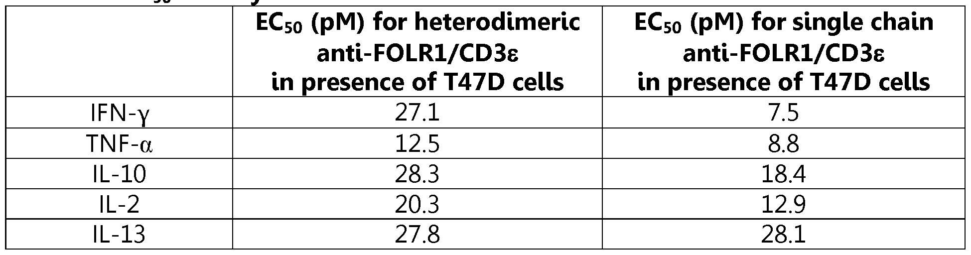


Claims
Priority Applications (7)
| Application Number | Priority Date | Filing Date | Title |
|---|---|---|---|
| AU2014236769A AU2014236769B2 (en) | 2013-03-15 | 2014-03-13 | Heterodimeric bispecific antibodies |
| MX2015012439A MX357011B (en) | 2013-03-15 | 2014-03-13 | Heterodimeric bispecific antibodies. |
| US14/774,320 US11634502B2 (en) | 2013-03-15 | 2014-03-13 | Heterodimeric bispecific antibodies |
| EP18168747.6A EP3424952A1 (en) | 2013-03-15 | 2014-03-13 | Heterodimeric bispecific antibodies |
| JP2016502206A JP6404313B2 (en) | 2013-03-15 | 2014-03-13 | Heterodimeric bispecific antibody |
| CA2903258A CA2903258C (en) | 2013-03-15 | 2014-03-13 | Heterodimeric bispecific antibodies |
| EP14722862.1A EP2970484B2 (en) | 2013-03-15 | 2014-03-13 | Heterodimeric bispecific antibodies |
Applications Claiming Priority (4)
| Application Number | Priority Date | Filing Date | Title |
|---|---|---|---|
| US201361791357P | 2013-03-15 | 2013-03-15 | |
| US61/791,357 | 2013-03-15 | ||
| US201461944841P | 2014-02-26 | 2014-02-26 | |
| US61/944,841 | 2014-02-26 |
Publications (1)
| Publication Number | Publication Date |
|---|---|
| WO2014151910A1 true WO2014151910A1 (en) | 2014-09-25 |
Family
ID=50686150
Family Applications (1)
| Application Number | Title | Priority Date | Filing Date |
|---|---|---|---|
| PCT/US2014/026658 WO2014151910A1 (en) | 2013-03-15 | 2014-03-13 | Heterodimeric bispecific antibodies |
Country Status (8)
| Country | Link |
|---|---|
| US (1) | US11634502B2 (en) |
| EP (2) | EP2970484B2 (en) |
| JP (2) | JP6404313B2 (en) |
| AU (1) | AU2014236769B2 (en) |
| CA (1) | CA2903258C (en) |
| MX (2) | MX357011B (en) |
| TW (1) | TW201439117A (en) |
| WO (1) | WO2014151910A1 (en) |
Cited By (93)
| Publication number | Priority date | Publication date | Assignee | Title |
|---|---|---|---|---|
| WO2016079076A1 (en) * | 2014-11-20 | 2016-05-26 | F. Hoffmann-La Roche Ag | T cell activating bispecific antigen binding molecules agiant folr1 and cd3 |
| WO2016100788A1 (en) * | 2014-12-19 | 2016-06-23 | Alkermes, Inc. | Single chain fc fusion proteins |
| WO2016094881A3 (en) * | 2014-12-11 | 2016-08-18 | Abbvie Inc. | Lrp-8 binding proteins |
| WO2016166360A1 (en) | 2015-04-17 | 2016-10-20 | Bayer Pharma Aktiengesellschaft | Bispecific antibody constructs for cdh3 and cd3 |
| WO2016127104A3 (en) * | 2015-02-06 | 2016-10-27 | University Of Maryland, Baltimore | Tetra-specific, octameric binding agents and antibodies against clostridium difficile toxin a and toxin b for treatment of c. difficile infection |
| WO2017021349A1 (en) | 2015-07-31 | 2017-02-09 | Amgen Research (Munich) Gmbh | Bispecific antibody constructs binding dll3 and cd3 |
| WO2017021354A1 (en) | 2015-07-31 | 2017-02-09 | Amgen Research (Munich) Gmbh | Antibody constructs for cd70 and cd3 |
| WO2017021370A1 (en) | 2015-07-31 | 2017-02-09 | Amgen Research (Munich) Gmbh | Bispecific antibody constructs binding egfrviii and cd3 |
| WO2017021362A1 (en) | 2015-07-31 | 2017-02-09 | Amgen Research (Munich) Gmbh | Antibody constructs for flt3 and cd3 |
| WO2017021356A1 (en) | 2015-07-31 | 2017-02-09 | Amgen Research (Munich) Gmbh | Bispecific antibody constructs binding mesothelin and cd3 |
| WO2017021526A1 (en) | 2015-08-05 | 2017-02-09 | Amgen Research (Munich) Gmbh | Immune-checkpoint inhibitors for use in the treatment of blood-borne cancers |
| US9708412B2 (en) | 2015-05-21 | 2017-07-18 | Harpoon Therapeutics, Inc. | Trispecific binding proteins and methods of use |
| WO2017162587A1 (en) * | 2016-03-22 | 2017-09-28 | F. Hoffmann-La Roche Ag | Protease-activated t cell bispecific molecules |
| JP2017186301A (en) * | 2016-02-03 | 2017-10-12 | アムゲン リサーチ (ミュンヘン) ゲーエムベーハーAMGEN Research(Munich)GmbH | Bcma and cd3 bispecific t cell engaging antibody constructs |
| JP2017184723A (en) * | 2016-02-03 | 2017-10-12 | アムゲン リサーチ (ミュンヘン) ゲーエムベーハーAMGEN Research(Munich)GmbH | Psma and cd3 bispecific t cell engaging antibody constructs |
| WO2017182427A1 (en) | 2016-04-19 | 2017-10-26 | Amgen Research (Munich) Gmbh | Administration of a bispecific construct binding to cd33 and cd3 for use in a method for the treatment of myeloid leukemia |
| JP2017536341A (en) * | 2014-10-09 | 2017-12-07 | エンクマフ アーゲー | Bispecific antibodies against CD3ε and ROR1 for use in the treatment of ovarian cancer |
| US9840554B2 (en) | 2015-06-15 | 2017-12-12 | Abbvie Inc. | Antibodies against platelet-derived growth factor (PDGF) |
| JP2018504092A (en) * | 2014-11-20 | 2018-02-15 | エフ.ホフマン−ラ ロシュ アーゲーF. Hoffmann−La Roche Aktiengesellschaft | Common light chain and methods of use |
| JP2018505849A (en) * | 2014-12-05 | 2018-03-01 | ジェネンテック, インコーポレイテッド | Anti-CD79b antibody and method of use |
| US9920115B2 (en) | 2016-05-20 | 2018-03-20 | Harpoon Therapeutics, Inc. | Single domain serum albumin binding protein |
| EP3302555A4 (en) * | 2015-05-29 | 2018-07-11 | Amphivena Therapeutics, Inc. | Methods of using bispecific cd33 and cd3 binding proteins |
| WO2018141910A1 (en) | 2017-02-02 | 2018-08-09 | Amgen Research (Munich) Gmbh | Low ph pharmaceutical composition comprising t cell engaging antibody constructs |
| JP2018524355A (en) * | 2015-07-06 | 2018-08-30 | ラボラトワール・フランセ・デュ・フラクシオンマン・エ・デ・ビョテクノロジーLaboratoire Francais Du Fractionnement Et Des Biotechnologies | Use of modified Fc fragments in immunotherapy |
| US10066016B2 (en) | 2016-05-20 | 2018-09-04 | Harpoon Therapeutics, Inc. | Single chain variable fragment CD3 binding proteins |
| WO2018204907A1 (en) | 2017-05-05 | 2018-11-08 | Amgen Inc. | Pharmaceutical composition comprising bispecific antibody constructs for improved storage and administration |
| WO2019118426A1 (en) | 2017-12-11 | 2019-06-20 | Amgen Inc. | Continuous manufacturing process for bispecific antibody products |
| US10335459B2 (en) | 2016-06-22 | 2019-07-02 | Alkermes, Inc. | Compositions for modulating IL-10 immunostimulatory and anti-inflammatory properties |
| CN109957026A (en) * | 2017-12-22 | 2019-07-02 | 成都恩沐生物科技有限公司 | Covalent multi-specificity antibody |
| WO2019133961A1 (en) | 2017-12-29 | 2019-07-04 | Amgen Inc. | Bispecific antibody construct directed to muc17 and cd3 |
| US10543271B2 (en) | 2017-05-12 | 2020-01-28 | Harpoon Therapeutics, Inc. | Mesothelin binding proteins |
| WO2020025792A1 (en) | 2018-08-03 | 2020-02-06 | Amgen Research (Munich) Gmbh | Antibody constructs for cldn18.2 and cd3 |
| WO2020025596A1 (en) | 2018-07-31 | 2020-02-06 | Amgen Research (Munich) Gmbh | Dosing regimen for bcma-cd3 bispecific antibodies |
| WO2020025532A1 (en) | 2018-07-30 | 2020-02-06 | Amgen Research (Munich) Gmbh | Prolonged administration of a bispecific antibody construct binding to cd33 and cd3 |
| WO2020069303A1 (en) | 2018-09-28 | 2020-04-02 | Amgen Inc. | Antibodies against soluble bcma |
| WO2020077212A1 (en) | 2018-10-11 | 2020-04-16 | Amgen Inc. | Downstream processing of bispecific antibody constructs |
| US10626190B2 (en) | 2014-07-01 | 2020-04-21 | Amphivena Therapeutics, Inc. | Bispecific CD33 and CD3 binding proteins |
| WO2020086635A1 (en) | 2018-10-23 | 2020-04-30 | Amgen Inc. | Automatic calibration and automatic maintenance of raman spectroscopic models for real-time predictions |
| US10730954B2 (en) | 2017-05-12 | 2020-08-04 | Harpoon Therapeutics, Inc. | MSLN targeting trispecific proteins and methods of use |
| WO2020159838A1 (en) | 2019-01-28 | 2020-08-06 | Amgen Inc. | A continuous manufacturing process for biologics manufacturing by integration of drug substance and drug product processes |
| US10781262B2 (en) | 2014-11-20 | 2020-09-22 | Hoffmann-La Roche Inc. | Combination therapy of T cell activating bispecific antigen binding molecules and PD-1 axis binding antagonists |
| US10815311B2 (en) | 2018-09-25 | 2020-10-27 | Harpoon Therapeutics, Inc. | DLL3 binding proteins and methods of use |
| US10844134B2 (en) | 2016-11-23 | 2020-11-24 | Harpoon Therapeutics, Inc. | PSMA targeting trispecific proteins and methods of use |
| US10849973B2 (en) | 2016-11-23 | 2020-12-01 | Harpoon Therapeutics, Inc. | Prostate specific membrane antigen binding protein |
| WO2020252442A1 (en) | 2019-06-13 | 2020-12-17 | Amgen Inc. | Automated biomass-based perfusion control in the manufacturing of biologics |
| US10906978B2 (en) | 2015-01-23 | 2021-02-02 | Sanofi | Anti-CD3 antibodies, anti-CD123 antibodies and bispecific antibodies specifically binding to CD3 and/or CD123 |
| EP3177645B1 (en) | 2014-08-08 | 2021-02-17 | Lindis Biotech GmbH | Subcutaneously administered bispecific antibodies for use in the treatment of cancer |
| US10927180B2 (en) | 2017-10-13 | 2021-02-23 | Harpoon Therapeutics, Inc. | B cell maturation antigen binding proteins |
| WO2021050640A1 (en) | 2019-09-10 | 2021-03-18 | Amgen Inc. | Purification method for bispecific antigen-binding polypeptides with enhanced protein l capture dynamic binding capacity |
| EP3819007A1 (en) | 2019-11-11 | 2021-05-12 | Amgen Research (Munich) GmbH | Dosing regimen for anti-bcma agents |
| WO2021091906A1 (en) | 2019-11-04 | 2021-05-14 | Amgen Inc. | Methods for treating leukemia |
| WO2021097344A1 (en) | 2019-11-13 | 2021-05-20 | Amgen Inc. | Method for reduced aggregate formation in downstream processing of bispecific antigen-binding molecules |
| WO2021127528A1 (en) | 2019-12-20 | 2021-06-24 | Amgen Inc. | Mesothelin-targeted cd40 agonistic multispecific antibody constructs for the treatment of solid tumors |
| WO2021150824A1 (en) | 2020-01-22 | 2021-07-29 | Amgen Research (Munich) Gmbh | Combinations of antibody constructs and inhibitors of cytokine release syndrome and uses thereof |
| WO2021158469A1 (en) | 2020-02-03 | 2021-08-12 | Amgen Inc. | Multivariate bracketing approach for sterile filter validation |
| WO2021183861A1 (en) | 2020-03-12 | 2021-09-16 | Amgen Inc. | Method for treatment and prophylaxis of crs in patients comprising a combination of bispecifc antibodies binding to cds x cancer cell and tnfalpha or il-6 inhibitor |
| WO2021188851A1 (en) | 2020-03-19 | 2021-09-23 | Amgen Inc. | Antibodies against mucin 17 and uses thereof |
| US11136403B2 (en) | 2017-10-13 | 2021-10-05 | Harpoon Therapeutics, Inc. | Trispecific proteins and methods of use |
| WO2021231969A1 (en) * | 2020-05-14 | 2021-11-18 | Xencor, Inc. | Heterodimeric antibodies that bind msln and cd3 |
| US11180563B2 (en) | 2020-02-21 | 2021-11-23 | Harpoon Therapeutics, Inc. | FLT3 binding proteins and methods of use |
| WO2021236638A1 (en) | 2020-05-19 | 2021-11-25 | Amgen Inc. | Mageb2 binding constructs |
| WO2021243320A2 (en) | 2020-05-29 | 2021-12-02 | Amgen Inc. | Adverse effects-mitigating administration of a bispecific antibody construct binding to cd33 and cd3 |
| US11242390B2 (en) | 2016-03-22 | 2022-02-08 | Hoffmann-La Roche Inc. | Protease-activated T cell bispecific molecules |
| WO2022060901A1 (en) | 2020-09-16 | 2022-03-24 | Amgen Inc. | Methods for administering therapeutic doses of bispecific t-cell engaging molecules for the treatment of cancer |
| WO2022060878A1 (en) | 2020-09-16 | 2022-03-24 | Amgen Inc. | Methods for treating prostate cancer |
| US11286300B2 (en) | 2015-10-01 | 2022-03-29 | Hoffmann-La Roche Inc. | Humanized anti-human CD19 antibodies and methods of use |
| WO2022096704A1 (en) | 2020-11-06 | 2022-05-12 | Amgen Inc. | Antigen binding domain with reduced clipping rate |
| WO2022096698A1 (en) | 2020-11-06 | 2022-05-12 | Amgen Inc. | Polypeptide constructs binding to cd3 |
| WO2022096716A2 (en) | 2020-11-06 | 2022-05-12 | Amgen Inc. | Multitargeting bispecific antigen-binding molecules of increased selectivity |
| WO2022096700A1 (en) | 2020-11-06 | 2022-05-12 | Amgen Research (Munich) Gmbh | Polypeptide constructs selectively binding to cldn6 and cd3 |
| WO2022103781A1 (en) | 2020-11-10 | 2022-05-19 | Amgen Inc. | Methods for administering a bcma x cd3 binding molecule |
| US11434302B2 (en) | 2016-02-03 | 2022-09-06 | Amgen Research (Munich) Gmbh | Bispecific T cell engaging antibody constructs |
| WO2022192504A1 (en) | 2021-03-10 | 2022-09-15 | Amgen Inc. | Methods for purification of recombinant proteins |
| WO2022191971A1 (en) | 2021-03-10 | 2022-09-15 | Amgen Inc. | Parallel chromatography systems and methods |
| WO2022212831A1 (en) | 2021-04-02 | 2022-10-06 | Amgen Inc. | Mageb2 binding constructs |
| KR20220142441A (en) | 2020-02-14 | 2022-10-21 | 쿄와 기린 가부시키가이샤 | Bispecific antibody that binds to CD3 |
| WO2022234102A1 (en) | 2021-05-06 | 2022-11-10 | Amgen Research (Munich) Gmbh | Cd20 and cd22 targeting antigen-binding molecules for use in proliferative diseases |
| US11535668B2 (en) | 2017-02-28 | 2022-12-27 | Harpoon Therapeutics, Inc. | Inducible monovalent antigen binding protein |
| US11623958B2 (en) | 2016-05-20 | 2023-04-11 | Harpoon Therapeutics, Inc. | Single chain variable fragment CD3 binding proteins |
| US11634506B2 (en) | 2013-01-14 | 2023-04-25 | Xencor, Inc. | Heterodimeric proteins |
| WO2023076318A1 (en) | 2021-10-27 | 2023-05-04 | Amgen Inc. | Deep learning-based prediction for monitoring of pharmaceuticals using spectroscopy |
| WO2023079493A1 (en) | 2021-11-03 | 2023-05-11 | Affimed Gmbh | Bispecific cd16a binders |
| WO2023078968A1 (en) | 2021-11-03 | 2023-05-11 | Affimed Gmbh | Bispecific cd16a binders |
| US11661462B2 (en) | 2014-07-31 | 2023-05-30 | Amgen Research (Munich) Gmbh | Optimized cross-species specific bispecific single chain antibody contructs |
| US11673972B2 (en) | 2014-11-26 | 2023-06-13 | Xencor, Inc. | Heterodimeric antibodies that bind CD3 and tumor antigens |
| US11739144B2 (en) | 2021-03-09 | 2023-08-29 | Xencor, Inc. | Heterodimeric antibodies that bind CD3 and CLDN6 |
| US11780920B2 (en) | 2020-06-19 | 2023-10-10 | Hoffmann-La Roche Inc. | Antibodies binding to CD3 and CD19 |
| US11814423B2 (en) | 2013-03-15 | 2023-11-14 | Xencor, Inc. | Heterodimeric proteins |
| WO2023218027A1 (en) | 2022-05-12 | 2023-11-16 | Amgen Research (Munich) Gmbh | Multichain multitargeting bispecific antigen-binding molecules of increased selectivity |
| US11859012B2 (en) | 2021-03-10 | 2024-01-02 | Xencor, Inc. | Heterodimeric antibodies that bind CD3 and GPC3 |
| US11919956B2 (en) | 2020-05-14 | 2024-03-05 | Xencor, Inc. | Heterodimeric antibodies that bind prostate specific membrane antigen (PSMA) and CD3 |
| WO2024059675A2 (en) | 2022-09-14 | 2024-03-21 | Amgen Inc. | Bispecific molecule stabilizing composition |
| US11945880B2 (en) | 2014-11-26 | 2024-04-02 | Xencor, Inc. | Heterodimeric antibodies that bind CD3 and tumor antigens |
Families Citing this family (15)
| Publication number | Priority date | Publication date | Assignee | Title |
|---|---|---|---|---|
| CN104302665B (en) * | 2011-12-21 | 2019-04-23 | 安姆根有限公司 | And the variant Fc polypeptide of the combination enhancing of neonatal Fc receptor |
| JO3519B1 (en) * | 2013-01-25 | 2020-07-05 | Amgen Inc | Antibody constructs for CDH19 and CD3 |
| KR20200064096A (en) | 2017-10-14 | 2020-06-05 | 싸이톰스 테라퓨틱스, 인크. | Antibodies, activatable antibodies, bispecific antibodies and bispecific activatable antibodies and methods of using them |
| CN108610417B (en) * | 2018-04-28 | 2021-01-26 | 暨南大学 | Anti-tetanus toxin neutralizing antibody, preparation method and application thereof |
| GB201811415D0 (en) | 2018-07-12 | 2018-08-29 | F Star Beta Ltd | Anti-Mesothelin Anti bodies |
| CN113784732B (en) | 2019-05-07 | 2024-03-22 | 亘喜生物科技(上海)有限公司 | Engineered immune cells targeting BCMA and uses thereof |
| CN116987197A (en) * | 2019-06-20 | 2023-11-03 | 成都恩沐生物科技有限公司 | Covalent multispecific antibodies |
| US20210032370A1 (en) | 2019-08-02 | 2021-02-04 | Immatics Biotechnologies Gmbh | Recruiting agent further binding an mhc molecule |
| DE102019121007A1 (en) | 2019-08-02 | 2021-02-04 | Immatics Biotechnologies Gmbh | Antigen binding proteins that specifically bind to MAGE-A |
| EP4090365A1 (en) | 2020-01-15 | 2022-11-23 | Immatics Biotechnologies GmbH | Antigen binding proteins specifically binding prame |
| US20240092890A1 (en) | 2020-08-27 | 2024-03-21 | Juntendo Educational Foundation | Anti-cleaved mutant calr-cd3 bispecific antibody and pharmaceutical composition |
| TW202309071A (en) | 2021-05-05 | 2023-03-01 | 德商英麥提克生物技術股份有限公司 | Antigen binding proteins specifically binding prame |
| US20230190806A1 (en) | 2021-10-06 | 2023-06-22 | Immatics Biotechnologies Gmbh | Methods of treating metastatic lesions and compositions thereof |
| US20230192886A1 (en) | 2021-11-08 | 2023-06-22 | Immatics Biotechnologies Gmbh | Adoptive cell therapy combination treatment and compositions thereof |
| WO2023209124A1 (en) | 2022-04-29 | 2023-11-02 | Immatics Biotechnologies Gmbh | Mammalian display platform for multispecific antigen binding proteins |
Citations (6)
| Publication number | Priority date | Publication date | Assignee | Title |
|---|---|---|---|---|
| WO2008024188A2 (en) * | 2006-08-18 | 2008-02-28 | Abbott Laboratories | Dual variable domain immunoglobulin and uses thereof |
| EP2050764A1 (en) * | 2007-10-15 | 2009-04-22 | sanofi-aventis | Novel polyvalent bispecific antibody format and uses thereof |
| WO2010037836A2 (en) * | 2008-10-01 | 2010-04-08 | Micromet Ag | Cross-species-specific psmaxcd3 bispecific single chain antibody |
| WO2012025530A1 (en) * | 2010-08-24 | 2012-03-01 | F. Hoffmann-La Roche Ag | Bispecific antibodies comprising a disulfide stabilized - fv fragment |
| WO2012118903A2 (en) * | 2011-03-01 | 2012-09-07 | Amgen Inc. | Bispecific binding agents |
| WO2012135345A1 (en) * | 2011-03-28 | 2012-10-04 | Sanofi | Dual variable region antibody-like binding proteins having cross-over binding region orientation |
Family Cites Families (32)
| Publication number | Priority date | Publication date | Assignee | Title |
|---|---|---|---|---|
| JP2919890B2 (en) | 1988-11-11 | 1999-07-19 | メディカル リサーチ カウンスル | Single domain ligand, receptor consisting of the ligand, method for producing the same, and use of the ligand and the receptor |
| DK0672142T3 (en) | 1992-12-04 | 2001-06-18 | Medical Res Council | Multivalent and multi-specific binding proteins as well as their preparation and use |
| US5731168A (en) | 1995-03-01 | 1998-03-24 | Genentech, Inc. | Method for making heteromultimeric polypeptides |
| US20020137890A1 (en) | 1997-03-31 | 2002-09-26 | Genentech, Inc. | Secreted and transmembrane polypeptides and nucleic acids encoding the same |
| US20020062010A1 (en) | 1997-05-02 | 2002-05-23 | Genentech, Inc. | Method for making multispecific antibodies having heteromultimeric and common components |
| JP2003531588A (en) | 2000-04-11 | 2003-10-28 | ジェネンテック・インコーポレーテッド | Multivalent antibodies and their uses |
| CA2410551A1 (en) * | 2000-06-30 | 2002-01-10 | Vlaams Interuniversitair Instituut Voor Biotechnologie Vzw (Vib) | Heterodimeric fusion proteins |
| AU2002248184C1 (en) | 2000-12-12 | 2018-01-04 | Board Of Regents, The University Of Texas System | Molecules with extended half-lives, compositions and uses thereof |
| US7053202B2 (en) | 2001-10-19 | 2006-05-30 | Millennium Pharmaceuticals, Inc. | Immunoglobulin DNA cassette molecules, monobody constructs, methods of production, and methods of use therefor |
| CN1678634A (en) * | 2002-06-28 | 2005-10-05 | 多曼蒂斯有限公司 | Immunoglobulin single variable antigen combination area and its opposite constituent |
| US8367805B2 (en) | 2004-11-12 | 2013-02-05 | Xencor, Inc. | Fc variants with altered binding to FcRn |
| KR100609578B1 (en) | 2004-12-28 | 2006-08-08 | 주식회사 하이닉스반도체 | Method of forming a floating gate electrode in flash memory device |
| US9284375B2 (en) | 2005-04-15 | 2016-03-15 | Macrogenics, Inc. | Covalent diabodies and uses thereof |
| US9963510B2 (en) | 2005-04-15 | 2018-05-08 | Macrogenics, Inc. | Covalent diabodies and uses thereof |
| EP1868650B1 (en) | 2005-04-15 | 2018-10-03 | MacroGenics, Inc. | Covalent diabodies and uses thereof |
| NZ612578A (en) | 2005-08-19 | 2014-11-28 | Abbvie Inc | Dual variable domain immunoglobin and uses thereof |
| AR056142A1 (en) | 2005-10-21 | 2007-09-19 | Amgen Inc | METHODS TO GENERATE THE MONOVALENT IGG ANTIBODY |
| MX2009010611A (en) | 2007-04-03 | 2010-03-26 | Micromet Ag | Cross-species-specific bispecific binders. |
| KR101589759B1 (en) | 2007-04-03 | 2016-01-29 | 암젠 리서치 (뮌헨) 게엠베하 | Cross-species-specific cd3-epsilon binding domain |
| ES2702087T3 (en) | 2007-06-21 | 2019-02-27 | Macrogenics Inc | Covalent diabodies and their uses |
| PT2235064E (en) * | 2008-01-07 | 2016-03-01 | Amgen Inc | Method for making antibody fc-heterodimeric molecules using electrostatic steering effects |
| EP2297209A4 (en) | 2008-06-03 | 2012-08-01 | Abbott Lab | Dual variable domain immunoglobulins and uses thereof |
| PL2786762T3 (en) | 2008-12-19 | 2019-09-30 | Macrogenics, Inc. | Covalent diabodies and uses thereof |
| CN103403025B (en) | 2011-02-28 | 2016-10-12 | 弗·哈夫曼-拉罗切有限公司 | Monovalent antigen binding protein |
| US20140112926A1 (en) | 2011-03-16 | 2014-04-24 | Amgen Inc. | Fc VARIANTS |
| CN104302665B (en) | 2011-12-21 | 2019-04-23 | 安姆根有限公司 | And the variant Fc polypeptide of the combination enhancing of neonatal Fc receptor |
| EP2938637A2 (en) | 2012-12-28 | 2015-11-04 | AbbVie Inc. | Multivalent binding protein compositions |
| US20160038588A1 (en) * | 2013-03-15 | 2016-02-11 | Amgen Inc. | Myostatin Antagonism in Human Subjects |
| DK3587448T3 (en) | 2013-03-15 | 2021-08-09 | Xencor Inc | HETERODIMER PROTEINS |
| US20140302037A1 (en) * | 2013-03-15 | 2014-10-09 | Amgen Inc. | BISPECIFIC-Fc MOLECULES |
| US20140308285A1 (en) * | 2013-03-15 | 2014-10-16 | Amgen Inc. | Heterodimeric bispecific antibodies |
| EP3733710A1 (en) * | 2013-09-25 | 2020-11-04 | Amgen, Inc | Hetrodimeric v-c-fc-v-c antibody |
-
2014
- 2014-03-13 JP JP2016502206A patent/JP6404313B2/en active Active
- 2014-03-13 MX MX2015012439A patent/MX357011B/en active IP Right Grant
- 2014-03-13 EP EP14722862.1A patent/EP2970484B2/en active Active
- 2014-03-13 EP EP18168747.6A patent/EP3424952A1/en active Pending
- 2014-03-13 CA CA2903258A patent/CA2903258C/en active Active
- 2014-03-13 WO PCT/US2014/026658 patent/WO2014151910A1/en active Application Filing
- 2014-03-13 AU AU2014236769A patent/AU2014236769B2/en active Active
- 2014-03-13 US US14/774,320 patent/US11634502B2/en active Active
- 2014-03-14 TW TW103109334A patent/TW201439117A/en unknown
-
2015
- 2015-09-11 MX MX2018007206A patent/MX2018007206A/en unknown
-
2018
- 2018-09-12 JP JP2018170391A patent/JP6700354B2/en active Active
Patent Citations (6)
| Publication number | Priority date | Publication date | Assignee | Title |
|---|---|---|---|---|
| WO2008024188A2 (en) * | 2006-08-18 | 2008-02-28 | Abbott Laboratories | Dual variable domain immunoglobulin and uses thereof |
| EP2050764A1 (en) * | 2007-10-15 | 2009-04-22 | sanofi-aventis | Novel polyvalent bispecific antibody format and uses thereof |
| WO2010037836A2 (en) * | 2008-10-01 | 2010-04-08 | Micromet Ag | Cross-species-specific psmaxcd3 bispecific single chain antibody |
| WO2012025530A1 (en) * | 2010-08-24 | 2012-03-01 | F. Hoffmann-La Roche Ag | Bispecific antibodies comprising a disulfide stabilized - fv fragment |
| WO2012118903A2 (en) * | 2011-03-01 | 2012-09-07 | Amgen Inc. | Bispecific binding agents |
| WO2012135345A1 (en) * | 2011-03-28 | 2012-10-04 | Sanofi | Dual variable region antibody-like binding proteins having cross-over binding region orientation |
Non-Patent Citations (1)
| Title |
|---|
| CHRISTIAN KLEIN ET AL: "Progress in overcoming the chain association issue in bispecific heterodimeric IgG antibodies", MABS, vol. 4, no. 6, 1 November 2012 (2012-11-01), pages 653 - 663, XP055106060, ISSN: 1942-0862, DOI: 10.4161/mabs.21379 * |
Cited By (154)
| Publication number | Priority date | Publication date | Assignee | Title |
|---|---|---|---|---|
| US11634506B2 (en) | 2013-01-14 | 2023-04-25 | Xencor, Inc. | Heterodimeric proteins |
| US11814423B2 (en) | 2013-03-15 | 2023-11-14 | Xencor, Inc. | Heterodimeric proteins |
| US10626190B2 (en) | 2014-07-01 | 2020-04-21 | Amphivena Therapeutics, Inc. | Bispecific CD33 and CD3 binding proteins |
| US11661462B2 (en) | 2014-07-31 | 2023-05-30 | Amgen Research (Munich) Gmbh | Optimized cross-species specific bispecific single chain antibody contructs |
| EP3177645B1 (en) | 2014-08-08 | 2021-02-17 | Lindis Biotech GmbH | Subcutaneously administered bispecific antibodies for use in the treatment of cancer |
| JP2017536341A (en) * | 2014-10-09 | 2017-12-07 | エンクマフ アーゲー | Bispecific antibodies against CD3ε and ROR1 for use in the treatment of ovarian cancer |
| JP2018504092A (en) * | 2014-11-20 | 2018-02-15 | エフ.ホフマン−ラ ロシュ アーゲーF. Hoffmann−La Roche Aktiengesellschaft | Common light chain and methods of use |
| US10781262B2 (en) | 2014-11-20 | 2020-09-22 | Hoffmann-La Roche Inc. | Combination therapy of T cell activating bispecific antigen binding molecules and PD-1 axis binding antagonists |
| US11613587B2 (en) | 2014-11-20 | 2023-03-28 | Hoffmann-La Roche Inc. | Combination therapy of T cell activating bispecific antigen binding molecules and PD-1 axis binding antagonists |
| CN107074955B (en) * | 2014-11-20 | 2021-06-22 | 豪夫迈·罗氏有限公司 | T cell activating bispecific antigen binding molecules against FolR1 and CD3 |
| AU2015348595B2 (en) * | 2014-11-20 | 2021-06-10 | F. Hoffmann-La Roche Ag | T cell activating bispecific antigen binding molecules against FOLR1 and CD3 |
| WO2016079076A1 (en) * | 2014-11-20 | 2016-05-26 | F. Hoffmann-La Roche Ag | T cell activating bispecific antigen binding molecules agiant folr1 and cd3 |
| CN107074955A (en) * | 2014-11-20 | 2017-08-18 | 豪夫迈·罗氏有限公司 | For FolR1 and CD3 T cell activation bispecific antigen binding molecules |
| EP3221357B1 (en) * | 2014-11-20 | 2020-06-03 | F.Hoffmann-La Roche Ag | Common light chains and methods of use |
| JP7146395B2 (en) | 2014-11-20 | 2022-10-04 | エフ・ホフマン-ラ・ロシュ・アクチェンゲゼルシャフト | Bispecific antibodies targeting FolR1 and CD3 |
| TWI756164B (en) | 2014-11-20 | 2022-03-01 | 瑞士商赫孚孟拉羅股份公司 | T cell activating bispecific antigen binding molecules |
| RU2747011C2 (en) * | 2014-11-20 | 2021-04-23 | Ф.Хоффманн-Ля Рош Аг | General light chains and methods of their application |
| JP7164949B2 (en) | 2014-11-20 | 2022-11-02 | エフ.ホフマン-ラ ロシュ アーゲー | Common Light Chains and Methods of Use |
| JP2017536121A (en) * | 2014-11-20 | 2017-12-07 | エフ・ホフマン−ラ・ロシュ・アクチェンゲゼルシャフト | Bispecific antibodies targeting FolR1 and CD3 |
| EP3747905A1 (en) * | 2014-11-20 | 2020-12-09 | F. Hoffmann-La Roche AG | Common light chains and methods of use |
| US11859011B2 (en) | 2014-11-26 | 2024-01-02 | Xencor, Inc. | Heterodimeric antibodies that bind CD3 and tumor antigens |
| US11945880B2 (en) | 2014-11-26 | 2024-04-02 | Xencor, Inc. | Heterodimeric antibodies that bind CD3 and tumor antigens |
| US11673972B2 (en) | 2014-11-26 | 2023-06-13 | Xencor, Inc. | Heterodimeric antibodies that bind CD3 and tumor antigens |
| JP2018505849A (en) * | 2014-12-05 | 2018-03-01 | ジェネンテック, インコーポレイテッド | Anti-CD79b antibody and method of use |
| US10941199B2 (en) | 2014-12-05 | 2021-03-09 | Genentech, Inc. | Anti-CD79b antibodies and methods of use |
| WO2016094881A3 (en) * | 2014-12-11 | 2016-08-18 | Abbvie Inc. | Lrp-8 binding proteins |
| US10093733B2 (en) | 2014-12-11 | 2018-10-09 | Abbvie Inc. | LRP-8 binding dual variable domain immunoglobulin proteins |
| WO2016100788A1 (en) * | 2014-12-19 | 2016-06-23 | Alkermes, Inc. | Single chain fc fusion proteins |
| US10881741B2 (en) | 2014-12-19 | 2021-01-05 | Alkermes, Inc. | Single chain Fc fusion proteins |
| US10906978B2 (en) | 2015-01-23 | 2021-02-02 | Sanofi | Anti-CD3 antibodies, anti-CD123 antibodies and bispecific antibodies specifically binding to CD3 and/or CD123 |
| US10961299B2 (en) | 2015-02-06 | 2021-03-30 | University Of Maryland, Baltimore | Tetra-specific, octameric binding agents and antibodies against Clostridium difficile toxin A and toxin B for treatment of C. difficile infection |
| WO2016127104A3 (en) * | 2015-02-06 | 2016-10-27 | University Of Maryland, Baltimore | Tetra-specific, octameric binding agents and antibodies against clostridium difficile toxin a and toxin b for treatment of c. difficile infection |
| US11926666B2 (en) | 2015-04-17 | 2024-03-12 | Amgen Research (Munich) Gmbh | Bispecific antibody constructs for CDH3 and CD3 |
| US11028171B2 (en) | 2015-04-17 | 2021-06-08 | Amgen Research (Munich) Gmbh | Bispecific antibody constructs for CDH3 and CD3 |
| EP4276116A2 (en) | 2015-04-17 | 2023-11-15 | Amgen Research (Munich) GmbH | Bispecific antibody constructs for cdh3 and cd3 |
| WO2016166360A1 (en) | 2015-04-17 | 2016-10-20 | Bayer Pharma Aktiengesellschaft | Bispecific antibody constructs for cdh3 and cd3 |
| JP2020125301A (en) * | 2015-05-21 | 2020-08-20 | ハープーン セラピューティクス,インク. | Tri-specific binding proteins and use methods thereof |
| US9708412B2 (en) | 2015-05-21 | 2017-07-18 | Harpoon Therapeutics, Inc. | Trispecific binding proteins and methods of use |
| JP2018517431A (en) * | 2015-05-21 | 2018-07-05 | ハープーン セラピューティクス,インク. | Trispecific binding proteins and methods of use |
| US10954311B2 (en) | 2015-05-21 | 2021-03-23 | Harpoon Therapeutics, Inc. | Trispecific binding proteins and methods of use |
| US10738118B2 (en) | 2015-05-29 | 2020-08-11 | Amphivena Therapeutics, Inc. | Methods of using bispecific CD33 and CD3 binding proteins |
| US11753469B2 (en) | 2015-05-29 | 2023-09-12 | Anji Bruno, Llc | Methods of using bispecific CD33 and CD3 binding proteins |
| EP3302555A4 (en) * | 2015-05-29 | 2018-07-11 | Amphivena Therapeutics, Inc. | Methods of using bispecific cd33 and cd3 binding proteins |
| CN108289949B (en) * | 2015-05-29 | 2022-04-12 | 安普希韦纳治疗公司 | Methods of use of bispecific CD33 and CD3 binding proteins |
| CN108289949A (en) * | 2015-05-29 | 2018-07-17 | 安普希韦纳治疗公司 | The application method of bispecific CD33 and CD3 conjugated protein |
| EP4190814A1 (en) * | 2015-05-29 | 2023-06-07 | Amphivena Therapeutics, Inc. | Methods of using bispecific cd33 and cd3 binding proteins |
| AU2016271113B2 (en) * | 2015-05-29 | 2022-09-08 | Amphivena Therapeutics, Inc. | Methods of using bispecific CD33 and CD3 binding proteins |
| US9840554B2 (en) | 2015-06-15 | 2017-12-12 | Abbvie Inc. | Antibodies against platelet-derived growth factor (PDGF) |
| JP7102496B2 (en) | 2015-07-06 | 2022-07-19 | ラボラトワール・フランセ・デュ・フラクシオンマン・エ・デ・ビョテクノロジー | Use of modified Fc fragments in immunotherapy |
| JP2021050235A (en) * | 2015-07-06 | 2021-04-01 | ラボラトワール・フランセ・デュ・フラクシオンマン・エ・デ・ビョテクノロジーLaboratoire Francais Du Fractionnement Et Des Biotechnologies | Use of modified Fc fragments in immunotherapy |
| JP2018524355A (en) * | 2015-07-06 | 2018-08-30 | ラボラトワール・フランセ・デュ・フラクシオンマン・エ・デ・ビョテクノロジーLaboratoire Francais Du Fractionnement Et Des Biotechnologies | Use of modified Fc fragments in immunotherapy |
| US11155629B2 (en) | 2015-07-31 | 2021-10-26 | Amgen Research (Munich) Gmbh | Method for treating glioblastoma or glioma with antibody constructs for EGFRVIII and CD3 |
| US11591396B2 (en) | 2015-07-31 | 2023-02-28 | Amgen Research (Munich) Gmbh | Antibody constructs for DLL3 and CD3 |
| WO2017021354A1 (en) | 2015-07-31 | 2017-02-09 | Amgen Research (Munich) Gmbh | Antibody constructs for cd70 and cd3 |
| US10294300B2 (en) | 2015-07-31 | 2019-05-21 | Amgen Research (Munich) Gmbh | Antibody constructs for DLL3 and CD3 |
| US11447567B2 (en) | 2015-07-31 | 2022-09-20 | Amgen Research (Munich) Gmbh | Antibody constructs for FLT3 and CD3 |
| WO2017021370A1 (en) | 2015-07-31 | 2017-02-09 | Amgen Research (Munich) Gmbh | Bispecific antibody constructs binding egfrviii and cd3 |
| US10519241B2 (en) | 2015-07-31 | 2019-12-31 | Amgen Research (Munich) Gmbh | Antibody constructs for EGFRVIII and CD3 |
| EP4219562A2 (en) | 2015-07-31 | 2023-08-02 | Amgen Research (Munich) GmbH | Antibody constructs for flt3 and cd3 |
| EP3865514A1 (en) | 2015-07-31 | 2021-08-18 | Amgen Research (Munich) GmbH | Bispecific antibody constructs binding dll3 and cd3 |
| EP3912999A1 (en) | 2015-07-31 | 2021-11-24 | Amgen Research (Munich) GmbH | Bispecific antibody constructs binding egfrviii and cd3 |
| US10851170B2 (en) | 2015-07-31 | 2020-12-01 | Amgen Research (Munich) Gmbh | Antibody constructs for CD70 and CD3 |
| WO2017021362A1 (en) | 2015-07-31 | 2017-02-09 | Amgen Research (Munich) Gmbh | Antibody constructs for flt3 and cd3 |
| WO2017021356A1 (en) | 2015-07-31 | 2017-02-09 | Amgen Research (Munich) Gmbh | Bispecific antibody constructs binding mesothelin and cd3 |
| EP4327885A2 (en) | 2015-07-31 | 2024-02-28 | Amgen Research (Munich) GmbH | Antibody constructs for msln and cd3 |
| US10683351B2 (en) | 2015-07-31 | 2020-06-16 | Amgen Research (Munich) Gmbh | Antibody constructs for DLL3 and CD3 |
| US11884720B2 (en) | 2015-07-31 | 2024-01-30 | Amgen Research (Munich) Gmbh | Antibody constructs for MSLN and CD3 |
| WO2017021349A1 (en) | 2015-07-31 | 2017-02-09 | Amgen Research (Munich) Gmbh | Bispecific antibody constructs binding dll3 and cd3 |
| WO2017021526A1 (en) | 2015-08-05 | 2017-02-09 | Amgen Research (Munich) Gmbh | Immune-checkpoint inhibitors for use in the treatment of blood-borne cancers |
| US11286300B2 (en) | 2015-10-01 | 2022-03-29 | Hoffmann-La Roche Inc. | Humanized anti-human CD19 antibodies and methods of use |
| US10781264B2 (en) | 2016-02-03 | 2020-09-22 | Amgen Research (Munich) Gmbh | PSMA and CD3 bispecific T cell engaging antibody constructs |
| EP4206228A1 (en) | 2016-02-03 | 2023-07-05 | Amgen Research (Munich) GmbH | Psma and cd3 bispecific t cell engaging constructs |
| EP4039709A1 (en) | 2016-02-03 | 2022-08-10 | Amgen Research (Munich) GmbH | Bcma and cd3 bispecific t cell engaging antibody constructs |
| US10301391B2 (en) | 2016-02-03 | 2019-05-28 | Amgen Research (Munich) Gmbh | BCMA and CD3 bispecific T cell engaging antibody constructs |
| US11434302B2 (en) | 2016-02-03 | 2022-09-06 | Amgen Research (Munich) Gmbh | Bispecific T cell engaging antibody constructs |
| JP2017184723A (en) * | 2016-02-03 | 2017-10-12 | アムゲン リサーチ (ミュンヘン) ゲーエムベーハーAMGEN Research(Munich)GmbH | Psma and cd3 bispecific t cell engaging antibody constructs |
| JP2017186301A (en) * | 2016-02-03 | 2017-10-12 | アムゲン リサーチ (ミュンヘン) ゲーエムベーハーAMGEN Research(Munich)GmbH | Bcma and cd3 bispecific t cell engaging antibody constructs |
| JP2022017277A (en) * | 2016-02-03 | 2022-01-25 | アムゲン リサーチ (ミュンヘン) ゲーエムベーハー | Bcma and cd3 bispecific t cell engaging antibody constructs |
| US11352433B2 (en) | 2016-02-03 | 2022-06-07 | Amgen Research (Munich) Gmbh | BCMA and CD3 bispecific T cell engaging antibody constructs |
| US11242390B2 (en) | 2016-03-22 | 2022-02-08 | Hoffmann-La Roche Inc. | Protease-activated T cell bispecific molecules |
| WO2017162587A1 (en) * | 2016-03-22 | 2017-09-28 | F. Hoffmann-La Roche Ag | Protease-activated t cell bispecific molecules |
| JP2019514871A (en) * | 2016-04-19 | 2019-06-06 | アムゲン リサーチ (ミュンヘン) ゲーエムベーハーAMGEN Research(Munich)GmbH | Methods of administration of bispecific constructs that bind CD33 and CD3 for use in a method of treating myeloid leukemia |
| US11053318B2 (en) | 2016-04-19 | 2021-07-06 | Amgen Research (Munich) Gmbh | Administration of a bispecific construct binding to CD33 and CD3 for use in a method for the treatment of myeloid leukemia |
| WO2017182427A1 (en) | 2016-04-19 | 2017-10-26 | Amgen Research (Munich) Gmbh | Administration of a bispecific construct binding to cd33 and cd3 for use in a method for the treatment of myeloid leukemia |
| US10066016B2 (en) | 2016-05-20 | 2018-09-04 | Harpoon Therapeutics, Inc. | Single chain variable fragment CD3 binding proteins |
| US11623958B2 (en) | 2016-05-20 | 2023-04-11 | Harpoon Therapeutics, Inc. | Single chain variable fragment CD3 binding proteins |
| US9920115B2 (en) | 2016-05-20 | 2018-03-20 | Harpoon Therapeutics, Inc. | Single domain serum albumin binding protein |
| US10100106B2 (en) | 2016-05-20 | 2018-10-16 | Harpoon Therapeutics, Inc. | Single domain serum albumin binding protein |
| US11453716B2 (en) | 2016-05-20 | 2022-09-27 | Harpoon Therapeutics, Inc. | Single domain serum albumin binding protein |
| US10544221B2 (en) | 2016-05-20 | 2020-01-28 | Harpoon Therapeutics, Inc. | Single chain variable fragment CD3 binding proteins |
| US11534480B2 (en) | 2016-06-22 | 2022-12-27 | Alkermes, Inc. | Compositions and methods for modulating IL-10 immunostimulatory and anti-inflammatory properties |
| US10335459B2 (en) | 2016-06-22 | 2019-07-02 | Alkermes, Inc. | Compositions for modulating IL-10 immunostimulatory and anti-inflammatory properties |
| US10849973B2 (en) | 2016-11-23 | 2020-12-01 | Harpoon Therapeutics, Inc. | Prostate specific membrane antigen binding protein |
| US10844134B2 (en) | 2016-11-23 | 2020-11-24 | Harpoon Therapeutics, Inc. | PSMA targeting trispecific proteins and methods of use |
| WO2018141910A1 (en) | 2017-02-02 | 2018-08-09 | Amgen Research (Munich) Gmbh | Low ph pharmaceutical composition comprising t cell engaging antibody constructs |
| US11535668B2 (en) | 2017-02-28 | 2022-12-27 | Harpoon Therapeutics, Inc. | Inducible monovalent antigen binding protein |
| WO2018204907A1 (en) | 2017-05-05 | 2018-11-08 | Amgen Inc. | Pharmaceutical composition comprising bispecific antibody constructs for improved storage and administration |
| US11918650B2 (en) | 2017-05-05 | 2024-03-05 | Amgen Inc. | Pharmaceutical composition comprising bispecific antibody constructs for improved storage and administration |
| US10730954B2 (en) | 2017-05-12 | 2020-08-04 | Harpoon Therapeutics, Inc. | MSLN targeting trispecific proteins and methods of use |
| US11607453B2 (en) | 2017-05-12 | 2023-03-21 | Harpoon Therapeutics, Inc. | Mesothelin binding proteins |
| US10543271B2 (en) | 2017-05-12 | 2020-01-28 | Harpoon Therapeutics, Inc. | Mesothelin binding proteins |
| US11136403B2 (en) | 2017-10-13 | 2021-10-05 | Harpoon Therapeutics, Inc. | Trispecific proteins and methods of use |
| US10927180B2 (en) | 2017-10-13 | 2021-02-23 | Harpoon Therapeutics, Inc. | B cell maturation antigen binding proteins |
| WO2019118426A1 (en) | 2017-12-11 | 2019-06-20 | Amgen Inc. | Continuous manufacturing process for bispecific antibody products |
| CN109957026A (en) * | 2017-12-22 | 2019-07-02 | 成都恩沐生物科技有限公司 | Covalent multi-specificity antibody |
| CN111655733A (en) * | 2017-12-22 | 2020-09-11 | 成都恩沐生物科技有限公司 | Covalent multispecific antibodies |
| WO2019133961A1 (en) | 2017-12-29 | 2019-07-04 | Amgen Inc. | Bispecific antibody construct directed to muc17 and cd3 |
| WO2020025532A1 (en) | 2018-07-30 | 2020-02-06 | Amgen Research (Munich) Gmbh | Prolonged administration of a bispecific antibody construct binding to cd33 and cd3 |
| WO2020025596A1 (en) | 2018-07-31 | 2020-02-06 | Amgen Research (Munich) Gmbh | Dosing regimen for bcma-cd3 bispecific antibodies |
| WO2020025792A1 (en) | 2018-08-03 | 2020-02-06 | Amgen Research (Munich) Gmbh | Antibody constructs for cldn18.2 and cd3 |
| US11692031B2 (en) | 2018-08-03 | 2023-07-04 | Amgen Research (Munich) Gmbh | Antibody constructs for CLDN18.2 and CD3 |
| US11807692B2 (en) | 2018-09-25 | 2023-11-07 | Harpoon Therapeutics, Inc. | DLL3 binding proteins and methods of use |
| US10815311B2 (en) | 2018-09-25 | 2020-10-27 | Harpoon Therapeutics, Inc. | DLL3 binding proteins and methods of use |
| US11505614B2 (en) | 2018-09-28 | 2022-11-22 | Amgen Inc. | Antibodies binding to soluble BCMA |
| WO2020069303A1 (en) | 2018-09-28 | 2020-04-02 | Amgen Inc. | Antibodies against soluble bcma |
| WO2020077212A1 (en) | 2018-10-11 | 2020-04-16 | Amgen Inc. | Downstream processing of bispecific antibody constructs |
| WO2020086635A1 (en) | 2018-10-23 | 2020-04-30 | Amgen Inc. | Automatic calibration and automatic maintenance of raman spectroscopic models for real-time predictions |
| WO2020159838A1 (en) | 2019-01-28 | 2020-08-06 | Amgen Inc. | A continuous manufacturing process for biologics manufacturing by integration of drug substance and drug product processes |
| WO2020252442A1 (en) | 2019-06-13 | 2020-12-17 | Amgen Inc. | Automated biomass-based perfusion control in the manufacturing of biologics |
| WO2021050640A1 (en) | 2019-09-10 | 2021-03-18 | Amgen Inc. | Purification method for bispecific antigen-binding polypeptides with enhanced protein l capture dynamic binding capacity |
| WO2021091906A1 (en) | 2019-11-04 | 2021-05-14 | Amgen Inc. | Methods for treating leukemia |
| EP3819007A1 (en) | 2019-11-11 | 2021-05-12 | Amgen Research (Munich) GmbH | Dosing regimen for anti-bcma agents |
| WO2021094000A1 (en) | 2019-11-11 | 2021-05-20 | Amgen Research (Munich) Gmbh | Dosing regimen for anti-bcma agents |
| WO2021097344A1 (en) | 2019-11-13 | 2021-05-20 | Amgen Inc. | Method for reduced aggregate formation in downstream processing of bispecific antigen-binding molecules |
| WO2021127528A1 (en) | 2019-12-20 | 2021-06-24 | Amgen Inc. | Mesothelin-targeted cd40 agonistic multispecific antibody constructs for the treatment of solid tumors |
| WO2021150824A1 (en) | 2020-01-22 | 2021-07-29 | Amgen Research (Munich) Gmbh | Combinations of antibody constructs and inhibitors of cytokine release syndrome and uses thereof |
| WO2021158469A1 (en) | 2020-02-03 | 2021-08-12 | Amgen Inc. | Multivariate bracketing approach for sterile filter validation |
| KR20220142441A (en) | 2020-02-14 | 2022-10-21 | 쿄와 기린 가부시키가이샤 | Bispecific antibody that binds to CD3 |
| US11180563B2 (en) | 2020-02-21 | 2021-11-23 | Harpoon Therapeutics, Inc. | FLT3 binding proteins and methods of use |
| WO2021183861A1 (en) | 2020-03-12 | 2021-09-16 | Amgen Inc. | Method for treatment and prophylaxis of crs in patients comprising a combination of bispecifc antibodies binding to cds x cancer cell and tnfalpha or il-6 inhibitor |
| WO2021188851A1 (en) | 2020-03-19 | 2021-09-23 | Amgen Inc. | Antibodies against mucin 17 and uses thereof |
| WO2021231969A1 (en) * | 2020-05-14 | 2021-11-18 | Xencor, Inc. | Heterodimeric antibodies that bind msln and cd3 |
| US11919956B2 (en) | 2020-05-14 | 2024-03-05 | Xencor, Inc. | Heterodimeric antibodies that bind prostate specific membrane antigen (PSMA) and CD3 |
| WO2021236638A1 (en) | 2020-05-19 | 2021-11-25 | Amgen Inc. | Mageb2 binding constructs |
| WO2021243320A2 (en) | 2020-05-29 | 2021-12-02 | Amgen Inc. | Adverse effects-mitigating administration of a bispecific antibody construct binding to cd33 and cd3 |
| US11780920B2 (en) | 2020-06-19 | 2023-10-10 | Hoffmann-La Roche Inc. | Antibodies binding to CD3 and CD19 |
| WO2022060878A1 (en) | 2020-09-16 | 2022-03-24 | Amgen Inc. | Methods for treating prostate cancer |
| WO2022060901A1 (en) | 2020-09-16 | 2022-03-24 | Amgen Inc. | Methods for administering therapeutic doses of bispecific t-cell engaging molecules for the treatment of cancer |
| WO2022096700A1 (en) | 2020-11-06 | 2022-05-12 | Amgen Research (Munich) Gmbh | Polypeptide constructs selectively binding to cldn6 and cd3 |
| WO2022096704A1 (en) | 2020-11-06 | 2022-05-12 | Amgen Inc. | Antigen binding domain with reduced clipping rate |
| WO2022096698A1 (en) | 2020-11-06 | 2022-05-12 | Amgen Inc. | Polypeptide constructs binding to cd3 |
| WO2022096716A2 (en) | 2020-11-06 | 2022-05-12 | Amgen Inc. | Multitargeting bispecific antigen-binding molecules of increased selectivity |
| WO2022103781A1 (en) | 2020-11-10 | 2022-05-19 | Amgen Inc. | Methods for administering a bcma x cd3 binding molecule |
| US11739144B2 (en) | 2021-03-09 | 2023-08-29 | Xencor, Inc. | Heterodimeric antibodies that bind CD3 and CLDN6 |
| WO2022192504A1 (en) | 2021-03-10 | 2022-09-15 | Amgen Inc. | Methods for purification of recombinant proteins |
| WO2022191971A1 (en) | 2021-03-10 | 2022-09-15 | Amgen Inc. | Parallel chromatography systems and methods |
| US11859012B2 (en) | 2021-03-10 | 2024-01-02 | Xencor, Inc. | Heterodimeric antibodies that bind CD3 and GPC3 |
| WO2022212831A1 (en) | 2021-04-02 | 2022-10-06 | Amgen Inc. | Mageb2 binding constructs |
| WO2022234102A1 (en) | 2021-05-06 | 2022-11-10 | Amgen Research (Munich) Gmbh | Cd20 and cd22 targeting antigen-binding molecules for use in proliferative diseases |
| WO2023076318A1 (en) | 2021-10-27 | 2023-05-04 | Amgen Inc. | Deep learning-based prediction for monitoring of pharmaceuticals using spectroscopy |
| WO2023078968A1 (en) | 2021-11-03 | 2023-05-11 | Affimed Gmbh | Bispecific cd16a binders |
| WO2023079493A1 (en) | 2021-11-03 | 2023-05-11 | Affimed Gmbh | Bispecific cd16a binders |
| WO2023218027A1 (en) | 2022-05-12 | 2023-11-16 | Amgen Research (Munich) Gmbh | Multichain multitargeting bispecific antigen-binding molecules of increased selectivity |
| WO2024059675A2 (en) | 2022-09-14 | 2024-03-21 | Amgen Inc. | Bispecific molecule stabilizing composition |
Also Published As
| Publication number | Publication date |
|---|---|
| US11634502B2 (en) | 2023-04-25 |
| CA2903258A1 (en) | 2014-09-25 |
| MX357011B (en) | 2018-06-22 |
| AU2014236769A1 (en) | 2015-09-10 |
| EP2970484B1 (en) | 2018-05-16 |
| JP2019022497A (en) | 2019-02-14 |
| TW201439117A (en) | 2014-10-16 |
| JP6404313B2 (en) | 2018-10-10 |
| AU2014236769B2 (en) | 2018-09-27 |
| US20160115241A1 (en) | 2016-04-28 |
| CA2903258C (en) | 2019-11-26 |
| EP2970484A1 (en) | 2016-01-20 |
| EP3424952A1 (en) | 2019-01-09 |
| JP6700354B2 (en) | 2020-05-27 |
| MX2018007206A (en) | 2021-11-16 |
| MX2015012439A (en) | 2016-04-19 |
| EP2970484B2 (en) | 2022-09-21 |
| JP2016513467A (en) | 2016-05-16 |
Similar Documents
| Publication | Publication Date | Title |
|---|---|---|
| US20230077648A1 (en) | Heterodimeric Bispecific Antibodies | |
| US20230212297A1 (en) | Bispecific fc molecules | |
| JP6700354B2 (en) | Heterodimeric bispecific antibody | |
| US20230055407A1 (en) | V-c-fc-v-c antibody | |
| US20230212318A1 (en) | Protease-activatable bispecific proteins | |
| RU2788521C2 (en) | Bispecific immunomodulatory antibodies that bind costimulatory receptors and checkpoint receptors |
Legal Events
| Date | Code | Title | Description |
|---|---|---|---|
| 121 | Ep: the epo has been informed by wipo that ep was designated in this application |
Ref document number: 14722862 Country of ref document: EP Kind code of ref document: A1 |
|
| ENP | Entry into the national phase |
Ref document number: 2903258 Country of ref document: CA |
|
| ENP | Entry into the national phase |
Ref document number: 2014236769 Country of ref document: AU Date of ref document: 20140313 Kind code of ref document: A |
|
| WWE | Wipo information: entry into national phase |
Ref document number: 14774320 Country of ref document: US |
|
| WWE | Wipo information: entry into national phase |
Ref document number: MX/A/2015/012439 Country of ref document: MX |
|
| ENP | Entry into the national phase |
Ref document number: 2016502206 Country of ref document: JP Kind code of ref document: A |
|
| NENP | Non-entry into the national phase |
Ref country code: DE |
|
| WWE | Wipo information: entry into national phase |
Ref document number: 2014722862 Country of ref document: EP |