WO2015091738A1 - Bispecific her2 antibodies and methods of use - Google Patents
Bispecific her2 antibodies and methods of use Download PDFInfo
- Publication number
- WO2015091738A1 WO2015091738A1 PCT/EP2014/078375 EP2014078375W WO2015091738A1 WO 2015091738 A1 WO2015091738 A1 WO 2015091738A1 EP 2014078375 W EP2014078375 W EP 2014078375W WO 2015091738 A1 WO2015091738 A1 WO 2015091738A1
- Authority
- WO
- WIPO (PCT)
- Prior art keywords
- seq
- antibody
- heavy chain
- light chain
- her2
- Prior art date
Links
- 238000000034 method Methods 0.000 title claims abstract description 104
- 101100314454 Caenorhabditis elegans tra-1 gene Proteins 0.000 title description 2
- 102100030086 Receptor tyrosine-protein kinase erbB-2 Human genes 0.000 claims abstract description 194
- 101001012157 Homo sapiens Receptor tyrosine-protein kinase erbB-2 Proteins 0.000 claims abstract description 192
- 238000004519 manufacturing process Methods 0.000 claims abstract description 23
- 239000008194 pharmaceutical composition Substances 0.000 claims abstract description 13
- 229960002087 pertuzumab Drugs 0.000 claims description 299
- 229960000575 trastuzumab Drugs 0.000 claims description 277
- 230000027455 binding Effects 0.000 claims description 262
- 239000000427 antigen Substances 0.000 claims description 131
- 108091007433 antigens Proteins 0.000 claims description 131
- 102000036639 antigens Human genes 0.000 claims description 131
- 125000003275 alpha amino acid group Chemical group 0.000 claims description 129
- 108010054477 Immunoglobulin Fab Fragments Proteins 0.000 claims description 58
- 102000001706 Immunoglobulin Fab Fragments Human genes 0.000 claims description 58
- 206010028980 Neoplasm Diseases 0.000 claims description 49
- 238000003556 assay Methods 0.000 claims description 41
- 239000013598 vector Substances 0.000 claims description 34
- 238000011282 treatment Methods 0.000 claims description 33
- 150000007523 nucleic acids Chemical group 0.000 claims description 32
- 201000011510 cancer Diseases 0.000 claims description 26
- 230000000295 complement effect Effects 0.000 claims description 23
- 239000003814 drug Substances 0.000 claims description 23
- 239000013604 expression vector Substances 0.000 claims description 20
- 206010006187 Breast cancer Diseases 0.000 claims description 19
- 208000026310 Breast neoplasm Diseases 0.000 claims description 18
- 108091028043 Nucleic acid sequence Proteins 0.000 claims description 17
- 230000004540 complement-dependent cytotoxicity Effects 0.000 claims description 17
- 230000009870 specific binding Effects 0.000 claims description 11
- 238000000338 in vitro Methods 0.000 claims description 7
- 238000002843 lactate dehydrogenase assay Methods 0.000 claims description 5
- 231100000416 LDH assay Toxicity 0.000 claims description 4
- 238000012258 culturing Methods 0.000 claims description 4
- 210000004027 cell Anatomy 0.000 description 261
- 241000282414 Homo sapiens Species 0.000 description 118
- 108090000623 proteins and genes Proteins 0.000 description 102
- 235000001014 amino acid Nutrition 0.000 description 82
- 230000035772 mutation Effects 0.000 description 73
- 108020004414 DNA Proteins 0.000 description 70
- 101150029707 ERBB2 gene Proteins 0.000 description 68
- 102000004169 proteins and genes Human genes 0.000 description 68
- 235000018102 proteins Nutrition 0.000 description 66
- 102100035361 Cerebellar degeneration-related protein 2 Human genes 0.000 description 64
- 101000737796 Homo sapiens Cerebellar degeneration-related protein 2 Proteins 0.000 description 64
- 108090000765 processed proteins & peptides Proteins 0.000 description 62
- 238000006467 substitution reaction Methods 0.000 description 59
- 230000002829 reductive effect Effects 0.000 description 56
- 108010087819 Fc receptors Proteins 0.000 description 51
- 102000009109 Fc receptors Human genes 0.000 description 51
- 239000012634 fragment Substances 0.000 description 51
- 229920001184 polypeptide Polymers 0.000 description 50
- 102000004196 processed proteins & peptides Human genes 0.000 description 50
- 229940024606 amino acid Drugs 0.000 description 47
- 150000001413 amino acids Chemical class 0.000 description 46
- FAPWRFPIFSIZLT-UHFFFAOYSA-M Sodium chloride Chemical compound [Na+].[Cl-] FAPWRFPIFSIZLT-UHFFFAOYSA-M 0.000 description 45
- 229940027941 immunoglobulin g Drugs 0.000 description 42
- 239000012636 effector Substances 0.000 description 40
- 102000040430 polynucleotide Human genes 0.000 description 40
- 108091033319 polynucleotide Proteins 0.000 description 40
- 239000002157 polynucleotide Substances 0.000 description 40
- 239000013612 plasmid Substances 0.000 description 37
- HNDVDQJCIGZPNO-UHFFFAOYSA-N Histidine Chemical compound OC(=O)C(N)CC1=CN=CN1 HNDVDQJCIGZPNO-UHFFFAOYSA-N 0.000 description 35
- 230000010056 antibody-dependent cellular cytotoxicity Effects 0.000 description 35
- 230000004048 modification Effects 0.000 description 35
- 238000012986 modification Methods 0.000 description 35
- 102220013081 rs397516454 Human genes 0.000 description 35
- 238000010494 dissociation reaction Methods 0.000 description 34
- 230000005593 dissociations Effects 0.000 description 34
- 108060003951 Immunoglobulin Proteins 0.000 description 33
- 239000000872 buffer Substances 0.000 description 33
- 230000006870 function Effects 0.000 description 33
- 102000018358 immunoglobulin Human genes 0.000 description 33
- 238000000746 purification Methods 0.000 description 32
- 230000014509 gene expression Effects 0.000 description 31
- 238000002198 surface plasmon resonance spectroscopy Methods 0.000 description 31
- 241001465754 Metazoa Species 0.000 description 28
- 125000000539 amino acid group Chemical group 0.000 description 27
- 239000000203 mixture Substances 0.000 description 27
- 230000015572 biosynthetic process Effects 0.000 description 26
- 102220565534 Lymphoid enhancer-binding factor 1_T31V_mutation Human genes 0.000 description 24
- 238000004458 analytical method Methods 0.000 description 24
- 230000000694 effects Effects 0.000 description 24
- 102000039446 nucleic acids Human genes 0.000 description 22
- 108020004707 nucleic acids Proteins 0.000 description 22
- 239000011780 sodium chloride Substances 0.000 description 22
- 238000002347 injection Methods 0.000 description 21
- 239000007924 injection Substances 0.000 description 21
- DHMQDGOQFOQNFH-UHFFFAOYSA-N Glycine Chemical compound NCC(O)=O DHMQDGOQFOQNFH-UHFFFAOYSA-N 0.000 description 20
- 239000002609 medium Substances 0.000 description 20
- 208000037265 diseases, disorders, signs and symptoms Diseases 0.000 description 18
- 125000002496 methyl group Chemical group [H]C([H])([H])* 0.000 description 18
- 102220044643 rs587781450 Human genes 0.000 description 18
- 238000004113 cell culture Methods 0.000 description 17
- 238000002474 experimental method Methods 0.000 description 17
- 102000005962 receptors Human genes 0.000 description 17
- 108020003175 receptors Proteins 0.000 description 17
- 230000009824 affinity maturation Effects 0.000 description 16
- 230000035755 proliferation Effects 0.000 description 16
- 102000008394 Immunoglobulin Fragments Human genes 0.000 description 15
- 108010021625 Immunoglobulin Fragments Proteins 0.000 description 15
- 108010076504 Protein Sorting Signals Proteins 0.000 description 15
- HEMHJVSKTPXQMS-UHFFFAOYSA-M Sodium hydroxide Chemical compound [OH-].[Na+] HEMHJVSKTPXQMS-UHFFFAOYSA-M 0.000 description 15
- 230000013595 glycosylation Effects 0.000 description 15
- 238000006206 glycosylation reaction Methods 0.000 description 15
- 238000001890 transfection Methods 0.000 description 15
- 230000003442 weekly effect Effects 0.000 description 15
- 102000003855 L-lactate dehydrogenase Human genes 0.000 description 14
- 108700023483 L-lactate dehydrogenases Proteins 0.000 description 14
- 241000699670 Mus sp. Species 0.000 description 14
- 108091007491 NSP3 Papain-like protease domains Proteins 0.000 description 14
- 238000001727 in vivo Methods 0.000 description 14
- 230000005764 inhibitory process Effects 0.000 description 14
- 238000005259 measurement Methods 0.000 description 14
- 239000002953 phosphate buffered saline Substances 0.000 description 14
- 230000004071 biological effect Effects 0.000 description 13
- 201000010099 disease Diseases 0.000 description 13
- 238000003780 insertion Methods 0.000 description 13
- 230000037431 insertion Effects 0.000 description 13
- 239000000047 product Substances 0.000 description 13
- 239000000243 solution Substances 0.000 description 13
- 210000004881 tumor cell Anatomy 0.000 description 13
- 230000004614 tumor growth Effects 0.000 description 13
- DCXYFEDJOCDNAF-REOHCLBHSA-N L-asparagine Chemical compound OC(=O)[C@@H](N)CC(N)=O DCXYFEDJOCDNAF-REOHCLBHSA-N 0.000 description 12
- KDXKERNSBIXSRK-UHFFFAOYSA-N Lysine Natural products NCCCCC(N)C(O)=O KDXKERNSBIXSRK-UHFFFAOYSA-N 0.000 description 12
- 238000001943 fluorescence-activated cell sorting Methods 0.000 description 12
- 229940022353 herceptin Drugs 0.000 description 12
- 230000001976 improved effect Effects 0.000 description 12
- 230000006698 induction Effects 0.000 description 12
- 238000007912 intraperitoneal administration Methods 0.000 description 12
- 238000003786 synthesis reaction Methods 0.000 description 12
- ROHFNLRQFUQHCH-YFKPBYRVSA-N L-leucine Chemical compound CC(C)C[C@H](N)C(O)=O ROHFNLRQFUQHCH-YFKPBYRVSA-N 0.000 description 11
- 239000003795 chemical substances by application Substances 0.000 description 11
- 230000001965 increasing effect Effects 0.000 description 11
- 108091026890 Coding region Proteins 0.000 description 10
- KZSNJWFQEVHDMF-UHFFFAOYSA-N Valine Natural products CC(C)C(N)C(O)=O KZSNJWFQEVHDMF-UHFFFAOYSA-N 0.000 description 10
- 150000001412 amines Chemical class 0.000 description 10
- 210000004899 c-terminal region Anatomy 0.000 description 10
- 230000003013 cytotoxicity Effects 0.000 description 10
- 231100000135 cytotoxicity Toxicity 0.000 description 10
- 238000012217 deletion Methods 0.000 description 10
- 230000037430 deletion Effects 0.000 description 10
- 238000009472 formulation Methods 0.000 description 10
- 230000003993 interaction Effects 0.000 description 10
- 230000001404 mediated effect Effects 0.000 description 10
- 230000002269 spontaneous effect Effects 0.000 description 10
- 229940099073 xolair Drugs 0.000 description 10
- CKLJMWTZIZZHCS-REOHCLBHSA-N L-aspartic acid Chemical compound OC(=O)[C@@H](N)CC(O)=O CKLJMWTZIZZHCS-REOHCLBHSA-N 0.000 description 9
- ZDXPYRJPNDTMRX-VKHMYHEASA-N L-glutamine Chemical compound OC(=O)[C@@H](N)CCC(N)=O ZDXPYRJPNDTMRX-VKHMYHEASA-N 0.000 description 9
- KDXKERNSBIXSRK-YFKPBYRVSA-N L-lysine Chemical compound NCCCC[C@H](N)C(O)=O KDXKERNSBIXSRK-YFKPBYRVSA-N 0.000 description 9
- COLNVLDHVKWLRT-QMMMGPOBSA-N L-phenylalanine Chemical compound OC(=O)[C@@H](N)CC1=CC=CC=C1 COLNVLDHVKWLRT-QMMMGPOBSA-N 0.000 description 9
- 241000283973 Oryctolagus cuniculus Species 0.000 description 9
- 239000012505 Superdex™ Substances 0.000 description 9
- 230000004075 alteration Effects 0.000 description 9
- 229940127089 cytotoxic agent Drugs 0.000 description 9
- 238000010790 dilution Methods 0.000 description 9
- 239000012895 dilution Substances 0.000 description 9
- 238000005516 engineering process Methods 0.000 description 9
- 229940127121 immunoconjugate Drugs 0.000 description 9
- 238000011534 incubation Methods 0.000 description 9
- 150000002482 oligosaccharides Chemical class 0.000 description 9
- 230000001737 promoting effect Effects 0.000 description 9
- 230000002285 radioactive effect Effects 0.000 description 9
- 230000028327 secretion Effects 0.000 description 9
- 238000001542 size-exclusion chromatography Methods 0.000 description 9
- 230000008685 targeting Effects 0.000 description 9
- 238000001712 DNA sequencing Methods 0.000 description 8
- 239000006144 Dulbecco’s modified Eagle's medium Substances 0.000 description 8
- 238000002965 ELISA Methods 0.000 description 8
- 241000196324 Embryophyta Species 0.000 description 8
- 241000588724 Escherichia coli Species 0.000 description 8
- 239000004471 Glycine Substances 0.000 description 8
- 102100026120 IgG receptor FcRn large subunit p51 Human genes 0.000 description 8
- XUJNEKJLAYXESH-REOHCLBHSA-N L-Cysteine Chemical compound SC[C@H](N)C(O)=O XUJNEKJLAYXESH-REOHCLBHSA-N 0.000 description 8
- 241000699666 Mus <mouse, genus> Species 0.000 description 8
- 230000037396 body weight Effects 0.000 description 8
- 239000006227 byproduct Substances 0.000 description 8
- 229940079593 drug Drugs 0.000 description 8
- 230000001900 immune effect Effects 0.000 description 8
- 230000002401 inhibitory effect Effects 0.000 description 8
- 229920001542 oligosaccharide Polymers 0.000 description 8
- 210000003819 peripheral blood mononuclear cell Anatomy 0.000 description 8
- 238000002360 preparation method Methods 0.000 description 8
- 230000010076 replication Effects 0.000 description 8
- 230000004044 response Effects 0.000 description 8
- 102220079427 rs780622172 Human genes 0.000 description 8
- 239000000523 sample Substances 0.000 description 8
- 238000002415 sodium dodecyl sulfate polyacrylamide gel electrophoresis Methods 0.000 description 8
- XLYOFNOQVPJJNP-UHFFFAOYSA-N water Chemical compound O XLYOFNOQVPJJNP-UHFFFAOYSA-N 0.000 description 8
- MTCFGRXMJLQNBG-REOHCLBHSA-N (2S)-2-Amino-3-hydroxypropansäure Chemical compound OC[C@H](N)C(O)=O MTCFGRXMJLQNBG-REOHCLBHSA-N 0.000 description 7
- NFGXHKASABOEEW-UHFFFAOYSA-N 1-methylethyl 11-methoxy-3,7,11-trimethyl-2,4-dodecadienoate Chemical compound COC(C)(C)CCCC(C)CC=CC(C)=CC(=O)OC(C)C NFGXHKASABOEEW-UHFFFAOYSA-N 0.000 description 7
- SHZGCJCMOBCMKK-UHFFFAOYSA-N D-mannomethylose Natural products CC1OC(O)C(O)C(O)C1O SHZGCJCMOBCMKK-UHFFFAOYSA-N 0.000 description 7
- PNNNRSAQSRJVSB-SLPGGIOYSA-N Fucose Natural products C[C@H](O)[C@@H](O)[C@H](O)[C@H](O)C=O PNNNRSAQSRJVSB-SLPGGIOYSA-N 0.000 description 7
- WHUUTDBJXJRKMK-UHFFFAOYSA-N Glutamic acid Natural products OC(=O)C(N)CCC(O)=O WHUUTDBJXJRKMK-UHFFFAOYSA-N 0.000 description 7
- SHZGCJCMOBCMKK-DHVFOXMCSA-N L-fucopyranose Chemical compound C[C@@H]1OC(O)[C@@H](O)[C@H](O)[C@@H]1O SHZGCJCMOBCMKK-DHVFOXMCSA-N 0.000 description 7
- 238000001042 affinity chromatography Methods 0.000 description 7
- 125000003295 alanine group Chemical group N[C@@H](C)C(=O)* 0.000 description 7
- 230000001580 bacterial effect Effects 0.000 description 7
- 102220368314 c.295G>A Human genes 0.000 description 7
- 230000008878 coupling Effects 0.000 description 7
- 238000010168 coupling process Methods 0.000 description 7
- 238000005859 coupling reaction Methods 0.000 description 7
- 239000012228 culture supernatant Substances 0.000 description 7
- 239000002254 cytotoxic agent Substances 0.000 description 7
- 238000006471 dimerization reaction Methods 0.000 description 7
- 239000000499 gel Substances 0.000 description 7
- 210000004602 germ cell Anatomy 0.000 description 7
- 239000001963 growth medium Substances 0.000 description 7
- 230000036541 health Effects 0.000 description 7
- 239000000463 material Substances 0.000 description 7
- 238000005457 optimization Methods 0.000 description 7
- 108091008146 restriction endonucleases Proteins 0.000 description 7
- 102200012488 rs111033670 Human genes 0.000 description 7
- 230000006641 stabilisation Effects 0.000 description 7
- 239000006228 supernatant Substances 0.000 description 7
- 108700012359 toxins Proteins 0.000 description 7
- HZAXFHJVJLSVMW-UHFFFAOYSA-N 2-Aminoethan-1-ol Chemical compound NCCO HZAXFHJVJLSVMW-UHFFFAOYSA-N 0.000 description 6
- 241000894006 Bacteria Species 0.000 description 6
- WQZGKKKJIJFFOK-GASJEMHNSA-N Glucose Natural products OC[C@H]1OC(O)[C@H](O)[C@@H](O)[C@@H]1O WQZGKKKJIJFFOK-GASJEMHNSA-N 0.000 description 6
- 241000701044 Human gammaherpesvirus 4 Species 0.000 description 6
- 101710177940 IgG receptor FcRn large subunit p51 Proteins 0.000 description 6
- 229930182816 L-glutamine Natural products 0.000 description 6
- AYFVYJQAPQTCCC-GBXIJSLDSA-N L-threonine Chemical compound C[C@@H](O)[C@H](N)C(O)=O AYFVYJQAPQTCCC-GBXIJSLDSA-N 0.000 description 6
- OUYCCCASQSFEME-QMMMGPOBSA-N L-tyrosine Chemical compound OC(=O)[C@@H](N)CC1=CC=C(O)C=C1 OUYCCCASQSFEME-QMMMGPOBSA-N 0.000 description 6
- -1 PCR Proteins 0.000 description 6
- 230000000259 anti-tumor effect Effects 0.000 description 6
- 230000005888 antibody-dependent cellular phagocytosis Effects 0.000 description 6
- 239000002246 antineoplastic agent Substances 0.000 description 6
- SCJNCDSAIRBRIA-DOFZRALJSA-N arachidonyl-2'-chloroethylamide Chemical compound CCCCC\C=C/C\C=C/C\C=C/C\C=C/CCCC(=O)NCCCl SCJNCDSAIRBRIA-DOFZRALJSA-N 0.000 description 6
- 239000006285 cell suspension Substances 0.000 description 6
- 238000012512 characterization method Methods 0.000 description 6
- 238000010367 cloning Methods 0.000 description 6
- 235000018417 cysteine Nutrition 0.000 description 6
- XUJNEKJLAYXESH-UHFFFAOYSA-N cysteine Natural products SCC(N)C(O)=O XUJNEKJLAYXESH-UHFFFAOYSA-N 0.000 description 6
- 125000000151 cysteine group Chemical group N[C@@H](CS)C(=O)* 0.000 description 6
- 230000009089 cytolysis Effects 0.000 description 6
- 231100000599 cytotoxic agent Toxicity 0.000 description 6
- 238000001514 detection method Methods 0.000 description 6
- 239000003937 drug carrier Substances 0.000 description 6
- 239000013613 expression plasmid Substances 0.000 description 6
- 238000000684 flow cytometry Methods 0.000 description 6
- 238000005734 heterodimerization reaction Methods 0.000 description 6
- 210000003734 kidney Anatomy 0.000 description 6
- 230000002147 killing effect Effects 0.000 description 6
- 125000001360 methionine group Chemical group N[C@@H](CCSC)C(=O)* 0.000 description 6
- 238000012544 monitoring process Methods 0.000 description 6
- 239000002245 particle Substances 0.000 description 6
- 239000013615 primer Substances 0.000 description 6
- 238000000159 protein binding assay Methods 0.000 description 6
- 230000022532 regulation of transcription, DNA-dependent Effects 0.000 description 6
- 238000011105 stabilization Methods 0.000 description 6
- 230000035882 stress Effects 0.000 description 6
- 239000000126 substance Substances 0.000 description 6
- 229940124597 therapeutic agent Drugs 0.000 description 6
- 238000002560 therapeutic procedure Methods 0.000 description 6
- 210000001519 tissue Anatomy 0.000 description 6
- 239000003053 toxin Substances 0.000 description 6
- 231100000765 toxin Toxicity 0.000 description 6
- 238000013518 transcription Methods 0.000 description 6
- 230000035897 transcription Effects 0.000 description 6
- 230000009261 transgenic effect Effects 0.000 description 6
- 108091003079 Bovine Serum Albumin Proteins 0.000 description 5
- 102220573536 C-C motif chemokine 5_T31A_mutation Human genes 0.000 description 5
- KCXVZYZYPLLWCC-UHFFFAOYSA-N EDTA Chemical compound OC(=O)CN(CC(O)=O)CCN(CC(O)=O)CC(O)=O KCXVZYZYPLLWCC-UHFFFAOYSA-N 0.000 description 5
- QNAYBMKLOCPYGJ-REOHCLBHSA-N L-alanine Chemical compound C[C@H](N)C(O)=O QNAYBMKLOCPYGJ-REOHCLBHSA-N 0.000 description 5
- HNDVDQJCIGZPNO-YFKPBYRVSA-N L-histidine Chemical compound OC(=O)[C@@H](N)CC1=CN=CN1 HNDVDQJCIGZPNO-YFKPBYRVSA-N 0.000 description 5
- 239000004472 Lysine Substances 0.000 description 5
- 239000002202 Polyethylene glycol Substances 0.000 description 5
- 108020004511 Recombinant DNA Proteins 0.000 description 5
- 241000251539 Vertebrata <Metazoa> Species 0.000 description 5
- 230000003213 activating effect Effects 0.000 description 5
- 239000004480 active ingredient Substances 0.000 description 5
- 235000004279 alanine Nutrition 0.000 description 5
- 101150073130 ampR gene Proteins 0.000 description 5
- 238000013459 approach Methods 0.000 description 5
- 229960001230 asparagine Drugs 0.000 description 5
- 238000012054 celltiter-glo Methods 0.000 description 5
- 210000004978 chinese hamster ovary cell Anatomy 0.000 description 5
- UQLDLKMNUJERMK-UHFFFAOYSA-L di(octadecanoyloxy)lead Chemical compound [Pb+2].CCCCCCCCCCCCCCCCCC([O-])=O.CCCCCCCCCCCCCCCCCC([O-])=O UQLDLKMNUJERMK-UHFFFAOYSA-L 0.000 description 5
- 208000035475 disorder Diseases 0.000 description 5
- 239000003623 enhancer Substances 0.000 description 5
- 210000003527 eukaryotic cell Anatomy 0.000 description 5
- 230000004927 fusion Effects 0.000 description 5
- 229960001031 glucose Drugs 0.000 description 5
- 238000004128 high performance liquid chromatography Methods 0.000 description 5
- 238000013507 mapping Methods 0.000 description 5
- 229910000402 monopotassium phosphate Inorganic materials 0.000 description 5
- 238000002823 phage display Methods 0.000 description 5
- 229920001223 polyethylene glycol Polymers 0.000 description 5
- 230000008929 regeneration Effects 0.000 description 5
- 238000011069 regeneration method Methods 0.000 description 5
- 239000012146 running buffer Substances 0.000 description 5
- 230000004083 survival effect Effects 0.000 description 5
- 206010002091 Anaesthesia Diseases 0.000 description 4
- DCXYFEDJOCDNAF-UHFFFAOYSA-N Asparagine Natural products OC(=O)C(N)CC(N)=O DCXYFEDJOCDNAF-UHFFFAOYSA-N 0.000 description 4
- 241000283707 Capra Species 0.000 description 4
- 108090000695 Cytokines Proteins 0.000 description 4
- 102000004127 Cytokines Human genes 0.000 description 4
- AOJJSUZBOXZQNB-TZSSRYMLSA-N Doxorubicin Chemical compound O([C@H]1C[C@@](O)(CC=2C(O)=C3C(=O)C=4C=CC=C(C=4C(=O)C3=C(O)C=21)OC)C(=O)CO)[C@H]1C[C@H](N)[C@H](O)[C@H](C)O1 AOJJSUZBOXZQNB-TZSSRYMLSA-N 0.000 description 4
- 102000003886 Glycoproteins Human genes 0.000 description 4
- 108090000288 Glycoproteins Proteins 0.000 description 4
- 241000238631 Hexapoda Species 0.000 description 4
- 241000701024 Human betaherpesvirus 5 Species 0.000 description 4
- 108700005091 Immunoglobulin Genes Proteins 0.000 description 4
- LRQKBLKVPFOOQJ-YFKPBYRVSA-N L-norleucine Chemical compound CCCC[C@H]([NH3+])C([O-])=O LRQKBLKVPFOOQJ-YFKPBYRVSA-N 0.000 description 4
- 206010058467 Lung neoplasm malignant Diseases 0.000 description 4
- OVRNDRQMDRJTHS-UHFFFAOYSA-N N-acelyl-D-glucosamine Natural products CC(=O)NC1C(O)OC(CO)C(O)C1O OVRNDRQMDRJTHS-UHFFFAOYSA-N 0.000 description 4
- MBLBDJOUHNCFQT-LXGUWJNJSA-N N-acetylglucosamine Natural products CC(=O)N[C@@H](C=O)[C@@H](O)[C@H](O)[C@H](O)CO MBLBDJOUHNCFQT-LXGUWJNJSA-N 0.000 description 4
- 108700026244 Open Reading Frames Proteins 0.000 description 4
- 230000002776 aggregation Effects 0.000 description 4
- 238000004220 aggregation Methods 0.000 description 4
- 230000037005 anaesthesia Effects 0.000 description 4
- 235000009582 asparagine Nutrition 0.000 description 4
- 239000012298 atmosphere Substances 0.000 description 4
- 102220359630 c.158A>T Human genes 0.000 description 4
- 102220352481 c.32G>A Human genes 0.000 description 4
- 238000013368 capillary electrophoresis sodium dodecyl sulfate analysis Methods 0.000 description 4
- 150000001720 carbohydrates Chemical group 0.000 description 4
- 230000004663 cell proliferation Effects 0.000 description 4
- 238000001516 cell proliferation assay Methods 0.000 description 4
- 238000003776 cleavage reaction Methods 0.000 description 4
- 230000004186 co-expression Effects 0.000 description 4
- 150000001875 compounds Chemical class 0.000 description 4
- 238000010276 construction Methods 0.000 description 4
- 210000004748 cultured cell Anatomy 0.000 description 4
- 230000001419 dependent effect Effects 0.000 description 4
- 229910000397 disodium phosphate Inorganic materials 0.000 description 4
- 230000002349 favourable effect Effects 0.000 description 4
- 230000002068 genetic effect Effects 0.000 description 4
- 239000008103 glucose Substances 0.000 description 4
- 230000005847 immunogenicity Effects 0.000 description 4
- 230000006872 improvement Effects 0.000 description 4
- 210000003292 kidney cell Anatomy 0.000 description 4
- 201000005202 lung cancer Diseases 0.000 description 4
- 208000020816 lung neoplasm Diseases 0.000 description 4
- 210000004962 mammalian cell Anatomy 0.000 description 4
- 239000003550 marker Substances 0.000 description 4
- 238000004949 mass spectrometry Methods 0.000 description 4
- 239000003094 microcapsule Substances 0.000 description 4
- 239000013642 negative control Substances 0.000 description 4
- 229960000470 omalizumab Drugs 0.000 description 4
- 238000010647 peptide synthesis reaction Methods 0.000 description 4
- 238000011002 quantification Methods 0.000 description 4
- 102200017973 rs12259370 Human genes 0.000 description 4
- 229920006395 saturated elastomer Polymers 0.000 description 4
- 230000007017 scission Effects 0.000 description 4
- 210000002966 serum Anatomy 0.000 description 4
- 238000002741 site-directed mutagenesis Methods 0.000 description 4
- 241000894007 species Species 0.000 description 4
- 230000000087 stabilizing effect Effects 0.000 description 4
- 238000007619 statistical method Methods 0.000 description 4
- 208000024891 symptom Diseases 0.000 description 4
- 230000001225 therapeutic effect Effects 0.000 description 4
- 238000000870 ultraviolet spectroscopy Methods 0.000 description 4
- 238000005406 washing Methods 0.000 description 4
- 108091032973 (ribonucleotides)n+m Proteins 0.000 description 3
- FPQQSJJWHUJYPU-UHFFFAOYSA-N 3-(dimethylamino)propyliminomethylidene-ethylazanium;chloride Chemical compound Cl.CCN=C=NCCCN(C)C FPQQSJJWHUJYPU-UHFFFAOYSA-N 0.000 description 3
- WVDDGKGOMKODPV-UHFFFAOYSA-N Benzyl alcohol Chemical compound OCC1=CC=CC=C1 WVDDGKGOMKODPV-UHFFFAOYSA-N 0.000 description 3
- 241000282693 Cercopithecidae Species 0.000 description 3
- 108020004705 Codon Proteins 0.000 description 3
- WQZGKKKJIJFFOK-QTVWNMPRSA-N D-mannopyranose Chemical compound OC[C@H]1OC(O)[C@@H](O)[C@@H](O)[C@@H]1O WQZGKKKJIJFFOK-QTVWNMPRSA-N 0.000 description 3
- 102000001301 EGF receptor Human genes 0.000 description 3
- 102000004190 Enzymes Human genes 0.000 description 3
- 108090000790 Enzymes Proteins 0.000 description 3
- 102000006496 Immunoglobulin Heavy Chains Human genes 0.000 description 3
- 108010019476 Immunoglobulin Heavy Chains Proteins 0.000 description 3
- 102000003960 Ligases Human genes 0.000 description 3
- 108090000364 Ligases Proteins 0.000 description 3
- 241000124008 Mammalia Species 0.000 description 3
- 206010027476 Metastases Diseases 0.000 description 3
- OVRNDRQMDRJTHS-RTRLPJTCSA-N N-acetyl-D-glucosamine Chemical compound CC(=O)N[C@H]1C(O)O[C@H](CO)[C@@H](O)[C@@H]1O OVRNDRQMDRJTHS-RTRLPJTCSA-N 0.000 description 3
- 108091034117 Oligonucleotide Proteins 0.000 description 3
- 229910019142 PO4 Inorganic materials 0.000 description 3
- 229920001213 Polysorbate 20 Polymers 0.000 description 3
- 239000007983 Tris buffer Substances 0.000 description 3
- 239000013504 Triton X-100 Substances 0.000 description 3
- 229920004890 Triton X-100 Polymers 0.000 description 3
- 102000004142 Trypsin Human genes 0.000 description 3
- 108090000631 Trypsin Proteins 0.000 description 3
- JLCPHMBAVCMARE-UHFFFAOYSA-N [3-[[3-[[3-[[3-[[3-[[3-[[3-[[3-[[3-[[3-[[3-[[5-(2-amino-6-oxo-1H-purin-9-yl)-3-[[3-[[3-[[3-[[3-[[3-[[5-(2-amino-6-oxo-1H-purin-9-yl)-3-[[5-(2-amino-6-oxo-1H-purin-9-yl)-3-hydroxyoxolan-2-yl]methoxy-hydroxyphosphoryl]oxyoxolan-2-yl]methoxy-hydroxyphosphoryl]oxy-5-(5-methyl-2,4-dioxopyrimidin-1-yl)oxolan-2-yl]methoxy-hydroxyphosphoryl]oxy-5-(6-aminopurin-9-yl)oxolan-2-yl]methoxy-hydroxyphosphoryl]oxy-5-(6-aminopurin-9-yl)oxolan-2-yl]methoxy-hydroxyphosphoryl]oxy-5-(6-aminopurin-9-yl)oxolan-2-yl]methoxy-hydroxyphosphoryl]oxy-5-(6-aminopurin-9-yl)oxolan-2-yl]methoxy-hydroxyphosphoryl]oxyoxolan-2-yl]methoxy-hydroxyphosphoryl]oxy-5-(5-methyl-2,4-dioxopyrimidin-1-yl)oxolan-2-yl]methoxy-hydroxyphosphoryl]oxy-5-(4-amino-2-oxopyrimidin-1-yl)oxolan-2-yl]methoxy-hydroxyphosphoryl]oxy-5-(5-methyl-2,4-dioxopyrimidin-1-yl)oxolan-2-yl]methoxy-hydroxyphosphoryl]oxy-5-(5-methyl-2,4-dioxopyrimidin-1-yl)oxolan-2-yl]methoxy-hydroxyphosphoryl]oxy-5-(6-aminopurin-9-yl)oxolan-2-yl]methoxy-hydroxyphosphoryl]oxy-5-(6-aminopurin-9-yl)oxolan-2-yl]methoxy-hydroxyphosphoryl]oxy-5-(4-amino-2-oxopyrimidin-1-yl)oxolan-2-yl]methoxy-hydroxyphosphoryl]oxy-5-(4-amino-2-oxopyrimidin-1-yl)oxolan-2-yl]methoxy-hydroxyphosphoryl]oxy-5-(4-amino-2-oxopyrimidin-1-yl)oxolan-2-yl]methoxy-hydroxyphosphoryl]oxy-5-(6-aminopurin-9-yl)oxolan-2-yl]methoxy-hydroxyphosphoryl]oxy-5-(4-amino-2-oxopyrimidin-1-yl)oxolan-2-yl]methyl [5-(6-aminopurin-9-yl)-2-(hydroxymethyl)oxolan-3-yl] hydrogen phosphate Polymers Cc1cn(C2CC(OP(O)(=O)OCC3OC(CC3OP(O)(=O)OCC3OC(CC3O)n3cnc4c3nc(N)[nH]c4=O)n3cnc4c3nc(N)[nH]c4=O)C(COP(O)(=O)OC3CC(OC3COP(O)(=O)OC3CC(OC3COP(O)(=O)OC3CC(OC3COP(O)(=O)OC3CC(OC3COP(O)(=O)OC3CC(OC3COP(O)(=O)OC3CC(OC3COP(O)(=O)OC3CC(OC3COP(O)(=O)OC3CC(OC3COP(O)(=O)OC3CC(OC3COP(O)(=O)OC3CC(OC3COP(O)(=O)OC3CC(OC3COP(O)(=O)OC3CC(OC3COP(O)(=O)OC3CC(OC3COP(O)(=O)OC3CC(OC3COP(O)(=O)OC3CC(OC3COP(O)(=O)OC3CC(OC3COP(O)(=O)OC3CC(OC3CO)n3cnc4c(N)ncnc34)n3ccc(N)nc3=O)n3cnc4c(N)ncnc34)n3ccc(N)nc3=O)n3ccc(N)nc3=O)n3ccc(N)nc3=O)n3cnc4c(N)ncnc34)n3cnc4c(N)ncnc34)n3cc(C)c(=O)[nH]c3=O)n3cc(C)c(=O)[nH]c3=O)n3ccc(N)nc3=O)n3cc(C)c(=O)[nH]c3=O)n3cnc4c3nc(N)[nH]c4=O)n3cnc4c(N)ncnc34)n3cnc4c(N)ncnc34)n3cnc4c(N)ncnc34)n3cnc4c(N)ncnc34)O2)c(=O)[nH]c1=O JLCPHMBAVCMARE-UHFFFAOYSA-N 0.000 description 3
- 230000004913 activation Effects 0.000 description 3
- 230000002411 adverse Effects 0.000 description 3
- 239000012491 analyte Substances 0.000 description 3
- 229940049595 antibody-drug conjugate Drugs 0.000 description 3
- 210000000612 antigen-presenting cell Anatomy 0.000 description 3
- 230000000890 antigenic effect Effects 0.000 description 3
- 239000011324 bead Substances 0.000 description 3
- 239000011230 binding agent Substances 0.000 description 3
- 230000008033 biological extinction Effects 0.000 description 3
- 229960002685 biotin Drugs 0.000 description 3
- YBJHBAHKTGYVGT-ZKWXMUAHSA-N biotin Natural products N1C(=O)N[C@@H]2[C@H](CCCCC(=O)O)SC[C@@H]21 YBJHBAHKTGYVGT-ZKWXMUAHSA-N 0.000 description 3
- 235000020958 biotin Nutrition 0.000 description 3
- 239000011616 biotin Substances 0.000 description 3
- 239000008280 blood Substances 0.000 description 3
- 102220408281 c.161G>T Human genes 0.000 description 3
- 235000014633 carbohydrates Nutrition 0.000 description 3
- 230000010261 cell growth Effects 0.000 description 3
- 238000005119 centrifugation Methods 0.000 description 3
- 230000008859 change Effects 0.000 description 3
- 238000006243 chemical reaction Methods 0.000 description 3
- 239000003153 chemical reaction reagent Substances 0.000 description 3
- 239000003638 chemical reducing agent Substances 0.000 description 3
- 238000002512 chemotherapy Methods 0.000 description 3
- 201000003478 cholangiolocellular carcinoma Diseases 0.000 description 3
- KRKNYBCHXYNGOX-UHFFFAOYSA-N citric acid Chemical compound OC(=O)CC(O)(C(O)=O)CC(O)=O KRKNYBCHXYNGOX-UHFFFAOYSA-N 0.000 description 3
- 238000004590 computer program Methods 0.000 description 3
- 239000000562 conjugate Substances 0.000 description 3
- 230000003247 decreasing effect Effects 0.000 description 3
- 229940088598 enzyme Drugs 0.000 description 3
- 230000007717 exclusion Effects 0.000 description 3
- 239000012091 fetal bovine serum Substances 0.000 description 3
- 238000001914 filtration Methods 0.000 description 3
- MHMNJMPURVTYEJ-UHFFFAOYSA-N fluorescein-5-isothiocyanate Chemical compound O1C(=O)C2=CC(N=C=S)=CC=C2C21C1=CC=C(O)C=C1OC1=CC(O)=CC=C21 MHMNJMPURVTYEJ-UHFFFAOYSA-N 0.000 description 3
- 230000033581 fucosylation Effects 0.000 description 3
- 238000002523 gelfiltration Methods 0.000 description 3
- ZDXPYRJPNDTMRX-UHFFFAOYSA-N glutamine Natural products OC(=O)C(N)CCC(N)=O ZDXPYRJPNDTMRX-UHFFFAOYSA-N 0.000 description 3
- 239000000710 homodimer Substances 0.000 description 3
- 210000004408 hybridoma Anatomy 0.000 description 3
- 229940072221 immunoglobulins Drugs 0.000 description 3
- 230000001939 inductive effect Effects 0.000 description 3
- 238000001802 infusion Methods 0.000 description 3
- 239000003446 ligand Substances 0.000 description 3
- 125000003588 lysine group Chemical group [H]N([H])C([H])([H])C([H])([H])C([H])([H])C([H])([H])C([H])(N([H])[H])C(*)=O 0.000 description 3
- 230000035800 maturation Effects 0.000 description 3
- 108020004999 messenger RNA Proteins 0.000 description 3
- 230000009401 metastasis Effects 0.000 description 3
- 238000002156 mixing Methods 0.000 description 3
- 210000000822 natural killer cell Anatomy 0.000 description 3
- 108010087904 neutravidin Proteins 0.000 description 3
- 230000003287 optical effect Effects 0.000 description 3
- 210000005259 peripheral blood Anatomy 0.000 description 3
- 239000011886 peripheral blood Substances 0.000 description 3
- NBIIXXVUZAFLBC-UHFFFAOYSA-K phosphate Chemical compound [O-]P([O-])([O-])=O NBIIXXVUZAFLBC-UHFFFAOYSA-K 0.000 description 3
- 239000010452 phosphate Substances 0.000 description 3
- 239000000256 polyoxyethylene sorbitan monolaurate Substances 0.000 description 3
- 235000010486 polyoxyethylene sorbitan monolaurate Nutrition 0.000 description 3
- 239000013641 positive control Substances 0.000 description 3
- 230000003389 potentiating effect Effects 0.000 description 3
- 230000005180 public health Effects 0.000 description 3
- 239000011541 reaction mixture Substances 0.000 description 3
- 238000003259 recombinant expression Methods 0.000 description 3
- 230000001105 regulatory effect Effects 0.000 description 3
- 230000000717 retained effect Effects 0.000 description 3
- 102220125995 rs144185168 Human genes 0.000 description 3
- 102220148488 rs201147592 Human genes 0.000 description 3
- 102220139130 rs570799464 Human genes 0.000 description 3
- 239000012723 sample buffer Substances 0.000 description 3
- 230000003248 secreting effect Effects 0.000 description 3
- 238000012163 sequencing technique Methods 0.000 description 3
- 235000000346 sugar Nutrition 0.000 description 3
- 239000000725 suspension Substances 0.000 description 3
- 230000001988 toxicity Effects 0.000 description 3
- 231100000419 toxicity Toxicity 0.000 description 3
- 238000012546 transfer Methods 0.000 description 3
- 230000014616 translation Effects 0.000 description 3
- LENZDBCJOHFCAS-UHFFFAOYSA-N tris Chemical compound OCC(N)(CO)CO LENZDBCJOHFCAS-UHFFFAOYSA-N 0.000 description 3
- 239000012588 trypsin Substances 0.000 description 3
- 238000001262 western blot Methods 0.000 description 3
- JWDFQMWEFLOOED-UHFFFAOYSA-N (2,5-dioxopyrrolidin-1-yl) 3-(pyridin-2-yldisulfanyl)propanoate Chemical compound O=C1CCC(=O)N1OC(=O)CCSSC1=CC=CC=N1 JWDFQMWEFLOOED-UHFFFAOYSA-N 0.000 description 2
- JKMHFZQWWAIEOD-UHFFFAOYSA-N 2-[4-(2-hydroxyethyl)piperazin-1-yl]ethanesulfonic acid Chemical compound OCC[NH+]1CCN(CCS([O-])(=O)=O)CC1 JKMHFZQWWAIEOD-UHFFFAOYSA-N 0.000 description 2
- VPFUWHKTPYPNGT-UHFFFAOYSA-N 3-(3,4-dihydroxyphenyl)-1-(5-hydroxy-2,2-dimethylchromen-6-yl)propan-1-one Chemical compound OC1=C2C=CC(C)(C)OC2=CC=C1C(=O)CCC1=CC=C(O)C(O)=C1 VPFUWHKTPYPNGT-UHFFFAOYSA-N 0.000 description 2
- STQGQHZAVUOBTE-UHFFFAOYSA-N 7-Cyan-hept-2t-en-4,6-diinsaeure Natural products C1=2C(O)=C3C(=O)C=4C(OC)=CC=CC=4C(=O)C3=C(O)C=2CC(O)(C(C)=O)CC1OC1CC(N)C(O)C(C)O1 STQGQHZAVUOBTE-UHFFFAOYSA-N 0.000 description 2
- 108010032595 Antibody Binding Sites Proteins 0.000 description 2
- 101710145634 Antigen 1 Proteins 0.000 description 2
- 239000004475 Arginine Substances 0.000 description 2
- CIWBSHSKHKDKBQ-JLAZNSOCSA-N Ascorbic acid Chemical compound OC[C@H](O)[C@H]1OC(=O)C(O)=C1O CIWBSHSKHKDKBQ-JLAZNSOCSA-N 0.000 description 2
- IJGRMHOSHXDMSA-UHFFFAOYSA-N Atomic nitrogen Chemical compound N#N IJGRMHOSHXDMSA-UHFFFAOYSA-N 0.000 description 2
- 101100437861 Caenorhabditis elegans brc-1 gene Proteins 0.000 description 2
- KRKNYBCHXYNGOX-UHFFFAOYSA-K Citrate Chemical compound [O-]C(=O)CC(O)(CC([O-])=O)C([O-])=O KRKNYBCHXYNGOX-UHFFFAOYSA-K 0.000 description 2
- 108010047041 Complementarity Determining Regions Proteins 0.000 description 2
- 241000699802 Cricetulus griseus Species 0.000 description 2
- 108090000204 Dipeptidase 1 Proteins 0.000 description 2
- 108060006698 EGF receptor Proteins 0.000 description 2
- YQYJSBFKSSDGFO-UHFFFAOYSA-N Epihygromycin Natural products OC1C(O)C(C(=O)C)OC1OC(C(=C1)O)=CC=C1C=C(C)C(=O)NC1C(O)C(O)C2OCOC2C1O YQYJSBFKSSDGFO-UHFFFAOYSA-N 0.000 description 2
- 101150050927 Fcgrt gene Proteins 0.000 description 2
- 241000233866 Fungi Species 0.000 description 2
- 239000007995 HEPES buffer Substances 0.000 description 2
- 208000017891 HER2 positive breast carcinoma Diseases 0.000 description 2
- 238000011993 High Performance Size Exclusion Chromatography Methods 0.000 description 2
- 101000958041 Homo sapiens Musculin Proteins 0.000 description 2
- XEEYBQQBJWHFJM-UHFFFAOYSA-N Iron Chemical compound [Fe] XEEYBQQBJWHFJM-UHFFFAOYSA-N 0.000 description 2
- PIWKPBJCKXDKJR-UHFFFAOYSA-N Isoflurane Chemical compound FC(F)OC(Cl)C(F)(F)F PIWKPBJCKXDKJR-UHFFFAOYSA-N 0.000 description 2
- AGPKZVBTJJNPAG-WHFBIAKZSA-N L-isoleucine Chemical compound CC[C@H](C)[C@H](N)C(O)=O AGPKZVBTJJNPAG-WHFBIAKZSA-N 0.000 description 2
- FFEARJCKVFRZRR-BYPYZUCNSA-N L-methionine Chemical compound CSCC[C@H](N)C(O)=O FFEARJCKVFRZRR-BYPYZUCNSA-N 0.000 description 2
- FBOZXECLQNJBKD-ZDUSSCGKSA-N L-methotrexate Chemical compound C=1N=C2N=C(N)N=C(N)C2=NC=1CN(C)C1=CC=C(C(=O)N[C@@H](CCC(O)=O)C(O)=O)C=C1 FBOZXECLQNJBKD-ZDUSSCGKSA-N 0.000 description 2
- QIVBCDIJIAJPQS-VIFPVBQESA-N L-tryptophane Chemical compound C1=CC=C2C(C[C@H](N)C(O)=O)=CNC2=C1 QIVBCDIJIAJPQS-VIFPVBQESA-N 0.000 description 2
- 241000829100 Macaca mulatta polyomavirus 1 Species 0.000 description 2
- 206010026673 Malignant Pleural Effusion Diseases 0.000 description 2
- 108010090665 Mannosyl-Glycoprotein Endo-beta-N-Acetylglucosaminidase Proteins 0.000 description 2
- 241001529936 Murinae Species 0.000 description 2
- NWIBSHFKIJFRCO-WUDYKRTCSA-N Mytomycin Chemical compound C1N2C(C(C(C)=C(N)C3=O)=O)=C3[C@@H](COC(N)=O)[C@@]2(OC)[C@@H]2[C@H]1N2 NWIBSHFKIJFRCO-WUDYKRTCSA-N 0.000 description 2
- NQTADLQHYWFPDB-UHFFFAOYSA-N N-Hydroxysuccinimide Chemical compound ON1C(=O)CCC1=O NQTADLQHYWFPDB-UHFFFAOYSA-N 0.000 description 2
- ZDZOTLJHXYCWBA-VCVYQWHSSA-N N-debenzoyl-N-(tert-butoxycarbonyl)-10-deacetyltaxol Chemical compound O([C@H]1[C@H]2[C@@](C([C@H](O)C3=C(C)[C@@H](OC(=O)[C@H](O)[C@@H](NC(=O)OC(C)(C)C)C=4C=CC=CC=4)C[C@]1(O)C3(C)C)=O)(C)[C@@H](O)C[C@H]1OC[C@]12OC(=O)C)C(=O)C1=CC=CC=C1 ZDZOTLJHXYCWBA-VCVYQWHSSA-N 0.000 description 2
- 238000005481 NMR spectroscopy Methods 0.000 description 2
- ISWSIDIOOBJBQZ-UHFFFAOYSA-N Phenol Natural products OC1=CC=CC=C1 ISWSIDIOOBJBQZ-UHFFFAOYSA-N 0.000 description 2
- 229920005372 Plexiglas® Polymers 0.000 description 2
- 241000288906 Primates Species 0.000 description 2
- ONIBWKKTOPOVIA-UHFFFAOYSA-N Proline Natural products OC(=O)C1CCCN1 ONIBWKKTOPOVIA-UHFFFAOYSA-N 0.000 description 2
- 239000012980 RPMI-1640 medium Substances 0.000 description 2
- 101710100968 Receptor tyrosine-protein kinase erbB-2 Proteins 0.000 description 2
- 102100029986 Receptor tyrosine-protein kinase erbB-3 Human genes 0.000 description 2
- 101710100969 Receptor tyrosine-protein kinase erbB-3 Proteins 0.000 description 2
- 102100029981 Receptor tyrosine-protein kinase erbB-4 Human genes 0.000 description 2
- 101710100963 Receptor tyrosine-protein kinase erbB-4 Proteins 0.000 description 2
- PLXBWHJQWKZRKG-UHFFFAOYSA-N Resazurin Chemical compound C1=CC(=O)C=C2OC3=CC(O)=CC=C3[N+]([O-])=C21 PLXBWHJQWKZRKG-UHFFFAOYSA-N 0.000 description 2
- 108010039491 Ricin Proteins 0.000 description 2
- 241000283984 Rodentia Species 0.000 description 2
- 239000006146 Roswell Park Memorial Institute medium Substances 0.000 description 2
- 240000004808 Saccharomyces cerevisiae Species 0.000 description 2
- 238000012300 Sequence Analysis Methods 0.000 description 2
- MTCFGRXMJLQNBG-UHFFFAOYSA-N Serine Natural products OCC(N)C(O)=O MTCFGRXMJLQNBG-UHFFFAOYSA-N 0.000 description 2
- 102220497176 Small vasohibin-binding protein_T47D_mutation Human genes 0.000 description 2
- VMHLLURERBWHNL-UHFFFAOYSA-M Sodium acetate Chemical compound [Na+].CC([O-])=O VMHLLURERBWHNL-UHFFFAOYSA-M 0.000 description 2
- 208000005718 Stomach Neoplasms Diseases 0.000 description 2
- 108010090804 Streptavidin Proteins 0.000 description 2
- 101100275845 Streptococcus pneumoniae (strain ATCC BAA-255 / R6) cshA gene Proteins 0.000 description 2
- AYFVYJQAPQTCCC-UHFFFAOYSA-N Threonine Natural products CC(O)C(N)C(O)=O AYFVYJQAPQTCCC-UHFFFAOYSA-N 0.000 description 2
- 239000004473 Threonine Substances 0.000 description 2
- 102000003978 Tissue Plasminogen Activator Human genes 0.000 description 2
- 108090000373 Tissue Plasminogen Activator Proteins 0.000 description 2
- QIVBCDIJIAJPQS-UHFFFAOYSA-N Tryptophan Natural products C1=CC=C2C(CC(N)C(O)=O)=CNC2=C1 QIVBCDIJIAJPQS-UHFFFAOYSA-N 0.000 description 2
- 206010050283 Tumour ulceration Diseases 0.000 description 2
- 208000002495 Uterine Neoplasms Diseases 0.000 description 2
- 210000000577 adipose tissue Anatomy 0.000 description 2
- 238000012867 alanine scanning Methods 0.000 description 2
- 229960000723 ampicillin Drugs 0.000 description 2
- AVKUERGKIZMTKX-NJBDSQKTSA-N ampicillin Chemical compound C1([C@@H](N)C(=O)N[C@H]2[C@H]3SC([C@@H](N3C2=O)C(O)=O)(C)C)=CC=CC=C1 AVKUERGKIZMTKX-NJBDSQKTSA-N 0.000 description 2
- 239000000611 antibody drug conjugate Substances 0.000 description 2
- 230000006907 apoptotic process Effects 0.000 description 2
- ODKSFYDXXFIFQN-UHFFFAOYSA-N arginine Natural products OC(=O)C(N)CCCNC(N)=N ODKSFYDXXFIFQN-UHFFFAOYSA-N 0.000 description 2
- 235000003704 aspartic acid Nutrition 0.000 description 2
- 238000002820 assay format Methods 0.000 description 2
- 125000004429 atom Chemical group 0.000 description 2
- 239000008228 bacteriostatic water for injection Substances 0.000 description 2
- OQFSQFPPLPISGP-UHFFFAOYSA-N beta-carboxyaspartic acid Natural products OC(=O)C(N)C(C(O)=O)C(O)=O OQFSQFPPLPISGP-UHFFFAOYSA-N 0.000 description 2
- 230000001588 bifunctional effect Effects 0.000 description 2
- 230000006287 biotinylation Effects 0.000 description 2
- 238000007413 biotinylation Methods 0.000 description 2
- 210000004369 blood Anatomy 0.000 description 2
- DEGAKNSWVGKMLS-UHFFFAOYSA-N calcein Chemical compound O1C(=O)C2=CC=CC=C2C21C1=CC(CN(CC(O)=O)CC(O)=O)=C(O)C=C1OC1=C2C=C(CN(CC(O)=O)CC(=O)O)C(O)=C1 DEGAKNSWVGKMLS-UHFFFAOYSA-N 0.000 description 2
- 238000004364 calculation method Methods 0.000 description 2
- 229940077731 carbohydrate nutrients Drugs 0.000 description 2
- 125000003178 carboxy group Chemical group [H]OC(*)=O 0.000 description 2
- YCIMNLLNPGFGHC-UHFFFAOYSA-N catechol Chemical compound OC1=CC=CC=C1O YCIMNLLNPGFGHC-UHFFFAOYSA-N 0.000 description 2
- 230000030833 cell death Effects 0.000 description 2
- 230000024245 cell differentiation Effects 0.000 description 2
- 230000001413 cellular effect Effects 0.000 description 2
- 210000003169 central nervous system Anatomy 0.000 description 2
- 208000019065 cervical carcinoma Diseases 0.000 description 2
- 239000002738 chelating agent Substances 0.000 description 2
- 238000007385 chemical modification Methods 0.000 description 2
- 229940044683 chemotherapy drug Drugs 0.000 description 2
- 230000002759 chromosomal effect Effects 0.000 description 2
- 238000011284 combination treatment Methods 0.000 description 2
- 239000013078 crystal Substances 0.000 description 2
- 231100000433 cytotoxic Toxicity 0.000 description 2
- 230000001472 cytotoxic effect Effects 0.000 description 2
- 238000002784 cytotoxicity assay Methods 0.000 description 2
- 231100000263 cytotoxicity test Toxicity 0.000 description 2
- 230000006378 damage Effects 0.000 description 2
- STQGQHZAVUOBTE-VGBVRHCVSA-N daunorubicin Chemical compound O([C@H]1C[C@@](O)(CC=2C(O)=C3C(=O)C=4C=CC=C(C=4C(=O)C3=C(O)C=21)OC)C(C)=O)[C@H]1C[C@H](N)[C@H](O)[C@H](C)O1 STQGQHZAVUOBTE-VGBVRHCVSA-N 0.000 description 2
- 230000007423 decrease Effects 0.000 description 2
- 230000002950 deficient Effects 0.000 description 2
- 239000003405 delayed action preparation Substances 0.000 description 2
- 238000003745 diagnosis Methods 0.000 description 2
- 235000005911 diet Nutrition 0.000 description 2
- 230000037213 diet Effects 0.000 description 2
- 238000009792 diffusion process Methods 0.000 description 2
- 239000000539 dimer Substances 0.000 description 2
- 229960003668 docetaxel Drugs 0.000 description 2
- 229960004679 doxorubicin Drugs 0.000 description 2
- 230000002255 enzymatic effect Effects 0.000 description 2
- 238000009585 enzyme analysis Methods 0.000 description 2
- 102000052116 epidermal growth factor receptor activity proteins Human genes 0.000 description 2
- 108700015053 epidermal growth factor receptor activity proteins Proteins 0.000 description 2
- 239000006167 equilibration buffer Substances 0.000 description 2
- 238000011156 evaluation Methods 0.000 description 2
- 239000002360 explosive Substances 0.000 description 2
- 239000012894 fetal calf serum Substances 0.000 description 2
- 235000013305 food Nutrition 0.000 description 2
- 230000005714 functional activity Effects 0.000 description 2
- 230000002538 fungal effect Effects 0.000 description 2
- 206010017758 gastric cancer Diseases 0.000 description 2
- 238000001502 gel electrophoresis Methods 0.000 description 2
- 238000010353 genetic engineering Methods 0.000 description 2
- 230000012010 growth Effects 0.000 description 2
- 208000014829 head and neck neoplasm Diseases 0.000 description 2
- 102000046949 human MSC Human genes 0.000 description 2
- 238000001597 immobilized metal affinity chromatography Methods 0.000 description 2
- 238000000099 in vitro assay Methods 0.000 description 2
- 230000002757 inflammatory effect Effects 0.000 description 2
- 239000003112 inhibitor Substances 0.000 description 2
- 238000001990 intravenous administration Methods 0.000 description 2
- 238000004255 ion exchange chromatography Methods 0.000 description 2
- 238000001155 isoelectric focusing Methods 0.000 description 2
- 229960002725 isoflurane Drugs 0.000 description 2
- 210000005229 liver cell Anatomy 0.000 description 2
- 238000004020 luminiscence type Methods 0.000 description 2
- RLSSMJSEOOYNOY-UHFFFAOYSA-N m-cresol Chemical compound CC1=CC=CC(O)=C1 RLSSMJSEOOYNOY-UHFFFAOYSA-N 0.000 description 2
- 210000002540 macrophage Anatomy 0.000 description 2
- 230000014759 maintenance of location Effects 0.000 description 2
- 239000012528 membrane Substances 0.000 description 2
- 229930182817 methionine Natural products 0.000 description 2
- 229960000485 methotrexate Drugs 0.000 description 2
- 239000013580 millipore water Substances 0.000 description 2
- 238000010369 molecular cloning Methods 0.000 description 2
- 210000001616 monocyte Anatomy 0.000 description 2
- 238000002703 mutagenesis Methods 0.000 description 2
- 231100000350 mutagenesis Toxicity 0.000 description 2
- YOHYSYJDKVYCJI-UHFFFAOYSA-N n-[3-[[6-[3-(trifluoromethyl)anilino]pyrimidin-4-yl]amino]phenyl]cyclopropanecarboxamide Chemical compound FC(F)(F)C1=CC=CC(NC=2N=CN=C(NC=3C=C(NC(=O)C4CC4)C=CC=3)C=2)=C1 YOHYSYJDKVYCJI-UHFFFAOYSA-N 0.000 description 2
- 108010068617 neonatal Fc receptor Proteins 0.000 description 2
- 230000007935 neutral effect Effects 0.000 description 2
- 210000002445 nipple Anatomy 0.000 description 2
- 231100000252 nontoxic Toxicity 0.000 description 2
- 230000003000 nontoxic effect Effects 0.000 description 2
- 239000002773 nucleotide Substances 0.000 description 2
- 125000003729 nucleotide group Chemical group 0.000 description 2
- 229960002378 oftasceine Drugs 0.000 description 2
- 210000001672 ovary Anatomy 0.000 description 2
- 230000037361 pathway Effects 0.000 description 2
- 239000008188 pellet Substances 0.000 description 2
- AQIXEPGDORPWBJ-UHFFFAOYSA-N pentan-3-ol Chemical compound CCC(O)CC AQIXEPGDORPWBJ-UHFFFAOYSA-N 0.000 description 2
- 230000008488 polyadenylation Effects 0.000 description 2
- 239000004926 polymethyl methacrylate Substances 0.000 description 2
- 230000004481 post-translational protein modification Effects 0.000 description 2
- 238000011533 pre-incubation Methods 0.000 description 2
- 239000003755 preservative agent Substances 0.000 description 2
- 230000002265 prevention Effects 0.000 description 2
- 230000008569 process Effects 0.000 description 2
- QELSKZZBTMNZEB-UHFFFAOYSA-N propylparaben Chemical compound CCCOC(=O)C1=CC=C(O)C=C1 QELSKZZBTMNZEB-UHFFFAOYSA-N 0.000 description 2
- 102000027426 receptor tyrosine kinases Human genes 0.000 description 2
- 108091008598 receptor tyrosine kinases Proteins 0.000 description 2
- 230000006798 recombination Effects 0.000 description 2
- 230000003362 replicative effect Effects 0.000 description 2
- GHMLBKRAJCXXBS-UHFFFAOYSA-N resorcinol Chemical compound OC1=CC=CC(O)=C1 GHMLBKRAJCXXBS-UHFFFAOYSA-N 0.000 description 2
- 238000012552 review Methods 0.000 description 2
- 102220024927 rs199472833 Human genes 0.000 description 2
- 238000012216 screening Methods 0.000 description 2
- 125000003607 serino group Chemical group [H]N([H])[C@]([H])(C(=O)[*])C(O[H])([H])[H] 0.000 description 2
- 230000019491 signal transduction Effects 0.000 description 2
- 229910052710 silicon Inorganic materials 0.000 description 2
- 239000010703 silicon Substances 0.000 description 2
- 239000001632 sodium acetate Substances 0.000 description 2
- 235000017281 sodium acetate Nutrition 0.000 description 2
- DAEPDZWVDSPTHF-UHFFFAOYSA-M sodium pyruvate Chemical compound [Na+].CC(=O)C([O-])=O DAEPDZWVDSPTHF-UHFFFAOYSA-M 0.000 description 2
- 239000007787 solid Substances 0.000 description 2
- 230000000392 somatic effect Effects 0.000 description 2
- 238000001228 spectrum Methods 0.000 description 2
- 239000007921 spray Substances 0.000 description 2
- 201000011549 stomach cancer Diseases 0.000 description 2
- 238000010254 subcutaneous injection Methods 0.000 description 2
- 239000007929 subcutaneous injection Substances 0.000 description 2
- 238000012360 testing method Methods 0.000 description 2
- 239000004308 thiabendazole Substances 0.000 description 2
- 125000000341 threoninyl group Chemical group [H]OC([H])(C([H])([H])[H])C([H])(N([H])[H])C(*)=O 0.000 description 2
- 229960000187 tissue plasminogen activator Drugs 0.000 description 2
- 238000004448 titration Methods 0.000 description 2
- 239000011573 trace mineral Substances 0.000 description 2
- 235000013619 trace mineral Nutrition 0.000 description 2
- 230000002103 transcriptional effect Effects 0.000 description 2
- 239000012096 transfection reagent Substances 0.000 description 2
- 230000009466 transformation Effects 0.000 description 2
- 238000000844 transformation Methods 0.000 description 2
- 230000010474 transient expression Effects 0.000 description 2
- 238000003146 transient transfection Methods 0.000 description 2
- 238000013519 translation Methods 0.000 description 2
- OUYCCCASQSFEME-UHFFFAOYSA-N tyrosine Natural products OC(=O)C(N)CC1=CC=C(O)C=C1 OUYCCCASQSFEME-UHFFFAOYSA-N 0.000 description 2
- 125000001493 tyrosinyl group Chemical group [H]OC1=C([H])C([H])=C(C([H])=C1[H])C([H])([H])C([H])(N([H])[H])C(*)=O 0.000 description 2
- 238000011144 upstream manufacturing Methods 0.000 description 2
- 206010046766 uterine cancer Diseases 0.000 description 2
- 230000035899 viability Effects 0.000 description 2
- 230000003612 virological effect Effects 0.000 description 2
- HDTRYLNUVZCQOY-UHFFFAOYSA-N α-D-glucopyranosyl-α-D-glucopyranoside Natural products OC1C(O)C(O)C(CO)OC1OC1C(O)C(O)C(O)C(CO)O1 HDTRYLNUVZCQOY-UHFFFAOYSA-N 0.000 description 1
- JKHVDAUOODACDU-UHFFFAOYSA-N (2,5-dioxopyrrolidin-1-yl) 3-(2,5-dioxopyrrol-1-yl)propanoate Chemical compound O=C1CCC(=O)N1OC(=O)CCN1C(=O)C=CC1=O JKHVDAUOODACDU-UHFFFAOYSA-N 0.000 description 1
- PVGATNRYUYNBHO-UHFFFAOYSA-N (2,5-dioxopyrrolidin-1-yl) 4-(2,5-dioxopyrrol-1-yl)butanoate Chemical compound O=C1CCC(=O)N1OC(=O)CCCN1C(=O)C=CC1=O PVGATNRYUYNBHO-UHFFFAOYSA-N 0.000 description 1
- BQWBEDSJTMWJAE-UHFFFAOYSA-N (2,5-dioxopyrrolidin-1-yl) 4-[(2-iodoacetyl)amino]benzoate Chemical compound C1=CC(NC(=O)CI)=CC=C1C(=O)ON1C(=O)CCC1=O BQWBEDSJTMWJAE-UHFFFAOYSA-N 0.000 description 1
- PMJWDPGOWBRILU-UHFFFAOYSA-N (2,5-dioxopyrrolidin-1-yl) 4-[4-(2,5-dioxopyrrol-1-yl)phenyl]butanoate Chemical compound O=C1CCC(=O)N1OC(=O)CCCC(C=C1)=CC=C1N1C(=O)C=CC1=O PMJWDPGOWBRILU-UHFFFAOYSA-N 0.000 description 1
- VLARLSIGSPVYHX-UHFFFAOYSA-N (2,5-dioxopyrrolidin-1-yl) 6-(2,5-dioxopyrrol-1-yl)hexanoate Chemical compound O=C1CCC(=O)N1OC(=O)CCCCCN1C(=O)C=CC1=O VLARLSIGSPVYHX-UHFFFAOYSA-N 0.000 description 1
- WCMOHMXWOOBVMZ-UHFFFAOYSA-N (2,5-dioxopyrrolidin-1-yl) 6-[3-(2,5-dioxopyrrol-1-yl)propanoylamino]hexanoate Chemical compound O=C1CCC(=O)N1OC(=O)CCCCCNC(=O)CCN1C(=O)C=CC1=O WCMOHMXWOOBVMZ-UHFFFAOYSA-N 0.000 description 1
- IHVODYOQUSEYJJ-UHFFFAOYSA-N (2,5-dioxopyrrolidin-1-yl) 6-[[4-[(2,5-dioxopyrrol-1-yl)methyl]cyclohexanecarbonyl]amino]hexanoate Chemical compound O=C1CCC(=O)N1OC(=O)CCCCCNC(=O)C(CC1)CCC1CN1C(=O)C=CC1=O IHVODYOQUSEYJJ-UHFFFAOYSA-N 0.000 description 1
- MFRNYXJJRJQHNW-DEMKXPNLSA-N (2s)-2-[[(2r,3r)-3-methoxy-3-[(2s)-1-[(3r,4s,5s)-3-methoxy-5-methyl-4-[methyl-[(2s)-3-methyl-2-[[(2s)-3-methyl-2-(methylamino)butanoyl]amino]butanoyl]amino]heptanoyl]pyrrolidin-2-yl]-2-methylpropanoyl]amino]-3-phenylpropanoic acid Chemical compound CN[C@@H](C(C)C)C(=O)N[C@@H](C(C)C)C(=O)N(C)[C@@H]([C@@H](C)CC)[C@H](OC)CC(=O)N1CCC[C@H]1[C@H](OC)[C@@H](C)C(=O)N[C@H](C(O)=O)CC1=CC=CC=C1 MFRNYXJJRJQHNW-DEMKXPNLSA-N 0.000 description 1
- UKAUYVFTDYCKQA-UHFFFAOYSA-N -2-Amino-4-hydroxybutanoic acid Natural products OC(=O)C(N)CCO UKAUYVFTDYCKQA-UHFFFAOYSA-N 0.000 description 1
- HNSDLXPSAYFUHK-UHFFFAOYSA-N 1,4-bis(2-ethylhexyl) sulfosuccinate Chemical compound CCCCC(CC)COC(=O)CC(S(O)(=O)=O)C(=O)OCC(CC)CCCC HNSDLXPSAYFUHK-UHFFFAOYSA-N 0.000 description 1
- DIYPCWKHSODVAP-UHFFFAOYSA-N 1-[3-(2,5-dioxopyrrol-1-yl)benzoyl]oxy-2,5-dioxopyrrolidine-3-sulfonic acid Chemical compound O=C1C(S(=O)(=O)O)CC(=O)N1OC(=O)C1=CC=CC(N2C(C=CC2=O)=O)=C1 DIYPCWKHSODVAP-UHFFFAOYSA-N 0.000 description 1
- CULQNACJHGHAER-UHFFFAOYSA-N 1-[4-[(2-iodoacetyl)amino]benzoyl]oxy-2,5-dioxopyrrolidine-3-sulfonic acid Chemical compound O=C1C(S(=O)(=O)O)CC(=O)N1OC(=O)C1=CC=C(NC(=O)CI)C=C1 CULQNACJHGHAER-UHFFFAOYSA-N 0.000 description 1
- PNDPGZBMCMUPRI-HVTJNCQCSA-N 10043-66-0 Chemical compound [131I][131I] PNDPGZBMCMUPRI-HVTJNCQCSA-N 0.000 description 1
- PXFBZOLANLWPMH-UHFFFAOYSA-N 16-Epiaffinine Natural products C1C(C2=CC=CC=C2N2)=C2C(=O)CC2C(=CC)CN(C)C1C2CO PXFBZOLANLWPMH-UHFFFAOYSA-N 0.000 description 1
- NVKAWKQGWWIWPM-ABEVXSGRSA-N 17-β-hydroxy-5-α-Androstan-3-one Chemical compound C1C(=O)CC[C@]2(C)[C@H]3CC[C@](C)([C@H](CC4)O)[C@@H]4[C@@H]3CC[C@H]21 NVKAWKQGWWIWPM-ABEVXSGRSA-N 0.000 description 1
- YBBNVCVOACOHIG-UHFFFAOYSA-N 2,2-diamino-1,4-bis(4-azidophenyl)-3-butylbutane-1,4-dione Chemical compound C=1C=C(N=[N+]=[N-])C=CC=1C(=O)C(N)(N)C(CCCC)C(=O)C1=CC=C(N=[N+]=[N-])C=C1 YBBNVCVOACOHIG-UHFFFAOYSA-N 0.000 description 1
- NHBKXEKEPDILRR-UHFFFAOYSA-N 2,3-bis(butanoylsulfanyl)propyl butanoate Chemical compound CCCC(=O)OCC(SC(=O)CCC)CSC(=O)CCC NHBKXEKEPDILRR-UHFFFAOYSA-N 0.000 description 1
- FZDFGHZZPBUTGP-UHFFFAOYSA-N 2-[[2-[bis(carboxymethyl)amino]-3-(4-isothiocyanatophenyl)propyl]-[2-[bis(carboxymethyl)amino]propyl]amino]acetic acid Chemical compound OC(=O)CN(CC(O)=O)C(C)CN(CC(O)=O)CC(N(CC(O)=O)CC(O)=O)CC1=CC=C(N=C=S)C=C1 FZDFGHZZPBUTGP-UHFFFAOYSA-N 0.000 description 1
- FBUTXZSKZCQABC-UHFFFAOYSA-N 2-amino-1-methyl-7h-purine-6-thione Chemical compound S=C1N(C)C(N)=NC2=C1NC=N2 FBUTXZSKZCQABC-UHFFFAOYSA-N 0.000 description 1
- AXAVXPMQTGXXJZ-UHFFFAOYSA-N 2-aminoacetic acid;2-amino-2-(hydroxymethyl)propane-1,3-diol Chemical compound NCC(O)=O.OCC(N)(CO)CO AXAVXPMQTGXXJZ-UHFFFAOYSA-N 0.000 description 1
- NDMPLJNOPCLANR-UHFFFAOYSA-N 3,4-dihydroxy-15-(4-hydroxy-18-methoxycarbonyl-5,18-seco-ibogamin-18-yl)-16-methoxy-1-methyl-6,7-didehydro-aspidospermidine-3-carboxylic acid methyl ester Natural products C1C(CC)(O)CC(CC2(C(=O)OC)C=3C(=CC4=C(C56C(C(C(O)C7(CC)C=CCN(C67)CC5)(O)C(=O)OC)N4C)C=3)OC)CN1CCC1=C2NC2=CC=CC=C12 NDMPLJNOPCLANR-UHFFFAOYSA-N 0.000 description 1
- BRMWTNUJHUMWMS-UHFFFAOYSA-N 3-Methylhistidine Natural products CN1C=NC(CC(N)C(O)=O)=C1 BRMWTNUJHUMWMS-UHFFFAOYSA-N 0.000 description 1
- ZMRMMAOBSFSXLN-UHFFFAOYSA-N 4-[4-(2,5-dioxopyrrol-1-yl)phenyl]butanehydrazide Chemical compound C1=CC(CCCC(=O)NN)=CC=C1N1C(=O)C=CC1=O ZMRMMAOBSFSXLN-UHFFFAOYSA-N 0.000 description 1
- 229940117976 5-hydroxylysine Drugs 0.000 description 1
- 108010066676 Abrin Proteins 0.000 description 1
- 102000007469 Actins Human genes 0.000 description 1
- 108010085238 Actins Proteins 0.000 description 1
- 208000031261 Acute myeloid leukaemia Diseases 0.000 description 1
- 208000010507 Adenocarcinoma of Lung Diseases 0.000 description 1
- HJCMDXDYPOUFDY-WHFBIAKZSA-N Ala-Gln Chemical compound C[C@H](N)C(=O)N[C@H](C(O)=O)CCC(N)=O HJCMDXDYPOUFDY-WHFBIAKZSA-N 0.000 description 1
- IPWKGIFRRBGCJO-IMJSIDKUSA-N Ala-Ser Chemical compound C[C@H]([NH3+])C(=O)N[C@@H](CO)C([O-])=O IPWKGIFRRBGCJO-IMJSIDKUSA-N 0.000 description 1
- 108010088751 Albumins Proteins 0.000 description 1
- 102000009027 Albumins Human genes 0.000 description 1
- 102100021266 Alpha-(1,6)-fucosyltransferase Human genes 0.000 description 1
- QGZKDVFQNNGYKY-OUBTZVSYSA-N Ammonia-15N Chemical compound [15NH3] QGZKDVFQNNGYKY-OUBTZVSYSA-N 0.000 description 1
- 101100067974 Arabidopsis thaliana POP2 gene Proteins 0.000 description 1
- OMLWNBVRVJYMBQ-YUMQZZPRSA-N Arg-Arg Chemical compound NC(N)=NCCC[C@H](N)C(=O)N[C@@H](CCCN=C(N)N)C(O)=O OMLWNBVRVJYMBQ-YUMQZZPRSA-N 0.000 description 1
- NPDLYUOYAGBHFB-WDSKDSINSA-N Asn-Arg Chemical compound NC(=O)C[C@H](N)C(=O)N[C@H](C(O)=O)CCCN=C(N)N NPDLYUOYAGBHFB-WDSKDSINSA-N 0.000 description 1
- IIFDPDVJAHQFSR-WHFBIAKZSA-N Asn-Glu Chemical compound NC(=O)C[C@H](N)C(=O)N[C@H](C(O)=O)CCC(O)=O IIFDPDVJAHQFSR-WHFBIAKZSA-N 0.000 description 1
- QJMCHPGWFZZRID-BQBZGAKWSA-N Asn-Lys Chemical compound NCCCC[C@@H](C(O)=O)NC(=O)[C@@H](N)CC(N)=O QJMCHPGWFZZRID-BQBZGAKWSA-N 0.000 description 1
- 101000669426 Aspergillus restrictus Ribonuclease mitogillin Proteins 0.000 description 1
- 206010003571 Astrocytoma Diseases 0.000 description 1
- 108091008875 B cell receptors Proteins 0.000 description 1
- 230000003844 B-cell-activation Effects 0.000 description 1
- 206010004593 Bile duct cancer Diseases 0.000 description 1
- 206010005949 Bone cancer Diseases 0.000 description 1
- 208000018084 Bone neoplasm Diseases 0.000 description 1
- BTBUEUYNUDRHOZ-UHFFFAOYSA-N Borate Chemical compound [O-]B([O-])[O-] BTBUEUYNUDRHOZ-UHFFFAOYSA-N 0.000 description 1
- 241000283690 Bos taurus Species 0.000 description 1
- 206010006143 Brain stem glioma Diseases 0.000 description 1
- 102100026094 C-type lectin domain family 12 member A Human genes 0.000 description 1
- 101710188619 C-type lectin domain family 12 member A Proteins 0.000 description 1
- OYPRJOBELJOOCE-UHFFFAOYSA-N Calcium Chemical compound [Ca] OYPRJOBELJOOCE-UHFFFAOYSA-N 0.000 description 1
- 241000282465 Canis Species 0.000 description 1
- 241000282472 Canis lupus familiaris Species 0.000 description 1
- 101710158575 Cap-specific mRNA (nucleoside-2'-O-)-methyltransferase Proteins 0.000 description 1
- OKTJSMMVPCPJKN-OUBTZVSYSA-N Carbon-13 Chemical compound [13C] OKTJSMMVPCPJKN-OUBTZVSYSA-N 0.000 description 1
- OKTJSMMVPCPJKN-NJFSPNSNSA-N Carbon-14 Chemical compound [14C] OKTJSMMVPCPJKN-NJFSPNSNSA-N 0.000 description 1
- 208000017897 Carcinoma of esophagus Diseases 0.000 description 1
- 102000000844 Cell Surface Receptors Human genes 0.000 description 1
- 108010001857 Cell Surface Receptors Proteins 0.000 description 1
- 238000003734 CellTiter-Glo Luminescent Cell Viability Assay Methods 0.000 description 1
- 241000282552 Chlorocebus aethiops Species 0.000 description 1
- 102000011413 Chondroitinases and Chondroitin Lyases Human genes 0.000 description 1
- 108010023736 Chondroitinases and Chondroitin Lyases Proteins 0.000 description 1
- 241000271274 Cleopatra Species 0.000 description 1
- 206010009944 Colon cancer Diseases 0.000 description 1
- 102000000989 Complement System Proteins Human genes 0.000 description 1
- 108010069112 Complement System Proteins Proteins 0.000 description 1
- 241000699800 Cricetinae Species 0.000 description 1
- 239000004971 Cross linker Substances 0.000 description 1
- 108700032819 Croton tiglium crotin II Proteins 0.000 description 1
- 241000701022 Cytomegalovirus Species 0.000 description 1
- FBPFZTCFMRRESA-FSIIMWSLSA-N D-Glucitol Natural products OC[C@H](O)[C@H](O)[C@@H](O)[C@H](O)CO FBPFZTCFMRRESA-FSIIMWSLSA-N 0.000 description 1
- FBPFZTCFMRRESA-KVTDHHQDSA-N D-Mannitol Chemical compound OC[C@@H](O)[C@@H](O)[C@H](O)[C@H](O)CO FBPFZTCFMRRESA-KVTDHHQDSA-N 0.000 description 1
- FBPFZTCFMRRESA-JGWLITMVSA-N D-glucitol Chemical compound OC[C@H](O)[C@@H](O)[C@H](O)[C@H](O)CO FBPFZTCFMRRESA-JGWLITMVSA-N 0.000 description 1
- 101150074155 DHFR gene Proteins 0.000 description 1
- 239000003155 DNA primer Substances 0.000 description 1
- WEAHRLBPCANXCN-UHFFFAOYSA-N Daunomycin Natural products CCC1(O)CC(OC2CC(N)C(O)C(C)O2)c3cc4C(=O)c5c(OC)cccc5C(=O)c4c(O)c3C1 WEAHRLBPCANXCN-UHFFFAOYSA-N 0.000 description 1
- 102000009058 Death Domain Receptors Human genes 0.000 description 1
- 108010049207 Death Domain Receptors Proteins 0.000 description 1
- 101710088194 Dehydrogenase Proteins 0.000 description 1
- 239000004375 Dextrin Substances 0.000 description 1
- 229920001353 Dextrin Polymers 0.000 description 1
- BWGNESOTFCXPMA-UHFFFAOYSA-N Dihydrogen disulfide Chemical compound SS BWGNESOTFCXPMA-UHFFFAOYSA-N 0.000 description 1
- 102000016607 Diphtheria Toxin Human genes 0.000 description 1
- 108010053187 Diphtheria Toxin Proteins 0.000 description 1
- 206010061818 Disease progression Diseases 0.000 description 1
- 102000005593 Endopeptidases Human genes 0.000 description 1
- 108010059378 Endopeptidases Proteins 0.000 description 1
- 241000283086 Equidae Species 0.000 description 1
- 208000000461 Esophageal Neoplasms Diseases 0.000 description 1
- 208000006168 Ewing Sarcoma Diseases 0.000 description 1
- 101710082714 Exotoxin A Proteins 0.000 description 1
- 241000282326 Felis catus Species 0.000 description 1
- PXGOKWXKJXAPGV-UHFFFAOYSA-N Fluorine Chemical compound FF PXGOKWXKJXAPGV-UHFFFAOYSA-N 0.000 description 1
- 102000006471 Fucosyltransferases Human genes 0.000 description 1
- 108010019236 Fucosyltransferases Proteins 0.000 description 1
- 229910052688 Gadolinium Inorganic materials 0.000 description 1
- 108010010803 Gelatin Proteins 0.000 description 1
- 108700004714 Gelonium multiflorum GEL Proteins 0.000 description 1
- 108700039691 Genetic Promoter Regions Proteins 0.000 description 1
- 201000010915 Glioblastoma multiforme Diseases 0.000 description 1
- TUTIHHSZKFBMHM-WHFBIAKZSA-N Glu-Asn Chemical compound OC(=O)CC[C@H](N)C(=O)N[C@@H](CC(N)=O)C(O)=O TUTIHHSZKFBMHM-WHFBIAKZSA-N 0.000 description 1
- SXRSQZLOMIGNAQ-UHFFFAOYSA-N Glutaraldehyde Chemical compound O=CCCCC=O SXRSQZLOMIGNAQ-UHFFFAOYSA-N 0.000 description 1
- 102000051366 Glycosyltransferases Human genes 0.000 description 1
- 108700023372 Glycosyltransferases Proteins 0.000 description 1
- 102000002812 Heat-Shock Proteins Human genes 0.000 description 1
- 108010004889 Heat-Shock Proteins Proteins 0.000 description 1
- 208000017604 Hodgkin disease Diseases 0.000 description 1
- 208000010747 Hodgkins lymphoma Diseases 0.000 description 1
- 241000282412 Homo Species 0.000 description 1
- 101000819490 Homo sapiens Alpha-(1,6)-fucosyltransferase Proteins 0.000 description 1
- 101100118549 Homo sapiens EGFR gene Proteins 0.000 description 1
- 101000935587 Homo sapiens Flavin reductase (NADPH) Proteins 0.000 description 1
- 101001041117 Homo sapiens Hyaluronidase PH-20 Proteins 0.000 description 1
- 101000878605 Homo sapiens Low affinity immunoglobulin epsilon Fc receptor Proteins 0.000 description 1
- 101000620894 Homo sapiens Lysophosphatidic acid phosphatase type 6 Proteins 0.000 description 1
- 101001135804 Homo sapiens Protein tyrosine phosphatase receptor type C-associated protein Proteins 0.000 description 1
- 108010003272 Hyaluronate lyase Proteins 0.000 description 1
- 102000001974 Hyaluronidases Human genes 0.000 description 1
- PMMYEEVYMWASQN-DMTCNVIQSA-N Hydroxyproline Chemical compound O[C@H]1CN[C@H](C(O)=O)C1 PMMYEEVYMWASQN-DMTCNVIQSA-N 0.000 description 1
- DGAQECJNVWCQMB-PUAWFVPOSA-M Ilexoside XXIX Chemical class C[C@@H]1CC[C@@]2(CC[C@@]3(C(=CC[C@H]4[C@]3(CC[C@@H]5[C@@]4(CC[C@@H](C5(C)C)OS(=O)(=O)[O-])C)C)[C@@H]2[C@]1(C)O)C)C(=O)O[C@H]6[C@@H]([C@H]([C@@H]([C@H](O6)CO)O)O)O.[Na+] DGAQECJNVWCQMB-PUAWFVPOSA-M 0.000 description 1
- 102000009786 Immunoglobulin Constant Regions Human genes 0.000 description 1
- 108010009817 Immunoglobulin Constant Regions Proteins 0.000 description 1
- 102000018071 Immunoglobulin Fc Fragments Human genes 0.000 description 1
- 108010091135 Immunoglobulin Fc Fragments Proteins 0.000 description 1
- 102000013463 Immunoglobulin Light Chains Human genes 0.000 description 1
- 108010065825 Immunoglobulin Light Chains Proteins 0.000 description 1
- 108010067060 Immunoglobulin Variable Region Proteins 0.000 description 1
- 102000017727 Immunoglobulin Variable Region Human genes 0.000 description 1
- 101710128560 Initiator protein NS1 Proteins 0.000 description 1
- 238000012695 Interfacial polymerization Methods 0.000 description 1
- 108020004684 Internal Ribosome Entry Sites Proteins 0.000 description 1
- 206010061252 Intraocular melanoma Diseases 0.000 description 1
- 108091092195 Intron Proteins 0.000 description 1
- ZCYVEMRRCGMTRW-AHCXROLUSA-N Iodine-123 Chemical compound [123I] ZCYVEMRRCGMTRW-AHCXROLUSA-N 0.000 description 1
- 241000710842 Japanese encephalitis virus Species 0.000 description 1
- 239000007836 KH2PO4 Substances 0.000 description 1
- 208000008839 Kidney Neoplasms Diseases 0.000 description 1
- AHLPHDHHMVZTML-BYPYZUCNSA-N L-Ornithine Chemical compound NCCC[C@H](N)C(O)=O AHLPHDHHMVZTML-BYPYZUCNSA-N 0.000 description 1
- ODKSFYDXXFIFQN-BYPYZUCNSA-P L-argininium(2+) Chemical compound NC(=[NH2+])NCCC[C@H]([NH3+])C(O)=O ODKSFYDXXFIFQN-BYPYZUCNSA-P 0.000 description 1
- UKAUYVFTDYCKQA-VKHMYHEASA-N L-homoserine Chemical compound OC(=O)[C@@H](N)CCO UKAUYVFTDYCKQA-VKHMYHEASA-N 0.000 description 1
- 125000000174 L-prolyl group Chemical group [H]N1C([H])([H])C([H])([H])C([H])([H])[C@@]1([H])C(*)=O 0.000 description 1
- 125000000510 L-tryptophano group Chemical group [H]C1=C([H])C([H])=C2N([H])C([H])=C(C([H])([H])[C@@]([H])(C(O[H])=O)N([H])[*])C2=C1[H] 0.000 description 1
- KZSNJWFQEVHDMF-BYPYZUCNSA-N L-valine Chemical compound CC(C)[C@H](N)C(O)=O KZSNJWFQEVHDMF-BYPYZUCNSA-N 0.000 description 1
- JVTAAEKCZFNVCJ-UHFFFAOYSA-M Lactate Chemical compound CC(O)C([O-])=O JVTAAEKCZFNVCJ-UHFFFAOYSA-M 0.000 description 1
- 108091026898 Leader sequence (mRNA) Proteins 0.000 description 1
- ROHFNLRQFUQHCH-UHFFFAOYSA-N Leucine Natural products CC(C)CC(N)C(O)=O ROHFNLRQFUQHCH-UHFFFAOYSA-N 0.000 description 1
- 102100038007 Low affinity immunoglobulin epsilon Fc receptor Human genes 0.000 description 1
- 206010025323 Lymphomas Diseases 0.000 description 1
- 239000012515 MabSelect SuRe Substances 0.000 description 1
- 239000004907 Macro-emulsion Substances 0.000 description 1
- PWHULOQIROXLJO-UHFFFAOYSA-N Manganese Chemical compound [Mn] PWHULOQIROXLJO-UHFFFAOYSA-N 0.000 description 1
- 229930195725 Mannitol Natural products 0.000 description 1
- 208000000172 Medulloblastoma Diseases 0.000 description 1
- 206010027406 Mesothelioma Diseases 0.000 description 1
- 244000302512 Momordica charantia Species 0.000 description 1
- 235000009811 Momordica charantia Nutrition 0.000 description 1
- 101000933447 Mus musculus Beta-glucuronidase Proteins 0.000 description 1
- 208000033776 Myeloid Acute Leukemia Diseases 0.000 description 1
- JDHILDINMRGULE-LURJTMIESA-N N(pros)-methyl-L-histidine Chemical compound CN1C=NC=C1C[C@H](N)C(O)=O JDHILDINMRGULE-LURJTMIESA-N 0.000 description 1
- OVRNDRQMDRJTHS-FMDGEEDCSA-N N-acetyl-beta-D-glucosamine Chemical compound CC(=O)N[C@H]1[C@H](O)O[C@H](CO)[C@@H](O)[C@@H]1O OVRNDRQMDRJTHS-FMDGEEDCSA-N 0.000 description 1
- 230000004988 N-glycosylation Effects 0.000 description 1
- WTBIAPVQQBCLFP-UHFFFAOYSA-N N.N.N.CC(O)=O.CC(O)=O.CC(O)=O.CC(O)=O.CC(O)=O Chemical compound N.N.N.CC(O)=O.CC(O)=O.CC(O)=O.CC(O)=O.CC(O)=O WTBIAPVQQBCLFP-UHFFFAOYSA-N 0.000 description 1
- 229930193140 Neomycin Natural products 0.000 description 1
- 101710144127 Non-structural protein 1 Proteins 0.000 description 1
- 239000012124 Opti-MEM Substances 0.000 description 1
- AHLPHDHHMVZTML-UHFFFAOYSA-N Orn-delta-NH2 Natural products NCCCC(N)C(O)=O AHLPHDHHMVZTML-UHFFFAOYSA-N 0.000 description 1
- UTJLXEIPEHZYQJ-UHFFFAOYSA-N Ornithine Natural products OC(=O)C(C)CCCN UTJLXEIPEHZYQJ-UHFFFAOYSA-N 0.000 description 1
- 206010033128 Ovarian cancer Diseases 0.000 description 1
- 206010061535 Ovarian neoplasm Diseases 0.000 description 1
- 229930012538 Paclitaxel Natural products 0.000 description 1
- 206010061902 Pancreatic neoplasm Diseases 0.000 description 1
- 208000000821 Parathyroid Neoplasms Diseases 0.000 description 1
- 241001494479 Pecora Species 0.000 description 1
- 208000002471 Penile Neoplasms Diseases 0.000 description 1
- 102000035195 Peptidases Human genes 0.000 description 1
- 108091005804 Peptidases Proteins 0.000 description 1
- FSXRLASFHBWESK-HOTGVXAUSA-N Phe-Tyr Chemical compound C([C@H](N)C(=O)N[C@@H](CC=1C=CC(O)=CC=1)C(O)=O)C1=CC=CC=C1 FSXRLASFHBWESK-HOTGVXAUSA-N 0.000 description 1
- 101100413173 Phytolacca americana PAP2 gene Proteins 0.000 description 1
- GLUUGHFHXGJENI-UHFFFAOYSA-N Piperazine Chemical compound C1CNCCN1 GLUUGHFHXGJENI-UHFFFAOYSA-N 0.000 description 1
- 208000007913 Pituitary Neoplasms Diseases 0.000 description 1
- 201000005746 Pituitary adenoma Diseases 0.000 description 1
- 206010061538 Pituitary tumour benign Diseases 0.000 description 1
- 206010035226 Plasma cell myeloma Diseases 0.000 description 1
- 239000004698 Polyethylene Substances 0.000 description 1
- 206010060862 Prostate cancer Diseases 0.000 description 1
- 208000000236 Prostatic Neoplasms Diseases 0.000 description 1
- 102100036937 Protein tyrosine phosphatase receptor type C-associated protein Human genes 0.000 description 1
- 241000589517 Pseudomonas aeruginosa Species 0.000 description 1
- 102220645475 RNA-binding motif protein, Y chromosome, family 1 member A1_P94Y_mutation Human genes 0.000 description 1
- 239000012979 RPMI medium Substances 0.000 description 1
- 241000700159 Rattus Species 0.000 description 1
- 241000700157 Rattus norvegicus Species 0.000 description 1
- 101100501698 Rattus norvegicus Erbb4 gene Proteins 0.000 description 1
- 208000015634 Rectal Neoplasms Diseases 0.000 description 1
- 208000035415 Reinfection Diseases 0.000 description 1
- 208000006265 Renal cell carcinoma Diseases 0.000 description 1
- 241000714474 Rous sarcoma virus Species 0.000 description 1
- 101100123851 Saccharomyces cerevisiae (strain ATCC 204508 / S288c) HER1 gene Proteins 0.000 description 1
- PPQRSMGDOHLTBE-UWVGGRQHSA-N Ser-Phe Chemical compound OC[C@H](N)C(=O)N[C@H](C(O)=O)CC1=CC=CC=C1 PPQRSMGDOHLTBE-UWVGGRQHSA-N 0.000 description 1
- XZKQVQKUZMAADP-IMJSIDKUSA-N Ser-Ser Chemical compound OC[C@H](N)C(=O)N[C@@H](CO)C(O)=O XZKQVQKUZMAADP-IMJSIDKUSA-N 0.000 description 1
- 108010071390 Serum Albumin Proteins 0.000 description 1
- 102000007562 Serum Albumin Human genes 0.000 description 1
- 208000000453 Skin Neoplasms Diseases 0.000 description 1
- 208000021712 Soft tissue sarcoma Diseases 0.000 description 1
- 241000256251 Spodoptera frugiperda Species 0.000 description 1
- 229930006000 Sucrose Natural products 0.000 description 1
- CZMRCDWAGMRECN-UGDNZRGBSA-N Sucrose Chemical compound O[C@H]1[C@H](O)[C@@H](CO)O[C@@]1(CO)O[C@@H]1[C@H](O)[C@@H](O)[C@H](O)[C@@H](CO)O1 CZMRCDWAGMRECN-UGDNZRGBSA-N 0.000 description 1
- 230000037453 T cell priming Effects 0.000 description 1
- 238000012288 TUNEL assay Methods 0.000 description 1
- 229940123237 Taxane Drugs 0.000 description 1
- 108020005038 Terminator Codon Proteins 0.000 description 1
- DSGIVWSDDRDJIO-ZXXMMSQZSA-N Thr-Thr Chemical compound C[C@@H](O)[C@H](N)C(=O)N[C@@H]([C@@H](C)O)C(O)=O DSGIVWSDDRDJIO-ZXXMMSQZSA-N 0.000 description 1
- 108091036066 Three prime untranslated region Proteins 0.000 description 1
- 208000024770 Thyroid neoplasm Diseases 0.000 description 1
- 101710120037 Toxin CcdB Proteins 0.000 description 1
- HDTRYLNUVZCQOY-WSWWMNSNSA-N Trehalose Natural products O[C@@H]1[C@@H](O)[C@@H](O)[C@@H](CO)O[C@@H]1O[C@@H]1[C@H](O)[C@@H](O)[C@@H](O)[C@@H](CO)O1 HDTRYLNUVZCQOY-WSWWMNSNSA-N 0.000 description 1
- JAQGKXUEKGKTKX-HOTGVXAUSA-N Tyr-Tyr Chemical compound C([C@H](N)C(=O)N[C@@H](CC=1C=CC(O)=CC=1)C(O)=O)C1=CC=C(O)C=C1 JAQGKXUEKGKTKX-HOTGVXAUSA-N 0.000 description 1
- 208000023915 Ureteral Neoplasms Diseases 0.000 description 1
- 206010046458 Urethral neoplasms Diseases 0.000 description 1
- 208000007097 Urinary Bladder Neoplasms Diseases 0.000 description 1
- 201000005969 Uveal melanoma Diseases 0.000 description 1
- 244000000188 Vaccinium ovalifolium Species 0.000 description 1
- 201000003761 Vaginal carcinoma Diseases 0.000 description 1
- 240000001866 Vernicia fordii Species 0.000 description 1
- JXLYSJRDGCGARV-WWYNWVTFSA-N Vinblastine Natural products O=C(O[C@H]1[C@](O)(C(=O)OC)[C@@H]2N(C)c3c(cc(c(OC)c3)[C@]3(C(=O)OC)c4[nH]c5c(c4CCN4C[C@](O)(CC)C[C@H](C3)C4)cccc5)[C@@]32[C@H]2[C@@]1(CC)C=CCN2CC3)C JXLYSJRDGCGARV-WWYNWVTFSA-N 0.000 description 1
- 229940122803 Vinca alkaloid Drugs 0.000 description 1
- 241000700605 Viruses Species 0.000 description 1
- IEDXPSOJFSVCKU-HOKPPMCLSA-N [4-[[(2S)-5-(carbamoylamino)-2-[[(2S)-2-[6-(2,5-dioxopyrrolidin-1-yl)hexanoylamino]-3-methylbutanoyl]amino]pentanoyl]amino]phenyl]methyl N-[(2S)-1-[[(2S)-1-[[(3R,4S,5S)-1-[(2S)-2-[(1R,2R)-3-[[(1S,2R)-1-hydroxy-1-phenylpropan-2-yl]amino]-1-methoxy-2-methyl-3-oxopropyl]pyrrolidin-1-yl]-3-methoxy-5-methyl-1-oxoheptan-4-yl]-methylamino]-3-methyl-1-oxobutan-2-yl]amino]-3-methyl-1-oxobutan-2-yl]-N-methylcarbamate Chemical compound CC[C@H](C)[C@@H]([C@@H](CC(=O)N1CCC[C@H]1[C@H](OC)[C@@H](C)C(=O)N[C@H](C)[C@@H](O)c1ccccc1)OC)N(C)C(=O)[C@@H](NC(=O)[C@H](C(C)C)N(C)C(=O)OCc1ccc(NC(=O)[C@H](CCCNC(N)=O)NC(=O)[C@@H](NC(=O)CCCCCN2C(=O)CCC2=O)C(C)C)cc1)C(C)C IEDXPSOJFSVCKU-HOKPPMCLSA-N 0.000 description 1
- 238000002835 absorbance Methods 0.000 description 1
- 238000009825 accumulation Methods 0.000 description 1
- 239000008351 acetate buffer Substances 0.000 description 1
- 230000002378 acidificating effect Effects 0.000 description 1
- 239000013543 active substance Substances 0.000 description 1
- 230000001464 adherent effect Effects 0.000 description 1
- 208000024447 adrenal gland neoplasm Diseases 0.000 description 1
- 239000011543 agarose gel Substances 0.000 description 1
- 150000001299 aldehydes Chemical class 0.000 description 1
- GZCGUPFRVQAUEE-SLPGGIOYSA-N aldehydo-D-glucose Chemical compound OC[C@@H](O)[C@@H](O)[C@H](O)[C@@H](O)C=O GZCGUPFRVQAUEE-SLPGGIOYSA-N 0.000 description 1
- 125000000217 alkyl group Chemical group 0.000 description 1
- HDTRYLNUVZCQOY-LIZSDCNHSA-N alpha,alpha-trehalose Chemical compound O[C@@H]1[C@@H](O)[C@H](O)[C@@H](CO)O[C@@H]1O[C@@H]1[C@H](O)[C@@H](O)[C@H](O)[C@@H](CO)O1 HDTRYLNUVZCQOY-LIZSDCNHSA-N 0.000 description 1
- WQZGKKKJIJFFOK-PHYPRBDBSA-N alpha-D-galactose Chemical compound OC[C@H]1O[C@H](O)[C@H](O)[C@@H](O)[C@H]1O WQZGKKKJIJFFOK-PHYPRBDBSA-N 0.000 description 1
- 108010001818 alpha-sarcin Proteins 0.000 description 1
- 150000003862 amino acid derivatives Chemical class 0.000 description 1
- 230000003698 anagen phase Effects 0.000 description 1
- 238000012436 analytical size exclusion chromatography Methods 0.000 description 1
- 238000010171 animal model Methods 0.000 description 1
- 229940045799 anthracyclines and related substance Drugs 0.000 description 1
- 239000003242 anti bacterial agent Substances 0.000 description 1
- 229940088710 antibiotic agent Drugs 0.000 description 1
- 230000009830 antibody antigen interaction Effects 0.000 description 1
- 238000010913 antigen-directed enzyme pro-drug therapy Methods 0.000 description 1
- 239000003963 antioxidant agent Substances 0.000 description 1
- 235000006708 antioxidants Nutrition 0.000 description 1
- 239000007864 aqueous solution Substances 0.000 description 1
- 108010068380 arginylarginine Proteins 0.000 description 1
- 125000003118 aryl group Chemical group 0.000 description 1
- 235000010323 ascorbic acid Nutrition 0.000 description 1
- 229960005070 ascorbic acid Drugs 0.000 description 1
- 239000011668 ascorbic acid Substances 0.000 description 1
- 238000011190 asparagine deamidation Methods 0.000 description 1
- 125000000613 asparagine group Chemical group N[C@@H](CC(N)=O)C(=O)* 0.000 description 1
- 229940009098 aspartate Drugs 0.000 description 1
- 108010044540 auristatin Proteins 0.000 description 1
- 230000008901 benefit Effects 0.000 description 1
- 229960000686 benzalkonium chloride Drugs 0.000 description 1
- 229960001950 benzethonium chloride Drugs 0.000 description 1
- UREZNYTWGJKWBI-UHFFFAOYSA-M benzethonium chloride Chemical compound [Cl-].C1=CC(C(C)(C)CC(C)(C)C)=CC=C1OCCOCC[N+](C)(C)CC1=CC=CC=C1 UREZNYTWGJKWBI-UHFFFAOYSA-M 0.000 description 1
- WPYMKLBDIGXBTP-UHFFFAOYSA-N benzoic acid Chemical compound OC(=O)C1=CC=CC=C1 WPYMKLBDIGXBTP-UHFFFAOYSA-N 0.000 description 1
- 235000019445 benzyl alcohol Nutrition 0.000 description 1
- CADWTSSKOVRVJC-UHFFFAOYSA-N benzyl(dimethyl)azanium;chloride Chemical compound [Cl-].C[NH+](C)CC1=CC=CC=C1 CADWTSSKOVRVJC-UHFFFAOYSA-N 0.000 description 1
- WQZGKKKJIJFFOK-VFUOTHLCSA-N beta-D-glucose Chemical compound OC[C@H]1O[C@@H](O)[C@H](O)[C@@H](O)[C@@H]1O WQZGKKKJIJFFOK-VFUOTHLCSA-N 0.000 description 1
- SQVRNKJHWKZAKO-UHFFFAOYSA-N beta-N-Acetyl-D-neuraminic acid Natural products CC(=O)NC1C(O)CC(O)(C(O)=O)OC1C(O)C(O)CO SQVRNKJHWKZAKO-UHFFFAOYSA-N 0.000 description 1
- 238000013357 binding ELISA Methods 0.000 description 1
- OWMVSZAMULFTJU-UHFFFAOYSA-N bis-tris Chemical compound OCCN(CCO)C(CO)(CO)CO OWMVSZAMULFTJU-UHFFFAOYSA-N 0.000 description 1
- HUTDDBSSHVOYJR-UHFFFAOYSA-H bis[(2-oxo-1,3,2$l^{5},4$l^{2}-dioxaphosphaplumbetan-2-yl)oxy]lead Chemical compound [Pb+2].[Pb+2].[Pb+2].[O-]P([O-])([O-])=O.[O-]P([O-])([O-])=O HUTDDBSSHVOYJR-UHFFFAOYSA-H 0.000 description 1
- 108010006025 bovine growth hormone Proteins 0.000 description 1
- LRHPLDYGYMQRHN-UHFFFAOYSA-N butyl alcohol Substances CCCCO LRHPLDYGYMQRHN-UHFFFAOYSA-N 0.000 description 1
- 125000000484 butyl group Chemical group [H]C([*])([H])C([H])([H])C([H])([H])C([H])([H])[H] 0.000 description 1
- BQRGNLJZBFXNCZ-UHFFFAOYSA-N calcein am Chemical compound O1C(=O)C2=CC=CC=C2C21C1=CC(CN(CC(=O)OCOC(C)=O)CC(=O)OCOC(C)=O)=C(OC(C)=O)C=C1OC1=C2C=C(CN(CC(=O)OCOC(C)=O)CC(=O)OCOC(=O)C)C(OC(C)=O)=C1 BQRGNLJZBFXNCZ-UHFFFAOYSA-N 0.000 description 1
- 229910052791 calcium Inorganic materials 0.000 description 1
- 239000011575 calcium Substances 0.000 description 1
- 239000001506 calcium phosphate Substances 0.000 description 1
- 229910000389 calcium phosphate Inorganic materials 0.000 description 1
- 235000011010 calcium phosphates Nutrition 0.000 description 1
- HXCHCVDVKSCDHU-LULTVBGHSA-N calicheamicin Chemical compound C1[C@H](OC)[C@@H](NCC)CO[C@H]1O[C@H]1[C@H](O[C@@H]2C\3=C(NC(=O)OC)C(=O)C[C@](C/3=C/CSSSC)(O)C#C\C=C/C#C2)O[C@H](C)[C@@H](NO[C@@H]2O[C@H](C)[C@@H](SC(=O)C=3C(=C(OC)C(O[C@H]4[C@@H]([C@H](OC)[C@@H](O)[C@H](C)O4)O)=C(I)C=3C)OC)[C@@H](O)C2)[C@@H]1O HXCHCVDVKSCDHU-LULTVBGHSA-N 0.000 description 1
- 229930195731 calicheamicin Natural products 0.000 description 1
- 238000005251 capillar electrophoresis Methods 0.000 description 1
- 239000006143 cell culture medium Substances 0.000 description 1
- 230000003915 cell function Effects 0.000 description 1
- 230000022534 cell killing Effects 0.000 description 1
- 230000006037 cell lysis Effects 0.000 description 1
- 230000003833 cell viability Effects 0.000 description 1
- 239000012094 cell viability reagent Substances 0.000 description 1
- JCKYGMPEJWAADB-UHFFFAOYSA-N chlorambucil Chemical compound OC(=O)CCCC1=CC=C(N(CCCl)CCCl)C=C1 JCKYGMPEJWAADB-UHFFFAOYSA-N 0.000 description 1
- 229960004630 chlorambucil Drugs 0.000 description 1
- 238000004587 chromatography analysis Methods 0.000 description 1
- 210000000349 chromosome Anatomy 0.000 description 1
- 230000001684 chronic effect Effects 0.000 description 1
- 239000007979 citrate buffer Substances 0.000 description 1
- 238000005354 coacervation Methods 0.000 description 1
- 208000029742 colonic neoplasm Diseases 0.000 description 1
- 238000002648 combination therapy Methods 0.000 description 1
- 230000001447 compensatory effect Effects 0.000 description 1
- 230000024203 complement activation Effects 0.000 description 1
- 239000002299 complementary DNA Substances 0.000 description 1
- 230000009918 complex formation Effects 0.000 description 1
- 238000010668 complexation reaction Methods 0.000 description 1
- 239000003636 conditioned culture medium Substances 0.000 description 1
- 230000021615 conjugation Effects 0.000 description 1
- 239000013068 control sample Substances 0.000 description 1
- 230000001276 controlling effect Effects 0.000 description 1
- 238000007796 conventional method Methods 0.000 description 1
- NKLPQNGYXWVELD-UHFFFAOYSA-M coomassie brilliant blue Chemical compound [Na+].C1=CC(OCC)=CC=C1NC1=CC=C(C(=C2C=CC(C=C2)=[N+](CC)CC=2C=C(C=CC=2)S([O-])(=O)=O)C=2C=CC(=CC=2)N(CC)CC=2C=C(C=CC=2)S([O-])(=O)=O)C=C1 NKLPQNGYXWVELD-UHFFFAOYSA-M 0.000 description 1
- 238000012937 correction Methods 0.000 description 1
- 239000007822 coupling agent Substances 0.000 description 1
- 230000009260 cross reactivity Effects 0.000 description 1
- 208000030381 cutaneous melanoma Diseases 0.000 description 1
- NZNMSOFKMUBTKW-UHFFFAOYSA-N cyclohexanecarboxylic acid Chemical compound OC(=O)C1CCCCC1 NZNMSOFKMUBTKW-UHFFFAOYSA-N 0.000 description 1
- HPXRVTGHNJAIIH-UHFFFAOYSA-N cyclohexanol Chemical compound OC1CCCCC1 HPXRVTGHNJAIIH-UHFFFAOYSA-N 0.000 description 1
- 238000013523 data management Methods 0.000 description 1
- 229960000975 daunorubicin Drugs 0.000 description 1
- 230000006240 deamidation Effects 0.000 description 1
- YSMODUONRAFBET-UHFFFAOYSA-N delta-DL-hydroxylysine Natural products NCC(O)CCC(N)C(O)=O YSMODUONRAFBET-UHFFFAOYSA-N 0.000 description 1
- 230000004041 dendritic cell maturation Effects 0.000 description 1
- 238000013461 design Methods 0.000 description 1
- 238000011161 development Methods 0.000 description 1
- 230000018109 developmental process Effects 0.000 description 1
- 235000019425 dextrin Nutrition 0.000 description 1
- 229940039227 diagnostic agent Drugs 0.000 description 1
- 239000000032 diagnostic agent Substances 0.000 description 1
- 238000010586 diagram Methods 0.000 description 1
- 229930191339 dianthin Natural products 0.000 description 1
- 125000005442 diisocyanate group Chemical group 0.000 description 1
- 239000003085 diluting agent Substances 0.000 description 1
- ZLFRJHOBQVVTOJ-UHFFFAOYSA-N dimethyl hexanediimidate Chemical compound COC(=N)CCCCC(=N)OC ZLFRJHOBQVVTOJ-UHFFFAOYSA-N 0.000 description 1
- FSXRLASFHBWESK-UHFFFAOYSA-N dipeptide phenylalanyl-tyrosine Natural products C=1C=C(O)C=CC=1CC(C(O)=O)NC(=O)C(N)CC1=CC=CC=C1 FSXRLASFHBWESK-UHFFFAOYSA-N 0.000 description 1
- 206010013023 diphtheria Diseases 0.000 description 1
- ZPWVASYFFYYZEW-UHFFFAOYSA-L dipotassium hydrogen phosphate Chemical compound [K+].[K+].OP([O-])([O-])=O ZPWVASYFFYYZEW-UHFFFAOYSA-L 0.000 description 1
- 229910000396 dipotassium phosphate Inorganic materials 0.000 description 1
- LOKCTEFSRHRXRJ-UHFFFAOYSA-I dipotassium trisodium dihydrogen phosphate hydrogen phosphate dichloride Chemical compound P(=O)(O)(O)[O-].[K+].P(=O)(O)([O-])[O-].[Na+].[Na+].[Cl-].[K+].[Cl-].[Na+] LOKCTEFSRHRXRJ-UHFFFAOYSA-I 0.000 description 1
- 150000002016 disaccharides Chemical class 0.000 description 1
- 230000005750 disease progression Effects 0.000 description 1
- 239000006185 dispersion Substances 0.000 description 1
- 238000009826 distribution Methods 0.000 description 1
- ZWIBGKZDAWNIFC-UHFFFAOYSA-N disuccinimidyl suberate Chemical compound O=C1CCC(=O)N1OC(=O)CCCCCCC(=O)ON1C(=O)CCC1=O ZWIBGKZDAWNIFC-UHFFFAOYSA-N 0.000 description 1
- PMMYEEVYMWASQN-UHFFFAOYSA-N dl-hydroxyproline Natural products OC1C[NH2+]C(C([O-])=O)C1 PMMYEEVYMWASQN-UHFFFAOYSA-N 0.000 description 1
- AMRJKAQTDDKMCE-UHFFFAOYSA-N dolastatin Chemical compound CC(C)C(N(C)C)C(=O)NC(C(C)C)C(=O)N(C)C(C(C)C)C(OC)CC(=O)N1CCCC1C(OC)C(C)C(=O)NC(C=1SC=CN=1)CC1=CC=CC=C1 AMRJKAQTDDKMCE-UHFFFAOYSA-N 0.000 description 1
- 229930188854 dolastatin Natural products 0.000 description 1
- 231100000673 dose–response relationship Toxicity 0.000 description 1
- 230000003828 downregulation Effects 0.000 description 1
- 238000012377 drug delivery Methods 0.000 description 1
- 230000036267 drug metabolism Effects 0.000 description 1
- 230000002900 effect on cell Effects 0.000 description 1
- 238000001962 electrophoresis Methods 0.000 description 1
- 238000010828 elution Methods 0.000 description 1
- 210000000750 endocrine system Anatomy 0.000 description 1
- 201000003914 endometrial carcinoma Diseases 0.000 description 1
- 108010028531 enomycin Proteins 0.000 description 1
- YSMODUONRAFBET-UHNVWZDZSA-N erythro-5-hydroxy-L-lysine Chemical compound NC[C@H](O)CC[C@H](N)C(O)=O YSMODUONRAFBET-UHNVWZDZSA-N 0.000 description 1
- 239000003797 essential amino acid Substances 0.000 description 1
- 235000020776 essential amino acid Nutrition 0.000 description 1
- 150000002148 esters Chemical class 0.000 description 1
- VJJPUSNTGOMMGY-MRVIYFEKSA-N etoposide Chemical compound COC1=C(O)C(OC)=CC([C@@H]2C3=CC=4OCOC=4C=C3[C@@H](O[C@H]3[C@@H]([C@@H](O)[C@@H]4O[C@H](C)OC[C@H]4O3)O)[C@@H]3[C@@H]2C(OC3)=O)=C1 VJJPUSNTGOMMGY-MRVIYFEKSA-N 0.000 description 1
- 229960005420 etoposide Drugs 0.000 description 1
- 230000001747 exhibiting effect Effects 0.000 description 1
- 201000001343 fallopian tube carcinoma Diseases 0.000 description 1
- 239000012467 final product Substances 0.000 description 1
- 229910052731 fluorine Inorganic materials 0.000 description 1
- 150000002222 fluorine compounds Chemical class 0.000 description 1
- 238000013467 fragmentation Methods 0.000 description 1
- 238000006062 fragmentation reaction Methods 0.000 description 1
- 239000012737 fresh medium Substances 0.000 description 1
- UIWYJDYFSGRHKR-UHFFFAOYSA-N gadolinium atom Chemical compound [Gd] UIWYJDYFSGRHKR-UHFFFAOYSA-N 0.000 description 1
- 229930182830 galactose Natural products 0.000 description 1
- 239000008273 gelatin Substances 0.000 description 1
- 229920000159 gelatin Polymers 0.000 description 1
- 235000019322 gelatine Nutrition 0.000 description 1
- 235000011852 gelatine desserts Nutrition 0.000 description 1
- 238000012637 gene transfection Methods 0.000 description 1
- 238000007429 general method Methods 0.000 description 1
- 239000011521 glass Substances 0.000 description 1
- 208000005017 glioblastoma Diseases 0.000 description 1
- 235000013922 glutamic acid Nutrition 0.000 description 1
- 239000004220 glutamic acid Substances 0.000 description 1
- 229940093915 gynecological organic acid Drugs 0.000 description 1
- 210000005003 heart tissue Anatomy 0.000 description 1
- 206010073071 hepatocellular carcinoma Diseases 0.000 description 1
- 125000005842 heteroatom Chemical group 0.000 description 1
- 239000000833 heterodimer Substances 0.000 description 1
- 210000005260 human cell Anatomy 0.000 description 1
- 229960002773 hyaluronidase Drugs 0.000 description 1
- 229920001477 hydrophilic polymer Polymers 0.000 description 1
- 230000002209 hydrophobic effect Effects 0.000 description 1
- 229920001600 hydrophobic polymer Polymers 0.000 description 1
- 229940031574 hydroxymethyl cellulose Drugs 0.000 description 1
- 229920003063 hydroxymethyl cellulose Polymers 0.000 description 1
- 229960002591 hydroxyproline Drugs 0.000 description 1
- 229940044700 hylenex Drugs 0.000 description 1
- 238000003384 imaging method Methods 0.000 description 1
- 150000002463 imidates Chemical class 0.000 description 1
- 239000012642 immune effector Substances 0.000 description 1
- 230000002163 immunogen Effects 0.000 description 1
- 230000016784 immunoglobulin production Effects 0.000 description 1
- 229940121354 immunomodulator Drugs 0.000 description 1
- 238000009169 immunotherapy Methods 0.000 description 1
- 230000002637 immunotoxin Effects 0.000 description 1
- 229940051026 immunotoxin Drugs 0.000 description 1
- 239000002596 immunotoxin Substances 0.000 description 1
- 231100000608 immunotoxin Toxicity 0.000 description 1
- 238000002513 implantation Methods 0.000 description 1
- 230000006882 induction of apoptosis Effects 0.000 description 1
- 208000015181 infectious disease Diseases 0.000 description 1
- 230000036512 infertility Effects 0.000 description 1
- 239000004615 ingredient Substances 0.000 description 1
- 230000010354 integration Effects 0.000 description 1
- 239000000138 intercalating agent Substances 0.000 description 1
- 238000001361 intraarterial administration Methods 0.000 description 1
- 238000007918 intramuscular administration Methods 0.000 description 1
- 238000010253 intravenous injection Methods 0.000 description 1
- 229940065638 intron a Drugs 0.000 description 1
- 229910052742 iron Inorganic materials 0.000 description 1
- 229960000310 isoleucine Drugs 0.000 description 1
- AGPKZVBTJJNPAG-UHFFFAOYSA-N isoleucine Natural products CCC(C)C(N)C(O)=O AGPKZVBTJJNPAG-UHFFFAOYSA-N 0.000 description 1
- 238000006317 isomerization reaction Methods 0.000 description 1
- 238000002372 labelling Methods 0.000 description 1
- 229950005692 larotaxel Drugs 0.000 description 1
- SEFGUGYLLVNFIJ-QDRLFVHASA-N larotaxel dihydrate Chemical compound O.O.O([C@@H]1[C@@]2(C[C@@H](C(C)=C(C2(C)C)[C@H](C([C@@]23[C@H]1[C@@]1(CO[C@@H]1C[C@@H]2C3)OC(C)=O)=O)OC(=O)C)OC(=O)[C@H](O)[C@@H](NC(=O)OC(C)(C)C)C=1C=CC=CC=1)O)C(=O)C1=CC=CC=C1 SEFGUGYLLVNFIJ-QDRLFVHASA-N 0.000 description 1
- 125000001909 leucine group Chemical group [H]N(*)C(C(*)=O)C([H])([H])C(C([H])([H])[H])C([H])([H])[H] 0.000 description 1
- 230000000670 limiting effect Effects 0.000 description 1
- 239000002502 liposome Substances 0.000 description 1
- 239000007788 liquid Substances 0.000 description 1
- 208000014018 liver neoplasm Diseases 0.000 description 1
- 238000011068 loading method Methods 0.000 description 1
- 210000004072 lung Anatomy 0.000 description 1
- 201000005249 lung adenocarcinoma Diseases 0.000 description 1
- 210000005265 lung cell Anatomy 0.000 description 1
- 210000004698 lymphocyte Anatomy 0.000 description 1
- 208000003747 lymphoid leukemia Diseases 0.000 description 1
- 239000006166 lysate Substances 0.000 description 1
- 238000002595 magnetic resonance imaging Methods 0.000 description 1
- 208000015486 malignant pancreatic neoplasm Diseases 0.000 description 1
- 208000026037 malignant tumor of neck Diseases 0.000 description 1
- 229910052748 manganese Inorganic materials 0.000 description 1
- 239000011572 manganese Substances 0.000 description 1
- 239000000594 mannitol Substances 0.000 description 1
- 235000010355 mannitol Nutrition 0.000 description 1
- 108010083819 mannosyl-oligosaccharide 1,3 - 1,6-alpha-mannosidase Proteins 0.000 description 1
- 239000011159 matrix material Substances 0.000 description 1
- 238000001840 matrix-assisted laser desorption--ionisation time-of-flight mass spectrometry Methods 0.000 description 1
- SGDBTWWWUNNDEQ-LBPRGKRZSA-N melphalan Chemical compound OC(=O)[C@@H](N)CC1=CC=C(N(CCCl)CCCl)C=C1 SGDBTWWWUNNDEQ-LBPRGKRZSA-N 0.000 description 1
- 229960001924 melphalan Drugs 0.000 description 1
- 206010027191 meningioma Diseases 0.000 description 1
- 230000002503 metabolic effect Effects 0.000 description 1
- 238000012269 metabolic engineering Methods 0.000 description 1
- 229910052751 metal Inorganic materials 0.000 description 1
- 239000002184 metal Chemical class 0.000 description 1
- 230000001394 metastastic effect Effects 0.000 description 1
- 206010061289 metastatic neoplasm Diseases 0.000 description 1
- MYWUZJCMWCOHBA-VIFPVBQESA-N methamphetamine Chemical compound CN[C@@H](C)CC1=CC=CC=C1 MYWUZJCMWCOHBA-VIFPVBQESA-N 0.000 description 1
- 239000004292 methyl p-hydroxybenzoate Substances 0.000 description 1
- 235000010270 methyl p-hydroxybenzoate Nutrition 0.000 description 1
- 229960002216 methylparaben Drugs 0.000 description 1
- 239000004530 micro-emulsion Substances 0.000 description 1
- 244000005700 microbiome Species 0.000 description 1
- 239000004005 microsphere Substances 0.000 description 1
- 229960004857 mitomycin Drugs 0.000 description 1
- 108010010621 modeccin Proteins 0.000 description 1
- 239000003607 modifier Substances 0.000 description 1
- 238000004264 monolayer culture Methods 0.000 description 1
- 239000000178 monomer Substances 0.000 description 1
- 150000002772 monosaccharides Chemical class 0.000 description 1
- 201000000050 myeloid neoplasm Diseases 0.000 description 1
- 229950006780 n-acetylglucosamine Drugs 0.000 description 1
- AEMBWNDIEFEPTH-UHFFFAOYSA-N n-tert-butyl-n-ethylnitrous amide Chemical compound CCN(N=O)C(C)(C)C AEMBWNDIEFEPTH-UHFFFAOYSA-N 0.000 description 1
- 239000002088 nanocapsule Substances 0.000 description 1
- 239000002105 nanoparticle Substances 0.000 description 1
- 229960004927 neomycin Drugs 0.000 description 1
- 231100000417 nephrotoxicity Toxicity 0.000 description 1
- 238000006386 neutralization reaction Methods 0.000 description 1
- 229910052757 nitrogen Inorganic materials 0.000 description 1
- 208000002154 non-small cell lung carcinoma Diseases 0.000 description 1
- 239000002736 nonionic surfactant Substances 0.000 description 1
- 230000009871 nonspecific binding Effects 0.000 description 1
- 238000010899 nucleation Methods 0.000 description 1
- 230000001293 nucleolytic effect Effects 0.000 description 1
- 201000002575 ocular melanoma Diseases 0.000 description 1
- 238000011275 oncology therapy Methods 0.000 description 1
- 150000007524 organic acids Chemical class 0.000 description 1
- 235000005985 organic acids Nutrition 0.000 description 1
- 229960003104 ornithine Drugs 0.000 description 1
- BWKDAMBGCPRVPI-ZQRPHVBESA-N ortataxel Chemical compound O([C@@H]1[C@]23OC(=O)O[C@H]2[C@@H](C(=C([C@@H](OC(C)=O)C(=O)[C@]2(C)[C@@H](O)C[C@H]4OC[C@]4([C@H]21)OC(C)=O)C3(C)C)C)OC(=O)[C@H](O)[C@@H](NC(=O)OC(C)(C)C)CC(C)C)C(=O)C1=CC=CC=C1 BWKDAMBGCPRVPI-ZQRPHVBESA-N 0.000 description 1
- 229950001094 ortataxel Drugs 0.000 description 1
- LXCFILQKKLGQFO-UHFFFAOYSA-N p-hydroxybenzoic acid methyl ester Natural products COC(=O)C1=CC=C(O)C=C1 LXCFILQKKLGQFO-UHFFFAOYSA-N 0.000 description 1
- 230000008650 pH stress Effects 0.000 description 1
- 229960001592 paclitaxel Drugs 0.000 description 1
- 201000002528 pancreatic cancer Diseases 0.000 description 1
- 208000008443 pancreatic carcinoma Diseases 0.000 description 1
- 238000004091 panning Methods 0.000 description 1
- 210000002990 parathyroid gland Anatomy 0.000 description 1
- 230000001575 pathological effect Effects 0.000 description 1
- 230000007170 pathology Effects 0.000 description 1
- 239000000546 pharmaceutical excipient Substances 0.000 description 1
- 108010076042 phenomycin Proteins 0.000 description 1
- COLNVLDHVKWLRT-UHFFFAOYSA-N phenylalanine Natural products OC(=O)C(N)CC1=CC=CC=C1 COLNVLDHVKWLRT-UHFFFAOYSA-N 0.000 description 1
- LFGREXWGYUGZLY-UHFFFAOYSA-N phosphoryl Chemical group [P]=O LFGREXWGYUGZLY-UHFFFAOYSA-N 0.000 description 1
- 229960005141 piperazine Drugs 0.000 description 1
- 208000021310 pituitary gland adenoma Diseases 0.000 description 1
- 239000013600 plasmid vector Substances 0.000 description 1
- 239000004033 plastic Substances 0.000 description 1
- 229920003023 plastic Polymers 0.000 description 1
- 108010054442 polyalanine Proteins 0.000 description 1
- 239000001267 polyvinylpyrrolidone Substances 0.000 description 1
- 229920000036 polyvinylpyrrolidone Polymers 0.000 description 1
- 235000013855 polyvinylpyrrolidone Nutrition 0.000 description 1
- GNSKLFRGEWLPPA-UHFFFAOYSA-M potassium dihydrogen phosphate Chemical compound [K+].OP(O)([O-])=O GNSKLFRGEWLPPA-UHFFFAOYSA-M 0.000 description 1
- 238000001556 precipitation Methods 0.000 description 1
- 238000004321 preservation Methods 0.000 description 1
- 230000002335 preservative effect Effects 0.000 description 1
- 230000003449 preventive effect Effects 0.000 description 1
- 238000004393 prognosis Methods 0.000 description 1
- 230000002062 proliferating effect Effects 0.000 description 1
- 125000001500 prolyl group Chemical group [H]N1C([H])(C(=O)[*])C([H])([H])C([H])([H])C1([H])[H] 0.000 description 1
- 230000001902 propagating effect Effects 0.000 description 1
- 230000000069 prophylactic effect Effects 0.000 description 1
- 238000011321 prophylaxis Methods 0.000 description 1
- 235000010232 propyl p-hydroxybenzoate Nutrition 0.000 description 1
- 239000004405 propyl p-hydroxybenzoate Substances 0.000 description 1
- 229960003415 propylparaben Drugs 0.000 description 1
- 235000019833 protease Nutrition 0.000 description 1
- 239000012460 protein solution Substances 0.000 description 1
- 231100000654 protein toxin Toxicity 0.000 description 1
- 230000004850 protein–protein interaction Effects 0.000 description 1
- 230000006337 proteolytic cleavage Effects 0.000 description 1
- 238000010188 recombinant method Methods 0.000 description 1
- 238000005215 recombination Methods 0.000 description 1
- 230000007115 recruitment Effects 0.000 description 1
- 206010038038 rectal cancer Diseases 0.000 description 1
- 201000001275 rectum cancer Diseases 0.000 description 1
- 230000000306 recurrent effect Effects 0.000 description 1
- 230000009467 reduction Effects 0.000 description 1
- 239000013643 reference control Substances 0.000 description 1
- 201000007444 renal pelvis carcinoma Diseases 0.000 description 1
- 238000011160 research Methods 0.000 description 1
- 230000001177 retroviral effect Effects 0.000 description 1
- 238000003757 reverse transcription PCR Methods 0.000 description 1
- 238000004007 reversed phase HPLC Methods 0.000 description 1
- 210000003935 rough endoplasmic reticulum Anatomy 0.000 description 1
- 230000035945 sensitivity Effects 0.000 description 1
- 238000013207 serial dilution Methods 0.000 description 1
- 210000000717 sertoli cell Anatomy 0.000 description 1
- 230000035939 shock Effects 0.000 description 1
- SQVRNKJHWKZAKO-OQPLDHBCSA-N sialic acid Chemical compound CC(=O)N[C@@H]1[C@@H](O)C[C@@](O)(C(O)=O)OC1[C@H](O)[C@H](O)CO SQVRNKJHWKZAKO-OQPLDHBCSA-N 0.000 description 1
- 230000011664 signaling Effects 0.000 description 1
- 201000000849 skin cancer Diseases 0.000 description 1
- 201000003708 skin melanoma Diseases 0.000 description 1
- 210000000813 small intestine Anatomy 0.000 description 1
- 150000003384 small molecules Chemical class 0.000 description 1
- 239000011734 sodium Substances 0.000 description 1
- 229910052708 sodium Inorganic materials 0.000 description 1
- 239000001509 sodium citrate Substances 0.000 description 1
- NLJMYIDDQXHKNR-UHFFFAOYSA-K sodium citrate Chemical compound O.O.[Na+].[Na+].[Na+].[O-]C(=O)CC(O)(CC([O-])=O)C([O-])=O NLJMYIDDQXHKNR-UHFFFAOYSA-K 0.000 description 1
- 229940054269 sodium pyruvate Drugs 0.000 description 1
- 229940074404 sodium succinate Drugs 0.000 description 1
- ZDQYSKICYIVCPN-UHFFFAOYSA-L sodium succinate (anhydrous) Chemical compound [Na+].[Na+].[O-]C(=O)CCC([O-])=O ZDQYSKICYIVCPN-UHFFFAOYSA-L 0.000 description 1
- MKNJJMHQBYVHRS-UHFFFAOYSA-M sodium;1-[11-(2,5-dioxopyrrol-1-yl)undecanoyloxy]-2,5-dioxopyrrolidine-3-sulfonate Chemical compound [Na+].O=C1C(S(=O)(=O)[O-])CC(=O)N1OC(=O)CCCCCCCCCCN1C(=O)C=CC1=O MKNJJMHQBYVHRS-UHFFFAOYSA-M 0.000 description 1
- ULARYIUTHAWJMU-UHFFFAOYSA-M sodium;1-[4-(2,5-dioxopyrrol-1-yl)butanoyloxy]-2,5-dioxopyrrolidine-3-sulfonate Chemical compound [Na+].O=C1C(S(=O)(=O)[O-])CC(=O)N1OC(=O)CCCN1C(=O)C=CC1=O ULARYIUTHAWJMU-UHFFFAOYSA-M 0.000 description 1
- VUFNRPJNRFOTGK-UHFFFAOYSA-M sodium;1-[4-[(2,5-dioxopyrrol-1-yl)methyl]cyclohexanecarbonyl]oxy-2,5-dioxopyrrolidine-3-sulfonate Chemical compound [Na+].O=C1C(S(=O)(=O)[O-])CC(=O)N1OC(=O)C1CCC(CN2C(C=CC2=O)=O)CC1 VUFNRPJNRFOTGK-UHFFFAOYSA-M 0.000 description 1
- 238000010532 solid phase synthesis reaction Methods 0.000 description 1
- 239000000600 sorbitol Substances 0.000 description 1
- 238000004611 spectroscopical analysis Methods 0.000 description 1
- 206010041823 squamous cell carcinoma Diseases 0.000 description 1
- 239000003381 stabilizer Substances 0.000 description 1
- 238000010186 staining Methods 0.000 description 1
- 238000010561 standard procedure Methods 0.000 description 1
- SFVFIFLLYFPGHH-UHFFFAOYSA-M stearalkonium chloride Chemical compound [Cl-].CCCCCCCCCCCCCCCCCC[N+](C)(C)CC1=CC=CC=C1 SFVFIFLLYFPGHH-UHFFFAOYSA-M 0.000 description 1
- 238000011146 sterile filtration Methods 0.000 description 1
- 238000003860 storage Methods 0.000 description 1
- 238000007920 subcutaneous administration Methods 0.000 description 1
- 239000000758 substrate Substances 0.000 description 1
- 239000005720 sucrose Substances 0.000 description 1
- 150000008163 sugars Chemical class 0.000 description 1
- 125000000472 sulfonyl group Chemical group *S(*)(=O)=O 0.000 description 1
- 230000008093 supporting effect Effects 0.000 description 1
- 230000001629 suppression Effects 0.000 description 1
- 239000004094 surface-active agent Substances 0.000 description 1
- 230000002459 sustained effect Effects 0.000 description 1
- DKPFODGZWDEEBT-QFIAKTPHSA-N taxane Chemical class C([C@]1(C)CCC[C@@H](C)[C@H]1C1)C[C@H]2[C@H](C)CC[C@@H]1C2(C)C DKPFODGZWDEEBT-QFIAKTPHSA-N 0.000 description 1
- RCINICONZNJXQF-MZXODVADSA-N taxol Chemical compound O([C@@H]1[C@@]2(C[C@@H](C(C)=C(C2(C)C)[C@H](C([C@]2(C)[C@@H](O)C[C@H]3OC[C@]3([C@H]21)OC(C)=O)=O)OC(=O)C)OC(=O)[C@H](O)[C@@H](NC(=O)C=1C=CC=CC=1)C=1C=CC=CC=1)O)C(=O)C1=CC=CC=C1 RCINICONZNJXQF-MZXODVADSA-N 0.000 description 1
- MODVSQKJJIBWPZ-VLLPJHQWSA-N tesetaxel Chemical compound O([C@H]1[C@@H]2[C@]3(OC(C)=O)CO[C@@H]3CC[C@@]2(C)[C@H]2[C@@H](C3=C(C)[C@@H](OC(=O)[C@H](O)[C@@H](NC(=O)OC(C)(C)C)C=4C(=CC=CN=4)F)C[C@]1(O)C3(C)C)O[C@H](O2)CN(C)C)C(=O)C1=CC=CC=C1 MODVSQKJJIBWPZ-VLLPJHQWSA-N 0.000 description 1
- 229950009016 tesetaxel Drugs 0.000 description 1
- 229940126622 therapeutic monoclonal antibody Drugs 0.000 description 1
- 125000003396 thiol group Chemical group [H]S* 0.000 description 1
- CNHYKKNIIGEXAY-UHFFFAOYSA-N thiolan-2-imine Chemical compound N=C1CCCS1 CNHYKKNIIGEXAY-UHFFFAOYSA-N 0.000 description 1
- 210000001685 thyroid gland Anatomy 0.000 description 1
- RUELTTOHQODFPA-UHFFFAOYSA-N toluene 2,6-diisocyanate Chemical compound CC1=C(N=C=O)C=CC=C1N=C=O RUELTTOHQODFPA-UHFFFAOYSA-N 0.000 description 1
- 231100000331 toxic Toxicity 0.000 description 1
- 230000002588 toxic effect Effects 0.000 description 1
- 230000005030 transcription termination Effects 0.000 description 1
- 230000014621 translational initiation Effects 0.000 description 1
- 238000012384 transportation and delivery Methods 0.000 description 1
- QORWJWZARLRLPR-UHFFFAOYSA-H tricalcium bis(phosphate) Chemical compound [Ca+2].[Ca+2].[Ca+2].[O-]P([O-])([O-])=O.[O-]P([O-])([O-])=O QORWJWZARLRLPR-UHFFFAOYSA-H 0.000 description 1
- LZAJKCZTKKKZNT-PMNGPLLRSA-N trichothecene Chemical compound C12([C@@]3(CC[C@H]2OC2C=C(CCC23C)C)C)CO1 LZAJKCZTKKKZNT-PMNGPLLRSA-N 0.000 description 1
- 229930013292 trichothecene Natural products 0.000 description 1
- 125000000430 tryptophan group Chemical group [H]N([H])C(C(=O)O*)C([H])([H])C1=C([H])N([H])C2=C([H])C([H])=C([H])C([H])=C12 0.000 description 1
- 229960001005 tuberculin Drugs 0.000 description 1
- 208000029729 tumor suppressor gene on chromosome 11 Diseases 0.000 description 1
- 108010003137 tyrosyltyrosine Proteins 0.000 description 1
- 241001430294 unidentified retrovirus Species 0.000 description 1
- 210000000626 ureter Anatomy 0.000 description 1
- 229960005486 vaccine Drugs 0.000 description 1
- 239000004474 valine Substances 0.000 description 1
- 125000002987 valine group Chemical group [H]N([H])C([H])(C(*)=O)C([H])(C([H])([H])[H])C([H])([H])[H] 0.000 description 1
- 229910052720 vanadium Inorganic materials 0.000 description 1
- 229960003048 vinblastine Drugs 0.000 description 1
- JXLYSJRDGCGARV-XQKSVPLYSA-N vincaleukoblastine Chemical compound C([C@@H](C[C@]1(C(=O)OC)C=2C(=CC3=C([C@]45[C@H]([C@@]([C@H](OC(C)=O)[C@]6(CC)C=CCN([C@H]56)CC4)(O)C(=O)OC)N3C)C=2)OC)C[C@@](C2)(O)CC)N2CCC2=C1NC1=CC=CC=C21 JXLYSJRDGCGARV-XQKSVPLYSA-N 0.000 description 1
- 229960004528 vincristine Drugs 0.000 description 1
- OGWKCGZFUXNPDA-XQKSVPLYSA-N vincristine Chemical compound C([N@]1C[C@@H](C[C@]2(C(=O)OC)C=3C(=CC4=C([C@]56[C@H]([C@@]([C@H](OC(C)=O)[C@]7(CC)C=CCN([C@H]67)CC5)(O)C(=O)OC)N4C=O)C=3)OC)C[C@@](C1)(O)CC)CC1=C2NC2=CC=CC=C12 OGWKCGZFUXNPDA-XQKSVPLYSA-N 0.000 description 1
- OGWKCGZFUXNPDA-UHFFFAOYSA-N vincristine Natural products C1C(CC)(O)CC(CC2(C(=O)OC)C=3C(=CC4=C(C56C(C(C(OC(C)=O)C7(CC)C=CCN(C67)CC5)(O)C(=O)OC)N4C=O)C=3)OC)CN1CCC1=C2NC2=CC=CC=C12 OGWKCGZFUXNPDA-UHFFFAOYSA-N 0.000 description 1
- 229960004355 vindesine Drugs 0.000 description 1
- UGGWPQSBPIFKDZ-KOTLKJBCSA-N vindesine Chemical compound C([C@@H](C[C@]1(C(=O)OC)C=2C(=CC3=C([C@]45[C@H]([C@@]([C@H](O)[C@]6(CC)C=CCN([C@H]56)CC4)(O)C(N)=O)N3C)C=2)OC)C[C@@](C2)(O)CC)N2CCC2=C1N=C1[C]2C=CC=C1 UGGWPQSBPIFKDZ-KOTLKJBCSA-N 0.000 description 1
- 208000013013 vulvar carcinoma Diseases 0.000 description 1
- XLYOFNOQVPJJNP-OUBTZVSYSA-N water-17o Chemical compound [17OH2] XLYOFNOQVPJJNP-OUBTZVSYSA-N 0.000 description 1
- 238000012447 xenograft mouse model Methods 0.000 description 1
- 210000005253 yeast cell Anatomy 0.000 description 1
Classifications
-
- C—CHEMISTRY; METALLURGY
- C07—ORGANIC CHEMISTRY
- C07K—PEPTIDES
- C07K16/00—Immunoglobulins [IGs], e.g. monoclonal or polyclonal antibodies
- C07K16/18—Immunoglobulins [IGs], e.g. monoclonal or polyclonal antibodies against material from animals or humans
- C07K16/32—Immunoglobulins [IGs], e.g. monoclonal or polyclonal antibodies against material from animals or humans against translation products of oncogenes
-
- A—HUMAN NECESSITIES
- A61—MEDICAL OR VETERINARY SCIENCE; HYGIENE
- A61K—PREPARATIONS FOR MEDICAL, DENTAL OR TOILETRY PURPOSES
- A61K31/00—Medicinal preparations containing organic active ingredients
- A61K31/70—Carbohydrates; Sugars; Derivatives thereof
- A61K31/7088—Compounds having three or more nucleosides or nucleotides
- A61K31/713—Double-stranded nucleic acids or oligonucleotides
-
- A—HUMAN NECESSITIES
- A61—MEDICAL OR VETERINARY SCIENCE; HYGIENE
- A61K—PREPARATIONS FOR MEDICAL, DENTAL OR TOILETRY PURPOSES
- A61K39/00—Medicinal preparations containing antigens or antibodies
- A61K39/395—Antibodies; Immunoglobulins; Immune serum, e.g. antilymphocytic serum
-
- A—HUMAN NECESSITIES
- A61—MEDICAL OR VETERINARY SCIENCE; HYGIENE
- A61P—SPECIFIC THERAPEUTIC ACTIVITY OF CHEMICAL COMPOUNDS OR MEDICINAL PREPARATIONS
- A61P35/00—Antineoplastic agents
-
- A—HUMAN NECESSITIES
- A61—MEDICAL OR VETERINARY SCIENCE; HYGIENE
- A61P—SPECIFIC THERAPEUTIC ACTIVITY OF CHEMICAL COMPOUNDS OR MEDICINAL PREPARATIONS
- A61P35/00—Antineoplastic agents
- A61P35/02—Antineoplastic agents specific for leukemia
-
- A—HUMAN NECESSITIES
- A61—MEDICAL OR VETERINARY SCIENCE; HYGIENE
- A61P—SPECIFIC THERAPEUTIC ACTIVITY OF CHEMICAL COMPOUNDS OR MEDICINAL PREPARATIONS
- A61P43/00—Drugs for specific purposes, not provided for in groups A61P1/00-A61P41/00
-
- C—CHEMISTRY; METALLURGY
- C07—ORGANIC CHEMISTRY
- C07K—PEPTIDES
- C07K16/00—Immunoglobulins [IGs], e.g. monoclonal or polyclonal antibodies
- C07K16/46—Hybrid immunoglobulins
- C07K16/468—Immunoglobulins having two or more different antigen binding sites, e.g. multifunctional antibodies
-
- C—CHEMISTRY; METALLURGY
- C12—BIOCHEMISTRY; BEER; SPIRITS; WINE; VINEGAR; MICROBIOLOGY; ENZYMOLOGY; MUTATION OR GENETIC ENGINEERING
- C12N—MICROORGANISMS OR ENZYMES; COMPOSITIONS THEREOF; PROPAGATING, PRESERVING, OR MAINTAINING MICROORGANISMS; MUTATION OR GENETIC ENGINEERING; CULTURE MEDIA
- C12N15/00—Mutation or genetic engineering; DNA or RNA concerning genetic engineering, vectors, e.g. plasmids, or their isolation, preparation or purification; Use of hosts therefor
- C12N15/09—Recombinant DNA-technology
- C12N15/63—Introduction of foreign genetic material using vectors; Vectors; Use of hosts therefor; Regulation of expression
-
- C—CHEMISTRY; METALLURGY
- C12—BIOCHEMISTRY; BEER; SPIRITS; WINE; VINEGAR; MICROBIOLOGY; ENZYMOLOGY; MUTATION OR GENETIC ENGINEERING
- C12N—MICROORGANISMS OR ENZYMES; COMPOSITIONS THEREOF; PROPAGATING, PRESERVING, OR MAINTAINING MICROORGANISMS; MUTATION OR GENETIC ENGINEERING; CULTURE MEDIA
- C12N5/00—Undifferentiated human, animal or plant cells, e.g. cell lines; Tissues; Cultivation or maintenance thereof; Culture media therefor
- C12N5/10—Cells modified by introduction of foreign genetic material
-
- A—HUMAN NECESSITIES
- A61—MEDICAL OR VETERINARY SCIENCE; HYGIENE
- A61K—PREPARATIONS FOR MEDICAL, DENTAL OR TOILETRY PURPOSES
- A61K39/00—Medicinal preparations containing antigens or antibodies
- A61K2039/505—Medicinal preparations containing antigens or antibodies comprising antibodies
-
- C—CHEMISTRY; METALLURGY
- C07—ORGANIC CHEMISTRY
- C07K—PEPTIDES
- C07K2317/00—Immunoglobulins specific features
- C07K2317/20—Immunoglobulins specific features characterized by taxonomic origin
- C07K2317/24—Immunoglobulins specific features characterized by taxonomic origin containing regions, domains or residues from different species, e.g. chimeric, humanized or veneered
-
- C—CHEMISTRY; METALLURGY
- C07—ORGANIC CHEMISTRY
- C07K—PEPTIDES
- C07K2317/00—Immunoglobulins specific features
- C07K2317/30—Immunoglobulins specific features characterized by aspects of specificity or valency
- C07K2317/31—Immunoglobulins specific features characterized by aspects of specificity or valency multispecific
-
- C—CHEMISTRY; METALLURGY
- C07—ORGANIC CHEMISTRY
- C07K—PEPTIDES
- C07K2317/00—Immunoglobulins specific features
- C07K2317/30—Immunoglobulins specific features characterized by aspects of specificity or valency
- C07K2317/35—Valency
-
- C—CHEMISTRY; METALLURGY
- C07—ORGANIC CHEMISTRY
- C07K—PEPTIDES
- C07K2317/00—Immunoglobulins specific features
- C07K2317/40—Immunoglobulins specific features characterized by post-translational modification
- C07K2317/41—Glycosylation, sialylation, or fucosylation
-
- C—CHEMISTRY; METALLURGY
- C07—ORGANIC CHEMISTRY
- C07K—PEPTIDES
- C07K2317/00—Immunoglobulins specific features
- C07K2317/50—Immunoglobulins specific features characterized by immunoglobulin fragments
- C07K2317/51—Complete heavy chain or Fd fragment, i.e. VH + CH1
-
- C—CHEMISTRY; METALLURGY
- C07—ORGANIC CHEMISTRY
- C07K—PEPTIDES
- C07K2317/00—Immunoglobulins specific features
- C07K2317/50—Immunoglobulins specific features characterized by immunoglobulin fragments
- C07K2317/515—Complete light chain, i.e. VL + CL
-
- C—CHEMISTRY; METALLURGY
- C07—ORGANIC CHEMISTRY
- C07K—PEPTIDES
- C07K2317/00—Immunoglobulins specific features
- C07K2317/50—Immunoglobulins specific features characterized by immunoglobulin fragments
- C07K2317/52—Constant or Fc region; Isotype
-
- C—CHEMISTRY; METALLURGY
- C07—ORGANIC CHEMISTRY
- C07K—PEPTIDES
- C07K2317/00—Immunoglobulins specific features
- C07K2317/50—Immunoglobulins specific features characterized by immunoglobulin fragments
- C07K2317/52—Constant or Fc region; Isotype
- C07K2317/522—CH1 domain
-
- C—CHEMISTRY; METALLURGY
- C07—ORGANIC CHEMISTRY
- C07K—PEPTIDES
- C07K2317/00—Immunoglobulins specific features
- C07K2317/50—Immunoglobulins specific features characterized by immunoglobulin fragments
- C07K2317/55—Fab or Fab'
-
- C—CHEMISTRY; METALLURGY
- C07—ORGANIC CHEMISTRY
- C07K—PEPTIDES
- C07K2317/00—Immunoglobulins specific features
- C07K2317/50—Immunoglobulins specific features characterized by immunoglobulin fragments
- C07K2317/56—Immunoglobulins specific features characterized by immunoglobulin fragments variable (Fv) region, i.e. VH and/or VL
- C07K2317/565—Complementarity determining region [CDR]
-
- C—CHEMISTRY; METALLURGY
- C07—ORGANIC CHEMISTRY
- C07K—PEPTIDES
- C07K2317/00—Immunoglobulins specific features
- C07K2317/50—Immunoglobulins specific features characterized by immunoglobulin fragments
- C07K2317/56—Immunoglobulins specific features characterized by immunoglobulin fragments variable (Fv) region, i.e. VH and/or VL
- C07K2317/567—Framework region [FR]
-
- C—CHEMISTRY; METALLURGY
- C07—ORGANIC CHEMISTRY
- C07K—PEPTIDES
- C07K2317/00—Immunoglobulins specific features
- C07K2317/60—Immunoglobulins specific features characterized by non-natural combinations of immunoglobulin fragments
- C07K2317/62—Immunoglobulins specific features characterized by non-natural combinations of immunoglobulin fragments comprising only variable region components
- C07K2317/622—Single chain antibody (scFv)
-
- C—CHEMISTRY; METALLURGY
- C07—ORGANIC CHEMISTRY
- C07K—PEPTIDES
- C07K2317/00—Immunoglobulins specific features
- C07K2317/60—Immunoglobulins specific features characterized by non-natural combinations of immunoglobulin fragments
- C07K2317/64—Immunoglobulins specific features characterized by non-natural combinations of immunoglobulin fragments comprising a combination of variable region and constant region components
-
- C—CHEMISTRY; METALLURGY
- C07—ORGANIC CHEMISTRY
- C07K—PEPTIDES
- C07K2317/00—Immunoglobulins specific features
- C07K2317/60—Immunoglobulins specific features characterized by non-natural combinations of immunoglobulin fragments
- C07K2317/66—Immunoglobulins specific features characterized by non-natural combinations of immunoglobulin fragments comprising a swap of domains, e.g. CH3-CH2, VH-CL or VL-CH1
-
- C—CHEMISTRY; METALLURGY
- C07—ORGANIC CHEMISTRY
- C07K—PEPTIDES
- C07K2317/00—Immunoglobulins specific features
- C07K2317/70—Immunoglobulins specific features characterized by effect upon binding to a cell or to an antigen
- C07K2317/73—Inducing cell death, e.g. apoptosis, necrosis or inhibition of cell proliferation
- C07K2317/732—Antibody-dependent cellular cytotoxicity [ADCC]
-
- C—CHEMISTRY; METALLURGY
- C07—ORGANIC CHEMISTRY
- C07K—PEPTIDES
- C07K2317/00—Immunoglobulins specific features
- C07K2317/70—Immunoglobulins specific features characterized by effect upon binding to a cell or to an antigen
- C07K2317/73—Inducing cell death, e.g. apoptosis, necrosis or inhibition of cell proliferation
- C07K2317/734—Complement-dependent cytotoxicity [CDC]
-
- C—CHEMISTRY; METALLURGY
- C07—ORGANIC CHEMISTRY
- C07K—PEPTIDES
- C07K2317/00—Immunoglobulins specific features
- C07K2317/90—Immunoglobulins specific features characterized by (pharmaco)kinetic aspects or by stability of the immunoglobulin
- C07K2317/92—Affinity (KD), association rate (Ka), dissociation rate (Kd) or EC50 value
-
- C—CHEMISTRY; METALLURGY
- C07—ORGANIC CHEMISTRY
- C07K—PEPTIDES
- C07K2317/00—Immunoglobulins specific features
- C07K2317/90—Immunoglobulins specific features characterized by (pharmaco)kinetic aspects or by stability of the immunoglobulin
- C07K2317/94—Stability, e.g. half-life, pH, temperature or enzyme-resistance
Definitions
- the present invention relates to bispecific HER2 antibodies, novel HER2 variants, methods for their production, pharmaceutical compositions containing said antibodies, and uses thereof.
- Antibodies specific for tumor-associated antigens are a valuable approach in cancer therapy because they mediate selective destruction of tumor cells, while leaving healthy cells and tissues undamaged.
- the receptor family includes four distinct members, including epidermal growth factor receptor (EGFR or ErbBl), HER2 (ErbB2 or pl85"e"), HER3 (ErbB3) and HER4 (ErbB4 or tyro2).
- EGFR epidermal growth factor receptor
- HER2 ErbB2 or pl85"e
- HER3 ErbB3
- HER4 ErbB4 or tyro2
- HER2 is a transmembrane surface-bound receptor tyrosine kinase and is normally involved in the signal transduction pathways leading to cell growth and differentiation.
- HER2 is a promising target for treatment of breast cancer as it was found to be overexpressed in about one-quarter of breast cancer patients (Bange et al, 2001, Nature Medicine 7:548).
- the murine monoclonal antibody 4D5 is targeting HER2 specifically in HER2 overexpressing cancer cells, while having no effect on cells expressing physiological levels of HER2.
- the humanized (4D5) monoclonal antibody (hu4D5) is commercially known as the drug Herceptin ® (trastuzumab, rhuMAb HER2, US Patent No 5,821,337), which gained FDA marketing approval in late 1998.
- Herceptin was the first monoclonal antibody developed for the treatment of HER2-positive breast cancer and has increased survival times for patients so that they are now the same as for patients with HER2-negative breast cancer. Before Herceptin treatment, shorter survival outcomes were expected for patients diagnosed with HER2-positive breast cancer, compared to patients with HER2-negative disease. In the CLEOPATRA study, PERJETA in combination with Herceptin and chemotherapy has shown the extension of survival times for patients with this aggressive disease even further than Herceptin. Pertuzumab (PERJETA , rhuMab 2C4, US Patent No.
- PERJETA is a humanized monoclonal antibody, which is designed specifically to prevent the HER2 receptor from pairing (dimerising) with other HER receptors (EGFR/HER1, HER3 and HER4) on the surface of cells, a process that is believed to play a role in tumor growth and survival.
- EGFR/HER1, HER3 and HER4 HER receptors
- the combination of PERJETA, Herceptin and chemotherapy is thought to provide a more comprehensive blockade of HER signaling pathways.
- PERJETA is approved in combination with Herceptin (trastuzumab) and docetaxel in adult patients with HER2-positive metastatic or locally recurrent unresectable breast cancer and gained FDA approval for neoadjuvant breast cancer treatment in September 2013.
- Pertuzumab binds to domain II of HER2, essential for dimerization, while Ttrastuzumab binds to extracellular domain IN of HER2.
- bispecific, bivalent antibodies to ErbB2 that overcome trastuzumab resistance.
- the bispecific, bivalent antibodies described therein are based on the native Trastuzumab and Pertuzumab sequences.
- the inventors of the present application found that optimizing the native Trastuzumab and Pertuzumab sequences and combining these optimized variants in two different improved bispecific, monovalent antibody formats leads to improved properties as compared to the combination of the monospecific antibodies rhuMab 2C4 and hu 4D5. Further the antibodies are superior to the bivalent antibody formats disclosed in Li et al, as they are monovalent and have the same molecular weight as the two monospecific antibodies Pertuzumab and
- the new bispecific format combines the superior characteristics of the bispecific HER2 antibodies known in the art with the advantages of a classical monospecific antibody:
- the novel bispecific HER2 antibodies of the present invention are monovalent for the two different HER2 epitopes, resulting in the same avidity effect as the bivalent parental antibodies.
- tetravalent antibodies may differ in their avidity for HER2 on cells.
- the avidity effect of the novel bispecific HER2 antibodies may result in a superior safety window on cell types with low HER2 expression such as in normal tissues or cardiac tissues where inhibition of HER2 and/or ADCC may not be desired.
- the bispecific antibodies described herein have the same diffusion constants as the bivalent parental antibodies due to their natural IgG architecture that matches to the diffusion constant of the parental 150KD IgGl antibody. Due to the natural IgG architecture furthermore the risk for immunogenicity and the formation of anti-drug antibodies can be expected to be reduced. Last but not least as compared to tetravalent bispecific antibodies the risk for the formation of immune complexes with shed HER2 extracellular domain is reduced by being monovalent and comparable to the parental antibodies. Immune complexes may result in the enhanced immunogenicity of the complex taken up by antigen presenting cells and ultimately can induce kidney toxicity if immune complexes are deposited in the kidney.
- a monovalent bispecific antibody wherein one of the Fab fragments of an IgG molecule is replaced by a crossover Fab fragment.
- Crossover Fab fragments are Fab fragments wherein either the variable regions or the constant regions of the heavy and light chain are exchanged.
- Bispecific antibody formats comprising crossover Fab fragments have been described, for example, in WO2009080252, WO2009080253, WO2009080251, WO2009080254, WO2010/136172, WO2010/145792 and WO2013/026831.
- the native Trastuzumab sequences has been optimized in their CDRs to improve the stability of the antibody CDRs against spontaneous chemical modification, the resulting sequences framework- grafted to avoid mispairing, and the bispecific antibody glycoengineered, resulting in highly potent bispecific antibodies that specifically bind to HER2 with enhanced FcgRIII binding resulting in enhanced recruitment of immune effector cells such as NK cells or monocytes/macrophages; finally they can be produced with high yield and only low percentage of side products comparable to the conventional parental Her2 antibodies.
- monovalent bispecific antibodies specifically binding to the extracellular domains IV and II of HER2 wherein the two binding moieties comprise identical light chains based on a consensus of the parental trastuzumab and pertuzumab light chains and the corresponding pertuzumab heavy chain has been remodeled.
- the use of this so-called 'common light chain' principle i.e. combining two binders that share one light chain but still have separate specificities, prevents light chain mispairing and in this particular case retains the epitope specificity of the parental antibodies. As a consequence, there are less side products during production, facilitating the homogenous preparation of HER2 bispecific antigen binding molecules at high yields.
- the inventors of the present invention found that the bispecific HER2 antibodies in the monovalent common light chain format have an increased affinity to the pertuzumab epitope, and show superior inhibitory effects on cell proliferation and induction of cell dependent cytotoxicity (CDC) as compared to the combination of the parental antibodies.
- CDC cell dependent cytotoxicity
- mAb monoclonal antibodies
- the present invention relates to bispecific antibodies specifically binding to HER2 comprising a first antigen binding site specific for the extracellular domain II of HER2 and a second antigen binding site specific for the extracellular domain IV of HER2, wherein the bispecific antibody is monovalent for both the extracellular domain II and IV of HER2.
- the bispecific antibody induces complement-dependent cytotoxicity (CDC) to a higher degree than the combination of Pertuzumab or Trastuzumab.
- the complement dependent cytotoxicity of the bispecific antibody is determined by a LDH assay or a complement assay and compared to the complement dependent cytotoxicity of the combination of Pertuzumab and Trastuzumab as determined by the same assay.
- the complement dependent cytotoxicity is determined in vitro on cancer cells, preferably on breast cancer cells.
- the bispecific antibody specifically binding to HER2 comprises a first Fab molecule capable of specific binding to extracellular domain II of HER2 and a second Fab molecule capable of specific binding to extracellular domain IV of HER2, wherein the sequence of the variable light chain of the first Fab molecule is identical to the sequence of the variable light chain of the second Fab molecule.
- the bispecific antibody specifically binding to HER2 comprises (a) a first heavy chain comprising a heavy chain CDR1 selected from the group consisting of SEQ ID NO: 55, SEQ ID NO: 58 and SEQ ID NO: 14; a heavy chain CDR 2 selected from the group consisting of SEQ ID NO: 77; SEQ ID NO: 15 and SEQ ID NO: 60 and a heavy chain CDR 3 selected from the group consisting of SEQ ID NO: 56 or SEQ ID NO: 59 and SEQ ID NO: 16, and (b) a second heavy chain comprising a heavy chain CDR1 of SEQ ID NO: 20, a heavy chain CDR2 of SEQ ID NO: 29 and a heavy chain CDR3 selected from the group consisting of SEQ ID NO: 30 and SEQ ID NO: 79; and (c) a first and a second light chain, wherein the variable light chains of the first and second light chain comprise the CDRs of SEQ ID NO: 89, SEQ ID NO: 90 and SEQ ID NO: 19.
- the bispecific antibody specifically binding to HER2 comprises two variable light chains comprising an amino acid sequence of SEQ ID NO: 54, a first heavy chain comprising a variable heavy chain comprising an amino acid sequence selected from the group consisting of SEQ ID NO: 64, SEQ ID NO: 70 and SEQ ID NO: 68, and a second heavy chain comprising a variable heavy chain comprising an amino acid sequence selected from the group consisting of SEQ ID NO: 92 and SEQ ID NO: 117 .
- the bispecific antibody specifically binding to HER2 comprises a first Fab molecule capable of specific binding to extracellular domain II of HER2 and a second Fab molecule capable of specific binding to extracellular domain IV of HER2, wherein either the variable regions or the constant regions of the heavy and light chain of at least one Fab fragment are exchanged.
- the bispecific antibody specifically binding to HER2 comprises a first Fab molecule comprising a heavy chain CDRl of SEQ ID NO: 14, a heavy chain CDR2 of SEQ ID NO: 15 and a heavy chain CDR3 of SEQ ID NO: 16; and a light chain CDRl of SEQ ID NO: 11; a light chain CDR2 of SEQ ID NO: 12 and a light chain CDR3 of SEQ ID NO: 13, and a second Fab molecule comprising a heavy chain CDRl of SEQ ID NO: 20; a heavy chain CDR2 of SEQ ID NO: 108; a heavy chain CDR3 of SEQ ID NO: 79; and a light chain CDRl of SEQ ID NO: 107, a light chain CDR2 of SEQ ID NO: 18 and a light chain CDR3 of SEQ ID NO: 19.
- the bispecific antibody specifically binding to HER2 comprises a first Fab molecule comprising a heavy chain CDRl of SEQ ID NO: 14, a heavy chain CDR2 of SEQ ID NO: 15 and a heavy chain CDR3 of SEQ ID NO: 16; and a light chain CDRl of SEQ ID NO: 11 ; a light chain CDR2 of SEQ ID NO: 12 and a light chain CDR3 of SEQ ID NO: 13, and a second Fab molecule comprising a heavy chain CDRl of SEQ ID NO: 20, a heavy chain CDR2 of SEQ ID NO: 29, and a heavy chain CDR3 selected from the group consisting of SEQ ID NO: 79, SEQ ID NO: 78, SEQ ID NO: 80, SEQ ID NO: 87, SEQ ID NO: 88; and a light chain CDRl selected from the group consisting of SEQ ID NO: 104, SEQ ID NO: 103 and SEQ ID NO: 158; a light chain CDR2 of SEQ ID NO:
- the bispecific antibody specifically binding to HER2 comprises a first Fab molecule comprising a variable heavy chain comprising an amino acid sequence of SEQ ID NO: 22 and a variable light chain comprising an amino acid sequence of SEQ ID NO: 24 and wherein a second Fab molecule comprising an amino acid sequence of SEQ ID NO: 105 and a light chain variable region comprising an amino acid sequence of SEQ ID NO: 106.
- the present invention relates to a pharmaceutical composition comprising a bispecific antibody of the present invention.
- the present invention relates to a bispecific antibody of the present invention for the treatment of cancer.
- use of the bispecific antibody as a medicament is provided.
- Preferably said use is for the treatment of cancer.
- the present invention relates to a nucleic acid sequence comprising a sequence encoding a heavy chain of a bispecific antibody of the present invention, a nucleic acid sequence comprising a sequence encoding a light chain of a bispecific antibody of the present invention, an expression vector comprising a nucleic acid sequence of the present invention and to a prokaryotic or eukaryotic host cell comprising a vector of the present invention.
- a method of producing an antibody comprising culturing the host cell so that the antibody is produced is provided.
- FIG. 1 Schematic drawing of Trastuzumab and Pertuzumab bispecific antibodies in a 2+2 IgG-scFv format.
- A The single chain Fv (scFv) is fused C-terminally to the heavy chain in the order VH- VL (TvAB12, SEQ ID NOs 123 and 124).
- FIG. 2 Purification of Trastuzumab and Pertuzumab bispecific antibodies in a 2+2 IgG- scFv format.
- A Size-exclusion purification of TvAbl2 (SEQ ID NOs 123 and 124) on a 26/60 Superdex 200 column.
- Figure 3 Purification of Trastuzumab and Pertuzumab bispecific antibodies in a 2+2 IgG- scFv format.
- Figure 4 Purification of Trastuzumab and Pertuzumab bispecific antibodies in a 2+2 IgG- scFv format.
- Figure 5 Off- rates of Trastuzumab variants as determined by SPR method (ProteOn instrument) after incubating the samples for 1, 2, or 3 months at 40° in buffer 40mM Histidin, 150mM NaCl, pH5.0. The off rates of the variant does not change over the investigated time period.
- "602” D98E mutation in heavy chain and T31V mutation in light chain.
- FIG. 7 Binding of Trastuzumab and Trastuzumab stabilization variants after stress to KPL-4 cells.
- Trastuzumab and 3 different stabilized Trastuzumab variants were incubated for one, two and three month in buffer with different pH values at 40°C.
- the stressed antibodies were tested compared to the antibody at time point zero for binding to KPL-4 cells by flow cytometry.
- "602" D98E mutation in heavy chain and T31V mutation in light chain
- N30T D98E mutation in heavy chain and N30T mutation in light chain
- N30S D98E mutation in heavy chain and N30S mutation in light chain.
- Figure 8 Binding of Trastuzumab and Trastuzumab stabilization variants after stress to KPL-4 cells.
- Trastuzumab and the 2 stabilization variants GA602 (D98E mutation in heavy chain and T31V mutation in light chain) and GA603 (D98E mutation in heavy chain and T31V mutation in light chain and FcRN mutation T307Q und N434A) were incubated for one, two and three month in buffer 1 (40 mM Histidin 150 mM NaCl, pH5.0) or buffer 2 (2. 40 mM Histidin 150 mM NaCl, pH6.0) at 40°C.
- the stressed antibodies were tested compared to the antibody at time point zero for binding to KPL-4 cells by flow cytometry.
- FIG. 9 ADCC induction with Trastuzumab, GA602 and GA603 after stress on KPL-4 cells.
- Trastuzumab and the 2 stabilization variants GA602 (D98E mutation in heavy chain and T31V mutation in light chain) and GA603 (D98E mutation in heavy chain and T31V mutation in light chain and FcRN mutation T307Q und N434A) were incubated for one, two and three month in buffer 1 (40 mM Histidin 150 mM NaCl, pH 5.0) or buffer 2 (2. 40 mM Histidin 150 mM NaCl, pH6.0) at 40°C.
- the stressed antibodies were tested compared to the antibody at time point zero for ADCC induction after 4 h on KPL-4 cells.
- FIG. 10 Schematic drawing of Trastuzumab and Pertuzumab bispecific antibodies in a 1+1 format.
- A single chain Fab (scFab) based molecules
- B cross-over Fab (xFab) based molecules.
- Figure 11 Purification of CrossMab-XPer (SEQ ID NOs 109, 110, 96, 86).
- A SDS-PAGE showing the purified antibody molecule under reduced and non-reduced conditions.
- B HP- SEC analysis of purified CrossMab-XPer.
- Figure 12 Q-TOF mass spectrometry comparison of the spectra of CrossMab-XTra (top, SEQ ID NOs 119, 120, 121,122) and CrossMab-CDRG (bottom, SEQ ID NOs 109, 110, 111, 112) estimating the integrity and purity of the antibody molecules.
- Figure 13 Proliferation inhibition by non-glycoengineered HER2 CrossMab (SEQ ID NOs 119, 120, 121,122) after 5 days of incubation as measured in an AlamarBlue® assay.
- SEQ ID NOs 119, 120, 121,122 Proliferation inhibition by non-glycoengineered HER2 CrossMab (SEQ ID NOs 119, 120, 121,122) after 5 days of incubation as measured in an AlamarBlue® assay.
- A BT474 cells
- B N87 cells.
- HER2 crossmab wt SEQ ID NOs 119, 120, 121,122, non glycoengineered
- HER2 crossmab g2 SEQ ID NOs 119, 120, 121,122, glycoengineered.
- Figure 15 Antitumor activity of different anti-Her2 antibodies in the Calu3 non-small cell lung cancer xenograft (Experiment: BispecHer2_PZ_Calu3_001). SCID beige mice with Calu3 xenograft tumors were treated i.p. once weekly at the indicated dosages for 7 weeks.
- Xolair a humanized IgGl antibody targeting human IgE was used as a control.
- Statistical analysis based on medians at endpoint (day 85) reveals that compared to Xolair the bispecific HER2 antibodies suppressed tumor growth by 87.5% (s.); OmniE (SEQ ID NOs 145, 146) by 43.7 % (n.s.); Crossmab_003 (SEQ ID NOs 119, 120, 121,122, non glycoengineered) by 92.1% (s.); TvAbl2 (SEQ ID NOs 123 and 124) by 59.8% (n.s.) and TvAb20 (SEQ ID NOs 131 and 132) by 12.6% (n.s.).
- Figure 16 Antitumor activity of different anti-Her2 antibodies in the KPL-4 breast cancer xenograft (Experiment: Bispec.Her2_PZ_KPL-4_002).
- SCID beige mice with KPL-4 xenograft tumors were treated i.p. once weekly at the indicated dosages for 5 weeks.
- Xolair a humanized IgGl antibody targeting human IgE was used as a control.
- Figure 17 Antitumor activity of anti-Her2_005 crossmab antibody (SEQ ID NOs 119, 120, 121,122, non glycoengineered) in the KPL-4 breast cancer xenograft (Experiment:
- FIG 18 Antitumor activity of different anti-Her2 antibodies in the KPL-4 breast cancer xenograft (Experiment: Bispec.Her2_PZ_KPL-4_009).
- SCID beige mice with KPL-4 xenograft tumors were treated i.p. once weekly with the different compounds for 4 weeks.
- Xolair a humanized IgGl antibody targeting human IgE was used as a control.
- Statistical analysis based on medians at endpoint (day 70) reveals that compared to Xolair the bispecific HER2 antibodies (each dosed at 5 mg/kg) suppressed tumor growth by 83.2% (s.) and both given at a dosage of 10 mg/kg each by 109.5% (s.).
- TvAb 16 (SEQ ID NOs 127 and 128) given at two different dosages (5 mg/kg and 10 mg/kg) had no significant anti-tumoral effect.
- FIG. 19 SPR analysis of initial Pertuzumab / Trastuzumab hybrid light chains. SPR- based kinetic analyses of Pertuzumab, Trastuzumab, and sequence combinations with the initial Pertuzumab hybrid LCs harboring amino acid residues of the Trastuzumab LCDR3 region. Smooth lines represent a global fit of the data to a 1: 1 interaction model.
- PertuzumabTrasL3 SEQ ID No: 26
- PertuzumabTras Y91H SEQ ID No: 28.
- Figure 20 SPR analysis of the Pertuzumab and Trastuzumab HCs in combination with the newly identified common light chain Pertuzumab (Tras.L3)(QM), SEQ ID No: 54. Shown is the binding of both antibodies to Her2 at different concentrations. Smooth lines represent a global fit of the data to a 1: 1 interaction model.
- Figure 21 Characterization of the affinity-matured Pertuzumab clones identified by phage display. SPR analysis of the identified affinity-matured clones. Shown is the binding of bacterial Fabs to Her2 at different concentrations. Smooth lines represent a global fit of the data to a 1: 1 interaction model.
- Figure 22 Schematic drawing of the bi-specific HER2 antibodies with a common light chain.
- Figure 23 Purification and analytical characterization of the bi-specific HER2 antibodies with a common light chain.
- the purification method involved an affinity step (protein A) followed by size exclusion chromatography (Superdex 200, GE Healthcare). The final product was analyzed and characterized by analytical size exclusion chromatography (Superdex 200 column).
- Figure 24 SPR analysis of the Her2 knock-out variants. Shown are the sensograms of Trastuzumab and Pertuzumab binding to both knock-out variants. Smooth lines represent a global fit of the data to a 1: 1 interaction model.
- FIG. 25 Binding of bi-specific HER2 antibodies with a common light chain clone variants to KPL-4 cells.
- KPL-4 cells were stained with increasing concentrations of the indicated antibodies. The antibodies were detected with a FITC labeled anti -human secondary and the fluorescence was determined by flow cytometry.
- "Herceptarg CLC Dl-der” SEQ ID NOs 64, 54, 92, "Herceptarg CLC G2/2”: SEQ ID NOs 70, 54, 92, "Herceptarg CLC El/1":
- FIG. 26 Proliferation inhibition of BT474, N87, and SkBr3 cells with bi-specific HER2 antibodies with common light chain clone variants.
- BT474 (A), N87 (B), and SkBr3 (C) cells were treated with the three different Herceptarg variants. As controls Trastuzumab, Pertuzumab and the combination of both were included. After 5 days, proliferation inhibition was determined with CellTiter Glo.
- Figure 27 Killing of KPL-4 cells and MDA-MB 231 with bi-specific HER2 antibodies with common light chain variants.
- A Antibody dependent killing of KPL-4 cells with PBMCs (E:T 25: 1) or was determined by measuring LDH release after 4 h.
- B Antibody dependent killing of MDA-MB 231 cells with PBMCs (E:T 5: 1) was determined by measuring LDH release after 24 h.
- Figure 28 Proliferation inhibition of BT474 cells with bi-specific HER2 antibodies with common light chain clone variants.
- BT474 cells were treated with the different Herceptarg -Invariants. As controls Trastuzumab, Pertuzumab and the combination of both were included.
- Herceptarg CrossMab SEQ ID NOs 109, 110, 111, 112 .
- Figure 29 Clq binding of Her2 antibodies on BT-474 cells.
- BT474 cells were incubated with the three Herceptarg variants.
- Trastuzumab, Pertuzumab and the combination of both were included.
- Herceptarg CLC Dl-der wt SEQ ID NOs 64, 54, 92
- Herceptarg CLC Dl-der G2 SEQ ID NOs 64, 54, 92 (glycoengineered variant)
- Herceptarg CrossMab SEQ ID NOs 109, 110, 111, 112 .
- FIG. 30 CDC activation on BT-474 cells (LDH release). BT474 cells were incubated with the three Herceptarg variants. As controls Trastuzumab, Pertuzumab and the combination of both were included.
- Herceptarg CLC Dl-der wt SEQ ID NOs 64, 54, 92
- Herceptarg CLC Dl-der G2 SEQ ID NOs 64, 54, 92 (glycoengineered variant)
- Herceptarg CrossMab SEQ ID NOs 109, 110, 111, 112.
- FIG 31 CDC mediated killing of BT-474 cells (ACEA).
- BT474 cells were incubated with the three Herceptarg variants. As controls Trastuzumab, Pertuzumab and the combination of both were included.
- Herceptarg CLC Dl-der wt SEQ ID NOs 64, 54, 92
- Herceptarg CLC Dl-der G2 SEQ ID NOs 64, 54, 92 (glycoengineered variant)
- Herceptarg CrossMab SEQ ID NOs 109, 110, 111, 112 .
- Figure 32 In vivo activity of bispecific antibodies. Tumor volume in mouse xenograft models after treatment with different Her2 bispecific molecules (10 mg/kg) was compared to treatment with Trastuzumab, Pertuzumab and the combination of both.
- Herceptarg SEQ ID NOs 64, 54, 92.
- Control Xolair, a non Her2 binding antibody.
- ErbB2 ErbB2 receptor
- c-Erb-B2 c-Erb-B2
- HER2 are used interchangeably, and, unless otherwise indicated, refer to a native sequence ErbB2 human polypeptide, or a functional derivative thereof. "ber2”, “erbB2” and “c-erb-B2” refer to the corresponding human gene.
- acceptor human framework for the purposes herein is a framework comprising the amino acid sequence of a light chain variable domain (VL) framework or a heavy chain variable domain (VH) framework derived from a human immunoglobulin framework or a human consensus framework, as defined below.
- An acceptor human framework "derived from” a human immunoglobulin framework or a human consensus framework may comprise the same amino acid sequence thereof, or it may contain amino acid sequence changes. In some embodiments, the number of amino acid changes are 10 or less, 9 or less, 8 or less, 7 or less, 6 or less, 5 or less, 4 or less, 3 or less, or 2 or less.
- the VL acceptor human framework is identical in sequence to the VL human immunoglobulin framework sequence or human consensus framework sequence.
- Bind refers to the strength of the sum total of noncovalent interactions between a single binding site of a molecule (e.g., an antibody) and its binding partner (e.g., an antigen).
- binding affinity refers to intrinsic binding affinity which reflects a 1: 1 interaction between members of a binding pair (e.g., antibody and antigen).
- the affinity of a molecule X for its partner Y can generally be represented by the dissociation constant (Kd). Affinity can be measured by common methods known in the art, including those described herein. Specific illustrative and exemplary embodiments for measuring binding affinity are described in the following.
- an “affinity matured” antibody refers to an antibody with one or more alterations in one or more hypervariable regions (HVRs), compared to a parent antibody which does not possess such alterations, such alterations resulting in an improvement in the affinity of the antibody for antigen.
- HVRs hypervariable regions
- a bispecific HER2 antibody and "a bispecific antibody that specifically binds to HER2” are used interchangeably and refer to a bispecific antibody that is capable of binding HER2 on both extracellular domains II and IV, respectively, with sufficient affinity such that the antibody is useful as a diagnostic and/or therapeutic agent in targeting cells expressing HER2.
- the extent of binding of a bispecific antibody that specifically binds to HER2 on both extracellular domains II and IV to an unrelated, non-HER2 protein is less than about 10% of the binding of the antibody to HER2 as measured, e.g., by a Enzyme-linked immunosorbent assay (ELISA), surface plasmon resonance (SPR) based assays (e.g.
- a bispecific antibody that specifically binds to HER2 has a dissociation constant (Kd) of ⁇ ⁇ , ⁇ 100 nM, ⁇ 10 nM, ⁇ 1 nM, ⁇ 0.1 nM, ⁇ 0.01 nM, or ⁇ 0.001 nM (e.g. 10 ⁇ 8 M or less, e.g. from 10 ⁇ 8 M to 10 "13 M, e.g., from 10 "9 M to 10 "13 M).
- Kd dissociation constant
- antibody herein is used in the broadest sense and encompasses various antibody structures, including but not limited to monoclonal antibodies, polyclonal antibodies, multispecific antibodies (e.g., bispecific antibodies), and antibody fragments so long as they exhibit the desired antigen-binding activity.
- antibody fragment refers to a molecule other than an intact antibody that comprises a portion of an intact antibody that binds the antigen to which the intact antibody binds.
- antibody fragments include but are not limited to Fv, Fab, Fab', Fab'-SH, F(ab') 2 ; diabodies, cross-Fab fragments; linear antibodies; single-chain antibody molecules (e.g. scFv); and multispecific antibodies formed from antibody fragments.
- scFv antibodies are, e.g. described in Houston, J.S., Methods in Enzymol. 203 (1991) 46-96).
- antibody fragments comprise single chain polypeptides having the characteristics of a VH domain, namely being able to assemble together with a VL domain, or of a VL domain, namely being able to assemble together with a VH domain to a functional antigen binding site and thereby providing the antigen binding property of full length antibodies.
- Fab fragment refers to an antibody fragment comprising a light chain fragment comprising a VL domain and a constant domain of a light chain (CL), and a VH domain and a first constant domain (CHI) of a heavy chain.
- the bispecific antibodies of the invention comprise at least one Fab fragment, wherein either the variable regions or the constant regions of the heavy and light chain are exchanged. Due to the exchange of either the variable regions or the constant regions, said Fab fragment is also referred to as "cross-Fab fragment” or "xFab fragment” or "crossover Fab fragment”.
- crossover Fab molecule comprises a peptide chain composed of the light chain variable region (VL) and the heavy chain constant region (CHI), and a peptide chain composed of the heavy chain variable region (VH) and the light chain constant region (CL).
- This crossover Fab molecule is also referred to as CrossFab ( VLVH>
- the crossover Fab molecule comprises a peptide chain composed of the heavy chain variable region (VH) and the light chain constant region (CL), and a peptide chain composed of the light chain variable region (VL) and the heavy chain constant region (CHI).
- This crossover Fab molecule is also referred to as CrossFab (C LCHI).
- a “single chain Fab fragment” or “scFab” is a polypeptide consisting of an antibody heavy chain variable domain (VH), an antibody constant domain 1 (CHI), an antibody light chain variable domain (VL), an antibody light chain constant domain (CL) and a linker, wherein said antibody domains and said linker have one of the following orders in N-terminal to C- terminal direction:
- linker is a polypeptide of at least 30 amino acids, preferably between 32 and 50 amino acids.
- Said single chain Fab fragments a) VH-CHl-linker- VL-CL, b) VL-CL-linker-VH-CHl, c) VH-CL-linker-VL-CHl and d) VL-CH1 -linker- VH-CL, are stabilized via the natural disulfide bond between the CL domain and the CHI domain.
- these single chain Fab molecules might be further stabilized by generation of interchain disulfide bonds via insertion of cysteine residues (e.g. position 44 in the variable heavy chain and positionn 100 in the variable light chain according to Kabat numbering).
- N-terminus denotes the last amino acid of the N-terminus.
- C-terminus denotes the last amino acid of the C-terminus.
- fused or “connected” is meant that the components (e.g. a Fab molecule and an Fc domain subunit) are linked by peptide bonds, either directly or via one or more peptide linkers.
- linker refers to a peptide linker and is preferably a peptide with an amino acid sequence with a length of at least 5 amino acids, preferably with a length of 5 to 100, more preferably of 10 to 50 amino acids.
- said peptide linker is (G 4 S) 2 .
- immunoglobulin molecule refers to a protein having the structure of a naturally occurring antibody.
- immunoglobulins of the IgG class are heterotetrameric glycoproteins of about 150,000 daltons, composed of two light chains and two heavy chains that are disulfide-bonded. From N- to C-terminus, each heavy chain has a variable region (VH), also called a variable heavy domain or a heavy chain variable domain, followed by three constant domains (CHI, CH2, and CH3), also called a heavy chain constant region.
- each light chain has a variable region (VL), also called a variable light domain or a light chain variable domain, followed by a constant light (CL) domain, also called a light chain constant region.
- VL variable region
- the heavy chain of an immunoglobulin may be assigned to one of five types, called a (IgA), ⁇ (IgD), ⁇ (IgE), ⁇ (IgG), or ⁇ (IgM), some of which may be further divided into subtypes, e.g. y 1 (IgGi), ⁇ 2 (IgG 2 ), ⁇ 3 (IgG 3 ), ⁇ 4 (IgG 4 ), i (IgAi) and a 2 (IgA 2 ).
- the light chain of an immunoglobulin may be assigned to one of two types, called kappa ( ⁇ ) and lambda ( ⁇ ), based on the amino acid sequence of its constant domain.
- An immunoglobulin essentially consists of two Fab molecules and an Fc domain, linked via the immunoglobulin hinge region.
- an "antibody that binds to the same epitope” as a reference antibody refers to an antibody that blocks binding of the reference antibody to its antigen in a competition assay by 50% or more, and conversely, the reference antibody blocks binding of the antibody to its antigen in a competition assay by 50% or more.
- An exemplary competition assay is provided herein.
- antigen binding domain refers to the part of an antigen binding molecule that comprises the area which specifically binds to and is complementary to part or all of an antigen. Where an antigen is large, an antigen binding molecule may only bind to a particular part of the antigen, which part is termed an epitope.
- An antigen binding domain may be provided by, for example, one or more antibody variable domains (also called antibody variable regions).
- an antigen binding domain comprises an antibody light chain variable region (VL) and an antibody heavy chain variable region (VH).
- VL antibody light chain variable region
- VH antibody heavy chain variable region
- chimeric antibody refers to an antibody in which a portion of the heavy and/or light chain is derived from a particular source or species, while the remainder of the heavy and/or light chain is derived from a different source or species, usually prepared by recombinant DNA techniques. Chimeric antibodies comprising a rabbit variable region and a human constant region are preferred. Other preferred forms of “chimeric antibodies” encompassed by the present invention are those in which the constant region has been modified or changed from that of the original antibody to generate the properties according to the invention, especially in regard to Clq binding and/or Fc receptor (FcR) binding. Such chimeric antibodies are also referred to as "class-switched antibodies”.
- Chimeric antibodies are the product of expressed immunoglobulin genes comprising DNA segments encoding immunoglobulin variable regions and DNA segments encoding immunoglobulin constant regions. Methods for producing chimeric antibodies involve conventional recombinant DNA and gene transfection techniques are well known in the art. See e.g. Morrison, S.L., et al., Proc. Natl. Acad. Sci. USA 81 (1984) 6851- 6855; US Patent Nos. 5,202,238 and 5,204,244.
- the term "cytotoxic agent” as used herein refers to a substance that inhibits or prevents a cellular function and/or causes cell death or destruction.
- Cytotoxic agents include, but are not limited to, radioactive isotopes (e.g., At 211 , 1 131 , 1 125 , Y 90 , Re 186 , Re 188 , Sm 153 , Bi 212 , P 32 , Pb 212 and radioactive isotopes of Lu); chemotherapeutic agents or drugs (e.g., methotrexate, adriamicin, vinca alkaloids (vincristine, vinblastine, etoposide), doxorubicin, melphalan, mitomycin C, chlorambucil, daunorubicin or other intercalating agents); growth inhibitory agents; enzymes and fragments thereof such as nucleolytic enzymes; antibiotics; toxins such as small molecule toxins or enzymatically active toxins of bacterial, fungal, plant or animal origin, including fragments and/or variants thereof; and the various antitumor or anticancer agents disclosed below.
- Antibody effector functions refer to those biological activities attributable to the Fc region of an antibody, which vary with the antibody isotype. Examples of antibody effector functions include: Clq binding and complement dependent cytotoxicity (CDC); Fc receptor binding; antibody- dependent cell-mediated cytotoxicity (ADCC); antibody-dependent cellular phagocytosis (ADCP), cytokine secretion, immune complex-mediated antigen uptake by antigen presenting cells; down regulation of cell surface receptors (e.g. B cell receptor); and B cell activation.
- CDC complement dependent cytotoxicity
- ADCC antibody- dependent cell-mediated cytotoxicity
- ADCP antibody-dependent cellular phagocytosis
- cytokine secretion immune complex-mediated antigen uptake by antigen presenting cells
- down regulation of cell surface receptors e.g. B cell receptor
- B cell activation e.g. B cell activation
- engine engineered, engineering
- engineering includes modifications of the amino acid sequence, of the glycosylation pattern, or of the side chain group of individual amino acids, as well as combinations of these approaches.
- amino acid mutation as used herein is meant to encompass amino acid substitutions, deletions, insertions, and modifications. Any combination of substitution, deletion, insertion, and modification can be made to arrive at the final construct, provided that the final construct possesses the desired characteristics, e.g., reduced binding to an Fc receptor, or increased association with another peptide.
- Amino acid sequence deletions and insertions include amino- and/or carboxy-terminal deletions and insertions of amino acids.
- Particular amino acid mutations are amino acid substitutions.
- non-conservative amino acid substitutions i.e. replacing one amino acid with another amino acid having different structural and/or chemical properties, are particularly preferred.
- Amino acid substitutions include replacement by non-naturally occurring amino acids or by naturally occurring amino acid derivatives of the twenty standard amino acids (e.g. 4-hydroxyproline, 3-methylhistidine, ornithine, homoserine, 5-hydroxylysine).
- Amino acid mutations can be generated using genetic or chemical methods well known in the art. Genetic methods may include site-directed mutagenesis, PCR, gene synthesis and the like. It is contemplated that methods of altering the side chain group of an amino acid by methods other than genetic engineering, such as chemical modification, may also be useful. Various designations may be used herein to indicate the same amino acid mutation. For example, a substitution from proline at position 329 of the Fc domain to glycine can be indicated as 329G, G329, G 329 , P329G, or Pro329Gly.
- an "effective amount" of an agent refers to an amount effective, at dosages and for periods of time necessary, to achieve the desired therapeutic or prophylactic result.
- Fc domain or "Fc region” herein is used to define a C-terminal region of an immunoglobulin heavy chain that contains at least a portion of the constant region.
- the term includes native sequence Fc regions and variant Fc regions.
- the boundaries of the Fc region of an IgG heavy chain might vary slightly, the human IgG heavy chain Fc region is usually defined to extend from Cys226, or from Pro230, to the carboxyl-terminus of the heavy chain.
- the C-terminal lysine (Lys447) of the Fc region may or may not be present.
- a "subunit" of an Fc domain as used herein refers to one of the two polypeptides forming the dimeric Fc domain, i.e. a polypeptide comprising C- terminal constant regions of an immunoglobulin heavy chain, capable of stable self-association.
- a subunit of an IgG Fc domain comprises an IgG CH2 and an IgG CH3 constant domain.
- a "modification promoting the association of the first and the second subunit of the Fc domain” is a manipulation of the peptide backbone or the post-translational modifications of an Fc domain subunit that reduces or prevents the association of a polypeptide comprising the Fc domain subunit with an identical polypeptide to form a homodimer.
- a modification promoting association as used herein particularly includes separate modifications made to each of the two Fc domain subunits desired to associate (i.e. the first and the second subunit of the Fc domain), wherein the modifications are complementary to each other so as to promote association of the two Fc domain subunits.
- a modification promoting association may alter the structure or charge of one or both of the Fc domain subunits so as to make their association sterically or electrostatically favorable, respectively.
- (hetero)dimerization occurs between a polypeptide comprising the first Fc domain subunit and a polypeptide comprising the second Fc domain subunit, which might be non-identical in the sense that further components fused to each of the subunits (e.g. antigen binding moieties) are not the same.
- the modification promoting association comprises an amino acid mutation in the Fc domain, specifically an amino acid substitution.
- the modification promoting association comprises a separate amino acid mutation, specifically an amino acid substitution, in each of the two subunits of the Fc domain.
- FR Framework or "FR” refers to variable domain residues other than hypervariable region (HVR) residues.
- the FR of a variable domain generally consists of four FR domains: FR1, FR2, FR3, and FR4. Accordingly, the HVR and FR sequences generally appear in the following sequence in VH (or VL): FR1-H1(L1)-FR2-H2(L2)-FR3-H3(L3)-FR4.
- full length antibody “intact antibody,” and “whole antibody” are used herein interchangeably to refer to an antibody having a structure substantially similar to a native antibody structure or having heavy chains that contain an Fc region as defined herein.
- host cell refers to cells into which exogenous nucleic acid has been introduced, including the progeny of such cells.
- Host cells include “transformants” and “transformed cells,” which include the primary transformed cell and progeny derived therefrom without regard to the number of passages. Progeny may not be completely identical in nucleic acid content to a parent cell, but may contain mutations. Mutant progeny that have the same function or biological activity as screened or selected for in the originally transformed cell are included herein.
- a “human antibody” is one which possesses an amino acid sequence which corresponds to that of an antibody produced by a human or a human cell or derived from a non-human source that utilizes human antibody repertoires or other human antibody-encoding sequences. This definition of a human antibody specifically excludes a humanized antibody comprising non- human antigen-binding residues.
- the term "human antibody” as used herein also comprises such antibodies which are modified in the constant region to generate the properties according to the invention, especially in regard to Clq binding and/or FcR binding, e.g. by "class switching" i.e. change or mutation of Fc parts (e.g. from IgGl to IgG4 and/or IgGl/IgG4 mutation.)
- recombinant human antibody is intended to include all human antibodies that are prepared, expressed, created or isolated by recombinant means, such as antibodies isolated from a host cell such as a NS0 or CHO cell or from an animal (e.g. a mouse) that is transgenic for human immunoglobulin genes or antibodies expressed using a recombinant expression vector transfected into a host cell.
- recombinant human antibodies have variable and constant regions in a rearranged form.
- the recombinant human antibodies according to the invention have been subjected to in vivo somatic hypermutation.
- the amino acid sequences of the VH and VL regions of the recombinant antibodies are sequences that, while derived from and related to human germ line VH and VL sequences, may not naturally exist within the human antibody germ line repertoire in vivo.
- a "human consensus framework” is a framework which represents the most commonly occurring amino acid residues in a selection of human immunoglobulin VL or VH framework sequences.
- the selection of human immunoglobulin VL or VH sequences is from a subgroup of variable domain sequences.
- the subgroup of sequences is a subgroup as in Kabat et al., Sequences of Proteins of Immunological Interest, Fifth Edition, NIH Publication 91-3242, Bethesda MD (1991), vols. 1-3.
- the subgroup is subgroup kappa I as in Kabat et al., supra.
- the subgroup is subgroup III as in Kabat et al., supra.
- a “humanized” antibody refers to a chimeric antibody comprising amino acid residues from non-human HVRs and amino acid residues from human FRs.
- a humanized antibody will comprise substantially all of at least one, and typically two, variable domains, in which all or substantially all of the HVRs (e.g., CDRs) correspond to those of a non- human antibody, and all or substantially all of the FRs correspond to those of a human antibody.
- a humanized antibody optionally may comprise at least a portion of an antibody constant region derived from a human antibody.
- a "humanized form" of an antibody, e.g., a non-human antibody refers to an antibody that has undergone humanization.
- Other forms of "humanized antibodies” encompassed by the present invention are those in which the constant region has been additionally modified or changed from that of the original antibody to generate the properties according to the invention, especially in regard to Clq binding and/or Fc receptor (FcR) binding.
- hypervariable region refers to each of the regions of an antibody variable domain which are hypervariable in sequence and/or form structurally defined loops ("hypervariable loops").
- native four-chain antibodies comprise six HVRs; three in the VH (HI, H2, H3), and three in the VL (LI, L2, L3).
- HVRs generally comprise amino acid residues from the hypervariable loops and/or from the "complementarity determining regions" (CDRs), the latter being of highest sequence variability and/or involved in antigen recognition.
- CDRs complementarity determining regions
- Exemplary hypervariable loops occur at amino acid residues 26-32 (LI), 50- 52 (L2), 91-96 (L3), 26-32 (HI), 53-55 (H2), and 96-101 (H3).
- Exemplary CDRs CDR-L1, CDR-L2, CDR-L3, CDR-H1, CDR-H2, and CDR-H3 occur at amino acid residues 24-34 of LI, 50-56 of L2, 89-97 of L3, 31-35B of HI, 50-65 of H2, and 95-102 of H3.
- Kabat et al. Sequences of Proteins of Immunological Interest, 5th Ed. Public Health Service, National Institutes of Health, Bethesda, MD (1991).
- Hypervariable regions are also referred to as complementarity determining regions (CDRs), and these terms are used herein interchangeably in reference to portions of the variable region that form the antigen binding regions.
- CDRs complementarity determining regions
- Kabat et al. also defined a numbering system for variable region sequences that is applicable to any antibody.
- One of ordinary skill in the art can unambiguously assign this system of "Kabat numbering" to any variable region sequence, without reliance on any experimental data beyond the sequence itself.
- Kabat numbering refers to the numbering system set forth by Kabat et al., U.S. Dept. of Health and Human Services, "Sequence of Proteins of Immunological Interest" (1983). Unless otherwise specified, references to the numbering of specific amino acid residue positions in an antibody variable region are according to the Kabat numbering system.
- CDRs generally comprise the amino acid residues that form the hypervariable loops.
- CDRs also comprise "specificity determining residues," or "SDRs,” which are residues that contact antigen. SDRs are contained within regions of the CDRs called abbreviated-CDRs, or a-CDRs.
- Exemplary a-CDRs (a-CDR-Ll, a-CDR-L2, a-CDR-L3, a- CDR-H1, a-CDR-H2, and a-CDR-H3) occur at amino acid residues 31-34 of LI, 50-55 of L2, 89-96 of L3, 31-35B of HI, 50-58 of H2, and 95-102 of H3.
- HVR residues and other residues in the variable domain are numbered herein according to Kabat et al., supra.
- an “immunoconjugate” is an antibody conjugated to one or more heterologous molecule(s), including but not limited to a cytotoxic agent.
- mammals include, but are not limited to, domesticated animals (e.g., cows, sheep, cats, dogs, and horses), primates (e.g., humans and non- human primates such as monkeys), rabbits, and rodents (e.g., mice and rats).
- domesticated animals e.g., cows, sheep, cats, dogs, and horses
- primates e.g., humans and non- human primates such as monkeys
- rabbits e.g., mice and rats
- rodents e.g., mice and rats.
- the individual or subject is a human.
- an “isolated” antibody is one which has been separated from a component of its natural environment.
- an antibody is purified to greater than 95% or 99% purity as determined by, for example, electrophoretic (e.g., SDS-PAGE, isoelectric focusing (IEF), capillary electrophoresis) or chromatographic (e.g., ion exchange or reverse phase HPLC).
- electrophoretic e.g., SDS-PAGE, isoelectric focusing (IEF), capillary electrophoresis
- chromatographic e.g., ion exchange or reverse phase HPLC
- nucleic acid refers to a nucleic acid molecule that has been separated from a component of its natural environment.
- An isolated nucleic acid includes a nucleic acid molecule contained in cells that ordinarily contain the nucleic acid molecule, but the nucleic acid molecule is present extrachromosomally or at a chromosomal location that is different from its natural chromosomal location.
- isolated nucleic acid encoding a bispecific antibody that specifically binds HER2 refers to one or more nucleic acid molecules encoding antibody heavy and light chains (or fragments thereof), including such nucleic acid molecule(s) in a single vector or separate vectors, and such nucleic acid molecule(s) present at one or more locations in a host cell.
- the term "monoclonal antibody” as used herein refers to an antibody obtained from a population of substantially homogeneous antibodies, i.e., the individual antibodies comprising the population are identical and/or bind the same epitope, except for possible variant antibodies, e.g., containing naturally occurring mutations or arising during production of a monoclonal antibody preparation, such variants generally being present in minor amounts.
- polyclonal antibody preparations typically include different antibodies directed against different determinants (epitopes)
- each monoclonal antibody of a monoclonal antibody preparation is directed against a single determinant on an antigen.
- monoclonal indicates the character of the antibody as being obtained from a substantially homogeneous population of antibodies, and is not to be construed as requiring production of the antibody by any particular method.
- the monoclonal antibodies to be used in accordance with the present invention may be made by a variety of techniques, including but not limited to the hybridoma method, recombinant DNA methods, phage-display methods, and methods utilizing transgenic animals containing all or part of the human immunoglobulin loci, such methods and other exemplary methods for making monoclonal antibodies being described herein.
- naked antibody refers to an antibody that is not conjugated to a heterologous moiety (e.g., a cytotoxic moiety) or radiolabel.
- the naked antibody may be present in a pharmaceutical formulation.
- Native antibodies refer to naturally occurring immunoglobulin molecules with varying structures.
- native IgG antibodies are heterotetrameric glycoproteins of about 150,000 daltons, composed of two identical light chains and two identical heavy chains that are disulfide-bonded. From N- to C-terminus, each heavy chain has a variable region (VH), also called a variable heavy domain or a heavy chain variable domain, followed by three constant domains (CHI, CH2, and CH3).
- VH variable region
- VL variable region
- the light chain of an antibody may be assigned to one of two types, called kappa ( ⁇ ) and lambda ( ⁇ ), based on the amino acid sequence of its constant domain.
- package insert is used to refer to instructions customarily included in commercial packages of therapeutic products, that contain information about the indications, usage, dosage, administration, combination therapy, contraindications and/or warnings concerning the use of such therapeutic products.
- No substantial cross -reactivity means that a molecule (e.g. , an antibody) does not recognize or specifically bind an antigen different from the actual target antigen of the molecule (e.g. an antigen closely related to the target antigen), particularly when compared to that target antigen.
- an antibody may bind less than about 10% to less than about 5% to an antigen different from the actual target antigen, or may bind said antigen different from the actual target antigen at an amount consisting of less than about 10%, 9%, 8% 7%, 6%, 5%, 4%, 3%, 2%, 1%, 0.5%, 0.2%, or 0.1%, preferably less than about 2%, 1%, or 0.5%, and most preferably less than about 0.2% or 0.1% antigen different from the actual target antigen.
- Percent (%) amino acid sequence identity with respect to a reference polypeptide sequence is defined as the percentage of amino acid residues in a candidate sequence that are identical with the amino acid residues in the reference polypeptide sequence, after aligning the sequences and introducing gaps, if necessary, to achieve the maximum percent sequence identity, and not considering any conservative substitutions as part of the sequence identity. Alignment for purposes of determining percent amino acid sequence identity can be achieved in various ways that are within the skill in the art, for instance, using publicly available computer software such as BLAST, BLAST-2, ALIGN or Megalign (DNASTAR) software. Those skilled in the art can determine appropriate parameters for aligning sequences, including any algorithms needed to achieve maximal alignment over the full length of the sequences being compared.
- % amino acid sequence identity values are generated using the sequence comparison computer program ALIGN-2.
- the ALIGN-2 sequence comparison computer program was authored by Genentech, Inc., and the source code has been filed with user documentation in the U.S. Copyright Office, Washington D.C., 20559, where it is registered under U.S. Copyright Registration No. TXU510087.
- the ALIGN-2 program is publicly available from Genentech, Inc., South San Francisco, California, or may be compiled from the source code.
- the ALIGN-2 program should be compiled for use on a UNIX operating system, including digital UNIX V4.0D. All sequence comparison parameters are set by the ALIGN-2 program and do not vary.
- % amino acid sequence identity of a given amino acid sequence A to, with, or against a given amino acid sequence B is calculated as follows:
- pharmaceutical formulation refers to a preparation which is in such form as to permit the biological activity of an active ingredient contained therein to be effective, and which contains no additional components which are unacceptably toxic to a subject to which the formulation would be administered.
- a “pharmaceutically acceptable carrier” refers to an ingredient in a pharmaceutical formulation, other than an active ingredient, which is nontoxic to a subject.
- a pharmaceutically acceptable carrier includes, but is not limited to, a buffer, excipient, stabilizer, or preservative.
- treatment and grammatical variations thereof such as “treat” or
- treating refers to clinical intervention in an attempt to alter the natural course of the individual being treated, and can be performed either for prophylaxis or during the course of clinical pathology. Desirable effects of treatment include, but are not limited to, preventing occurrence or recurrence of disease, alleviation of symptoms, diminishment of any direct or indirect pathological consequences of the disease, preventing metastasis, decreasing the rate of disease progression, amelioration or palliation of the disease state, and remission or improved prognosis.
- antibodies of the invention are used to delay development of a disease or to slow the progression of a disease.
- cancer refers to proliferative diseases, such as lymphomas, lymphocytic leukemias, lung cancer, non small cell lung (NSCL) cancer, bronchioloalviolar cell lung cancer, bone cancer, pancreatic cancer, skin cancer, cancer of the head or neck, cutaneous or intraocular melanoma, uterine cancer, ovarian cancer, rectal cancer, cancer of the anal region, stomach cancer, gastric cancer, colon cancer, breast cancer, uterine cancer, carcinoma of the fallopian tubes, carcinoma of the endometrium, carcinoma of the cervix, carcinoma of the vagina, carcinoma of the vulva, Hodgkin's Disease, cancer of the esophagus, cancer of the small intestine, cancer of the endocrine system, cancer of the thyroid gland, cancer of the parathyroid gland, cancer of the adrenal gland, sarcoma of soft tissue, cancer of the urethra, cancer of the penis, prostate cancer, cancer of the bladder, cancer of the kidney or ure
- variable region refers to the domain of an antibody heavy or light chain that is involved in binding the antibody to antigen.
- the variable domains of the heavy chain and light chain (VH and VL, respectively) of a native antibody generally have similar structures, with each domain comprising four conserved framework regions (FRs) and three hypervariable regions (HVRs).
- FRs conserved framework regions
- HVRs hypervariable regions
- antibodies that bind a particular antigen may be isolated using a VH or VL domain from an antibody that binds the antigen to screen a library of complementary VL or VH domains, respectively. See, e.g., Portolano et al., J. Immunol.
- antigen-binding site of an antibody when used herein refer to the amino acid residues of an antibody which are responsible for antigen-binding.
- the antigen-binding portion of an antibody comprises amino acid residues from the "complementary determining regions" or "CDRs".
- CDRs complementary determining regions
- FR Framework regions are those variable domain regions other than the hypervariable region residues as herein defined. Therefore, the light and heavy chain variable domains of an antibody comprise from N- to C-terminus the domains FR1, CDR1, FR2, CDR2, FR3, CDR3, and FR4.
- CDR3 of the heavy chain is the region which contributes most to antigen binding and defines the antibody's properties.
- CDR and FR regions are determined according to the standard definition of Kabat et al., Sequences of Proteins of Immunological Interest, 5th ed., Public Health Service, National Institutes of Health, Bethesda, MD (1991) and/or those residues from a "hypervariable loop".
- Antibody specificity refers to selective recognition of the antibody for a particular epitope of an antigen. Natural antibodies, for example, are monospecific.
- the term "mono specific” antibody as used herein denotes an antibody that has one or more binding sites each of which bind to the same epitope of the same antigen.
- Bispecific antibodies are antibodies which have two different antigen-binding specificities. Antibodies of the present invention are specific for two different epitopes of HER2, i.e. the extracellular domains II and IV of HER2. The term
- bispecific antibody as used herein denotes an antibody that has at least two binding sites each of which bind to different epitopes of the same antigen.
- Bispecific antibodies may also be used to localize cytotoxic agents to cells which express
- Bispecific antibodies can be prepared as full length antibodies or antibody fragments.
- valent as used within the current application denotes the presence of a specified number of binding sites in an antibody molecule. As such, the terms “bivalent”,
- bispecific antibodies according to the invention are at least “bivalent” and may be “trivalent” or “multivalent” (e.g. "tetravalent” or "hexavalent”).
- Antibodies of the present invention have two or more binding sites and are bispecific. That is, the antibodies may be bispecific even in cases where there are more than two binding sites (i.e. that the antibody is trivalent or multivalent).
- Bispecific antibodies of the invention include, for example, multivalent single chain antibodies, diabodies and triabodies, as well as antibodies having the constant domain structure of full length antibodies to which further antigen-binding sites (e.g., single chain Fv, a VH domain and/or a VL domain, Fab, or (Fab)2) are linked via one or more pep tide-linkers.
- the antibodies can be full length from a single species, or be chimerized or humanized.
- vector refers to a nucleic acid molecule capable of propagating another nucleic acid to which it is linked.
- the term includes the vector as a self- replicating nucleic acid structure as well as the vector incorporated into the genome of a host cell into which it has been introduced.
- Certain vectors are capable of directing the expression of nucleic acids to which they are operatively linked. Such vectors are referred to herein as
- amino acid denotes the group of naturally occurring carboxy a-amino acids comprising alanine (three letter code: ala, one letter code: A), arginine (arg, R), asparagine (asn, N), aspartic acid (asp, D), cysteine (cys, C), glutamine (gin, Q), glutamic acid (glu, E), glycine (gly, G), histidine (his, H), isoleucine (ile, I), leucine (leu, L), lysine (lys, K), methionine (met, M), phenylalanine (phe, F), proline (pro, P), serine (ser, S), threonine (thr, T), tryptophan (trp, W), tyrosine (tyr, Y), and valine (val, V).
- progeny include progeny.
- transfectants and transfected cells include the primary subject cell and cultures derived there from without regard for the number of transfers. It is also understood that all progeny may not be precisely identical in DNA content, due to deliberate or inadvertent mutations. Variant progeny that have the same function or biological activity as screened for in the originally transformed cell are included.
- Bind refers to the strength of the sum total of noncovalent interactions between a single binding site of a molecule (e.g., an antibody) and its binding partner (e.g., an antigen).
- binding affinity refers to intrinsic binding affinity which reflects a 1: 1 interaction between members of a binding pair (e.g., antibody and antigen).
- the affinity of a molecule X for its partner Y can generally be represented by the dissociation constant (Kd). Affinity can be measured by common methods known in the art, including those described herein. Specific illustrative and exemplary embodiments for measuring binding affinity are described in the following.
- binding refers to the binding of the antibody to an epitope of the antigen in an in-vitro assay, preferably in a surface plasmon resonance assay (SPR, BIAcore, GE-Healthcare Uppsala, Sweden).
- SPR surface plasmon resonance assay
- BIAcore surface plasmon resonance assay
- GE-Healthcare Uppsala Sweden
- the affinity of the binding is defined by the terms ka (rate constant for the association of the antibody from the
- Binding or specifically binding means a binding affinity (KD) of 10-8 mol/1 or less, preferably 10-9 M to 10-13 mol/1.
- Binding of the antibody to the death receptor can be investigated by a BIAcore assay (GE-Healthcare Uppsala, Sweden).
- the affinity of the binding is defined by the terms ka (rate constant for the association of the antibody from the antibody/antigen complex), kD (dissociation constant), and KD (kD/ka)
- epitope includes any polypeptide determinant capable of specific binding to an antibody.
- epitope determinant include chemically active surface groupings of molecules such as amino acids, sugar side chains, phosphoryl, or sulfonyl, and, in certain embodiments, may have specific three dimensional structural characteristics, and or specific charge characteristics.
- An epitope is a region of an antigen that is bound by an antibody.
- engine, engineered, engineering particularly with the prefix “glyco-,” as well as the term “glycosylation engineering” are considered to include any manipulation of the glycosylation pattern of a naturally occurring or recombinant polypeptide or fragment thereof.
- Glycosylation engineering includes metabolic engineering of the glycosylation machinery of a cell, including genetic manipulations of the oligosaccharide synthesis pathways to achieve altered glycosylation of glycoproteins expressed in cells. Furthermore, glycosylation engineering includes the effects of mutations and cell environment on glycosylation. In one embodiment, the glycosylation engineering is an alteration in glycosyltransferase activity. In a particular embodiment, the engineering results in altered glucosaminyltransferase activity and/or fucosyltransferase activity.
- the invention is based on bispecific antibodies specifically binding to HER2.
- Antibodies of the invention are useful, e.g., for the treatment or diagnosis of cancer.
- bispecific antibodies specifically binding to HER2 with a common light chain
- a bispecific antibody specifically binding to HER2 wherein the antibody comprises a first binding moiety that specifically binds the extracellular domain II of HER2 and a second binding moiety that specifically binds the extracellular domain IV of HER2.
- the inventors of the present invention surprisingly generated a monovalent bispecific antibody wherein both binding moieties share a common light chain that retains the efficacy of the parent monospecific antibodies in terms of inhibition of tumor cell proliferation and has an increased affinity to the extracellular domain II of HER2.
- the heavy chain of Pertuzumab was further optimized, resulting in a more potent molecule in terms of affinity to the extracellular domain II of HER2.
- the Trastuzumab heavy chain has been stabilized by introducing certain mutations into the CDRs .
- the resulting molecule is superior to the parent Pertuzumab and Trastuzumab monospecific antibodies.
- the bispecific HER2 antibodies in the monovalent common light chain format have an increased affinity to the pertuzumab epitope, and show superior inhibitory effects on cell proliferation as compared to the combination of the parental antibodies.
- a bispecific antibody comprising a first Fab molecule capable of specific binding to extracellular domain II of HER2 and a second Fab molecule capable of specific binding to extracellular domain IV of HER2, wherein the sequence of the variable light chain of the first Fab molecule is identical to the sequence of the variable light chain of the second Fab molecule (i.e. the first and the second Fab molecule comprise a common light chain).
- the invention provides a bispecific antibody that specifically binds to extracellular domains II and IV of HER2 comprising
- the invention provides a bispecific antibody that specifically binds to extracellular domains II and IV of HER2 comprising
- the invention provides a bispecific antibody that specifically binds to extracellular domains II and IV of HER2 comprising
- the second heavy chain of any of the embodiments above bears at least one modification in the amino acid sequence that confers higher chemical stability to the CDR , resulting in retained binding to HER2 under stress conditions.
- Modifications useful herein are e.g. D98E, D98N, D98T, G99A or G99S.
- the invention provides a bispecific antibody that specifically binds to extracellular domains II and IV of HER2 comprising
- the invention provides a bispecific antibody that specifically binds to extracellular domains II and IV of HER2 comprising
- the invention provides a bispecific antibody that specifically binds to extracellular domains II and IV of HER2 comprising a first heavy chain comprising
- the bispecific antibody comprises two variable light chains comprising an amino acid sequence of SEQ ID NO 54 (i.e. a common light chain), a first heavy chain comprising a variable heavy chain comprising an amino acid sequence of SEQ ID NO: 64, and a second heavy chain comprising a variable heavy chain comprising an amino acid sequence of SEQ ID NO 92.
- the bispecific antibody comprises two variable light chains comprising an amino acid sequence of SEQ ID NO 54 (i.e. a common light chain), a first heavy chain comprising a variable heavy chain comprising an amino acid sequence of SEQ ID NO: 70, and a second heavy chain comprising a variable heavy chain comprising an amino acid sequence of SEQ ID NO 92.
- the bispecific antibody comprises two variable light chains comprising an amino acid sequence of SEQ ID NO 54 (i.e. a common light chain), a first heavy chain comprising a variable heavy chain comprising an amino acid sequence of SEQ ID NO: 68, and a second heavy chain comprising a variable heavy chain comprising an amino acid sequence of SEQ ID NO: 92.
- the second heavy chain of any of the embodiments above bears at least one modification in the amino acid sequence that confers stability to the CDRs and the binding to the target, e.g. D98E, D98N, D98T, G99A or G99S.
- the bispecific antibody comprises two variable light chains comprising an amino acid sequence of SEQ ID NO: 54 (i.e. a common light chain), a first heavy chain comprising a variable heavy chain comprising an amino acid sequence of SEQ ID NO: 64, and a second heavy chain comprising a variable heavy chain comprising an amino acid sequence of SEQ ID NO: 117.
- the bispecific antibody comprises two variable light chains comprising an amino acid sequence of SEQ ID NO:54 (i.e. a common light chain), a first heavy chain comprising a variable heavy chain comprising an amino acid sequence of SEQ ID NO: 70, and a second heavy chain comprising a variable heavy chain comprising an amino acid sequence of SEQ ID NO: 117.
- the bispecific antibody comprises two variable light chains comprising an amino acid sequence of SEQ ID NO:54 (i.e. a common light chain), a first heavy chain comprising a variable heavy chain comprising an amino acid sequence of SEQ ID NO: 68, and a second heavy chain comprising a variable heavy chain comprising an amino acid sequence of SEQ ID NO: 117.
- the bispecific antibody of the invention comprises a first heavy chain constant region comprising the amino acid sequence of SEQ ID NO: 114. In one embodiment, the bispecific antibody of the invention comprises a second heavy chain constant region comprising the amino acid sequence of SEQ ID NO: 115.
- the bispecific antibody of the invention comprises a first light chain constant region comprising the amino acid sequence of SEQ ID NO: 113.
- the bispecific antibody of the invention comprises a second light chain constant region comprising the amino acid sequence of SEQ ID NO: 116.
- a bispecific antibody comprising SEQ ID NOs: 54, 113, 64, 114, 82, 116, 92 and 115.
- a bispecific antibody comprising SEQ ID NOs: 54, 113, 64, 114, 82, 116, 96 and 115.
- the bispecific antibody of any of the above embodiments is glycoengineered, as outlined in section F below.
- the bispecific antibody of any of the above embodiments comprises a Fc domain modification that promotes heterodimerization as outlined in section D below.
- HER2 bispecific antibodies comprising a crossover Fab fragment
- a bispecific antibody specifically binding to HER2 wherein the antibody comprises a first binding moiety that specifically binds to the extracellular domain II of HER2 and a second binding moiety that specifically binds to the extracellular domain IV of HER2.
- the inventors of the present invention generated a second monovalent bispecific antibody format wherein one of the binding moieties is a crossover Fab fragment.
- a monovalent bispecific antibody is provided, wherein one of the Fab fragments of an IgG molecule is replaced by a crossover Fab fragment.
- Crossover Fab fragments are Fab fragments wherein either the variable regions or the constant regions of the heavy and light chain are exchanged.
- Bispecific antibody formats comprising crossover Fab fragments have been described, for example, in WO2009080252, WO2009080253, WO2009080251, WO2009080254, WO2010/136172, WO2010/145792 and WO2013/026831.
- the native Trastuzumab sequence has been optimized by introducing modifications into the CDRs of both the variable heavy chain and the variable light chain to improve stability and affinity, the resulting sequences framework- grafted to avoid mispairing of the light chains in the bispecific molecule, and the bispecific antibody glycoengineered, resulting in highly potent bispecific antibodies that target HER2 that can be produced with high yield and only low percentage of side products. In addition it shows superior inhibition of tumor cell proliferation as compared to the combination of the respective parentnal antibodies.
- the invention provides a bispecific antibody specifically binding to HER2 comprising
- a first antigen binding site specific for extracellular domain II of HER2 comprising
- the bispecific antibody comprises a first antigen binding site specific for the extracellular domain II of HER2 comprising a variable heavy chain comprising an amino acid sequence of SEQ ID NO: 22 and a variable light chain comprising an amino acid sequence of SEQ ID NO: 24; and a second antigen binding site specific for the extracellular domain IV of HER2, comprising a heavy chain variable region comprising an amino acid sequence of SEQ ID NO: 105 and a light chain variable region comprising an amino acid sequence of SEQ ID NO: 106.
- the invention provides a bispecific antibody that specifically binds to HER2 comprising
- a first antigen binding site specific for extracellular domain II of HER2 comprising
- the bispecific antibody comprises a first antigen binding site specific for the extracellular domain II of HER2 comprising a variable heavy chain comprising an amino acid sequence of SEQ ID NO: 22 and a variable light chain comprising an amino acid sequence of SEQ ID NO: 24; and a second antigen binding site specific for the extracellular domain IV of HER2, comprising a heavy chain variable region comprising an amino acid sequence of SEQ ID NO: 117 and a light chain variable region comprising an amino acid sequence of SEQ ID NO: 118.
- the invention provides a bispecific antibody that specifically binds to HER2 comprising
- a first antigen binding site specific for extracellular domain II of HER2 comprising
- a heavy chain CDR3 selected from the group consisting of SEQ ID NO: 79, SEQ ID NO: 78, SEQ ID NO: 80, SEQ ID NO: 87, SEQ ID NO: 88;
- a light chain CDR1 selected from the group consisting of SEQ ID NO: 104, SEQ ID NO: 103 and SEQ ID NO: 158;
- the bispecific antibody of the invention comprises a first heavy chain constant region comprising the amino acid sequence of SEQ ID NO: 114. In one embodiment, the bispecific antibody of the invention comprises a first heavy chain constant region comprising the amino acid sequence of SEQ ID NO: 114, wherein the C-terminal Lysine has been removed.
- the bispecific antibody of the invention comprises a second heavy chain constant region comprising the amino acid sequence of SEQ ID NO: 115.
- the bispecific antibody of the invention comprises a second heavy chain constant region comprising the amino acid sequence of SEQ ID NO: 115, wherein the C- terminal Lysine has been removed.
- the bispecific antibody of the invention comprises a first light chain constant region comprising the amino acid sequence of SEQ ID NO: 113.
- the bispecific antibody of the invention comprises a second light chain constant region comprising the amino acid sequence of SEQ ID NO: 116.
- a bispecific antibody comprising SEQ ID NOs: 109, 110, 111 and 112.
- a bispecific antibody that specifically binds to HER2 comprises
- one Fab fragment comprising a first antigen binding site specific for the extracellular domain II of HER2,
- Fab fragment comprising a second antigen binding site specific for the extracellular domain IV of HER2,
- variable regions or the constant regions of the heavy and light chain of at least one Fab fragment are exchanged.
- bispecific antibody Since the above bispecific antibody is bivalent with one binding site for the extracellular domain II of HER2 and one binding site for the extracellular domain IV of HER2, this format is also referred to as "1+1" format. Hence the bispecific antibodies described in this section are monovalent for the extracellular domain II of HER2 and monovalent for the extracellular domain IV of HER2.
- An exemplary structure of a bispecific antibody with a 1 +1 format is depicted in Figure 10. Due to the exchange of either the variable regions or the constant regions, the Fab fragment above is also referred to as “cross-Fab fragment” or “xFab fragment” or “crossover Fab fragment".
- the IgG molecule in a 1+1 format is also referred to as Crossmab format (see Schaefer et al. Proc Natl Acad Sci USA 2011; 108: 11187-92).
- a bispecific antibody that specifically binds to HER2 comprises
- one Fab fragment comprising an antigen binding site specific for the extracellular domain II of HER2, wherein either the variable regions or the constant regions of the heavy and light chain are exchanged.
- Fab fragment comprising an antigen binding site specific for the extracellular domain IV of HER2.
- a bispecific antibody that specifically binds to HER2 comprises
- Fab fragment comprising an antigen binding site specific for the extracellular domain II of HER2,
- Fab fragment comprising an antigen binding site specific for the extracellular domain IV of HER2, wherein either the variable regions or the constant regions of the heavy and light chain are exchanged.
- a bispecific antibody that specifically binds to HER2 comprises
- Fab fragment comprising an antigen binding site specific for the extracellular domain II of HER2,
- Fab fragment comprising an antigen binding site specific for the extracellular domain IV of HER2 , wherein the variable regions of the heavy and light chain of the Fab fragment are exchanged.
- a bispecific antibody that specifically binds to HER2 comprises an Fc domain
- Fab fragments comprising an antigen binding site specific for the extracellular domain II of HER2,
- Fab fragment comprising an antigen binding site specific for the extracellular domain IV of HER2, wherein the constant regions of the heavy and light chain are exchanged.
- said bispecific antibody that specifically binds to HER2 comprises an Fc domain to which two Fab fragments are fused to the N-terminus, wherein either the variable regions or the constant regions of the heavy and light chain of at least one Fab fragment are exchanged.
- the two Fab fragments are fused to the N-terminus of the Fc domain through an immunoglobulin hinge region.
- the immunoglobulin hinge region is a human IgGl hinge region.
- the Fab fragment comprising an antigen binding site specific for the extracellular domain II of HER2 the Fab fragment comprising an antigen binding site specific for the extracellular domain IV of HER2 and the Fc domain are part of an immunoglobulin molecule.
- the immunoglobulin molecule is an IgG class immunoglobulin. In an even more particular embodiment the immunoglobulin is an IgGl subclass immunoglobulin. In another embodiment the immunoglobulin is an IgG4 subclass immunoglobulin. In a further particular embodiment the immunoglobulin is a human immunoglobulin. In other embodiments the immunoglobulin is a chimeric immunoglobulin or a humanized immunoglobulin.
- a bispecific antibody that specifically binds to HER2 comprises an Immunoglobulin G (IgG) molecule with one binding site specific for the extracellular domain II of HER2 and one binding site specific for the extracellular domain IV of HER2, wherein either the variable regions or the constant regions of the heavy and light chain of one arm (Fab fragment) of the IgG molecule are exchanged.
- IgG Immunoglobulin G
- a bispecific antibody that specifically binds to HER2 comprises an Immunoglobulin G (IgG) molecule with one binding site specific for the extracellular domain II of HER2 and one binding site specific for the extracellular domain IV of HER2, wherein the variable regions of the heavy and light chain of one arm (Fab fragment) of the IgG molecule are exchanged.
- This antibody format is also referred to as CrossMab(vHVL)-
- the variable regions of the heavy and light chain of the one arm (Fab fragment) of the IgG molecule which comprises the binding site specific for the extracellular domain IV of HER2 are exchanged.
- a bispecific antibody that specifically binds to HER2 comprises an Immunoglobulin G (IgG) molecule with one binding site specific for the extracellular domain II of HER2 and one binding site specific for the extracellular domain IV of HER2, wherein the constant regions of the heavy and light chain of one arm (Fab fragment) of the IgG molecule are exchanged.
- IgG Immunoglobulin G
- Fab fragment constant regions of the heavy and light chain of one arm
- the constant regions of the heavy and light chain of the one arm (Fab fragment) of the IgG molecule which comprises the binding site specific for the extracellular domain IV of HER2 are exchanged.
- a bispecific antibody that specifically binds to HER2 comprises an Immunoglobulin G (IgG) molecule with one binding site specific for the extracellular domain II of HER2 and one binding site specific for the extracellular domain IV of HER2, wherein the complete VH-CH1 and VL-CL domains of one arm (Fab fragment) of the IgG molecule are exchanged.
- IgG Immunoglobulin G
- Fab fragment fragment of one arm
- VLCL light chain
- the other Fab fragment is fused to the the N-terminus of the Fc domain via the heavy chain (VHCH1).
- This antibody format is also referred to as CrossMab Fat ,.
- both Fab fragments are fused to the N-terminus of the Fc domain through an immunoglobulin hinge region.
- the bispecific antibody of any of the above embodiments is glycoengineered, as outlined in section F below. In one embodiment the bispecific antibody of any of the above embodiments comprises a Fc domain modification that promotes heterodimerization as outlined in section D below.
- the bispecific HER2 antibodies of the invention comprise different antigen binding moieties, fused to one or the other of the two subunits of the Fc domain, thus the two subunits of the Fc domain are typically comprised in two non-identical polypeptide chains. Recombinant co- expression of these polypeptides and subsequent dimerization leads to several possible combinations of the two polypeptides. To improve the yield and purity of the bispecific antibodies of the invention in recombinant production, it will thus be advantageous to introduce in the Fc domain of the bispecific antibodies of the invention a modification promoting the association of the desired polypeptides.
- the Fc domain of the bispecific antibodies of the invention comprises a modification promoting the association of the first and the second subunit of the Fc domain.
- the site of most extensive protein-protein interaction between the two subunits of a human IgG Fc domain is in the CH3 domain of the Fc domain.
- said modification is in the CH3 domain of the Fc domain.
- said modification is a so-called "knob-into-hole” modification, comprising a "knob” modification in one of the two subunits of the Fc domain and a "hole” modification in the other one of the two subunits of the Fc domain.
- the method involves introducing a protuberance ("knob") at the interface of a first polypeptide and a corresponding cavity ("hole") in the interface of a second polypeptide, such that the protuberance can be positioned in the cavity so as to promote heterodimer formation and hinder homodimer formation.
- Protuberances are constructed by replacing small amino acid side chains from the interface of the first polypeptide with larger side chains (e.g. tyrosine or tryptophan).
- Compensatory cavities of identical or similar size to the protuberances are created in the interface of the second polypeptide by replacing large amino acid side chains with smaller ones (e.g. alanine or threonine).
- an amino acid residue is replaced with an amino acid residue having a larger side chain volume, thereby generating a protuberance within the CH3 domain of the first subunit which is positionable in a cavity within the CH3 domain of the second subunit, and in the CH3 domain of the second subunit of the Fc domain an amino acid residue is replaced with an amino acid residue having a smaller side chain volume, thereby generating a cavity within the CH3 domain of the second subunit within which the protuberance within the CH3 domain of the first subunit is positionable.
- the protuberance and cavity can be made by altering the nucleic acid encoding the polypeptides, e.g. by site-specific mutagenesis, or by peptide synthesis.
- the threonine residue at position 366 is replaced with a tryptophan residue (T366W)
- T366W tryptophan residue
- Y407V valine residue
- the threonine residue at position 366 is replaced with a serine residue (T366S) and the leucine residue at position 368 is replaced with an alanine residue (L368A).
- the serine residue at position 354 is replaced with a cysteine residue (S354C)
- the tyrosine residue at position 349 is replaced by a cysteine residue (Y349C).
- a modification promoting association of the first and the second subunit of the Fc domain comprises a modification mediating electrostatic steering effects, e.g. as described in PCT publication WO 2009/089004.
- this method involves replacement of one or more amino acid residues at the interface of the two Fc domain subunits by charged amino acid residues so that homodimer formation becomes electrostatically unfavorable but heterodimerization electrostatically favorable.
- a bispecific antibody that specifically binds to HER2 comprises an Immunoglobulin G (IgG) molecule comprising a first antigen binding site specific for extracellular domain II of HER2 and a second antigen binding site specific for extracellular domain IV of HER2, wherein the Fc part of the first heavy chain comprises a first dimerization module and the Fc part of the second heavy chain comprises a second dimerization module allowing a heterodimerization of the two heavy chains of the IgG molecule.
- IgG Immunoglobulin G
- the first dimerization module comprises knobs and the second dimerization module comprises holes according to the knobs into holes strategy (see Carter P.; Ridgway J.B.B.; Presta L.G.: Immunotechnology, Volume 2, Number 1, February 1996 , pp. 73-73(1)).
- the invention further provides isolated polynucleotides encoding a bispecific antibody specifically binding to HER2 as described herein or a fragment thereof.
- the polynucleotides encoding bispecific antibodies of the invention may be expressed as a single polynucleotide that encodes the entire bispecific antigen binding molecule or as multiple (e.g., two or more) polynucleotides that are co-expressed. Polypeptides encoded by polynucleotides that are co- expressed may associate through, e.g., disulfide bonds or other means to form a functional bispecific antibody.
- the light chain portion of a Fab fragment may be encoded by a separate polynucleotide from the portion of the bispecific antibody comprising the heavy chain portion of the Fab fragment, an Fc domain subunit and optionally (part of) another Fab fragment.
- the heavy chain polypeptides will associate with the light chain polypeptides to form the Fab fragment.
- the portion of the bispecific antibody provided therein comprising one of the two Fc domain subunits and optionally (part of) one or more Fab fragments could be encoded by a separate polynucleotide from the portion of the bispecific antibody provided therein comprising the other of the two Fc domain subunits and optionally (part of) a Fab fragment. When co-expressed, the Fc domain subunits will associate to form the Fc domain.
- the present invention is directed to an isolated polynucleotide encoding a bispecific antibody of the invention or a fragment thereof, wherein the polynucleotide comprises a sequence that encodes a first variable heavy chain sequence as shown in SEQ ID NOs 63, 67 and 69.
- the present invention is directed to an isolated polynucleotide encoding a bispecific antibody of the invention or a fragment thereof, wherein the polynucleotide comprises a sequence that encodes a second variable heavy chain sequence as shown in SEQ ID NOs 91 and 133.
- the present invention is directed to an isolated polynucleotide encoding a bispecific antibody of the invention or a fragment thereof, wherein the polynucleotide comprises a sequence that encodes a variable light chain sequence as shown in SEQ ID NO: 53.
- the present invention is directed to an isolated polynucleotide encoding a bispecific antibody or fragment thereof, wherein the polynucleotide comprises a sequence that encodes a polypeptide sequence as shown in SEQ ID NOs 83, 85, 91, 93, 95, 97, 99, 101, 63, 67, 69, 53, 21 and 23.
- the invention is directed to an isolated polynucleotide encoding a bispecific antibody of the invention or a fragment thereof, wherein the polynucleotide comprises a sequence that encodes a first variable heavy chain sequence that is at least about 80%, 85%, 90%, 95%, 96%, 97%, 98%, or 99% identical to an amino acid sequence in SEQ ID NOs 63, 67 and 69.
- the invention is directed to an isolated polynucleotide encoding a bispecific antibody of the invention or a fragment thereof, wherein the polynucleotide comprises a sequence that encodes a second variable heavy chain sequence that is at least about 80%, 85%, 90%, 95%, 96%, 97%, 98%, or 99% identical to an amino acid sequence in SEQ ID NOs 91 and 133.
- the invention is directed to an isolated polynucleotide encoding a bispecific antibody of the invention or a fragment thereof, wherein the polynucleotide comprises a sequence that encodes a variable light chain sequence that is at least about 80%, 85%, 90%, 95%, 96%, 97%, 98%, or 99% identical to an amino acid sequence in SEQ ID NO: 53.
- RNA for example, in the form of messenger RNA (mRNA).
- mRNA messenger RNA
- RNA of the present invention may be single stranded or double stranded.
- the present invention relates to an expression vector comprising a nucleic acid sequence of the present invention and to a prokaryotic or eukaryotic host cell comprising a vector of the present invention.
- a method of producing an antibody comprising culturing the host cell so that the antibody is produced is provided.
- a bispecific antibody that specifically binds to HER2 comprises an Immunoglobulin G (IgG) molecule wherein the Fc part is modified.
- the modified Fc part has a reduced binding affinity for the Fey receptors compared to a wildtype Fc part.
- the Fc domain of the bispecific antibodies of the invention consists of a pair of polypeptide chains comprising heavy chain domains of an immunoglobulin molecule.
- the Fc domain of an immunoglobulin G (IgG) molecule is a dimer, each subunit of which comprises the CH2 and CH3 IgG heavy chain constant domains.
- the two subunits of the Fc domain are capable of stable association with each other.
- the Fc domain of the bispecific antibodies of the invention is an IgG Fc domain.
- the Fc domain is an IgGi Fc domain.
- the Fc domain is an IgG 4 Fc domain.
- the Fc domain is an IgG 4 Fc domain comprising an amino acid substitution at position S228 (Kabat numbering), particularly the amino acid substitution S228P.
- the Fc domain is an IgG 4 Fc domain comprising amino acid substitutions L235E and S228P and P329G. This amino acid substitution reduces in vivo Fab arm exchange of IgG 4 antibodies (see Stubenrauch et al., Drug Metabolism and Disposition 38, 84-91 (2010)).
- the Fc domain is human.
- the Fc domain confers favorable pharmacokinetic properties to the bispecific antibodies of the invention, including a long serum half-life which contributes to good accumulation in the target tissue and a favorable tissue-blood distribution ratio. At the same time it may, however, lead to undesirable targeting of the bispecific antibodies of the invention to cells expressing Fc receptors rather than to the preferred antigen -bearing cells. Accordingly, in particular embodiments the Fc domain of the the bispecific antibodies of the invention exhibits reduced binding affinity to an Fc receptor and/or reduced effector function, as compared to a native IgGi Fc domain.
- the Fc domain exhibits less than 50%, preferably less than 20%, more preferably less than 10% and most preferably less than 5% of the binding affinity to an Fc receptor, as compared to a native IgGi Fc domain (or a bispecific antibodies of the invention comprising a native IgGi Fc domain), and/or less than 50%, preferably less than 20%, more preferably less than 10% and most preferably less than 5% of the effector function, as compared to a native IgGi Fc domain domain (or a bispecific antibodies of the invention comprising a native IgGi Fc domain).
- the Fc domain does not substantially bind to an Fc receptor and/or induce effector function.
- the Fc receptor is an Fey receptor.
- the Fc receptor is a human Fc receptor.
- the Fc receptor is an activating Fc receptor.
- the Fc receptor is an activating human Fey receptor, more specifically human FcyRIIIa, FcyRI or FcyRIIa, most specifically human FcyRIIIa.
- the Fc receptor is an inhibitory Fc receptor.
- the Fc receptor is an inhibitory human Fey receptor, more specifically human FcgRIIB.
- the effector function is one or more of CDC, ADCC, ADCP, and cytokine secretion. In a particular embodiment the effector function is ADCC.
- the Fc domain domain exhibits substantially similar binding affinity to neonatal Fc receptor (FcRn), as compared to a native IgGi Fc domain domain. Substantially similar binding to FcRn is achieved when the Fc domain (or the bispecific antibodies of the invention comprising said Fc domain) exhibits greater than about 70%, particularly greater than about 80%, more particularly greater than about 90% of the binding affinity of a native IgGi Fc domain (or the bispecific antibodies of the invention comprising a native IgGi Fc domain) to FcRn.
- the Fc domain is engineered to have reduced binding affinity to an Fc receptor and/or reduced effector function, as compared to a non-engineered Fc domain.
- the Fc domain of the bispecific antibodies of the invention comprises one or more amino acid mutation that reduces the binding affinity of the Fc domain to an Fc receptor and/or effector function. Typically, the same one or more amino acid mutation is present in each of the two subunits of the Fc domain.
- the amino acid mutation reduces the binding affinity of the Fc domain to an Fc receptor.
- the amino acid mutation reduces the binding affinity of the Fc domain to an Fc receptor by at least 2- fold, at least 5-fold, or at least 10-fold.
- the combination of these amino acid mutations may reduce the binding affinity of the Fc domain to an Fc receptor by at least 10-fold, at least 20-fold, or even at least 50-fold.
- the bispecific antibodies of the invention comprising an engineered Fc domain exhibits less than 20%, particularly less than 10%, more particularly less than 5% of the binding affinity to an Fc receptor as compared to a bispecific antibodies of the invention comprising a non-engineered Fc domain.
- the Fc receptor is an Fey receptor.
- the Fc receptor is a human Fc receptor.
- the Fc receptor is an inhibitory Fc receptor.
- the Fc receptor is an inhibitory human Fey receptor, more specifically human FcgRIIB. In some embodiments the Fc receptor is an activating Fc receptor. In a specific embodiment the Fc receptor is an activating human Fey receptor, more specifically human FcyRIIIa, FcyRI or FcyRIIa, most specifically human FcyRIIIa. Preferably, binding to each of these receptors is reduced. In some embodiments binding affinity to a complement component, specifically binding affinity to Clq, is also reduced. In one embodiment binding affinity to neonatal Fc receptor (FcRn) is not reduced. Substantially similar binding to FcRn, i.e.
- the Fc domain or the bispecific antibodies of the invention comprising said Fc domain
- the Fc domain, or the bispecific antibodies of the invention of the invention comprising said Fc domain may exhibit greater than about 80% and even greater than about 90% of such affinity.
- the Fc domain of the bispecific antibodies of the invention is engineered to have reduced effector function, as compared to a non-engineered Fc domain.
- the reduced effector function can include, but is not limited to, one or more of the following: reduced complement dependent cytotoxicity (CDC), reduced antibody-dependent cell-mediated cytotoxicity (ADCC), reduced antibody-dependent cellular phagocytosis (ADCP), reduced cytokine secretion, reduced immune complex-mediated antigen uptake by antigen-presenting cells, reduced binding to NK cells, reduced binding to macrophages, reduced binding to monocytes, reduced binding to polymorphonuclear cells, reduced direct signaling inducing apoptosis, reduced dendritic cell maturation, or reduced T cell priming.
- the reduced effector function is one or more of reduced CDC, reduced ADCC, reduced ADCP, and reduced cytokine secretion.
- the reduced effector function is reduced ADCC.
- the reduced ADCC is less than 20% of the ADCC induced by a non- engineered Fc domain (or a bispecific antibody of the invention comprising a non-engineered Fc domain).
- the amino acid mutation that reduces the binding affinity of the Fc domain to an Fc receptor and/or effector function is an amino acid substitution.
- the Fc domain comprises an amino acid substitution at a position of E233, L234, L235, N297, P331 and P329.
- the Fc domain comprises an amino acid substitution at a position of L234, L235 and P329.
- the Fc domain comprises the amino acid substitutions L234A and L235A.
- the Fc domain is an IgGi Fc domain, particularly a human IgGi Fc domain.
- the Fc domain comprises an amino acid substitution at position P329.
- the amino acid substitution is P329A or P329G, particularly P329G.
- the Fc domain comprises an amino acid substitution at position P329 and a further amino acid substitution at a position selected from E233, L234, L235, N297 and P331.
- the further amino acid substitution is E233P, L234A, L235A, L235E, N297A, N297D or P331S.
- the Fc domain comprises amino acid substitutions at positions P329, L234 and L235.
- the Fc domain comprises the amino acid mutations L234A, L235A and P329G ("P329G LALA").
- the Fc domain is an IgGi Fc domain, particularly a human IgGi Fc domain.
- the "P329G LALA" combination of amino acid substitutions almost completely abolishes Fey receptor binding of a human IgGi Fc domain, as described in PCT patent application no. PCT/EP2012/055393, incorporated herein by reference in its entirety.
- PCT/EP2012/055393 also describes methods of preparing such mutant Fc domains and methods for determining its properties such as Fc receptor binding or effector functions.
- the Fc domain of the bispecific antibodies of the invention is an IgG 4 Fc domain, particularly a human IgG 4 Fc domain.
- the IgG 4 Fc domain comprises amino acid substitutions at position S228, specifically the amino acid substitution S228P.
- the IgG 4 Fc domain comprises an amino acid substitution at position L235, specifically the amino acid substitution L235E.
- the IgG 4 Fc domain comprises an amino acid substitution at position P329, specifically the amino acid substitution P329G.
- the IgG 4 Fc domain comprises amino acid substitutions at positions S228, L235 and P329, specifically amino acid substitutions S228P, L235E and P329G.
- IgG 4 Fc domain mutants and their Fey receptor binding properties are described in PCT patent application no. PCT/EP2012/055393, incorporated herein by reference in its entirety.
- the Fc domain exhibiting reduced binding affinity to an Fc receptor and/or reduced effector function, as compared to a native IgGi Fc domain is a human IgGi Fc domain comprising the amino acid substitutions L234A, L235A and optionally P329G, or a human IgG 4 Fc domain comprising the amino acid substitutions S228P, L235E and optionally P329G.
- N-glycosylation of the Fc domain has been eliminated.
- the Fc domain comprises an amino acid mutation at position N297, particularly an amino acid substitution replacing asparagine by alanine (N297A) or aspartic acid (N297D).
- Fc domains with reduced Fc receptor binding and/or effector function also include those with substitution of one or more of Fc domain residues 238, 265, 269, 270, 297, 327 and 329 (U.S. Patent No. 6,737,056).
- Fc mutants include Fc mutants with substitutions at two or more of amino acid positions 265, 269, 270, 297 and 327, including the so-called
- Mutant Fc domains can be prepared by amino acid deletion, substitution, insertion or modification using genetic or chemical methods well known in the art. Genetic methods may include site-specific mutagenesis of the encoding DNA sequence, PCR, gene synthesis, and the like. The correct nucleotide changes can be verified for example by sequencing.
- Binding to Fc receptors can be easily determined e.g. by ELISA, or by Surface Plasmon Resonance (SPR) using standard instrumentation such as a BIAcore instrument (GE Healthcare), and Fc receptors such as may be obtained by recombinant expression. A suitable such binding assay is described herein. Alternatively, binding affinity of Fc domains or cell activating bispecific antigen binding molecules comprising an Fc domain for Fc receptors may be evaluated using cell lines known to express particular Fc receptors, such as human NK cells expressing Fcyllla receptor.
- Effector function of an Fc domain, or bispecific antibodies of the invention comprising an Fc domain can be measured by methods known in the art.
- a suitable assay for measuring ADCC is described herein.
- Other examples of in vitro assays to assess ADCC activity of a molecule of interest are described in U.S. Patent No. 5,500,362; Hellstrom et al. Proc Natl Acad Sci USA 83, 7059-7063 (1986) and Hellstrom et al., Proc Natl Acad Sci USA 82, 1499-1502 (1985); U.S. Patent No. 5,821,337; Bruggemann et al., J Exp Med 166, 1351-1361 (1987).
- nonradioactive assays methods may be employed (see, for example, ACTF M non-radioactive cytotoxicity assay for flow cytometry (CellTechnology, Inc. Mountain View, CA); and CytoTox 96 ® non-radioactive cytotoxicity assay (Promega, Madison, WI)).
- Useful effector cells for such assays include peripheral blood mononuclear cells (PBMC) and Natural Killer (NK) cells.
- PBMC peripheral blood mononuclear cells
- NK Natural Killer
- ADCC activity of the molecule of interest may be assessed in vivo, e.g. in a animal model such as that disclosed in Clynes et al., Proc Natl Acad Sci USA 95, 652- 656 (1998).
- binding of the Fc domain to a complement component, specifically to Clq is reduced.
- said reduced effector function includes reduced CDC.
- Clq binding assays may be carried out to determine whether the bispecific antibodies of the invention is able to bind Clq and hence has CDC activity. See e.g., Clq and C3c binding ELISA in WO 2006/029879 and WO 2005/100402.
- a CDC assay may be performed (see, for example, Gazzano-Santoro et al., J Immunol Methods 202, 163 (1996); Cragg et al., Blood 101, 1045-1052 (2003); and Cragg and Glennie, Blood 103, 2738-2743 (2004)).
- the following section describes preferred embodiments of the bispecific antibodies of the invention comprising Fc domain modifications reducing Fc receptor binding and/or effector function.
- amino acid sequence variants of the bispecific antibodies provided herein are contemplated, in addition to those described above. For example, it may be desirable to improve the binding affinity and/or other biological properties of the bispecific antibody.
- Amino acid sequence variants of a bispecific antibody may be prepared by introducing appropriate modifications into the nucleotide sequence encoding the bispecific antibody, or by peptide synthesis. Such modifications include, for example, deletions from, and/or insertions into and/or substitutions of residues within the amino acid sequences of the antibody. Any
- antibody variants having one or more amino acid substitutions are provided.
- Sites of interest for substitutional mutagenesis include the HVRs and FRs.
- amino acid side chain classes As further described below in reference to amino acid side chain classes. Amino acid substitutions may be introduced into an antibody of interest and the products screened for a desired activity, e.g., retained/improved antigen binding, decreased
- Amino acids may be grouped according to common side-chain properties:
- Non-conservative substitutions will entail exchanging a member of one of these classes for another class.
- substitutional variant involves substituting one or more hypervariable region residues of a parent antibody (e.g. a humanized or human antibody).
- a parent antibody e.g. a humanized or human antibody
- the resulting variant(s) selected for further study will have modifications (e.g., improvements) in certain biological properties (e.g., increased affinity, reduced immunogenicity) relative to the parent antibody and/or will have substantially retained certain biological properties of the parent antibody.
- An exemplary substitutional variant is an affinity matured antibody, which may be conveniently generated, e.g., using phage display-based affinity maturation techniques such as those described herein. Briefly, one or more HVR residues are mutated and the variant antibodies displayed on phage and screened for a particular biological activity (e.g. binding affinity).
- Alterations may be made in HVRs, e.g., to improve antibody affinity. Such alterations may be made in HVR "hotspots," i.e., residues encoded by codons that undergo mutation at high frequency during the somatic maturation process (see, e.g., Chowdhury, Methods Mol. Biol. 207: 179-196 (2008)), and/or SDRs (a-CDRs), with the resulting variant VH or VL being tested for binding affinity.
- HVR "hotspots” i.e., residues encoded by codons that undergo mutation at high frequency during the somatic maturation process (see, e.g., Chowdhury, Methods Mol. Biol. 207: 179-196 (2008)), and/or SDRs (a-CDRs), with the resulting variant VH or VL being tested for binding affinity.
- Affinity maturation by constructing and reselecting from secondary libraries has been described, e.g., in Hoogenboo
- affinity maturation diversity is introduced into the variable genes chosen for maturation by any of a variety of methods (e.g., error-prone PCR, chain shuffling, or oligonucleotide-directed mutagenesis).
- a secondary library is then created. The library is then screened to identify any antibody variants with the desired affinity.
- Another method to introduce diversity involves HVR- directed approaches, in which several HVR residues (e.g., 4-6 residues at a time) are
- HVR residues involved in antigen binding may be specifically identified, e.g., using alanine scanning mutagenesis or modeling.
- CDR-H3 and CDR-L3 in particular are often targeted.
- substitutions, insertions, or deletions may occur within one or more HVRs so long as such alterations do not substantially reduce the ability of the antibody to bind antigen.
- conservative alterations e.g., conservative substitutions as provided herein
- Such alterations may be outside of HVR "hotspots" or SDRs.
- each HVR either is unaltered, or contains no more than one, two or three amino acid substitutions.
- a useful method for identification of residues or regions of an antibody that may be targeted for mutagenesis is called "alanine scanning mutagenesis" as described by Cunningham and Wells (1989) Science, 244: 1081-1085.
- a residue or group of target residues e.g., charged residues such as arg, asp, his, lys, and glu
- a neutral or negatively charged amino acid e.g., alanine or polyalanine
- a crystal structure of an antigen-antibody complex to identify contact points between the antibody and antigen.
- Such contact residues and neighboring residues may be targeted or eliminated as candidates for substitution.
- Variants may be screened to determine whether they contain the desired properties.
- Amino acid sequence insertions include amino- and/or carboxyl-terminal fusions ranging in length from one residue to polypeptides containing a hundred or more residues, as well as intrasequence insertions of single or multiple amino acid residues.
- terminal insertions include an antibody with an N-terminal methionyl residue.
- Other insertional variants of the antibody molecule include the fusion to the N- or C-terminus of the antibody to an enzyme (e.g. for ADEPT) or a polypeptide which increases the serum half-life of the antibody.
- a bispecific antibody provided herein is altered to increase or decrease the extent to which the antibody is glycosylated. Addition or deletion of glycosylation sites to an antibody may be conveniently accomplished by altering the amino acid sequence such that one or more glycosylation sites is created or removed.
- the carbohydrate attached thereto may be altered.
- Native antibodies produced by mammalian cells typically comprise a branched, biantennary oligosaccharide that is generally attached by an N-linkage to Asn297 of the CH2 domain of the Fc region. See, e.g., Wright et al. TIBTECH 15:26-32 (1997).
- the oligosaccharide may include various carbohydrates, e.g., mannose, N-acetyl glucosamine (GlcNAc), galactose, and sialic acid, as well as a fucose attached to a GlcNAc in the "stem" of the biantennary oligosaccharide structure.
- modifications of the oligosaccharide in a bispecific antibody of the invention may be made in order to create antibody variants with certain improved properties.
- bispecific antibody variants having a carbohydrate structure that lacks fucose attached (directly or indirectly) to an Fc region.
- the amount of fucose in such antibody may be from 1% to 80%, from 1% to 65%, from 5% to 65% or from 20% to 40%.
- the amount of fucose is determined by calculating the average amount of fucose within the sugar chain at Asn297, relative to the sum of all glycostructures attached to Asn 297 (e. g. complex, hybrid and high mannose structures) as measured by MALDI-TOF mass spectrometry, as described in WO 2008/077546, for example.
- Asn297 refers to the asparagine residue located at about position 297 in the Fc region (Eu numbering of Fc region residues); however, Asn297 may also be located about + 3 amino acids upstream or downstream of position 297, i.e., between positions 294 and 300, due to minor sequence variations in antibodies. Such fucosylation variants may have improved ADCC function. See, e.g., US Patent Publication Nos. US 2003/0157108 (Presta, L.); US 2004/0093621 (Kyowa Hakko Kogyo Co., Ltd).
- Examples of cell lines capable of producing defucosylated antibodies include Led 3 CHO cells deficient in protein fucosylation (Ripka et al. Arch. Biochem. Biophys. 249:533-545 (1986); US Pat Appl No US 2003/0157108 Al, Presta, L; and WO 2004/056312 Al, Adams et al., especially at Example 11), and knockout cell lines, such as alpha- 1,6-fucosyltransferase gene, FUT8, knockout CHO cells (see, e.g., Yamane-Ohnuki et al. Biotech. Bioeng. 87: 614 (2004); Kanda, Y. et al., Biotechnol. Bioeng., 94(4):680-688 (2006); and WO2003/085107).
- Bispecific antibodies variants are further provided with bisected oligosaccharides, e.g., in which a biantennary oligosaccharide attached to the Fc region of the bispecific antibody is bisected by GlcNAc.
- Such bispecific antibody variants may have reduced fucosylation and/or improved ADCC function. Examples of such antibody variants are described, e.g., in WO 2003/011878 (Jean-Mairet et al.); US Patent No. 6,602,684 (Umana et al.); and US
- Antibody variants with at least one galactose residue in the oligosaccharide attached to the Fc region are also provided. Such antibody variants may have improved CDC function. Such antibody variants are described, e.g., in WO 1997/30087 (Patel et al.); WO 1998/58964 (Raju, S.); and WO 1999/22764 (Raju, S.).
- cysteine engineered bispecific antibodies e.g., "thioMAbs”
- one or more residues of a bispecific antibody are substituted with cysteine residues.
- the substituted residues occur at accessible sites of the bispecific antibody.
- reactive thiol groups are thereby positioned at accessible sites of the antibody and may be used to conjugate the antibody to other moieties, such as drug moieties or linker-drug moieties, to create an immunoconjugate.
- any one or more of the following residues may be substituted with cysteine: V205 (Kabat numbering) of the light chain; A118 (EU numbering) of the heavy chain; and S400 (EU numbering) of the heavy chain Fc region.
- Cysteine engineered antibodies may be generated as described, e.g., in U.S. Patent No. 7,521,541.
- Bispecific antibodies of the invention may be obtained, for example, by solid-state peptide synthesis (e.g. Merrifield solid phase synthesis) or recombinant production.
- polynucleotide encoding the bispecific antibodies (or fragments), e.g., as described above, is isolated and inserted into one or more vectors for further cloning and/or expression in a host cell.
- Such polynucleotide may be readily isolated and sequenced using conventional procedures.
- a vector, preferably an expression vector, comprising one or more of the polynucleotides of the invention is provided.
- the expression vector can be part of a plasmid, virus, or may be a nucleic acid fragment.
- the expression vector includes an expression cassette into which the polynucleotide encoding the bispecific antibody (fragment) (i.e. the coding region) is cloned in operable association with a promoter and/or other transcription or translation control elements.
- a "coding region" is a portion of nucleic acid which consists of codons translated into amino acids.
- a "stop codon" (TAG, TGA, or TAA) is not translated into an amino acid, it may be considered to be part of a coding region, if present, but any flanking sequences, for example promoters, ribosome binding sites, transcriptional terminators, introns, 5' and 3' untranslated regions, and the like, are not part of a coding region.
- Two or more coding regions can be present in a single polynucleotide construct, e.g. on a single vector, or in separate polynucleotide constructs, e.g. on separate (different) vectors.
- any vector may contain a single coding region, or may comprise two or more coding regions, e.g.
- a vector of the present invention may encode one or more polypeptides, which are post- or co-translationally separated into the final proteins via proteolytic cleavage.
- a vector, polynucleotide, or nucleic acid of the invention may encode heterologous coding regions, either fused or unfused to a polynucleotide encoding the bispecific antibody (fragment) of the invention, or variant or derivative thereof.
- Heterologous coding regions include without limitation specialized elements or motifs, such as a secretory signal peptide or a heterologous functional domain.
- An operable association is when a coding region for a gene product, e.g.
- a polypeptide is associated with one or more regulatory sequences in such a way as to place expression of the gene product under the influence or control of the regulatory sequence(s).
- Two DNA fragments (such as a polypeptide coding region and a promoter associated therewith) are "operably associated” if induction of promoter function results in the transcription of mRNA encoding the desired gene product and if the nature of the linkage between the two DNA fragments does not interfere with the ability of the expression regulatory sequences to direct the expression of the gene product or interfere with the ability of the DNA template to be transcribed.
- a promoter region would be operably associated with a nucleic acid encoding a polypeptide if the promoter was capable of effecting transcription of that nucleic acid.
- the promoter may be a cell- specific promoter that directs substantial transcription of the DNA only in predetermined cells.
- Other transcription control elements besides a promoter, for example enhancers, operators, repressors, and transcription termination signals, can be operably associated with the polynucleotide to direct cell- specific transcription.
- Suitable promoters and other transcription control regions are disclosed herein.
- a variety of transcription control regions are known to those skilled in the art. These include, without limitation, transcription control regions, which function in vertebrate cells, such as, but not limited to, promoter and enhancer segments from cytomegaloviruses (e.g. the immediate early promoter, in conjunction with intron-A), simian virus 40 (e.g.
- transcription control regions include those derived from vertebrate genes such as actin, heat shock protein, bovine growth hormone and rabbit a-globin, as well as other sequences capable of controlling gene expression in eukaryotic cells. Additional suitable transcription control regions include tissue-specific promoters and enhancers as well as inducible promoters (e.g. promoters inducible tetracyclins). Similarly, a variety of translation control elements are known to those of ordinary skill in the art.
- the expression cassette may also include other features such as an origin of replication, and/or chromosome integration elements such as retroviral long terminal repeats (LTRs), or adeno-associated viral (AAV) inverted terminal repeats (ITRs).
- LTRs retroviral long terminal repeats
- AAV adeno-associated viral
- ITRs inverted terminal repeats
- Polynucleotide and nucleic acid coding regions of the present invention may be associated with additional coding regions which encode secretory or signal peptides, which direct the secretion of a polypeptide encoded by a polynucleotide of the present invention.
- DNA encoding a signal sequence may be placed upstream of the nucleic acid encoding a bispecific antibody of the invention or a fragment thereof.
- proteins secreted by mammalian cells have a signal peptide or secretory leader sequence which is cleaved from the mature protein once export of the growing protein chain across the rough endoplasmic reticulum has been initiated.
- polypeptides secreted by vertebrate cells generally have a signal peptide fused to the N-terminus of the polypeptide, which is cleaved from the translated polypeptide to produce a secreted or "mature" form of the polypeptide.
- the native signal peptide e.g. an immunoglobulin heavy chain or light chain signal peptide is used, or a functional derivative of that sequence that retains the ability to direct the secretion of the polypeptide that is operably associated with it.
- a heterologous mammalian signal peptide, or a functional derivative thereof may be used.
- the wild- type leader sequence may be substituted with the leader sequence of human tissue plasminogen activator (TPA) or mouse ⁇ - glucuronidase.
- DNA encoding a short protein sequence that could be used to facilitate later purification (e.g. a histidine tag) or assist in labeling the bispecific antibody may be included within or at the ends of the bispecific antibody (fragment) encoding polynucleotide.
- a host cell comprising one or more polynucleotides of the invention.
- a host cell comprising one or more vectors of the invention.
- the polynucleotides and vectors may incorporate any of the features, singly or in combination, described herein in relation to polynucleotides and vectors, respectively.
- a host cell comprises (e.g. has been transformed or transfected with) a vector comprising a polynucleotide that encodes (part of) a bispecific antibody of the invention.
- the term "host cell” refers to any kind of cellular system which can be engineered to generate the bispecific antibodies of the invention or fragments thereof.
- Host cells suitable for replicating and for supporting expression of bispecific antibodies are well known in the art. Such cells may be transfected or transduced as appropriate with the particular expression vector and large quantities of vector containing cells can be grown for seeding large scale fermenters to obtain sufficient quantities of the bispecific antibody for clinical applications.
- Suitable host cells include prokaryotic microorganisms, such as E. coli, or various eukaryotic cells, such as Chinese hamster ovary cells (CHO), insect cells, or the like.
- polypeptides may be produced in bacteria in particular when glycosylation is not needed. After expression, the polypeptide may be isolated from the bacterial cell paste in a soluble fraction and can be further purified.
- eukaryotic microbes such as filamentous fungi or yeast are suitable cloning or expression hosts for polypeptide-encoding vectors, including fungi and yeast strains whose glycosylation pathways have been "humanized", resulting in the production of a polypeptide with a partially or fully human glycosylation pattern.
- fungi and yeast strains whose glycosylation pathways have been "humanized", resulting in the production of a polypeptide with a partially or fully human glycosylation pattern.
- Suitable host cells for the expression of (glycosylated) polypeptides are also derived from multicellular organisms (invertebrates and vertebrates). Examples of invertebrate cells include plant and insect cells.
- baculoviral strains have been identified which may be used in conjunction with insect cells, particularly for transfection of Spodoptera frugiperda cells.
- Plant cell cultures can also be utilized as hosts. See e.g. US Patent Nos. 5,959,177, 6,040,498, 6,420,548, 7,125,978, and 6,417,429 (describing PLANTIBODIESTM technology for producing antibodies in transgenic plants).
- Vertebrate cells may also be used as hosts.
- mammalian cell lines that are adapted to grow in suspension may be useful.
- TM4 cells as described, e.g., in Mather, Biol Reprod 23, 243-251 (1980)
- monkey kidney cells CV1
- African green monkey kidney cells VERO-76
- human cervical carcinoma cells HELA
- canine kidney cells MDCK
- buffalo rat liver cells BBL 3A
- human lung cells W138
- human liver cells Hep G2
- mouse mammary tumor cells MMT 060562
- TRI cells as described, e.g., in Mather et al., Annals N.Y.
- MRC 5 cells MRC 5 cells
- FS4 cells Other useful mammalian host cell lines include Chinese hamster ovary (CHO) cells, including dhfr " CHO cells (Urlaub et al., Proc Natl Acad Sci USA 77, 4216 (1980)); and myeloma cell lines such as YO, NSO, P3X63 and Sp2/0.
- CHO Chinese hamster ovary
- CHO Chinese hamster ovary
- myeloma cell lines such as YO, NSO, P3X63 and Sp2/0.
- Host cells include cultured cells, e.g., mammalian cultured cells, yeast cells, insect cells, bacterial cells and plant cells, to name only a few, but also cells comprised within a transgenic animal, transgenic plant or cultured plant or animal tissue.
- the host cell is a eukaryotic cell, preferably a mammalian cell, such as a Chinese Hamster Ovary (CHO) cell, a human embryonic kidney (HEK) cell or a lymphoid cell (e.g., Y0, NSO, Sp20 cell). Standard technologies are known in the art to express foreign genes in these systems.
- Cells expressing a polypeptide comprising either the heavy or the light chain of an antigen binding domain such as an antibody may be engineered so as to also express the other of the antibody chains such that the expressed product is an antibody that has both a heavy and a light chain.
- a method of producing a bispecific antibody according to the invention comprises culturing a host cell comprising a polynucleotide encoding the bispecific antibody, as provided herein, under conditions suitable for expression of the bispecific antibody, and recovering the bispecific antibody from the host cell (or host cell culture medium).
- bispecific antibodies are genetically fused to each other.
- Bispecific antibodies can be designed such that its components are fused directly to each other or indirectly through a linker sequence.
- the composition and length of the linker may be determined in accordance with methods well known in the art and may be tested for efficacy. Examples of linker sequences between different components of bispecific antibodies are found in the sequences provided herein. Additional sequences may also be included to incorporate a cleavage site to separate the individual components of the fusion if desired, for example an endopeptidase recognition sequence.
- the Fab fragments forming part of the bispecific antibody comprise at least an antibody variable region capable of binding an antigenic determinant.
- Variable regions can form part of and be derived from naturally or non-naturally occurring antibodies and fragments thereof.
- Methods to produce polyclonal antibodies and monoclonal antibodies are well known in the art (see e.g. Harlow and Lane, "Antibodies, a laboratory manual", Cold Spring Harbor Laboratory, 1988).
- Non-naturally occurring antibodies can be constructed using solid phase-peptide synthesis, can be produced recombinantly (e.g. as described in U.S. patent No. 4,186,567) or can be obtained, for example, by screening combinatorial libraries comprising variable heavy chains and variable light chains (see e.g. U.S. Patent. No. 5,969,108 to McCafferty).
- any animal species of antibody, antibody fragment, antigen binding domain or variable region can be used in the bispecific antibodies of the invention.
- Non-limiting antibodies, antibody fragments, antigen binding domains or variable regions useful in the present invention can be of murine, primate, or human origin. If the bispecific antibody is intended for human use, a chimeric form of antibody may be used wherein the constant regions of the antibody are from a human.
- a humanized or fully human form of the antibody can also be prepared in accordance with methods well known in the art (see e. g. U.S. Patent No. 5,565,332 to Winter). Humanization may be achieved by various methods including, but not limited to (a) grafting the non-human (e.g., donor antibody) CDRs onto human (e.g.
- recipient antibody framework and constant regions with or without retention of critical framework residues (e.g. those that are important for retaining good antigen binding affinity or antibody functions), (b) grafting only the non-human specificity-determining regions (SDRs or a-CDRs; the residues critical for the antibody-antigen interaction) onto human framework and constant regions, or (c) transplanting the entire non-human variable domains, but "cloaking" them with a human-like section by replacement of surface residues.
- critical framework residues e.g. those that are important for retaining good antigen binding affinity or antibody functions
- Human antibodies and human variable regions can be produced using various techniques known in the art. Human antibodies are described generally in van Dijk and van de Winkel, Curr Opin Pharmacol 5, 368- 74 (2001) and Lonberg, Curr Opin Immunol 20, 450-459 (2008). Human variable regions can form part of and be derived from human monoclonal antibodies made by the hybridoma method (see e.g. Monoclonal Antibody Production Techniques and Applications, pp. 51-63 (Marcel Dekker, Inc., New York, 1987)). Human antibodies and human variable regions may also be prepared by administering an immunogen to a transgenic animal that has been modified to produce intact human antibodies or intact antibodies with human variable regions in response to antigenic challenge (see e.g.
- Human antibodies and human variable regions may also be generated by isolating Fv clone variable region sequences selected from human-derived phage display libraries (see e.g., Hoogenboom et al. in Methods in Molecular Biology 178, 1-37 (O'Brien et al., ed., Human Press, Totowa, NJ, 2001); and McCafferty et al., Nature 348, 552-554; Clackson et al., Nature 352, 624-628 (1991)). Phage typically display antibody fragments, either as single-chain Fv (scFv) fragments or as Fab fragments.
- scFv single-chain Fv
- the Fab fragments useful in the present invention are engineered to have enhanced binding affinity according to, for example, the methods disclosed in U.S. Pat. Appl. Publ. No. 2004/0132066, the entire contents of which are hereby incorporated by reference.
- the ability of the bispecific antibody of the invention to bind to a specific antigenic determinant can be measured either through an enzyme-linked immunosorbent assay (ELISA) or other techniques familiar to one of skill in the art, e.g. surface plasmon resonance technique (analyzed on a BIACORE T100 system) (Liljeblad, et al., Glyco J 17, 323-329 (2000)), and traditional binding assays (Heeley, Endocr Res 28, 217-229 (2002)).
- ELISA enzyme-linked immunosorbent assay
- Competition assays may be used to identify an antibody, antibody fragment, antigen binding domain or variable domain that competes with a reference antibody for binding to a particular antigen.
- a competing antibody binds to the same epitope (e.g. a linear or a conformational epitope) that is bound by the reference antibody.
- epitope e.g. a linear or a conformational epitope
- Detailed exemplary methods for mapping an epitope to which an antibody binds are provided in Morris (1996) "Epitope Mapping Protocols," in Methods in Molecular Biology vol. 66 (Humana Press, Totowa, NJ).
- immobilized antigen is incubated in a solution comprising a first labeled antibody that binds to the antigen and a second unlabeled antibody that is being tested for its ability to compete with the first antibody for binding to the antigen.
- the second antibody may be present in a hybridoma supernatant.
- immobilized antigen is incubated in a solution comprising the first labeled antibody but not the second unlabeled antibody. After incubation under conditions permissive for binding of the first antibody to the antigen, excess unbound antibody is removed, and the amount of label associated with immobilized antigen is measured.
- Bispecific antibodies prepared as described herein may be purified by art-known techniques such as high performance liquid chromatography, ion exchange chromatography, gel electrophoresis, affinity chromatography, size exclusion chromatography, and the like.
- the actual conditions used to purify a particular protein will depend, in part, on factors such as net charge, hydrophobicity, hydrophilicity etc., and will be apparent to those having skill in the art.
- affinity chromatography purification an antibody, ligand, receptor or antigen can be used to which the bispecific antibody binds.
- a matrix with protein A or protein G may be used.
- Sequential Protein A or G affinity chromatography and size exclusion chromatography can be used to isolate a bispecific antibody essentially as described in the Examples.
- the purity of the bispecific antibody can be determined by any of a variety of well known analytical methods including gel electrophoresis, high pressure liquid chromatography, and the like.
- Bispecific antibodies that specifically bind to HER2 provided herein may be identified, screened for, or characterized for their physical/chemical properties and/or biological activities by various assays known in the art.
- the affinity of the bispecific antibody specifically binding to HER2 can be determined in accordance with the methods set forth in the Examples by surface plasmon resonance (SPR), using standard instrumentation such as a BIAcore instrument (GE Healthcare), and receptors or target proteins such as may be obtained by recombinant expression.
- SPR surface plasmon resonance
- BIAcore instrument GE Healthcare
- receptors or target proteins such as may be obtained by recombinant expression.
- binding of bispecific antibody provided therein to HER2 may be evaluated using cell lines expressing the particular receptor or target antigen, for example by flow cytometry (FACS).
- FACS flow cytometry
- K D is measured by surface plasmon resonance using a BIACORE® T100 machine (GE Healthcare) at 25 °C.
- CM5 chips To analyze the interaction between the Fc-portion and Fc receptors, His-tagged recombinant Fc- receptor is captured by an anti-Penta His antibody (Qiagen) immobilized on CM5 chips and the bispecific constructs are used as analytes. Briefly, carboxymethylated dextran biosensor chips (CM5, GE Healthcare) are activated with N-ethyl-N'-(3-dimethylaminopropyl)-carbodiimide hydrochloride (EDC) and N-hydroxysuccinimide (NHS) according to the supplier's instructions.
- EDC N-ethyl-N'-(3-dimethylaminopropyl)-carbodiimide hydrochloride
- NHS N-hydroxysuccinimide
- Anti Penta-His antibody is diluted with 10 mM sodium acetate, pH 5.0, to 40 ⁇ g/ml before injection at a flow rate of 5 ⁇ /min to achieve approximately 6500 response units (RU) of coupled protein. Following the injection of the ligand, 1 M ethanolamine is injected to block unreacted groups. Subsequently the Fc -receptor is captured for 60 s at 4 or 10 nM.
- HBS-EP GE Healthcare, 10 mM HEPES, 150 mM NaCl, 3 mM EDTA, 0.05 % Surfactant P20, pH 7.4
- HBS-EP GE Healthcare, 10 mM HEPES, 150 mM NaCl, 3 mM EDTA, 0.05 % Surfactant P20, pH 7.4
- bispecific constructs are captured by an anti human Fab specific antibody (GE Healthcare) that is immobilized on an activated CM5-sensor chip surface as described for the anti Penta-His antibody.
- the final amount of coupled protein is approximately 12000 RU.
- the bispecific constructs are captured for 90 s at 300 nM.
- the target antigens are passed through the flow cells for 180 s at a concentration range from 250 to 1000 nM with a flowrate of 30 ⁇ /min. The dissociation is monitored for 180 s.
- a bispecific antibody of the invention is tested for its antigen binding activity, e.g., by known methods such as ELISA, Western blot, etc.
- competition assays may be used to identify an antibody that competes with a specific anti-HER2 for binding to HER2.
- a competing antibody binds to the same epitope (e.g., a linear or a conformational epitope) that is bound by a specific anti-HER2 antibody.
- epitope e.g., a linear or a conformational epitope
- mapping an epitope to which an antibody binds are provided in Morris (1996) "Epitope Mapping Protocols," in Methods in Molecular Biology vol. 66 (Humana Press, Totowa, NJ). Further methods are described in the example section.
- assays are provided for identifying bispecific antibodies that bind to HER2 thereof having biological activity.
- Biological activity may include, e.g., DNA fragmentation, induction of apoptosis and lysis of targeted cells.
- Antibodies having such biological activity in vivo and/or in vitro are also provided. In one embodiment said activity is induction of
- said bispecific antibodies induce CDC to a higher degree than pertuzumab or trastuzumab alone. In one embodiment said bispecific antibodies induce CDC to at least an about 10 times higher degree, an about 20 times higher degree or an about 30 times higher degree than pertuzumab or trastuzumab alone. In another embodiment said bispecific antibodies induce CDC to a higher degree than the combination of pertuzumab or trastuzumab. In another embodiment said bispecific antibodies induce CDC to about a 30%, 40%, 50 % or 60 %, or at least a 30% to 70 %, or at least a 40% to 60 % higher degree than the combination of pertuzumab or trastuzumab.
- the complement- dependent cytotoxicity (CDC) assay can be performed with non heat-treated serum or commercially available complement fractions (see e.g. Lazar, G. A. et al. Engineered antibody Fc variants with enhanced effector function. Proc. Natl Acad. Sci. USA 103, 4005-4010 (2006)).
- Target cell killing can be assessed by several cell viability reagents such as Alamar Blue (Lazar, G. A. et al. Engineered antibody Fc variants with enhanced effector function. Proc. Natl Acad. Sci. USA 103, 4005-4010 (2006), Idusogie, E. E. et al. Engineered antibodies with increased activity to recruit complement. J. Immunol.
- said degree of induction of CDC is determined by a LDH release assay or a complement assay measuring binding of complement protein Clq to the antibodies of the invention bound to a cellular antigen.
- the CDC induction of the bispecific antibody is then compared to the CDC induction of either Pertuzumab or Trastuzumab alone, or the combination of Pertuzumab and Trastuzumab, with all values for CDC induction being assayed in the same assay, with the same cell line and the same respective antibody concentration.
- the capability of the bispecific antibodies and the controls (either Pertuzumab or Trastuzumab alone, or the combination of Pertuzumab and Trastuzumab) to induce CDC are preferably measured in the same microtiterplate using the same assay.
- Exemplary assays are disclosed, e.g. in example 18 or 19.
- said induction of CDC is determined on cancer cells, e.g. breast cancer cells.
- a bispecific antibody of the invention is tested for such biological activity.
- Assays for detecting cell lysis e.g. by measurement of LDH release
- apoptosis e.g. using the TUNEL assay
- Assays for measuring ADCC or CDC are also described in WO 2004/065540 (see Example 1 therein), the entire content of which is incorporated herein by reference.
- compositions of a bispecific antibody that specifically binds to HER2 as described herein are prepared by mixing such bispecific antibody having the desired degree of purity with one or more optional pharmaceutically acceptable carriers (Remington's
- Pharmaceutically acceptable carriers are generally nontoxic to recipients at the dosages and concentrations employed, and include, but are not limited to:
- buffers such as phosphate, citrate, and other organic acids; antioxidants including ascorbic acid and methionine; preservatives (such as octadecyldimethylbenzyl ammonium chloride;
- hexamethonium chloride benzalkonium chloride; benzethonium chloride; phenol, butyl or benzyl alcohol; alkyl parabens such as methyl or propyl paraben; catechol; resorcinol;
- polypeptides such as serum albumin, gelatin, or immunoglobulins
- proteins such as serum albumin, gelatin, or immunoglobulins
- hydrophilic polymers such as polyvinylpyrrolidone
- amino acids such as glycine, glutamine, asparagine, histidine, arginine, or lysine
- monosaccharides, disaccharides, and other carbohydrates including glucose, mannose, or dextrins chelating agents such as EDTA; sugars such as sucrose, mannitol, trehalose or sorbitol; salt-forming counter-ions such as sodium; metal complexes (e.g.
- Zn-protein complexes Zn-protein complexes); and/or non-ionic surfactants such as polyethylene glycol (PEG).
- exemplary pharmaceutically acceptable carriers herein further include insterstitial drug dispersion agents such as soluble neutral- active hyaluronidase glycoproteins (sHASEGP), for example, human soluble PH-20 hyaluronidase glycoproteins, such as rHuPH20 (HYLENEX ® , Baxter
- sHASEGPs and methods of use including rHuPH20, are described in US Patent Publication Nos. 2005/0260186 and 2006/0104968.
- a sHASEGP is combined with one or more additional glycosaminoglycanases such as
- Exemplary lyophilized antibody formulations are described in US Patent No. 6,267,958.
- Aqueous antibody formulations include those described in US Patent No. 6,171,586 and
- the formulation herein may also contain more than one active ingredients as necessary for the particular indication being treated, preferably those with complementary activities that do not adversely affect each other.
- active ingredients are suitably present in combination in amounts that are effective for the purpose intended.
- Active ingredients may be entrapped in microcapsules prepared, for example, by coacervation techniques or by interfacial polymerization, for example, hydroxymethylcellulose or gelatin-microcapsules and poly-(methylmethacylate) microcapsules, respectively, in colloidal drug delivery systems (for example, liposomes, albumin microspheres, microemulsions, nano- particles and nanocapsules) or in macroemulsions.
- colloidal drug delivery systems for example, liposomes, albumin microspheres, microemulsions, nano- particles and nanocapsules
- Sustained-release preparations may be prepared. Suitable examples of sustained-release preparations include semipermeable matrices of solid hydrophobic polymers containing the antibody, which matrices are in the form of shaped articles, e.g. films, or microcapsules.
- the formulations to be used for in vivo administration are generally sterile. Sterility may be readily accomplished, e.g., by filtration through sterile filtration membranes.
- bispecific antibodies that bind to HER2 may be used in therapeutic methods.
- a bispecific antibody that specifically binds to HER2 for use as a medicament is provided.
- a bispecific antibody that specifically binds to HER2 for use in treating cancer is provided.
- a bispecific antibody that specifically binds to HER2 for use in a method of treatment is provided.
- the invention provides a bispecific antibody that specifically binds to HER2 for use in a method of treating an individual having cancer comprising administering to the individual an effective amount of the bispecific antibody that specifically binds to HER2.
- the method further comprises administering to the individual an effective amount of at least one additional therapeutic agent, e.g., as described below.
- An "individual" according to any of the above embodiments is preferably a human.
- the invention provides for the use of a bispecific antibody that specifically binds to HER2 in the manufacture or preparation of a medicament.
- the medicament is for treatment of cancer.
- the medicament is for treatment of cancer.
- medicament is for use in a method of treating cancer comprising administering to an individual having cancer an effective amount of the medicament.
- the method further comprises administering to the individual an effective amount of at least one additional therapeutic agent, e.g., as described below.
- An "individual" according to any of the above embodiments may be a human.
- the invention provides a method for treating cancer.
- the invention provides a method for treating cancer.
- the method comprises administering to an individual having cancer an effective amount of a bispecific antibody that binds to HER2. In one such embodiment, the method further comprises administering to the individual an effective amount of at least one additional therapeutic agent, as described below.
- embodiments may be a human.
- the invention provides pharmaceutical formulations comprising any of the bispecific antibodies that bind to HER2 provided herein, e.g., for use in any of the above therapeutic methods.
- a pharmaceutical formulation comprises any of the bispecific antibodies that bind to HER2 provided herein and a pharmaceutically acceptable carrier.
- a pharmaceutical formulation comprises any of the bispecific antibodies that bind to HER2 provided herein and at least one additional therapeutic agent, e.g., as described below.
- a bispecific antibody of the invention can be administered by any suitable means, including parenteral, intrapulmonary, and intranasal, and, if desired for local treatment, intralesional administration.
- Parenteral infusions include intramuscular, intravenous, intraarterial, intraperitoneal, or subcutaneous administration. Dosing can be by any suitable route, e.g. by injections, such as intravenous or subcutaneous injections, depending in part on whether the administration is brief or chronic.
- Various dosing schedules including but not limited to single or multiple administrations over various time-points, bolus administration, and pulse infusion are contemplated herein.
- Bispecific antibodies of the invention would be formulated, dosed, and administered in a fashion consistent with good medical practice.
- Factors for consideration in this context include the particular disorder being treated, the particular mammal being treated, the clinical condition of the individual patient, the cause of the disorder, the site of delivery of the agent, the method of administration, the scheduling of administration, and other factors known to medical
- the bispecific antibody need not be, but is optionally formulated with one or more agents currently used to prevent or treat the disorder in question.
- the effective amount of such other agents depends on the amount of antibody present in the formulation, the type of disorder or treatment, and other factors discussed above. These are generally used in the same dosages and with administration routes as described herein, or about from 1 to 99% of the dosages described herein, or in any dosage and by any route that is empirically/clinically determined to be appropriate.
- the appropriate dosage of a bispecific antibody of the invention will depend on the type of disease to be treated, the type of antibody, the severity and course of the disease, whether the bispecific antibody is administered for preventive or therapeutic purposes, previous therapy, the patient's clinical history and response to the bispecific antibody, and the discretion of the attending physician.
- the bispecific antibody is suitably administered to the patient at one time or over a series of treatments.
- about 1 ⁇ g/kg to 15 mg/kg (e.g. O. lmg/kg- lOmg/kg) of the bispecific antibody can be an initial candidate dosage for administration to the patient, whether, for example, by one or more separate administrations, or by continuous infusion.
- One typical daily dosage might range from about 1 ⁇ g/kg to 100 mg/kg or more, depending on the factors mentioned above. For repeated administrations over several days or longer, depending on the condition, the treatment would generally be sustained until a desired suppression of disease symptoms occurs.
- One exemplary dosage of the bispecific antibody would be in the range from about 0.05 mg/kg to about 10 mg/kg.
- one or more doses of about 0.5 mg/kg, 2.0 mg/kg, 4.0 mg/kg or 10 mg/kg may be administered to the patient.
- Such doses may be administered intermittently, e.g. every week or every three weeks (e.g. such that the patient receives from about two to about twenty, or e.g. about six doses of the bispecific antibody).
- An initial higher loading dose, followed by one or more lower doses may be administered.
- other dosage regimens may be useful. The progress of this therapy is easily monitored by conventional techniques and assays.
- any of the above formulations or therapeutic methods may be carried out using an immunoconjugate of the invention in place of or in addition to a bispecific antibody that specifically binds to HER2 of the invention.
- an article of manufacture containing materials useful for the treatment, prevention and/or diagnosis of the disorders described above comprises a container and a label or package insert on or associated with the container.
- Suitable containers include, for example, bottles, vials, syringes, IV solution bags, etc.
- the containers may be formed from a variety of materials such as glass or plastic.
- the container holds a composition which is by itself or combined with another composition effective for treating, preventing and/or diagnosing the condition and may have a sterile access port (for example the container may be an intravenous solution bag or a vial having a stopper pierceable by a hypodermic injection needle).
- At least one active agent in the composition is a bispecific antibody of the invention.
- the label or package insert indicates that the composition is used for treating the condition of choice.
- the article of manufacture may comprise (a) a first container with a composition contained therein, wherein the composition comprises a bispecific antibody of the invention; and (b) a second container with a composition contained therein, wherein the composition comprises a further cytotoxic or otherwise therapeutic agent.
- the article of manufacture in this embodiment of the invention may further comprise a package insert indicating that the compositions can be used to treat a particular condition.
- the article of manufacture may further comprise a second (or third) container comprising a pharmaceutically-acceptable buffer, such as bacteriostatic water for injection (BWFI), phosphate-buffered saline, Ringer's solution and dextrose solution. It may further include other materials desirable from a commercial and user standpoint, including other buffers, diluents, filters, needles, and syringes.
- BWFI bacteriostatic water for injection
- phosphate-buffered saline such
- immunoconjugate of the invention in place of or in addition to a bispecific antibody that specifically binds to HER2 of the invention.
- the invention also provides immunoconjugates comprising an bispecific antibody that specifically binds to HER2 herein conjugated to one or more cytotoxic agents, such as chemotherapeutic agents or drugs, growth inhibitory agents, toxins (e.g., protein toxins, enzymatically active toxins of bacterial, fungal, plant, or animal origin, or fragments thereof), or radioactive isotopes.
- cytotoxic agents such as chemotherapeutic agents or drugs, growth inhibitory agents, toxins (e.g., protein toxins, enzymatically active toxins of bacterial, fungal, plant, or animal origin, or fragments thereof), or radioactive isotopes.
- an immunoconjugate is an antibody-drug conjugate (ADC) in which an antibody is conjugated to one or more drugs, including but not limited to a maytansinoid (see U.S. Patent Nos. 5,208,020, 5,416,064 and European Patent EP 0 425 235 Bl); an auristatin such as monomethylauristatin drug moieties DE and DF (MMAE and MMAF) (see U.S. Patent Nos. 5,635,483 and 5,780,588, and 7,498,298); a dolastatin; a calicheamicin or derivative thereof (see U.S. Patent Nos.
- ADC antibody-drug conjugate
- drugs including but not limited to a maytansinoid (see U.S. Patent Nos. 5,208,020, 5,416,064 and European Patent EP 0 425 235 Bl); an auristatin such as monomethylauristatin drug moieties DE and DF (MMAE and MMAF) (
- an immunoconjugate comprises an antibody as described herein conjugated to an enzymatically active toxin or fragment thereof, including but not limited to diphtheria A chain, nonbinding active fragments of diphtheria toxin, exotoxin A chain (from
- Pseudomonas aeruginosa Pseudomonas aeruginosa
- ricin A chain abrin A chain, modeccin A chain, alpha-sarcin, Aleurites fordii proteins, dianthin proteins, Phytolaca americana proteins (PAPI, PAPII, and
- PAP-S momordica charantia inhibitor
- curcin crotin
- sapaonaria officinalis inhibitor gelonin
- mitogellin mitogellin
- restrictocin phenomycin, enomycin, and the tricothecenes.
- an immunoconjugate comprises an antibody as described herein conjugated to a radioactive atom to form a radioconjugate.
- a radioactive atom to form a radioconjugate.
- radioactive isotopes are available for the production of radioconjugates. Examples include At 211 , 1 131 , 1 125 , Y 90 , Re 186 ,
- radioconjugate When used for detection, it may comprise a radioactive atom for scintigraphic studies, for example tc99m or 1123, or a spin label for nuclear magnetic resonance (NMR) imaging (also known as magnetic resonance imaging, mri), such as iodine-123 again, iodine-131, indium-I l l, fluorine-19, carbon- 13, nitrogen-15, oxygen-17, gadolinium, manganese or iron.
- NMR nuclear magnetic resonance
- Conjugates of an antibody and cytotoxic agent may be made using a variety of bifunctional protein coupling agents such as N-succinimidyl-3-(2-pyridyldithio) propionate (SPDP), succinimidyl-4-(N-maleimidomethyl) cyclohexane-l-carboxylate (SMCC),
- SPDP N-succinimidyl-3-(2-pyridyldithio) propionate
- SMCC succinimidyl-4-(N-maleimidomethyl) cyclohexane-l-carboxylate
- iminothiolane ⁇
- bifunctional derivatives of imidoesters such as dimethyl adipimidate HC1
- active esters such as disuccinimidyl suberate
- aldehydes such as glutaraldehyde
- bis-azido compounds such as bis (p-azidobenzoyl) hexanediamine
- bis-diazonium derivatives such as bis-(p-diazoniumbenzoyl)-ethylenediamine
- diisocyanates such as toluene 2,6-diisocyanate
- bis-active fluorine compounds such as l,5-difluoro-2,4-dinitrobenzene.
- a ricin immunotoxin can be prepared as described in Vitetta et al., Science 238: 1098 (1987).
- Carbon- 14-labeled l-isothiocyanatobenzyl-3-methyldiethylene triaminepentaacetic acid is an exemplary chelating agent for conjugation of radionucleotide to the antibody. See W094/11026.
- the linker may be a "cleavable linker" facilitating release of a cytotoxic drug in the cell.
- an acid-labile linker, peptidase-sensitive linker, photolabile linker, dimethyl linker or disulfide-containing linker (Chari et al., Cancer Res. 52:127-131 (1992); U.S. Patent No. 5,208,020) may be used.
- the immunuoconjugates or ADCs herein expressly contemplate, but are not limited to such conjugates prepared with cross-linker reagents including, but not limited to, BMPS, EMCS, GMBS, HBVS, LC-SMCC, MBS, MPBH, SBAP, SIA, SIAB, SMCC, SMPB, SMPH, sulfo- EMCS, sulfo-GMBS, sulfo-KMUS, sulfo-MBS, sulfo-SIAB, sulfo-SMCC, and sulfo-SMPB, and SVSB (succinimidyl-(4-vinylsulfone)benzoate) which are commercially available (e.g., from Pierce Biotechnology, Inc., Rockford, IL., U.S. A).
- cross-linker reagents including, but not limited to, BMPS, EMCS, GMBS, HBVS,
- DNA sequences were determined by double strand sequencing performed at SequiServe (Vaterstetten, Germany) and Geneart AG (Regensburg, Germany).
- Desired gene segments were prepared by Geneart AG (Regensburg, Germany) from synthetic oligonucleotides and PCR products by automated gene synthesis.
- the gene segments which are flanked by singular restriction endonuclease cleavage sites were cloned into pGA18 (ampR) plasmids.
- the plasmid DNA was purified from transformed bacteria and concentration determined by UV spectroscopy. The DNA sequence of the subcloned gene fragments was confirmed by DNA sequencing.
- the following expression vector was used for the construction of all heavy and light chain encoding expression plasmids.
- the vector is composed of the following elements:
- oriP an origin of replication
- EBV Epstein-Barr virus
- HCMV human cytomegalovirus
- poly A human 1 -immunoglobulin polyadenylation
- the immunoglobulin genes comprising the heavy or light chain were prepared by gene synthesis and cloned into pGA18 (ampR) plasmids as described above. Variable heavy chain constructs were constructed by directional cloning using unique restriction sites. Variable light chain constructs were ordered as gene synthesis comprising VL and CL and constructed by directional cloning using unique restriction sites. The final expression vectors were transformed into E. coli cells, expression plasmid DNA was isolated (Miniprep) and subjected to restriction enzyme analysis and DNA sequencing. Correct clones were grown in 150 ml LB-Amp medium, again plasmid DNA was isolated (Maxiprep) and sequence integrity confirmed by DNA sequencing. Transient expression of immunoglobulin variants in HEK293 cells
- Recombinant immunoglobulin variants were expressed by transient transfection of human embryonic kidney 293-F cells using the FreeStyleTM 293 Expression System according to the manufacturer's instruction (Invitrogen, USA). For small scale test expressions 30 ml of 0.5 x 10 6 HEK293F cells/ml were seeded one day prior to transfection. The next day, plasmid DNA (1 ⁇ g DNA per ml culture volume) was mixed with 1.2 ml Opti-MEM® I Reduced Serum Medium (Invitrogen, Carlsbad, CA, USA) followed by addition of 40 ⁇ of 293FectinTM Transfection Reagent (Invitrogen, Carlsbad, CA, USA).
- each flask was fed with 300 ⁇ L-Glutamine (200 mM, Sigma-Aldrich, Steinheim, Germany) and 600 ⁇ feed7 containing L-asparagine, amino acids, trace elements, ammonium-Fe(III) citrate, ethanolamine, trace elements, D-glucose, FreeStyle medium without RPMI.
- 300 ⁇ L-Glutamine 200 mM, Sigma-Aldrich, Steinheim, Germany
- 600 ⁇ feed7 containing L-asparagine, amino acids, trace elements, ammonium-Fe(III) citrate, ethanolamine, trace elements, D-glucose, FreeStyle medium without RPMI.
- Three days post-transfection cell concentration, viability and glucose concentration in the medium were determined using an automated cell viability analyzer (Vi-CELLTM XR, Beckman Coulter, Fullerton, CA, USA) and a glucose meter (Accu-CHEK® Sensor comfort, Roche Diagnostics GmbH, Mannheim, Germany).
- each flask was fed with 300 ⁇ of L-glutamine, 300 ⁇ non-essential amino acids solution (PANTM Biotech, Aidenbach, Germany), 300 ⁇ sodium pyruvate (100 mM, Gibco, Invitrogen), 1.2 ml feed7 and ad 5 g/L glucose (D-(+)-Glucose solution 45%, Sigma).
- Bispecific antibodies were purified from cell culture supernatants by affinity chromatography using Protein A-SepharoseTM (GE Healthcare, Sweden) and Superdex200 size exclusion chromatography. Briefly, sterile filtered cell culture supernatants were applied on a HiTrap ProteinA HP (5 ml) column equilibrated with PBS buffer (10 mM Na 2 HP0 4 , 1 mM KH 2 P0 4 , 137 mM NaCl and 2.7 mM KC1, pH 7.4). Unbound proteins were washed out with equilibration buffer.
- Antibody and antibody variants were eluted with 0.1 M citrate buffer, pH 2.8, and the protein containing fractions were neutralized with 0.1 ml 1 M Tris, pH 8.5. Eluted protein fractions were pooled, concentrated with an Amicon Ultra centrifugal filter device (MWCO: 30 K, Millipore) to a volume of 3 ml and loaded on a Superdex200 HiLoad 120 ml 16/60 gel filtration column (GE Healthcare, Sweden) equilibrated with 20mM Histidin, 140 mM NaCl, pH 6.0. Fractions containing purified bispecific and control antibodies with less than 5 % high molecular weight aggregates were pooled and stored as 1.0 mg/ml aliquots at -80°C. Protein Quantification
- Proteins were quantified by affinity chromatography using the automated Ultimate 3000 system (Dionex, Idstein, Germany) with a pre-packed Poros ® A protein A column (Applied Biosystems, Foster City, CA, USA). All samples were loaded in buffer A (0.2 M Na 2 HP0 4 [2H 2 0], pH 7.4) and eluted in buffer B (0.1 M citric acid, 0.2 M NaCl, pH 2.5). In order to determine the protein concentration an extinction coefficient of 1.62 was used for all samples.
- the protein concentration of purified protein samples was determined by measuring the optical density (OD) at 280 nm, using the molar extinction coefficient calculated on the basis of the amino acid sequence.
- Purity and molecular weight of bispecific and control antibodies were analyzed by SDS-PAGE in the presence and absence of a reducing agent (5 mM 1,4-dithiotreitol) and staining with Coomassie brilliant blue.
- the NuPAGE® Pre-Cast gel system (Invitrogen, USA) was used according to the manufacturer's instruction (4-20 % Tris-Glycine gels).
- the aggregate content of bispecific and control antibody samples was analyzed by high-performance SEC using a Superdex 200 analytical size-exclusion column (GE Healthcare, Sweden) in 200 mM KH 2 P0 4 , 250 mM KC1, pH 7.0 running buffer at 25°C. 25 ⁇ g protein were injected on the column at a flow rate of 0.5 ml/min and eluted isocratic over 50 minutes. Integrity of the amino acid backbone of reduced bispecific antibody light and heavy chains was verified by NanoElectro spray Q-TOF mass spectrometry after removal of N-glycans by enzymatic treatment with Peptide-N-Glycosidase F (Roche Molecular Biochemicals). Analytical HPLC
- Antibodies were analyzed using a Agilent HPLC 1100 (Agilent Technologies, Palo Alto, CA, USA) with a TSK-GEL G3000SW gel filtration column (7.5 mm ID x 30 cm, TosoHaas Corp., MontgomeryviUe, PA, USA). 18 ⁇ of the eluted proteins were loaded onto the column in Buffer A (0.05 M K 2 HPO 4 /KH 2 PO 4 in 300 mM NaCl, pH 7.5) and separated based on size.
- Buffer A 0.05 M K 2 HPO 4 /KH 2 PO 4 in 300 mM NaCl, pH 7.5
- Figures 2 to 4 show exemplary size-exclusion purification graphs, SDS-PAGE analysis and analytical HPLC of variants of Trastuzumab and Pertuzumab bispecific antibodies in a 2+2 IgG- scFv format.
- Table la Sequences of Trastuzumab and Pertuzumab bispecific antibodies in a 2+2 IgG-scFv format
- TvAB13 [TvAbl3_12 £0scFvTrastuzumab(LC_HC)OmnitargLC_IntronA_cDNA]
- TvAB17 [TvAbl7_24 l31_TrastuzumabHCscFvOmnitarg(LC_HC)]
- Example 3 Proliferation inhibition assay with Trastuzumab and Pertuzumab bispecific antibodies in a 2+2 IgG-scFv format Cell lines
- MDA-MB 175 VII cells were maintained in DMEM/F12 medium (Gibco) supplemented with 10% fetal calf serum and 2mL L-glutamine. Propagation of cell lines followed standard cell culture protocols.
- MDA-MB-175 VII The ability of the bispecific antibodies to inhibit proliferation was assessed in the cell line MDA- MB-175 VII.
- MDA-MB-175 VII were cultured in DMEM/F12 medium (Gibco) supplemented with 10% fetal calf serum, 2 mM L-glutamine. Cells in the logarithmic growth phase were detached, counted and 2x10e4 cells were seeded in 100 ⁇ _, medium per well of a 96- well cell culture plate. Cells were maintained overnight in the incubator and the following day 100 ⁇ _, of the respective antibodies diluted in medium were added in form of a dilution series to the cells. After a total incubation time of 6 days cell growth was assessed in an Alamar Blue (Invitrogen) assay. The assay was performed as recommended by the manufacturer.
- Alamar Blue Invitrogen
- Table 2 shows the potency of selected bispecific antibodies in the proliferation assay.
- the aspartate isomerization site at position 98 of the heavy chain and the asparagine deamidation site at position 30 of the light chain are stability hotspots of trastuzumab. Those two positions affect the stability and integrity of the antigen binding capacity of the antibody. This problem was overcome by introducing the lyophilized formulations using either sodium succinate or histidine buffer. In order to increase stability and storage half-life we intended to replace those known sources of instability by amino acids, or amino acid streches that should have a higher intrinsic stability. We tested herefore the replacement of Asp98 by Glu in the heavy chain and the replacement of Asn30 by Ser, as well Thr31 by Val in the light chain.
- T31 V does not directly influence the deamidation of N30, but it was assumed that the residue adjacent on the C-terminal side of an Asparagine would influence the stability properties of a polypeptide chain.
- the antibody samples were incubated over a period of either 1, 2, or 3 months at 40°C in one of the three buffers: 40mM Histidin, 150mM NaCl, pH5.0, 40mM Histidin, 150mM NaCl, pH6.0, or 40mM Histidin, 150mM NaCL, pH7.4.
- the protein concentration during this period was always 1 mg/ml.
- samples were taken and shock frozen in liquid nitrogen, and then kept at -80°C until further analysis. This analysis was carried out on a
- Trastuzumab variants were measured in duplicates at 6 different analyte concentrations (100, 50, 25, 12.5, 6.25, 0 nM) by injections in horizontal orientation at 100 ⁇ /min. Association rate were recorded for 180s, the dissociation rate for 600s. Regeneration was performed by two pulses of lOmM glycine pH 1.5 and 50mM NaOH for 60s at 150 ⁇ 1/ ⁇ (horizontal orientation). In total four different Trastuzumab variants were measured. In the first experiment (Tables 3 and 4), only the unmodified Trastuzumab and variant 602 (D98E of heavy chain and T31V of light chain) were tested.
- Trastuzumab molecule After exposure to pH5 or pH6 at 40°C, Trastuzumab lost affinity by a factor ⁇ 5, mainly driven by increasing the off-rate and keeping the on-rate unchanged. Variant 602 showed almost undistinguishable affinities before and after pH stress. Variant N30S had a higher affinity from the beginning compared to the parental Trastuzumab, which stayed approximately constant during the stress conditions.
- the variants D98E (VH) together with either N30S, N30T, or T31V (VL) were used in the further experiments. Results are shown in Figures 5 and 6 and the tables below.
- Table 4 Kinetic affinity parameters of Trastuzumab variants similar to table 3. Here the samples are incubated at 40° in 40mM Histidin, 150mM pH6.0 for the same time intervals as before.
- Table 6 Kinetic affinity parameters of Trastuzumab variants similar to table 3.
- the samples are incubated at 40° in 40mM Histidin, 150mM pH6.0 for the same time intervals as before.
- Samples included are the resynthesized Trastuzumab (wt), the D98E T31V variant in the heavy and lig chain, respectively (named clone 602. This was produced in CHO instead of HEK).
- the light chain variants N30S, and N30T both have the D heavy chain variant
- Table 7 Kinetic affinity parameters of Trastuzumab variants similar to table 3.
- the samples are incubated at 40° in 40mM Histidin, 150mM pH7.4 for the same time intervals as before.
- Samples included are the resynthesized Trastuzumab (wt), the D98E T31V variant in the heavy and lig chain, respectively (named clone 602. This was produced in CHO instead of HEK).
- the light chain variants N30S, and N30T both have the D heavy chain variant
- KPL-4 cells were harvested and resuspended in FACS buffer.
- 0.2 Mio cells were seeded into a 96 well round bottom plate. The plate was centrifuged at 400 g for 3 min to pellet the cells. The supernatant was removed and the cells were resuspended in 40 ⁇ of the diluted antibodies. The plate was incubated for 30 min at 4°C to allow binding of the antibodies. To remove unbound antibodies the cells were centrifuged again and washed twice with FACS buffer. To detect the antibodies the cells were resuspended in 12 ⁇ diluted secondary goat anti-human Fc specific FITC-labeled secondary antibody (Jackson ImmunoResearch # 109-096-098) and incubated again for 30 min at 4°C. Afterwards the cells were washed twice with FACS buffer, resuspended in 200 ⁇ FACS buffer and the fluorescence was measured with BD CantoII. ADCC
- Target cells were harvested, washed, stained with calcein (Invitrogen), resuspended in AIM V® medium (Life Technologies), and plated at a concentration of 3 x 10 4 cells/well. The respective antibody dilutions were added in triplicates to the cells and incubated for 10 min before addition of the effector cells (peripheral blood mononuclear effector cells [PBMCs]). Effector (E) and target (T) cells were then incubated for the indicated time at 37°C at the indicated E:T ratio (triplicates for all samples). After incubation the cells were washed once with PBS and then lysed with borate buffer. Calcein retention was measured in a Wallac Victor3 1420 Multilabel Counter. ADCC was calculated using the following formula: f sample release - spontaneous release
- Spontaneous release corresponding to target cells incubated with effector cells without antibody, was defined as 0% cytotoxicity, and maximal release (target cells lysed with 1% Triton X-100) was defined as 100% cytotoxicity.
- the average percentage of ADCC and standard deviations of the triplicates of each experiment were calculated.
- Desired gene segments were prepared by Geneart AG (Regensburg, Germany) from synthetic oligonucleotides and PCR products by automated gene synthesis.
- MGWSCIILFLVATATGVHS which targets proteins for secretion in eukaryotic cells.
- the expression vector that was used for the construction of all "knobs-into-hole" heavy chain as well as antibody light chain encoding expression plasmids comprises the following elements:
- oriP an origin of replication
- EBV Epstein-Barr virus
- poly A human 1 -immunoglobulin polyadenylation
- immunoglobulin genes comprising heavy or light chains with C-terminal attachment of scFv antibody fragments, "knobs-into-hole” heavy chains with unmodified VH domains, crossed C kappa domains or scFab fragments as well as unmodified light chains or CHI domain exchanged light chains were prepared by gene synthesis and cloned into pGA18 (ampR) plasmids as described.
- the pG18 (ampR) plasmids carrying the synthesized DNA segments and the expression vector were digested with BamHI and Xbal, BamHI and Xmnl or BamHI and Kpnl restriction enzymes (Roche Molecular Biochemicals) and subjected to agarose gel
- Recombinant bispecific antibodies were expressed by transient transfection of human embryonic kidney 293-F cells using the FreeStyleTM 293 Expression System according to the manufacturer's instruction (Invitrogen, USA). Briefly, suspension FreeStyleTM 293-F cells were cultivated in FreeStyleTM 293 Expression medium at 37°C/8 % C0 2 and the cells were seeded in fresh medium at a density of lxlO 6 viable cells/ml one day before transfection. For transfection, DNA was prepared in 10 ml Dulbecco's PBS (PAA, Austria) using 162.5 ⁇ of 293-FreeTM
- a plasmid ratio of 1: 1: 1: 1, 1: 1: 1:2, 1: 1: 1:4, 1: 1: 1:8 of "Knobs-into-hole" heavy chain 1 : unmodified light chain : C kappa domain exchanged "Knobs-into-hole” heavy chain 2 : CHI domain exchanged light chain was prepared.
- the CH1-VL light chain plasmid ration was used at lx, 2x, 4x and 8x molar ratios to assess the optimization of chain pairing using a combination of CE-SDS and Q-TOF spectrometry.
- Antibody containing cell culture supematants were harvested 7 days after transfection by centrifugation at 14000 g for 30 minutes and filtered through a sterile filter (0.22 ⁇ ). Supematants were stored at -20° C until purification. The sequences of the resulting antibodies are shown below in table 8. Preparation of the glycoengineered derivatives of bispecific ⁇ Her2GlyMab> antibodies
- Glycoengineered derivatives of bispecific ⁇ Her2GlyMab> antibodies were produced by co- transfecting HEK293-EBNA cells with the mammalian antibody heavy and light chain expression vectors using a calcium phosphate-transfection approach. Exponentially growing HEK293-EBNA cells were transfected by the calcium phosphate method. For the production of the glycoengineered antibody, the cells were co-transfected with a plasmid for a fusion GnTIII polypeptide expression and a second plasmid for mannosidase II expression, respectively.
- Plasmid ratios of bispecific antibodies were added as described in the material and methods section above.
- Cells were grown as adherent monolayer cultures in T flasks using DMEM culture medium supplemented with 10 % FCS, and were transfected when they were between 50 and 80 % confluent.
- DMEM culture medium supplemented with 10 % FCS
- For the transfection of a T75 flask 7.5 (to 8) million cells were seeded 24 hours before transfection in ca 14 ml DMEM culture medium supplemented with FCS (at 10 % V/V final), (eventually 250 ⁇ g/ml neomycin,) and cells were placed at 37°C in an incubator with a 5 % C02 atmosphere overnight.
- a solution of DNA, CaC12 and water was prepared by mixing 47 ⁇ g total plasmid vector DNA, 235 ⁇ of a 1M CaC12 solution, and adding water to a final volume of 469 ⁇ .
- 469 ⁇ of a 50mM HEPES, 280 mM NaCl, 1.5 mM Na2HP04 solution at pH 7.05 were added, mixed immediately for 10 sec and left to stand at room temperature for 20 sec.
- the suspension was diluted with ca. 12 ml of DMEM supplemented with 2 % FCS, and added to the T75 in place of the existing medium.
- the cells were incubated at 37°C, 5 % C02 for about 17 to 20 hours, then medium was replaced with ca. 12 ml DMEM, 10 % FCS.
- the conditioned culture medium was harvested 5 to 7 days post- transfection centrifuged for 5 min at 210 - 300 *g , sterile filtered through a 0.22 ⁇ filter (or alternatively centrifuged for 5 min at 1200 rpm, followed by a second centrifugation for 10 min at 4000 rpm) and kept at 4°C.
- Glycoengineered antibodies were purified and formulated as described above for the non- glycoengineered antibodies.
- the oligosaccharides attached to the Fc region of the antibodies were analysed as described below to determine the amount of fucose.
- trastuzumab and pertuzumab allow mis-pairing of light chains: Both Trastuzumab and Pertuzumab have a V H UI Vikl framework and therefore the light chain interface affinity to both heavy chains is identical.
- the Trastuzumab framework of "CrossMabXPer Her2GlyMab” was exchanged with the framework of the non- related antibody LC06.
- Trastuzumab is related to germlines hVH3_66 and
- Pertuzumab is related to hVH3_23 and hVKlD_13 showing a very similar framework system compared to Trastuzumab.
- Both germline acceptor frameworks of LC06 are different from the Trastuzumab framework, especially, the LC06 lambda light chain, compared to the Trastzumab kappa light chain.
- the antibody Fab crystal structure of Trastuzumab has been superimposed and the compatibility of the framewords have been structrally evaluated.
- CDRI, II and III of the Trastuzumab light chain were grafted onto the new Lambda framework of LC06. Mutations included were D98E and
- the N30S was used in favour of T31V because of the increase in affinity to the Her2 extracellular domain with the N30S modification. It was thought that any reduction of the Kd caused by the CDR grafting could be compensated by the use of the N30S mutation.
- Some accomodations in the acceptor frameworks were required in order to get the CDRs in their biological active conformation; for example A(LC06 lambda) at the Kabat position 71 of the light chain has been backmutated to F(Trastuzumab kappa).
- the original VHIII - VLkl (Kappa I family) framework of Pertuzumab was maintained.
- the resulting bispecific antibody is depicted as "CrossMab-CDRG Her2GlyMab", with sequences as shown in Table 8 below.
- Table 8 Sequences of Herceptarg CrossMab bispecific antibodies.
- Pertuzumab 134 evqlvesggglvqpggslrlscaasgftftdytmdwvrqapgkglewvadvnpnsggsiyn qrfkgrftlsvdrskntlylqmnslraedtavyycarnlgpsfyfdywgqgtlvtvssastkgps heavy chain 2
- traszumab 142 evqlvesggglvqpggslrlscaasgfnikdtyihwvrqapgkglewvariyptngytryads vkgrftisadtskntaylqmnslraedtavyycsrwggegfyamdywgqgtlvtvssastkg heavy chain 1
- XPertuzumab 109 evqlvesggglvqpggslrlscaasgftftdytmdwvrqapgkglewvadvnpnsggsiyn qrfkgrftlsvdrskntlylqmnslraedtavyycarnlgpsfyfdywgqgtlvtvssasvaaps heavy chain
- XPertuzumab 110 diqmtqspsslsasvgdrvtitckasqdvsigvawyqqkpgkapklliysasyrytgvpsrfsg sgsgtdftltisslqpedfatyycqqyyiypytfgqgtkveikssastkgpsvfplapsskstsggt light chain
- Pertuzumab 119 evqlvesggglvqpggslrlscaasgftftdytmdwvrqapgkglewvadvnpnsggsiyn qrfkgrftlsvdrskntlylqmnslraedtavyycarnlgpsfyfdywgqgtlvtvssasvaaps heavy chain
- Optimized Trastuzumab sequences CrossMab-XTra Her2GlyMab and CrossMab-XPer Her2GlyMab
- XPertuzumab 109 evqlvesggglvqpggslrlscaasgftftdytmdwvrqapgkglewvadvnpnsggsiyn qrfkgrftlsvdrskntlylqmnslraedtavyycarnlgpsfyfdywgqgtlvtvssasvaaps heavy chain
- Pertuzumab 110 diqmtqspsslsasvgdrvtitckasqdvsigvawyqqkpgkapklliysasyrytgvpsrfsg sgsgtdftltisslqpedfatyycqqyyiypytfgqgtkveikssastkgpsvfplapsskstsggt light chain
- trasstuzumab 111 qvqlvqsgaevkkpgasvkvsckasgfnikdtyihwvrqapgqglewmgriyptngytrya qkfqgrvtmtrdtsistaymelsrlrsddtavyycsrwggegfyamdywgqgtmvtvssast CDRG heavy
- Bispecific antibodies were purified from cell culture supematants by affinity chromatography using MabSelectSure-SepharoseTM (GE Healthcare, Sweden) and Superdex 200 size exclusion (GE Healthcare, Sweden) chromatography. Briefly, sterile filtered cell culture supematants were captured on a MabSelect SuRe resin equilibrated with PBS buffer (10 mM Na 2 HP0 4 , 1 mM KH 2 P0 4 , 137 mM NaCl and 2.7 mM KC1, pH 7.4), washed with equilibration buffer and eluted with 25 mM sodium citrate at pH 3.0.
- PBS buffer 10 mM Na 2 HP0 4 , 1 mM KH 2 P0 4 , 137 mM NaCl and 2.7 mM KC1, pH 7.4
- the eluted protein fractions were pooled, neutralized with 2M Tris, pH 9.0 and further purified by size exclusion chromatography using a Superdex 200 26/60 GL (GE Healthcare, Sweden) column equilibrated with 20 mM histidine, 140 mM NaCl, pH 6.0. Size exclusion chromatography fractions were analysed by CE-SDS (Caliper Life Science, USA) and bispecific antibody containing fractions were pooled and stored at -80°C.
- the protein concentration of purified protein samples was determined by measuring the optical density (OD) at 280 nm, using the molar extinction coefficient calculated on the basis of the amino acid sequence.
- Purity, antibody integrity and molecular weight of bispecific and control antibodies were analyzed by CE-SDS using microfluidic Labchip technology (Caliper Life Science, USA). 5 ⁇ of protein solution was prepared for CE-SDS analysis using the HT Protein Express Reagent Kit according manufacturer's instructions and analysed on LabChip GXII system using a HT Protein Express Chip. Data were analyzed using LabChip GX Software version 3.0.618.0.
- the aggregate content of bispecific and control antibody samples was analyzed by high-performance SEC using a Superdex 200 analytical size-exclusion column (GE Healthcare, Sweden) in 200 mM KH 2 P0 4 , 250 mM KC1, pH 7.0 running buffer at 25°C. 25 ⁇ g protein were injected on the column at a flow rate of 0.5 ml/min and eluted isocratic over 50 minutes.
- Biacore B3000 (Biacore)
- Chip surface on flow cell 1 and 3 (with either amine coupled parental mAb "Trastuzumab” or “Pertuzumab”) were used as reference control surface for correction of possible buffer-effects or non-specific binding.
- the bispecific, bivalent ⁇ Her2GlyMab> antibody molecules OAscFabl, OAscFab2, OAscFabPerl, OAscFabPer2, CrossMab-XPer, CrossMab-XTra and CrossMab-CDRG were expressed and purified.
- the VH and VL of part are based on optimized Trastuzumab sequences with mutations T31V or N30T in the variable light chain and mutation D98E in the heavy chain and the
- Pertuzumab parent sequence respectively.
- the schematic structure of the antibodies is shown in Figure 10.
- the sequences are shown in Table 8 above.
- the bispecific, bivalent ⁇ Her2GlyMab> antibody molecule CrossMab-XTra was expressed with molar plasmid ratios of 1: 1: 1:1, 1: 1: 1:2, 1: 1: 1:4 and 1: 1: 1:8 and purified. Expression of CrossMab-XTra was confirmed by Western blot.
- the construct showed at a 1: 1: 1: 1 equimolar plasmid ratio approximately 73% of bispecific antibody with the expected molecular weight of approximately 148 kDa as detected by Q-TOF MS; 11% containing 2 x Pertuzumab light chains paired with Pertuzumab and XTrastuzumab heavy chains; 9% intact XTrastuzumab antibodies (both heavy and light chains originating from XTrastuzumab) with the formation of the heavy chain hole-hole association; 4% XTrastuzumab heavy chain hole-hole association combined with Pertuzumab light chains only; and 3% XTrastuzumab heavy chain hole-hole association with 1 x XTrastuzumab light chain and 1 x Pertuzumab light chain.
- XTrastuzumab light chains paired with Pertuzumab and XTrastuzumab heavy chains was detected for the first time at this ratio; 24% intact XTrastuzumab antibodies (both heavy and light chains originating from XTrastuzumab) with the formation of the heavy chain hole-hole association; XTrastuzumab heavy chain hole-hole association combined with Pertuzumab light chains only were not detected; XTrastuzumab heavy chain hole-hole association with 1 x XTrastuzumab light chain and 1 x Pertuzumab light chain were not detected.
- XTrastuzumab light chains paired with Pertuzumab and XTrastuzumab heavy chains was detected for the first time at this ratio; 27% intact XTrastuzumab antibodies (both heavy and light chains originating from XTrastuzumab) with the formation of the heavy chain hole-hole association; XTrastuzumab heavy chain hole-hole association combined with Pertuzumab light chains only were not detected; XTrastuzumab heavy chain hole-hole association with 1 x XTrastuzumab light chain and 1 x Pertuzumab light chain were not detected.
- the construct After Protein A purification of cell culture supernatants the construct showed at a 1: 1: 1: 1 equimolar plasmid ratio approximately 85% of bispecific antibody with the expected molecular weight of approximately 148 kDa as detected by Q-TOF MS; 2% containing 2 x XPertuzumab light chains paired with XPertuzumab and Trastuzumab heavy chains; 1% intact Trastuzumab antibodies (both heavy and light chains originating from Trastuzumab) with the formation of the heavy chain hole-hole association; 12% 2 x Trastuzumab light chains paired with XPertuzumab and Trastuzumab heavy chains. No further species were detected.
- the 1: 1: 1:4 plasmid ratio where the molar ratio of the crossed Pertuzumab light chain (XPerLC) was expressed 4-fold, showed approximately 74% of bispecific antibody with the expected molecular weight of approximately 148 kDa as detected by Q-TOF MS; 25% containing 2 x XPertuzumab light chains paired with XPertuzumab and Trastuzumab heavy chains; intact Trastuzumab antibodies (both heavy and light chains originating from Trastuzumab) with the formation of the heavy chain hole-hole association were not detected; 1% 2 x Trastuzumab light chains paired with XPertuzumab and Trastuzumab heavy chains.
- the construct After Protein A purification of cell culture supernatants the construct showed at a 1: 1: 1: 1 equimolar plasmid ratio approximately 95% of bispecific antibody with the expected molecular weight of approximately 148 kDa as detected by Q-TOF MS and 5% containing 2 x XPertuzumab light chains paired with XPertuzumab and Trastuzumab (HerCDRG) heavy chains. No further species were detected, therefore, further optimization of the plamid ratios was not performed.
- the MS spectra performed on the antibodies CrossMab-XHer and CrossMab-CDRG to compare the byproduct profile can be seen in Figure 12.
- the binding of the bispecific antibody was analyzed via BIAcore as described above.
- Partly bivalent binding of the positive controls -depending on the ligand density on the CM5- Chip may cause the variation of the dissociation rate constants in the two experiments depicted in this example.
- AlamarBlue® (Invitrogen) was used for the measurement of the metabolic activity and proliferation of (A) BT474 and (B) N87 cells after a 5 day incubation in presence of HER2 CrossMab (CrossMab-XTra Her2GlyMab, SEQ ID NOs 119, 120, 121, 122), Trastuzumab, Pertuzumab or the combination of Trastuzumab/Pertuzumab.
- the bioreduction of the dye reduces the amount of the oxidized form (blue) and concomitantly increases the fluorescent intermediate (red).
- Target cells were harvested, washed, resuspended in RPMI 1640 (Gibco) + 10 % FCS + 1 % GlutaMAXTM (Gibco) and plated at a concentration of 1 x 10 4 cells/well. Cells were incubated for 3 hours in the cell incubator before respective antibody dilutions were added. Plates were gently shaked and incubated for 5 days in the cell incubator.
- ADCC mediated by HER2 CrossMab (CrossMab-XTra Her2GlyMab, SEQ ID NOs 119, 120, 121, 122), Trastuzumab, Pertuzumab or the combo of Trastuzumab/Pertuzumab was assessed on KPL-4 (A), T47D (B) and Calu-3 (C) cells.
- Target cells were harvested, washed, resuspended in AIM V® medium (Life Technologies), and plated at a concentration of 3 x 10 4 cells/well. The respective antibody dilutions were added in triplicates to the cells and incubated for 10 min before addition of the effector cells (peripheral blood mononuclear effector cells [PBMCs]). Effector (E) and target (T) cells were then incubated for 4 h at 37°C at an E:T ratio of 25: 1 (triplicates for all samples). Lactate
- LDH release was measured using the LDH Cytotoxicity Detection Kit (Roche Applied Science). ADCC was calculated using the following formula: sample release - spontaneous release
- Example 10 In vivo characterization of HER2 CrossMab: Effect of bispecific antibodies targeting HER2 on tumor growth in Calu3 lung cancer and KPL4 breast cancer xenograft
- This human lung adenocarcinoma cancer cell line has been established from a human Caucasian male with lung cancer.
- Cells were obtained from Chugai Pharmaceuticals Co., Ltd. and passaged in house for working cell bank.
- Tumor cells are routinely cultured in RPMI medium (PAN Biotech, Germany) supplemented with 10 % fetal bovine serum glutamine (PAN Biotech, Germany) at 37 °C in a water- saturated atmosphere at 5 % C02. Culture passage is performed with trypsin / EDTA lx (PAN) splitting twice / week.
- Cell passage P6 is used for in vivo study.
- This human breast cancer cell line has been established from the malignant pleural effusion of a breast cancer patient with an inflammatory skin metastasis.
- Cells have been provided by Professor J. Kurebayashi (Kawasaki Medical School, Kurashiki, Japan). Tumor cells are routinely cultured in DMEM medium (PAN Biotech, Germany) supplemented with 10 % fetal bovine serum (PAN Biotech, Germany) and 2 mM L-glutamine (PAN Biotech, Germany) at 37 °C in a water- saturated atmosphere at 5 % C02. Culture passage is performed with trypsin / EDTA lx (PAN) splitting twice / week. Cell passage P6 is used for in vivo study.
- mice Female SCID beige (C.B.-17) mice; age 10-12 weeks; body weight 18-20 g (Charles River Germany, Sulzfeld) or female BALB/C nu/nu mice; age 8-10 weeks; body weight >20 g (Bomholtgard, Denmark) are maintained under specific -pathogen-free condition with daily cycles of 12 h light /12 h darkness according to international guidelines (GV-Solas; Felasa; TierschG). After arrival animals are housed in the quarantine part of the animal facility for one week to get accustomed to new environment and for observation. Continuous health monitoring is carried out on regular basis. Diet food (AUtromin) and water (acidified pH 2.5-3) are provided ad libitum. The experimental study was reviewed and approved by local government; registration no. 55.2-1-54-2531.2-3-08 Scid-beige orthotop rodent breast cancer model and 211-2531.2- 16/00. 1.2.2 subcutan tumor model.
- tumor cells are harvested (trypsin-EDTA) from culture flasks (Greiner TriFlask) and transferred into 50 ml culture medium, washed once and resuspended in PBS. After an additional washing step with PBS and filtration (cell strainer; Falcon 0 ⁇ ) the final cell titer is adjusted to 1.5 x 10⁸ / ml. Tumor cell suspension is carefully mixed with transfer pipette to avoid cell aggregation.
- Cell suspension is filled into a 1.0 ml tuberculin syringe (Braun, Melsungen) using a wide injection needle (0.45 x 25 mm).
- KPL-4 cells (3 x 10e6 cells) are injected orthotopically in a volume of 20 ⁇ into the right penultimate inguinal mammary fat pad of each anesthetized mouse.
- the cell suspension is injected through the skin under the nipple using a using a Hamilton microliter syringe and a 30Gxl/2" needle.
- mice were randomized for tumor volume, for KPL-4 a mean of 80mm 3 , for Calu3 a mean of 100mm 3 . Mice were treated once weekly with a volume of lOml/kg intra peritoneal. For combination treatment Trastuzumab was given first and Pertuzumab was given 24 hrs thereafter. Results are shown in tables 14 to 17 and figures 15 to 18.
- CrossMab-XTra Her2GlyMab (SEQ ID NOs 119, 120, 121, 122), negative control: anti-IgE antibody (Omalizumab).
- Table 17 Exploratory_PZ_KPL-4_009 (Figure 18): negative control: anti-IgE antibody (Omalizumab), TvAbl6 and TvAb20: scFv 2+2 HER2 bispecific antibodies, see example 2.
- Example 11 Generation of a common light chain for Trastuzumab and Pertuzumab
- Desired gene segments were either generated by PCR using appropriate templates or were synthesized at Geneart AG (Regensburg, Germany) from synthetic
- oligonucleotides and PCR products by automated gene synthesis.
- oligonucleotide primers were designed based on sequences from closest homologues and the genes were isolated by RT-PCR from RNA originating from the appropriate tissue.
- the gene segments flanked by singular restriction endonuclease cleavage sites were cloned into standard cloning / sequencing vectors.
- the plasmid DNA was purified from transformed bacteria and concentration determined by UV spectroscopy.
- the DNA sequence of the subcloned gene fragments was confirmed by DNA sequencing.
- Gene segments were designed with suitable restriction sites to allow subcloning into the respective expression vectors. All constructs were designed with a 5 '-end DNA sequence coding for a leader peptide which targets proteins for secretion in eukaryotic cells. SEQ ID NOs: 155, 156, and 157 give exemplary leader peptides.
- a DNA fragment encoding amino acids 1 to 629 of matured Tyrosine kinase-type cell surface receptor HER2 (Her2, Uniprot: P04626) was cloned in frame into a mammalian recipient vector containing an N-terminal leader sequence.
- the construct contains a C-terminal avi- tag allowing specific biotinylation during co-expression with Bir A biotin ligase and a His-tag used for purification by immobilized-metal affinity chromatography (IMAC) (SEQ ID NOs 1 and 2).
- IMAC immobilized-metal affinity chromatography
- each vector contains an EBV oriP sequence for autonomous replication in EBV-EBNA expressing cell lines.
- Both antigens and antibodies were transiently transfected into HEK 293 cells, stably expressing the EBV-derived protein EBNA.
- a simultaneously co-transfected plasmid encoding biotin ligase Bir A allowed avi tag- specific biotinlylation in vivo.
- the proteins were then purified using a protein A column followed by gel filtration.
- a hybrid LC construct was created encoding Pertuzumab-derived LCDR1 and 2 but harboring Trastuzumab-derived LCDR3 amino acid residues.
- the resulting CLCs named either "Pertuzumab (Tras.L3) LC” (DNA sequence of variable domains listed as SEQ ID NO: 25) or "Pertuzumab (Tras.Y91H) LC” (SEQ ID NO: 27), were co-expressed with either the Trastuzumab or the Pertuzumab HC.
- the Affinity (K D ) of the new antibody chain combinations was measured by surface plasmon resonance using a ProteOn XPR36 instrument (Biorad) at 25°C.
- 6500 RU of anti- human IgG (Sigma 12136, polyclonal goat antibody) recognizing hu IgG (Fc-specific) was immobilized on all 6 channels of a GLM chip by Amine coupling( NaAcetate pH4, 30 ⁇ 1/ ⁇ , 300s) (vertical orientation).
- Each antibody was diluted with PBST (10 mM phosphate, 150 mM sodium chloride pH 7.4, 0.005% Tween 20) to 2 ⁇ g/ml, and then injected for 60s at 30 ⁇ /minute to achieve
- immobilization levels of about 400 response units (RU) in vertical orientation.
- Injection of Her2 For one-shot kinetics measurements, injection direction was changed to horizontal orientation, three-fold dilution series of purified Her2 (varying concentration ranges between 300 and 3.7 nM) were injected simultaneously at ⁇ /min along separate channels 1-5, with association times of 180s, and dissociation times of 600s. Buffer (PBST) was injected along the sixth channel to provide an "in-line" blank for referencing. Regeneration was performed by two pulses of lOmM glycine pH 1.5 and 50mM NaOH for 30s at ⁇ /min (horizontal orientation).
- association rate constants (k on ) and dissociation rate constants (k off ) were calculated using a simple one-to-one Langmuir binding model in ProteOn Manager v3.1 software by simultaneously fitting the association and dissociation sensorgrams.
- the equilibrium dissociation constant (K D ) was calculated as the ratio k off /k on .
- thermodynamic measurements is given in Figure 19 and table 18. Based on this finding, the CLC "Pertuzumab LC (Trast. L3)" was further modified in order to restore Her2 binding in combination with Trastuzumab HC while affecting the affinity as little as possible when co- expressed with Pertuzumab HC.
- Table 18 Kinetic and thermodynamic parameters of Trastuzumab, Pertuzumab, and hybrid molecules thereof
- Pertuzumab (Trast. L3) LC in combination with the Pertuzumab HC led to a reduced but still well detectable binding.
- co-expression of the CLC with Trastuzumab HC resulted in a very weak affinity in the micromolar range and was significantly reduced compared to the parental Trastuzumab antibody.
- the resulting constructs (protein sequence of variable domains listed as SEQ ID NO: 32, 34, 36, 38, 40, 42, 44, 46, 48, 50, and 52) were co-expressed with the Trastuzumab HC (SEQ ID NO: 92) or the
- Pertuzumab HC (SEQ ID NO: 22) and purified from mammalian-derived cell culture supernatant. Binding to Her2 was measured and compared with the respective parental antibodies. Affinity-determination of the "Pertuzumab (Trast.L3) LC" variants by SPR
- Pertuzumab (Tras.L3) (QM) LC as a CLC for both Her2 specificities and restore binding to the Pertuzumab- specific epitope by affinity-maturation of the Pertuzumab HC.
- Table 19 Kinetic and thermodynamic parameters of antibodies comprising either a Trastuzumab or Pertuzumab HC in combination of additional CLC hybrid molecules
- Pertuzumab HC Trastuzumab HC
- Pertuzumab-derived HCs with improved affinity when jointly expressed with "Pertuzumab (Tras.L3) (QM) LC”
- a maturation library randomized in CDRl and 3 or in CDR2 was generated.
- the sequence of the Pertuzumab HC (SEQ ID NO: 22) and of the
- Pertuzumab (Tras.L3) (QM) LC” (SEQ ID NO: 54) was cloned into a phagemid and used as a template for the randomization.
- Pertuzumab HC affinity maturation library randomized in CDRl and 3
- three fragments were assembled by "splicing by overlapping extension” (SOE) PCR and cloned into the phage vector.
- SOE splicing by overlapping extension
- AM_omni_Hl_TN-ba (SEQ ID NO: 148), fragment 2 (RJH108 (omni_3'Hl_fo) (SEQ ID NO: 149) and RJH109 (omni_5'H3_re) (SEQ ID NO: 150), and fragment 3 (AM_omni_H3_TN_fo (SEQ ID NO: 151) and RJH99 (SEQ ID NO: 152).
- Phagemid particles displaying the Pertuzumab affinity maturation library were rescued and purified by PEG/NaCl purification to be used for selections.
- the generation of the CDR2-randomized Pertuzumab HC affinity maturation library was done similarly, but only 2 fragments were generated, assembled and cloned into the phagemid using the same restriction enzymes as before.
- the following primer combinations were used to generate the library fragments: fragment 1 (LMB3 (SEQ ID NO: 147) and RJH110 (omni_5'H2_ba) (SEQ ID NO: 153) and fragment 2 (AM_omni_h2_TN_fo (SEQ ID NO: 154) and RJH99 (SEQ ID NO: 152).
- Purified ligations were used for 60 transformations resulting in 4 x 10 exp9 transformants.
- Phagemid particles displaying the Pertuzumab affinity maturation library were rescued and purified by PEG/NaCl purification to be used for selections.
- binders were identified by ELISA as follows: 100 ⁇ of 30nM biotinylated Her2 ECD per well were coated on neutravidin plates. Fab- containing bacterial supernatants were added and binding Fabs were detected via their Flag-tags by using an anti-Flag/HRP secondary antibody. ELISA-positive clones were bacterially expressed as soluble Fab fragments in 96-well format and supernatants were subjected to a kinetic screening experiment by SPR-analysis using Proteon XPR36. Affinity-determination of affinity-matured Pertuzumab HC variants by SPR
- the Affinity (K D ) of the new Pertuzumab HC variants was measured by surface plasmon resonance.
- 7000 RU of polyclonal anti-human Fab antibody were immobilized on all 6 channels of a GLM chip by Amine coupling (NaAcetate pH4.5, 30 ⁇ 1/ ⁇ , 300s) (vertical orientation).
- association rate constants (k on ) and dissociation rate constants (k Q ff) were calculated using a simple one-to-one Langmuir binding model in ProteOn Manager v3.1 software by simultaneously fitting the association and dissociation sensorgrams.
- the equilibrium dissociation constant (K D ) was calculated as the ratio koff/kon- Clones expressing Fabs with the highest affinity constants were identified and the heavy chains of the corresponding phagemids were sequenced.
- K D The thermodynamic measurement of the most affine Pertuzumab HC variants (protein sequence of variable domains listed as SEQ ID NOs: 62, 66, 68, 70, 72, and 74) is summarized in table 23 and Figure 21.
- the affinity-matured Pertuzumab HC as well as the Trastuzumab HC were expressed in combination with the CLC named "Pertuzumab (Tras.L3) (QM) LC" in a bispecific antibody format.
- Pertuzumab (Tras.L3) (QM) LC the CLC named "Pertuzumab (Tras.L3) (QM) LC” in a bispecific antibody format.
- Variable domains of the Pertuzumab affinity- matured clones El and G2 protein sequence of variable domains listed as SEQ ID NOs: 68 and 70
- a clone "Dl -derived" (Dl-der) sequence SEQ ID NOs: 64
- SEQ ID NOs: 64 a clone "Dl -derived” sequence
- variable domain of the Tratuzumab HC (SEQ ID NO: 92) was cloned into a human IgGl HC harboring the "knob” mutations in domain CH3.
- the resulting constructs called “Herceptarg” constructs, were co-expressed and purified from mammalian-derived cell culture supernatant.
- a summary of the analytical data for all three bi-specific antibodies is shown clones in figure 23 and table 24.
- Her2 knock-out variants were designed. In these variants, either of the two specific epitopes was deleted by mutation of the amino acids that interact with the respective antibody chains.
- the respective DNA fragments were fused in frame to an N- terminal leader sequence and a C-terminal human IgGl Fc coding fragment serving as solubility- and purification tag.
- An avi-tag at the C-terminal end of the Fc fragment allowed in vivo biotinylation.
- these Fc chains contained the "knob" mutations (SEQ ID NOs: 5 and 6, Her2 ECD-Fc(knob); SEQ ID NOs: 7 and 8, Her2 ECD (Pertuzumab KO)-Fc(knob); SEQ ID NOs: 9 and 10, Her2 ECD (Trastuzumab KO)- Fc(knob)) and was co-expressed in combination with an "Fc-hole” counterpart (SEQ ID NOs: 3 and 4).
- SEQ ID NOs: 5 and 6 Her2 ECD-Fc(knob); SEQ ID NOs: 7 and 8, Her2 ECD (Pertuzumab KO)-Fc(knob); SEQ ID NOs: 9 and 10, Her2 ECD (Trastuzumab KO)- Fc(knob)) and was co-expressed in combination with an "Fc-hole” counterpart (SEQ ID NOs: 3 and 4).
- the Affinity (K D ) of the new bi-specific antibodies to each of their epitopes in her2 was measured by SPR using a ProteOn XPR36 instrument (Biorad) at 25°C.
- 11000 RU of a polyclonal goat anti-human IgG (Sigma 12136,) recognizing human IgG (Fc-specific) was immobilized on all 6 channels of a GLM chip by Amine coupling( NaAcetate pH4, 30 ⁇ 1/ ⁇ , 300s) (vertical orientation).
- Each antibody was diluted with PBST (10 mM phosphate, 150 mM sodium chloride pH 7.4, 0.005% Tween 20) to 2 ⁇ g/ml, and then injected for 60s at 30 ⁇ /minute to achieve immobilization levels of about 400 response units (RU) in vertical orientation.
- PBST mM phosphate, 150 mM sodium chloride pH 7.4, 0.005% Tween 20
- injection direction was changed to horizontal orientation, two-fold dilution series of purified monovalent Her2-Fc protein constructs (varying
- concentration ranges between 100 and 6.25 nM were injected simultaneously at ⁇ /min along separate channels 1-5, with association times of 180s, and dissociation times of 600s.
- Buffer (PBST) was injected along the sixth channel to provide an "in-line" blank for referencing. Regeneration was performed by two pulses of lOmM glycine pH 1.5 and 50mM NaOH for 30s at ⁇ /min (horizontal orientation). Association rate constants (k on ) and dissociation rate constants (k 0ff ) were calculated using a simple one-to-one Langmuir binding model in ProteOn Manager v3.1 software by simultaneously fitting the association and dissociation sensorgrams. The equilibrium dissociation constant (K D ) was calculated as the ratio k off /k on .
- KPL-4 cells were harvested and resuspended in FACS buffer.
- 0.2 Mio cells were seeded into a 96 well round bottom plate. The plate was centrifuged at 400 g for 3 min to pellet the cells. The supernatant was removed and the cells were resuspended in 40 ⁇ of the diluted antibodies. The plate was incubated for 30 min at 4°C to allow binding of the antibodies. To remove unbound antibodies the cells were centrifuged again and washed twice with FACS buffer. To detect the antibodies the cells were resuspended in 12 ⁇ diluted secondary goat anti-human Fc specific FITC-labeled secondary antibody (Jackson ImmunoResearch # 109-096-098) and incubated again for 30 min at 4°C. Afterwards the cells were washed twice with FACS buffer, resuspended in 200 ⁇ FACS buffer and the fluorescence was measured with BD CantoII. Results are shown in Figure 25.
- Table 25 Affinity of selected bi-specific antibodies affinity matured Pertuzumab clone variants in combination with the CLC
- Target cells were harvested, washed, resuspended in RPMI 1640 (Gibco) + 10 % FCS + 1 %
Abstract
Description


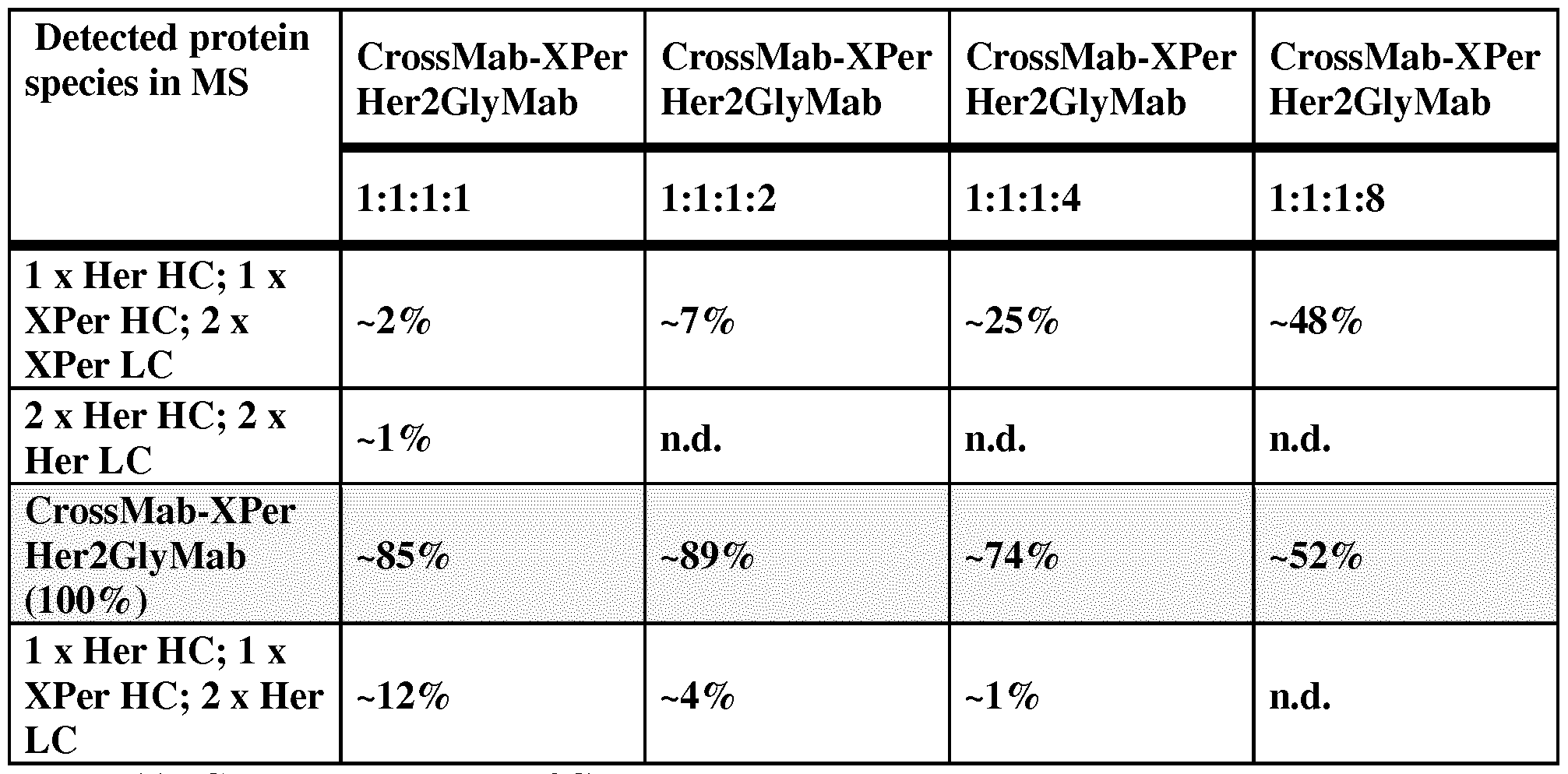


Claims
Priority Applications (13)
| Application Number | Priority Date | Filing Date | Title |
|---|---|---|---|
| EP14821603.9A EP3083696B1 (en) | 2013-12-20 | 2014-12-18 | Bispecific her2 antibodies and methods of use |
| CA2925677A CA2925677A1 (en) | 2013-12-20 | 2014-12-18 | Bispecific her2 antibodies and methods of use |
| MX2016008098A MX2016008098A (en) | 2013-12-20 | 2014-12-18 | Bispecific her2 antibodies and methods of use. |
| CN201480067305.2A CN105829347B (en) | 2013-12-20 | 2014-12-18 | Bispecific HER2 antibodies and methods of use |
| JP2016541217A JP6510532B2 (en) | 2013-12-20 | 2014-12-18 | Bispecific HER2 antibodies and methods of use |
| KR1020167016099A KR20160099087A (en) | 2013-12-20 | 2014-12-18 | Bispecific her2 antibodies and methods of use |
| CN202010780777.XA CN112062853B (en) | 2013-12-20 | 2014-12-18 | Bispecific HER2 antibodies and methods of use |
| EP17203203.9A EP3327038B1 (en) | 2013-12-20 | 2014-12-18 | Bispecific her2 antibodies and methods of use |
| RU2016129517A RU2016129517A (en) | 2013-12-20 | 2014-12-18 | SPECIFIC ANTIBODIES TO HER2 AND METHODS OF APPLICATION |
| BR112016010706A BR112016010706A2 (en) | 2013-12-20 | 2014-12-18 | bispecific antibody, pharmaceutical composition, use of bispecific antibody, nucleic acid sequence, expression vector, host cell and method of producing an antibody |
| US15/186,098 US10584178B2 (en) | 2013-12-20 | 2016-06-17 | Bispecific HER2 antibodies and methods of use |
| HK16111380.7A HK1223115A1 (en) | 2013-12-20 | 2016-09-29 | Bispecific her2 antibodies and methods of use her2 |
| US16/744,697 US11787873B2 (en) | 2013-12-20 | 2020-01-16 | Bispecific HER2 antibodies and methods of use |
Applications Claiming Priority (2)
| Application Number | Priority Date | Filing Date | Title |
|---|---|---|---|
| EP13198819 | 2013-12-20 | ||
| EP13198819.8 | 2013-12-20 |
Related Child Applications (2)
| Application Number | Title | Priority Date | Filing Date |
|---|---|---|---|
| EP17203203.9A Previously-Filed-Application EP3327038B1 (en) | 2013-12-20 | 2014-12-18 | Bispecific her2 antibodies and methods of use |
| US15/186,098 Continuation US10584178B2 (en) | 2013-12-20 | 2016-06-17 | Bispecific HER2 antibodies and methods of use |
Publications (1)
| Publication Number | Publication Date |
|---|---|
| WO2015091738A1 true WO2015091738A1 (en) | 2015-06-25 |
Family
ID=49876448
Family Applications (1)
| Application Number | Title | Priority Date | Filing Date |
|---|---|---|---|
| PCT/EP2014/078375 WO2015091738A1 (en) | 2013-12-20 | 2014-12-18 | Bispecific her2 antibodies and methods of use |
Country Status (11)
| Country | Link |
|---|---|
| US (2) | US10584178B2 (en) |
| EP (2) | EP3327038B1 (en) |
| JP (2) | JP6510532B2 (en) |
| KR (1) | KR20160099087A (en) |
| CN (2) | CN105829347B (en) |
| BR (1) | BR112016010706A2 (en) |
| CA (1) | CA2925677A1 (en) |
| HK (1) | HK1223115A1 (en) |
| MX (1) | MX2016008098A (en) |
| RU (1) | RU2016129517A (en) |
| WO (1) | WO2015091738A1 (en) |
Cited By (31)
| Publication number | Priority date | Publication date | Assignee | Title |
|---|---|---|---|---|
| WO2017053856A1 (en) * | 2015-09-23 | 2017-03-30 | Regeneron Pharmaceuticals, Inc. | Optimized anti-cd3 bispecific antibodies and uses thereof |
| CN106554419A (en) * | 2015-09-28 | 2017-04-05 | 上海抗体药物国家工程研究中心有限公司 | Recombinate anti-HER2 bispecific antibodies, its preparation method and application |
| US9745382B1 (en) | 2016-07-22 | 2017-08-29 | Beijing Mabworks Biotech Co. Ltd. | Bispecific anti-HER2 antibody |
| JP2018504113A (en) * | 2015-01-08 | 2018-02-15 | 蘇州康寧傑瑞生物科技有限公司 | Bispecific antibody or antibody mixture having a common light chain |
| US10000576B1 (en) | 2013-11-27 | 2018-06-19 | Zymeworks Inc. | Bispecific antigen-binding constructs targeting HER2 |
| WO2018114728A1 (en) | 2016-12-20 | 2018-06-28 | F. Hoffmann-La Roche Ag | Combination therapy with a bispecific anti-ang2/vegf antibody and a bispecific anti-her2 antibody |
| WO2018191188A1 (en) * | 2017-04-09 | 2018-10-18 | Xiao Shouhua | Biparatopic and multiparatopic antibodies with common light chain and method of use |
| US10106610B2 (en) | 2013-02-01 | 2018-10-23 | Regeneron Pharmaceuticals, Inc. | Antibodies comprising chimeric constant domains |
| US10377833B2 (en) | 2016-07-22 | 2019-08-13 | Beijing Mabworks Biotech Co., Ltd. | Bispecific anti-HER2 antibody |
| US10377825B2 (en) * | 2015-06-17 | 2019-08-13 | Genentech, Inc. | Anti-HER2 antibodies and methods of use |
| WO2019175125A1 (en) | 2018-03-13 | 2019-09-19 | F. Hoffmann-La Roche Ag | Combination therapy with targeted 4-1bb (cd137) agonists |
| WO2019197600A1 (en) | 2018-04-13 | 2019-10-17 | F. Hoffmann-La Roche Ag | Her2-targeting antigen binding molecules comprising 4-1bbl |
| US10550193B2 (en) | 2014-03-19 | 2020-02-04 | Regeneron Pharmaceuticals, Inc. | Methods and antibody compositions for tumor treatment |
| US10556952B2 (en) | 2015-03-30 | 2020-02-11 | Regeneron Pharmaceuticals, Inc. | Heavy chain constant regions with reduced binding to Fc gamma receptors |
| US10584178B2 (en) | 2013-12-20 | 2020-03-10 | Hoffmann-La Roche Inc. | Bispecific HER2 antibodies and methods of use |
| WO2020074469A1 (en) * | 2018-10-08 | 2020-04-16 | Universität Zürich | Her2-binding tetrameric polypeptides |
| US10662244B2 (en) | 2014-11-17 | 2020-05-26 | Regeneron Pharmaceuticals, Inc. | Methods for tumor treatment using CD3XCD20 bispecific antibody |
| WO2020208049A1 (en) | 2019-04-12 | 2020-10-15 | F. Hoffmann-La Roche Ag | Bispecific antigen binding molecules comprising lipocalin muteins |
| EP3733714A1 (en) * | 2019-04-30 | 2020-11-04 | Universität Zürich | Her2-binding tetrameric polypeptides |
| WO2021174113A1 (en) * | 2020-02-28 | 2021-09-02 | Regeneron Pharmaceuticals, Inc. | Bispecific antigen binding molecules that bind her2, and methods of use thereof |
| WO2021220215A1 (en) * | 2020-05-01 | 2021-11-04 | Novartis Ag | Engineered immunoglobulins |
| WO2021259890A1 (en) | 2020-06-23 | 2021-12-30 | F. Hoffmann-La Roche Ag | Agonistic cd28 antigen binding molecules targeting her2 |
| US20220153869A1 (en) * | 2020-11-16 | 2022-05-19 | Tavotek Biotherapeutics (Hong Kong) Limited | Shielded and homing bispecific antibody that simultaneously inhibits angiogenic pathway targets and her2 family proteins and uses thereof |
| US11359194B2 (en) | 2008-04-11 | 2022-06-14 | Chugai Seiyaku Kabushiki Kaisha | Antigen-binding molecule capable of binding two or more antigen molecules repeatedly |
| WO2022188743A1 (en) * | 2021-03-08 | 2022-09-15 | Genequantum Healthcare (Suzhou) Co., Ltd. | Anti-her2 antibody-immune agonist conjugate and applications thereof |
| US11590223B2 (en) | 2018-08-31 | 2023-02-28 | Regeneron Pharmaceuticals, Inc. | Dosing strategy that mitigates cytokine release syndrome for therapeutic antibodies |
| US11608374B2 (en) | 2017-01-30 | 2023-03-21 | Chugai Seiyaku Kabushiki Kaisha | Anti-sclerostin antibodies and methods of use |
| US11891434B2 (en) | 2010-11-30 | 2024-02-06 | Chugai Seiyaku Kabushiki Kaisha | Antigen-binding molecule capable of binding to plurality of antigen molecules repeatedly |
| WO2024028732A1 (en) * | 2022-08-05 | 2024-02-08 | Janssen Biotech, Inc. | Cd98 binding constructs for treating brain tumors |
| WO2024028731A1 (en) * | 2022-08-05 | 2024-02-08 | Janssen Biotech, Inc. | Transferrin receptor binding proteins for treating brain tumors |
| US11958910B2 (en) | 2021-02-26 | 2024-04-16 | Regeneron Pharmaceuticals, Inc. | Bispecific antigen binding molecules that bind HER2, and methods of use thereof |
Families Citing this family (27)
| Publication number | Priority date | Publication date | Assignee | Title |
|---|---|---|---|---|
| US20090162359A1 (en) | 2007-12-21 | 2009-06-25 | Christian Klein | Bivalent, bispecific antibodies |
| US9676845B2 (en) | 2009-06-16 | 2017-06-13 | Hoffmann-La Roche, Inc. | Bispecific antigen binding proteins |
| AR085403A1 (en) | 2011-02-28 | 2013-09-25 | Hoffmann La Roche | MONOVALENT PROTEINS THAT JOIN ANTIGENS |
| RU2607038C2 (en) | 2011-02-28 | 2017-01-10 | Ф. Хоффманн-Ля Рош Аг | Antigen-binding proteins |
| JP6422956B2 (en) * | 2013-10-11 | 2018-11-14 | エフ.ホフマン−ラ ロシュ アーゲーF. Hoffmann−La Roche Aktiengesellschaft | Multispecific domain exchange common variable light chain antibody |
| US11299751B2 (en) | 2016-04-29 | 2022-04-12 | Voyager Therapeutics, Inc. | Compositions for the treatment of disease |
| EP3448874A4 (en) | 2016-04-29 | 2020-04-22 | Voyager Therapeutics, Inc. | Compositions for the treatment of disease |
| KR20190118172A (en) | 2017-02-08 | 2019-10-17 | 드래곤플라이 쎄라퓨틱스, 인크. | Multispecific Binding Proteins for Activation of Natural Killer Cells and Their Therapeutic Uses in Cancer Treatment |
| CN108395480A (en) * | 2017-02-08 | 2018-08-14 | 广州百尼夫生物科技有限公司 | Chimeric antigen receptor and its gene and recombinant expression carrier, CARHER2-NKT cells and its preparation method and application |
| FI3582806T3 (en) | 2017-02-20 | 2023-09-07 | Dragonfly Therapeutics Inc | Proteins binding her2, nkg2d and cd16 |
| WO2018166527A1 (en) * | 2017-03-17 | 2018-09-20 | 本康生物制药(深圳)有限公司 | Multispecific antibody, antibody conjugate and related pharmaceutical composition and use |
| SG11201912865VA (en) * | 2017-06-25 | 2020-01-30 | Systimmune Inc | Multi-specific antibodies and methods of making and using thereof |
| US20210206882A1 (en) * | 2017-08-01 | 2021-07-08 | Ab Studio Inc. | Bispecific antibodies and uses thereof |
| WO2019051308A1 (en) * | 2017-09-07 | 2019-03-14 | Dragonfly Therapeutics, Inc. | Proteins binding nkg2d, cd16 and a tumor-associated antigen |
| CA3090244A1 (en) | 2018-02-08 | 2019-08-15 | Dragonfly Therapeutics, Inc. | Antibody variable domains targeting the nkg2d receptor |
| CR20200391A (en) | 2018-02-08 | 2020-10-19 | Genentech Inc | Bispecific antigen-binding molecules and methods of use |
| ES2958933T3 (en) | 2018-03-13 | 2024-02-16 | Zymeworks Bc Inc | Biparatopic anti-HER2 antibody-drug conjugates and methods of use |
| WO2020156555A1 (en) | 2019-02-03 | 2020-08-06 | 苏州康聚生物科技有限公司 | Anti-her2 bispecific antibody and application thereof |
| CN114040928A (en) * | 2020-03-27 | 2022-02-11 | 江苏康宁杰瑞生物制药有限公司 | Effective dose of HER2 bispecific antibody |
| CN114057882B (en) * | 2020-07-31 | 2023-01-06 | 山东博安生物技术股份有限公司 | Multivalent multispecific antibodies |
| EP4183421A1 (en) * | 2020-08-13 | 2023-05-24 | Chia Tai Tianqing Pharmaceutical Group Co., Ltd. | Antibody drug conjugate |
| CN114539413A (en) * | 2020-11-25 | 2022-05-27 | 三生国健药业(上海)股份有限公司 | Multivalent bispecific antibodies that bind HER2, methods of making, and uses thereof |
| WO2023280092A1 (en) * | 2021-07-05 | 2023-01-12 | 江苏康宁杰瑞生物制药有限公司 | Antibody-drug conjugate and application thereof |
| CN113817065B (en) * | 2021-09-06 | 2023-04-18 | 深圳市乐土生物医药有限公司 | anti-HER 2 polypeptide and application thereof |
| WO2023088295A1 (en) * | 2021-11-17 | 2023-05-25 | 江苏先声药业有限公司 | Multi-specific antibody and pharmaceutical use thereof |
| WO2024061224A1 (en) * | 2022-09-20 | 2024-03-28 | 普米斯生物技术(珠海)有限公司 | Anti-her2 antibodies and use thereof |
| CN116514998B (en) * | 2023-05-12 | 2023-09-15 | 再少年(北京)生物科技有限公司 | Chimeric antigen receptor, chimeric antigen receptor-natural killer cell and application thereof in preparation of antitumor drugs |
Citations (4)
| Publication number | Priority date | Publication date | Assignee | Title |
|---|---|---|---|---|
| WO1998050431A2 (en) * | 1997-05-02 | 1998-11-12 | Genentech, Inc. | A method for making multispecific antibodies having heteromultimeric and common components |
| WO2011069104A2 (en) * | 2009-12-04 | 2011-06-09 | Genentech, Inc. | Multispecific antibodies, antibody analogs, compositions, and methods |
| WO2012085113A1 (en) * | 2010-12-23 | 2012-06-28 | Roche Diagnostics Gmbh | Binding agent |
| WO2012143523A1 (en) * | 2011-04-20 | 2012-10-26 | Genmab A/S | Bispecifc antibodies against her2 |
Family Cites Families (33)
| Publication number | Priority date | Publication date | Assignee | Title |
|---|---|---|---|---|
| DE69233254T2 (en) | 1991-06-14 | 2004-09-16 | Genentech, Inc., South San Francisco | Humanized Heregulin antibody |
| US6733752B1 (en) | 1994-03-30 | 2004-05-11 | The Trustees Of The University Of Pennsylvania | Prevention of tumors with monoclonal antibodies against neu |
| US6949245B1 (en) | 1999-06-25 | 2005-09-27 | Genentech, Inc. | Humanized anti-ErbB2 antibodies and treatment with anti-ErbB2 antibodies |
| US7125843B2 (en) | 2001-10-19 | 2006-10-24 | Neose Technologies, Inc. | Glycoconjugates including more than one peptide |
| US20040110226A1 (en) * | 2002-03-01 | 2004-06-10 | Xencor | Antibody optimization |
| US7332585B2 (en) | 2002-04-05 | 2008-02-19 | The Regents Of The California University | Bispecific single chain Fv antibody molecules and methods of use thereof |
| ES2429112T3 (en) | 2002-04-10 | 2013-11-13 | Genentech, Inc. | Anti-HER2 antibody variants |
| US7709610B2 (en) * | 2003-05-08 | 2010-05-04 | Facet Biotech Corporation | Therapeutic use of anti-CS1 antibodies |
| US20060003412A1 (en) * | 2003-12-08 | 2006-01-05 | Xencor, Inc. | Protein engineering with analogous contact environments |
| UA90480C2 (en) | 2004-07-22 | 2010-05-11 | Джэнэнтэк, Инк. | HER2 antibody composition exhibiting anticancer activity |
| GB0624500D0 (en) * | 2006-12-07 | 2007-01-17 | Istituto Superiore Di Sanito | A novel passive vaccine for candida infections |
| US9266967B2 (en) | 2007-12-21 | 2016-02-23 | Hoffmann-La Roche, Inc. | Bivalent, bispecific antibodies |
| US8242247B2 (en) | 2007-12-21 | 2012-08-14 | Hoffmann-La Roche Inc. | Bivalent, bispecific antibodies |
| US8227577B2 (en) | 2007-12-21 | 2012-07-24 | Hoffman-La Roche Inc. | Bivalent, bispecific antibodies |
| US20090162359A1 (en) | 2007-12-21 | 2009-06-25 | Christian Klein | Bivalent, bispecific antibodies |
| CN102448985B (en) | 2009-05-27 | 2015-08-05 | 霍夫曼-拉罗奇有限公司 | Three or four specific antibodies |
| US9676845B2 (en) | 2009-06-16 | 2017-06-13 | Hoffmann-La Roche, Inc. | Bispecific antigen binding proteins |
| TWI449821B (en) * | 2009-08-11 | 2014-08-21 | Academia Sinica | Phage displaying system expressing single chain antibody |
| EP2482212A4 (en) | 2009-09-25 | 2014-02-26 | Sh Nat Eng Res Ct Nanotech Co | Method of acquiring proteins with high affinity by computer aided design |
| PL2330131T3 (en) * | 2009-12-07 | 2015-05-29 | Fundacio Privada Inst Catalana De Recerca I Estudis Avancants | Antibodies against HER2 truncated variant CTF-611 |
| ES2594893T3 (en) * | 2009-12-16 | 2016-12-23 | Abbvie Biotherapeutics Inc. | Anti HER2 antibodies and their uses |
| TW201138821A (en) * | 2010-03-26 | 2011-11-16 | Roche Glycart Ag | Bispecific antibodies |
| NZ604007A (en) * | 2010-05-27 | 2015-03-27 | Genmab As | Monoclonal antibodies against her2 epitope |
| US10208129B2 (en) * | 2010-12-06 | 2019-02-19 | National Research Council Of Canada | Antibodies selective for cells presenting ErbB2 at high density |
| WO2012085111A1 (en) | 2010-12-23 | 2012-06-28 | F. Hoffmann-La Roche Ag | Polypeptide-polynucleotide-complex and its use in targeted effector moiety delivery |
| US20140170149A1 (en) | 2011-04-20 | 2014-06-19 | Genmab A/S | Bispecific antibodies against her2 and cd3 |
| SI2748202T1 (en) | 2011-08-23 | 2018-10-30 | Roche Glycart Ag | Bispecific antigen binding molecules |
| JP6324974B2 (en) * | 2012-10-12 | 2018-05-23 | アリゾナ ボード オブ リージェンツ オン ビハーフ オブ アリゾナ ステート ユニバーシティ | Antibody-based reagents that specifically recognize toxic oligomeric forms of tau |
| SG10201809385RA (en) | 2013-12-17 | 2018-11-29 | Genentech Inc | Anti-cd3 antibodies and methods of use |
| EP3327038B1 (en) | 2013-12-20 | 2020-09-23 | F. Hoffmann-La Roche AG | Bispecific her2 antibodies and methods of use |
| US9629801B2 (en) * | 2014-01-10 | 2017-04-25 | Wisconsin Alumni Research Foundation | Blood-brain barrier targeting antibodies |
| CA2874083C (en) * | 2014-12-05 | 2024-01-02 | Universite Laval | Tdp-43-binding polypeptides useful for the treatment of neurodegenerative diseases |
| CN110658340B (en) | 2015-01-08 | 2023-10-31 | 苏州康宁杰瑞生物科技有限公司 | Bispecific antibodies or antibody mixtures with a common light chain |
-
2014
- 2014-12-18 EP EP17203203.9A patent/EP3327038B1/en active Active
- 2014-12-18 EP EP14821603.9A patent/EP3083696B1/en active Active
- 2014-12-18 KR KR1020167016099A patent/KR20160099087A/en not_active Application Discontinuation
- 2014-12-18 MX MX2016008098A patent/MX2016008098A/en unknown
- 2014-12-18 CN CN201480067305.2A patent/CN105829347B/en active Active
- 2014-12-18 RU RU2016129517A patent/RU2016129517A/en unknown
- 2014-12-18 WO PCT/EP2014/078375 patent/WO2015091738A1/en active Application Filing
- 2014-12-18 JP JP2016541217A patent/JP6510532B2/en active Active
- 2014-12-18 CA CA2925677A patent/CA2925677A1/en not_active Abandoned
- 2014-12-18 BR BR112016010706A patent/BR112016010706A2/en not_active Application Discontinuation
- 2014-12-18 CN CN202010780777.XA patent/CN112062853B/en active Active
-
2016
- 2016-06-17 US US15/186,098 patent/US10584178B2/en active Active
- 2016-09-29 HK HK16111380.7A patent/HK1223115A1/en unknown
-
2019
- 2019-04-03 JP JP2019071526A patent/JP7077263B2/en active Active
-
2020
- 2020-01-16 US US16/744,697 patent/US11787873B2/en active Active
Patent Citations (4)
| Publication number | Priority date | Publication date | Assignee | Title |
|---|---|---|---|---|
| WO1998050431A2 (en) * | 1997-05-02 | 1998-11-12 | Genentech, Inc. | A method for making multispecific antibodies having heteromultimeric and common components |
| WO2011069104A2 (en) * | 2009-12-04 | 2011-06-09 | Genentech, Inc. | Multispecific antibodies, antibody analogs, compositions, and methods |
| WO2012085113A1 (en) * | 2010-12-23 | 2012-06-28 | Roche Diagnostics Gmbh | Binding agent |
| WO2012143523A1 (en) * | 2011-04-20 | 2012-10-26 | Genmab A/S | Bispecifc antibodies against her2 |
Non-Patent Citations (2)
| Title |
|---|
| KLEIN CHRISTIAN ET AL: "Progress in overcoming the chain association issue in bispecific heterodimeric IgG antibodies", MABS, LANDES BIOSCIENCE, US, vol. 4, no. 6, 1 November 2012 (2012-11-01), pages 653 - 663, XP009170958, ISSN: 1942-0862, DOI: 10.4161/MABS.21379 * |
| LI BOHUA ET AL: "Bispecific antibody to ErbB2 overcomes trastuzumab resistance through comprehensive blockade of ErbB2 heterodimerization.", CANCER RESEARCH 1 NOV 2013, vol. 73, no. 21, 1 November 2013 (2013-11-01), pages 6471 - 6483, XP002726071, ISSN: 1538-7445 * |
Cited By (51)
| Publication number | Priority date | Publication date | Assignee | Title |
|---|---|---|---|---|
| US11371039B2 (en) | 2008-04-11 | 2022-06-28 | Chugai Seiyaku Kabushiki Kaisha | Antigen-binding molecule capable of binding to two or more antigen molecules repeatedly |
| US11359194B2 (en) | 2008-04-11 | 2022-06-14 | Chugai Seiyaku Kabushiki Kaisha | Antigen-binding molecule capable of binding two or more antigen molecules repeatedly |
| US11891434B2 (en) | 2010-11-30 | 2024-02-06 | Chugai Seiyaku Kabushiki Kaisha | Antigen-binding molecule capable of binding to plurality of antigen molecules repeatedly |
| US10988537B2 (en) | 2013-02-01 | 2021-04-27 | Regeneren Pharmaceuticals, Inc. | Antibodies comprising chimeric constant domains |
| US10106610B2 (en) | 2013-02-01 | 2018-10-23 | Regeneron Pharmaceuticals, Inc. | Antibodies comprising chimeric constant domains |
| US11325981B2 (en) | 2013-11-27 | 2022-05-10 | Zymeworks Inc. | Bispecific antigen-binding constructs targeting Her2 |
| US10947319B2 (en) | 2013-11-27 | 2021-03-16 | Zymeworks Inc. | Bispecific antigen-binding constructs targeting HER2 |
| US10000576B1 (en) | 2013-11-27 | 2018-06-19 | Zymeworks Inc. | Bispecific antigen-binding constructs targeting HER2 |
| US11787873B2 (en) | 2013-12-20 | 2023-10-17 | Hoffmann-La Roche Inc. | Bispecific HER2 antibodies and methods of use |
| US10584178B2 (en) | 2013-12-20 | 2020-03-10 | Hoffmann-La Roche Inc. | Bispecific HER2 antibodies and methods of use |
| US11434300B2 (en) | 2014-03-19 | 2022-09-06 | Regeneron Pharmaceuticals, Inc. | Methods and antibody compositions for tumor treatment |
| US10550193B2 (en) | 2014-03-19 | 2020-02-04 | Regeneron Pharmaceuticals, Inc. | Methods and antibody compositions for tumor treatment |
| US10662244B2 (en) | 2014-11-17 | 2020-05-26 | Regeneron Pharmaceuticals, Inc. | Methods for tumor treatment using CD3XCD20 bispecific antibody |
| EP3243840A4 (en) * | 2015-01-08 | 2018-09-12 | Suzhou Alphamab Co., Ltd | Bispecific antibody or antibody mixture with common light chains |
| US10808043B2 (en) | 2015-01-08 | 2020-10-20 | Jiangsu Alphamab Biopharmaceuticals Co., Ltd. | Bispecific antibody or antibody mixture with common light chains |
| JP2018504113A (en) * | 2015-01-08 | 2018-02-15 | 蘇州康寧傑瑞生物科技有限公司 | Bispecific antibody or antibody mixture having a common light chain |
| JP7041516B2 (en) | 2015-01-08 | 2022-03-24 | 江蘇康寧杰瑞生物制薬有限公司 | Bispecific antibodies or antibody mixtures with common light chains |
| US11396557B2 (en) | 2015-01-08 | 2022-07-26 | Jiangsu Alphamab Biopharmaceuticals Co., Ltd. | Bispecific antibody or antibody mixture with common light chains |
| US10556952B2 (en) | 2015-03-30 | 2020-02-11 | Regeneron Pharmaceuticals, Inc. | Heavy chain constant regions with reduced binding to Fc gamma receptors |
| US11518807B2 (en) | 2015-03-30 | 2022-12-06 | Regeneron Pharmaceuticals, Inc. | Heavy chain constant regions with reduced binding to Fc gamma receptors |
| US10377825B2 (en) * | 2015-06-17 | 2019-08-13 | Genentech, Inc. | Anti-HER2 antibodies and methods of use |
| CN108290951B (en) * | 2015-09-23 | 2022-04-01 | 瑞泽恩制药公司 | Optimized anti-CD 3 bispecific antibodies and uses thereof |
| WO2017053856A1 (en) * | 2015-09-23 | 2017-03-30 | Regeneron Pharmaceuticals, Inc. | Optimized anti-cd3 bispecific antibodies and uses thereof |
| CN108290951A (en) * | 2015-09-23 | 2018-07-17 | 瑞泽恩制药公司 | Optimize AntiCD3 McAb bispecific antibody and its purposes |
| CN106554419A (en) * | 2015-09-28 | 2017-04-05 | 上海抗体药物国家工程研究中心有限公司 | Recombinate anti-HER2 bispecific antibodies, its preparation method and application |
| CN109715671B (en) * | 2016-07-22 | 2020-08-21 | 北京天广实生物技术股份有限公司 | Bispecific anti-HER 2 antibodies |
| CN109715671A (en) * | 2016-07-22 | 2019-05-03 | 北京天广实生物技术股份有限公司 | Bispecific Anti-HER 2 |
| WO2018014864A1 (en) * | 2016-07-22 | 2018-01-25 | Beijing Mabworks Biotech Co. Ltd. | Bispecific anti-her2 antibody |
| US9745382B1 (en) | 2016-07-22 | 2017-08-29 | Beijing Mabworks Biotech Co. Ltd. | Bispecific anti-HER2 antibody |
| AU2017298251B2 (en) * | 2016-07-22 | 2020-02-06 | Beijing Mabworks Biotech Co. Ltd. | Bispecific anti-HER2 antibody |
| US10377833B2 (en) | 2016-07-22 | 2019-08-13 | Beijing Mabworks Biotech Co., Ltd. | Bispecific anti-HER2 antibody |
| WO2018114728A1 (en) | 2016-12-20 | 2018-06-28 | F. Hoffmann-La Roche Ag | Combination therapy with a bispecific anti-ang2/vegf antibody and a bispecific anti-her2 antibody |
| US11608374B2 (en) | 2017-01-30 | 2023-03-21 | Chugai Seiyaku Kabushiki Kaisha | Anti-sclerostin antibodies and methods of use |
| US11325982B2 (en) | 2017-04-09 | 2022-05-10 | Xuanzhu Biopharmaceutical Co., Ltd. | Biparatopic and multiparatopic antibodies with common light chain and method of use |
| EP3609580A4 (en) * | 2017-04-09 | 2021-03-24 | Combio Pharmaceutical, Inc. | Biparatopic and multiparatopic antibodies with common light chain and method of use |
| WO2018191188A1 (en) * | 2017-04-09 | 2018-10-18 | Xiao Shouhua | Biparatopic and multiparatopic antibodies with common light chain and method of use |
| WO2019175125A1 (en) | 2018-03-13 | 2019-09-19 | F. Hoffmann-La Roche Ag | Combination therapy with targeted 4-1bb (cd137) agonists |
| CN111741979A (en) * | 2018-04-13 | 2020-10-02 | 豪夫迈·罗氏有限公司 | Her2 targeting antigen binding molecule comprising 4-1BBL |
| WO2019197600A1 (en) | 2018-04-13 | 2019-10-17 | F. Hoffmann-La Roche Ag | Her2-targeting antigen binding molecules comprising 4-1bbl |
| US11590223B2 (en) | 2018-08-31 | 2023-02-28 | Regeneron Pharmaceuticals, Inc. | Dosing strategy that mitigates cytokine release syndrome for therapeutic antibodies |
| WO2020074469A1 (en) * | 2018-10-08 | 2020-04-16 | Universität Zürich | Her2-binding tetrameric polypeptides |
| WO2020208049A1 (en) | 2019-04-12 | 2020-10-15 | F. Hoffmann-La Roche Ag | Bispecific antigen binding molecules comprising lipocalin muteins |
| EP3733714A1 (en) * | 2019-04-30 | 2020-11-04 | Universität Zürich | Her2-binding tetrameric polypeptides |
| WO2021174113A1 (en) * | 2020-02-28 | 2021-09-02 | Regeneron Pharmaceuticals, Inc. | Bispecific antigen binding molecules that bind her2, and methods of use thereof |
| WO2021220215A1 (en) * | 2020-05-01 | 2021-11-04 | Novartis Ag | Engineered immunoglobulins |
| WO2021259890A1 (en) | 2020-06-23 | 2021-12-30 | F. Hoffmann-La Roche Ag | Agonistic cd28 antigen binding molecules targeting her2 |
| US20220153869A1 (en) * | 2020-11-16 | 2022-05-19 | Tavotek Biotherapeutics (Hong Kong) Limited | Shielded and homing bispecific antibody that simultaneously inhibits angiogenic pathway targets and her2 family proteins and uses thereof |
| US11958910B2 (en) | 2021-02-26 | 2024-04-16 | Regeneron Pharmaceuticals, Inc. | Bispecific antigen binding molecules that bind HER2, and methods of use thereof |
| WO2022188743A1 (en) * | 2021-03-08 | 2022-09-15 | Genequantum Healthcare (Suzhou) Co., Ltd. | Anti-her2 antibody-immune agonist conjugate and applications thereof |
| WO2024028732A1 (en) * | 2022-08-05 | 2024-02-08 | Janssen Biotech, Inc. | Cd98 binding constructs for treating brain tumors |
| WO2024028731A1 (en) * | 2022-08-05 | 2024-02-08 | Janssen Biotech, Inc. | Transferrin receptor binding proteins for treating brain tumors |
Also Published As
| Publication number | Publication date |
|---|---|
| EP3083696A1 (en) | 2016-10-26 |
| CN105829347B (en) | 2020-09-08 |
| RU2016129517A (en) | 2018-01-25 |
| EP3327038A2 (en) | 2018-05-30 |
| US20200291131A1 (en) | 2020-09-17 |
| MX2016008098A (en) | 2017-01-11 |
| EP3327038B1 (en) | 2020-09-23 |
| JP2017501706A (en) | 2017-01-19 |
| CN112062853A (en) | 2020-12-11 |
| JP7077263B2 (en) | 2022-05-30 |
| US10584178B2 (en) | 2020-03-10 |
| CN112062853B (en) | 2024-01-09 |
| CA2925677A1 (en) | 2015-06-25 |
| JP6510532B2 (en) | 2019-05-08 |
| CN105829347A (en) | 2016-08-03 |
| US20170029529A1 (en) | 2017-02-02 |
| BR112016010706A2 (en) | 2017-12-05 |
| EP3083696B1 (en) | 2018-02-14 |
| EP3327038A3 (en) | 2018-06-13 |
| KR20160099087A (en) | 2016-08-19 |
| US11787873B2 (en) | 2023-10-17 |
| JP2019141066A (en) | 2019-08-29 |
| HK1223115A1 (en) | 2017-07-21 |
Similar Documents
| Publication | Publication Date | Title |
|---|---|---|
| US11787873B2 (en) | Bispecific HER2 antibodies and methods of use | |
| US20230212312A1 (en) | Trispecific antibodies specific for her2 and a blood brain barrier receptor and methods of use | |
| US10538585B2 (en) | Anti-ANG2 antibodies and methods of use | |
| US11827711B2 (en) | Antibodies binding to NKG2D | |
| US10087246B2 (en) | Anti-PDGF-B antibodies and methods of use | |
| JP7248761B2 (en) | Anti-BRDU Antibodies and Methods of Use | |
| US11685790B2 (en) | Antibodies binding to STEAP-1 |
Legal Events
| Date | Code | Title | Description |
|---|---|---|---|
| 121 | Ep: the epo has been informed by wipo that ep was designated in this application |
Ref document number: 14821603 Country of ref document: EP Kind code of ref document: A1 |
|
| ENP | Entry into the national phase |
Ref document number: 2925677 Country of ref document: CA |
|
| REEP | Request for entry into the european phase |
Ref document number: 2014821603 Country of ref document: EP |
|
| WWE | Wipo information: entry into national phase |
Ref document number: 2014821603 Country of ref document: EP |
|
| REG | Reference to national code |
Ref country code: BR Ref legal event code: B01A Ref document number: 112016010706 Country of ref document: BR |
|
| ENP | Entry into the national phase |
Ref document number: 20167016099 Country of ref document: KR Kind code of ref document: A |
|
| ENP | Entry into the national phase |
Ref document number: 2016541217 Country of ref document: JP Kind code of ref document: A |
|
| WWE | Wipo information: entry into national phase |
Ref document number: MX/A/2016/008098 Country of ref document: MX |
|
| NENP | Non-entry into the national phase |
Ref country code: DE |
|
| ENP | Entry into the national phase |
Ref document number: 2016129517 Country of ref document: RU Kind code of ref document: A |
|
| ENP | Entry into the national phase |
Ref document number: 112016010706 Country of ref document: BR Kind code of ref document: A2 Effective date: 20160512 |