WO2015107025A1 - Fc-region variants with modified fcrn-binding properties - Google Patents
Fc-region variants with modified fcrn-binding properties Download PDFInfo
- Publication number
- WO2015107025A1 WO2015107025A1 PCT/EP2015/050425 EP2015050425W WO2015107025A1 WO 2015107025 A1 WO2015107025 A1 WO 2015107025A1 EP 2015050425 W EP2015050425 W EP 2015050425W WO 2015107025 A1 WO2015107025 A1 WO 2015107025A1
- Authority
- WO
- WIPO (PCT)
- Prior art keywords
- polypeptide
- mutations
- antibody
- domain
- region
- Prior art date
Links
Classifications
-
- C—CHEMISTRY; METALLURGY
- C07—ORGANIC CHEMISTRY
- C07K—PEPTIDES
- C07K16/00—Immunoglobulins [IGs], e.g. monoclonal or polyclonal antibodies
- C07K16/18—Immunoglobulins [IGs], e.g. monoclonal or polyclonal antibodies against material from animals or humans
- C07K16/22—Immunoglobulins [IGs], e.g. monoclonal or polyclonal antibodies against material from animals or humans against growth factors ; against growth regulators
-
- A—HUMAN NECESSITIES
- A61—MEDICAL OR VETERINARY SCIENCE; HYGIENE
- A61K—PREPARATIONS FOR MEDICAL, DENTAL OR TOILETRY PURPOSES
- A61K39/00—Medicinal preparations containing antigens or antibodies
- A61K39/395—Antibodies; Immunoglobulins; Immune serum, e.g. antilymphocytic serum
-
- A—HUMAN NECESSITIES
- A61—MEDICAL OR VETERINARY SCIENCE; HYGIENE
- A61P—SPECIFIC THERAPEUTIC ACTIVITY OF CHEMICAL COMPOUNDS OR MEDICINAL PREPARATIONS
- A61P27/00—Drugs for disorders of the senses
- A61P27/02—Ophthalmic agents
-
- A—HUMAN NECESSITIES
- A61—MEDICAL OR VETERINARY SCIENCE; HYGIENE
- A61P—SPECIFIC THERAPEUTIC ACTIVITY OF CHEMICAL COMPOUNDS OR MEDICINAL PREPARATIONS
- A61P43/00—Drugs for specific purposes, not provided for in groups A61P1/00-A61P41/00
-
- A—HUMAN NECESSITIES
- A61—MEDICAL OR VETERINARY SCIENCE; HYGIENE
- A61K—PREPARATIONS FOR MEDICAL, DENTAL OR TOILETRY PURPOSES
- A61K39/00—Medicinal preparations containing antigens or antibodies
- A61K2039/505—Medicinal preparations containing antigens or antibodies comprising antibodies
-
- C—CHEMISTRY; METALLURGY
- C07—ORGANIC CHEMISTRY
- C07K—PEPTIDES
- C07K2317/00—Immunoglobulins specific features
- C07K2317/30—Immunoglobulins specific features characterized by aspects of specificity or valency
- C07K2317/31—Immunoglobulins specific features characterized by aspects of specificity or valency multispecific
-
- C—CHEMISTRY; METALLURGY
- C07—ORGANIC CHEMISTRY
- C07K—PEPTIDES
- C07K2317/00—Immunoglobulins specific features
- C07K2317/30—Immunoglobulins specific features characterized by aspects of specificity or valency
- C07K2317/33—Crossreactivity, e.g. for species or epitope, or lack of said crossreactivity
-
- C—CHEMISTRY; METALLURGY
- C07—ORGANIC CHEMISTRY
- C07K—PEPTIDES
- C07K2317/00—Immunoglobulins specific features
- C07K2317/50—Immunoglobulins specific features characterized by immunoglobulin fragments
- C07K2317/52—Constant or Fc region; Isotype
- C07K2317/524—CH2 domain
-
- C—CHEMISTRY; METALLURGY
- C07—ORGANIC CHEMISTRY
- C07K—PEPTIDES
- C07K2317/00—Immunoglobulins specific features
- C07K2317/50—Immunoglobulins specific features characterized by immunoglobulin fragments
- C07K2317/52—Constant or Fc region; Isotype
- C07K2317/526—CH3 domain
-
- C—CHEMISTRY; METALLURGY
- C07—ORGANIC CHEMISTRY
- C07K—PEPTIDES
- C07K2317/00—Immunoglobulins specific features
- C07K2317/50—Immunoglobulins specific features characterized by immunoglobulin fragments
- C07K2317/52—Constant or Fc region; Isotype
- C07K2317/53—Hinge
-
- C—CHEMISTRY; METALLURGY
- C07—ORGANIC CHEMISTRY
- C07K—PEPTIDES
- C07K2317/00—Immunoglobulins specific features
- C07K2317/60—Immunoglobulins specific features characterized by non-natural combinations of immunoglobulin fragments
- C07K2317/64—Immunoglobulins specific features characterized by non-natural combinations of immunoglobulin fragments comprising a combination of variable region and constant region components
-
- C—CHEMISTRY; METALLURGY
- C07—ORGANIC CHEMISTRY
- C07K—PEPTIDES
- C07K2317/00—Immunoglobulins specific features
- C07K2317/90—Immunoglobulins specific features characterized by (pharmaco)kinetic aspects or by stability of the immunoglobulin
- C07K2317/92—Affinity (KD), association rate (Ka), dissociation rate (Kd) or EC50 value
-
- C—CHEMISTRY; METALLURGY
- C07—ORGANIC CHEMISTRY
- C07K—PEPTIDES
- C07K2317/00—Immunoglobulins specific features
- C07K2317/90—Immunoglobulins specific features characterized by (pharmaco)kinetic aspects or by stability of the immunoglobulin
- C07K2317/94—Stability, e.g. half-life, pH, temperature or enzyme-resistance
Definitions
- IgG Fc-regions that have been modified with respect to Fc- receptor binding without impairing their purification properties.
- the ligands most adopted to bind selectively IgG are Staphylococcal protein A and protein G, which are able to establish highly selective interactions with the Fc- region of most IgGs in a region known as "consensus binding site” (CBS) (DeLano, W.L., et al, Science 287 (2000) 1279), which is located at the hinge region between the CH2 and CH3 domains of the Fc-region.
- CBS consensus binding site
- Staphylococcal protein A is a cell wall associated protein domain exposed on the surface of the Gram-positive bacterium Staphylococcus aureus.
- SPA has high affinity to IgG from various species, for instance human, rabbit and guinea pig IgG but only weak interaction with bovine and mouse IgG (see the following Table) (see Hober supra; Duhamel, R.C., et al, J. Immunol. Methods 31 (1979) 211; Bjork, L. and Kronvall, G., Immunol. J. 133 (1984) 969; Richman, D.D., et al, J. Immunol. 128 (1982) 2300; Amersham Pharmacia Biotech, Handbook, Antibody Purification (2000)).
- the heavy chain hinge-region between the CH2 and CH3 domains of IgG is able to bind several proteins beyond protein A, such as the neonatal Fc receptor (FcRn)
- the SPA CBS comprehends a hydrophobic pocket on the surface of the antibody.
- the residues composing the IgG CBS are lie 253, Ser 254, Met 252, Met 423, Tyr 326, His 435, Asn 434, His 433, Arg 255 and Glu 380 (numbering of the IgG heavy chain residues according to the Kabat EU index numbering system).
- the charged amino acids (Arg 255, Glu 380) are placed around a hydrophobic knob formed by He 253 and Ser 254. This (can) result in the establishment of polar and hydrophilic interactions (see Salvalaglio supra).
- the protein A-IgG interaction can be described using two main binding sites: the first is positioned in the heavy chain CH2 domain and is characterized by hydrophobic interactions between Phe 132, Leu 136, He 150 (of protein A) and the IgG hydrophobic knob constituted by He 253 and Ser 254, and by one electrostatic interaction between Lys 154 (protein A) and Thr 256 (IgG).
- the second site is located in the heavy chain CH3 domain and is dominated by electrostatic interactions between Gin 129 and Tyr 133 (protein A) and His 433, Asn 434, and His 435 (IgG) (see Salvalaglio supra).
- the basic challenge in generating multispecific heterodimeric IgG antibodies from four antibody chains (two different heavy chains and two different light chains) in one expression cell line is the so-called chain association issue (see Klein, C, et al, mAbs 4 (2012) 653-663).
- chain association issue see Klein, C, et al, mAbs 4 (2012) 653-663.
- Carter et al. from Genentech invented an approach termed "knobs-into-holes" (KiH) (see Carter, P., J. Immunol. Meth. 248 (2001) 7-15; Merchant, A.M., et al, Nat. Biotechnol. 16 (1998) 677-681; Zhu, Z., et al, Prot. Sci. 6 (1997) 781-788; Ridgway, J.B., et al, Prot. Eng. 9 (1996) 617-621; Atwell, S., et al, J. Mol. Biol.
- Hole -hole dimers can either be depleted by selective purification procedures or by procedures as outlined below.
- Zhu et al. introduced several sterically complementary mutations, as well as disulfide bridges, in the two VL/VH interfaces of diabody variants.
- VL Y87A/F98M and VH V37F/L45W were introduced into the anti- pi 85HER2 VL/VH interface, a heterodimeric diabody was recovered with > 90 % yield while maintaining overall yield and affinity compared with the parental diabody (see Zhu supra).
- WO2010151792 a bispecific antibody format providing ease of isolation is provided, comprising immunoglobulin heavy chain variable domains that are differentially modified, i.e. heterodimeric, in the CH3 domain, wherein the differential modifications are non-immunogenic or substantially non-immunogenic with respect to the CH3 modifications, and at least one of the modifications results in a differential affinity for the bispecific antibody for an affinity reagent such as protein A, and the bispecific antibody is isolable from a disrupted cell, from medium, or from a mixture of antibodies based on its affinity for protein A.
- the neonatal Fc-receptor (FcRn) is important for the metabolic fate of antibodies of the IgG class in vivo.
- the FcRn functions to salvage IgG from the lysosomal degradation pathway, resulting in reduced clearance and increased half-life. It is a heterodimeric protein consisting of two polypeptides: a 50 kDa class I major histocompatibility complex-like protein (a-FcRn) and a 15 kDa p2-microglobulin ( ⁇ 2 ⁇ ). FcRn binds with high affinity to the CH2-CH3 portion of the Fc-region of an antibody of the class IgG. The interaction between an antibody of the class IgG and the FcRn is pH dependent and occurs in a 1 :2 stoichiometry, i.e.
- one IgG antibody molecule can interact with two FcRn molecules via its two heavy chain Fc-region polypeptides (see e.g. Huber, A.H., et al, J. Mol. Biol. 230 (1993) 1077- 1083).
- an IgGs in vitro FcRn binding properties/characteristics are indicative of its in vivo pharmacokinetic properties in the blood circulation.
- FcRn and the Fc-region of an antibody of the IgG class different amino acid residues of the heavy chain CH2- and CH3 -domain are participating.
- Fc-region residues critical to the mouse Fc-region- mouse FcRn interaction have been identified by site-directed mutagenesis (see e.g. Dall'Acqua, W.F., et al. J. Immunol 169 (2002) 5171-5180).
- Residues 1253, H310, H433, N434 and H435 are involved in the interaction (Medesan, C, et al., Eur. J. Immunol. 26 (1996) 2533-2536; Firan, M., et al, Int. Immunol.
- Residues 1253, H310, and H435 were found to be critical for the interaction of human Fc-region with murine FcRn (Kim, J.K., et al, Eur. J. Immunol. 29 (1999) 2819-2885).
- variant Fc-regions that specifically bind to Staphylococcus protein A and that do not bind to human FcRn.
- These variant Fc-regions contain specific amino acid mutations in the CH2- and CH3-domain. It has been found that these mutations when used either in the hole chain or the knob chain of a heterodimeric Fc-region allow for the purification of the heterodimeric Fc-region, i.e. the separation of a heterodimeric Fc-region from a homodimeric Fc-region.
- a (dimeric) polypeptide comprising a first polypeptide comprising in N-terminal to C-terminal direction at least a portion of an immunoglobulin hinge region, which comprises one or more cysteine residues, an immunoglobulin CH2-domain and an immunoglobulin CH3-domain, and a second polypeptide comprising in N-terminal to C- terminal direction at least a portion of an immunoglobulin hinge region, which comprises one or more cysteine residues, an immunoglobulin CH2- domain and an immunoglobulin CH3 -domain, wherein (numbering according to the Kabat EU index numbering system) i) the first and the second polypeptide each comprise the mutations
- the first and the second polypeptide each comprise the mutations L251D, L314D and L432D, or iii) the first and the second polypeptide each comprise the mutations L251S, L314S and L432S, and, wherein the first polypeptide and the second polypeptide are connected by one or more disulfide bridges in the at least a portion of an immunoglobulin hinge region.
- the (dimeric) polypeptide does not specifically bind to the human FcRn and does specifically bind to Staphylococcal protein A.
- the (dimeric) polypeptide is a homodimeric polypeptide.
- the (dimeric) polypeptide is a heterodimeric polypeptide.
- the first polypeptide further comprises the mutations Y349C, T366S, L368A and Y407V Communityhole" and the second polypeptide comprises the mutations S354C and T366W disorderknob").
- the first polypeptide further comprises the mutations S354C, T366S, L368A and Y407V Communityhole" and the second polypeptide comprises the mutations Y349C and T366W disorderknob").
- the immunoglobulin hinge region, the immunoglobulin CH2- domain and the immunoglobulin CH3 -domain of the first and the second polypeptide are of the human IgGl subclass.
- the first polypeptide and the second polypeptide each further comprise the mutations L234A and L235A.
- the first polypeptide and the second polypeptide each further comprise the mutation P329G.
- the first polypeptide and the second polypeptide each further comprise the mutations
- the immunoglobulin hinge region, the immunoglobulin CH2- domain and the immunoglobulin CH3 -domain of the first and the second polypeptide are of the human IgG4 subclass. In one embodiment the first polypeptide and the second polypeptide each further comprise the mutations S228P and L235E. In one embodiment the first polypeptide and the second polypeptide each further comprise the mutation P329G. In one embodiment the first polypeptide and the second polypeptide each further comprise the mutations S228P, L235E and P329G. In one embodiment the immunoglobulin hinge region, the immunoglobulin CH2- domain and the immunoglobulin CH3 -domain of the first and the second polypeptide are of the human IgG2 subclass. In one embodiment the first polypeptide and the second polypeptide each further comprise the mutations H268Q, V309L, A330S and P331S.
- the immunoglobulin hinge region, the immunoglobulin CHI- domain and the immunoglobulin CH3 -domain of the first and the second polypeptide are of the human IgG2 subclass.
- the first polypeptide and the second polypeptide each further comprise the mutations V234A, G237A, P238S, H268A, V309L, A330S and P331S.
- the immunoglobulin hinge region, the immunoglobulin CH2- domain and the immunoglobulin CH3 -domain of the first and the second polypeptide are of the human IgG4 subclass.
- the first polypeptide and the second polypeptide each further comprise the mutations S228P, L234A and L235A.
- the first polypeptide and the second polypeptide each further comprise the mutation P329G.
- the first polypeptide and the second polypeptide each further comprise the mutations S228P, L234A, L235A and P329G.
- first and the second polypeptide comprise the mutation Y436A.
- the (dimeric) polypeptide is an Fc-region fusion polypeptide.
- the (dimeric) polypeptide is an (full length) antibody. In one embodiment the (full length) antibody is a monospecific antibody. In one embodiment the monospecific antibody is a monovalent monospecific antibody. In one embodiment the monospecific antibody is a bivalent monospecific antibody.
- the (full length) antibody is a bispecific antibody. In one embodiment the bispecific antibody is a bivalent bispecific antibody. In one embodiment the bispecific antibody is a tetravalent bispecific antibody.
- the (full length) antibody is a trispecific antibody. In one embodiment the trispecific antibody is a trivalent trispecific antibody. In one embodiment the trispecific antibody is a tetravalent trispecific antibody.
- One aspect as reported herein is the use of the mutation Y436A for increasing the binding of a (dimeric) polypeptide comprising an immunoglobulin Fc-region to protein A.
- a (dimeric) polypeptide comprising a first polypeptide comprising in N-terminal to C-terminal direction at least a portion of an immunoglobulin hinge region, which comprises one or more cysteine residues, an immunoglobulin CH2-domain and an immunoglobulin CH3-domain, and a second polypeptide comprising in N-terminal to C- terminal direction at least a portion of an immunoglobulin hinge region, which comprises one or more cysteine residues, an immunoglobulin CHI- domain and an immunoglobulin CH3 -domain, wherein the first, the second or the first and the second polypeptide comprise the mutation Y436A (numbering according to the Kabat EU index numbering system), and wherein the first polypeptide and the second polypeptide are connected by one or more disul
- first and the second polypeptide comprise the mutation Y436A.
- an antibody comprising a first polypeptide comprising in N-terminal to C-terminal direction a first heavy chain variable domain, an immunoglobulin CHI -domain of the subclass IgGl, an immunoglobulin hinge region of the subclass IgGl, an immunoglobulin CH2-domain of the subclass IgGl and an immunoglobulin CH3-domain of the subclass IgGl, a second polypeptide comprising in N-terminal to C-terminal direction a second heavy chain variable domain, an immunoglobulin CHI -domain of the subclass IgGl, an immunoglobulin hinge region of the subclass IgGl, an immunoglobulin CH2-domain of the subclass IgGl and an immunoglobulin CH3-domain of the subclass IgGl, a third polypeptide comprising in N-terminal to C-terminal direction a first light chain variable domain and a light chain constant domain, a fourth polypeptide comprising in N-terminal to C-
- an antibody comprising a first polypeptide comprising in N-terminal to C-terminal direction a first heavy chain variable domain, an immunoglobulin light chain constant domain, an immunoglobulin hinge region of the subclass IgGl, an immunoglobulin CH2-domain of the subclass IgGl and an immunoglobulin CH3-domain of the subclass IgGl, a second polypeptide comprising in N-terminal to C-terminal direction a second heavy chain variable domain, an immunoglobulin CHI -domain of the subclass IgGl, an immunoglobulin hinge region of the subclass IgGl, an immunoglobulin CH2-domain of the subclass IgGl and an immunoglobulin CH3-domain of the subclass IgGl, a third polypeptide comprising in N-terminal to C-terminal direction a first light chain variable domain and an immunoglobulin CHI -domain of the subclass IgGl, a fourth polypeptide comprising in N-terminal
- an antibody comprising a first polypeptide comprising in N-terminal to C-terminal direction a first heavy chain variable domain, an immunoglobulin CHI -domain of the subclass IgG4, an immunoglobulin hinge region of the subclass IgG4, an immunoglobulin CH2-domain of the subclass IgG4 and an immunoglobulin CH3-domain of the subclass IgG4, a second polypeptide comprising in N-terminal to C-terminal direction a second heavy chain variable domain, an immunoglobulin CHI -domain of the subclass IgG4, an immunoglobulin hinge region of the subclass IgG4, an immunoglobulin CH2-domain of the subclass IgG4 and an immunoglobulin CH3-domain of the subclass IgG4, a third polypeptide comprising in N-terminal to C-terminal direction a first light chain variable domain and a light chain constant domain, a fourth polypeptide comprising in N-terminal to C-terminal direction a second
- an antibody comprising a first polypeptide comprising in N-terminal to C-terminal direction a first heavy chain variable domain, an immunoglobulin light chain constant domain, an immunoglobulin hinge region of the subclass IgG4, an immunoglobulin CH2-domain of the subclass IgG4 and an immunoglobulin CH3-domain of the subclass IgG4, a second polypeptide comprising in N-terminal to C-terminal direction a second heavy chain variable domain, an immunoglobulin CHI -domain of the subclass IgG4, an immunoglobulin hinge region of the subclass IgG4, an immunoglobulin CH2-domain of the subclass IgG4 and an immunoglobulin CH3-domain of the subclass IgG4, a third polypeptide comprising in N-terminal to C-terminal direction a first light chain variable domain and an immunoglobulin CHI -domain of the subclass IgG4, a fourth polypeptide comprising in N-terminal to C-terminal direction
- an antibody comprising a first polypeptide comprising in N-terminal to C-terminal direction a first heavy chain variable domain, an immunoglobulin CHI -domain of the subclass IgGl, an immunoglobulin hinge region of the subclass IgGl, an immunoglobulin CH2-domain of the subclass IgGl, an immunoglobulin CH3-domain of the subclass IgGl, a peptidic linker and a first scFv, a second polypeptide comprising in N-terminal to C-terminal direction a second heavy chain variable domain, an immunoglobulin CHI -domain of the subclass IgGl, an immunoglobulin hinge region of the subclass IgGl, an immunoglobulin CH2-domain of the subclass IgGl, an immunoglobulin CH3-domain of the subclass IgGl, a peptidic linker and a second scFv, a third polypeptide comprising in N-terminal to C-
- an antibody comprising a first polypeptide comprising in N-terminal to C-terminal direction a first heavy chain variable domain, an immunoglobulin light chain constant domain, an immunoglobulin hinge region of the subclass IgGl, an immunoglobulin CH2-domain of the subclass IgGl, an immunoglobulin CH3-domain of the subclass IgGl, a peptidic linker and a first scFv, a second polypeptide comprising in N-terminal to C-terminal direction a second heavy chain variable domain, an immunoglobulin CHI -domain of the subclass IgGl, an immunoglobulin hinge region of the subclass IgGl, an immunoglobulin CH2-domain of the subclass IgGl, an immunoglobulin CH3-domain of the subclass IgGl, a peptidic linker and a second scFv, a third polypeptide comprising in N-terminal to C-terminal direction a first light chain variable
- One aspect as reported herein is a method for producing a (dimeric) polypeptide as reported herein comprising the following steps: a) cultivating a mammalian cell comprising one or more nucleic acids encoding the (dimeric) polypeptide, b) recovering the (dimeric) polypeptide from the cultivation medium, and c) purifying the (dimeric) polypeptide with a protein A affinity chromatography and thereby producing the (dimeric) polypeptide.
- One aspect as reported herein is the use of the combination of the mutations
- H310A, H433A and Y436A for separating heterodimeric polypeptides from homodimeric polypeptides.
- One aspect as reported herein is the use of the combination of the mutations L251D, L314D and L432D for separating heterodimeric polypeptides from homodimeric polypeptides.
- One aspect as reported herein is the use of the combination of the mutations L251S, L314S and L432S for separating heterodimeric polypeptides from homodimeric polypeptides.
- One aspect as reported herein is method of treatment of a patient suffering from ocular vascular diseases by administering a (dimeric) polypeptide or an antibody as reported herein to a patient in the need of such treatment.
- One aspect as reported herein is a (dimeric) polypeptide or an antibody as reported herein for intravitreal application.
- One aspect as reported herein is a (dimeric) polypeptide or an antibody as reported herein for use as a medicament.
- One aspect as reported herein is a (dimeric) polypeptide or an antibody as reported herein for the treatment of vascular eye diseases.
- One aspect as reported herein is a pharmaceutical formulation comprising a (dimeric) polypeptide or an antibody as reported herein and optionally a pharmaceutically acceptable carrier.
- a short systemic half-live after passage of the blood- ocular-barrier from the eye into the blood is beneficial in order to avoid systemic side effects.
- an antibody that specifically binds to ligands of a receptor is only effective in the treatment of eye-diseases if the antibody-antigen complex is removed from the eye, i.e. the antibody functions as a transport vehicle for receptor ligands out of the eye and thereby inhibits receptor signaling.
- an antibody comprising an Fc-region that does not bind to the human neonatal Fc-receptor, i.e. a (dimeric) polypeptide as reported herein, is transported across the blood-ocular barrier. This is surprising as the antibody does not bind to human FcRn although binding to FcRn is considered to be required for transport across the blood-ocular-barrier.
- One aspect as reported herein is the use of a (dimeric) polypeptide or an antibody as reported herein for the transport of a soluble receptor ligand from the eye over the blood-ocular-barrier into the blood circulation.
- One aspect as reported herein is the use of a (dimeric) polypeptide or an antibody as reported herein for the removal of one or more soluble receptor ligands from the eye.
- One aspect as reported herein is the use of a (dimeric) polypeptide or an antibody as reported herein for the treatment of eye diseases, especially of ocular vascular diseases.
- One aspect as reported herein is the use of a (dimeric) polypeptide or an antibody as reported herein for the transport of one or more soluble receptor ligands from the intravitreal space to the blood circulation.
- One aspect as reported herein is a (dimeric) polypeptide or an antibody as reported herein for use in treating an eye disease.
- One aspect as reported herein is a (dimeric) polypeptide or an antibody as reported herein for use in the transport of a soluble receptor ligand from the eye over the blood-ocular-barrier into the blood circulation.
- One aspect as reported herein is a (dimeric) polypeptide or an antibody as reported herein for use in the removal of one or more soluble receptor ligands from the eye.
- One aspect as reported herein is a (dimeric) polypeptide or an antibody as reported herein for use in treating eye diseases, especially ocular vascular diseases.
- One aspect as reported herein is a (dimeric) polypeptide or an antibody as reported herein for use in the transport of one or more soluble receptor ligands from the intravitreal space to the blood circulation.
- One aspect as reported herein is a method of treating an individual having an ocular vascular disease comprising administering to the individual an effective amount of a (dimeric) polypeptide or an antibody as reported herein.
- One aspect as reported herein is a method for transporting a soluble receptor ligand from the eye over the blood-ocular-barrier into the blood circulation in an individual comprising administering to the individual an effective amount of a (dimeric) polypeptide or an antibody as reported herein to transport a soluble receptor ligand from the eye over the blood-ocular-barrier into the blood circulation.
- One aspect as reported herein is a method the removal of one or more soluble receptor ligands from the eye in an individual comprising administering to the individual an effective amount of a (dimeric) polypeptide or an antibody as reported herein to remove one or more soluble receptor ligands from the eye.
- One aspect as reported herein is a method for the transport of one or more soluble receptor ligands from the intravitreal space to the blood circulation in an individual comprising administering to the individual an effective amount of a (dimeric) polypeptide or an antibody as reported herein to transport of one or more soluble receptor ligands from the intravitreal space to the blood circulation.
- One aspect as reported herein is a method for transporting a soluble receptor ligand from the intravitreal space or the eye over the blood-ocular-barrier into the blood circulation in an individual comprising administering to the individual an effective amount of a (dimeric) polypeptide or an antibody as reported herein to transport a soluble receptor ligand from the eye over the blood-ocular-barrier into the blood circulation.
- the (dimeric) polypeptide is a bispecific antibody.
- the bispecific antibody is a bivalent bispecific antibody.
- the bispecific antibody is a tetravalent bispecific antibody.
- the (dimeric) polypeptide is a trispecific antibody.
- the trispecific antibody is a trivalent trispecific antibody.
- the trispecific antibody is a tetravalent trispecific antibody.
- the (dimeric) polypeptide is a CrossMab. In one embodiment the (dimeric) polypeptide is an Fc-region fusion polypeptide.
- the first polypeptide further comprises the mutations Y349C, T366S, L368A and Y407V and the second polypeptide further comprises the mutations S354C and T366W.
- the first polypeptide further comprises the mutations S354C, T366S, L368A and Y407V and the second polypeptide further comprises the mutations Y349C and T366W.
- the antibody or the Fc-region fusion polypeptide is of the subclass IgGl . In one embodiment the antibody or the Fc-region fusion polypeptide further comprise the mutations L234A and L235A. In one embodiment the antibody or the Fc-region fusion polypeptide further comprise the mutation P329G.
- the antibody or the Fc-region fusion polypeptide is of the subclass IgG2. In one embodiment the antibody or the Fc-region fusion polypeptide further comprise the mutations V234A, G237A, P238S, H268A, V309L, A330S and P331S.
- the antibody or the Fc-region fusion polypeptide is of the subclass IgG4. In one embodiment the antibody or the Fc-region fusion polypeptide further comprise the mutations S228P and L235E. In one embodiment the antibody or the Fc-region fusion polypeptide further comprise the mutation P329G.
- Figure 1 Scheme of concept and advantages of anti-VEGF/ANG2 antibodies of the IgGl or IgG4 subclass with IHH-AAA mutation (combination of mutations 1253 A, H310A and H435A (numbering according to the Kabat EU index numbering system)).
- VEGF/ANG2 antibody as reported herein VEGF/ANG2- 0016 (with IHH-AAA mutation) with reference antibody VEGF/ANG2-0015 (without such IHH-AAA mutation)).
- Figure 4 Seven day storage at 40 °C at 100 mg/mL (decrease of
- B VEGF/ANG2-0016 (with IHH-AAA mutation).
- P329G LALA mutations as controls an anti-digoxygenin antibody (anti-Dig antibody) of IgGl subclass and an IgG4 based antibody were used).
- Figure 7A Schematic pharmacokinetic (PK) ELISA assay principle for determination of concentrations of anti-VEGF/ANG2 antibodies in serum and whole eye lysates.
- Figure 7B Serum concentration after intravenous (i.v.) application: comparison of VEGF/ANG2-0015 without IHH-AAA mutation and VEGF/ANG2-0016 with IHH-AAA mutation.
- Figure 7C Serum concentration after intravitreal application:
- IHH-AAA mutation in right and left eye (after intravitreal application only into the right eye in comparison to intravenous application): significant concentrations could be detected only in the right eye after intravitreal application; after intravenous application no concentration in eye lysates could be detected due to the low serum half- life of VEGF/ANG2-0016 (with IHH-AAA mutation).
- Figure 7E Eye lysates concentration of VEGF/ANG2-0015 (without
- IHH-AAA mutation in right and left eye (after intravitreal application only into the right eye in comparison to intravenous application): in the right eye (and to some extent in the left eye) after intravitreal application concentrations of VEGF/ANG2-0015 could be detected; this indicates the diffusion from the right eye into serum and from there into the left eye, which can be explained by the long half-life of VEGF/ANG2-0015 (without IHH- AAA mutation); after intravenous application also significant concentrations in eye lysates of both eyes could be detected due to diffusion into the eyes of the serum- stable VEGF/ANG2-0015 (without IHH-AAA mutation).
- Figure 8 Antibodies engineered with respect to their ability to bind
- FcRn display prolonged (YTE mutation) or shortened (IHH-AAA mutation) in vivo half-lives, enhanced (YTE mutation) or reduced binding (IHH-AAA mutation) compared to the reference wild-type (wt) antibody in SPR analysis as well as enhanced or reduced retention time in FcRn column chromatography; a) PK data after single i.v. bolus application of 10 mg/kg into huFcRn transgenic male
- C57BL/6J mice +/- 276 AUC data for wt IgG as well as YTE and IHH-AAA Fc-region-modified IgGs; b) BIAcore sensorgram; c) FcRn affinity column elution; wild-type anti-IGF-lR antibody (reference), YTE-mutant of anti-IGF- 1R antibody, IHH-AAA-mutant of anti-IGF-lR antibody.
- Figure 9 Change of retention time in an FcRn affinity chromatography depending on the number of mutations introduced into the Fc-region.
- Figure 10 Change of FcRn-binding depending on asymmetric distribution of mutations introduced into the Fc-region.
- Figure 11 Elution chromatogram of a bispecific anti-VEGF/ANG2 antibody (VEGF/ANG2-0121) with the combination of the mutations H310A, H433A and Y436A in both heavy chains from two consecutive protein A affinity chromatography columns.
- Figure 12 Elution chromatogram of an anti-IGF-lR antibody (IGF- 1R-0045) with the mutations H310A, H433A and Y436A in both heavy chains from a protein A affinity chromatography column.
- Figure 13 Binding of IgG Fc-region modified anti-VEGF/ANG2 antibodies to immobilized protein A on a CM5 chip.
- Figure 14 Elution chromatogram of different anti-VEGF/ANG2 antibodies on an FcRn affinity column.
- Figure 15 Binding of different fusion polypeptides to Staphylococcal protein A (SPR).
- Figure 16 Binding of different anti-VEGF/ANG2 antibody and anti- IGF-1R antibody mutants to immobilized protein A (SPR).
- Figure 17 Comparison of serum concentrations after intravenous application of antibodies IGF-1R 0033, 0035 and 0045.
- Figure 18 Comparison of eye lysate concentration after intra vitreal and intravenous application of antibody IGF-1R 0033.
- Figure 19 Comparison of eye lysate concentration after intravitreal and intravenous application of antibody IGF-1R 0035.
- Figure 20 Comparison of eye lysate concentration after intravitreal and intravenous application of antibody IGF-1R 0045.
- the term "about” denotes a range of +/- 20 % of the thereafter following numerical value. In one embodiment the term about denotes a range of +/- 10 % of the thereafter following numerical value. In one embodiment the term about denotes a range of +/- 5 % of the thereafter following numerical value.
- acceptor human framework for the purposes herein is a framework comprising the amino acid sequence of a light chain variable domain (VL) framework or a heavy chain variable domain (VH) framework derived from a human immunoglobulin framework or a human consensus framework, as defined below.
- An acceptor human framework "derived from” a human immunoglobulin framework or a human consensus framework may comprise the same amino acid sequence thereof, or it may contain amino acid sequence alterations. In some embodiments, the number of amino acid alterations are 10 or less, 9 or less, 8 or less, 7 or less, 6 or less, 5 or less, 4 or less, 3 or less, or 2 or less.
- the VL acceptor human framework is identical in sequence to the VL human immunoglobulin framework sequence or human consensus framework sequence.
- an “affinity matured” antibody refers to an antibody with one or more alterations in one or more hypervariable regions (HVRs), compared to a parent antibody which does not possess such alterations, such alterations resulting in an improvement in the affinity of the antibody for antigen.
- HVRs hypervariable regions
- alteration denotes the mutation (substitution), insertion (addition), or deletion of one or more amino acid residues in a parent antibody or fusion polypeptide, e.g. a fusion polypeptide comprising at least an FcRn binding portion of an Fc-region, to obtain a modified antibody or fusion polypeptide.
- the termticianmutation denotes that the specified amino acid residue is substituted for a different amino acid residue.
- mutation L234A denotes that the amino acid residue lysine at position 234 in an antibody Fc-region (polypeptide) is substituted by the amino acid residue alanine (substitution of lysine with alanine)
- the amino acid positions of all constant regions and domains of the heavy and light chain are numbered according to the Kabat numbering system described in Kabat, et al, Sequences of Proteins of Immunological Interest, 5th ed., Public Health Service, National Institutes of Health, Bethesda, MD (1991) and is referred to as "numbering according to Kabat” herein.
- Kabat numbering system see pages 647-660 of Kabat, et al, Sequences of Proteins of Immunological Interest, 5th ed., Public Health Service, National Institutes of
- a "naturally occurring amino acid residues” denotes an amino acid residue from the group consisting of alanine (three letter code: Ala, one letter code: A), arginine (Arg, R), asparagine (Asn, N), aspartic acid (Asp, D), cysteine (Cys, C), glutamine (Gin, Q), glutamic acid (Glu, E), glycine (Gly, G), histidine (His, H), isoleucine (He, I), leucine (Leu, L), lysine (Lys, K), methionine (Met, M), phenylalanine (Phe, F), proline (Pro, P), serine (Ser, S), threonine (Thr, T), tryptophane (Trp, W), tyrosine (Tyr, Y), and valine (Val, V).
- alanine three letter code: Ala, one letter code: A
- arginine Arg,
- the replacing amino acid residue may be a "naturally occurring amino acid residues" and selected from the group consisting of alanine (three letter code: ala, one letter code: A), arginine (arg, R), asparagine (asn, N), aspartic acid (asp, D), cysteine (cys, C), glutamine (gin, Q), glutamic acid (glu, E), glycine (gly, G), histidine (his, H), isoleucine (ile, I), leucine (leu, L), lysine (lys, K), methionine (met, M), phenylalanine (phe, F), proline (pro, P), serine (ser, S), threonine (thr, T), tryptophan (trp, W), tyrosine (tyr, Y),
- the replacing amino acid residue may be a "non-naturally occurring amino acid residue". See e.g. US 6,586,207, WO 98/48032, WO 03/073238, US 2004/0214988, WO 2005/35727, WO 2005/74524, Chin, J.W., et al, J. Am. Chem. Soc. 124 (2002) 9026-9027; Chin, J.W. and Schultz, P.G., ChemBioChem 11 (2002) 1135-1137; Chin, J.W., et al, PICAS United States of America 99 (2002) 11020-11024; and, Wang, L. and
- amino acid insertion denotes the (additional) incorporation of at least one amino acid residue at a predetermined position in an amino acid sequence. In one embodiment the insertion will be the insertion of one or two amino acid residues.
- the inserted amino acid residue(s) can be any naturally occurring or non- naturally occurring amino acid residue.
- amino acid deletion denotes the removal of at least one amino acid residue at a predetermined position in an amino acid sequence.
- ANG-2 refers to human angiopoietin-2 (ANG-2)
- ANGPT2 (alternatively abbreviated with ANGPT2 or ANG2) (SEQ ID NO: 31) which is described e.g. in Maisonpierre, P.C., et al, Science 277 (1997) 55-60 and Cheung, A.H., et al, Genomics 48 (1998) 389-91.
- the angiopoietins-1 (SEQ ID NO: 32) and -2 were discovered as ligands for the Ties, a family of tyrosine kinases that is selectively expressed within the vascular endothelium (Yancopoulos, G.D., et al,
- Angiopoietin-3 and -4 may represent widely diverged counterparts of the same gene locus in mouse and man (Kim, I., et al, FEBS Let, 443 (1999) 353-356; Kim, I., et al, J. Biol. Chem. 274 (1999) 26523-26528).
- ANG-1 and ANG-2 were originally identified in tissue culture experiments as agonist and antagonist, respectively (see for ANG-1 : Davis, S., et al, Cell 87 (1996) 1161-1169; and for ANG-2: Maisonpierre, P.C., et al, Science 277 (1997) 55-60). All of the known angiopoietins bind primarily to Tie2 (SEQ ID NO: 33), and both ANG-1 and -2 bind to Tie2 with an affinity of 3 nM (Kd) (Maisonpierre, P.C., et al, Science 277 (1997) 55-60).
- antibody herein is used in the broadest sense and encompasses various antibody structures, including but not limited to monoclonal antibodies, multispecific antibodies (e.g. bispecific antibodies, trispecific antibodies), and antibody fragments so long as they exhibit the desired antigen-, and/or protein A and/or FcRn-binding activity.
- asymmetric Fc-region denotes a pair of Fc-region polypeptides that have different amino acid residues at corresponding positions according to the Kabat EU index numbering system.
- asymmetric Fc-region with respect to FcRn binding denotes an Fc- region that consists of two polypeptide chains that have different amino acid residues at corresponding positions, whereby the positions are determined according to the Kabat EU index numbering system, whereby the different positions affect the binding of the Fc-region to the human neonatal Fc-receptor (FcRn).
- FcRn human neonatal Fc-receptor
- the differences between the two polypeptide chains of the Fc-region in an "asymmetric Fc-region with respect to FcRn binding" do not include differences that have been introduced to facilitate the formation of heterodimeric Fc-regions, e.g. for the production of bispecific antibodies. These differences can also be asymmetric, i.e.
- the two chains have differences at non corresponding amino acid residues according to the Kabat EU index numbering system. These differences facilitate heterodimerization and reduce homodimerization. Examples of such differences are the so-called "knobs into holes” substitutions (see, e.g., US 7,695,936 and US 2003/0078385).
- heterodimerization facilitating amino acid changes are the so-called “charge pair substitutions” (see, e.g., WO 2009/089004).
- charge pair substitutions in the individual polypeptide chains of an Fc-region of an IgG antibody of subclass
- IgGl have been found to increase heterodimer formation: 1) K409D or K409E in one chain and D399K or D399R in the other chain; 2) K392D or K392E in one chain and D399K or D399R in the other chain; 3) K439D or K439E in one chain and E356K or E356R in the other chain; 4) K370D or K370E in one chain and E357K or E357R in the other chain; 5) K409D and K360D in one chain plus
- K409D and K370D in one chain plus D399K and E357K in the other chain K409D and K370D in one chain plus D399K and E357K in the other chain
- K409D and K392D in one chain plus D399K, E356K, and E357K in the other chain K409D and K392D in one chain and D399K in the other chain
- binding denotes the binding of an antibody to its antigen in an in vitro assay, in one embodiment in a binding assay in which the antibody is bound to a surface and binding of the antigen to the antibody is measured by Surface Plasmon Resonance (SPR).
- Binding means a binding affinity (K D ) of 10 "8 M or less, in some embodiments of 10 "13 to 10 "8 M, in some embodiments of 10 "13 to 10 "9 M.
- Binding can be investigated by a BIAcore assay (GE Healthcare Biosensor AB, Uppsala, Sweden).
- the affinity of the binding is defined by the terms k a (rate constant for the association of the antibody from the antibody/antigen complex), kj (dissociation constant), and K D (k d /k a ).
- chimeric antibody refers to an antibody in which a portion of the heavy and/or light chain is derived from a particular source or species, while the remainder of the heavy and/or light chain is derived from a different source or species.
- CH2 domain denotes the part of an antibody heavy chain polypeptide that extends approximately from EU position 231 to EU position 340 (EU numbering system according to Kabat).
- a CH2 domain has the amino acid sequence of SEQ ID NO: 09: APELLGG PSVFLFPPKP
- KDTLMISRTP EVTCVWDVS HEDPEVKFNW YVDGVEVHNA KTKPREEQ E STYRWSVLT VLHQDWLNGK EYKCKVSNKA LPAPIEKTIS KAK.
- CH3 -domain denotes the part of an antibody heavy chain polypeptide that extends approximately from EU position 341 to EU position 446.
- the CH3 domain has the amino acid sequence of SEQ ID NO: 10:
- the "class" of an antibody refers to the type of constant domain or constant region possessed by its heavy chain.
- the heavy chain constant domains that correspond to the different classes of immunoglobulins are called ⁇ , ⁇ , ⁇ , ⁇ , and ⁇ , respectively.
- the term "comparable length” denotes that two polypeptides comprise the identical number of amino acid residues or can be different in length by one or more and up to 10 amino acid residues at most.
- the (Fc-region) polypeptides comprise the identical number of amino acid residues or differ by a number of from
- the (Fc-region) polypeptides comprise the identical number of amino acid residues or differ by a number of from 1 to 5 amino acid residues. In one embodiment the (Fc-region) polypeptides comprise the identical number of amino acid residues or differ by a number of from 1 to 3 amino acid residues.
- Antibody effector functions refer to those biological activities attributable to the Fc-region of an antibody, which vary with the antibody class. Examples of antibody effector functions include: Clq binding and complement dependent cytotoxicity (CDC); Fc receptor binding; antibody-dependent cell-mediated cytotoxicity (ADCC); phagocytosis; down regulation of cell surface receptors (e.g. B cell receptor); and B-cell activation.
- an “effective amount” of an agent refers to an amount effective, at dosages and for periods of time necessary, to achieve the desired therapeutic or prophylactic result.
- the term "Fc-fusion polypeptide” denotes a fusion of a binding domain (e.g. an antigen binding domain such as a single chain antibody, or a polypeptide such as a ligand of a receptor) with an antibody Fc-region that exhibits the desired target-, protein A- and FcRn-binding activity.
- Fc-region of human origin denotes the C-terminal region of an immunoglobulin heavy chain of human origin that contains at least a part of the hinge region, the CH2 domain and the CH3 domain.
- a human IgG heavy chain Fc-region extends from Cys226, or from Pro230, to the carboxyl- terminus of the heavy chain.
- the Fc-region has the amino acid sequence of SEQ ID NO: 60.
- the C-terminal lysine (Lys447) of the Fc- region may or may not be present.
- FcRn denotes the human neonatal Fc-receptor. FcRn functions to salvage IgG from the lysosomal degradation pathway, resulting in reduced clearance and increased half-life.
- the FcRn is a heterodimeric protein consisting of two polypeptides: a 50 kDa class I major histocompatibility complex-like protein (a-FcRn) and a 15 kDa p2-microglobulin ( ⁇ 2 ⁇ ). FcRn binds with high affinity to the CH2-CH3 portion of the Fc-region of IgG.
- IgG and FcRn The interaction between IgG and FcRn is strictly pH dependent and occurs in a 1 :2 stoichiometry, with one IgG binding to two FcRn molecules via its two heavy chains (Huber, A.H., et al., J. Mol. Biol. 230 (1993) 1077-1083). FcRn binding occurs in the endosome at acidic pH (pH ⁇ 6.5) and IgG is released at the neutral cell surface (pH of about 7.4).
- the pH- sensitive nature of the interaction facilitates the FcRn-mediated protection of IgGs pinocytosed into cells from intracellular degradation by binding to the receptor within the acidic environment of endosomes. FcRn then facilitates the recycling of
- IgG to the cell surface and subsequent release into the blood stream upon exposure of the FcRn-IgG complex to the neutral pH environment outside the cell.
- FcRn binding portion of an Fc-region denotes the part of an antibody heavy chain polypeptide that extends approximately from EU position 243 to EU position 261 and approximately from EU position 275 to EU position 293 and approximately from EU position 302 to EU position 319 and approximately from EU position 336 to EU position 348 and approximately from EU position 367 to EU position 393 and EU position 408 and approximately from EU position 424 to EU position 440.
- one or more of the following amino acid residues according to the EU numbering of Kabat are altered F243, P244, P245 P,
- FR Framework or "FR” refers to variable domain residues other than hypervariable region (HVR) residues.
- the FR of a variable domain generally consists of four FR domains: FR1, FR2, FR3, and FR4. Accordingly, the HVR and FR sequences generally appear in the following sequence in VH (or VL): FR1-H1(L1)-FR2- H2(L2)-FR3-H3(L3)-FR4.
- full length antibody denotes an antibody having a structure substantially similar to a native antibody structure comprising four polypeptides or having heavy chains that contain an Fc-region as defined herein.
- a full length antibody may comprise further domains, such as e.g. a scFv or a scFab conjugated to one or more of the chains of the full length antibody. These conjugates are also encompassed by the term full length antibody.
- dimeric polypeptide denotes a complex comprising at least two polypeptides that are associated covalently.
- the complex may comprise further polypeptides that are also associated covalently or non-covalently with the other polypeptides.
- the dimeric polypeptide comprises two or four polypeptides.
- heterodimer or “heterodimeric” denote a molecule that comprises two polypeptides (e.g. of comparable length), wherein the two polypeptides have an amino acid sequence that have at least one different amino acid residue in a corresponding position, whereby corresponding position is determined according to the Kabat EU index numbering system.
- homodimer and “homodimeric” denote a molecule that comprises two polypeptides of comparable length, wherein the two polypeptides have an amino acid sequence that is identical in corresponding positions, whereby corresponding positions are determined according to the Kabat EU index numbering system.
- a dimeric polypeptide as reported herein can be homodimeric or heterodimeric which is determined with respect to mutations or properties in focus. For example, with respect to FcRn and/or protein A binding (i.e. the focused on properties) a dimeric polypeptide is homodimeric (i.e.
- both polypeptides of the dimeric polypeptide comprise these mutations) with respect to the mutations H310A, H433A and Y436A (these mutations are in focus with respect to FcRn and/or protein A binding property of the dimeric polypeptide) but at the same time heterodimeric with respect to the mutations Y349C, T366S, L368A and Y407V (these mutations are not in focus as these mutations are directed to the heterodimerization of the dimeric polypeptide and not to the FcRn/protein A binding properties) as well as the mutations S354C and T366W, respectively (the first set is comprised only in the first polypeptide whereas the second set is comprised only in the second polypeptide).
- a dimeric polypeptide as reported herein can be heterodimeric with respect to the mutations
- polypeptide comprises the mutations 1253 A, H310A and H435A, whereas the other polypeptide comprises the mutations H310A, H433A and Y436A.
- host cell refers to cells into which exogenous nucleic acid has been introduced, including the progeny of such cells.
- Host cells include “transformants” and “transformed cells,” which include the primary transformed cell and progeny derived therefrom without regard to the number of passages. Progeny may not be completely identical in nucleic acid content to a parent cell, but may contain mutations. Mutant progeny that have the same function or biological activity as screened or selected for in the originally transformed cell are included herein.
- a "human antibody” is one which possesses an amino acid sequence which corresponds to that of an antibody produced by a human or a human cell or derived from a non-human source that utilizes human antibody repertoires or other human antibody-encoding sequences. This definition of a human antibody specifically excludes a humanized antibody comprising non-human antigen-binding residues.
- a "human consensus framework” is a framework which represents the most commonly occurring amino acid residues in a selection of human immunoglobulin VL or VH framework sequences.
- the selection of human immunoglobulin VL or VH sequences is from a subgroup of variable domain sequences.
- the subgroup of sequences is a subgroup as in Kabat, E.A. et al, Sequences of Proteins of Immunological Interest, 5th ed., Bethesda MD (1991), NIH Publication 91-3242, Vols. 1-3.
- the subgroup is subgroup kappa I as in Kabat et al, supra.
- the subgroup is subgroup III as in Kabat et al, supra.
- derived from denotes that an amino acid sequence is derived from a parent amino acid sequence by introducing alterations at at least one position.
- a derived amino acid sequence differs from the corresponding parent amino acid sequence at at least one corresponding position (numbering according to Kabat EU index for antibody Fc-regions).
- an amino acid sequence derived from a parent amino acid sequence differs by one to fifteen amino acid residues at corresponding positions.
- an amino acid sequence derived from a parent amino acid sequence differs by one to ten amino acid residues at corresponding positions.
- an amino acid sequence derived from a parent amino acid sequence differs by one to six amino acid residues at corresponding positions.
- a derived amino acid sequence has a high amino acid sequence identity to its parent amino acid sequence.
- an amino acid sequence derived from a parent amino acid sequence has 80 % or more amino acid sequence identity.
- an amino acid sequence derived from a parent amino acid sequence has 90 % or more amino acid sequence identity.
- an amino acid sequence derived from a parent amino acid sequence has 95 % or more amino acid sequence identity.
- human Fc-region polypeptide denotes an amino acid sequence which is identical to a "native" or "wild-type" human Fc-region polypeptide.
- variant (human) Fc-region polypeptide denotes an amino acid sequence which derived from a "native” or “wild-type” human Fc-region polypeptide by virtue of at least one "amino acid alteration".
- a "human Fc-region” is consisting of two human Fc-region polypeptides.
- a “variant (human) Fc-region” is consisting of two Fc- region polypeptides, whereby both can be variant (human) Fc-region polypeptides or one is a human Fc-region polypeptide and the other is a variant (human) Fc- region polypeptide.
- the human Fc-region polypeptide has the amino acid sequence of a human IgGl Fc-region polypeptide of SEQ ID NO: 60, or of a human IgG2
- Fc-region polypeptide of SEQ ID NO: 61 or of a human IgG4 Fc-region polypeptide of SEQ ID NO: 63 with the mutations as reported herein.
- the variant (human) Fc-region polypeptide is derived from an Fc- region polypeptide of SEQ ID NO: 60, or 61, or 63 and has at least one amino acid mutation compared to the Fc-region polypeptide of SEQ ID NO: 60, or 61, or 63.
- the variant (human) Fc-region polypeptide comprises/has from about one to about ten amino acid mutations, and in one embodiment from about one to about five amino acid mutations. In one embodiment the variant (human) Fc-region polypeptide has at least about 80 % homology with a human Fc-region polypeptide of SEQ ID NO: 60, or 61, or 63. In one embodiment the variant
- (human) Fc-region polypeptide has least about 90 % homology with a human Fc- region polypeptide of SEQ ID NO: 60, or 61, or 63. In one embodiment the variant (human) Fc-region polypeptide has at least about 95 % homology with a human Fc- region polypeptide of SEQ ID NO: 60, or 61, or 63.
- the variant (human) Fc-region polypeptide derived from a human Fc-region polypeptide of SEQ ID NO: 60, or 61, or 63 is defined by the amino acid alterations that are contained.
- P329G denotes a variant (human) Fc-region polypeptide derived human Fc-region polypeptide with the mutation of proline to glycine at amino acid position 329 relative to the human Fc- region polypeptide of SEQ ID NO: 60, or 61, or 63.
- numbering is according to the
- a human IgGl Fc-region polypeptide has the following amino acid sequence:
- a human IgGl Fc-region derived Fc-region polypeptide with the mutations L234A, L235A has the following amino acid sequence: DKTHTCPPCPAPEAAGGPSVFLFPPKPKDTLMISRTPEVTCVVVDVSHEDPE VKFNWYVDGVEVHNAKTKPREEQYNSTYRVVSVLTVLHQDWLNGKEYK CKVSNKALPAPIEKTISKAKGQPREPQVYTLPPSRDELTKNQVSLTCLVKGF YPSDIAVEWESNGQPENNYKTTPPVLDSDGSFFLYSKLTVDKSRWQQGNV FSCSVMHEALHNHYTQKSLSPGK (SEQ ID NO: 64).
- L368A and Y407V mutations has the following amino acid sequence:
- a human IgGl Fc-region derived Fc-region polypeptide with S354C, T366W mutations has the following amino acid sequence:
- a human IgGl Fc-region derived Fc-region polypeptide with L234A, L235A mutations and Y349C, T366S, L368A, Y407V mutations has the following amino acid sequence:
- a human IgGl Fc-region derived Fc-region polypeptide with a L234A, L235A and S354C, T366W mutations has the following amino acid sequence:
- a human IgGl Fc-region derived Fc-region polypeptide with a P329G mutation has the following amino acid sequence:
- a human IgGl Fc-region derived Fc-region polypeptide with L234A, L235A mutations and P329G mutation has the following amino acid sequence: DKTHTCPPCPAPEAAGGPSVFLFPPKPKDTLMISRTPEVTCVVVDVSHEDPE VKFNWYVDGVEVHNAKTKPREEQYNSTYRVVSVLTVLHQDWLNGKEYK CKVSNKALGAPIEKTISKAKGQPREPQVYTLPPSRDELTKNQVSLTCLVKG FYPSDIAVEWESNGQPENNYKTTPPVLDSDGSFFLYSKLTVDKSRWQQGN VFSCSVMHEALHNHYTQKSLSPGK (SEQ ID NO: 70).
- a human IgGl Fc-region derived Fc-region polypeptide with a P239G mutation and Y349C, T366S, L368A, Y407V mutations has the following amino acid sequence:
- a human IgGl Fc-region derived Fc-region polypeptide with a P329G mutation and S354C, T366W mutation has the following amino acid sequence:
- a human IgGl Fc-region derived Fc-region polypeptide with L234A, L235A, P329G and Y349C, T366S, L368A, Y407V mutations has the following amino acid sequence:
- a human IgGl Fc-region derived Fc-region polypeptide with L234A, L235A, P329G mutations and S354C, T366W mutations has the following amino acid sequence: DKTHTCPPCPAPEAAGGPSVFLFPPKPKDTLMISRTPEVTCVVVDVSHEDPE VKFNWYVDGVEVHNAKTKPREEQYNSTYRVVSVLTVLHQDWLNGKEYK CKVSNKALGAPIEKTISKAKGQPREPQVYTLPPCRDELTK QVSLWCLVKG FYPSDIAVEWESNGQPENNYKTTPPVLDSDGSFFLYSKLTVDKSRWQQGN VFSCSVMHEALHNHYTQKSLSPGK (SEQ ID NO: 74).
- a human IgG4 Fc-region polypeptide has the following amino acid sequence:
- a human IgG4 Fc-region derived Fc-region polypeptide with S228P and L235E mutations has the following amino acid sequence:
- a human IgG4 Fc-region derived Fc-region polypeptide with S228P, L235E mutations and P329G mutation has the following amino acid sequence:
- a human IgG4 Fc-region derived Fc-region polypeptide with S354C, T366W mutations has the following amino acid sequence:
- a human IgG4 Fc-region derived Fc-region polypeptide with Y349C, T366S, L368A, Y407V mutations has the following amino acid sequence:
- a human IgG4 Fc-region derived Fc-region polypeptide with a S228P, L235E and S354C, T366W mutations has the following amino acid sequence: ESKYGPPCPPCPAPEFEGGPSVFLFPPKPKDTLMISRTPEVTC VVVDVSQED
- Y349C, T366S, L368A, Y407V mutations has the following amino acid sequence:
- a human IgG4 Fc-region derived Fc-region polypeptide with a P329G mutation has the following amino acid sequence:
- a human IgG4 Fc-region derived Fc-region polypeptide with a P239G and Y349C, T366S, L368A, Y407V mutations has the following amino acid sequence: ESKYGPPCPSCPAPEFLGGPSVFLFPPKPKDTLMISRTPEVTCVVVDVSQED PEVQFNWYVDGVEVHNAKTKPREEQFNSTYRVVSVLTVLHQDWLNGKEY KCKVSNKGLGSSIEKTISKAKGQPREPQVCTLPPSQEEMTK QVSLSCAVK GFYPSDIAVEWESNGQPENNYKTTPPVLDSDGSFFLVSRLTVDKSRWQEGN VFSCSVMHEALHNHYTQKSLSLSLGK (SEQ ID NO: 82).
- a human IgG4 Fc-region derived Fc-region polypeptide with a P329G and S354C, T366W mutations has the following amino acid sequence:
- a human IgG4 Fc-region derived Fc-region polypeptide with a S228P, L235E, P329G and Y349C, T366S, L368A, Y407V mutations has the following amino acid sequence:
- a human IgG4 Fc-region derived Fc-region polypeptide with a S228P, L235E, P329G and S354C, T366W mutations has the following amino acid sequence:
- a “humanized” antibody refers to a chimeric antibody comprising amino acid residues from non-human HVRs and amino acid residues from human FRs.
- a humanized antibody will comprise substantially all of at least one, and typically two, variable domains, in which all or substantially all of the HVRs (e.g., the CDRs) correspond to those of a non-human antibody, and all or substantially all of the FRs correspond to those of a human antibody.
- a humanized antibody optionally may comprise at least a portion of an antibody constant region derived from a human antibody.
- a "humanized form" of an antibody, e.g., a non- human antibody refers to an antibody that has undergone humanization.
- hypervariable region refers to each of the regions of an antibody variable domain which are hypervariable in sequence ("complementarity determining regions” or “CDRs”) and form structurally defined loops (“hypervariable loops”), and/or contain the antigen-contacting residues
- antibodies comprise six HVRs; three in the VH (HI, H2, H3), and three in the VL (LI, L2, L3).
- HVRs as denoted herein include
- HVR residues and other residues in the variable domain are numbered herein according to the Kabat EU index numbering system (Kabat et al., supra).
- IGF-1R refers to any native IGF-1R from any vertebrate source, including mammals such as primates (e.g. humans) and rodents (e.g., mice and rats), unless otherwise indicated.
- the term encompasses "full-length", unprocessed IGF-1R as well as any form of IGF-1R that results from processing in the cell.
- the term also encompasses naturally occurring variants of IGF-1R, e.g., splice variants or allelic variants.
- the amino acid sequence of human IGF-1R is shown in SEQ ID NO: 11.
- An "individual” or “subject” is a mammal. Mammals include, but are not limited to, domesticated animals (e.g.
- an "isolated" antibody is one which has been separated from a component of its natural environment.
- an antibody is purified to greater than 95 % or 99 % purity as determined by, for example, electrophoretic (e.g., SDS- PAGE, isoelectric focusing (IEF), capillary electrophoresis) or chromatographic (e.g., size exclusion chromatography, ion exchange or reverse phase HPLC).
- electrophoretic e.g., SDS- PAGE, isoelectric focusing (IEF), capillary electrophoresis
- chromatographic e.g., size exclusion chromatography, ion exchange or reverse phase HPLC.
- nucleic acid refers to a nucleic acid molecule that has been separated from a component of its natural environment.
- An isolated nucleic acid includes a nucleic acid molecule contained in cells that ordinarily contain the nucleic acid molecule, but the nucleic acid molecule is present extrachromosomally or at a chromosomal location that is different from its natural chromosomal location.
- isolated nucleic acid encoding an anti-IGF-lR antibody refers to one or more nucleic acid molecules encoding antibody heavy and light chains (or fragments thereof), including such nucleic acid molecule(s) in a single vector or separate vectors, and such nucleic acid molecule(s) present at one or more locations in a host cell.
- monoclonal antibody refers to an antibody obtained from a population of substantially homogeneous antibodies, i.e., the individual antibodies comprising the population are identical and/or bind the same epitope, except for possible variant antibodies, e.g., containing naturally occurring mutations or arising during production of a monoclonal antibody preparation, such variants generally being present in minor amounts.
- polyclonal antibody preparations typically include different antibodies directed against different determinants (epitopes)
- each monoclonal antibody of a monoclonal antibody preparation is directed against a single determinant on an antigen.
- the modifier "monoclonal” indicates the character of the antibody as being obtained from a substantially homogeneous population of antibodies, and is not to be construed as requiring production of the antibody by any particular method.
- the monoclonal antibodies to be used in accordance with the present invention may be made by a variety of techniques, including but not limited to the hybridoma method, recombinant DNA methods, phage-display methods, and methods utilizing transgenic animals containing all or part of the human immunoglobulin loci, such methods and other exemplary methods for making monoclonal antibodies being described herein.
- Native antibodies refer to naturally occurring immunoglobulin molecules with varying structures.
- native IgG antibodies are heterotetrameric glycoproteins of about 150,000 daltons, composed of two identical light chains and two identical heavy chains that are disulfide-bonded. From N- to C-terminus, each heavy chain has a variable region (VH), also called a variable heavy domain or a heavy chain variable domain, followed by three constant domains (CHI, CH2, and CH3).
- VH variable region
- VL variable region
- the light chain of an antibody may be assigned to one of two types, called kappa ( ⁇ ) and lambda ( ⁇ ), based on the amino acid sequence of its constant domain.
- package insert is used to refer to instructions customarily included in commercial packages of therapeutic products, that contain information about the indications, usage, dosage, administration, combination therapy, contraindications and/or warnings concerning the use of such therapeutic products.
- Percent (%) amino acid sequence identity with respect to a reference polypeptide sequence is defined as the percentage of amino acid residues in a candidate sequence that are identical with the amino acid residues in the reference polypeptide sequence, after aligning the sequences and introducing gaps, if necessary, to achieve the maximum percent sequence identity, and not considering any conservative substitutions as part of the sequence identity. Alignment for purposes of determining percent amino acid sequence identity can be achieved in various ways that are within the skill in the art, for instance, using publicly available computer software such as BLAST, BLAST-2, ALIGN or Megalign (DNASTAR) software. Those skilled in the art can determine appropriate parameters for aligning sequences, including any algorithms needed to achieve maximal alignment over the full length of the sequences being compared.
- % amino acid sequence identity values are generated using the sequence comparison computer program ALIGN-2.
- the ALIGN-2 sequence comparison computer program was authored by Genentech, Inc., and the source code has been filed with user documentation in the U.S. Copyright Office, Washington D.C., 20559, where it is registered under U.S. Copyright Registration No. TXU510087.
- the ALIGN-2 program is publicly available from Genentech, Inc., South San Francisco, California, or may be compiled from the source code.
- the ALIGN-2 program should be compiled for use on a UNIX operating system, including digital UNIX V4.0D. All sequence comparison parameters are set by the ALIGN-2 program and do not vary.
- % amino acid sequence identity of a given amino acid sequence A to, with, or against a given amino acid sequence B is calculated as follows:
- pharmaceutical formulation refers to a preparation which is in such form as to permit the biological activity of an active ingredient contained therein to be effective, and which contains no additional components which are unacceptably toxic to a subject to which the formulation would be administered.
- a “pharmaceutically acceptable carrier” refers to an ingredient in a pharmaceutical formulation, other than an active ingredient, which is nontoxic to a subject.
- a pharmaceutically acceptable carrier includes, but is not limited to, a buffer, excipient, stabilizer, or preservative.
- peptidic linker denotes a peptide with amino acid sequences, which is in one embodiment of synthetic origin.
- the peptidic linker is in one embodiment a peptide with an amino acid sequence with a length of at least 30 amino acids, in one embodiment with a length of 32 to 50 amino acids.
- the peptidic linker is a peptide with an amino acid sequence with a length of 32 to 40 amino acids.
- the peptidic linker is (G 4 S) 6 G 2 .
- recombinant antibody denotes all antibodies (chimeric, humanized and human) that are prepared, expressed, created or isolated by recombinant means. This includes antibodies isolated from a host cell such as a NS0 or CHO cell, or from an animal (e.g. a mouse) that is transgenic for human immunoglobulin genes, or antibodies expressed using a recombinant expression vector transfected into a host cell. Such recombinant antibodies have variable and constant regions in a rearranged form. The recombinant antibodies can be subjected to in vivo somatic hypermutation. Thus, the amino acid sequences of the VH and VL regions of the recombinant antibodies are sequences that, while derived from and related to human germ line VH and VL sequences, may not naturally exist within the human antibody germ line repertoire in vivo.
- treatment refers to clinical intervention in an attempt to alter the natural course of the individual being treated, and can be performed either for prophylaxis or during the course of clinical pathology. Desirable effects of treatment include, but are not limited to, preventing occurrence or recurrence of disease, alleviation of symptoms, diminishment of any direct or indirect pathological consequences of the disease, preventing metastasis, decreasing the rate of disease progression, amelioration or palliation of the disease state, and remission or improved prognosis.
- antibodies or Fc-region fusion polypeptides as reported herein are used to delay development of a disease or to slow the progression of a disease.
- bivalent as used within the current application denotes the presence of a specified number of binding sites in a (antibody) molecule.
- bivalent tetravalent
- hexavalent denote the presence of two binding site, four binding sites, and six binding sites, respectively, in a (antibody) molecule.
- the bispecific antibodies as reported herein are in one preferred embodiment "bivalent”.
- variable region or “variable domain” refer to the domain of an antibody heavy or light chain that is involved in binding of the antibody to its antigen.
- the variable domains of the heavy chain and light chain (VH and VL, respectively) of an antibody generally have similar structures, with each domain comprising four framework regions (FRs) and three hypervariable regions (HVRs) (see, e.g., Kindt, T.J. et al. Kuby Immunology, 6th ed., W.H. Freeman and Co., N.Y. (2007), page 91).
- a single VH or VL domain may be sufficient to confer antigen-binding specificity.
- antibodies that bind a particular antigen may be isolated using a VH or VL domain from an antibody that binds the antigen to screen a library of complementary VL or VH domains, respectively (see, e.g., Porto lano, S. et al, J. Immunol. 150 (1993) 880-887; Clackson, T. et al, Nature 352 (1991) 624- 628).
- ocular vascular disease includes, but is not limited to intraocular neovascular syndromes such as diabetic retinopathy, diabetic macular edema, retinopathy of prematurity, neovascular glaucoma, retinal vein occlusions, central retinal vein occlusions, macular degeneration, age-related macular degeneration, retinitis pigmentosa, retinal angiomatous proliferation, macular telangectasia, ischemic retinopathy, iris neovascularization, intraocular neovascularization, corneal neovascularization, retinal neovascularization, choroidal neovascularization, and retinal degeneration (see e.g.
- Garner, A. Vascular diseases, In: Pathobiology of ocular disease, A dynamic approach, Garner, A., and Klintworth, G.K., (eds.), 2nd edition, Marcel Dekker, New York (1994), pp. 1625-1710).
- vector refers to a nucleic acid molecule capable of propagating another nucleic acid to which it is linked.
- the term includes the vector as a self-replicating nucleic acid structure as well as the vector incorporated into the genome of a host cell into which it has been introduced.
- Certain vectors are capable of directing the expression of nucleic acids to which they are operatively linked. Such vectors are referred to herein as "expression vectors”.
- VEGF refers to human vascular endothelial growth factor (VEGF/VEGF-A) the 165 -amino acid human vascular endothelial cell growth factor (amino acid 27-191 of precursor sequence of human VEGF 165: SEQ ID NO: 30; amino acids 1-26 represent the signal peptide), and related 121, 189, and 206 vascular endothelial cell growth factor isoforms, as described by Leung, D.W., et al, Science 246 (1989) 1306-1309; Houck et al, Mol. Endocrin.
- VEGF vascular endothelial growth factor
- VEGF is a homodimeric glycoprotein that has been isolated from several sources and includes several isoforms. VEGF shows highly specific mitogenic activity for endothelial cells.
- the term "with (the) mutation IHH-AAA” as used herein refers to the combination of the mutations 1253 A (Ile253Ala), H310A (His3 lOAla), and H435A (His435Ala) and the term "with (the) mutation HHY-AAA” as used herein refers to the combination of the mutations H310A (His310Ala), H433A (His433Ala), and Y436A (Tyr436Ala) and the term "with (the) mutation YTE” as used herein refers to the combination of mutations M252Y (Met252Tyr), S254T (Ser254Thr), and T256E (Thr256Glu) in the constant heavy chain region of IgGl or IgG4 subclass, wherein the numbering is according to the Kabat EU index numbering system.
- the term "with (the) mutations P329G LALA” as used herein refers to the combination of the mutations L234A (Leu235Ala), L235A (Leu234Ala) and P329G (Pro329Gly) in the constant heavy chain region of IgGl subclass, wherein the numbering is according to the Kabat EU index numbering system.
- the term "with (the) mutation SPLE” as used herein refers to the combination of the mutations S228P (Ser228Pro) and L235E (Leu235Glu) in the constant heavy chain region of IgG4 subclass, wherein the numbering is according to the Kabat EU index numbering system.
- the term “with (the) mutation SPLE and P329G” as used herein refers to the combination of the mutations S228P (Ser228Pro), L235E
- the invention is based, in part, on the finding that specific mutations or combination of mutations which influence the binding of an immunoglobulin Fc-region to the neonatal Fc-receptor (FcRn), i.e. which reduce or even eliminate the binding of the Fc-region to FcRn, do not simultaneously eliminate the binding of the Fc-region to Staphylococcal protein A.
- FcRn neonatal Fc-receptor
- KappaSelect which only binds to antibodies comprising a kappa light chain
- the invention is based, in part, on the finding that by using different mutations in the Fc-regions of each heavy chain of a heterodimeric molecule, such as e.g. a bispecific antibody, can be provided that on the one hand has a reduced or even eliminated binding to FcRn but on the other hand maintains the ability to bind to Staphylococcal protein A.
- This binding to Staphylococcal protein A can be used to separate the heterodimeric molecule from homodimeric by-products.
- a heterodimeric Fc-region can be obtained that on the one hand does not bind to FcRn (both sets of mutations are silent with respect to the human FcRn) but maintains binding to Staphylococcal protein A (the heavy chain Fc-region with the mutations 1253 A, H310A and H435A does not bind to FcRn and does not bind to Staphylococcal protein A, whereas the heavy chain Fc-region with the mutations H310A, H433A and Y436A does not bind to FcRn but does still bind to Staphylococcal protein A).
- the invention is based, in part, on the finding that antibodies for intravitreal application are beneficial that do not have FcRn-binding as these antibodies can cross the blood-retinal-barrier, do not have substantially prolonged or shortened half-lives in the eye and are cleared fast from the blood circulation resulting in no or very limited systemic side effects outside the eye.
- Antibodies of the invention are useful, e.g., for the diagnosis or treatment of ocular vascular diseases.
- the invention is based, at least in part, on the finding that by using different mutations in each of the Fc-region polypeptides of an Fc-region a heterodimeric molecule, such as e.g. a bispecific antibody, can be provided that has tailor-made FcRn-binding and therewith antibodies can be provided that have a tailor-made systemic half-life.
- M252Y/T256Q increase; M252F/T256D increase; M252Y/S254T/T256E increase
- the modifications as reported herein alter the binding specificity for one or more Fc receptors such as the human FcRn. At the same time some of the mutations which alter the binding to human FcRn do not alter the binding to Staphylococcal protein A.
- the combination of mutations as reported herein does alter or does substantially alter the serum half-life of the dimeric polypeptide as compared with a corresponding dimeric polypeptide that lacks this combination of mutations. In one embodiment the combination of mutations further does not alter or does not substantially alter the binding of the dimeric polypeptide to Staphylococcal protein A as compared with a corresponding dimeric polypeptide that lacks this combination of mutations.
- the neonatal Fc-receptor (FcRn) is important for the metabolic fate of antibodies of the IgG class in vivo.
- the FcRn functions to salvage wild-type IgG from the lysosomal degradation pathway, resulting in reduced clearance and increased half- life. It is a heterodimeric protein consisting of two polypeptides: a 50 kDa class I major histocompatibility complex-like protein (a-FcRn) and a 15 kDa ⁇ 2- microglobulin ( ⁇ 2 ⁇ ).
- a-FcRn major histocompatibility complex-like protein
- ⁇ 2 ⁇ microglobulin
- an antibody of the class IgG and the FcRn is pH dependent and occurs in a 1 :2 stoichiometry, i.e. one IgG antibody molecule can interact with two FcRn molecules via its two heavy chain Fc-region polypeptides (see e.g. Huber, A.H., et al, J. Mol. Biol. 230 (1993) 1077-1083).
- an IgGs in vitro FcRn binding properties/characteristics are indicative of its in vivo pharmacokinetic properties in the blood circulation.
- the amino acid residues interacting with the FcRn are located approximately between EU position 243 and EU position 261, approximately between EU position 275 and EU position 293, approximately between EU position 302 and EU position 319, approximately between EU position 336 and EU position 348, approximately between EU position 367 and EU position 393, at EU position 408, and approximately between EU position 424 and EU position 440.
- antibodies with reduced half-life in the blood circulation are desired.
- drugs for intravitreal application should have a long half-live in the eye and a short half-life in the blood circulation of the patient.
- Such antibodies also have the advantage of increased exposure to a disease site, e.g. in the eye.
- Fc-region residues critical to the mouse Fc-region- -mouse FcRn interaction have been identified by site-directed mutagenesis (see e.g. Dall'Acqua, W.F., et al. J. Immunol 169 (2002) 5171-5180).
- Residues 1253, H310, H433, N434, and H435 are involved in the interaction (Medesan, C, et al, Eur. J. Immunol. 26 (1996) 2533-2536; Firan, M., et al, Int. Immunol.
- Residues 1253, H310, and H435 were found to be critical for the interaction of human Fc with murine FcRn (Kim, J.K., et al., Eur. J. Immunol. 29 (1999) 2819-2855).
- Residues M252Y, S254T, T256E have been described by Dall'Acqua et al. to improve FcRn binding by protein-protein interaction studies (DallAcqua, W.F., et al. J. Biol. Chem. 281 (2006) 23514-23524).
- Studies of the human Fc-human FcRn complex have shown that residues 1253, S254, H435, and
- Y436 are crucial for the interaction (Firan, M., et al., Int. Immunol. 13 (2001) 993- 1002; Shields, R.L., et al, J. Biol. Chem. 276 (2001) 6591-6604).
- Yeung, Y.A., et al. J. Immunol. 182 (2009) 7667-7671
- various mutants of residues 248 to 259 and 301 to 317 and 376 to 382 and 424 to 437 have been reported and examined. Exemplary mutations and their effect on FcRn binding are listed in the following
- T252L/T254S/T256F increased increased Ghetie, V. and T252A/T254S/T256A (murine) (in mouse) Ward, E.S., (murine IgGl) Immunol.
- H435A rel. binding to Immunol. 29 H435R muFcRn (1999) 2819- (human IgGl) (murine) 2825
- T307A/E380A/N434A increased increased in Pop, L.M., et
- E380A human transgenic mouse et al., Int.
- T250Q/M428L increased no change in Datta- P257I/Q311I (mouse and Cynomolgus Mannan, A., et (humanized IgGl) Cynomolgus) increased in mouse al., J. Biol.
- N434A increased increased in Yeung, Y.A.
- one mutation one-sided in one Fc-region polypeptide is sufficient to weaken the binding significantly. The more mutations are introduced into the Fc-region the weaker the binding to the FcRn becomes. But one-sided asymmetric mutations are not sufficient to completely inhibit FcRn binding. Mutations on both sides are necessary to completely inhibit FcRn binding.
- the single mutation H310A is the most silent symmetrical mutation to delete any FcRn-binding.
- the symmetric single mutation 1253 A and H435A result in a relative shift of retention time of 0.3 to 0.4 min. This can be generally regarded as a non-detectable binding.
- the single mutation Y436A results in detectable interaction strength to the FcRn affinity column. Without being bound by this theory this mutation could have an effect on FcRn mediated half-life in vivo which can be differentiated from a zero interaction such as the combination of the 1253 A, H310A and H435A mutations (IHH-AAA mutation).
- FcRn affinity column have a longer half-life in vivo, and vice versa.
- LLL-DDD mutation combination of the mutations L251D, L314D and L432D
- H433A and Y436A results in an Fc-region that does no longer bind to the human FcRn whereas the binding to protein A is maintained (see Figures 11, 12, 13 and 14).
- IGF-1R none none none none yes yes n.d. yes 0033
- P329G antibody further further FcR FcRn FcRn protein A protein A mutation mutation binding binding binding binding binding binding binding in knob in hole affecting (SPR) (column) (SPR) (column) chain chain mutations
- One aspect as reported herein is an antibody or Fc-region fusion polypeptide comprising the variant human IgG class Fc-region as reported herein.
- the Fc-region (dimeric polypeptide) as reported herein when contained in an Fc- region fusion polypeptide or a full length antibody confers the above described characteristics to the molecule.
- the fusion partner can be any molecules having a biological activity who's in vivo half-live shall be reduced or increased, i.e. who's in vivo half-live shall be clearly defined and tailor-made for its intended application.
- TNFR human tumor necrosis factor receptor
- IL-IR human interleukin-1 receptor
- VEGFR-Fc-region fusion polypeptides VEGFR-Fc-region fusion polypeptides
- VEGFR-Fc-region fusion polypeptides human vascular
- Fc-region fusion polypeptides may comprise e.g. a variant (human) IgG class Fc- region as reported herein and an antibody fragment that binds to a target including, such as, for example, an antibody Fab fragment, scFvs (see e.g. Nat. Biotechnol. 23 (2005) 1126-1136), or domain antibodies (dAbs) (see e.g. WO 2004/058821, WO 2003/002609).
- Fc-region fusion polypeptides may comprise e.g. a variant (human) human IgG class Fc-region as reported herein and a receptor ligand (either naturally occurring or artificial).
- Antibodies e.g. full length antibodies or CrossMabs, can comprise a variant (human) human IgG class Fc-region as reported herein.
- Ocular vascular diseases are any pathological condition characterized by altered or unregulated proliferation and invasion of new blood vessels into the structures of ocular tissues such as the retina or cornea.
- the ocular vascular disease is selected from the group consisting of wet age-related macular degeneration (wet AMD), dry age-related macular degeneration (dry AMD), diabetic macular edema (DME), cystoid macular edema (CME), non-proliferative diabetic retinopathy (NPDR), proliferative diabetic retinopathy (PDR), cystoid macular edema, vasculitis (e.g.
- central retinal vein occlusion central retinal vein occlusion
- papilledema retinitis, conjunctivitis, uveitis, choroiditis, multifocal choroiditis, ocular histoplasmosis, blepharitis, dry eye (Sjogren's disease) and other ophthalmic diseases wherein the eye disease or disorder is associated with ocular neovascularization, vascular leakage, and/or retinal edema.
- the antibody comprising the dimeric polypeptide as reported herein is useful in the prevention and treatment of wet AMD, dry AMD, CME, DME, NPDR, PDR, blepharitis, dry eye and uveitis, in one preferred embodiment wet AMD, dry AMD, blepharitis, and dry eye, also in one preferred embodiment CME, DME, NPDR and PDR, also in one preferred embodiment blepharitis, and dry eye, in particular wet AMD and dry AMD, and also particularly wet AMD.
- the ocular vascular disease is selected from the group consisting of wet age-related macular degeneration (wet AMD), macular edema, retinal vein occlusions, retinopathy of prematurity, and diabetic retinopathy.
- wet AMD wet age-related macular degeneration
- macular edema macular edema
- retinal vein occlusions retinal vein occlusions
- retinopathy of prematurity retinopathy of prematurity
- diabetic retinopathy diabetic retinopathy
- corneal neovascularization include, but are not limited to, epidemic keratoconjunctivitis, Vitamin A deficiency, contact lens overwear, atopic keratitis, superior limbic keratitis, pterygium keratitis sicca, Sjogren's disease, acne rosacea, phylectenulosis, syphilis, Mycobacteria infections, lipid degeneration, chemical burns, bacterial ulcers, fungal ulcers, Herpes simplex infections, Herpes zoster infections, protozoan infections, Kaposi sarcoma, Mooren ulcer, Terrien's marginal degeneration, mariginal keratolysis, rheumatoid arthritis, systemic lupus, polyarteritis, trauma, Wegener's sarcoidosis, Scleritis, Steven's Johnson disease, periphigoid radial keratotomy, and corneal graph rejection.
- Diseases associated with retinal/choroidal neovascularization include, but are not limited to, diabetic retinopathy, macular degeneration, sickle cell anemia, sarcoid, syphilis, pseudoxanthoma elasticum, Paget' s disease, vein occlusion, artery occlusion, carotid obstructive disease, chronic uveitis/vitritis, mycobacterial infections, Lyme's disease, systemic lupus erythematosis, retinopathy of prematurity, retinitis pigmentosa, retina edema (including macular edema), Eale's disease, Bechet's disease, infections causing a retinitis or choroiditis, presumed ocular histoplasmosis, Best's disease, myopia, optic pits, Stargart's disease, pars planitis, chronic retinal detachment, hyperviscosity syndromes, toxoplasmosis
- diseases include, but are not limited to, diseases associated with rubeosis (neovascularization of the angle) and diseases caused by the abnormal proliferation of fibrovascular or fibrous tissue including all forms of proliferative vitreoretinopathy.
- Retinopathy of prematurity is a disease of the eye that affects prematurely born babies. It is thought to be caused by disorganized growth of retinal blood vessels which may result in scarring and retinal detachment. ROP can be mild and may resolve spontaneously, but may lead to blindness in serious cases. As such, all preterm babies are at risk for ROP, and very low birth weight is an additional risk factor. Both oxygen toxicity and relative hypoxia can contribute to the development of ROP. Macular degeneration is a medical condition predominantly found in elderly adults in which the center of the inner lining of the eye, known as the macula area of the retina, suffers thinning, atrophy, and in some cases, bleeding.
- age-related macular degeneration begins with characteristic yellow deposits in the macula (central area of the retina which provides detailed central vision, called fovea) called drusen between the retinal pigment epithelium and the underlying choroid. Most people with these early changes (referred to as age-related maculopathy) have good vision.
- drusen People with drusen can go on to develop advanced AMD. The risk is considerably higher when the drusen are large and numerous and associated with disturbance in the pigmented cell layer under the macula. Large and soft drusen are related to elevated cholesterol deposits and may respond to cholesterol lowering agents or the Rheo Procedure.
- Advanced AMD which is responsible for profound vision loss, has two forms: dry and wet.
- vitamin supplements with high doses of antioxidants, lutein and zeaxanthin have been demonstrated by the National Eye Institute and others to slow the progression of dry macular degeneration and in some patients, improve visual acuity.
- Retinitis pigmentosa is a group of genetic eye conditions. In the progression of symptoms for RP, night blindness generally precedes tunnel vision by years or even decades. Many people with RP do not become legally blind until their 40s or 50s and retain some sight all their life. Others go completely blind from RP, in some cases as early as childhood. Progression of RP is different in each case. RP is a type of hereditary retinal dystrophy, a group of inherited disorders in which abnormalities of the photoreceptors (rods and cones) or the retinal pigment epithelium (RPE) of the retina lead to progressive visual loss. Affected individuals first experience defective dark adaptation or nyctalopia (night blindness), followed by reduction of the peripheral visual field (known as tunnel vision) and, sometimes, loss of central vision late in the course of the disease.
- Macular edema occurs when fluid and protein deposits collect on or under the macula of the eye, a yellow central area of the retina, causing it to thicken and swell. The swelling may distort a person's central vision, as the macula is near the center of the retina at the back of the eyeball. This area holds tightly packed cones that provide sharp, clear central vision to enable a person to see form, color, and detail that is directly in the line of sight. Cystoid macular edema is a type of macular edema that includes cyst formation.
- a dimeric polypeptide comprising a first polypeptide and a second polypeptide each comprising in N-terminal to C-terminal direction at least a portion of an immunoglobulin hinge region, which comprises one or more cysteine residues, an immunoglobulin CHI- domain and an immunoglobulin CH3 -domain, wherein i) the first and the second polypeptide each comprise the mutations H310A, H433A and Y436A, or ii) the first and the second polypeptide each comprise the mutations
- the first and the second polypeptide each comprise the mutations L251S, L314S and L432S, or iv) the first polypeptide comprises the mutations 1253 A, H310A and H435A and the second polypeptide comprises the mutations H310A,
- the first polypeptide comprises the mutations 1253 A, H310A and H435A and the second polypeptide comprises the mutations L251D, L314D and L432D, or vi) the first polypeptide comprises the mutations 1253 A, H310A and H435A and the second polypeptide comprises the mutations L251S, L314S and L432S is provided.
- dimeric polypeptides have due to the mutations the properties of not binding to human FcRn whereas the binding to Staphylococcal protein A is maintained.
- these antibodies can be purified, i.e. separated from unwanted by-products by using conventional protein A affinity materials, such as MabSelectSure. It is not required to use highly sophisticated but species limited affinity materials, such as e.g. KappaSelect, which is only useable with antibodies comprising a light chain of the kappa subclass. Additionally it is not required to adopt the purification method if a modification/exchange of the light chain subclass is made (see Figure 11 and 12, respectively).
- One aspect as reported herein is a method for producing a dimeric polypeptide as reported herein comprising the following steps: a) cultivating a mammalian cell comprising one or more nucleic acids encoding a dimeric polypeptide as reported herein, b) recovering the dimeric polypeptide from the cultivation medium, and c) purifying the dimeric polypeptide with a protein A affinity chromatography and thereby producing the dimeric polypeptide.
- One aspect as reported herein is the use of the mutations H310A, H433A and Y436A for separating heterodimeric polypeptides from homodimeric polypeptides.
- One aspect as reported herein is the use of the mutations 1253 A, H310A and H435A in a first Fc-region polypeptide in combination with the mutations H310A, H433A and Y436A in a second Fc-region polypeptide for separating heterodimeric Fc-regions comprising the first and the second Fc-region polypeptide from homodimeric Fc-regions.
- One aspect as reported herein is the use of the mutations 1253 A, H310A and H435A in a first Fc-region polypeptide in combination with the mutations L251D, L314D and L432D in a second Fc-region polypeptide for separating heterodimeric
- Fc-regions comprising the first and the second Fc-region polypeptide from homodimeric Fc-regions.
- One aspect as reported herein is the use of the mutations 1253 A, H310A and H435A in a first Fc-region polypeptide in combination with the mutations L251S, L314S and L432S in a second Fc-region polypeptide for separating heterodimeric
- Fc-regions comprising the first and the second Fc-region polypeptide from homodimeric Fc-regions.
- the first Fc-region polypeptide further comprises the mutations Y349C, T366S, L368A and Y407V and the second Fc-region polypeptide further comprises the mutations S354C and T366W.
- the first Fc-region polypeptide further comprises the mutations S354C, T366S, L368A and Y407V and the second Fc-region polypeptide further comprises the mutations Y349C and T366W.
- One aspect as reported herein is the use of the mutation Y436A for increasing the binding of a dimeric Fc-region polypeptide to protein A.
- One aspect as reported herein is a dimeric polypeptide comprising a first polypeptide and a second polypeptide each comprising in N-terminal to C-terminal direction at least a portion of an immunoglobulin hinge region, which comprises one or more cysteine residues, an immunoglobulin CH2- domain and an immunoglobulin CH3 -domain, wherein the first, the second or the first and the second polypeptide comprise the mutation Y436A (numbering according to the Kabat EU index numbering system).
- first and the second polypeptide comprise the mutation Y436A.
- bispecific antibody providing ease of isolation/purification comprising immunoglobulin heavy chain Fc-regions that are differentially modified, wherein at least one of the modifications results in i) a differential affinity of the bispecific antibody for protein A and ii) a differential affinity of the bispecific antibody for the human FcRn, and the bispecific antibody is isolable from a disrupted cell, from medium, or from a mixture of antibodies based on its affinity for protein A.
- the bispecific antibody elutes at a pH value above pH 4.0.
- the bispecific antibody is isolated using a protein A affinity chromatography and a pH gradient or pH step, wherein the pH gradient or pH step includes the addition of a salt.
- the salt is present at a concentration of about 0.5 molar to about 1 molar.
- the salt is selected from the group consisting of lithium, sodium, and potassium salts of acetate; sodium and potassium bicarbonates; lithium, sodium, and potassium carbonates; lithium, sodium, potassium, and magnesium chlorides; sodium and potassium fluorides; sodium, potassium, and calcium nitrates; sodium and potassium phosphates; and calcium and magnesium sulfates.
- the salt is a halide salt of an alkaline metal or alkaline earth metal.
- the salt is sodium chloride.
- the dimeric polypeptide comprises a first polypeptide that is modified as reported herein and a second polypeptide that is not modified regarding protein A and FcRn binding, so as to form a heterodimeric polypeptide, wherein the differential modification results in the dimeric polypeptide eluting from a protein A affinity material at 0.5, 0.6, 0.7, 0.8, 0.9, 1.0, 1.2, 1.3, or 1.4 pH unit(s) higher than a corresponding dimeric polypeptide that lacks the differential modification.
- the differentially modified dimeric polypeptide elutes at a pH of 4 or higher, whereas the unmodified dimeric polypeptide elutes at a pH of 3.5 or lower. In one embodiment, the differentially modified dimeric polypeptide elutes at a pH of about 4, whereas the unmodified dimeric polypeptide elutes at a pH of about 2.8-3.5, 2.8-3.2, or 2.8-3. In these embodiments, "unmodified” refers to lack of the modification H310A, H433A and Y436A (Kabat EU index numbering system) in both of the polypeptides.
- 0.5 molar to 1 molar salt e.g. NaCl
- the addition of salt to the elution solution increasing the pH value can broaden the pH range for elution such that e.g. a pH step gradient could successfully separate the two species.
- a method for separating a bispecific antibody comprising a heterodimeric IgG Fc-region with one chain comprising mutations as reported herein comprises a step of employing a pH gradient in the presence of a salt.
- the salt is present at a concentration sufficient to maximize the pH difference between elution from a protein A chromatography material of an IgG Fc-region homodimer and an IgG Fc-region heterodimer.
- the salt is present at a concentration of about 0.5 molar to about 1 molar.
- the salt is a salt of an alkaline metal or an alkaline earth metal and a halogen.
- the salt is a chloride salt of an alkaline metal or an alkaline earth metal, such as e.g. NaCl, KC1, LiCl, CaCl 2 , or MgCl 2 .
- the pH gradient is from about pH 4 to about pH 5.
- the gradient is a linear gradient.
- the pH gradient is a step gradient.
- the method comprises applying to an equilibrated protein A affinity column a solution of about pH 4.
- the bispecific antibody comprising the heterodimeric IgG Fc-region with respect to the modifications as reported herein elutes from the protein A affinity chromatography material in one or more fractions substantially free of non- heterodimeric bispecific antibody.
- the dimeric polypeptide as reported herein is produced by recombinant means.
- one aspect of the current invention is a nucleic acid encoding the dimeric polypeptide as reported herein and a further aspect is a cell comprising the nucleic acid encoding the dimeric polypeptide as reported herein.
- Methods for recombinant production are widely known in the state of the art and comprise protein expression in prokaryotic and eukaryotic cells with subsequent isolation of the dimeric polypeptide and usually purification to a pharmaceutically acceptable purity.
- nucleic acids encoding the respective first and second polypeptides are inserted into expression vectors by standard methods.
- Expression is performed in appropriate prokaryotic or eukaryotic host cells like CHO cells, NSO cells, SP2/0 cells, HEK293 cells, COS cells, PER.C6 cells, yeast, or E.coli cells, and the dimeric polypeptide is recovered from the cells (cultivation supernatant or cells after lysis).
- one aspect as reported herein is a method for the production of a dimeric polypeptide as reported herein, comprising the steps of a) transforming a host cell with one or more vectors comprising nucleic acid molecules encoding a dimeric polypeptide as reported herein, b) culturing the host cell under conditions that allow synthesis of the dimeric polypeptide, and
- the recovering step under c) includes the use of an immunoglobulin Fc-region specific capture reagent.
- this Fc- region specific capture reagent is used in a bind-and-elute-mode.
- Fc-region specific capture reagents are e.g. Staphylococcus protein A-based affinity chromatography columns, which are based on a highly rigid agarose base matrix that allows high flow rates and low back pressure at large scale. They feature a ligand that binds to the dimeric polypeptide, i.e. its Fc-region. The ligands are attached to the matrix via a long hydrophilic spacer arm to make it easily available for binding to the target molecule.
- the dimeric polypeptides as reported herein are suitably separated from the culture medium by conventional immunoglobulin purification procedures such as, for example, protein A-Sepharose, hydroxylapatite chromatography, gel electrophoresis, dialysis, or affinity chromatography.
- B-cells or hybridoma cells can serve as a source of DNA and RNA encoding the dimeric polypeptide.
- DNA and RNA encoding the monoclonal antibodies are readily isolated and sequenced using conventional procedures.
- the DNA may be inserted into expression vectors, which are then transfected into host cells such as HEK 293 cells, CHO cells, or myeloma cells that do not otherwise produce dimeric polypeptides, to obtain the synthesis of recombinant monoclonal dimeric polypeptides in the host cells.
- host cells such as HEK 293 cells, CHO cells, or myeloma cells that do not otherwise produce dimeric polypeptides, to obtain the synthesis of recombinant monoclonal dimeric polypeptides in the host cells.
- Purification of antibodies is performed in order to eliminate cellular components or other contaminants, e.g. other cellular nucleic acids or proteins, by standard techniques, including alkaline/SDS treatment, CsCl banding, column chromatography, agarose gel electrophoresis, and others well known in the art (see Ausubel, F., et al, ed. Current Protocols in Molecular Biology, Greene Publishing and Wiley Interscience, New York (1987)).
- Different methods are well established and widespread used for protein purification, such as affinity chromatography with microbial proteins (e.g. protein A or protein G affinity chromatography), ion exchange chromatography (e.g.
- cation exchange (carboxymethyl resins), anion exchange (amino ethyl resins) and mixed-mode exchange), thiophilic adsorption (e.g. with beta-mercaptoethanol and other SH ligands), hydrophobic interaction or aromatic adsorption chromatography (e.g. with phenyl-sepharose, aza-arenophilic resins, or m-aminophenylboronic acid), metal chelate affinity chromatography (e.g.
- One aspect of the invention is a pharmaceutical formulation comprising a dimeric polypeptide or an antibody as reported herein.
- Another aspect of the invention is the use of a dimeric polypeptide or an antibody as reported herein for the manufacture of a pharmaceutical formulation.
- a further aspect of the invention is a method for the manufacture of a pharmaceutical formulation comprising a dimeric polypeptide or an antibody as reported herein.
- the present invention provides a formulation, e.g. a pharmaceutical formulation, containing a dimeric polypeptide or an antibody as reported herein, formulated together with a pharmaceutical carrier.
- a formulation as reported herein can be administered by a variety of methods known in the art.
- the route and/or mode of administration will vary depending upon the desired results.
- the compound may be administered to a subject in an appropriate carrier, for example, liposomes, or a diluent.
- Pharmaceutically acceptable diluents include saline and aqueous buffer solutions.
- Pharmaceutical carriers include sterile aqueous solutions or dispersions and sterile powders for the extemporaneous preparation of sterile injectable solutions or dispersion. The use of such media and agents for pharmaceutically active substances is known in the art.
- the application is intraocular and includes, but it's not limited to subconjunctival injection, intracanieral injection, injection into the anterior chamber via the termporai limbus, intrastromal injection, intracorneal injection, subretinal injection, aqueous humor injection, subtenon injection or sustained delivery device, intravitreal injection (e.g., front, mid or back vitreal injection).
- the application is topical and includes, but it's not limited to eye drops to the cornea.
- the dimeric polypeptide as reported herein or the pharmaceutical formulation as reported herein is administered via intravitreal application, e.g. via intravitreal injection.
- intravitreal application e.g. via intravitreal injection.
- This can be performed in accordance with standard procedures known in the art (see, e.g., Ritter et al, J. Clin. Invest. 116 (2006) 3266-3276; Russelakis-Carneiro et al, Neuropathol. Appl. Neurobiol. 25 (1999) 196-206; and Wray et al, Arch. Neurol. 33 (1976) 183-185).
- therapeutic kits of the invention can contain one or more doses of a dimeric polypeptide as reported herein present in a pharmaceutical formulation as described herein, a suitable device for intravitreal injection of the pharmaceutical formulation, and an instruction detailing suitable subjects and protocols for carrying out the injection.
- the formulations are typically administered to the subject in need of treatment via intravitreal injection.
- the formulation may also contain adjuvants such as preservatives, wetting agents, emulsifying agents and dispersing agents.
- Actual dosage levels of the active ingredients in the pharmaceutical formulation as reported herein may be varied so as to obtain an amount of the active ingredient which is effective to achieve the desired therapeutic response for a particular patient, composition, and mode of administration, without being toxic to the patient.
- the selected dosage level will depend upon a variety of pharmacokinetic factors including the activity of the particular compositions of the present invention employed, the route of administration, the time of administration, the rate of excretion of the particular compound being employed, the duration of the treatment, other drugs, compounds and/or materials used in combination with the particular compositions employed, the age, sex, weight, condition, general health and prior medical history of the patient being treated, and like factors well known in the medical arts.
- the formulation must be sterile and fluid to the extent that the formulation is deliverable by syringe.
- the carrier in one preferred embodiment is an isotonic buffered saline solution.
- Proper fluidity can be maintained, for example, by use of coating such as lecithin, by maintenance of required particle size in the case of dispersion and by use of surfactants.
- isotonic agents for example, sugars, polyalcohols such as mannitol or sorbitol, and sodium chloride in the composition.
- the formulation can comprise an ophthalmic depot formulation comprising an active agent for subconjunctival administration.
- the ophthalmic depot formulation comprises microparticles of essentially pure active agent, e.g., a dimeric polypeptide as reported herein.
- the microparticles comprising a dimeric polypeptide as reported herein can be embedded in a biocompatible pharmaceutically acceptable polymer or a lipid encapsulating agent.
- the depot formulations may be adapted to release all of substantially all the active material over an extended period of time.
- the polymer or lipid matrix if present, may be adapted to degrade sufficiently to be transported from the site of administration after release of all or substantially all the active agent.
- the depot formulation can be liquid formulation, comprising a pharmaceutical acceptable polymer and a dissolved or dispersed active agent. Upon injection, the polymer forms a depot at the injections site, e.g. by gelifying or precipitating.
- Another aspect of the invention is a dimeric polypeptide or an antibody as reported herein for use in the treatment of ocular vascular diseases.
- One embodiment of the invention is a dimeric polypeptide or an antibody as reported herein for use in the treatment of ocular vascular diseases.
- Another aspect of the invention is the pharmaceutical formulation for use in the treatment of ocular vascular diseases.
- Another aspect of the invention is the use of a dimeric polypeptide or an antibody as reported herein for the manufacture of a medicament for the treatment of ocular vascular disease.
- Another aspect of the invention is method of treatment of patient suffering from ocular vascular diseases by administering a dimeric polypeptide or an antibody as reported herein to a patient in the need of such treatment.
- the term “comprising” as used herein comprises the term “consisting of.
- all aspects and embodiments that contain the term “comprising” are likewise disclosed with the term “consisting of.
- a dimeric polypeptide according to any of the above embodiments may incorporate any of the features, singly or in combination, as described in Sections 1-6 below:
- Kd is measured using a BIACORE ® surface plasmon resonance assay.
- a BIACORE ® surface plasmon resonance assay For example, an assay using a BIACORE -2000 or a BIACORE ® -3000 (GE Healthcare Inc., Piscataway, NJ) is performed at 25 °C with immobilized binding partner CM5 chips at ⁇ 10 response units (RU).
- CM5 chips ⁇ 10 response units
- carboxymethylated dextran biosensor chips CM5, GE Healthcare Inc.
- EDC N-ethyl-N'-(3-dimethylaminopropyl)-carbodiimide hydrochloride
- NHS N-hydroxysuccinimide
- Binding partner is diluted with 10 mM sodium acetate, pH 4.8, to 5 ⁇ g/mL ( ⁇ 0.2 ⁇ ) before injection at a flow rate of 5 ⁇ /minute to achieve approximately 10 response units (RU) of coupled binding partner. Following the injection of the binding partner, 1 M ethanolamine is injected to block non-reacted groups. For kinetics measurements, two-fold serial dilutions of the dimeric polypeptide containing fusion polypeptide or antibody (0.78 nM to 500 nM) are injected in PBS with 0.05 % polysorbate 20 (TWEEN-20TM) surfactant (PBST) at 25 °C at a flow rate of approximately 25 ⁇ / ⁇ .
- TWEEN-20TM polysorbate 20
- a 20 nM anti-antigen antibody (Fab form) in PBS, pH 7.2 in the presence of increasing concentrations of antigen as measured in a spectrometer, such as a stop-flow equipped spectrophotometer (Aviv Instruments) or a 8000-series SLM- AMINCOTM spectrophotometer (ThermoSpectronic) with a stirred cuvette.
- a spectrometer such as a stop-flow equipped spectrophotometer (Aviv Instruments) or a 8000-series SLM- AMINCOTM spectrophotometer (ThermoSpectronic) with a stirred cuvette.
- a dimeric polypeptide as reported herein is a chimeric antibody.
- Certain chimeric antibodies are described, e.g., in US 4,816,567; and Morrison, S.L., et al, Proc. Natl. Acad. Sci. USA 81 (1984) 6851-6855).
- a chimeric antibody comprises a non-human variable region (e.g., a variable region derived from a mouse, rat, hamster, rabbit, or non-human primate, such as a monkey) and a human constant region.
- a chimeric antibody is a "class switched" antibody in which the class or subclass has been changed from that of the parent antibody.
- Chimeric antibodies include antigen- binding fragments thereof.
- a chimeric antibody is a humanized antibody.
- a non-human antibody is humanized to reduce immunogenicity to humans, while retaining the specificity and affinity of the parental non-human antibody.
- a humanized antibody comprises one or more variable domains in which HVRs, e.g., CDRs, (or portions thereof) are derived from a non-human antibody, and FRs
- a humanized antibody optionally will also comprise at least a portion of a human constant region.
- some FR residues in a humanized antibody are substituted with corresponding residues from a non-human antibody (e.g., the antibody from which the HVR residues are derived), e.g., to restore or improve antibody specificity or affinity.
- Human framework regions that may be used for humanization include but are not limited to: framework regions selected using the "best-fit" method(see, e.g., Sims, M.J., et al., J. Immunol. 151 (1993) 2296-2308; framework regions derived from the consensus sequence of human antibodies of a particular subgroup of light or heavy chain variable regions (see, e.g., Carter, P., et al, Proc. Natl. Acad. Sci. USA 89 (1992) 4285-4289; and Presta, L.G., et al, J. Immunol.
- a dimeric polypeptide as reported herein is a human antibody.
- Human antibodies can be produced using various techniques known in the art. Human antibodies are described generally in van Dijk, M.A. and van de Winkel, J.G., Curr. Opin. Pharmacol. 5 (2001) 368-374 and Lonberg, N., Curr.
- Human antibodies maybe prepared by administering an immunogen to a transgenic animal that has been modified to produce intact human antibodies or intact antibodies with human variable regions in response to antigenic challenge.
- Such animals typically contain all or a portion of the human immunoglobulin loci, which replace the endogenous immunoglobulin loci, or which are present extrachromosomally or integrated randomly into the animal's chromosomes. In such transgenic mice, the endogenous immunoglobulin loci have generally been inactivated.
- Human antibodies can also be made by hybridoma-based methods. Human myeloma and mouse-human heteromyeloma cell lines for the production of human monoclonal antibodies have been described. (See, e.g., Kozbor, D., J. Immunol.133 (1984) 3001 -3005; Brodeur, B.R., et al, Monoclonal Antibody Production
- Trioma technology Human hybridoma technology (Trioma technology) is also described in Vollmers, H.P. and Brandlein, S., Histology and Histopathology 20 (2005) 927-937 and Vollmers, H.P. and Brandlein, S., Methods and Findings in Experimental and Clinical Pharmacology 27 (2005) 185-191.
- Human antibodies may also be generated by isolating Fv clone variable domain sequences selected from human-derived phage display libraries. Such variable domain sequences may then be combined with a desired human constant domain.
- a dimeric polypeptide as reported herein is a library- derived antibody.
- Library-derived antibodies may be isolated by screening combinatorial libraries for antibodies with the desired activity or activities. For example, a variety of methods are known in the art for generating phage display libraries and screening such libraries for antibodies possessing the desired binding characteristics. Such methods are reviewed, e.g., in Hoogenboom, H.R. et al, Methods in Molecular Biology 178 (2001) 1-37 and further described, e.g., in the
- repertoires of VH and VL genes are separately cloned by polymerase chain reaction (PCR) and recombined randomly in phage libraries, which can then be screened for antigen-binding phage as described in Winter, G., et al, Ann. Rev. Immunol. 12 (1994) 433-455.
- Phage typically display antibody fragments, either as single-chain Fv (scFv) fragments or as Fab fragments.
- Libraries from immunized sources provide high-affinity antibodies to the immunogen without the requirement of constructing hybridomas.
- naive repertoire can be cloned (e.g., from human) to provide a single source of antibodies to a wide range of non-self and also self-antigens without any immunization as described by Griffiths, A.D., et al, EMBO J. 12 (1993) 725-734.
- naive libraries can also be made synthetically by cloning non-rearranged V-gene segments from stem cells, and using PCR primers containing random sequence to encode the highly variable CDR3 regions and to accomplish rearrangement in vitro, as described by Hoogenboom, H.R. and Winter, G., J. Mol. Biol. 227 (1992) 381-388.
- Patent publications describing human antibody phage libraries include, for example: US 5,750,373, and US 2005/0079574, US 2005/0119455, US 2005/0266000, US 2007/0117126, US 2007/0160598, US 2007/0237764, US 2007/0292936, and US 2009/0002360.
- Antibodies or antibody fragments isolated from human antibody libraries are considered human antibodies or human antibody fragments herein.
- a dimeric polypeptide as reported herein is a multispecific antibody, e.g. a bispecific antibody.
- Multispecific antibodies are monoclonal antibodies that have binding specificities for at least two different sites. In certain embodiments, one of the binding specificities is for a first antigen and the other is for a different second antigen. In certain embodiments, bispecific antibodies may bind to two different epitopes of the same antigen. Bispecific antibodies may also be used to localize cytotoxic agents to cells which express at least one of the antigens. Bispecific antibodies can be prepared as full length antibodies or antibody fragments.
- Techniques for making multispecific antibodies include, but are not limited to, recombinant co-expression of two immunoglobulin heavy chain-light chain pairs having different specificities (see Milstein, C. and Cuello, A.C., Nature 305 (1983)
- Multi-specific antibodies may also be made by engineering electrostatic steering effects for making antibody Fc-heterodimeric molecules (WO 2009/089004); cross-linking two or more antibodies or fragments (see, e.g., US 4,676,980, and Brennan, M. et al, Science229 (1985) 81-83); using leucine zippers to produce bi-specific antibodies (see, e.g., Kostelny, S.A., et al, J.
- the antibody or fragment herein also includes a "Dual Acting Fab” or “DAF” (see, US 2008/0069820, for example).
- the antibody or fragment herein also includes multispecific antibodies described in WO 2009/080251, WO 2009/080252, WO 2009/080253, WO 2009/080254, WO 2010/112193, WO 2010/115589, WO 2010/136172, WO 2010/145792, and WO 2010/145793. 6. Antibody Variants
- a dimeric polypeptide as reported herein is an antibody.
- amino acid sequence variants of the antibodies provided herein are contemplated. For example, it may be desirable to improve the binding affinity and/or other biological properties of the antibody.
- Amino acid sequence variants of an antibody may be prepared by introducing appropriate modifications into the nucleotide sequence encoding the antibody, or by peptide synthesis. Such modifications include, for example, deletions from, and/or insertions into and/or substitutions of residues within the amino acid sequences of the antibody. Any combination of deletion, insertion, and substitution can be made to arrive at the final construct, provided that the final construct possesses the desired characteristics, e.g., antigen-binding.
- antibody variants having one or more amino acid substitutions are provided.
- Sites of interest for substitutional mutagenesis include the HVRs and FRs.
- Conservative substitutions are shown in the Table below under the heading of "preferred substitutions”. More substantial changes are provided in the following Table under the heading of "exemplary substitutions", and as further described below in reference to amino acid side chain classes.
- Amino acid substitutions may be introduced into an antibody of interest and the products screened for a desired activity, e.g., retained/improved antigen binding, decreased immunogenicity, or improved ADCC or CDC. TABLE.
- Amino acids may be grouped according to common side-chain properties:
- substitutional variant involves substituting one or more hypervariable region residues of a parent antibody (e.g. a humanized or human antibody). Generally, the resulting variant(s) selected for further study will have modifications
- An exemplary substitutional variant is an affinity matured antibody, which may be conveniently generated, e.g., using phage display-based affinity maturation techniques such as those described herein. Briefly, one or more HVR residues are mutated and the variant antibodies displayed on phage and screened for a particular biological activity (e.g. binding affinity).
- Alterations may be made in HVRs, e.g., to improve antibody affinity. Such alterations may be made in HVR "hotspots," i.e., residues encoded by codons that undergo mutation at high frequency during the somatic maturation process (see, e.g., Chowdhury, P.S., Methods Mol. Biol. 207 (2008) 179-196), and/or residues that contact antigen, with the resulting variant VH or VL being tested for binding affinity.
- Affinity maturation by constructing and reselecting from secondary libraries has been described, e.g., in Hoogenboom, H.R. et al. in
- affinity maturation diversity is introduced into the variable genes chosen for maturation by any of a variety of methods (e.g., error-prone PCR, chain shuffling, or oligonucleotide-directed mutagenesis).
- a secondary library is then created. The library is then screened to identify any antibody variants with the desired affinity.
- HVR-directed approaches in which several HVR residues (e.g., 4-6 residues at a time) are randomized.
- HVR residues involved in antigen binding may be specifically identified, e.g., using alanine scanning mutagenesis or modeling.
- CDR-H3 and CDR-L3 in particular are often targeted.
- substitutions, insertions, or deletions may occur within one or more HVRs so long as such alterations do not substantially reduce the ability of the antibody to bind antigen.
- conservative alterations e.g., conservative substitutions as provided herein
- Such alterations may, for example, be outside of antigen contacting residues in the HVRs.
- each HVR either is unaltered, or contains no more than one, two or three amino acid substitutions.
- a useful method for identification of residues or regions of an antibody that may be targeted for mutagenesis is called "alanine scanning mutagenesis" as described by
- a residue or group of target residues e.g., charged residues such as Arg, Asp, His, Lys, and Glu
- a neutral or negatively charged amino acid e.g., alanine or polyalanine
- Further substitutions may be introduced at the amino acid locations demonstrating functional sensitivity to the initial substitutions.
- a crystal structure of an antigen-antibody complex to identify contact points between the antibody and antigen can be used. Such contact residues and neighboring residues may be targeted or eliminated as candidates for substitution.
- Variants may be screened to determine whether they contain the desired properties.
- Amino acid sequence insertions include amino- and/or carboxyl-terminal fusions ranging in length from one residue to polypeptides containing a hundred or more residues, as well as intrasequence insertions of single or multiple amino acid residues.
- terminal insertions include an antibody with an N-terminal methionyl residue.
- Other insertional variants of the antibody molecule include the fusion to the N- or C-terminus of the antibody to an enzyme (e.g. for ADEPT) or a polypeptide which increases the serum half-life of the antibody.
- an antibody provided herein is altered to increase or decrease the extent to which the antibody is glycosylated.
- Addition or deletion of glycosylation sites to an antibody may be conveniently accomplished by altering the amino acid sequence such that one or more glycosylation sites is created or removed.
- the antibody comprises an Fc-region
- the carbohydrate attached thereto may be altered.
- Native antibodies produced by mammalian cells typically comprise a branched, biantennary oligosaccharide that is generally attached by an N-linkage to Asn297 of the CH2 domain of the Fc-region. See, e.g., Wright, A. and Morrison, S.L., TIBTECH 15 (1997) 26-32.
- the oligosaccharide may include various carbohydrates, e.g., mannose, N-acetyl glucosamine (GlcNAc), galactose, and sialic acid, as well as a fucose attached to a GlcNAc in the "stem" of the biantennary oligosaccharide structure.
- modifications of the oligosaccharide in an antibody of the invention may be made in order to create antibody variants with certain improved properties.
- antibody variants having a carbohydrate structure that lacks fucose attached (directly or indirectly) to an Fc-region.
- the amount of fucose in such antibody may be from 1% to 80%, from 1% to 65%, from 5 % to 65 % or from 20 % to 40 %.
- the amount of fucose is determined by calculating the average amount of fucose within the sugar chain at Asn297, relative to the sum of all glycostructures attached to Asn 297 (e. g. complex, hybrid and high mannose structures) as measured by MALDI-TOF mass spectrometry, as described in WO 2008/077546, for example.
- Asn297 refers to the asparagine residue located at about position 297 in the Fc-region (EU numbering of Fc-region residues); however, Asn297 may also be located about ⁇ 3 amino acids upstream or downstream of position 297, i.e., between positions 294 and 300, due to minor sequence variations in antibodies.
- Such fucosylation variants may have improved ADCC function. See, e.g., US 2003/0157108; US 2004/0093621.
- Examples of publications related to "defucosylated” or "fucose-deficient" antibody variants include: US 2003/0157108; WO 2000/61739; WO 2001/29246; US 2003/0115614;
- Antibodies variants are further provided with bisected oligosaccharides, e.g., in which a biantennary oligosaccharide attached to the Fc-region of the antibody is bisected by GlcNAc. Such antibody variants may have reduced fucosylation and/or improved ADCC function. Examples of such antibody variants are described, e.g., in WO 2003/011878; US 6,602,684; and US 2005/0123546. Antibody variants with at least one galactose residue in the oligosaccharide attached to the Fc-region are also provided. Such antibody variants may have improved CDC function. Such antibody variants are described, e.g., in WO 1997/30087; WO 1998/58964; and WO 1999/22764. c) Fc-region variants
- one or more further amino acid modifications may be introduced into a dimeric polypeptide as reported herein, thereby generating an Fc- region variant.
- the Fc-region variant may comprise a human Fc-region sequence (e.g., a human IgGl, IgG2, IgG3 or IgG4 Fc-region) comprising an amino acid modification (e.g. a substitution/mutation) at one or more amino acid positions.
- the invention contemplates a dimeric polypeptide that possesses some but not all effector functions, which make it a desirable candidate for applications in which the half-life of the dimeric polypeptide in vivo is important yet certain effector functions (such as CDC and ADCC) are unnecessary or deleterious.
- In vitro and/or in vivo cytotoxicity assays can be conducted to confirm the reduction/depletion of CDC and/or ADCC activities.
- Fc receptor (FcR) binding assays can be conducted to ensure that the dimeric polypeptide antibody lacks FcyR binding (hence likely lacking ADCC activity), but retains FcRn binding ability.
- NK cells express FcyRIII only, whereas monocytes express FcyRI, FcyRII and FcyRIII.
- FcR expression on hematopoietic cells is summarized in Table 3 on page 464 of Ravetch, J.V. and Kinet, J.P., Annu. Rev. Immunol. 9 (1991) 457-492.
- Non- limiting examples of in vitro assays to assess ADCC activity of a molecule of interest are described in US 5,500,362 (see, e.g. Hellstrom, I. et al, Proc. Natl. Acad. Sci. USA 83 (1986) 7059-7063; and Hellstrom, I. et al, Proc. Natl. Acad.
- non-radioactive assays methods may be employed (see, for example, ACTITM non-radioactive cytotoxicity assay for flow cytometry (CellTechnology, Inc. Mountain View, CA; and CytoTox 96 ® non- radioactive cytotoxicity assay (Promega, Madison, WI).
- Useful effector cells for such assays include peripheral blood mononuclear cells (PBMC) and Natural Killer (NK) cells.
- ADCC activity of the molecule of interest may be assessed in vivo, e.g., in an animal model such as that disclosed in Clynes, R. et al, Proc. Natl. Acad. Sci. USA 95 (1998) 652-656.
- Clq binding assays may also be carried out to confirm that the dimeric polypeptide is unable to bind Clq and hence lacks CDC activity. See, e.g., Clq and C3c binding ELISA in WO 2006/029879 and WO 2005/100402.
- a CDC assay may be performed (see, for example, Gazzano-Santoro, H. et al., J. Immunol.
- FcRn binding and in vivo clearance/half-life determinations can also be performed using methods known in the art (see, e.g., Petkova, S.B. et al, Int. Immunol. 18 (2006) 1759-1769).
- Dimeric polypeptides with reduced effector function include those with substitution of one or more of Fc-region residues 238, 265, 269, 270, 297, 327 and 329 (US 6,737,056).
- Fc-region variants include Fc-regions with substitutions at two or more of amino acid positions 265, 269, 270, 297 and 327, including the so- called "DANA" Fc-region mutant with substitution of residues 265 and 297 to alanine (US 7,332,581).
- a dimeric polypeptide variant comprises an Fc-region with one or more amino acid substitutions which improve ADCC, e.g., substitutions at positions 298, 333, and/or 334 of the Fc-region (EU numbering of residues).
- alterations are made in the Fc-region that result in altered
- Such Fc-region variants include those with substitutions at one or more of Fc-region residues: 238, 256, 265, 272, 286, 303, 305, 307, 311, 312, 317, 340, 356, 360, 362, 376, 378, 380, 382, 413, 424 or 434, e.g., substitution of Fc-region residue 434 (US 7,371,826). See also Duncan, A.R. and Winter, G., Nature 322 (1988) 738-740; US 5,648,260; US 5,624,821; and WO 94/29351 concerning other examples of Fc-region variants. d) Cysteine engineered antibody variants
- cysteine engineered dimeric polypeptides e.g., in analogy to "thioMAbs," in which one or more residues of an antibody are substituted with cysteine residues.
- the substituted residues occur at accessible sites of the dimeric polypeptide.
- reactive thiol groups are thereby positioned at accessible sites of the dimeric polypeptide and may be used to conjugate the dimeric polypeptide to other moieties, such as drug moieties or linker-drug moieties, to create an immunoconjugate, as described further herein.
- any one or more of the following residues may be substituted with cysteine: V205 (Kabat numbering) of the light chain; A118 (EU numbering) of the heavy chain; and S400 (EU numbering) of the heavy chain Fc-region.
- Cysteine engineered dimeric polypeptides may be generated as described, e.g., in
- a dimeric polypeptide as reported herein may be further modified to contain additional non-proteinaceous moieties that are known in the art and readily available.
- the moieties suitable for derivatization of the dimeric polypeptide include but are not limited to water soluble polymers.
- Non- limiting examples of water soluble polymers include, but are not limited to, polyethylene glycol (PEG), copolymers of ethylene glycol/propylene glycol, carboxymethylcellulose, dextran, polyvinyl alcohol, polyvinyl pyrrolidone, poly- 1,3-dioxolane, poly-l,3,6-trioxane, ethylene/maleic anhydride copolymer, polyaminoacids (either homopolymers or random copolymers), and dextran or poly(n-vinyl pyrrolidone)polyethylene glycol, propropylene glycol homopolymers, prolypropylene oxide/ethylene oxide co-polymers, polyoxyethylated polyols (e.g., glycerol), polyvinyl alcohol, and mixtures thereof.
- PEG polyethylene glycol
- copolymers of ethylene glycol/propylene glycol carboxymethylcellulose
- dextran polyvinyl alcohol
- Polyethylene glycol propionaldehyde may have advantages in manufacturing due to its stability in water.
- the polymer may be of any molecular weight, and may be branched or non- branched.
- the number of polymers attached to the dimeric polypeptide may vary, and if more than one polymer is attached, they can be the same or different molecules. In general, the number and/or type of polymers used for derivatization can be determined based on considerations including, but not limited to, the particular properties or functions of the dimeric polypeptide to be improved, whether the dimeric polypeptide derivative will be used in a therapy under defined conditions, etc.
- conjugates of a dimeric polypeptide as reported herein and non-proteinaceous moiety that may be selectively heated by exposure to radiation are provided.
- the non-proteinaceous moiety is a carbon nanotube (Kam, N.W. et al, Proc. Natl. Acad. Sci. USA 102 (2005) 11600-11605).
- the radiation may be of any wavelength, and includes, but is not limited to, wavelengths that do not harm ordinary cells, but which heat the non-proteinaceous moiety to a temperature at which cells proximal to the dimeric polypeptide-non- proteinaceous moiety are killed. f) Heterodimerization
- first CH3 domain and the second CH3 domains are both engineered in a complementary manner so that each CH3 domain (or the heavy chain comprising it) cannot longer homodimerize with itself but is forced to heterodimerize with the complementary engineered other CH3 domain ( so that the first and second CH3 domain heterodimerize and no homodimers between the two first or the two second CH3 domains are formed).
- the CH3 domains of said multispecific antibody according to the invention can be altered by the "knob-into- holes" technology which is described in detail with several examples in e.g. WO 96/027011, Ridgway, J.B., et al, Protein Eng. 9 (1996) 617-621; and Merchant, A.M., et al, Nat. Biotechnol. 16 (1998) 677-681; WO 98/ 050431.
- the interaction surfaces of the two CH3 domains are altered to increase the heterodimerization of both heavy chains containing these two CH3 domains.
- Each of the two CH3 domains (of the two heavy chains) can be the "knob", while the other is the "hole”.
- the introduction of a disulfide bridge further stabilizes the heterodimers (Merchant, A.M., et al, Nature Biotech. 16 (1998) 677-681; Atwell, S., et al, J. Mol. Biol. 270 (1997) 26-35) and increases the yield.
- said multispecific antibody (comprises a CH3 domain in each heavy chain and) is further characterized in that the first CH3 domain of the first heavy chain of the antibody under a) and the second CH3 domain of the second heavy chain of the antibody under b) each meet at an interface which comprises an original interface between the antibody CH3 domains.
- said interface is altered to promote the formation of the multispecific antibody, wherein the alteration is characterized in that:
- the CH3 domain of one heavy chain is altered, so that within the original interface of the CH3 domain of one heavy chain that meets the original interface of the CH3 domain of the other heavy chain within the multispecific antibody, an amino acid residue is replaced with an amino acid residue having a larger side chain volume, thereby generating a protuberance within the interface of the CH3 domain of one heavy chain which is positionable in a cavity within the interface of the CH3 domain of the other heavy chain
- the CH3 domain of the other heavy chain is altered, so that within the original interface of the second CH3 domain that meets the original interface of the first CH3 domain within the multispecific antibody
- an amino acid residue is replaced with an amino acid residue having a smaller side chain volume, thereby generating a cavity within the interface of the second CH3 domain within which a protuberance within the interface of the first CH3 domain is positionable.
- said amino acid residue having a larger side chain volume is selected from the group consisting of arginine (R), phenylalanine (F), tyrosine (Y), tryptophan (W).
- said amino acid residue having a smaller side chain volume is selected from the group consisting of alanine (A), serine (S), threonine (T), valine (V).
- both CH3 domains are further altered by the introduction of cysteine (C) as amino acid in the corresponding positions of each CH3 domain such that a disulfide bridge between both CH3 domains can be formed.
- C cysteine
- said multispecific antibody comprises a amino acid T366W mutation in the first CH3 domain of the "knobs chain” and amino acid T366S, L368A, Y407V mutations in the second CH3 domain of the "hole chain”.
- An additional interchain disulfide bridge between the CH3 domains can also be used (Merchant, A.M., et al, Nature Biotech. 16 (1998) 677-681) e.g. by introducing an amino acid Y349C mutation into the CH3 domain of the "hole chain” and an amino acid E356C mutation or an amino acid S354C mutation into the CH3 domain of the "knobs chain”.
- said multispecific antibody (which comprises a CH3 domain in each heavy chain) comprises amino acid S354C, T366W mutations in one of the two CH3 domains and amino acid Y349C, T366S, L368A, Y407V mutations in the other of the two CH3 domains (the additional amino acid S354C mutation in one CH3 domain and the additional amino acid Y349C mutation in the other CH3 domain forming an interchain disulfide bridge) (numbering according to Kabat).
- CH3 -modifications to enforcing the heterodimerization are contemplated as alternatives of the invention and described e.g. in WO 96/27011, WO 98/050431, EP 1870459, WO 2007/110205, WO 2007/147901, WO 2009/089004, WO 2010/129304, WO 2011/90754, WO 2011/143545, WO 2012/058768, WO 2013/157954, WO 2013/096291.
- the heterodimerization approach described in EP 1 870 459A1 can be used alternatively.
- This approach is based on the by the introduction of substitutions/mutations of charged amino acids with the opposite charge at specific amino acid positions of the in the CH3/ CH3 domain interface between both heavy chains.
- One preferred embodiment for said multispecific antibody are amino acid R409D; K370E mutations in the first CH3 domain of the (of the multispecific antibody) and amino acid D399K; E357K mutations in the seconds CH3 domain of the multispecific antibody (numbering according to Kabat).
- said multispecific antibody comprises a amino acid T366W mutation in the CH3 domain of the "knobs chain” and amino acid T366S, L368A, Y407V mutations in the CH3 domain of the "hole chain” and additionally amino acid R409D; K370E mutations in the CH3 domain of the "knobs chain” and amino acid D399K; E357K mutations in the CH3 domain of the "hole chain”.
- said multispecific antibody comprises amino acid S354C, T366W mutations in one of the two CH3 domains and amino acid Y349C, T366S, L368A, Y407V mutations in the other of the two CH3 domains or said multispecific antibody comprises amino acid Y349C, T366W mutations in one of the two CH3 domains and amino acid S354C, T366S, L368A, Y407V mutations in the other of the two CH3 domains and additionally amino acid R409D; K370E mutations in the CH3 domain of the "knobs chain” and amino acid D399K; E357K mutations in the CH3 domain of the "hole chain”.
- a first CH3 domain comprises amino acid T366K mutation and a second CH3 domain polypeptide comprises amino acid L351D mutation.
- the first CH3 domain comprises further amino acid L351K mutation.
- the second CH3 domain comprises further amino acid mutation selected from Y349E, Y349D and L368E (preferably L368E).
- a first CH3 domain comprises amino acid L351Y, Y407A mutations and a second CH3 domain comprises amino acid T366A, K409F mutations.
- the second CH3 domain comprises a further amino acid mutation at position T411, D399, S400, F405,
- K392 e.g. selected from a) T411 N, T411 R, T411Q, T411 K, T411D, T411E or T411W, b) D399R, D399W, D399Y or D399K, c S400E, S400D, S400R, or S400K F405I, F405M, F405T, F405S, F405V or F405W N390R, N390K or N390D K392V, K392M, K392R, K392L, K392F or K392E.
- a first CH3 domain comprises amino acid L351Y, Y407A mutations and a second CH3 domain comprises amino acid T366V, K409F mutations.
- a first CH3 domain comprises amino acid Y407A mutations and a second CH3 domain comprises amino acid T366A, K409F mutations.
- the second CH3 domain comprises a further amino acid
- a first CH3 domain comprises amino acid T366W mutations and a second CH3 domain comprises amino acid Y407A mutations.
- a first CH3 domain comprises amino acid T366Y mutations and a second CH3 domain comprises amino acid Y407T mutations.
- the multispecific antibody is of IgG2 isotype and the heterodimerization approach described in WO2010/129304 can be used alternatively.
- a first CH3 domain comprises amino acid substitution of K392 or N392 with a negative-charged amino acid (e.g. glutamic acid (E), or aspartic acid (D), preferably K392D or N392D) and a second CH3 domain comprises amino acid substitution of D399, E356, D356, or E357 with a positive-charged amino acid (e.g. Lysine (K) or arginine (R), preferably D399K, E356K, D356K, or E357K and more preferably D399K and E356K.
- a negative-charged amino acid e.g. glutamic acid (E), or aspartic acid (D), preferably K392D or N392D
- a second CH3 domain comprises amino acid substitution of D399, E356, D356, or E357 with a positive-charged amino acid (e.g. Lysine (K) or arginine (R), preferably D399K, E35
- the first CH3 domain further comprises amino acid substitution of K409 or R409 with a negative-charged amino acid (e.g. glutamic acid (E), or aspartic acid (D), preferably K409D or R409D).
- a negative-charged amino acid e.g. glutamic acid (E), or aspartic acid (D)
- the first CH3 domain further or alternatively comprises amino acid substitution of K439 and/or K370 with a negative-charged amino acid (e.g. glutamic acid (E), or aspartic acid (D)).
- a first CH3 domain comprises amino acid K253E, D282K, and K322D mutations and a second CH3 domain comprises amino acid D239K, E240K, and K292D mutations.
- Antibodies may be produced using recombinant methods and compositions, e.g., as described in US 4,816,567.
- isolated nucleic acid(s) encoding a dimeric polypeptide as reported herein is(are) provided.
- Such nucleic acid may encode an amino acid sequence comprising the first polypeptide and/or an amino acid sequence comprising the second polypeptide of the dimeric polypeptide.
- one or more vectors e.g., expression vectors
- a host cell comprising such nucleic acid is provided.
- a host cell comprises (e.g., has been transformed with): (1) a vector comprising a nucleic acid that encodes an amino acid sequence comprising the first polypeptide of the dimeric polypeptide and an amino acid sequence comprising the second polypeptide of the dimeric polypeptide, or (2) a first vector comprising a nucleic acid that encodes an amino acid sequence comprising the first polypeptide of the dimeric polypeptide and a second vector comprising a nucleic acid that encodes an amino acid sequence comprising the second polypeptide of the dimeric polypeptide.
- the host cell is eukaryotic, e.g.
- a method of making a dimeric polypeptide as reported herein comprises culturing a host cell comprising a nucleic acid encoding the dimeric polypeptide, as provided above, under conditions suitable for expression of the dimeric polypeptide, and optionally recovering the antibody from the host cell (or host cell culture medium).
- nucleic acid encoding a dimeric polypeptide is isolated and inserted into one or more vectors for further cloning and/or expression in a host cell.
- Such nucleic acid may be readily isolated and sequenced using conventional procedures (e.g., by using oligonucleotide probes that are capable of binding specifically to genes encoding the variant Fc-region polypeptide(s) and the heavy and light chains of the antibody).
- Suitable host cells for cloning or expression of dimeric polypeptide-encoding vectors include prokaryotic or eukaryotic cells described herein.
- dimeric polypeptides may be produced in bacteria, in particular when glycosylation and Fc effector function are not needed.
- expression of antibody fragments and polypeptides in bacteria see, e.g., US 5,648,237, US 5,789,199, and US 5,840,523.
- the dimeric polypeptide may be isolated from the bacterial cell paste in a soluble fraction and can be further purified.
- eukaryotic microbes such as filamentous fungi or yeast are suitable cloning or expression hosts for dimeric polypeptide-encoding vectors, including fungi and yeast strains whose glycosylation pathways have been "humanized” resulting in the production of a dimeric polypeptide with a partially or fully human glycosylation pattern. See Gerngross, T.U., Nat. Biotech. 22 (2004)
- Suitable host cells for the expression of glycosylated a dimeric polypeptide are also derived from multicellular organisms (invertebrates and vertebrates). Examples of invertebrate cells include plant and insect cells. Numerous baculoviral strains have been identified which may be used in conjunction with insect cells, particularly for transfection of Spodoptera frugiperda cells.
- Plant cell cultures can also be utilized as hosts. See, e.g., US 5,959,177, US 6,040,498, US 6,420,548, US 7,125,978, and US 6,417,429 (describing PLANTIBODIESTM technology for producing antibodies in transgenic plants).
- Vertebrate cells may also be used as hosts.
- mammalian cell lines that are adapted to grow in suspension may be useful.
- Other examples of useful mammalian host cell lines are monkey kidney CV1 line transformed by SV40 (COS-7); human embryonic kidney line (HEK293 or 293 cells as described, e.g., in Graham, F.L., et al, J. Gen Virol. 36 (1977) 59-74); baby hamster kidney cells (BHK); mouse Sertoli cells (TM4 cells as described, e.g., in Mather, J.P., Biol.
- monkey kidney cells (CV1); African green monkey kidney cells (VERO-76); human cervical carcinoma cells (HELA); canine kidney cells (MDCK); buffalo rat liver cells (BRL 3A); human lung cells (W138); human liver cells (Hep G2); mouse mammary tumor (MMT 060562); TRI cells, as described, e.g., in Mather, J.P., et al, Annals N.Y. Acad. Sci. 383 (1982) 44-68; MRC 5 cells; and FS4 cells.
- Other useful mammalian host cell lines include Chinese hamster ovary (CHO) cells, including DHFR " CHO cells (Urlaub, G., et al, Proc.
- the dimeric polypeptide as reported herein or pharmaceutical formulation as reported herein is administered alone (without an additional therapeutic agent) for the treatment of one or more ocular vascular diseases described herein.
- dimeric polypeptide antibody or pharmaceutical formulation as reported herein is administered in combination with one or more additional therapeutic agents or methods for the treatment of one or more vascular eye diseases described herein.
- the dimeric polypeptide or pharmaceutical formulation as reported herein is formulated in combination with one or more additional therapeutic agents and administered for the treatment of one or more vascular eye diseases described herein.
- the combination treatments provided herein include that the dimeric polypeptide or pharmaceutical formulation as reported herein is administered sequentially with one or more additional therapeutic agents for the treatment of one or more ocular vascular diseases described herein.
- the additional therapeutic agents include, but are not limited to, Tryptophanyl- tRNA synthetase (TrpRS), EyeOOl (anti-VEGF PEGylated aptamer), squalamine, RETAANE(TM) (anecortave acetate for depot suspension; Alcon, Inc.), Combretastatin A4 Prodrug (CA4P), MACUGEN(TM), MIFEPREX(TM) (mifepristone-ru486), subtenon triamcinolone acetonide, intravitreal crystalline triamcinolone acetonide, Prinomastat (AG3340- synthetic matrix metalloproteinase inhibitor, Pfizer), fluocinolone acetonide (including fluocinolone intraocular implant, Bausch & Lomb/Control Delivery Systems), VEGFR inhibitors (Sugen), VEGF-Trap (Regeneron/Aventis), VEGF receptor tyrosine kinase
- Opt-24 Opt-24 (OPTIS France SA), retinal cell ganglion neuroprotectants (Cogent Neurosciences), N-nitropyrazole derivatives (Texas A&M University System), KP-102 (Krenitsky Pharmaceuticals), cyclosporin A, Timited retinal translocation, photodynamic therapy, (including, by way of example only, receptor- targeted PDT, Bristol-Myers Squibb, Co.; porfimer sodium for injection with PDT; verteporfin, QLT Inc.; rostaporfm with PDT, Miravent Medical Technologies; talaporfm sodium with PDT, Nippon Petroleum; motexafm lutetium, Pharmacy clips, Inc.), antisense oligonucleotides (including, by way of example, products tested by Novagali Pharma SA and ISIS- 13650, Isis Pharmaceuticals), laser photocoagulation, drusen lasering, macular hole surgery, macular translocation surgery, implantable miniature telescopes, Phi-Motion Angiography
- any anti-angiogenic agent can be used in combination with the dimeric polypeptide or pharmaceutical formulation as reported herein, including, but not limited to, those listed by Carmeliet and Jain (Nature 407 (2000) 249-257).
- the anti-angiogenic agent is another VEGF antagonist or a VEGF receptor antagonist such as VEGF variants, soluble VEGF receptor fragments, aptamers capable of blocking VEGF or VEGFR, neutralizing anti- VEGFR antibodies, low molecule weight inhibitors of VEGFR tyrosine kinases and any combinations thereof and these include anti-VEGF aptamers (e.g. Pegaptanib), soluble recombinant decoy receptors (e.g. VEGF Trap).
- anti-VEGF aptamers e.g. Pegaptanib
- soluble recombinant decoy receptors e.g. VEGF Trap
- the anti-angiogenic agent is include corticosteroids, angiostatic steroids, anecortave acetate, angiostatin, endostatin, small interfering RNA's decreasing expression of VEGFR or VEGF ligand, post- VEGFR blockade with tyrosine kinase inhibitors, MMP inhibitors, IGFBP3, SDF-1 blockers, PEDF, gamma-secretase, Delta-like ligand 4, integrin antagonists, HIF-1 alpha blockade, protein kinase CK2 blockade, and inhibition of stem cell (i.e.
- vascular endothelial cadherin CD- 1444
- SDF stromal derived factor
- Small molecule RTK inhibitors targeting VEGF receptors including PTK787 can also be used.
- Agents that have activity against neovascularization that are not necessarily anti- VEGF compounds can also be used and include anti-inflammatory drugs, m-Tor inhibitors, rapamycin, everolismus, temsirolismus, cyclospohne, anti-TNF agents, anti-complement agents, and non-steroidal anti-inflammatory agents.
- Agents that are neuroprotective and can potentially reduce the progression of dry macular degeneration can also be used, such as the class of drugs called the summonneurosteroids". These include drugs such as dehydroepiandrosterone (DHEA) (Brand names:
- Any AMD (age-related macular degeneration) therapeutic agent can be used in combination with the dimeric polypeptide or pharmaceutical formulation as reported herein, including but not limited to verteporfm in combination with PDT, pegaptanib sodium, zinc, or an antioxidant(s), alone or in any combination.
- Any AMD (age-related macular degeneration) therapeutic agent can be used in combination with the dimeric polypeptide or pharmaceutical formulation as reported herein, including but not limited to verteporfm in combination with PDT, pegaptanib sodium, zinc, or an antioxidant(s), alone or in any combination.
- compositions of a dimeric polypeptide as reported herein are prepared by mixing such dimeric polypeptide having the desired degree of purity with one or more optional pharmaceutically acceptable carriers (Remington's Pharmaceutical Sciences, 16th edition, Osol, A. (ed.) (1980)), in the form of lyophilized formulations or aqueous solutions.
- Pharmaceutically acceptable carriers are generally nontoxic to recipients at the dosages and concentrations employed, and include, but are not limited to: buffers such as phosphate, citrate, and other organic acids; antioxidants including ascorbic acid and methionine; preservatives (such as octadecyl dimethylbenzyl ammonium chloride; hexamethonium chloride; benzalkonium chloride; benzethonium chloride; phenol, butyl or benzyl alcohol; alkyl parabens such as methyl or propyl paraben; catechol; resorcinol; cyclohexanol; 3-pentanol; and m-cresol); low molecular weight (less than about 10 residues) polypeptides; proteins, such as serum albumin, gelatin, or immunoglobulins; hydrophilic polymers such as polyvinylpyrrolidone); amino acids such as glycine, glutamine, asparagine, histidine, argin
- sHASEGP soluble neutral-active hyaluronidase glycoproteins
- rhuPH20 HYLENEX®, Baxter International, Inc.
- Certain exemplary sHASEGPs and methods of use, including rhuPH20, are described in US 2005/0260186 and US 2006/0104968.
- a sHASEGP is combined with one or more additional glycosaminoglycanases such as chondroitinases.
- Exemplary lyophilized antibody formulations are described in US 6,267,958.
- Aqueous antibody formulations include those described in US 6,171 ,586 and WO 2006/044908, the latter formulations including a histidine-acetate buffer.
- the formulation herein may also contain more than one active ingredients as necessary for the particular indication being treated, preferably those with complementary activities that do not adversely affect each other. Such active ingredients are suitably present in combination in amounts that are effective for the purpose intended.
- Active ingredients may be entrapped in microcapsules prepared, for example, by coacervation techniques or by interfacial polymerization, for example, hydroxymethylcellulose or gelatin-microcapsules and poly-(methyl methacrylate) microcapsules, respectively, in colloidal drug delivery systems (for example, liposomes, albumin microspheres, microemulsions, nanoparticles and nanocapsules) or in macroemulsions.
- colloidal drug delivery systems for example, liposomes, albumin microspheres, microemulsions, nanoparticles and nanocapsules
- Sustained-release preparations may be prepared. Suitable examples of sustained- release preparations include semi-permeable matrices of solid hydrophobic polymers containing the antibody, which matrices are in the form of shaped articles, e.g. films, or microcapsules.
- the formulations to be used for in vivo administration are generally sterile. Sterility may be readily accomplished, e.g., by filtration through sterile filtration membranes.
- a dimeric polypeptide as reported herein for use as a medicament is provided.
- a dimeric polypeptide for use in treating ocular vascular diseases is provided.
- a dimeric polypeptide for use in a method of treatment is provided.
- the invention provides a dimeric polypeptide for use in a method of treating an individual having an ocular vascular disease comprising administering to the individual an effective amount of the dimeric polypeptide as reported herein.
- the method further comprises administering to the individual an effective amount of at least one additional therapeutic agent, e.g., as described above in section D.
- the invention provides a dimeric polypeptide for use in inhibiting angiogenesis in the eye.
- the invention provides a dimeric polypeptide for use in a method of inhibiting angiogenesis in an individual comprising administering to the individual an effective of the dimeric polypeptide to inhibit angiogenesis.
- An "individual” according to any of the above embodiments is in one preferred embodiment a human.
- the invention provides for the use of a dimeric polypeptide in the manufacture or preparation of a medicament.
- the medicament is for treatment of an ocular vascular disease.
- the medicament is for use in a method of treating an ocular vascular disease comprising administering to an individual having an ocular vascular disease an effective amount of the medicament.
- the method further comprises administering to the individual an effective amount of at least one additional therapeutic agent, e.g., as described above.
- the medicament is for inhibiting angiogenesis.
- the medicament is for use in a method of inhibiting angiogenesis in an individual comprising administering to the individual an amount effective of the medicament to inhibit angiogenesis.
- An "individual" according to any of the above embodiments may be a human.
- the invention provides a method for treating a vascular eye disease.
- the method comprises administering to an individual having such a vascular eye disease an effective amount of a dimeric polypeptide as reported herein.
- the method further comprises administering to the individual an effective amount of at least one additional therapeutic agent, as described below.
- An "individual" according to any of the above embodiments may be a human.
- the invention provides a method for inhibiting angiogenesis in the eye in an individual.
- the method comprises administering to the individual an effective amount of a dimeric polypeptide as reported herein to inhibit angiogenesis.
- an "individual" is a human.
- the invention provides pharmaceutical formulations comprising any of the dimeric polypeptides as reported herein, e.g., for use in any of the above therapeutic methods.
- a pharmaceutical formulation comprises any of the dimeric polypeptides as reported herein and a pharmaceutically acceptable carrier.
- a pharmaceutical formulation comprises any of the dimeric polypeptides as reported herein and at least one additional therapeutic agent, e.g., as described below.
- Dimeric polypeptide as reported herein can be used either alone or in combination with other agents in a therapy. For instance, a dimeric polypeptide as reported herein may be co-administered with at least one additional therapeutic agent
- a dimeric polypeptide as reported herein can be administered by any suitable means, including parenteral, intrapulmonary, and intranasal, and, if desired for local treatment, intralesional administration.
- Parenteral infusions include intramuscular, intravenous, intraarterial, intraperitoneal, or subcutaneous administration. Dosing can be by any suitable route, e.g. by injections, such as intravenous or subcutaneous injections, depending in part on whether the administration is brief or chronic.
- Various dosing schedules including but not limited to single or multiple administrations over various time- points, bolus administration, and pulse infusion are contemplated herein.
- Dimeric polypeptides as reported herein would be formulated, dosed, and administered in a fashion consistent with good medical practice. Factors for consideration in this context include the particular disorder being treated, the particular mammal being treated, the clinical condition of the individual patient, the cause of the disorder, the site of delivery of the agent, the method of administration, the scheduling of administration, and other factors known to medical practitioners.
- the dimeric polypeptide need not be, but is optionally formulated with one or more agents currently used to prevent or treat the disorder in question. The effective amount of such other agents depends on the amount of dimeric polypeptide present in the formulation, the type of disorder or treatment, and other factors discussed above. These are generally used in the same dosages and with administration routes as described herein, or about from 1 to 99 % of the dosages described herein, or in any dosage and by any route that is empirically/clinically determined to be appropriate.
- a dimeric polypeptide as reported herein when used alone or in combination with one or more other additional therapeutic agents, will depend on the type of disease to be treated, the type of dimeric polypeptide, the severity and course of the disease, whether the dimeric polypeptide is administered for preventive or therapeutic purposes, previous therapy, the patient's clinical history and response to the dimeric polypeptide, and the discretion of the attending physician.
- the dimeric polypeptide is suitably administered to the patient at one time or over a series of treatments. Depending on the type and severity of the disease, about 1 ⁇ g/kg to 15 mg/kg (e.g.
- 0.5 mg/kg - 10 mg/kg) of dimeric polypeptide can be an initial candidate dosage for administration to the patient, whether, for example, by one or more separate administrations, or by continuous infusion.
- One typical daily dosage might range from about 1 ⁇ g/kg to 100 mg/kg or more, depending on the factors mentioned above.
- the treatment would generally be sustained until a desired suppression of disease symptoms occurs.
- One exemplary dosage of the dimeric polypeptide would be in the range from about 0.05 mg/kg to about 10 mg/kg.
- one or more doses of about 0.5 mg/kg, 2.0 mg/kg, 4.0 mg/kg or 10 mg/kg (or any combination thereof) may be administered to the patient.
- Such doses may be administered intermittently, e.g. every week or every three weeks (e.g. such that the patient receives from about two to about twenty, or e.g. about six doses of the dimeric polypeptide).
- An initial higher loading dose, followed by one or more lower doses may be administered. The progress of this therapy is easily monitored by conventional techniques and assays.
- an article of manufacture containing materials useful for the treatment, prevention and/or diagnosis of the disorders described above comprises a container and a label or package insert on or associated with the container.
- Suitable containers include, for example, bottles, vials, syringes, IV solution bags, etc.
- the containers may be formed from a variety of materials such as glass or plastic.
- the container holds a composition which is by itself or combined with another composition effective for treating, preventing and/or diagnosing the condition and may have a sterile access port (for example the container may be an intravenous solution bag or a vial having a stopper pierceable by a hypodermic injection needle).
- At least one active agent in the composition is a dimeric polypeptide as reported herein.
- the label or package insert indicates that the composition is used for treating the condition of choice.
- the article of manufacture may comprise (a) a first container with a composition contained therein, wherein the composition comprises a dimeric polypeptide as reported herein; and (b) a second container with a composition contained therein, wherein the composition comprises a further cytotoxic or otherwise therapeutic agent.
- the article of manufacture in this embodiment of the invention may further comprise a package insert indicating that the compositions can be used to treat a particular condition.
- the article of manufacture may further comprise a second (or third) container comprising a pharmaceutically-acceptable buffer, such as bacteriostatic water for injection (BWFI), phosphate-buffered saline, Ringer's solution and dextrose solution. It may further include other materials desirable from a commercial and user standpoint, including other buffers, diluents, filters, needles, and syringes.
- BWFI bacteriostatic water for injection
- phosphate-buffered saline such as bacteriostatic water for injection (BWFI), phosphate-buffered saline, Ringer's solution and dextrose solution.
- BWFI bacteriostatic water for injection
- phosphate-buffered saline such as bacteriostatic water for injection (BWFI), phosphate-buffered saline, Ringer's solution and dextrose solution.
- BWFI bacteriostatic water for injection
- Ringer's solution such as phosphate
- any of the above articles of manufacture may include an immunoconjugate as reported herein in place of or in addition to a dimeric polypeptide as reported herein.
- a dimeric polypeptide comprising a first polypeptide and a second polypeptide each comprising in N-terminal to C-terminal direction at least a portion of an immunoglobulin hinge region, which comprises one or more cysteine residues, an immunoglobulin CHI- domain and an immunoglobulin CH3 -domain, wherein i) the first and the second polypeptide comprise the mutations H310A, H433A and Y436A, or ii) the first and the second polypeptide comprise the mutations L251D, L314D and L432D, or iii) the first and the second polypeptide comprise the mutations L251S, L314S and L432S, or iv) the first polypeptide comprises the mutations 1253 A, H310A and H435A and the second polypeptide comprises the mutations H310A, H433A and Y436A, or v) the first polypeptide comprises the mutations 1253 A, H310A and H435A
- the dimeric polypeptide according to any one of items 1 to 2, characterized in that the dimeric polypeptide is a homodimeric polypeptide.
- the dimeric polypeptide according to any one of items 1 to 2, characterized in that the dimeric polypeptide is a heterodimeric polypeptide.
- the dimeric polypeptide according to any one of items 1 to 4, characterized in that i) the first polypeptide further comprises the mutations Y349C, T366S, L368A and Y407V and the second polypeptide comprises the mutations S354C and T366W, or ii) the first polypeptide further comprises the mutations S354C, T366S, L368A and Y407V and the second polypeptide comprises the mutations Y349C and T366W.
- the dimeric polypeptide according to any one of items 1 to 5, characterized in that the immunoglobulin hinge region, the immunoglobulin CH2-domain and the immunoglobulin CH3 -domain are of the human IgGl subclass.
- the dimeric polypeptide according to any one of items 1 to 6, characterized in that the first polypeptide and the second polypeptide further comprise the mutations L234A and L235A.
- the dimeric polypeptide according to any one of items 1 to 5, characterized in that the immunoglobulin hinge region, the immunoglobulin CH2-domain and the immunoglobulin CH3 -domain are of the human IgG2 subclass optionally with the mutations V234A, G237A, P238S, H268A, V309L, A330S and P331S.
- the dimeric polypeptide according to any one of items 1 to 5, characterized in that the immunoglobulin hinge region, the immunoglobulin CH2-domain and the immunoglobulin CH3 -domain are of the human IgG4 subclass.
- dimeric polypeptide according to any one of items 1 to 11, characterized in that the dimeric polypeptide is an Fc-region fusion polypeptide.
- dimeric polypeptide according to any one of items 1 to 11, characterized in that the dimeric polypeptide is an (full length) antibody.
- the dimeric polypeptide according to any one of items 1 to 11 and 13 and 20 to 21, characterized in that the trispecific antibody is a tetravalent trispecific antibody.
- a dimeric polypeptide comprising a first polypeptide and a second polypeptide each comprising in N-terminal to C-terminal direction at least a portion of an immunoglobulin hinge region, which comprises one or more cysteine residues, an immunoglobulin CH2- domain and an immunoglobulin CH3 -domain, wherein the first, the second or the first and the second polypeptide comprise the mutation Y436A (numbering according to the Kabat EU index numbering system).
- the second polypeptide comprises the mutations S354C and T366W,
- the first polypeptide further comprises the mutations S354C, T366S, L368A and Y407V and the second polypeptide comprises the mutations Y349C and T366W, and/or b) i) the first and the second polypeptide comprise the mutations H310A, H433A and Y436A, or ii) the first and the second polypeptide comprise the mutations L251D, L314D and L432D, or - I l l - iii) the first and the second polypeptide comprise the mutations L251S, L314S and L432S, or iv) the first polypeptide comprises the mutations 1253 A, H310A and H435A and the second polypeptide comprises the mutations H310A, H433A and Y436A, or v) the first polypeptide comprises the mutations 1253 A, H310A and H435A and the second polypeptide comprises the mutations L251D, L3
- the dimeric polypeptide according to any one of items 23 to 28, characterized in that the immunoglobulin hinge region, the immunoglobulin CH2-domain and the immunoglobulin CH3 -domain are of the human IgG4 subclass.
- dimeric polypeptide according to any one of items 23 to 34, characterized in that the dimeric polypeptide is an Fc-region fusion polypeptide.
- dimeric polypeptide according to any one of items 23 to 34, characterized in that the dimeric polypeptide is an (full length) antibody.
- the dimeric polypeptide according to any one of items 23 to 34 and 36 to 37, characterized in that the monospecific antibody is a monovalent monospecific antibody.
- dimeric polypeptide according to any one of items 23 to 34 and 36 to 38, characterized in that the monospecific antibody is a bivalent monospecific antibody.
- a dimeric polypeptide comprising a first polypeptide comprising in N-terminal to C-terminal direction a first heavy chain variable domain, an immunoglobulin CHI -domain of the subclass IgGl, an immunoglobulin hinge region of the subclass IgGl, an immunoglobulin CH2-domain of the subclass IgGl and an immunoglobulin CH3-domain of the subclass IgGl, a second polypeptide comprising in N-terminal to C-terminal direction a second heavy chain variable domain, an immunoglobulin CHI -domain of the subclass IgGl, an immunoglobulin hinge region of the subclass IgGl, an immunoglobulin CH2-domain of the subclass IgGl and an immunoglobulin CH3-domain of the subclass IgGl, a third polypeptide comprising in N-terminal to C-terminal direction a first light chain variable domain and a light chain constant domain, a fourth polypeptide comprising in N-terminal to C-terminal
- a dimeric polypeptide comprising a first polypeptide comprising in N-terminal to C-terminal direction a first heavy chain variable domain, an immunoglobulin light chain constant domain, an immunoglobulin hinge region of the subclass IgGl, an immunoglobulin CH2-domain of the subclass IgGl and an immunoglobulin CH3-domain of the subclass IgGl, a second polypeptide comprising in N-terminal to C-terminal direction a second heavy chain variable domain, an immunoglobulin CHI -domain of the subclass IgGl, an immunoglobulin hinge region of the subclass IgGl, an immunoglobulin CH2-domain of the subclass IgGl and an immunoglobulin CH3-domain of the subclass IgGl, a third polypeptide comprising in N-terminal to C-terminal direction a first light chain variable domain and an immunoglobulin CHI -domain of the subclass IgGl, a fourth polypeptide comprising in N-terminal to C
- a dimeric polypeptide comprising a first polypeptide comprising in N-terminal to C-terminal direction a first heavy chain variable domain, an immunoglobulin CHI -domain of the subclass IgG4, an immunoglobulin hinge region of the subclass IgG4, an immunoglobulin CH2-domain of the subclass IgG4 and an immunoglobulin CH3-domain of the subclass IgG4, a second polypeptide comprising in N-terminal to C-terminal direction a second heavy chain variable domain, an immunoglobulin CHI -domain of the subclass IgG4, an immunoglobulin hinge region of the subclass IgG4, an immunoglobulin CH2-domain of the subclass IgG4 and an immunoglobulin CH3-domain of the subclass IgG4, a third polypeptide comprising in N-terminal to C-terminal direction a first light chain variable domain and a light chain constant domain, a fourth polypeptide comprising in N-terminal to C-terminal direction a second light chain
- a dimeric polypeptide comprising a first polypeptide comprising in N-terminal to C-terminal direction a first heavy chain variable domain, an immunoglobulin light chain constant domain, an immunoglobulin hinge region of the subclass IgG4, an immunoglobulin CH2-domain of the subclass IgG4 and an immunoglobulin CH3-domain of the subclass IgG4, a second polypeptide comprising in N-terminal to C-terminal direction a second heavy chain variable domain, an immunoglobulin CHI -domain of the subclass IgG4, an immunoglobulin hinge region of the subclass IgG4, an immunoglobulin CH2-domain of the subclass IgG4 and an immunoglobulin CH3-domain of the subclass IgG4, a third polypeptide comprising in N-terminal to C-terminal direction a first light chain variable domain and an immunoglobulin CHI -domain of the subclass IgG4, a fourth polypeptide comprising in N-terminal to C-terminal direction a
- a dimeric polypeptide comprising a first polypeptide comprising in N-terminal to C-terminal direction a first heavy chain variable domain, an immunoglobulin CHI -domain of the subclass IgGl, an immunoglobulin hinge region of the subclass IgGl, an immunoglobulin CH2-domain of the subclass IgGl,.
- an immunoglobulin CH3-domain of the subclass IgGl, a peptidic linker and a first scFv a second polypeptide comprising in N-terminal to C-terminal direction a second heavy chain variable domain, an immunoglobulin CHI -domain of the subclass IgGl, an immunoglobulin hinge region of the subclass IgGl, an immunoglobulin CH2-domain of the subclass IgGl, an immunoglobulin CH3-domain of the subclass IgGl, a peptidic linker and a second scFv, a third polypeptide comprising in N-terminal to C-terminal direction a first light chain variable domain and a light chain constant domain, a fourth polypeptide comprising in N-terminal to C-terminal direction a second light chain variable domain and a light chain constant domain, wherein the first heavy chain variable domain and the first light chain variable domain form a first binding site that specifically binds to a first antigen, the second heavy chain variable
- a dimeric polypeptide comprising a first polypeptide comprising in N-terminal to C-terminal direction a first heavy chain variable domain, an immunoglobulin light chain constant domain, an immunoglobulin hinge region of the subclass IgGl, an immunoglobulin CH2-domain of the subclass IgGl, an immunoglobulin CH3-domain of the subclass IgGl, a peptidic linker and a first scFv, a second polypeptide comprising in N-terminal to C-terminal direction a second heavy chain variable domain, an immunoglobulin CHI -domain of the subclass IgGl, an immunoglobulin hinge region of the subclass IgGl, an immunoglobulin CH2-domain of the subclass IgGl, an immunoglobulin CH3-domain of the subclass IgGl, a peptidic linker and a second scFv, a third polypeptide comprising in N-terminal to C-terminal direction a first light chain variable domain and
- a method for producing a dimeric polypeptide according to any one of items 1 to 51 comprising the following steps: a) cultivating a mammalian cell comprising one or more nucleic acids encoding the dimeric polypeptide according to any one of items 1 to 51 , b) recovering the dimeric polypeptide from the cultivation medium, and c) purifying the dimeric polypeptide with a protein A affinity chromatography.
- Use of the mutation Y436A for increasing the binding of a dimeric polypeptide to protein A.
- Use of the mutations H310A, H433A and Y436A for separating heterodimeric polypeptides from homodimeric polypeptides.
- a method of treatment of a patient suffering from ocular vascular diseases by administering a dimeric polypeptide according to any one of items 1 to 51 to a patient in the need of such treatment.
- a pharmaceutical formulation comprising a dimeric polypeptide according to any one of items 1 to 51 and optionally a pharmaceutically acceptable carrier.
- a dimeric polypeptide according to any one of items 1 to 51 for the transport of a soluble receptor ligand from the eye over the blood-ocular- barrier into the blood circulation.
- a dimeric polypeptide according to any one of items 1 to 51 for the transport of one or more soluble receptor ligands from the intravitreal space to the blood circulation.
- a method of treating an individual having an ocular vascular disease comprising administering to the individual an effective amount of a dimeric polypeptide according to any one of items 1 to 51.
- a method for transporting a soluble receptor ligand from the eye over the blood-ocular-barrier into the blood circulation in an individual comprising administering to the individual an effective amount of a dimeric polypeptide according to any one of items 1 to 51 to transport a soluble receptor ligand from the eye over the blood-ocular-barrier into the blood circulation.
- a method the removal of one or more soluble receptor ligands from the eye in an individual comprising administering to the individual an effective amount of a dimeric polypeptide according to any one of items 1 to 51 to remove one or more soluble receptor ligands from the eye.
- a method for the transport of one or more soluble receptor ligands from the intravitreal space to the blood circulation in an individual comprising administering to the individual an effective amount of a dimeric polypeptide according to any one of items 1 to 51 to transport of one or more soluble receptor ligands from the intravitreal space to the blood circulation.
- a method for transporting a soluble receptor ligand from the intravitreal space or the eye over the blood-ocular-barrier into the blood circulation in an individual comprising administering to the individual an effective amount of a dimeric polypeptide according to any one of items 1 to 51 to transport a soluble receptor ligand from the eye over the blood-ocular-barrier into the blood circulation.
- Protein aliquots (50 ⁇ g) were deglycosylated by adding 0.5 N-Glycanase plus (Roche) and sodium phosphate buffer (0.1 M, pH 7.1) to obtain a final sample volume of 115 ⁇ . The mixture was incubated at 37 °C for 18 h. Afterwards for reduction and denaturing 60 ⁇ ⁇ 0.5 M TCEP (Pierce) in 4 M guanidine * HC1 (Pierce) and 50 8 M guanidine * HC1 were added. The mixture was incubated at
- the FcRn receptor was immobilized onto a BIAcore CM5 -biosensor chip (GE Healthcare Bioscience, Uppsala, Sweden) via amine coupling to a level of 400 Response units (RU).
- the assay was carried out at room temperature with PBS, 0.05 % Tween20 pH 6.0 (GE Healthcare Bioscience) as running and dilution buffer. 200 nM of samples were injected at a flow rate of 50 ⁇ / ⁇ at room temperature. Association time was 180 sec, dissociation phase took 360 sec. Regeneration of the chip surface was reached by a short injection of HBS-P, pH 8.0. Evaluation of SPR-data was performed by comparison of the biological response signal height at
- the corresponding parameters are the RU max level (180 sec. after injection) and late stability (300 sec. after end of injection).
- Protein A surface plasmon resonance (SPR) analysis The assay is based on surface plasmon resonance spectroscopy. Protein A is immobilized onto the surface of a SPR biosensor. By injecting the sample into the flow cells of the SPR spectrometer it forms a complex with the immobilized protein A resulting in an increasing mass on the sensor chip surface, and therefore to a higher response (as 1 RU is defined as 1 pg/mm 2 ). Afterwards the sensor chip is regenerated by dissolving the sample-protein A-complex. The gained responses are then evaluated for the signal high in response units (RU) and the dissociation behavior
- the sample and system buffer was HBS-P+ (0.01 M HEPES, 0.15 M NaCl, 0.005 % Surfactant P20 Sterile-filtered, pH 7.4).
- Flow cell temperature was set to 25 °C and sample compartment temperature to 12 °C.
- the system was primed with running buffer. Then, a 5 nM solutions of the sample constructs were injected for 120 seconds with a flow rate of 30 ⁇ / ⁇ , followed by a 300 seconds dissociation phase. Then the sensor chip surface was regenerated by two 30 seconds long injections of Glycine-HCl pH 1.5 at a flow rate of 30 ⁇ / ⁇ . Each sample was measured as a triplicate.
- anti-VEGF/ANG2 SEQ ID NO: 34 SEQ ID CrossMab IgGl with NO: 35, SEQ ID NO: 36, IHH-AAA mutations SEQ ID NO: 37 anti-VEGF/ANG2 SEQ ID NO: 52, SEQ ID CrossMab IgGl wild type NO: 53, SEQ ID NO: 54, (without IHH-AAA SEQ ID NO: 55
- anti-VEGF/ANG2 SEQ ID NO: 38, SEQ ID CrossMab IgGl with NO: 39, SEQ ID NO: 40, IHH-AAA mutations and SEQ ID NO: 41
- P329G LALA mutations anti-VEGF/ANG2 SEQ ID NO: 56, SEQ ID CrossMab IgGl with NO: 57, SEQ ID NO: 58, P329G LALA mutations SEQ ID NO: 59
- anti-VEGF/ANG2 SEQ ID NO: 42, SEQ ID CrossMab IgG4 with NO: 43, SEQ ID NO: 44, IHH-AAA mutations and SEQ ID NO: 45
- ⁇ VEGF-ANG-2> SEQ ID NO: 49, SEQ ID OAscFab IgG4 with IHH- NO: 50, SEQ ID NO: 51 AAA mutations and with
- anti-VEGF/ANG2 SEQ ID NO: 102, SEQ ID CrossMab IgGl with NO: 103, SEQ ID NO: 36, HHY-AAA mutations SEQ ID NO: 37 anti-VEGF/ANG2 SEQ ID NO: 104, SEQ ID CrossMab IgGl with NO: 105, SEQ ID NO: 36, HHY-AAA mutations and SEQ ID NO: 37
- P329G LALA mutations anti-VEGF/ANG2 SEQ ID NO: 106, SEQ ID CrossMab IgG4 with NO: 107, SEQ ID NO: 58, HHY-AAA mutations and SEQ ID NO: 59
- ⁇ VEGF-ANG-2> SEQ ID NO: 110, SEQ ID OAscFab IgG4 with NO: 111, SEQ ID NO: 51 HHY-AAA mutations
- the term "with (the) mutation IHH-AAA” as used herein refers the combination of the mutations I253A (Ile253Ala), H310A (His310Ala), and H435A (His435Ala) in a constant heavy chain region of IgGl or IgG4 subclass (numbering according to the Kabat EU index numbering system), the term “with (the) mutation HHY-AAA” as used herein refers the combination of the mutations H310A (His310Ala),
- the term "with (the) mutation P329G LALA” as used herein refers to the combination of the mutations L234A (Leu234Ala), L235A (Leu235Ala) and P329G (Pro329Gly) in a constant heavy chain region of IgGl subclass (numbering according to the Kabat EU index numbering system), and the term "with (the) mutation SPLE” as used herein refers to the combination of the mutations S228P (Ser228Pro) and L235E (Leu235Glu) in a constant heavy chain region of IgG4 subclass (numbering according to the Kabat EU index numbering system).
- Desired gene segments were ordered according to given specifications at Geneart (Regensburg, Germany).
- DNA sequences were determined by double strand sequencing performed at MediGenomix GmbH (Martinsried, Germany) or SequiServe GmbH (Vaterstetten,
- the transcription unit of the antibody gene was composed of the following elements:
- nucleic acid encoding an immunoglobulin heavy chain signal sequence
- nucleic acid encoding the human antibody chain (wild-type or with domain exchange) either as cDNA or in genomic organization with the immunoglobulin exon-intron organization,
- nucleic acids encoding the antibody chains were generated by PCR and/or gene synthesis and assembled by known recombinant methods and techniques by connection of the according nucleic acid segments e.g. using unique restriction sites in the respective vectors.
- the subcloned nucleic acid sequences were verified by DNA sequencing.
- larger quantities of the vectors were prepared by vector preparation from transformed E. coli cultures (Nucleobond
- the bispecific antibodies were expressed by transient co-transfection of the respective expression vectors in HEK29-F cells growing in suspension as described below.
- Example 1 The bispecific antibodies were expressed by transient co-transfection of the respective expression vectors in HEK29-F cells growing in suspension as described below.
- the monospecific and bispecific antibodies were generated by transient transfection with the respective vectors (e.g. encoding the heavy and modified heavy chain, as well as the corresponding light and modified light chain) using the HEK293-F system (Invitrogen) according to the manufacturer's instruction. Briefly, HEK293-F cells (Invitrogen) growing in suspension either in a shake flask or in a stirred fermenter in serum-free FreeStyleTM 293 expression medium (Invitrogen) were trans fected with a mix of the respective expression vectors and 293fectinTM or fectin (Invitrogen).
- HEK293-F cells Invitrogen growing in suspension either in a shake flask or in a stirred fermenter in serum-free FreeStyleTM 293 expression medium (Invitrogen) were trans fected with a mix of the respective expression vectors and 293fectinTM or fectin (Invitrogen).
- HEK293-F cells were seeded at a density of 1 * 10 6 cells/mL in 600 mL and incubated at 120 rpm, 8 % C0 2 .
- Bispecific antibodies were purified from cell culture supernatants by affinity chromatography using MabSelectSure-SepharoseTM (for non-IHH-AAA mutants) (GE Healthcare, Sweden) or KappaSelect-Agarose (for IHH-AAA mutants) (GE).
- sterile filtered cell culture supernatants were captured on a MabSelectSuRe resin equilibrated (non-IHH-AAA mutations and wild-type antibodies) with PBS buffer (10 mM Na 2 HP0 4 , 1 mM KH 2 P0 4 , 137 mM NaCl and 2.7 mM KC1, pH 7.4), washed with equilibration buffer and eluted with 25 mM sodium citrate at pH 3.0.
- PBS buffer 10 mM Na 2 HP0 4 , 1 mM KH 2 P0 4 , 137 mM NaCl and 2.7 mM KC1, pH 7.4
- the IHH-AAA mutants were captured on a KappaSelect resin equilibrated with 25 mM Tris, 50 mM NaCl, pH 7.2, washed with equilibration buffer and eluted with 25 mM sodium citrate pH 2.9.
- the eluted antibody fractions were pooled and neutralized with 2 M Tris, pH 9.0.
- the antibody pools were prepared for hydrophobic interaction chromatography by adding 1.6 M ammonium sulfate solution to a final concentration of 0.8 M ammonium sulfate and the pH adjusted to pH 5.0 using acetic acid. After equilibration of the butyl-Sepharose resin with
- the (monospecific or bispecific) antibody containing fractions were pooled and further purified by size exclusion chromatography using a Superdex 200 26/60 GL (GE Healthcare, Sweden) column equilibrated with 20 mM histidine, 140 mM NaCl, pH 6.0.
- the (monospecific or bispecific) antibody containing fractions were pooled, concentrated to the required concentration using Vivaspin ultrafiltration devices (Sartorius Stedim Biotech S.A., France) and stored at -80 °C.
- the aggregate content of antibody samples was analyzed by high-performance SEC using a Superdex 200 analytical size-exclusion column (GE Healthcare, Sweden) in 2xPBS (20 mM Na 2 HP0 4 , 2 mM KH 2 P0 4 , 274 mM NaCl and 5.4 mM KCl, pH 7.4) running buffer at 25 °C. 25 ⁇ g protein were injected on the column at a flow rate of 0.75 mL/min and eluted isocratic over 50 minutes.
- anti-VEGF/ANG2 bispecific antibodies anti-VEGF/ANG2 CrossMAb IgG4 with IHH-AAA mutation and with SPLE mutation SEQ ID NO: 42, SEQ ID NO: 43, SEQ ID NO: 44, SEQ ID NO: 45
- anti-VEGF/ANG2 OAscFab IgGl with IHH-AAA mutation SEQ ID NO: 46, SEQ ID NO: 47, SEQ ID NO: 48
- anti-VEGF/ANG2 OAscFab IgG4 with IHH-AAA mutation and with SPLE mutation SEQ ID NO: 49, SEQ ID NO: 50, SEQ ID NO: 51
- anti-VEGF/ANG2 CrossMab IgGl with HHY-AAA mutation and P329G LALA mutation SEQ ID NO: 90,
- SEQ ID NO: 96 SEQ ID NO: 97, SEQ ID NO: 51
- anti-IGF-lR monospecific antibodies anti-IGF-lR wild-type (SEQ ID NO: 88, SEQ ID NO: 89), anti-IGF-lR IgGl with IHH-AAA mutation (SEQ ID NO: 88, SEQ ID NO: 90), anti-IGF-lR IgGl with YTE mutation (SEQ ID NO: 88, SEQ ID NO: 91), anti- IGF-IR IgGl wild-type with KiH mutation (SEQ ID NO: 88, SEQ ID NO: 92,
- SEQ ID NO: 93 anti-IGF-lR IgGl with KiH mutation and the IHH-AAA mutation in the hole chain
- SEQ ID NO: 88, SEQ ID NO: 94, SEQ ID NO: 95 anti-IGF-lR IgGl with KiH mutation and the HHY-AAA mutation in the hole chain
- SEQ ID NO: 88, SEQ ID NO: 96, SEQ ID NO: 97 anti-IGF-lR IgGl with KiH mutation and the YTE mutation
- SEQ ID NO: 88, SEQ ID NO: 98 SEQ ID NO:
- anti-IGF-lR IgGl with KiH mutation and the DDD mutation SEQ ID NO: 88, SEQ ID NO: 100, SEQ ID NO: 101
- anti-IGF-lR IgGl with HHY- AAA mutation SEQ ID NO: 88, SEQ ID NO: 112
- Viscosity measurement was essentially performed as described in (He, F. et al, Analytical Biochemistry 399 (2009) 141-143). Briefly, samples are concentrated to various protein concentrations in 200 mM arginine succinate, pH 5.5, before polystyrene latex beads (300 nm diameter) and Polysorbate 20 (0.02 % v/v) are added. Samples are transferred into an optical 384-well plate by centrifugation through a 0.4 ⁇ filter plate and covered with paraffin oil. The apparent diameter of the latex beads is determined by dynamic light scattering at 25 °C.
- Equation 1 Mooney, M., Colloid. Sci., 6 (1951) 162-170; Monkos, K., Biochem. Biophys. Acta 304 (1997) 1339) and data interpolated accordingly. Equation 1
- VEGF/ANG2-0016 with IHH-AAA mutation in the Fc-region shows a lower viscosity at all measured temperatures compared to VEGF/ANG2-0015 without the IHH-AAA mutation in the Fc-region.
- Samples are prepared at a concentration of 1 mg/mL in 20 mM histidine/histidine hydrochloride, 140 mM NaCl, pH 6.0, transferred into an optical 384-well plate by centrifugation through a 0.4 ⁇ filter plate and covered with paraffin oil.
- the hydrodynamic radius is measured repeatedly by dynamic light scattering while the samples are heated with a rate of 0.05 °C/min from 25 °C to 80 °C.
- the aggregation onset temperature is defined as the temperature at which the hydrodynamic radius starts to increase. Results are shown in Figure 3.
- VEGF/ANG2-0015 without the IHH-AAA mutation versus VEGF/ANG2-0016 with IHH-AAA mutation in the Fc-region is shown.
- VEGF/ANG2-0016 showed an aggregation onset temperature of 61 °C whereas VEGF/ANG2-0015 without the IHH-AAA mutation showed an onset temperature of 60 °C.
- Samples are prepared at a concentration of 1 mg/mL in 20 mM histidine/histidine hydrochloride, 140 mM NaCl, pH 6.0, transferred into an optical 384-well plate by centrifugation through a 0.4 ⁇ filter plate and covered with paraffin oil.
- the hydrodynamic radius is measured repeatedly by dynamic light scattering while the samples are kept at a constant temperature of 50 °C for up to 145 hours.
- aggregation tendencies of the native, unfolded protein at elevated temperature would lead to an increase of the average particle diameter over time.
- This DLS-based method is very sensitive for aggregates because these contribute over-proportionally to the scattered light intensity. Even after 145 hours at 50 °C (a temperature close to the aggregation-onset temperature, see above), an average particle diameter increase of only less than 0.5 nm was found for both VEGF/ANG2-0015 and VEGF/ANG2-0016.
- HMWs and LMWs are determined by size-exclusion chromatography. The difference in HMW and LMW content between the stored sample and a sample measured immediately after preparation is reported as "HMW increase" and
- VEGF/ANG2-0015 (without IHH-AAA mutation).
- the mass increases if molecules bind immobilized ligands on the surface, and vice versa, the mass decreases in case of dissociation of the analyte from the immobilized ligand (reflecting complex dissociation).
- SPR allows a continuous real-time monitoring of ligand/analyte binding and thus the determination of the association rate constant (ka), the dissociation rate constant (kd), and of the equilibrium constant (KD).
- Solution affinity measures the affinity of an interaction by determining the concentration of free interaction partners in an equilibrium mixture.
- Maximum possible resonance units e.g. 17,000 resonance units (RU)
- RU resonance units
- ANG2 To generate a calibration curve increasing concentrations of ANG2 were injected into a BIAcore flow-cell containing the immobilized anti-VEGF/ANG2 antibody. The amount of bound ANG2 was determined as resonance units (RU) and plotted against the concentration. Solutions of each ligand (11 concentrations from 0 to 200 nM for the anti-VEGF/ANG2 antibody) were incubated with 10 nM ANG2 and allowed to reach equilibrium at room temperature. Free ANG2 concentrations were determined from calibration curve generated before and after measuring the response of solutions with known amounts of ANG2. A 4-parameter fit was set with XLfit4 (IDBS Software) using Model 201 using free ANG2 concentration as y-axis and used concentration of antibody for inhibition as x-axis.
- the affinity was calculated by determining the inflection point of this curve.
- the surface was regenerated by one time 30 seconds washing with a 0.85 % H 3 P0 4 solution at a flow rate of 30 ⁇ / ⁇ .
- Bulk refractive index differences were corrected by subtracting the response obtained from a blank-coupled surface. Results are shown in below.
- Flow rate was set to 30 ⁇ / ⁇ and the different samples were injected consecutively onto the chip surface choosing 180 seconds association time.
- the surface was regenerated by injected PBS-T pH 8 for 60 seconds at a flow rate of 30 ⁇ / ⁇ .
- For calculation of steady state affinity the method from the BIA-Evaluation software was used. Briefly, the RU values were plotted against the analyzed concentrations, yielding a dose-response curve. Based on a 2-parametric fit, the upper asymptote is calculated, allowing the determination of the half-maximal RU value and hence the affinity. Results are shown in Figure 5 and the Table below. Analogously the affinity to Cynomolgus, mouse and rabbit FcRn can be determined.
- FcgammaRIIIa measurement a direct binding assay was used. Around 3,000 resonance units (RU) of the capturing system (1 ⁇ g/mL Penta-His; Qiagen) were coupled on a CM5 chip (GE Healthcare BR- 1005-30) at pH 5.0 by using an amine coupling kit supplied by GE Healthcare. The sample and system buffer was HBS- P+ pH 7.4. The flow cell was set to 25 °C - and sample block to 12 °C - and primed with running buffer twice. The FcgammaRIIIa-His-receptor was captured by injecting a 100 nM solution for 60 seconds at a flow of 5 ⁇ / ⁇ .
- Binding was measured by injection of 100 nM of bispecific antibody or monospecific control antibodies (anti-digoxygenin antibody for IgGl subclass and an IgG4 subclass antibody) for 180 seconds at a flow of 30 ⁇ / ⁇ . The surface was regenerated by 120 seconds washing with Glycine pH 2.5 solution at a flow rate of 30 ⁇ / ⁇ . Because FcgammaRIIIa binding differs from the Langmuir 1 : 1 model, only binding/no binding was determined with this assay. In a similar manner FcgammaRIa and FcgammaRIIa binding can be determined. Results are shown in Figure 6, where it follows that by introduction of the mutations P329G LALA no more binding to FcgammaRIIIa could be detected. Assessment of independent VEGF- and ANG2-binding to the anti-VEGF/ANG2 antibodies
- the bispecific antibody was captured by injecting a 10 nM solution for 60 seconds at a flow of 5 ⁇ / ⁇ . Independent binding of each ligand to the bispecific antibody was analyzed by determining the active binding capacity for each ligand, either added sequentially or simultaneously (flow of 30 ⁇ / ⁇ ): 1. Injection of human VEGF with a concentration of 200 nM for 180 seconds (identifies the single binding of the antigen).
- the surface was regenerated by 60 seconds washing with a 3 M MgCl 2 solution at a flow rate of 30 ⁇ / ⁇ . Bulk refractive index differences were corrected by subtracting the response obtained from a goat anti-human IgG surface.
- the bispecific antibody is able to bind both antigens mutual independently if the resulting final signal of the approaches 3, 4 & 5 equals or is similar to the sum of the individual final signals of the approaches 1 and 2. Results are shown in the Table below, where both antibodies VEGF/ANG2-0016, VEGF/ANG2-0012 are shown to be able to bind mutual independently to VEGF and ANG2.
- VEGF vascular endothelial growth factor
- the binding response of hANG2 depends from the amount of the bispecific antibody bound to VEGF and shows simultaneous binding.
- the surface was regenerated by 60 seconds washing with a 0.85 % H 3 P0 4 solution at a flow rate of 30 ⁇ / ⁇ . Simultaneous binding is shown by an additional specific binding signal of hANG2 to the previous VEGF bound anti-VEGF/ANG2 antibodies.
- VEGF/ANG2-0015 and VEGF/ANG2-0016 simultaneous VEGF- and ANG2-binding to the anti-VEGF/ANG2 antibodies could be detected (data not shown).
- This section describes the characterization of anti-VEGF/ANG2 antibodies with emphasis on the correct assembly.
- the expected primary structures were confirmed by electrospray ionization mass spectrometry (ESI-MS) of the deglycosylated, and intact or IdeS-digested (IgG-degrading enzyme of S. pyogenes) anti-VEGF/ANG2 antibodies.
- ESI-MS electrospray ionization mass spectrometry
- IgG-degrading enzyme of S. pyogenes IdeS-digested anti-VEGF/ANG2 antibodies.
- the IdeS-digestion was performed with 100 ⁇ g purified antibody incubated with 2 ⁇ g IdeS protease (Fabricator) in 100 mmol/L NaH 2 P0 4 / Na 2 HP0 4 , pH 7.1 at 37 °C for 5 h.
- the antibodies were deglycosylated with N-Glycosidase F, Neuraminidase and O-glycosidase (Roche) in 100 mmol/L NaH 2 P0 4 / Na 2 HP0 4 , pH 7.1 at 37 °C for up to 16 hours at a protein concentration of 1 mg/rnL and subsequently desalted via HPLC on a
- the masses obtained for the IdeS-digested, deglycosylated (Table below), or intact, deglycosylated (Table below) molecules correspond to the predicted masses deduced from the amino acid sequences for the anti-VEGF/ANG2 antibodies consisting of two different light chains LCA N G 2 and LC LuC entis, and two different heavy chains HCANG2 and HCmcentis-
- VEGF/ANG2-0016 (with IHH-AAA mutation) and VEGF/ANG2-0015
- equilibration buffer 20 mM MES, with 150 mM NaCl, adjusted to pH 5.5 elution buffer: 20 mM Tris/HCl, with 150 mM NaCl, adjusted to pH 8.8 elution: 7.5 CV equilibration buffer, in 30 CV to 100 % elution buffer, 10 CV elution buffer
- mice All mice were injected once intravitreally into the right eye with 2 of the appropriate solution (i.e. 21 ⁇ g compound/animal (VEGF/ANG2-0015 (without IHH-AAA mutation)) or 23.6 ⁇ g compound/animal (VEGF/ANG2-0016 (with IHH-AAA mutation)). Mice were allocated to 2 groups with 6 animals each. Blood samples are taken from group 1 at 2, 24 and 96 hours and from group 2 at 7, 48 and 168 hours after dosing.
- the appropriate solution i.e. 21 ⁇ g compound/animal (VEGF/ANG2-0015 (without IHH-AAA mutation)
- VEGF/ANG2-0016 with IHH-AAA mutation
- mice were anesthetized with 2.5 % Isoflurane and for visualization of the mouse eye a Leica MZFL 3 microscope with a 40 fold magnification and a ring-light with a Leica KL 2500 LCD lightning was used. Subsequently, 2 ⁇ _, of the compound were injected using a 35-gauge needle. Blood was collected via the retrobulbar venous plexus of the contralateral eye from each animal for the determination of the compound levels in serum.
- Serum samples of at least 50 were obtained from blood after 1 hour at RT by centrifugation (9,300 x g) at 4 °C for 3 min. Serum samples were frozen directly after centrifugation and stored frozen at -80 °C until analysis. Treated eyes of the animals of group 1 were isolated 96 hours after treatment and of the animals of group 2 168 hours after treatment. Samples were stored frozen at -80 °C until analysis.
- mice were injected once intravenously via the tail vein with 200 ⁇ of the appropriate solution (i.e. 21 ⁇ g compound/animal (VEGF/ANG2-0015 (without IHH-AAA mutation)) or 23.6 ⁇ g compound/animal (VEGF/ANG2-0016 (with IHH-AAA mutation)).
- the appropriate solution i.e. 21 ⁇ g compound/animal (VEGF/ANG2-0015 (without IHH-AAA mutation)
- VEGF/ANG2-0016 with IHH-AAA mutation
- mice were allocated to 2 groups with 5 animals each. Blood samples are taken from group 1 at 1, 24 and 96 hours and from group 2 at 7, 48 and 168 hours after dosing. Blood was collected via the retrobulbar venous plexus from each animal for the determination of the compound levels in serum.
Abstract
Description


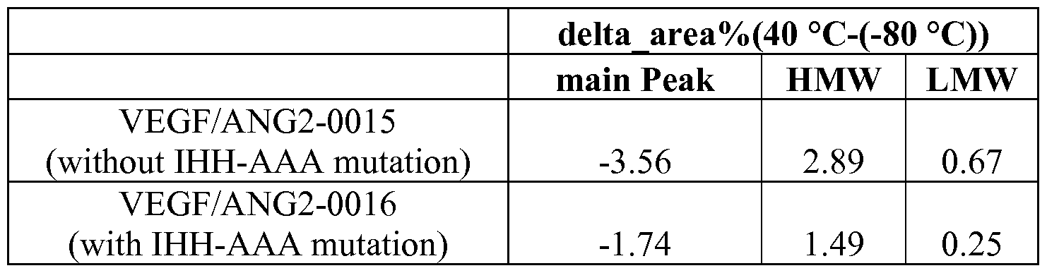


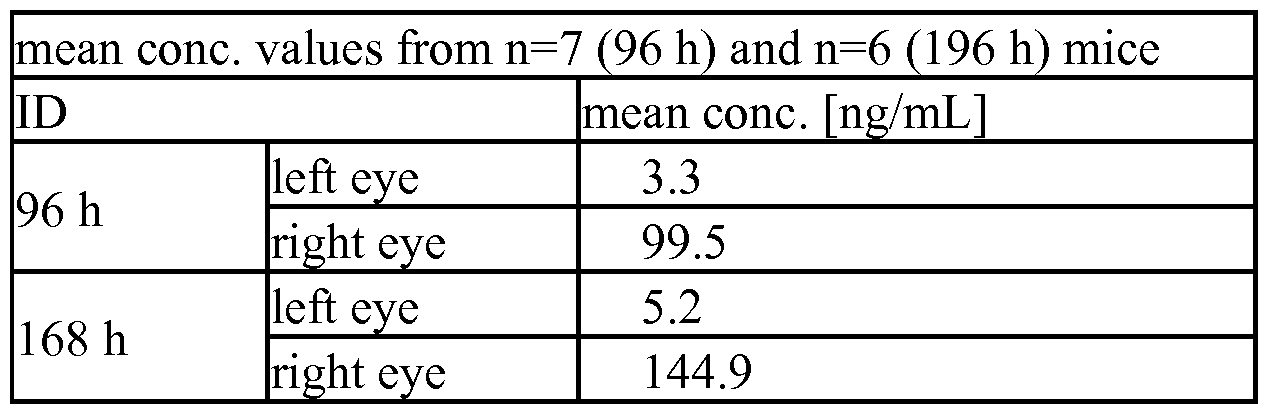


Claims
Priority Applications (11)
| Application Number | Priority Date | Filing Date | Title |
|---|---|---|---|
| RU2016133345A RU2730592C2 (en) | 2014-01-15 | 2015-01-12 | Versions of the fc-domain with modified abilities to bind to fcrn |
| MX2016008540A MX2016008540A (en) | 2014-01-15 | 2015-01-12 | Fc-region variants with modified fcrn-binding properties. |
| BR112016016411A BR112016016411A2 (en) | 2014-01-15 | 2015-01-12 | "Fc"-REGION VARIANTS WITH MODIFIED "FcRn" BINDING PROPERTIES |
| CN201580003633.0A CN105873948B (en) | 2014-01-15 | 2015-01-12 | Fc region variants with modified FCRN binding properties |
| CA2931979A CA2931979A1 (en) | 2014-01-15 | 2015-01-12 | Fc-region variants with modified fcrn-binding properties |
| KR1020167019016A KR20160104009A (en) | 2014-01-15 | 2015-01-12 | Fc-region variants with modified fcrn-binding properties |
| JP2016546942A JP6873701B2 (en) | 2014-01-15 | 2015-01-12 | Fc region mutant with modified FcRn binding properties |
| EP15703229.3A EP3094649A1 (en) | 2014-01-15 | 2015-01-12 | Fc-region variants with modified fcrn-binding properties |
| US15/210,218 US20170037121A1 (en) | 2014-01-15 | 2016-07-14 | Fc-region variants with modified fcrn-binding properties |
| HK16112240.5A HK1223951A1 (en) | 2014-01-15 | 2016-10-25 | Fc-region variants with modified fcrn-binding properties fcrn fc |
| US15/947,377 US20190016792A1 (en) | 2014-01-15 | 2018-04-06 | Fc-region variants with modified fcrn-binding properties |
Applications Claiming Priority (4)
| Application Number | Priority Date | Filing Date | Title |
|---|---|---|---|
| EP14151319 | 2014-01-15 | ||
| EP14151319.2 | 2014-01-15 | ||
| EP14165922.7 | 2014-04-25 | ||
| EP14165922 | 2014-04-25 |
Related Child Applications (1)
| Application Number | Title | Priority Date | Filing Date |
|---|---|---|---|
| US15/210,218 Continuation US20170037121A1 (en) | 2014-01-15 | 2016-07-14 | Fc-region variants with modified fcrn-binding properties |
Publications (1)
| Publication Number | Publication Date |
|---|---|
| WO2015107025A1 true WO2015107025A1 (en) | 2015-07-23 |
Family
ID=52462893
Family Applications (1)
| Application Number | Title | Priority Date | Filing Date |
|---|---|---|---|
| PCT/EP2015/050425 WO2015107025A1 (en) | 2014-01-15 | 2015-01-12 | Fc-region variants with modified fcrn-binding properties |
Country Status (12)
| Country | Link |
|---|---|
| US (2) | US20170037121A1 (en) |
| EP (1) | EP3094649A1 (en) |
| JP (2) | JP6873701B2 (en) |
| KR (1) | KR20160104009A (en) |
| CN (2) | CN105873948B (en) |
| AR (1) | AR099079A1 (en) |
| BR (1) | BR112016016411A2 (en) |
| CA (1) | CA2931979A1 (en) |
| HK (1) | HK1223951A1 (en) |
| MX (1) | MX2016008540A (en) |
| RU (1) | RU2730592C2 (en) |
| WO (1) | WO2015107025A1 (en) |
Cited By (27)
| Publication number | Priority date | Publication date | Assignee | Title |
|---|---|---|---|---|
| EP2970508A4 (en) * | 2013-03-15 | 2016-12-14 | Permeon Biologics Inc | Charge-engineered antibodies or compositions of penetration-enhanced targeting proteins and methods of use |
| WO2017072210A1 (en) * | 2015-10-29 | 2017-05-04 | F. Hoffmann-La Roche Ag | Anti-variant fc-region antibodies and methods of use |
| WO2017165464A1 (en) | 2016-03-21 | 2017-09-28 | Elstar Therapeutics, Inc. | Multispecific and multifunctional molecules and uses thereof |
| WO2018129255A1 (en) * | 2017-01-06 | 2018-07-12 | Momenta Pharmaceuticals, Inc. | Compositions and methods related to engineered fc constructs |
| WO2018151820A1 (en) | 2017-02-16 | 2018-08-23 | Elstar Therapeutics, Inc. | Multifunctional molecules comprising a trimeric ligand and uses thereof |
| WO2018222901A1 (en) | 2017-05-31 | 2018-12-06 | Elstar Therapeutics, Inc. | Multispecific molecules that bind to myeloproliferative leukemia (mpl) protein and uses thereof |
| WO2019035938A1 (en) | 2017-08-16 | 2019-02-21 | Elstar Therapeutics, Inc. | Multispecific molecules that bind to bcma and uses thereof |
| US10239944B2 (en) | 2014-05-02 | 2019-03-26 | Momenta Pharmaceuticals, Inc. | Compositions and methods related to engineered Fc constructs |
| EP3514171A1 (en) * | 2018-01-18 | 2019-07-24 | Molecular Cloning Laboratories (MCLAB) LLC | Long-acting therapeutic fusion proteins |
| WO2019178362A1 (en) | 2018-03-14 | 2019-09-19 | Elstar Therapeutics, Inc. | Multifunctional molecules that bind to calreticulin and uses thereof |
| WO2019178364A2 (en) | 2018-03-14 | 2019-09-19 | Elstar Therapeutics, Inc. | Multifunctional molecules and uses thereof |
| WO2020010250A2 (en) | 2018-07-03 | 2020-01-09 | Elstar Therapeutics, Inc. | Anti-tcr antibody molecules and uses thereof |
| WO2020172605A1 (en) | 2019-02-21 | 2020-08-27 | Elstar Therapeutics, Inc. | Antibody molecules that bind to nkp30 and uses thereof |
| WO2020172596A1 (en) | 2019-02-21 | 2020-08-27 | Elstar Therapeutics, Inc. | Anti-tcr antibody molecules and thereof |
| WO2020172571A1 (en) | 2019-02-21 | 2020-08-27 | Elstar Therapeutics, Inc. | Multifunctional molecules that bind to t cell related cancer cells and uses thereof |
| WO2020172598A1 (en) | 2019-02-21 | 2020-08-27 | Elstar Therapeutics, Inc. | Multifunctional molecules that bind to t cells and uses thereof to treat autoimmune disorders |
| WO2020172601A1 (en) | 2019-02-21 | 2020-08-27 | Elstar Therapeutics, Inc. | Multifunctional molecules that bind to calreticulin and uses thereof |
| US10947295B2 (en) * | 2017-08-22 | 2021-03-16 | Sanabio, Llc | Heterodimers of soluble interferon receptors and uses thereof |
| WO2021138407A2 (en) | 2020-01-03 | 2021-07-08 | Marengo Therapeutics, Inc. | Multifunctional molecules that bind to cd33 and uses thereof |
| US11155640B2 (en) | 2016-05-23 | 2021-10-26 | Janssen Biotech, Inc. | Compositions and methods related to engineered Fc constructs |
| WO2021217085A1 (en) | 2020-04-24 | 2021-10-28 | Marengo Therapeutics, Inc. | Multifunctional molecules that bind to t cell related cancer cells and uses thereof |
| US11254744B2 (en) | 2015-08-07 | 2022-02-22 | Imaginab, Inc. | Antigen binding constructs to target molecules |
| WO2022046920A2 (en) | 2020-08-26 | 2022-03-03 | Marengo Therapeutics, Inc. | Multifunctional molecules that bind to calreticulin and uses thereof |
| WO2022047046A1 (en) | 2020-08-26 | 2022-03-03 | Marengo Therapeutics, Inc. | Methods of detecting trbc1 or trbc2 |
| WO2022046922A2 (en) | 2020-08-26 | 2022-03-03 | Marengo Therapeutics, Inc. | Antibody molecules that bind to nkp30 and uses thereof |
| US11266745B2 (en) | 2017-02-08 | 2022-03-08 | Imaginab, Inc. | Extension sequences for diabodies |
| WO2022216993A2 (en) | 2021-04-08 | 2022-10-13 | Marengo Therapeutics, Inc. | Multifuntional molecules binding to tcr and uses thereof |
Families Citing this family (12)
| Publication number | Priority date | Publication date | Assignee | Title |
|---|---|---|---|---|
| US20090162359A1 (en) | 2007-12-21 | 2009-06-25 | Christian Klein | Bivalent, bispecific antibodies |
| US9676845B2 (en) | 2009-06-16 | 2017-06-13 | Hoffmann-La Roche, Inc. | Bispecific antigen binding proteins |
| AR085403A1 (en) | 2011-02-28 | 2013-09-25 | Hoffmann La Roche | MONOVALENT PROTEINS THAT JOIN ANTIGENS |
| RU2607038C2 (en) | 2011-02-28 | 2017-01-10 | Ф. Хоффманн-Ля Рош Аг | Antigen-binding proteins |
| JP6422956B2 (en) * | 2013-10-11 | 2018-11-14 | エフ.ホフマン−ラ ロシュ アーゲーF. Hoffmann−La Roche Aktiengesellschaft | Multispecific domain exchange common variable light chain antibody |
| CA3040823A1 (en) * | 2016-10-17 | 2018-04-26 | Vetoquinol Sa | Modified antibody constant region |
| WO2019122054A1 (en) * | 2017-12-22 | 2019-06-27 | F. Hoffmann-La Roche Ag | Depletion of light chain mispaired antibody variants by hydrophobic interaction chromatography |
| CN112955240B (en) * | 2018-10-25 | 2022-09-16 | 豪夫迈·罗氏有限公司 | Modification of antibody FcRn binding |
| KR102605376B1 (en) | 2018-12-31 | 2023-11-23 | 삼성디스플레이 주식회사 | Display device |
| JP2023529443A (en) * | 2020-06-08 | 2023-07-10 | シアトル チルドレンズ ホスピタル (ディービーエイ シアトル チルドレンズ リサーチ インスティテュート) | Anti-CD171 chimeric antigen receptor |
| JP2024512240A (en) | 2021-02-18 | 2024-03-19 | エフ. ホフマン-ラ ロシュ アーゲー | Methods for elucidating complex multistep antibody interactions |
| WO2022265331A1 (en) * | 2021-06-14 | 2022-12-22 | 고려대학교 산학협력단 | Fc variants with controlled immune mechanism and increased blood half-life |
Citations (4)
| Publication number | Priority date | Publication date | Assignee | Title |
|---|---|---|---|---|
| US6277375B1 (en) * | 1997-03-03 | 2001-08-21 | Board Of Regents, The University Of Texas System | Immunoglobulin-like domains with increased half-lives |
| US20100272720A1 (en) * | 2009-04-22 | 2010-10-28 | Merck Patent Gmbh | Antibody Fusion Proteins with a Modified FcRn Binding Site |
| WO2013004842A2 (en) * | 2011-07-06 | 2013-01-10 | Genmab A/S | Antibody variants and uses thereof |
| WO2014006217A1 (en) * | 2012-07-06 | 2014-01-09 | Genmab B.V. | Dimeric protein with triple mutations |
Family Cites Families (11)
| Publication number | Priority date | Publication date | Assignee | Title |
|---|---|---|---|---|
| JP4685764B2 (en) * | 2003-04-10 | 2011-05-18 | アボット バイオセラピューティクス コーポレイション | Modification of antibody FcRn binding affinity or serum half-life by mutagenesis |
| US20050249723A1 (en) * | 2003-12-22 | 2005-11-10 | Xencor, Inc. | Fc polypeptides with novel Fc ligand binding sites |
| US7923538B2 (en) * | 2005-07-22 | 2011-04-12 | Kyowa Hakko Kirin Co., Ltd | Recombinant antibody composition |
| EP2407548A1 (en) * | 2006-10-16 | 2012-01-18 | MedImmune, LLC | Molecules with reduced half-lives, compositions and uses thereof |
| EP2853542A1 (en) * | 2010-11-24 | 2015-04-01 | Glaxo Group Limited | Multispecific antigen binding proteins targeting HGF |
| SG191762A1 (en) * | 2011-01-06 | 2013-08-30 | Glaxo Group Ltd | Ligands that bind tgf-beta receptor ii |
| CN107080843A (en) * | 2011-10-13 | 2017-08-22 | 爱尔皮奥治疗有限公司 | The treatment of illness in eye |
| RU2675319C2 (en) * | 2011-11-04 | 2018-12-18 | Займворкс Инк. | STABLE HETERODIMERIC ANTIBODY DESIGN WITH MUTATIONS IN Fc DOMAIN |
| AU2013258834B2 (en) * | 2012-05-10 | 2017-09-07 | Zymeworks Bc Inc. | Heteromultimer constructs of immunoglobulin heavy chains with mutations in the Fc domain |
| CN105143262A (en) * | 2013-04-29 | 2015-12-09 | 豪夫迈·罗氏有限公司 | Human fcrn-binding modified antibodies and methods of use |
| MX364861B (en) * | 2013-04-29 | 2019-05-09 | Hoffmann La Roche | Fcrn-binding abolished anti-igf-1r antibodies and their use in the treatment of vascular eye diseases. |
-
2015
- 2015-01-12 WO PCT/EP2015/050425 patent/WO2015107025A1/en active Application Filing
- 2015-01-12 CN CN201580003633.0A patent/CN105873948B/en active Active
- 2015-01-12 RU RU2016133345A patent/RU2730592C2/en active
- 2015-01-12 KR KR1020167019016A patent/KR20160104009A/en not_active Application Discontinuation
- 2015-01-12 JP JP2016546942A patent/JP6873701B2/en active Active
- 2015-01-12 CA CA2931979A patent/CA2931979A1/en active Pending
- 2015-01-12 BR BR112016016411A patent/BR112016016411A2/en not_active IP Right Cessation
- 2015-01-12 EP EP15703229.3A patent/EP3094649A1/en active Pending
- 2015-01-12 CN CN202110445639.0A patent/CN113248613A/en active Pending
- 2015-01-12 MX MX2016008540A patent/MX2016008540A/en unknown
- 2015-01-13 AR ARP150100075A patent/AR099079A1/en unknown
-
2016
- 2016-07-14 US US15/210,218 patent/US20170037121A1/en not_active Abandoned
- 2016-10-25 HK HK16112240.5A patent/HK1223951A1/en unknown
-
2018
- 2018-04-06 US US15/947,377 patent/US20190016792A1/en not_active Abandoned
-
2021
- 2021-04-21 JP JP2021071749A patent/JP2021113214A/en active Pending
Patent Citations (4)
| Publication number | Priority date | Publication date | Assignee | Title |
|---|---|---|---|---|
| US6277375B1 (en) * | 1997-03-03 | 2001-08-21 | Board Of Regents, The University Of Texas System | Immunoglobulin-like domains with increased half-lives |
| US20100272720A1 (en) * | 2009-04-22 | 2010-10-28 | Merck Patent Gmbh | Antibody Fusion Proteins with a Modified FcRn Binding Site |
| WO2013004842A2 (en) * | 2011-07-06 | 2013-01-10 | Genmab A/S | Antibody variants and uses thereof |
| WO2014006217A1 (en) * | 2012-07-06 | 2014-01-09 | Genmab B.V. | Dimeric protein with triple mutations |
Non-Patent Citations (6)
| Title |
|---|
| KIM H ET AL: "FcRn receptor-mediated pharmacokinetics of therapeutic IgG in the eye", vol. 15, 16 December 2009 (2009-12-16), pages 2803 - 2812, XP002688851, ISSN: 1090-0535, Retrieved from the Internet <URL:http://www.molvis.org/molvis/v15/a296> * |
| KIM J-K ET AL: "Mapping the site on human IgG for binding of the MHC class I-related receptor, FcRn", EUROPEAN JOURNAL OF IMMUNOLOGY, WILEY - V C H VERLAG GMBH & CO. KGAA, DE, vol. 29, no. 9, 1 September 1999 (1999-09-01), pages 2819 - 2825, XP002300286, ISSN: 0014-2980, DOI: 10.1002/(SICI)1521-4141(199909)29:09<2819::AID-IMMU2819>3.0.CO;2-6 * |
| MARTIN W L ET AL: "Crystal Structure at 2.8A of an FcRn/Heterodimeric Fc Complex: Mechanism of pH-Dependent Binding", MOLECULAR CELL, CELL PRESS, CAMBRIDGE, MA, US, vol. 7, 1 April 2001 (2001-04-01), pages 867 - 877, XP003027710, ISSN: 1097-2765, DOI: 10.1016/S1097-2765(01)00230-1 * |
| MEDESAN C ET AL: "Delineation of the amino acid residues involved in transcytosis and catabolism of mouse IgG1", THE JOURNAL OF IMMUNOLOGY, THE AMERICAN ASSOCIATION OF IMMUNOLOGISTS, US, vol. 158, no. 5, 1 March 1997 (1997-03-01), pages 2211 - 2217, XP002532767, ISSN: 0022-1767 * |
| SHIELDS R L ET AL: "High resolution mapping of the binding site on human IgG1 for Fc gamma RI, Fc gamma RII, Fc gamma RIII, and FcRn and design of IgG1 variants with improved binding to the Fc gamma R", JOURNAL OF BIOLOGICAL CHEMISTRY, AMERICAN SOCIETY FOR BIOCHEMISTRY AND MOLECULAR BIOLOGY, US, vol. 276, no. 9, 2 March 2001 (2001-03-02), pages 6591 - 6604, XP002495886, ISSN: 0021-9258, [retrieved on 20001128], DOI: 10.1074/JBC.M009483200 * |
| TIMOTHY T KUO ET AL: "Neonatal Fc Receptor: From Immunity to Therapeutics", JOURNAL OF CLINICAL IMMUNOLOGY, KLUWER ACADEMIC PUBLISHERS-PLENUM PUBLISHERS, NE, vol. 30, no. 6, 1 October 2010 (2010-10-01), pages 777 - 789, XP019858481, ISSN: 1573-2592, DOI: 10.1007/S10875-010-9468-4 * |
Cited By (43)
| Publication number | Priority date | Publication date | Assignee | Title |
|---|---|---|---|---|
| EP2970508A4 (en) * | 2013-03-15 | 2016-12-14 | Permeon Biologics Inc | Charge-engineered antibodies or compositions of penetration-enhanced targeting proteins and methods of use |
| US10239944B2 (en) | 2014-05-02 | 2019-03-26 | Momenta Pharmaceuticals, Inc. | Compositions and methods related to engineered Fc constructs |
| US11124573B2 (en) | 2014-05-02 | 2021-09-21 | Janssen Biotech, Inc. | Compositions and methods related to engineered Fc constructs |
| US11254744B2 (en) | 2015-08-07 | 2022-02-22 | Imaginab, Inc. | Antigen binding constructs to target molecules |
| AU2016344665C1 (en) * | 2015-10-29 | 2023-07-27 | F. Hoffmann-La Roche Ag | Anti-variant Fc-region antibodies and methods of use |
| AU2016344665B2 (en) * | 2015-10-29 | 2023-01-19 | F. Hoffmann-La Roche Ag | Anti-variant Fc-region antibodies and methods of use |
| JP2018535670A (en) * | 2015-10-29 | 2018-12-06 | エフ.ホフマン−ラ ロシュ アーゲーF. Hoffmann−La Roche Aktiengesellschaft | Anti-variant Fc region antibodies and methods of use |
| WO2017072210A1 (en) * | 2015-10-29 | 2017-05-04 | F. Hoffmann-La Roche Ag | Anti-variant fc-region antibodies and methods of use |
| EP4015533A1 (en) * | 2015-10-29 | 2022-06-22 | F. Hoffmann-La Roche AG | Anti-variant fc-region antibodies and methods of use |
| CN108350066A (en) * | 2015-10-29 | 2018-07-31 | 豪夫迈·罗氏有限公司 | Resistance body Fc domain antibodies and application method |
| US10899845B2 (en) | 2015-10-29 | 2021-01-26 | Hoffmann-La Roche Inc. | Anti-variant Fc-region antibodies and method of use |
| JP2022025063A (en) * | 2015-10-29 | 2022-02-09 | エフ.ホフマン-ラ ロシュ アーゲー | Anti-variant fc-region antibodies and methods of use |
| JP7305719B2 (en) | 2015-10-29 | 2023-07-10 | エフ. ホフマン-ラ ロシュ アーゲー | Anti-variant Fc region antibodies and methods of use |
| IL258245B (en) * | 2015-10-29 | 2022-10-01 | Hoffmann La Roche | Anti-variant fc-region antibodies and methods of use |
| IL258245B2 (en) * | 2015-10-29 | 2023-02-01 | Hoffmann La Roche | Anti-variant fc-region antibodies and methods of use |
| WO2017165464A1 (en) | 2016-03-21 | 2017-09-28 | Elstar Therapeutics, Inc. | Multispecific and multifunctional molecules and uses thereof |
| US11155640B2 (en) | 2016-05-23 | 2021-10-26 | Janssen Biotech, Inc. | Compositions and methods related to engineered Fc constructs |
| US11623964B2 (en) | 2016-05-23 | 2023-04-11 | Momenta Pharmaceuticals, Inc. | Compositions and methods related to engineered Fc constructs |
| WO2018129255A1 (en) * | 2017-01-06 | 2018-07-12 | Momenta Pharmaceuticals, Inc. | Compositions and methods related to engineered fc constructs |
| US11827682B2 (en) | 2017-01-06 | 2023-11-28 | Momenta Pharmaceuticals, Inc. | Engineered Fc constructs |
| US11220531B2 (en) | 2017-01-06 | 2022-01-11 | Janssen Biotech, Inc. | Engineered Fc constructs |
| US11266745B2 (en) | 2017-02-08 | 2022-03-08 | Imaginab, Inc. | Extension sequences for diabodies |
| WO2018151820A1 (en) | 2017-02-16 | 2018-08-23 | Elstar Therapeutics, Inc. | Multifunctional molecules comprising a trimeric ligand and uses thereof |
| WO2018222901A1 (en) | 2017-05-31 | 2018-12-06 | Elstar Therapeutics, Inc. | Multispecific molecules that bind to myeloproliferative leukemia (mpl) protein and uses thereof |
| WO2019035938A1 (en) | 2017-08-16 | 2019-02-21 | Elstar Therapeutics, Inc. | Multispecific molecules that bind to bcma and uses thereof |
| US10947295B2 (en) * | 2017-08-22 | 2021-03-16 | Sanabio, Llc | Heterodimers of soluble interferon receptors and uses thereof |
| EP3514171A1 (en) * | 2018-01-18 | 2019-07-24 | Molecular Cloning Laboratories (MCLAB) LLC | Long-acting therapeutic fusion proteins |
| WO2019178362A1 (en) | 2018-03-14 | 2019-09-19 | Elstar Therapeutics, Inc. | Multifunctional molecules that bind to calreticulin and uses thereof |
| WO2019178364A2 (en) | 2018-03-14 | 2019-09-19 | Elstar Therapeutics, Inc. | Multifunctional molecules and uses thereof |
| DE202019005887U1 (en) | 2018-07-03 | 2023-06-14 | Marengo Therapeutics, Inc. | Anti-TCR antibody molecules and uses thereof |
| US11845797B2 (en) | 2018-07-03 | 2023-12-19 | Marengo Therapeutics, Inc. | Anti-TCR antibody molecules and uses thereof |
| WO2020010250A2 (en) | 2018-07-03 | 2020-01-09 | Elstar Therapeutics, Inc. | Anti-tcr antibody molecules and uses thereof |
| WO2020172571A1 (en) | 2019-02-21 | 2020-08-27 | Elstar Therapeutics, Inc. | Multifunctional molecules that bind to t cell related cancer cells and uses thereof |
| WO2020172598A1 (en) | 2019-02-21 | 2020-08-27 | Elstar Therapeutics, Inc. | Multifunctional molecules that bind to t cells and uses thereof to treat autoimmune disorders |
| WO2020172596A1 (en) | 2019-02-21 | 2020-08-27 | Elstar Therapeutics, Inc. | Anti-tcr antibody molecules and thereof |
| WO2020172605A1 (en) | 2019-02-21 | 2020-08-27 | Elstar Therapeutics, Inc. | Antibody molecules that bind to nkp30 and uses thereof |
| WO2020172601A1 (en) | 2019-02-21 | 2020-08-27 | Elstar Therapeutics, Inc. | Multifunctional molecules that bind to calreticulin and uses thereof |
| WO2021138407A2 (en) | 2020-01-03 | 2021-07-08 | Marengo Therapeutics, Inc. | Multifunctional molecules that bind to cd33 and uses thereof |
| WO2021217085A1 (en) | 2020-04-24 | 2021-10-28 | Marengo Therapeutics, Inc. | Multifunctional molecules that bind to t cell related cancer cells and uses thereof |
| WO2022046922A2 (en) | 2020-08-26 | 2022-03-03 | Marengo Therapeutics, Inc. | Antibody molecules that bind to nkp30 and uses thereof |
| WO2022047046A1 (en) | 2020-08-26 | 2022-03-03 | Marengo Therapeutics, Inc. | Methods of detecting trbc1 or trbc2 |
| WO2022046920A2 (en) | 2020-08-26 | 2022-03-03 | Marengo Therapeutics, Inc. | Multifunctional molecules that bind to calreticulin and uses thereof |
| WO2022216993A2 (en) | 2021-04-08 | 2022-10-13 | Marengo Therapeutics, Inc. | Multifuntional molecules binding to tcr and uses thereof |
Also Published As
| Publication number | Publication date |
|---|---|
| CN113248613A (en) | 2021-08-13 |
| RU2016133345A (en) | 2018-02-20 |
| US20190016792A1 (en) | 2019-01-17 |
| JP2021113214A (en) | 2021-08-05 |
| RU2730592C2 (en) | 2020-08-24 |
| CN105873948B (en) | 2021-04-13 |
| KR20160104009A (en) | 2016-09-02 |
| HK1223951A1 (en) | 2017-08-11 |
| US20170037121A1 (en) | 2017-02-09 |
| JP6873701B2 (en) | 2021-05-19 |
| JP2017505768A (en) | 2017-02-23 |
| MX2016008540A (en) | 2016-09-26 |
| EP3094649A1 (en) | 2016-11-23 |
| RU2016133345A3 (en) | 2018-10-31 |
| CN105873948A (en) | 2016-08-17 |
| BR112016016411A2 (en) | 2017-10-03 |
| CA2931979A1 (en) | 2015-07-23 |
| AR099079A1 (en) | 2016-06-29 |
Similar Documents
| Publication | Publication Date | Title |
|---|---|---|
| US20230183330A1 (en) | Fc-region variants with improved protein a-binding | |
| AU2020201429B2 (en) | Human FcRn-binding modified antibodies and methods of use | |
| US20190016828A1 (en) | Fc-region variants with modified fcrn- and maintained protein a-binding properties | |
| US20190016792A1 (en) | Fc-region variants with modified fcrn-binding properties | |
| US10899846B2 (en) | Fc-region variants with modified FcRn- and protein A-binding properties | |
| WO2014177461A1 (en) | Fcrn-binding abolished anti-igf-1r antibodies and their use in the treatment of vascular eye diseases |
Legal Events
| Date | Code | Title | Description |
|---|---|---|---|
| 121 | Ep: the epo has been informed by wipo that ep was designated in this application |
Ref document number: 15703229 Country of ref document: EP Kind code of ref document: A1 |
|
| ENP | Entry into the national phase |
Ref document number: 2931979 Country of ref document: CA |
|
| REEP | Request for entry into the european phase |
Ref document number: 2015703229 Country of ref document: EP |
|
| WWE | Wipo information: entry into national phase |
Ref document number: 2015703229 Country of ref document: EP |
|
| WWE | Wipo information: entry into national phase |
Ref document number: MX/A/2016/008540 Country of ref document: MX |
|
| ENP | Entry into the national phase |
Ref document number: 20167019016 Country of ref document: KR Kind code of ref document: A |
|
| ENP | Entry into the national phase |
Ref document number: 2016546942 Country of ref document: JP Kind code of ref document: A |
|
| NENP | Non-entry into the national phase |
Ref country code: DE |
|
| REG | Reference to national code |
Ref country code: BR Ref legal event code: B01A Ref document number: 112016016411 Country of ref document: BR |
|
| ENP | Entry into the national phase |
Ref document number: 2016133345 Country of ref document: RU Kind code of ref document: A |
|
| ENP | Entry into the national phase |
Ref document number: 112016016411 Country of ref document: BR Kind code of ref document: A2 Effective date: 20160714 |