WO2016020332A1 - Bispecific antibodies against cd3epsilon and bcma - Google Patents
Bispecific antibodies against cd3epsilon and bcma Download PDFInfo
- Publication number
- WO2016020332A1 WO2016020332A1 PCT/EP2015/067841 EP2015067841W WO2016020332A1 WO 2016020332 A1 WO2016020332 A1 WO 2016020332A1 EP 2015067841 W EP2015067841 W EP 2015067841W WO 2016020332 A1 WO2016020332 A1 WO 2016020332A1
- Authority
- WO
- WIPO (PCT)
- Prior art keywords
- bcma
- antibody
- cells
- amino acid
- domain
- Prior art date
Links
Classifications
-
- C—CHEMISTRY; METALLURGY
- C07—ORGANIC CHEMISTRY
- C07K—PEPTIDES
- C07K16/00—Immunoglobulins [IGs], e.g. monoclonal or polyclonal antibodies
- C07K16/18—Immunoglobulins [IGs], e.g. monoclonal or polyclonal antibodies against material from animals or humans
- C07K16/28—Immunoglobulins [IGs], e.g. monoclonal or polyclonal antibodies against material from animals or humans against receptors, cell surface antigens or cell surface determinants
- C07K16/2878—Immunoglobulins [IGs], e.g. monoclonal or polyclonal antibodies against material from animals or humans against receptors, cell surface antigens or cell surface determinants against the NGF-receptor/TNF-receptor superfamily, e.g. CD27, CD30, CD40, CD95
-
- A—HUMAN NECESSITIES
- A61—MEDICAL OR VETERINARY SCIENCE; HYGIENE
- A61P—SPECIFIC THERAPEUTIC ACTIVITY OF CHEMICAL COMPOUNDS OR MEDICINAL PREPARATIONS
- A61P35/00—Antineoplastic agents
-
- C—CHEMISTRY; METALLURGY
- C07—ORGANIC CHEMISTRY
- C07K—PEPTIDES
- C07K16/00—Immunoglobulins [IGs], e.g. monoclonal or polyclonal antibodies
- C07K16/18—Immunoglobulins [IGs], e.g. monoclonal or polyclonal antibodies against material from animals or humans
- C07K16/28—Immunoglobulins [IGs], e.g. monoclonal or polyclonal antibodies against material from animals or humans against receptors, cell surface antigens or cell surface determinants
- C07K16/2803—Immunoglobulins [IGs], e.g. monoclonal or polyclonal antibodies against material from animals or humans against receptors, cell surface antigens or cell surface determinants against the immunoglobulin superfamily
- C07K16/2809—Immunoglobulins [IGs], e.g. monoclonal or polyclonal antibodies against material from animals or humans against receptors, cell surface antigens or cell surface determinants against the immunoglobulin superfamily against the T-cell receptor (TcR)-CD3 complex
-
- C—CHEMISTRY; METALLURGY
- C07—ORGANIC CHEMISTRY
- C07K—PEPTIDES
- C07K16/00—Immunoglobulins [IGs], e.g. monoclonal or polyclonal antibodies
- C07K16/18—Immunoglobulins [IGs], e.g. monoclonal or polyclonal antibodies against material from animals or humans
- C07K16/28—Immunoglobulins [IGs], e.g. monoclonal or polyclonal antibodies against material from animals or humans against receptors, cell surface antigens or cell surface determinants
- C07K16/30—Immunoglobulins [IGs], e.g. monoclonal or polyclonal antibodies against material from animals or humans against receptors, cell surface antigens or cell surface determinants from tumour cells
- C07K16/3061—Blood cells
-
- C—CHEMISTRY; METALLURGY
- C07—ORGANIC CHEMISTRY
- C07K—PEPTIDES
- C07K16/00—Immunoglobulins [IGs], e.g. monoclonal or polyclonal antibodies
- C07K16/46—Hybrid immunoglobulins
- C07K16/468—Immunoglobulins having two or more different antigen binding sites, e.g. multifunctional antibodies
-
- A—HUMAN NECESSITIES
- A61—MEDICAL OR VETERINARY SCIENCE; HYGIENE
- A61K—PREPARATIONS FOR MEDICAL, DENTAL OR TOILETRY PURPOSES
- A61K39/00—Medicinal preparations containing antigens or antibodies
- A61K2039/505—Medicinal preparations containing antigens or antibodies comprising antibodies
-
- C—CHEMISTRY; METALLURGY
- C07—ORGANIC CHEMISTRY
- C07K—PEPTIDES
- C07K2317/00—Immunoglobulins specific features
- C07K2317/20—Immunoglobulins specific features characterized by taxonomic origin
- C07K2317/21—Immunoglobulins specific features characterized by taxonomic origin from primates, e.g. man
-
- C—CHEMISTRY; METALLURGY
- C07—ORGANIC CHEMISTRY
- C07K—PEPTIDES
- C07K2317/00—Immunoglobulins specific features
- C07K2317/30—Immunoglobulins specific features characterized by aspects of specificity or valency
- C07K2317/31—Immunoglobulins specific features characterized by aspects of specificity or valency multispecific
-
- C—CHEMISTRY; METALLURGY
- C07—ORGANIC CHEMISTRY
- C07K—PEPTIDES
- C07K2317/00—Immunoglobulins specific features
- C07K2317/30—Immunoglobulins specific features characterized by aspects of specificity or valency
- C07K2317/33—Crossreactivity, e.g. for species or epitope, or lack of said crossreactivity
-
- C—CHEMISTRY; METALLURGY
- C07—ORGANIC CHEMISTRY
- C07K—PEPTIDES
- C07K2317/00—Immunoglobulins specific features
- C07K2317/30—Immunoglobulins specific features characterized by aspects of specificity or valency
- C07K2317/35—Valency
-
- C—CHEMISTRY; METALLURGY
- C07—ORGANIC CHEMISTRY
- C07K—PEPTIDES
- C07K2317/00—Immunoglobulins specific features
- C07K2317/50—Immunoglobulins specific features characterized by immunoglobulin fragments
- C07K2317/52—Constant or Fc region; Isotype
- C07K2317/522—CH1 domain
-
- C—CHEMISTRY; METALLURGY
- C07—ORGANIC CHEMISTRY
- C07K—PEPTIDES
- C07K2317/00—Immunoglobulins specific features
- C07K2317/50—Immunoglobulins specific features characterized by immunoglobulin fragments
- C07K2317/52—Constant or Fc region; Isotype
- C07K2317/526—CH3 domain
-
- C—CHEMISTRY; METALLURGY
- C07—ORGANIC CHEMISTRY
- C07K—PEPTIDES
- C07K2317/00—Immunoglobulins specific features
- C07K2317/50—Immunoglobulins specific features characterized by immunoglobulin fragments
- C07K2317/55—Fab or Fab'
-
- C—CHEMISTRY; METALLURGY
- C07—ORGANIC CHEMISTRY
- C07K—PEPTIDES
- C07K2317/00—Immunoglobulins specific features
- C07K2317/50—Immunoglobulins specific features characterized by immunoglobulin fragments
- C07K2317/56—Immunoglobulins specific features characterized by immunoglobulin fragments variable (Fv) region, i.e. VH and/or VL
- C07K2317/565—Complementarity determining region [CDR]
-
- C—CHEMISTRY; METALLURGY
- C07—ORGANIC CHEMISTRY
- C07K—PEPTIDES
- C07K2317/00—Immunoglobulins specific features
- C07K2317/60—Immunoglobulins specific features characterized by non-natural combinations of immunoglobulin fragments
- C07K2317/64—Immunoglobulins specific features characterized by non-natural combinations of immunoglobulin fragments comprising a combination of variable region and constant region components
-
- C—CHEMISTRY; METALLURGY
- C07—ORGANIC CHEMISTRY
- C07K—PEPTIDES
- C07K2317/00—Immunoglobulins specific features
- C07K2317/60—Immunoglobulins specific features characterized by non-natural combinations of immunoglobulin fragments
- C07K2317/66—Immunoglobulins specific features characterized by non-natural combinations of immunoglobulin fragments comprising a swap of domains, e.g. CH3-CH2, VH-CL or VL-CH1
-
- C—CHEMISTRY; METALLURGY
- C07—ORGANIC CHEMISTRY
- C07K—PEPTIDES
- C07K2317/00—Immunoglobulins specific features
- C07K2317/70—Immunoglobulins specific features characterized by effect upon binding to a cell or to an antigen
- C07K2317/73—Inducing cell death, e.g. apoptosis, necrosis or inhibition of cell proliferation
-
- C—CHEMISTRY; METALLURGY
- C07—ORGANIC CHEMISTRY
- C07K—PEPTIDES
- C07K2317/00—Immunoglobulins specific features
- C07K2317/90—Immunoglobulins specific features characterized by (pharmaco)kinetic aspects or by stability of the immunoglobulin
- C07K2317/92—Affinity (KD), association rate (Ka), dissociation rate (Kd) or EC50 value
Definitions
- the present invention relates to novel bispecific antibodies against CD3s and BCMA, their manufacture and use.
- BCMA Human B cell maturation antigen also known as BCMA; TR17 HUMAN, TNFRSF17 (UniProt Q02223), is a member of the tumor necrosis receptor superfamily that is preferentially expressed in differentiated plasma cells (Laabi et al. 1992; Madry et al. 1998).
- BCMA is a non glycosylated type III transmembrane protein, which is involved in B cell maturation, growth and survival.
- BCMA is a receptor for two ligands of the TNF superfamily: APRIL (a proliferation-inducing ligand), the high- affinity ligand to BCMA and the B cell activation factor BAFF, the low-affinity ligand to BCMA (THANK, BlyS, B lymphocyte stimulator, TALL-1 and zTNF4).
- APRIL and BAFF show structural similarity and overlapping yet distinct receptor binding specificity.
- the negative regulator TACI also binds to both BAFF and APRIL.
- the coordinate binding of APRIL and BAFF to BCMA and/or TACI activates transcription factor NF- ⁇ and increases the expression of pro-survival Bcl-2 family members (e.g.
- Bcl-2, Bcl-xL, Bcl-w, Mcl-1, Al the downregulation of pro-apoptotic factors (e.g. Bid, Bad, Bik, Bim, etc.), thus inhibiting apoptosis and promoting survival.
- pro-apoptotic factors e.g. Bid, Bad, Bik, Bim, etc.
- This combined action promotes B cell differentiation, proliferation, survival and antibody production (as reviewed in Rickert RC et al., Immunol Rev (2011) 244 (1): 115-133).
- the TCR/CD3 complex of T-lymphocytes consists of either a TCR alpha (a)/beta ( ⁇ ) or TCR gamma (y)/delta ( ⁇ ) heterodimer coexpressed at the cell surface with the invariant subunits of CD3 labeled gamma ( ⁇ ), delta ( ⁇ ), epsilon ( ⁇ ), zeta ( ⁇ ), and eta ( ⁇ ).
- Human CD3s is described under UniProt P07766 (CD3E HUMAN).
- An anti CD3s antibody described in the state of the art is SP34 (Yang SJ, The Journal of Immunology (1986) 137; 1097-1100). SP34 reacts with both primate and human CD3.
- SP34 is available from PharMingen.
- a further anti CD3 antibody described in the state of the art is UCHT-1 (see WO2000041474).
- a further anti CD3 antibody described in the state of the art is BC-3 (Fred Hutchinson Cancer Research Institute; used in Phase I/II trials of GvHD, Anasetti et al., Transplantation 54: 844 (1992)).
- bispecific antibody formats have been developed in the recent past, e.g. by fusion of, e.g. an IgG antibody format and single chain domains (see Kontermann RE, mAbs 4:2, (2012) 1-16).
- Bispecific antibodies wherein the variable domains VL and VH or the constant domains CL and CHI are replaced by each other are described in WO2009080251 and WO2009080252.
- An approach to circumvent the problem of mispaired byproducts which is known as 'knobs-into-holes', aims at forcing the pairing of two different antibody heavy chains by introducing mutations into the CH3 domains to modify the contact interface.
- the percentage of heterodimer could be further increased by remodeling the interaction surfaces of the two CH3 domains using a phage display approach and the introduction of a disulfide bridge to stabilize the heterodimers (Merchant A.M, et al, Nature Biotech 16 (1998) 677-681; Axwell S, Ridgway JB, Wells JA, Carter P., J Mol Biol 270 (1997) 26-35).
- New approaches for the knobs-into-holes technology are described in e.g. in EP 1870459A1. Although this format appears very attractive, no data describing progression towards the clinic are currently available.
- One important constraint of this strategy is that the light chains of the two parent antibodies have to be identical to prevent mispairing and formation of inactive molecules.
- WO 201302362 relate to heterodimerized polypeptides.
- WO201312733 relates to polypeptides comprising heterodimeric Fc regions.
- WO2012131555 relates to engineered heterodimeric immunoglobulins.
- EP 2647707 relates to engineered hetero-dimeric immunoglobulins.
- WO2009080251, WO 2009080252, WO 2009080253, WO 2009080254 and Schaefer, W. et al, PNAS, 108 (2011) 11187- 1191 relate to bivalent, bispecific IgG antibodies with a domain crossover.
- the multispecific antibodies with VH/VL replacement/exchange in one binding to prevent light chain mispairing (CrossMabVH-VL) which are described in WO2009080252, (see also Schaefer, W. et al, PNAS, 108 (2011) 11187-1191) clearly reduce the byproducts caused by the mismatch of a light chain against a first antigen with the wrong heavy chain against the second antigen (compared to approaches without such domain exchange). However their preparation is not completely free of side products. The main side product is based on a Bence-Jones -type interaction (Schaefer, W. et al, PNAS, 108 (2011) 11187-1191).
- Antibodies against BCMA are described e.g. in Gras M-P. et al. Int Immunol. 7 (1995) 1093-1106, WO200124811, WO200124812, WO2010104949 and WO2012163805.
- Antibodies against BCMA and their use for the treatment of lymphomas and multiple myeloma are mentioned e.g. in WO2002066516 and WO2010104949.
- WO2013154760 relates to chimeric antigen receptors (CAR) comprising a BCMA recognition moiety and a T-cell activation moiety.
- CAR chimeric antigen receptors
- BCMA antibodies can act on myeloma cell lines through multiple mechanisms that include inhibition of APRIL-dependent NF- ⁇ activation, promotion of tumor cell lysis by natural killer cell-mediated ADCC activity, and induction of cytotoxicity by ADCs.
- Bispecific antibodies against CD3 and BCMA are mentioned in WO2007117600, WO2009132058, WO2012066058, WO2012143498, and WO2013072415.
- PCT/EP2014/052189 and PCT/EP2014/052190 describe antibodies against BCMA and bispecific antibodies against CD3 and BCMA comprising certain CDRs and variable domains disclosed also in the present invention.
- the invention relates to a bispecific bi- or trivalent antibody specifically binding to the two targets which are the extracellular domain of human BCMA (further named also as "BCMA”) and human CD3s (further named also as "CD3") wherein the variable domains VL and VH in a light chain and the respective heavy chain are replaced by each other, characterized in comprising a constant domain CL wherein the amino acid at position 124 is substituted independently by lysine (K), arginine (R) or histidine (H) (numbering according to Kabat), and in the respective constant domain CHI the amino acid at position 147 and the amino acid at position 213 is substituted independently by glutamic acid (E), or aspartic acid (D) (numbering according to Kabat).
- BCMA extracellular domain of human BCMA
- CD3s human CD3s
- the antibody is monovalent for CD3 binding.
- the amino acid at position 123 is substituted independently by lysine (K), arginine (R) or histidine (H).
- the antibody is monovalent for CD3 binding and amino acid 124 is K, amino acid 147 is E, amino acid 213 is E, and amino acid 123 is R.
- the invention relates to a bispecific antibody specifically binding to the two targets which are the extracellular domain of human BCMA (further named also as "BCMA”) and human CD3s (further named also as "CD3”), characterized in comprising a) the first light chain and the first heavy chain of a first antibody which specifically binds to BCMA; and b) the second light chain and the second heavy chain of a second antibody which specifically binds to CD3, and wherein the variable domains VL and VH in the second light chain and second heavy chain of the second antibody are replaced by each other; and c) wherein in the constant domain CL of the first light chain under a) the amino acid at position 124 is substituted independently by lysine (K), arginine (R) or histidine (H) (numbering according to Kabat), and wherein in the constant domain CHI of the first heavy chain under a) the amino acid at position 147 and the amino acid at position 213 is substituted independently by glutamic acid (E), or aspartic acid
- said bispecific antibody described in the last preceding paragraph is further characterized in that said bispecific antibody comprises in addition a Fab fragment of said first antibody (further named also as "BCMA-Fab") and in the constant domain CL said BCMA-Fab the amino acid at position 124 is substituted independently by lysine (K), arginine (R) or histidine (H) (numbering according to Kabat), and wherein in the constant domain CHI of said BCMA-Fab the amino acid at positions 147 and the amino acid at position 213 is substituted independently by glutamic acid (E), or aspartic acid (D) (numbering according to Kabat) (see e.g. Figures 2A, 2C).
- BCMA-Fab Fab fragment of said first antibody
- the invention further relates to a bispecific antibody specifically binding to the two targets which are the extracellular domain of human BCMA (further named also as "BCMA”) and human CD3s (further named also as "CD3”), characterized in comprising a) the first light chain and the first heavy chain of a first antibody which specifically binds to BCMA; and b) the second light chain and the second heavy chain of a second antibody which specifically binds to CD3, and wherein the variable domains VL and VH in the second light chain and second heavy chain of the second antibody are replaced by each other; and wherein c) in the constant domain CL of the second light chain under b) the amino acid at position 124 is substituted independently by lysine (K), arginine (R) or histidine (H) (numbering according to Kabat), and wherein in the constant domain CHI of the second heavy chain under b) the amino acid at positions 147 and the amino acid at position 213 is substituted independently by glutamic acid (E), or aspartic
- said bispecific antibody described in the last preceding paragraph is further characterized in that said bispecific antibody comprises in addition a second Fab fragment of said first antibody ("BCMA-Fab") (see e.g. Figure 2B, 2D).
- BCMA-Fab second Fab fragment of said first antibody
- Amino acid numbering is according to Kabat (see below).
- amino acid replacement at position 124 in the constant domain CL of the first or second light chain the amino acid at position 123 is substituted independently by lysine (K), arginine (R) or histidine (H).
- the amino acid at position 124 is substituted by lysine (K)
- the amino acid at position 147 and the amino acid at position 213 are substituted by glutamic acid (E).
- the constant domain CL in the amino acid at position 123 is substituted by arginine (R).
- the antibody according to the invention consists of one Fab fragment of an antibody specifically binding to CD3 (further named also as "CD3-Fab”), and one Fab fragment of an antibody specifically binding to BCMA (further named also as "BCMA-Fab(s)”) and a Fc part, wherein the CD3-Fab and the BCMA-Fab are linked via their C-termini to the hinge region of said Fc part.
- CD3-Fab or the BCMA-Fab comprises aa substitution and the CD3-Fab comprises crossover ( Figures 1A and IB).
- the antibody according to the invention consists of one CD3- Fab, and one BCMA-Fab and a Fc part, wherein the CD3-Fab and the BCMA-Fab are linked via their C-termini to the hinge region of said Fc part and a second BCMA-Fab, which is linked with its C- terminus to the N-terminus of the CD3-Fab.
- the CD3-Fab comprises crossover and either the CD3-Fab or both BCMA-Fabs comprise aa substitution ( Figures 2A and 2B).
- a bispecific antibody comprising BCMA-Fab-Fc-CD3-Fab-BCMA-Fab, wherein both BCMA-Fabs comprise aa substitution and the CD3-Fab comprises VL/VH crossover ( Figures 2A).
- a bispecific antibody consisting of BCMA-Fab-Fc-CD3-Fab-BCMA-Fab, wherein both BCMA-Fabs comprise aa substitution Q124K, E123R, K147E and K213E and the CD3-Fab comprises VL/VH crossover.
- both BCMA-Fabs comprise as CDRs the CDRs of antibody 83A10, or as VH/VL the VH/VL of 83A10.
- both BCMA-Fabs comprise as CDRs the CDRs of antibody 17A5, or as VH/VL the VH/VL of 17A5. Further preferred is that both BCMA-Fabs comprise as CDRs the CDRs of antibody 13A4, or as VH/VL the VH/VL of 13A4.
- the antibody according to the invention consists of two BCMA-Fabs and a Fc part, wherein the BCMA-Fabs are linked via their C-termini to the hinge region of said Fc part and a CD3-Fab, which is linked with its C-terminus to the N-terminus of one BCMA- Fab.
- the CD3-Fab comprises crossover and either the CD3-Fab or both BCMA-Fabs comprise aa substitution ( Figures 2C and 2D).
- the antibody according to the invention consists of one CD3- Fab, which is linked via its C-terminus to the hinge region of said Fc part and a BCMA-Fab, which is linked with its C-terminus to the N-terminus of the CD3-Fab.
- the CD3-Fab comprises crossover and either the CD3-Fab or the BCMA-Fab comprise aa substitution ( Figures 3A and 3B).
- the antibody according to the invention consists of one BCMA-Fab, which is linked via its C-terminus to the hinge region of said Fc part and a CD3-Fab, which is linked with its C-terminus to the N-terminus of the BCMA-Fab.
- the CD3-Fab comprises crossover and either the CD3-Fab or the BCMA-Fab comprise aa substitution ( Figures 3C and 3D).
- the Fab fragments are chemically linked together by the use of an appropriate linker according to the state of the art.
- a linker Preferably a (Gly4-Serl)3 linker is used (Desplancq DK et al., Protein Eng. 1994 Aug;7(8): 1027-33 and Mack M. et al., PNAS July 18, 1995 vol. 92 no. 15 7021 -7025).
- Linkage between two Fab fragments is performed between the heavy chains. Therefore the C-terminus of CHI of a first Fab fragment is linked to the N-terminus of VH of the second Fab fragment (no crossover) or to VL (crossover).
- Linkage between a Fab fragment and the Fc part is performed according to the invention as linkage between CHI and CH2.
- the first and a second Fab fragment of an antibody specifically binding to BCMA are preferably derived from the same antibody and preferably identical in the CDR sequences, variable domain sequences VH and VL and/or the constant domain sequences CHI and CL.
- the amino acid sequences of the first and a second Fab fragment of an antibody specifically binding to BCMA are identical.
- the BCMA antibody is an antibody comprising the CDR sequences of antibody 83A10, 17A5 or 13A4, an antibody comprising the VH and VL sequences of antibody 83A10, 17A5 or 13A4, or an antibody comprising the VH, VL, CHI, and CL sequences of antibody 83A10, 17A5 or 13A4.
- the bispecific antibody comprises as Fab fragments and Fc part, not more than one Fab fragment of an anti-CD3 antibody, not more than two Fab fragments of an anti-BCMA antibody and not more than one Fc part, preferably a human Fc part.
- the second Fab fragment of an anti- BCMA antibody is linked via its C-terminus either to the N-terminus of the Fab fragment of an anti- CD3 antibody or to the hinge region of the Fc part.
- linkage is performed between CHI of BCMA-Fab and VL of CD3 -Fab (VL/VH crossover).
- the antibody according to the invention is further characterized in that it binds also specifically to cynomolgus BCMA.
- a preferred antibody according to the invention is the antibody characterized in comprising a heavy and light chain set of polypeptides SEQ ID NO:43, SEQ ID NO:44, SEQ ID NO:45, and SEQ ID NO:46 (set 1).
- the antibody portion specifically binding to human CD3, preferably the Fab fragment is characterized in comprising a variable domain VH comprising the heavy chain CDRs of SEQ ID NO: 1 , 2 and 3 as respectively heavy chain CDR1, CDR2 and CDR3 and a variable domain VL comprising the light chain CDRs of SEQ ID NO: 4, 5 and 6 as respectively light chain CDR1, CDR2 and CDR3 of the anti-CD3s antibody (CDR MAB CD3).
- the antibody portion specifically binding to human CD3 is characterized in that the variable domains are of SEQ ID NO:7 and 8 (VHVL MAB CD3).
- the antibody portion, preferably the Fab fragment, specifically binding to human BCMA is characterized in comprising a variable domain VH comprising the heavy chain CDRs CDR1H of SEQ ID NO:21, a CDR2H of SEQ ID NO:24, a CDR3H of SEQ ID NO: 27 and comprising a variable domain VL comprising the light chain CDRs CDR1L of SEQ ID NO:30, a CDR2L of SEQ ID NO:33, a CDR3L of SEQ ID NO: 36 (CDR MAB 83A10).
- the antibody portion, preferably the Fab fragment, specifically binding to human BCMA is characterized in comprising a variable domain VH comprising the heavy chain CDRs CDR1H of SEQ ID NO:22, a CDR2H of SEQ ID NO:25, a CDR3H of SEQ ID NO: 28 and a variable domain VL comprising the light chain CDR1L of SEQ ID NO:31 , a CDR2L of SEQ ID NO:34, a CDR3L of SEQ ID NO: 37 (CDR MAB 17A5) .
- the antibody portion, preferably the Fab fragment, specifically binding to human BCMA is characterized in comprising a variable domain VH comprising the heavy chain CDRs CDR1 H of SEQ ID NO:23, a CDR2H of SEQ ID NO:26, a CDR3H of SEQ ID NO: 29 and a variable domain VL comprising the light chain CDR1L of SEQ ID NO:32, a CDR2L of SEQ ID NO:35, a CDR3L of SEQ ID NO: 38 (CDR MAB 13A4).
- VH comprising the heavy chain CDRs CDR1 H of SEQ ID NO:23, a CDR2H of SEQ ID NO:26, a CDR3H of SEQ ID NO: 29
- VL comprising the light chain CDR1L of SEQ ID NO:32, a CDR2L of SEQ ID NO:35, a CDR3L of SEQ ID NO: 38 (CDR MAB 13A4).
- the antibody portion, preferably the Fab fragment, specifically binding to human BCMA is characterized in comprising a VH of SEQ ID NO: 15 and a VL of SEQ ID NO: 18 (VHVL MAB 83A10).
- the antibody portion, preferably the Fab fragment, specifically binding to human BCMA is characterized in comprising a VH of SEQ ID NO: 16 and a VL of SEQ ID NO: 19 (VHVL MAB 17A5).
- the antibody portion, preferably the Fab fragment, specifically binding to human BCMA is characterized in comprising a VH of SEQ ID NO: 17 and a VL of SEQ ID NO: 20 (VHVL MAB 13A4).
- the invention relates to a bispecific antibody specifically binding to the extracellular domain of human BCMA and to human CD3s, characterized in comprising a heavy and light chain set selected from the group consisting of polypeptides i) SEQ ID NO:43, SEQ ID NO:44, SEQ ID NO:45, and SEQ ID NO:46 (set 1), ii) SEQ ID NO:45, SEQ ID NO:47, SEQ ID NO:48, and SEQ ID NO:49 (set 2), and iii) SEQ ID NO:45, SEQ ID NO:50, SEQ ID NO:51, and SEQ ID NO:52 (set 3).
- a heavy and light chain set selected from the group consisting of polypeptides i) SEQ ID NO:43, SEQ ID NO:44, SEQ ID NO:45, and SEQ ID NO:46 (set 1), ii) SEQ ID NO:45, SEQ ID NO:47, SEQ ID NO:48, and SEQ ID NO:49 (set 2), and i
- an antibody according to the invention comprises as anti-BCMA antibody an antibody selected from the group of the BCMA antibody variants of 13A4 and 83A10.
- an antibody to the invention comprising sequences of 13A4, comprises an amino acid replacement at respective position 95 (N95) or 96 (G96) within CDR3H of SEQ ID NO:29 by either a single amino acid change in position 95, either N95S, N95T, N95E, N95Q, N95A, or N95G, or by a single amino acid change in position 96, either G96A, G96E, or G96Q.
- an antibody according to the invention comprises an amino acid replacement selected from the group consisting of amino acid replacements at respective positions 27 (N27f) and 28 (G28) within CDR1L of SEQ ID NO:32 by either a single amino acid change in position 27, either N27fS, ⁇ 27 ⁇ , N27fE, N27fQ, N27fA, or N27fG or by a single amino acid change in position 28 either G28A, G28E, or G28Q.
- an antibody according to the invention comprises an amino acid replacement selected from the group consisting of amino acid replacements at respective positions 54 (D54) and 55 (S55) within CDR2H of SEQ ID NO:26 by either a single amino acid change in position 54, either D54S, D54T, D54E, D54Q, D54A, or D54G or by a single amino acid change in position 55, either S55A, S55E, or S55Q.
- an antibody according to the invention comprising sequences of 13A4, comprises an amino acid replacement selected from the group consisting of amino acid replacements W at position 33 (W33) within CDR1H of SEQ ID NO:23 by either W33F, W33Y, W33V, W33I, W33L, or W33A.
- an antibody according to the invention comprising sequences of 13A4, comprises up to two 5 amino acid replacements in N95 and/or W33 or up to two amino acid replacements in G96 and/or W33.
- an antibody according to the invention, comprising sequences of 13A4 comprises one amino acid replacement in N95 or G96.
- an antibody according to the invention comprising sequences of 13A4, comprises up to four amino acid replacements in N95 and/or W33 and/or N27 or G28 and/or D54 or S55or up to four amino 10 acid replacements in G96 and/or W33 and/or N27 or G28 and/or D54 or S55.
- an antibody according to the invention, comprising sequences of 13A4 comprises one amino acid replacement in N95 or G96.
- an antibody according to the invention comprises the first light chain and the first heavy chain of a first antibody specifically binding to BCMA, wherein said light chain comprising as CDRs,
- an antibody according to the invention comprises within the first light chain of a first antibody specifically binding to BCMA a variable light chain domain VL selected from the group consisting of SEQ ID NO: 20, 72, 74, 76, 78, 80, 82, 84, 86, and 88, and within the first heavy chain of a first antibody specifically binding to BCMA a variable heavy chain domain VH selected from the group 25 consisting of SEQ ID NO: 17, 54, 56, 58, 60, 62, 64, 66, 68, 70, 90, 92, 94, 96, 98, 100, 102, 104, 106, 108, 110, 112, 114, 116, and 118.
- an antibody according to the invention comprising sequences of 83A10, comprises an amino acid replacement selected from the group consisting of amino acid replacements at position 98 (W98) within CDR3H of SEQ ID NO:27 by either W98F, W98Y, W98V, W98I, W98L, or W98A.
- an antibody according to the invention comprises the first light chain and the first heavy chain of a first antibody specifically binding to BCMA, wherein said light chain comprising as CDRs CDRIL of SEQ ID NO:30, CDR2L of SEQ ID NO:33, CDR3L of SEQ ID NO: 36 and said heavy chain comprising as heavy chain CDRs CDR1H of SEQ ID NO:21, CDR2H of SEQ ID NO:24, and CDR3H selected from the group consisting of SEQ ID NO: 27, 119, 121, 123, 125, 127, and 129.
- an antibody according to the invention comprises within the first light chain of a first antibody specifically binding to BCMA a variable light chain domain VL of SEQ ID NO: 18 and within the first heavy chain of a first antibody specifically binding to BCMA a variable heavy chain domain VH selected from the group consisting of SEQ ID NO: 15, 120, 122, 124, 126, 128, and 130.
- the invention relates further to a bispecific antibody specifically binding to the extracellular domain of BCMA and to CD3s, characterized in comprising as heavy and light chains the polypeptides of SEQ ID NO: 45, 50, 51, and 52 wherein one or more CDRs are replaced by the respective CDRs of the "BCMA antibody variants".
- the invention relates further to a bispecific antibody specifically binding to the extracellular domain of BCMA and to CD3s, characterized in comprising as heavy and light chains the polypeptides of SEQ ID NO: 45, 50, 51, and 52 wherein one or more VHs and/or VLs are replaced by the respective VHs and/or VLs of the "BCMA antibody variants".
- the invention relates further to a bispecific antibody specifically binding to the extracellular domain of BCMA and to CD3s, characterized in comprising as heavy and light chains the polypeptides of SEQ ID NO: 43, 44, 45, and 46 wherein one or more CDRs are replaced by the respective CDRs of the "BCMA antibody variants".
- the invention relates further to a bispecific antibody specifically binding to the extracellular domain of BCMA and to CD3s, characterized in comprising as heavy and light chains the polypeptides of SEQ ID NO: 43, 44, 45, and 46 wherein one or more VHs and/or VLs are replaced by the respective VHs and/or VLs of the "BCMA antibody variants".
- an antibody according to the invention shows high affinity to BCMA and low aggregation.
- the invention further relates to a nucleic acid set encoding a respective heavy and light chain set.
- the bispecific antibody according to the invention comprising constant heavy regions CH2/CH3 of IgGl subclass is characterized in comprising the mutations L234A, L235A and P239G (numbering according to Kabat) to avoid FcR and Clq binding and minimizing ADCC/CDC.
- L234A, L235A and P239G numbering according to Kabat
- the invention comprises preferably a heavy chain of an antibody according to the invention consisting of (from N-to-C-terminus) VH(BCMA)-CH1(BCMA)-VL(CD3)-CH1(CD3)-CH2-CH3 of SEQ ID NO: 43, 47, or 50, as well as the respective encoding nucleic acids.
- These polypeptides and respective nucleic acids are useful for the production of a bispecific antibody according to the invention.
- the amino acid (aa) exchanges outside of the CDRs of the bispecific antibodies according to the invention provide considerably improved production/purification without changing biological properties like binding to BCMA. By introduction of the aa exchanges according to the invention light chain LC mispairing and the formation of side products in production is significantly reduced and therefore purification is facilitated.
- an antibody according to the invention is characterized by showing tumor growth inhibition of more than 70%, preferably of more than 85%, preferably of close to 100%> in a multiple myeloma xenograft model (e.g. xenograft with NCI-H929 cells or RPMI8226 cells or U266B1 cells or L-363 cells) at a dose of 1 mg/kg body weight (BW) administered intravenously (i.v.) or subcutaneously (s.c.) or intraperitoneal (i.p.) twice a week or once a week, preferably 0.5 mg/kg BW administered i.v. or i.p. or s.c.
- BW body weight
- s.c. subcutaneously
- i.p. intraperitoneal
- twice a week or once a week preferably at 0.1 mg/kg BW administered i.v. or i.p. or s.c. twice a week or once a week, preferably at 0.05 mg/kg BW administered i.v. or i.p. or s.c. twice a week or once a week, preferably at 0.01 mg/kg BW administered i.v. or i.p. or s.c twice a week or once a week, preferably at 5 ⁇ g/kg BW administered i.v. or i.p. or s.c. twice a week or once a week.
- an antibody according to the invention is characterized by an elimination half-life in mice, preferably cynomolgus monkeys of longer than 24 hours, preferably 3 days or longer, preferably half- life is measured for the doses which are effective in the xenograft model at twice or once a week administration.
- Bispecific antibodies binding to a target on tumor cells and to CD3 and having the molecular format (scFv) 2 have very short elimination half-life of 1 to 4 hours.
- this compound had to be administered via a pump carried by the patients over weeks and months (Topp et al. J Clin Oncol 2011 ; 29(18): 2493-8).
- treatment administered via a pump is much less convenient for the patients and also much more risky (e.g. failure of pump, issues with the catheter).
- an antibody according to the invention is characterized in showing an EC50 value for binding to NCI-H929 cells (ATCC® CRL-9068TM) of 30 nM or lower, preferably an EC50 value of 15 nM and lower.
- an antibody according to the invention is characterized by its capability to induce redirected killing of NCI-H929 tumor cells in the presence of human T cells with an EC50 lower than 0.1 nM, preferably 0.05 nM, preferably 0.02 nM, preferably 0.002 nM and lower.
- the potency of tumor cell killing of a bispecific antibody according to the invention is not or only minimally reduced by clinically relevant concentrations of APRIL; specifically the bispecific antibody of the invention is characterized in that addition of 100 ng/ml APRIL changes the EC50 for redirected T-cell killing of tumor cells by less than a factor of 4, preferably less than a factor of 2, preferably less than a factor of 1.5; In one preferred embodiment, the bispecific antibody of the invention is characterized in that addition of 1000 ng/niL APRIL changes the EC50 for tumor cell killing by less than a factor of 6.5, preferably less than a factor of 5, preferably less than a factor of 4, preferably less than a factor of 3, preferably less than a factor of 2.
- APRIL and BAFF have been shown to be important in multiple myeloma pathogenesis. Patients suffering from multiple myeloma have a high variability of plasma concentrations of APRIL and BAFF. In healthy subjects, APRIL plasma concentrations are usually 10 ng/ml or less. In myeloma patients, plasma concentrations of APRIL and/or BAFF range from 10 ng/ml to 100 ng/ml and in a low percentage of patients even up to 300 ng/ml and more (Moreaux et al. 2004; Blood 103(8): 3148-3157).
- APRIL is constitutively expressed in the bone marrow microenvironment being an important survival factor to malignant myeloma cells and also being mainly produced and secreted by bone marrow myeloid precursor cells (Matthes et al. Blood 2011; 118 (7): 1838-1844).
- concentrations of APRIL in the bone marrow of myeloma patients which are expected to be of higher magnitude, up to 1000 ng/mL or even more, are of high relevance in this context. If the concentrations for redirected T-cell killing of tumor cells by a BCMAxCD3 bispecific antibody are e.g.
- T-cell bispecific antibodies can be associated with dose-dependent side- effects, e.g. as reported for blinatumomab in clinical phase 1 and 2 trials).
- T-cell bispecific antibodies can be associated with dose-dependent side- effects, e.g. as reported for blinatumomab in clinical phase 1 and 2 trials.
- Such a shift towards higher effective concentrations of the BCMAxCD3 antibody by high levels of APRIL would more likely be caused if the BCMAxCD3 antibody and APRIL ligand compete for the same binding sites on the BCMA receptor. In that case, BCMA receptor occupancy would be reduced by the competing APRIL. Less receptor occupancy with the BCMAxCD3bispecific antibody means lower efficacy.
- BAFF could also cause such a shift, but given the much lower binding affinity of BAFF to BCMA than of APRIL to BCMA (i.e. up to 1000-fold lower affinity), APRIL plasma concentrations are in this context more relevant than BAFF plasma concentrations.
- the antibody according to the invention is not competing with the binding of a natural ligand of BCMA, preferably not with APRIL.
- an antibody according to the invention is characterized in that the binding of said antibody in a concentration of 1000 nM to human BCMA is not reduced by 140 ng/ml (6.25nM) murine ⁇ APRIL for more than 10%, measured in an ELISA assay as OD at 450 nm, compared to the binding of said binder/antibody to human BCMA without APRIL.
- an antibody according to the invention is characterized in that the binding of said antibody in a concentration of 500nM to human BCMA is not reduced by 1120 ng/ml (50nM) AAPRIL for more than 35%, preferably not reduced by for more than 15% measured in an ELISA assay as OD at 450 nm compared to the binding of said binder/antibody to human BCMA without APRIL.
- the antibody according to the invention is characterized in that the binding of said binder/antibody in a concentration of ⁇ is not reduced by 1120 ng/ml (50nM) ⁇ APRIL for more than 35%>, preferably not reduced by for more than 15% measured in an ELISA assay as OD at 450 nm, compared to the binding of said binder/antibody to human BCMA without APRIL.
- an antibody according to the invention is characterized in that the binding of said antibody to BCMA on cells of multiple myeloma cell line NCI-H929 is not reduced by APRIL for more than 25%>, preferably not more than 20%, preferably not more than 10%, measured as binding of said binder/antibody in a concentration of 200nM, preferably ⁇ , preferably 50nM, and preferably 5nM to NCI-H929 cells (ATCC® CRL-9068TM) in presence of APRIL in a concentration of 2 ⁇ g/ml compared to the binding of said antibody to NCI-H929 cells without APRIL.
- an antibody according to the invention is characterized in that said antibody does not alter (induce or reduce) APRIL-induced (APRIL concentration 1000 ng/niL) NF- ⁇ activation in a concentration of InM, preferably ⁇ , preferably 400nM for more than 30%, preferably does not alter more than 20%, preferably does not alter more than 10% as compared to APRIL alone, and/or said antibody does not induce NF- ⁇ activation without APRIL in a concentration of InM, preferably ⁇ , preferably 400nM for more than 20%, preferably for not more than 10%, preferably for not more than 5%> as compared without said antibody.
- APRIL-induced (APRIL concentration 1000 ng/niL) NF- ⁇ activation in a concentration of InM, preferably ⁇ , preferably 400nM for more than 30%, preferably does not alter more than 20%, preferably does not alter more than 10% as compared to APRIL alone, and/or said antibody does not induce NF- ⁇ activation without APRIL in
- an antibody according to this invention is characterized in that said antibody stored in standard formulation buffer at 37 °C preferably at 40 ° C, for 10 days, preferably up to 2 weeks, preferably up to 4 weeks, does not result in more than 10% changes ( ⁇ ), preferably not more than 5% changes ( ⁇ ), in high molecular weight (HMW) species and/or low molecular weight (LMW) species and/or monomer content as compared to the said antibody stored in the same formulation buffer at -80 ° C for the same period of storage.
- HMW high molecular weight
- LMW low molecular weight
- a bispecific antibody according to the invention is characterized by its capability to induce redirected killing of multiple myeloma patient primary myeloma cells in the presence of human T cells.
- the bispecific antibody according to the invention is characterized in that the CH3 domain of one heavy chain and the CH3 domain of the other heavy chain each meet at an interface which comprises an original interface between the antibody CH3 domains; wherein said interface is altered to promote the formation of the bispecific antibody, wherein the alteration is characterized in that: a) the CH3 domain of one heavy chain is altered, so that within the original interface the CH3 domain of one heavy chain that meets the original interface of the CH3 domain of the other heavy chain within the bispecific antibody, an amino acid residue is replaced with an amino acid residue having a larger side chain volume, thereby generating a protuberance within the interface of the CH3 domain of one heavy chain which is positionable in a cavity within the interface of the CH3 domain of the other heavy chain and b) the CH3 domain of the other heavy chain is altered, so
- the antibody according to the invention is characterized in that said amino acid residue having a larger side chain volume is selected from the group consisting of arginine (R), phenylalanine (F), tyrosine (Y), tryptophan (W).
- said amino acid residue having a smaller side chain volume is selected from the group consisting of alanine (A), serine (S), threonine (T), valine (V).
- the antibody according to the invention is characterized in that both CH3 domains are further altered by the introduction of cysteine (C) as amino acid in the corresponding positions of each CH3 domain.
- the antibody according to the invention is characterized in that one of the constant heavy chain domains CH3 of both heavy chains is replaced by a constant heavy chain domain CHI; and the other constant heavy chain domain CH3 is replaced by a constant light chain domain CL.
- the antibody according to the invention is further characterized in that it binds also specifically to cynomolgus BCMA.
- a further embodiment of the invention is one or more nucleic acids encoding the amino acid sequences of an antibody according to the invention.
- inventions are expression vectors comprising nucleic acids according to the invention capable of expressing said nucleic acid in a host cell.
- a further embodiment of the invention is a method for the preparation of a bispecific antibody according to the invention comprising the steps of a) transforming a host cell with vectors comprising nucleic acid molecules encoding the light chains and heavy chains of an antibody according to this invention
- a further embodiment of the invention is a host cell comprising vectors comprising nucleic acid molecules encoding the light chains and heavy chains of an antibody according to the invention.
- a further preferred embodiment of the invention is a pharmaceutical composition comprising an antibody according to the invention and a pharmaceutically acceptable excipient.
- a further embodiment of the invention is a diagnostic composition comprising an antibody according to the invention.
- a further embodiment of the invention is a method for the treatment of a patient in need of therapy, characterized by administering to the patient a therapeutically effective amount of a bispecific antibody according to the invention.
- a further preferred embodiment of the invention is a pharmaceutical composition comprising an antibody according to the invention for use as a medicament.
- a further preferred embodiment of the invention is a pharmaceutical composition comprising an antibody according to the invention for use as a medicament in the treatment of plasma cell disorders.
- a further preferred embodiment of the invention is a pharmaceutical composition comprising an antibody according to the invention for use as a medicament in the treatment of multiple myeloma.
- a further embodiment of the invention is an antibody according to the invention for the treatment of plasma cell disorders like multiple myeloma or other B- cell disorders expressing BCMA.
- Multiple myeloma is a B-cell malignancy characterized by a monoclonal expansion and accumulation of abnormal plasma cells in the bone marrow compartment. Multiple myeloma also involves circulating clonal B cells with same IgG gene rearrangement and somatic hypermutation. Multiple myeloma arises from an asymptomatic, premalignant condition called monoclonal gammopathy of unknown significance (MGUS), characterized by low levels of bone marrow plasma cells and a monoclonal protein. Multiple myeloma cells proliferate at low rate. Multiple myeloma results from a progressive occurrence of multiple structural chromosomal changes (e.g. unbalanced translocations).
- MGUS monoclonal gammopathy of unknown significance
- Multiple myeloma involves the mutual interaction of malignant plasma cells and bone marrow microenvironment (e.g. normal bone marrow stromal cells).
- Clinical signs of active Multiple myeloma include monoclonal antibody spike, plasma cells overcrowding the bone marrow, lytic bone lesions and bone destruction resulting from overstimulation of osteoclasts (Dimopulos & Terpos, Ann Oncol 2010; 21 suppl 7: viil43-150).
- Another B-cell disorder involving plasma cells i.e. expressing BCMA is systemic lupus erythematosus (SLE), also known as lupus.
- SLE systemic lupus erythematosus
- SLE is a systemic, autoimmune disease that can affect any part of the body and is represented with the immune system attacking the body's own cells and tissue, resulting in chronic inflammation and tissue damage. It is a Type III hypersensitivity reaction in which antibody-immune complexes precipitate and cause a further immune response (Inaki & Lee, Nat Rev Rheumatol 2010; 6: 326-337).
- a further preferred embodiment of the invention is pharmaceutical composition comprising an antibody according to the invention for use as a medicament in the treatment of systemic lupus erythematosus. Description of the Figures
- Bispecific bivalent antibodies comprising only the Fab fragments (specific to CD3 and BCMA) and the Fc part as specified: (A) Fab BCMA(RK/EE)-Fc-Fab CD3; (B) Fab BCMA-Fc-Fab CD3(RK/EE). aa substitutions for RK/EE introduced in CL-CH1 to reduce LC mispairing/side products in production.
- the Fab CD3 includes a VL-VH crossover to reduce LC mispairing and side-products.
- Preferred bispecific trivalent antibodies comprising only the Fab fragments (specific to CD3 and BCMA) and the Fc part as specified: (A) Fab BCMA(RK/EE)-Fc-Fab CD3-Fab BCMA(RK/EE); (B) Fab BCMA-Fc-Fab CD3(RK/EE)-Fab BCMA; (C) Fab BCMA(RK/EE)-Fc-Fab BCMA(RK/EE)- Fab CD3; (D) Fab BCMA-Fc-Fab BCMA-Fab CD3 (RK/EE). aa substitutions for RK/EE introduced in CL-CH1 to reduce LC mispairing/side -products in production.
- the Fab CD3 includes a VL- VH crossover to reduce LC mispairing and side-products.
- Fab CD3 and Fab BCMA are linked to each other with flexible linkers.
- Bispecific bivalent antibodies comprising only the Fab fragments (specific to CD3 and BCMA) and the Fc part as specified: (A) Fc-Fab CD3-Fab BCMA(RK/EE); (B) Fc-Fab CD3(RK/EE)- Fab BCMA; (C) Fc-Fab BCMA(RK/EE)-Fab CD3; (D) Fc-Fab BCMA-Fab CD3 (RK/EE).
- the Fabs CD3 include a VL-VH crossover to reduce LC mispairing and side-products.
- Fab CD3 and Fab BCMA are linked to each other with flexible linkers.
- Figure 4 Binding of anti-BCMA antibodies on BCMA-positive multiple myeloma cells. Mean fluorescence intensity for anti-BCMA IgG clones plotted in function of anti-BCMA antibody concentrations (from 0.2 to 40 ⁇ g/mL); (A) clones 17A5 and 83A10 on H929 cells, (B) clones 17A5, 83A10 on MK 45 cells, (C) clones 13A4 on H929 cells (D) clones 13A4 on MK 45 cells (see Example 4).
- FIG. 5 Competition ELISA. ELISA results of selected anti-BCMA Fab clones (17A5, 83A10, and 13A4), at saturating concentrations of 500 or 1000 nM, binding to immobilized human BCMA in the presence of a concentration range of murine APRIL (from 1.56 to 100 nM) are shown. In case of noncompetition, signals remained constant within the variability of the assay across the concentration range and signals in the presence of murine APRIL were comparable to those from the control wells where no murine APRIL was added. In case of competition a concentration dependent reduction of the signal was measured (see Example 5a).
- FIG. 6 Binding competition by FACS in H929 cells. Competition of murine ⁇ -APRIL with anti- BCMA antibodies detected by flow cytometry. Relative median fluorescence intensity of ⁇ -APRIL (FLTC signal) used at a concentration of 1000 ng/mL detected in function of concentrations (land 16 ⁇ g/mL) of anti-BCMA antibody clones 83A10, 17A5, and 13A4 on H929 cells. The median fluorescence intensity upon binding of ⁇ -APRIL in presence of the isotype control was set to one; the other signals were normalized to it. The detection of APRIL binding to BCMA-positive H929 cells in the presence of anti-BCMA antibodies was measured via anti-HA fluorochrome -conjugated antibody (see Example 5b).
- FIG. 7 Binding competition by FACS on RPMI-8226 cells. Competition of anti-BCMA antibodies with ⁇ -APRIL detected by flow cytometry. The relative median fluorescence intensity of anti-BCMA antibody (Alexa.Fluor 647 signal) used at a concentration of 40 ⁇ g/mL for anti-BCMA antibody clones
- the median fluorescence intensity upon binding of anti-BCMA antibodies in absence of ⁇ -APRIL was set to one; the other signals respective to the anti-BCMA antibody in presence of ⁇ -APRIL were normalized to it.
- the detection of anti-BCMA antibodies binding to BCMA-positive RPMI-8226 cells in the presence of ⁇ -APRIL was measured via anti-human Fc fluorochrome-conjugated antibody (see
- Figure 8 Competition of anti-BCMA antibodies with ⁇ -APRIL in H929 cells after simultaneous incubation as detected by flow cytometry.
- A The median fluorescence intensity and the relative fluorescence signal (Alexa.Fluor 647 signal) of the anti-BCMA antibody clones 13A4, 17A5, 83A10 at the concentration of 20 ⁇ g/mL in presence or absence of 2.5 ⁇ g/mL ⁇ -APRIL or
- B the mean fluorescence intensity and the relative fluorescence signal of ⁇ -APRIL (FITC signal) at a concentration of 2.5 ⁇ g/mL ⁇ -APRIL and the anti-BCMA antibody clone 83A10 (20 ⁇ g/mL) (Alexa.Fluor 647 signal) were measured.
- Detection of anti-BCMA antibody in presence of ⁇ -APRIL with FITC conjugated anti- human Fc antibody was normalized to the signal of anti-BCMA antibody clone in absence ⁇ -APRIL.
- Detection of ⁇ -APRIL in presence of the anti-BCMA antibody clone with Alexa.Fluor 647 conjugated anti-HA antibody was normalized to ⁇ -APRIL signal in presence of the isotype control (see Example 5d).
- Figure 9 Effect of anti-BCMA antibodies upon binding to H929 cells on NF- ⁇ activation in absence of APRIL as detected by chemiluminescence ELISA-based assay. Detection of NF- ⁇ activation upon binding of anti-BCMA antibodies (17A5, 83A10) and isotype control antibodies to BCMA-expressing H929 cells was measured using chemiluminescence ELISA-based assay (see Example 6).
- Figure 10. Production and purification of 83A10-TCB without charge variant vs. 83A10-TCBcv with charge variant. CE-SDS (non-reduced) graphs of the final protein preparations after different methods of purification for 83A10-TCB and 83A10-TCBcv antibodies.
- Protein A affinity chromatography and size exclusion chromatographic (SEC) purification steps applied to 83A10-TCB antibody (A).
- B Additional purification steps: cation exchange chromatography (cIEX) and a final size exclusion chromatographic (re-SEC) steps applied to the final protein preparations in (A).
- C 83A10-TCBcv antibody after PA + cIEX + SEC purification steps.
- 83A10-TCB and 83A10-TCBcv molecules are both of molecular format as described in Figure 2a (see Example 8).
- PA affinity chromatography purification step applied to 83A10-TCB antibody.
- B PA affinity chromatography purification step applied to 83A10-TCBcv antibody.
- C PA affinity chromatography + SEC purification steps applied to 83A10-TCB antibody.
- D PA affinity chromatography + SEC purification steps applied to 83A10-TCBcv antibody.
- E PA affinity chromatography +/- SEC + cIEX + SEC purification steps applied to 83A10-TCB antibody.
- FIG. 13 Binding of anti-BCMA/anti-CD3 -TCBcv antibodies on BCMA-positive H929 cells by flow cytometry.
- the mean fluorescence intensity of anti-BCMA/anti-CD3 TCB antibodies were plotted in function of antibody concentrations; (A) 83A10-TCBcv on H929 cells and MKN45 cells, (B) 17A5- TCBcv on H929 cells and MKN45 cells (see Example 11).
- Figure 14 Binding of anti-BCMA/anti-CD3 -TCBcv antibodies on CD3-positive Jurkat T cells as measured by flow cytometry. Median fluorescence intensity for anti-BCMA/anti-CD3 TCB antibodies (83A10-TCBcv (A); 17A5-TCBcv (B)) binding to Jurkat T cells and plotted in function of antibody concentration. Non-binding to BCMA-negative and CD3-negative MKN45 cells (see Example 12).
- Figure 15 Effect of anti-BCMA/anti-CD3-TCBcv antibodies on APRIL-induced NF- ⁇ activation as detected by phosphoflow cytometry.
- A Effect of APRIL non-competing 83A10-TCBcv compared to APRIL competing J6M0-TCB on APRIL (1000 ng/mL) mediated NF- ⁇ activation in H929 cells.
- B Effect of APRIL non-competing 83A10-TCBcv compared to APRIL competing J6M0-TCB on APRIL (saturating concentration of 5000 ng/mL) mediated NF- ⁇ activation in H929 cells. Detection of intracellular phosphorylated NF- ⁇ by phosphoflow cytometry (see Example 13).
- FIG. 16 Effect of anti-BCMA/anti-CD3 TCBcv antibodies on NF- ⁇ activation in H929 cells in absence of APRIL as detected by phosphoflow cytometry.
- A Effect of APRIL non-competing 83A10- TCBcv on NF- ⁇ activation in H929 cells in absence of APRIL (experiment 1).
- B Effect of APRIL non-competing 83A10-TCB on NF- ⁇ activation in H929 cells in absence of APRIL (experiment 2). Detection of intracellular phosphorylated NF- ⁇ by phosphoflow cytometry (see Example 14).
- FIG. 17 T-cell activation mediated by anti-BCMA/anti-CD3 TCBcv antibodies in presence of H929 cells as detected by flow cytometry. Expression level of the early activation marker CD69 (C, D), and the late activation marker CD25 (A, B) on CD4 + and CD8 + T cells after 48 hours of incubation. 83A10- TCBcv antibody induced an up-regulation of CD69 and CD25 activation markers in a concentration- dependent and specific manner in the presence of BCMA-positive target cells. E:T ratio used as 10 PBMCs: l H929 cell; cells were incubated for 48h before measurement of CD69 and CD25 upregulation. Representative results from two independent experiments (see Example 15). Figure 18.
- Anti-BCMA/anti-CD3 TCBcv antibodies induce T-cell redirected killing of BCMA-positive H929 myeloma cells as detected by colorimetric LDH release assay.
- Anti-BCMA/anti-CD3 TCB antibodies ((A) 83A10-TCBcv, (B) 17A5-TCBcv) induced a concentration-dependent killing of BCMA- positive H929 myeloma cells as measured by LDH release. E:T ratio used as 10 PBMCs: l H929 cell; cells were incubated for 24h before measurement of LDH release (see Example 18).
- FIG. 18-1 Redirected T-cell lysis of H929 MM cells induced by anti-BCMA/anti-CD3 T-cell bispecific antibodies as measured by LDH release. Concentration response curves for lysis of H929 MM cells induced by 83A10-TCBcv (open circle, dotted line). There was a concentration-dependent killing of H929 cells for 83A10-TCBcv antibody while no killing was observed with the control-TCB. Experiments were performed with PBMC donor 1 (A), donor 3 (B), donor 4 (C), donor 5 (D) using an effector cell to tumor target cell (E:T) ratio of 10 PBMCs to 1 MM cell (see Example 18).
- E:T effector cell to tumor target cell
- FIG 18-2 Redirected T-cell lysis of L363 MM cells induced by anti-BCMA/anti-CD3 T-cell bispecific antibodies as measured by LDH release. Concentration response curves for lysis of L363 MM cells induced by 83A10-TCBcv (open circle, dotted line). A concentration-dependent killing of L363 cells was observed for 83A10-TCBcv antibody while no killing was observed with the control- TCB. Experiments were performed with PBMC donor 1 (A), donor 2 (B), donor 3 (C), donor 4 (D), donor 5 (E) using an E:T ratio of 10 PBMCs to 1 MM cell (see Example 19). Figure 18-3.
- FIG. 18-4 Redirected T-cell lysis of JJN-3 MM cells induced by anti-BCMA/anti-CD3 T-cell bispecific antibodies as measured by flow cytometry. Concentration-dependent killing of JJN-3 MM cells by 83A10-TCBcv (open circle, dotted line). Percentage of annexin-V positive JJN-3 cells (A, C) and tumor cell lysis (B, D) were determined and plotted.
- the percentage of lysis of JJN-3 cells induced by a specific concentration of anti-BCMA/anti-CD3 T cell bispecific antibody determined as the following: the absolute count of annexin-V-negative JJN-3 cells at a given TCB concentration and subtracting it from the absolute count of annexin-V-negative JJN-3 cells without TCB; divided by the absolute count of annexin-V-negative JJN-3 cells without TCB.
- Experiments were performed with 2 PBMC donors: donor 1 (A, B) and donor 2 (C, D) using an E:T ratio of 10 PBMCs to 1 MM cell (see Example 19B).
- Anti-BCMA/anti-CD3 TCB antibodies induce T-cell redirected killing of BCMA-positive H929 myeloma cells in presence of APRIL as detected by colorimetric LDH release assay.
- APRIL non-blocking/non-competing 83A10-TCBcv in the absence of exogenous APRIL and in presence of 100 ng/mL or 1000 ng/mL of exogenous APRIL.
- B APRIL blocking/competing J6M0-TCB absence of exogenous APRIL and in presence of 100 ng/mL or 1000 ng/mL of exogenous APRIL.
- FIG. 5 Comparison between 83A10-TCB without charge variant and 83A10-TCBcv with charge variant on biological properties.
- A Head-to-head comparison: binding of 83A10-TCB and 83A10- TCBcv antibodies to H929 cells as detected by flow cytometry (experiment 1);
- B Head-to-head comparison: binding of 83A10-TCB and 83A10-TCBcv antibodies to H929 cells and MK 45 cells as detected by flow cytometry (experiment 2);
- C-F Comparison of 83A10-TCB antibody (C, D) and 10 83A10-TCBcv antibody (E, F) to induce T-cell redirected killing of BCMA-positive H929 myeloma cells.
- FIG 21 Redirected T-cell lysis of multiple myeloma patient bone marrow myeloma plasma cells in presence of autologous bone marrow infiltrating T cells (patient's whole bone marrow aspirates)
- FIG. 22 Redirected T-cell lysis of multiple myeloma patient bone marrow myeloma plasma cells in presence of autologous bone marrow infiltrating T cells induced by anti-BCMA/anti-CD3 T-cell bispecific antibodies as measured by flow cytometry. Percentage of viable myeloma plasma cells was determined by gating on annexin-V negative cell population and plotted against the concentration of
- 83A10-TCBcv anti-BCMA/anti-CD3 T cell bispecific antibody for Patient 001 (A), Patient 002 (B) and Patient 003 (c).
- 83A10-TCBcv induced lysis of myeloma plasma cells in myeloma patient bone marrow aspirate samples. Concentration-dependent reduction of viable myeloma cells was observed in 3/3 patient samples treated with 83A10-TCBcv.
- Comparison of 83A10-TCBcv with J6M0-TCBcv an antibody reported to be competing with APRIL on the binding to BCMA (Tai et al., Blood 2014): In
- FIG 23 Redirected T-cell lysis of multiple myeloma patient bone marrow myeloma plasma cells in presence of autologous bone marrow infiltrating T cells induced by anti-BCMA/anti-CD3 T-cell bispecific antibodies as measured by flow cytometry. Percentage of propidium iodide negative myeloma plasma cells was determined and the percentage of viable bone marrow plasma cells relative to the medium control (MC) was plotted against TCB concentrations. Concentration-dependent and specific lysis of patient myeloma plasma cells were observed with 83A10-TCBcv (A - G) while lysis of bone marrow microenvironment (BMME) was not observed (H).
- MC medium control
- FIG 24 Activation of myeloma patient bone marrow T cells in presence of bone marrow plasma cells (patient whole bone marrow aspirates) induced by 83A10-TCBcv anti-BCMA/anti-CD3 T-cell bispecific antibody as measured by multiparameter flow cytometry (8-color staining panel). CD4 T-cell activation (top) and CD8 T-cell activation (bottom) (see Example 23A).
- FIG. 25 Concentrations of 83A10-TCBcv measured from serum samples after single intravenous (IV) injection in immunodeficient NOD/Shi-scid IL2rgamma(null) (NOG) mice with 0.0082, 0.082 and 0.82 mg/kg of 83A10-TCBcv. Serum samples collection was performed at pre -dose and 0.25, 0.5, 1, 3, 7, 24, 48, 96, 168, 240 h after dosing (see Example 24). Figure 26.
- Serum samples collection was performed at pre-dose and 30, 90, 180 min, 7, 24, 48, 96, 168, 336, 504 h after dosing.
- Bone marrow samples were collected at pre-dose, and 96 and 336 h after dosing (see Example 24A).
- Figure 27 Figure 27.
- PCs Plasma cells
- Figure 29 Antitumoral activity induced by 83A10-TCBcv anti-BCMA/anti-CD3 T cell bispecific antibody in the H929 human myeloma xenograft model using PBMC-humanized NOG mice.
- Immunodeficient NOD/Shi-scid IL2rgamma(null) (NOG) received on day 0 (dO) human multiple myeloma H929 cells as a subcutaneous (SC) injection into the right dorsal flank.
- NOG mice received a single intraperitoneal (IP) injection of human PBMCs.
- IP intraperitoneal
- the experimental groups were the control untreated group, control-TCB treated group, 83A10-TCBcv 2.6 nM/kg treated group and BCMA50-BiTE® (BCMAxCD3 (scFv) 2 ) 2.6 nM/kg treated group.
- Antibody treatment given by tail vein injection started on day 19 (dl9), i.e. 19 days after SC injection of H929 tumor cells.
- the TCB antibody treatment schedule consisted of a once a week IV administration for up to 3 weeks (i.e. total of 3 injections of TCB antibody).
- Tumor volume was measured by caliper during the study and progress evaluated by intergroup comparison of TV.
- TV (mm3) plotted against day post tumor injection.
- the mean tumor volume had reached 300 ⁇ 161 mm3 for the vehicle treated control group (A), 315 ⁇ 148 mm3 for the 2.6 nM/kg control-TCB treated group (A), 293 ⁇ 135 mm3 for the 2.6 nM/kg 83A10-TCBcv group (B) and 307 ⁇ 138 mm3 for the 2.6 nM/kg BCMA50-BiTE® group (C).
- bispecific antibodies against CD3s and BCMA with VH/VL exchange can be produced in high yield and easily purified if in the light chain CL of either the antibody portion against CD3s or BCMA the amino acid at position 124 is substituted independently by lysine (K), arginine (R) or histidine (H) (numbering according to Kabat), and in the respective constant domain CHI the amino acid at position 147 and the amino acid at position 213 is substituted independently by glutamic acid (E), or aspartic acid (D) (numbering according to Kabat).
- the VH/VL exchange is in the CD3 binding portion.
- the bispecific antibody is monovalent for CD3 binding.
- the amino acid substitutions described above can be either in the BCMA binding portion or in the CD3 binding portion. Therefore in a certain embodiment of the invention the CD3 binding portion can comprise the VH/VL exchange and the amino acid substitutions. In this case the BCMA binding portion does not comprise any VH/VL exchange or amino acid substitutions at amino acids 124, 147, 213, or 123.
- the bispecific antibody is monovalent for CD3 binding and bivalent for BCMA binding. As described, the bispecific antibody can therefore comprise a second BCMA binding portion, which is identical to the first one.
- the second BCMA binding portion comprises the same substitutions and if the first BCMA binding portion does not comprise the amino acid substitutions, the second BCMA binding portion does also not comprise the substitutions.
- amino acid 124 is K
- amino acid 147 is E
- amino acid 213 is E
- amino acid 123 is R.
- the CD3 binding portion and the BCMA binding portion are Fab fragments, whereby when two BCMA binding portions are present one BCMA portion is chemically linked to the CD3 binding portion via CH1/VL (C -terminus of BCMA binding portion (CHI) to N-terminus of crossover CD3 binding portion(VL)) or CH1/VH (C-terminus of crossover CD3 binding portion (CHI) to N- terminus of BCMA binding portion(VH)).
- the bispecific antibody can comprise or not comprise an Fc part.
- target means either BCMA or CD3.
- first target and second target means either CD3 as first target and BCMA as second target or means BCMA as first target and CD3 as second target.
- BCMA human B cell maturation antigen
- TR17 HUMAN TR17 HUMAN
- TNFRSF17 TNFRSF17
- antibody against BCMA, anti-BCMA antibody relates to an antibody specifically binding to BCMA.
- CD3s or CD3 as used herein relates to human CD3s described under UniProt P07766 (CD3E HUMAN).
- antibody against CD3, anti CD3 antibody relates to an antibody binding to CD3s.
- the antibody comprises a variable domain VH comprising the heavy chain CDRs of SEQ ID NO: 1, 2 and 3 as respectively heavy chain CDR1, CDR2 and CDR3 and a variable domain VL comprising the light chain CDRs of SEQ ID NO: 4, 5 and 6 as respectively light chain CDR1, CDR2 and CDR3.
- the antibody comprises the variable domains of SEQ ID NO: 7 (VH) and SEQ ID NO:8 (VL).
- the term "antibody against CD3, anti CD3 antibody” as used herein relates to an antibody specifically binding to CD3.
- Specifically binding to CD3 or BCMA refer to an antibody that is capable of binding CD3 or BCMA (the targets) with sufficient affinity such that the antibody is useful as a therapeutic agent in targeting CD3 or BCMA.
- the extent of binding of an anti-CD3 or BCMA antibody to an unrelated, non-CD3 or non-BCMA protein is about 10-fold preferably >100-fold less than the binding of the antibody to CD3 or BCMA as measured, e.g., by surface plasmon resonance (SPR) e.g. Biacore®, enzyme -linked immunosorbent (ELISA) or flow cytometry (FACS).
- SPR surface plasmon resonance
- ELISA enzyme -linked immunosorbent
- FACS flow cytometry
- the antibody that binds to CD3 or BCMA has a dissociation constant (Kd) of 10 ⁇ 8 M or less, preferably from 10 ⁇ 8 M to 10 ⁇ 13 M, preferably from 10 "9 M to 10 ⁇ 13 M.
- Kd dissociation constant
- the anti-CD3 and/or anti-BCMA antibody binds to an epitope of CD3 and/or BCMA that is conserved among CD3 and/or BCMA from different species, preferably among human and cynomolgus.
- Bispecific antibody specifically binding to CD3 and BCMA or “antibody according to the invention” or "bispecific antibody against CD3 and BCMA” refers to a respective definition for binding to both targets.
- OD values for such unrelated targets will be equal or lower to that of the limit of detection of the specific assay, preferably > 0.3 ng/mL, or equal or lower to OD values of control samples without plate-bound-BCMA or with untransfected HEK293 cells.
- BCMA antibody variant as used herein relates to an anti-BCMA antibody, which comprises the sequences of antibody 13A4 with an amino acid replacement selected from the group consisting of amino acid replacements at respective position 95 (N95) or 96 (G96) within CDR3H of SEQ ID NO:29 by either a single amino acid change in position 95, either N95S, N95T, N95E, N95Q, N95A, or N95G, or by a single amino acid change in position 96, either G96A, G96E, or G96Q.
- an antibody of the invention which comprises the sequences of antibody 13A4 with amino acid replacement selected from the group consisting of amino acid replacements at respective positions 27 (N27f) and 28 (G28) within CDR1L of SEQ ID NO:32 by either a single amino acid change in position 27, either N27fS, N27fT, N27fE, N27fQ, N27fA, or N27fG or by a single amino acid change in position 28 either G28A, G28E, or G28Q.
- an antibody of the invention which comprises the sequences of antibody 13A4 with amino acid replacement selected from the group consisting of amino acid replacements at respective positions 54 (D54) and 55 (S55) within CDR2H of SEQ ID NO:26 by either a single amino acid change in position 54, either D54S, D54T, D54E, D54Q, D54A, or D54G or by a single amino acid change in position 55, either S55A, S55E, or S55Q.
- an antibody of the invention which comprises the sequences of antibody 13A4 with amino acid replacement selected from the group consisting of amino acid replacements W at position 33 (W33) within CDR1H of SEQ ID NO:23 by either W33F, W33Y, W33V, W33I, W33L, or W33A.
- the term "BCMA antibody variant” as used herein relates also to an antibody of the invention, which comprises the sequences of antibody 83A10, with an amino acid replacement selected from the group consisting of amino acid replacements at position 98 (W98) within CDR3H of SEQ ID NO:27 by either W98F, W98Y, W98V, W98I, W98L, or W98A.
- APRIL as used herein relates to recombinant, truncated murine APRIL ( ⁇ -APRIL) (amino acids 106-241 ; NP 076006).
- APRIL can be produced as described in Ryan, 2007 (Mol Cancer Ther; 6 (11): 3009-18) and is also commercially available (R&D Systems Europe).
- Anti-BCMA antibodies are analyzed by ELISA for binding to human BCMA using plate-bound BCMA in the presence and absence of APRIL.
- an amount of plate-bound BCMA preferably 1.5 ⁇ g/mL and concentration(s) preferably ranging from 1 pM to 200 nM of anti-BCMA antibody are used.
- a BCMA antibody for which its BCMA binding is not inhibited according to the invention is an anti- BCMA antibody "not inhibiting the binding of APRIL to human BCMA in an ELISA assay".
- NF- ⁇ as used herein relates to recombinant NF- ⁇ p50 (accession number (P19838).
- NF- ⁇ activity is measured by a DNA-binding ELISA of an extract of NCI-H929 MM cells (CRL- 9068TM). NCI-H929 MM cells, untreated or treated with 0.1 pM to 200 nM isotype control or with 0.1 pM to 200 nM of anti-BCMA antibodies and incubated for 20 min in the absence of APRIL. NF-KB activity is assayed using a functional ELISA that detects chemiluminescent signal from p65 bound to the NF-KB consensus sequence (US6150090).
- NF- ⁇ activity is measured by phosphoflow cytometry measuring intracellular phosphorylated NF- ⁇ p65 (pS529) in NCI-H929 MM cells (CRL-9068TM).
- NCI-H929 MM cells untreated or treated with 1000 ng/mL, preferably 3000 ng/mL, preferably 5000 ng/mL APRIL for 1 min to 30 min, preferably 15 min, and previously incubated with 0.1 pM to 200 nM of anti-BCMA/anti-CD3 TCB antibodies or isotype control antibodies for 20 min without APRIL or concomitantly with APRIL.
- NF- ⁇ activity is assayed using a functional phosphoflow assay that detects the intracellular signal of phosphorylated S529 of from p65 bound to the NF- ⁇ consensus sequence (US6150090).
- binding of said antibody to BCMA is not reduced by 140 ng/ml APRIL for more than 10%, 5 preferably not more than 6%>, preferably not more than 1 %>.
- binding of 1000 ng/ml APRIL to NCI-H929 cells is not reduced by more than 10%, preferably not more than 5%, preferably not more than 1 %> in presence of said antibody.
- an antibody according to the invention is used in large excess, preferably up to 267 nM, 10 binding of said antibody to RPMI8226 (CCL-155TM) is not reduced by 1000 ng/ml APRIL for more than 10%, preferably not more than 5%, preferably not more than 1%.
- an antibody according to the invention is used in large excess, preferably up to 133 nM, binding of said antibody to NCI-H929 cells (CRL-9068TM) is not reduced by 2500 ng/ml APRIL for more than 25%, preferably not more than 20%, preferably not more than 10%.
- an antibody according to the invention is used in large excess, preferably up to 400 nM, said antibody does not alter APRIL-induced NF- ⁇ activation in NCI-H929 cells (CRL-9068TM) for more than 30% in presence of 1000 ng/niL, preferably 3000 ng/mL, preferably 5000 ng/niL APRIL.
- an antibody according to the invention if used in large excess, preferably up to 400 nM, said antibody does not induce NF- ⁇ activation in NCI-H929 cells (CRL- 20 9068TM) for more than 5% in absence of APRIL.
- an antibody according to the invention is characterized in showing an EC50 value for binding to NCI-H929 cells (ATCC® CRL-9068TM) of 30 nM or lower, preferably an EC50 value of 15 nM and lower.
- an antibody according to this invention is characterized by its capability to bind to 25 RPMI8226 (CCL-155TM) cells.
- an antibody according to the invention is characterized by its capability to bind to human T cells.
- TB refers to a bispecific antibody specifically binding to BCMA and CD3.
- an antibody according to this invention is characterized by its capability to bind to cynomolgus monkey BCMA transiently expressed on HEK-cells.
- an antibody according to this invention is characterized by its capability to induce CD4 + and CD8 + T-cell activation in the presence of tumor cells expressing BCMA.
- an antibody according to the invention is characterized by its capability to induce redirected killing of NCI-H929 tumor cells in the presence of human T cells with an EC50 lower than 0.1 nM, preferably 0.05 nM, preferably 0.02 nM, preferably 0.002 nM and lower.
- the potency (e.g. EC50) to induce redirected T-cell killing of NCI-H929 cells of an antibody according to the invention is defined by not being reduced or being only minimally reduced by clinically relevant concentrations of APRIL; characterized in that addition of 100 ng/ml APRIL changes the EC50 for NCI-H929 cell killing by less than a factor of 4, preferably less than a factor of 2, preferably less than a factor of 1.5; more preferably that addition of 1000 ng/mL APRIL changes the EC50 for NCI- H929 cell killing by less than a factor of 6.5, preferably less than a factor of 5, preferably less than a factor of 4, preferably less than a factor of 3, preferably less than a factor of 2, preferably less than a factor of 1.5.
- antibody refers to a monoclonal antibody.
- An antibody consists of two pairs of a "light chain” (LC) and a “heavy chain” (HC) (such light chain (LC) /heavy chain pairs are abbreviated herein as LC/HC).
- the light chains and heavy chains of such antibodies are polypeptides consisting of several domains.
- Each heavy chain comprises a heavy chain variable region (abbreviated herein as HCVR or VH) and a heavy chain constant region.
- the heavy chain constant region comprises the heavy chain constant domains CHI, CH2 and CH3 (antibody classes IgA, IgD, and IgG) and optionally the heavy chain constant domain CH4 (antibody classes IgE and IgM).
- Each light chain comprises a light chain variable domain VL and a light chain constant domain CL.
- the variable domains VH and VL can be further subdivided into regions of hypervariability, termed complementarity determining regions (CDR), interspersed with regions that are more conserved, termed framework regions (FR).
- CDR complementarity determining regions
- FR framework regions
- Each VH and VL is composed of three CDRs and four FRs, arranged from amino- terminus to carboxy-terminus in the following order: FR1 , CDR1, FR2, CDR2, FR3, CDR3, FR4.
- the "constant domains" of the heavy chain and of the light chain are not involved directly in binding of an antibody to a target, but exhibit various effector functions.
- the "light chain of an antibody” as used herein is a polypeptide comprising in N-terminal to C-terminal direction a light chain variable domain (VL), and a light chain constant domain (CL), abbreviated as VL-CL.
- VL-CL light chain variable domain
- a “crossover light chain (VH-CL)” as used herein is a light chain wherein the VL domain is replaced by the respective VH domain.
- the “heavy chain of an antibody” as used herein is a polypeptide comprising in N-terminal to C-terminal direction a heavy chain variable domain (VH) and a constant heavy chain domain 1 (CHI).
- a “crossover heavy chain (VL-CH1)” as used herein is a heavy chain wherein the VH domain is replaced by the respective VL domain.
- first CH3 domain and the second CH3 domains are both engineered in a complementary manner so that each CH3 domain (or the heavy chain comprising it) cannot longer homodimerize with itself but is forced to heterodimerize with the complementary engineered other CH3 domain ( so that the first and second CH3 domain heterodimerize and no homodimers between the two first or the two second CH3 domains are formed).
- These different approaches for improved heavy chain heterodimerization are contemplated as different alternatives in combination with the heavy -light chain modifications (VH and VL exchange/replacement in one binding arm and the introduction of substitutions of charged amino acids with opposite charges in the CHl/CL interface) in the antibodies according to the invention which reduce light chain mispairing, e.g. Bence-Jones
- the CH3 domains of said multispecific antibody according to the invention can be altered by the "knob-into-holes" technology which is described in detail with several examples in e.g. WO 96/027011 , Ridgway, J.B., et al., Protein Eng. 9 (1996) 617-621 ; and Merchant, A.M., et al., Nat. Biotechnol. 16 (1998) 677-681 ; W098/ 050431.
- the interaction surfaces of the two CH3 domains are altered to increase the heterodimerisation of both heavy chains containing these two CH3 domains.
- each of the two CH3 domains (of the two heavy chains) can be the "knob", while the other is the "hole”.
- said antibody according to the invention (comprises a CH3 domain in each heavy chain and) is further characterized in that the first CH3 domain of the first heavy chain of the antibody under a) and the second CH3 domain of the second heavy chain of the antibody under b) each meet at an interface which comprises an original interface between the antibody CH3 domains, wherein said interface is altered to promote the formation of the antibody according to the invention, wherein the alteration is characterized in that: i) the CH3 domain of one heavy chain is altered, so that within the original interface of the CH3 domain of one heavy chain that meets the original interface of the CH3 domain of the other heavy chain within the antibody according to the invention, an amino acid residue is replaced with an amino acid residue having a larger side chain volume, thereby generating a protuberance within the interface of the CH3 domain of one heavy chain which is positionable in a cavity within the interface of the CH3 domain of
- amino acid residue having a larger side chain volume is selected from the group consisting of arginine (R), phenylalanine (F), tyrosine (Y), tryptophan (W).
- both CH3 domains are further altered by the introduction of cysteine (C) as amino acid in the corresponding positions of each CH3 domain such that a disulfide bridge between both CH3 domains can be formed.
- C cysteine
- CH3 -modifications to enforcing the heterodimerization are contemplated as alternatives of the invention and described e.g. in WO96/27011, WO98/050431, EP1870459, WO2007/110205, WO2007/147901, WO2009/089004, WO2010/129304, WO2011/90754, WO2011/143545, WO2012/058768, WO2013/157954, WO2013/157953, WO2013/096291.
- the antibody according to the invention is of IgG2 isotype and the heterodimerization approach described in WO2010/129304 can be used alternatively.
- the term “antibody” includes e.g. mouse antibodies, human antibodies, chimeric antibodies, humanized antibodies and genetically engineered antibodies (variant or mutant antibodies) as long as their characteristic properties are retained. Especially preferred are human or humanized antibodies, especially as recombinant human or humanized antibodies.
- the terms "monoclonal antibody” or “monoclonal antibody composition” as used herein refer to a preparation of antibody molecules of a single amino acid composition.
- bispecific antibody and “antibody according to the invention” refer to an antibody in which one of the two pairs of heavy chain and light chain (HC/LC) is specifically binding to BCMA and the other one is specifically binding to CD3 or preferably to CD3 and BCMA.
- the term “valent” as used within the current application denotes the presence of a specified number of binding sites in an antibody molecule.
- a bivalent antibody according to this invention has two binding sites, one for CD3 and the other for BCMA.
- the term “trivalent” denote the presence of three binding sites in an antibody according to the invention, which are two binding sites for BCMA and one binding site for CD3.
- the type of heavy chain present defines the class of antibody; these chains are found in IgA, IgD, IgE, IgG, and IgM antibodies, respectively (Rhoades RA, Rooer RG (2002). Human Physiology, 4th ed., Thomson Learning). Distinct heavy chains differ in size and composition; a and ⁇ contain approximately 450 amino acids, while ⁇ and ⁇ have approximately 550 amino acids. Each heavy chain has two regions, the constant region and the variable region.
- the constant region is identical in all antibodies of the same isotype, but differs in antibodies of different isotype.
- Heavy chains ⁇ , a and ⁇ have a constant region composed of three constant domains CHI , CH2, and CH3 (in a line) , and a hinge region for added flexibility (Woof J, Burton D Nat Rev Immunol 4 (2004) 89-99); heavy chains ⁇ and ⁇ have a constant region composed of four constant domains CHI , CH2, CH3, and CH4 (Janeway CA, Jr et al (2001). Immunobiology. 5th ed., Garland Publishing).
- the variable region of the heavy chain differs in antibodies produced by different B cells, but is the same for all antibodies produced by a single B cell or B cell clone.
- the variable region of each heavy chain is approximately 110 amino acids long and is composed of a single antibody domain.
- a light chain has two successive domains: one constant domain CL and one variable domain VL.
- the approximate length of a light chain is 211 to 217 amino acids.
- the light chain is a kappa ( ⁇ ) light chain
- the constant domain CL is preferably derived from a kappa (K) light chain (the constant domain CK).
- the heavy and light chains constant domains of the antibody according to the invention are human domains.
- the "antibodies” according to the invention can be of any class (e.g. IgA, IgD, IgE, IgG, and IgM, preferably IgG or IgE), or subclass (e.g., IgGl, IgG2, IgG3, IgG4, IgAl and IgA2, preferably IgGl), whereby both antibodies, from which the bivalent bispecific antibody according to the invention is derived, have an Fc part of the same subclass( e.g. IgGl, IgG4 and the like, preferably IgGl), preferably of the same allotype (e.g. Caucasian).
- a “Fab fragment of an antibody” as used herein is a fragment on an antibody that binds to antigens.
- a Fab fragment of an antibody consists of two pairs of domains. In a wild-type antibody it is composed of one constant and one variable domain of each of the heavy chain (CHI and VH) and the light chain (CL and VL). According to the invention such domain pairs can be, due to a crossover, also VH-CL and VL- CH1. In a wild-type antibody and according to the invention the domain of the heavy and light chain domain pairs of a Fab fragment are not chemically linked together and are therefore not scFvs (single chain variable fragments). "Crossover" according to the invention means that preferably in one Fab the domains VL and VH are replaced by each other.
- a substitution or charge variant means amino acid substitution according to the invention in that in a constant domain CL the amino acid at position 124 is substituted independently by lysine (K), arginine (R) or histidine (H) (numbering according to Kabat), and wherein in the respective constant domain CHI the amino acid at position 147 and the amino acid at position 213 is substituted independently by glutamic acid (E), or aspartic acid (D), and preferably in addition in the constant domain CL in the amino acid at position 123 is substituted independently by lysine (K), arginine (R) or histidine (H) and preferably by arginine (R).
- a preferred combination of aa substitution is Q124K, E123R, K147E and K213E (for example: E123R means that glutamic acid (E) at position 123 is replaced by arginine (R).
- Fc part of an antibody is a term well known to the skilled artisan and defined on the basis of papain cleavage of antibodies.
- the antibodies according to the invention contain as Fc part, preferably a Fc part derived from human origin and preferably all other parts of the human constant regions.
- the Fc part of an antibody is directly involved in complement activation, Clq binding, C3 activation and Fc receptor binding. While the influence of an antibody on the complement system is dependent on certain conditions, binding to Clq is caused by defined binding sites in the Fc part. Such binding sites are known in the state of the art and described e.g. by Lukas, TJ., et al., J. Immunol.
- binding sites are e.g. L234, L235, D270, N297, E318, K320, K322, P331 and P329 (numbering according to EU index of Kabat, see below).
- Antibodies of subclass IgGl , IgG2 and IgG3 usually show complement activation, Clq binding and C3 activation, whereas IgG4 do not activate the complement system, do not bind Clq and do not activate C3.
- the Fc part is a human Fc part.
- the Fc part is a human IgGlFc part.
- the antibody according to the invention comprises in the human IgGl Fc part amino acid substitution of Pro329 with glycine or arginine and/or substitutions L234A and L235A, preferably Pro329 with glycine and substitutions L234A and L235A.
- the antibody according to the invention comprises as Fc part an Fc variant of a wild-type human IgG Fc region, said Fc variant comprising an amino acid substitution at position Pro329 and at least one further amino acid substitution, wherein the residues are numbered according to the EU index of Kabat, and wherein said antibody exhibits a reduced affinity to the human FcyRIIIA and/or FcyRIIA and /or FcyRI compared to an antibody comprising the wildtype IgG Fc region, and wherein the ADCC induced by said antibody is reduced to at least 20% of the ADCC induced by the antibody comprising a wild-type human IgG Fc region.
- Pro329 of a wild-type human Fc region in the antibody according to the invention is substituted with glycine or arginine or an amino acid residue large enough to destroy the proline sandwich within the Fc/Fcy receptor interface, that is formed between the proline329 of the Fc and tryptophane residues Trp 87 and Tip 110 of FcyRHI (Sondermann et al.: Nature 406, 267-273 (20 July 2000)).
- At least one further amino acid substitution in the Fc variant is S228P, E233P, L234A, L235A, L235E, N297A, N297D, or P331S and still in another embodiment said at least one further amino acid substitution is L234A (denotes that leucine 234 is substituted by alanine) and L235A of the human IgGl Fc region or S228P and L235E of the human IgG4 Fc region.
- L234A denotes that leucine 234 is substituted by alanine
- chimeric antibody refers to an antibody comprising a variable region, i.e., binding region, from one source or species and at least a portion of a constant region derived from a different source or species, usually prepared by recombinant DNA techniques. Chimeric antibodies comprising a murine variable region and a human constant region are preferred. Other preferred forms of “chimeric antibodies” encompassed by the present invention are those in which the constant region has been modified or changed from that of the original antibody to generate the properties according to the invention, especially in regard to Clq binding and/or Fc receptor (FcR) binding. Such chimeric antibodies are also referred to as "class-switched antibodies”.
- Chimeric antibodies are the product of expressed immunoglobulin genes comprising DNA segments encoding immunoglobulin variable regions and DNA segments encoding immunoglobulin constant regions. Methods for producing chimeric antibodies involve conventional recombinant DNA and gene transfection techniques are well known in the art. See, e.g., Morrison, S.L., et al., Proc. Natl. Acad. Sci. USA 81 (1984) 6851 -6855; US Patent Nos. 5,202,238 and 5,204,244.
- the term "humanized antibody” refers to antibodies in which the framework or "complementarity determining regions" (CDR) have been modified to comprise the CDR of an immunoglobulin of different specificity as compared to that of the parent immunoglobulin.
- CDR complementarity determining regions
- a murine CDR is grafted into the framework region of a human antibody to prepare the "humanized antibody.” See, e.g., Riechmann, L., et al., Nature 332 (1988) 323-327; and Neuberger, M.S., et al., Nature 314 (1985) 268-270. Particularly preferred CDRs correspond to those representing sequences recognizing the targets noted above for chimeric antibodies.
- Other forms of "humanized antibodies” encompassed by the present invention are those in which the constant region has been additionally modified or changed from that of the original antibody to generate the properties according to the invention, especially in regard to Clq binding and/or Fc receptor (FcR) binding.
- human antibody is intended to include antibodies having variable and constant regions derived from human germ line immunoglobulin sequences.
- Human antibodies are well-known in the state of the art (van Dijk, M.A., and van de Winkel, J.G., Curr. Opin. Chem. Biol. 5 (2001) 368-374).
- Human antibodies can also be produced in transgenic animals (e.g., mice) that are capable, upon immunization, of producing a full repertoire or a selection of human antibodies in the absence of endogenous immunoglobulin production.
- Human antibodies can also be produced in phage display libraries (Hoogenboom, H.R., and Winter, G., J. Mol. Biol.
- human antibody as used herein also comprises such antibodies which are modified in the constant region to generate the properties according to the invention, especially in regard to Clq binding and/or FcR binding, e.g. by "class switching” i.e. change or mutation of Fc parts (e.g. from IgGl to IgG4 and/or IgGl/IgG4 mutation).
- recombinant human antibody is intended to include all human antibodies that are prepared, expressed, created or isolated by recombinant means, such as antibodies isolated from a host cell such as a NSO or CHO cell or from an animal (e.g. a mouse) that is transgenic for human immunoglobulin genes or antibodies expressed using a recombinant expression vector transfected into a host cell.
- recombinant human antibodies have variable and constant regions in a rearranged form.
- the recombinant human antibodies according to the invention have been subjected to in vivo somatic hypermutation.
- the amino acid sequences of the VH and VL regions of the recombinant antibodies are sequences that, while derived from and related to human germ line VH and VL sequences, may not naturally exist within the human antibody germ line repertoire in vivo.
- variable domain denotes each of the pair of light and heavy chains which is involved directly in binding the antibody to the target.
- the domains of variable human light and heavy chains have the same general structure and each domain comprises four framework (FR) regions whose sequences are widely conserved, connected by three "hypervariable regions” (or complementarity determining regions, CDRs).
- the framework regions adopt a ⁇ -sheet conformation and the CDRs may form loops connecting the ⁇ -sheet structure.
- the CDRs in each chain are held in their three-dimensional structure by the framework regions and form together with the CDRs from the other chain the target binding site.
- the antibody heavy and light chain CDR3 regions play a particularly important role in the binding specificity/affinity of the antibodies according to the invention and therefore provide a further object of the invention.
- hypervariable region or "target-binding portion of an antibody” when used herein refer to the amino acid residues of an antibody which are responsible for target-binding.
- the hypervariable region comprises amino acid residues from the "complementarity determining regions” or "CDRs".
- “Framework” or "FR” regions are those variable domain regions other than the hypervariable region residues as herein defined. Therefore, the light and heavy chains of an antibody comprise from N- to C- terminus the domains FR1 , CDR1 , FR2, CDR2, FR3, CDR3, and FR4. CDRs on each chain are separated by such framework amino acids. Especially, CDR3 of the heavy chain is the region which contributes most to target binding.
- CDR and FR regions are determined according to the standard definition of Kabat et al., Sequences of Proteins of Immunological Interest, 5th ed., Public Health Service, National Institutes of Health, Bethesda, MD (1991).
- epitope includes any polypeptide determinant capable of specific binding to an antibody.
- epitope determinant include chemically active surface groupings of molecules such as amino acids, sugar side chains, phosphoryl, or sulfonyl, and, in certain embodiments, may have specific three dimensional structural characteristics, and or specific charge characteristics.
- An epitope is a region of a target that is bound by an antibody.
- bispecific antibody For the preparation of a bispecific antibody according to the invention there could be used separate vectors encoding each light and heavy chain or another appropriate amount of vectors. Such vectors can be used in transforming the host cell.
- nucleic acid or nucleic acid molecule is intended to include DNA molecules and RNA molecules.
- a nucleic acid molecule may be single-stranded or double-stranded, but preferably is double-stranded DNA.
- the expressions "cell,” “cell line,” and “cell culture” are used interchangeably and all such designations include progeny.
- the words “transformants” and “transformed cells” include the primary subject cell and cultures derived therefrom without regard for the number of transfers. It is also understood that all progeny may not be precisely identical in DNA content, due to deliberate or inadvertent mutations. Variant progeny that have the same function or biological activity as screened for in the originally transformed cell are included.
- transfection refers to process of transfer of a vectors/nucleic acid into a host cell. If cells without daunting cell wall barriers are used as host cells, transfection is carried out e.g. by the calcium phosphate precipitation method as described by Graham and Van der Eh, Virology 52 (1978) 546ff. However, other methods for introducing DNA into cells such as by nuclear injection or by protoplast fusion may also be used. If prokaryotic cells or cells which contain substantial cell wall constructions are used, e.g. one method of transfection is calcium treatment using calcium chloride as described by Cohen SN, et al, PNAS 1972, 69 (8): 2110-2114.
- expression refers to the process by which a nucleic acid is transcribed into mRNA and/or to the process by which the transcribed mRNA (also referred to as transcript) is subsequently being translated into peptides, polypeptides, or proteins.
- the transcripts and the encoded polypeptides are collectively referred to as gene product. If the polynucleotide is derived from genomic DNA, expression in a eukaryotic cell may include splicing of the mRNA.
- a "vector” is a nucleic acid molecule, in particular self-replicating, which transfers an inserted nucleic acid molecule into and/or between host cells.
- the term includes vectors that function primarily for insertion of DNA or RNA into a cell (e.g., chromosomal integration), replication of vectors that function primarily for the replication of DNA or RNA, and expression vectors that function for transcription and/or translation of the DNA or RNA. Also included are vectors that provide more than one of the functions as described.
- an "expression vector” is a polynucleotide which, when introduced into an appropriate host cell, can be transcribed and translated into a polypeptide.
- An “expression system” usually refers to a suitable host cell comprised of an expression vector that can function to yield a desired expression product.
- the bispecific antibodies according to the invention are preferably produced by recombinant means. Such methods are widely known in the state of the art and comprise protein expression in prokaryotic and eukaryotic cells with subsequent isolation of the antibody polypeptide and usually purification to a pharmaceutically acceptable purity.
- nucleic acids encoding light and heavy chains or fragments thereof are inserted into expression vectors by standard methods.
- the bispecific antibodies may be present in whole cells, in a cell lysate, or in a partially purified or substantially pure form. Purification is performed in order to eliminate other cellular components or other contaminants, e.g. other cellular nucleic acids or proteins, by standard techniques, including alkaline/SDS treatment, column chromatography and others well known in the art. See Ausubel, F., et al., ed., Current Protocols in Molecular Biology, Greene Publishing and Wiley Interscience, New York (1987).
- NSO cells Expression in NSO cells is described by, e.g., Barnes, L.M., et al., Cytotechnology 32 (2000) 109-123; and Barnes, L.M., et al., Biotech. Bioeng. 73 (2001) 261 -270.
- Transient expression is described by, e.g., Durocher, Y., et al., Nucl. Acids. Res. 30 (2002) E9.
- Cloning of variable domains is described by Orlandi, R., et al., Proc. Natl. Acad. Sci. USA 86 (1989) 3833-3837; Carter, P., et al., Proc. Natl. Acad. Sci.
- a preferred transient expression system (HEK293) is described by Schlaeger, E.- J., and Christensen, K., in Cytotechnology 30 (1999) 71-83 and by Schlaeger, E.-J., in J. Immunol. Methods 194 (1996) 191 - 199.
- the control sequences that are suitable for prokaryotes include a promoter, optionally an operator sequence, and a ribosome binding site. Eukaryotic cells are known to utilize promoters, enhancers and polyadenylation signals.
- Nucleic acid is "operably linked" when it is placed into a functional relationship with another nucleic acid sequence.
- DNA for a presequence or secretory leader is operably linked to DNA for a polypeptide if it is expressed as a preprotein that participates in the secretion of the polypeptide;
- a promoter or enhancer is operably linked to a coding sequence if it affects the transcription of the sequence; or
- a ribosome binding site is operably linked to a coding sequence if it is positioned so as to facilitate translation.
- "operably linked" means that the
- DNA sequences being linked are contiguous, and, in the case of a secretory leader, contiguous and in reading frame. However, enhancers do not have to be contiguous. Linking is accomplished by ligation at convenient restriction sites. If such sites do not exist, the synthetic oligonucleotide adaptors or linkers are used in accordance with conventional practice.
- the bispecific antibodies are suitably separated from the culture medium by conventional immunoglobulin purification procedures such as, for example, protein A-Sepharose, hydroxylapatite chromatography, gel electrophoresis, dialysis, or affinity chromatography.
- DNA or RNA encoding the monoclonal antibodies is readily isolated and sequenced using conventional procedures.
- the hybridoma cells can serve as a source of such DNA and RNA.
- the DNA may be inserted into expression vectors, which are then transfected into host cells such as HEK293 cells, CHO cells, or myeloma cells that do not otherwise produce immunoglobulin protein, to obtain the synthesis of recombinant monoclonal antibodies in the host cells.
- Amino acid sequence variants (or mutants) of the bispecific antibody are prepared by introducing appropriate nucleotide changes into the antibody DNA, or by nucleotide synthesis. Such modifications can be performed, however, only in a very limited range, e.g. as described above. For example, the modifications do not alter the above mentioned antibody characteristics such as the IgG isotype and target binding, but may improve the yield of the recombinant production, protein stability or facilitate the purification.
- T cell bispecific (TCB) binders have very high concentration/tumor-cell-receptor-occupancy dependent potency in cell killing (e.g. EC50 in in vitro cell killing assays in the sub- or low picomolar range; Dreier et al. Int J Cancer 2002), T-cell bispecific binder (TCB) are given at much lower doses than conventional monospecific antibodies. For example, blinatumomab (CD19xCD3) is given at a continuous intravenous dose of 5 to 15 ⁇ g/m 2 /day (i.e.
- TCB can cause considerable adverse events (Klinger et al., Blood 2012). Therefore it is critical to control tumor cell occupancy/coverage.
- APRIL the high affinity ligand which binds to human BCMA with 1000-fold higher affinity than to BAFF.
- Another advantage of the antibody according to the invention is based on the inclusion of an Fc portion, which increases the elimination half-life up to -12 days or even more and offers the opportunity of once or twice/week administrations as compared to TCBs without an Fc portion (e.g. blinatumomab) which are required to be given intravenously and continuously with a pump carried by patients.
- the antibodies according to the invention have unique features and advantages to e.g. blinatumomab and to published BCMAxCD3 TCB antibodies: - long elimination half life (days instead of hours)
- VL-VH crossover to reduce light chain mispairing
- knob into hole technology to improve correct heavy chain pairing
- a bispecific bi- or trivalent antibody specifically binding to the two targets which are the extracellular domain of human BCMA (further named also as "BCMA”) and human CD3s (further named also as "CD3") wherein the variable domains VL and VH in a light chain and the respective heavy chain are replaced by each other, characterized in comprising a constant domain CL wherein the amino acid at position 124 is substituted independently by lysine (K), arginine (R) or histidine (H) (numbering according to Kabat), and in the respective constant domain CHI the amino acid at position 147 and the amino acid at position 213 is substituted independently by glutamic acid (E), or aspartic acid (D) (numbering according to Kabat).
- BCMA extracellular domain of human BCMA
- CD3s human CD3s
- a bispecific antibody according to embodiment 2 above characterized in that said bispecific antibody comprises in addition a Fab fragment of said first antibody (further named also as "BCMA-Fab") and in the constant domain CL said BCMA-Fab the amino acid at position 124 is substituted independently by lysine (K), arginine (R) or histidine (H) (numbering according to Kabat), and wherein in the constant domain CHI of said BCMA-Fab the amino acid at positions 147 and the amino acid at position 213 is substituted independently by glutamic acid (E), or aspartic acid (D) (numbering according to Kabat). 5.
- a bispecific antibody according to embodiment 3 above characterized in that said bispecific antibody comprises in addition a second Fab fragment of said first antibody ("BCMA- Fab").
- a bispecific antibody according to any one of embodiment 1 to 5, characterized in consisting of one CD3-Fab, which is linked via its C-terminus to the hinge region of said Fc part and a BCMA-Fab, which is linked with its C-terminus to the N-terminus of the CD3-Fab and either the CD3-Fab or the BCMA- Fab comprise aa substitution ( Figures 3A and 3B).
- a bispecific antibody according to any one of embodiment 1 to 6, characterized in consisting of one BCMA-Fab, which is linked via its C-terminus to the hinge region of said Fc part and a CD3-Fab, which is linked with its C-terminus to the N-terminus of the BCMA-Fab and either the CD3-Fab or the BCMA-Fab comprise aa substitution ( Figures 3C and 3D).
- CDR 25 comprising the heavy chain CDRs CDRIH of SEQ ID NO:21, a CDR2H of SEQ ID NO:24, a CDR3H of SEQ ID NO: 27 and comprising a variable domain VL comprising the light chain CDRs CDR1L of SEQ ID NO:30, a CDR2L of SEQ ID NO:33, a CDR3L of SEQ ID NO: 36 (CDR MAB 83A10).
- VHVL MAB 83A10 a VL of SEQ ID NO: 18
- a bispecific antibody according to any one of embodiment 1 to 21 characterized in that in addition to the amino acid replacement at position 124 in the constant domain CL the amino acid at position 123 is substituted independently by lysine (K), arginine (R) or histidine (H).
- SEQ ID NO:43, SEQ ID NO:44, SEQ ID NO:45, and SEQ ID NO:46 set 1
- ii) SEQ ID NO:45, SEQ ID NO:47, SEQ ID NO:48, and SEQ ID NO:49 set 2
- iii) SEQ ID NO:45, SEQ ID NO:50, SEQ ID NO:51, and SEQ ID NO:52 set 3
- variable domain VH is replaced by a variable domain VH comprising the heavy chain CDRs of SEQ ID NO: 1, 2 and 3 as respectively heavy chain CDR1, CDR2 and CDR3 and the variable domain VL is replaced by a variable domain VL comprising the light chain CDRs of SEQ ID NO: 4, 5 and 6 as respectively light chain CDR1, CDR2 and CDR3 of the anti CD3s antibody.
- an amino acid residue is replaced with an amino acid residue having a larger side chain volume, thereby generating a protuberance within the interface of the CH3 domain of one heavy chain which is positionable in a cavity within the interface of the CH3 domain of the other heavy chain and b) the CH3 domain of the other heavy chain is altered, so that within the original interface of the second 15 CH3 domain that meets the original interface of the first CH3 domain within the bispecific antibody an amino acid residue is replaced with an amino acid residue having a smaller side chain volume, thereby generating a cavity within the interface of the second CH3 domain within which a protuberance within the interface of the first CH3 domain is positionable.
- a method for the preparation of an a bispecific antibody according to any one of embodiments 1 to 20 26 comprising the steps of a) transforming a host cell with vectors comprising nucleic acid molecules encoding the light chain and heavy chain of an antibody according to any one of embodiments 1 to 26, b) culturing the host cell under conditions that allow synthesis of said antibody molecule; and c) recovering said antibody molecule from said culture.
- a host cell comprising vectors comprising nucleic acid molecules encoding the light chain and heavy chains of an antibody according to any one of embodiments 1 to 26.
- a pharmaceutical composition comprising an antibody according to any one of embodiments 1 to 26 and a pharmaceutically acceptable excipient.
- Amino acids of antibody chains were numbered and referred to according to 25 Kabat, E.A., et al., Sequences of Proteins of Immunological Interest, 5th ed., Public Health Service, National Institutes of Health, Bethesda, MD, (1991).
- Desired gene segments were prepared from oligonucleotides made by chemical synthesis.
- the 600 - 1800 bp long gene segments, which were flanked by singular restriction endonuclease cleavage sites, 30 were assembled by annealing and ligation of oligonucleotides including PCR amplification and subsequently cloned via the indicated restriction sites e.g. Kpnl/ Sad or Ascl/Pacl into a pPCRScript (Stratagene) based pGA4 cloning vector.
- the DNA sequences of the subcloned gene fragments were confirmed by DNA sequencing. Gene synthesis fragments were ordered according to given specifications at Geneart (Regensburg, Germany).
- Desired gene segments where required were either generated by PCR using appropriate templates or were synthesized by Geneart AG (Regensburg, Germany) from synthetic oligonucleotides and PCR products by automated gene synthesis.
- the gene segments flanked by singular restriction endonuclease cleavage sites were cloned into standard expression vectors or into sequencing vectors for further analysis.
- the plasmid DNA was purified from transformed bacteria using commercially available plasmid purification kits. Plasmid concentration was determined by UV spectroscopy.
- the DNA sequence of the subcloned gene fragments was confirmed by DNA sequencing. Gene segments were designed with suitable restriction sites to allow sub-cloning into the respective expression vectors. If required, protein coding genes were designed with a 5 '-end DNA sequence coding for a leader peptide which targets proteins for secretion in eukaryotic cells. DNA sequence determination
- DNA sequences were determined by double strand sequencing.
- the Clone Manager (Scientific & Educational Software) software package version 9.2 was used for sequence mapping, analysis, annotation and illustration.
- variable regions of heavy and light chain DNA sequences were subcloned in frame with either the human IgGl constant heavy chain or the hum IgGl constant light chain pre-inserted into the respective generic recipient expression vector optimized for expression in mammalian cell lines.
- the antibody expression is driven by a chimeric MPSV promoter comprising a CMV enhancer and a MPSV promoter followed by a 5 ' UTR, an intron and a Ig kappa MAR element.
- the transcription is terminated by a synthetic polyA signal sequence at the 3' end of the CDS.
- the IgGl derived bispecific molecules consist at least of two antigen binding moieties capable of binding specifically to two distinct antigenic determinants CD3 and BCMA.
- the antigen binding moieties are Fab fragments composed of a heavy and a light chain, each comprising a variable and a constant region. At least one of the Fab fragments was a "Crossfab" fragment, wherein VH and VL were exchanged.
- the exchange of VH and VL within the Fab fragment assures that Fab fragments of different specificity do not have identical domain arrangements.
- the bispecific molecule design was monovalent for CD3 and bivalent for BCMA where one Fab fragment was fused to the N-terminus of the inner CrossFab (2+1).
- the bispecific molecule contained an Fc part in order for the molecule to have a long half-life.
- a schematic representation of the constructs is given in Figure 2; the preferred sequences of the constructs are shown in SEQ ID NOs 39 to 52.
- the molecules were produced by co-transfecting HEK293 EBNA cells growing in suspension with the mammalian expression vectors using polymer-based transfection.
- HEK293-EBNA system Bispecific antibodies were expressed by transient co-transfection of the respective mammalian expression vectors in HEK293-EBNA cells, which were cultivated in suspension, using polymer-based transfection.
- HEK293-EBNA cells were seeded at 1.5 Mio viable cells/mL in Ex-Cell medium, supplemented with 6 niM of L-Glutamine.
- niM of L-Glutamine For every mL of final production volume 2.0 Mio viable cells were centrifuged (5 minutes at 210 x g). The supernatant was aspirated and the cells resuspended in 100 of CD CHO medium.
- the recombinant anti-BCMA human antibody and bispecific antibodies were produced in suspension by co-transfecting HEK293-EBNA cells with the mammalian expression vectors using polymer-based transfection.
- the cells were transfected with two or four vectors, depending in the format.
- For the human IgGl one plasmid encoded the heavy chain and the other plasmid the light chain.
- the bispecific antibodies four plasmids were co-transfected. Two of them encoded the two different heavy chains and the other two encoded the two different light chains.
- the HEK293-EBNA cells were seeded at 1.5 Mio viable cells/mL in F17 Medium, supplemented with 6 mM of L-Glutamine.
- Determination of the antibody concentration was done by measurement of the absorbance at 280 nm, using the theoretical value of the absorbance of a 0.1% solution of the antibody. This value was based on the amino acid sequence and calculated by GPMAW software (Lighthouse data).
- the NuPAGE® Pre-Cast gel system (Invitrogen) is used according to the manufacturer's instruction. In particular, 10% or 4-12% NuPAGE® Novex® Bis-TRIS Pre-Cast gels (pH 6.4) and a NuPAGE® MES (reduced gels, with NuPAGE® Antioxidant running buffer additive) or MOPS (non-reduced gels) running buffer is used.
- NuPAGE® MES reduced gels, with NuPAGE® Antioxidant running buffer additive
- MOPS non-reduced gels
- the supernatant was loaded on a protein A column (HiTrap Protein A FF , 5 mL, GE Healthcare) equilibrated with 6 CV 20 mM sodium phosphate, 20 mM sodium citrate, pH 7.5. After a washing step with the same buffer the antibody was eluted from the column by step elution with 20 mM sodium phosphate, 100 mM sodium chloride, 100 mM Glycine, pH 3.0. The fractions with the desired antibody were immediately neutralized by 0.5 M Sodium Phosphate, pH 8.0 (1 :10), pooled and concentrated by centrifugation. The concentrate was sterile filtered and processed further by cation exchange chromatography and/or size exclusion chromatography.
- the concentrated protein was diluted 1 : 10 with the elution buffer used for the affinity step and loaded onto a cation exchange colume (Poros 50 HS, Applied Biosystems). After two washing steps with the equilibration buffer and a washing buffer resp.
- the concentrated protein was injected in a XK16/60 HiLoad Superdex 200 column (GE Healthcare), and 20 mM Histidine, 140 mM Sodium Chloride, pH 6.0 with or without Tween20 as formulation buffer.
- the fractions containing the monomers were pooled, concentrated by centrifugation and sterile filtered into a sterile vial.
- the LC-MS method was performed on an Agilent HPLC 1200 coupled to a TOF 6441 mass spectrometer (Agilent).
- the chromatographic separation was performed on a Macherey Nagel Polysterene column; RP1000-8 (8 ⁇ particle size, 4.6 x 250 mm; cat. No. 719510).
- Eluent A was 5 % acetonitrile and 0.05 % (v/v) formic acid in water
- eluent B was 95 % acetonitrile, 5 % water and 0.05 % formic acid.
- the flow rate was 1 ml/min, the separation was performed at 40°C and 6 ⁇ g (15 ⁇ ) of a protein sample obtained with a treatment as described before.
- the ESI-source was running with a drying gas flow of 12 1/min, a temperature of 350 °C and a nebulizer pressure of 60psi.
- the MS spectra were acquired using a fragmentor voltage of 380 V and a mass range 700 to 3200 m/z in positive ion mode using. MS data were acquired by the instrument software from 4 to 17 minutes.
- PBMCs Peripheral blood mononuclear ceils
- Histopaque density centrifugation from enriched lymphocyte preparations (buffy coats) obtained from local blood banks or from fresh blood from healthy human donors. Briefly, blood was diluted with sterile PBS and carefully layered over a Histopaque gradient ( Sigma, H8889). After centrifugation for 30 minutes at 450 x g at room temperature (brake switched off), part of the plasma above the PBMC containing interphase was discarded. The PBMCs were transferred into new 50 ml Falcon tubes and tubes were filled up with PBS to a total volume of 50 mi.
- the mixture was centrifuged at room temperature for 10 minutes at 400 x g (brake switched on). The supernatant was discarded and the PBMC pellet washed twice with sterile PBS (centrifugation steps at 4°C for 10 minutes at 350 x g). The resulting PBMC population was counted automatically (ViCell) and stored in RPMI1640 medium, containing 10% PCS and 1 % L-alanyl-L- glutamine (Biochrom, K0302) at 37°C, 5% C0 2 in the incubator until assay start. T cell enrichment from PBMCs was performed using the Pan T ( ell Isolation Kit I I (Miltenyi Biotec
- the cell pellets were diluted in 40 ⁇ cold buffer per 10 million cells (PBS with 0.5% BSA, 2 niM EDTA, sterile filtered) and incubated with 10 ⁇ ' ⁇ ' Biotin- Antibody Cocktail per 10 million cells for 10 min at 4°C. 30 ⁇ cold buffer and 20 ⁇ ' ⁇ " Anti-Biotin magnetic beads per 10 mi llion cells were added, and the mixture incubated for another 15 min at 4°C. Cells were washed by adding 10-20x the current volume and a subsequent centrifugation step at 300 x g for 10 min. Up to 100 million cells were resuspended in 500 ⁇ buffer.
- Magnetic separation of unlabeled human pan T cells was performed using LS columns (Miltenyi Biotec #130-042- 401) according to the manufacturer's instructions. The resulting T cell population was counted automatically (ViCell) and stored in AIM-V medium at 37°C, 5% C0 2 in the incubator until assay start (not longer than 24 h).
- PBMCs Peri heral blood mononuclar cells
- enriched lymphocyte preparations obtained from local blood banks or from fresh blood from healthy human donors.
- T-cell enrichment from PBMCs was performed using the Naive CDS T cell isolation Kit from Miltenyi Biotec (#130-093-244), according to the manufacturer's instructions, but skipping the last isolation step of CDS ' T cells (also see description for the isolation of primary human pan T cells).
- PBMCs Peripheral blood mononuclar cells
- Heparinized blood was diluted 1 :3 with sterile PBS
- Lymphoprep medium (Axon Lab #1 1 14545) was diluted to 90% with sterile PBS.
- Two volumes of the di luted blood were layered over one volume f the di luted density gradient and the PBMC fraction was separated by centrifugation for 30 min at 520 x g, without brake, at room temperature.
- the PBMC band was transferred into a fresh 50 ml Falcon tube and washed with sterile PBS by centrifugation for 10 min at 400 g at 4°C.
- Example 1 Generation of anti-BCMA antibodies
- Example 1.1 Production of antigens and tool reagents
- Example 1.1.1 Recombinant, soluble, human BCMA extracellular domain
- the extracellular domains of human, cynomolgus and murine BCMA that were used as antigens for phage display selections were transiently expressed as N-terminal monomeric Fc-fusion in HEK EBNA cells and in vivo site-specifically biotinylated via co-expression of BirA biotin ligase at the avi-tag recognition sequence located at the C-terminus of the Fc portion carrying the receptor chain (Fc knob chain).
- the extracellular domains of human, cynomolgus and murine BCMA comprised methionine 4 to asparagine 53, methionine 4 to asparagine 52, and alanine 2 to threonine 49, respectively. These were N- terminally fused to the hinge of a human IgGl enabling heterodimerization with an unfused human IgGl Fc portion (hole chain) by knobs-into-holes technology.
- AAGCTTGGATCCATGTTGCAGATGGCTGGGCAGTGCTCC-3 (SEQ ID NO:9) incorporating a BamHl site (bold, underlined) and reverse primer 5-GAATTCGCGGCCGCTCATCCTTTCACTGAATTGGTCACACTTGCATTAC-3 (SEQ ID NO: 10) primer 5-ACGTTAGATCTCCACTCAGTCCTGCATCTTGTTCCAGTTAAC-3 (SEQ ID NO: 11) and reverse primer 5-AACGTTGCGGCCGCTAGTTTCACAAACCCCAGG-3 (SEQ ID NO: 12)
- GAATTCAAGCTTGCCACCATGTTGCAGATGGCTGGGCAGTGCTCC-3 (SEQ ID NO: 13) including a Hindlll restriction site (bold, underlined) and Kozak consensus sequence and reverse primer 5-GAATTCTCTAGATTACCTAGCAGAAATTGATTTCTCTATCTCCGTAGC-3 (SEQ ID NO: 14)
- Example 1.1.2 Recombinant, truncated murine APRIL Recombinant, truncated, murine APRIL that was used as tool (competitor) for the phage display selections and ELISAs was transiently expressed as N-terminal monomeric Fc-fusion in HEK EBNA cells.
- Murine APRIL comprised histidine 106 to leucine 241. It was N-terminally fused to the hinge of a human IgGl enabling heterodimerization with an unfused human IgGl Fc portion (hole chain) by knobs-into-holes technology.
- Example 1.2 BCMA-expressing cells as tools
- Example 1.2.1 Recombinant cells expressing human, cyno or mouse BCMA on their surface a) Production of HEK293-EBNA cells, transiently expressing full length BCMA
- Transiently expressing BCMA HEK293-EBNA cells were produced in suspension by transfecting HEK293-EBNA cells with the mammalian expression vectors using polymer-based transfection. The cells were transfected with vectors containing the gene coding either full length human, murine or cynomolgus BCMA. One day prior to transfection the HEK293-EBNA cells were seeded at 1.5 Mio viable cells/mL in Ex-cell® GTM-3 medium, supplemented with 6 mM of L-Glutamine.
- the transiently transfected HEK293-EBNA cells were harvested by centrifugation (200 *g; 10 min.). After aspiration of the supernatant the cell pellet was gently resuspended with Ex-cell® GTM-3 medium at the required density (30 Mio cells/mL). Cells were transferred into cryo vials (1 mL/vial), placed in a cryo preservation box, which was previously cooled at four degrees centigrade for at least 12 hours, and stored at -80 degrees centigrade. After 72 hours at - 80 degrees centrigrade the cells were transferred into liquid nitrogen.
- CHO cell line CHO cell lines overexpressing human, cyno or murine BCMA were generated by transduction with virus-like particles (VLP).
- VLP virus-like particles
- Lentivirus-based virus-like particles were produced by co-transfection of HEK293T (ATCC CRL11268) cells with ViraSafeTM Lentiviral Packaging plasmids (Cell Biolabs, USA) and lentiviral expression vectors coding for either human, murine or cyno BCMA. Plasmid transfections into HEK293T cells were performed with Lipofectamine LTX (Life Technologies, USA) according the manufacturer's instructions.
- Transfections were done in 6-well plates seeded with 6 x 10 5 cells/well the day before transfection and 2.5 ⁇ g of plasmid DNA. Each transfection contained 0.4 ⁇ g of pRSV-Rev packaging vector, 0.4 ⁇ g of pCgpV packaging vector, 0.4 ⁇ g of pCMV-VSV-G envelop vector, and 1.3 ⁇ g of either human (pETR14305), murine (pETR14304) or cyno (pETR14306) BCMA expression vector. The VLP-containing supernatant was collected after 48 h and filtered through 0.45 ⁇ pore-sized polyethersulfone membrane.
- CHO cells were seeded at 1.0 x 10 6 cells/well in 6-well plates and overlaid with 2 mL of VLP-containing supernatant. Transductions were carried out by spinoculation at 800 x g and at 32 °C for 30 min in an Eppendorf centrifuge 5810 table-top centrifuge (Eppendorf, Germany). Viral supernatant was exchanged for fresh media 12 h after spinoculation. 3 days after transduction, puromycin was added to 6 ⁇ g/mL and the cells were cultured for several passages.
- a BCMA-positive cell pool was obtained by FACS sorting (FACS ARIA, Becton, Dickinson and Company, USA) using a cross-reactive Alexa Fluor 448-labeled anti-BCMA antibody.
- FACS ARIA Becton, Dickinson and Company, USA
- a cross-reactive Alexa Fluor 448-labeled anti-BCMA antibody a cross-reactive Alexa Fluor 448-labeled anti-BCMA antibody.
- HEK293T ATCC CRL11268 cell lines overexpressing human or cynomolgus BCMA were generated by transduction with virus-like particles (VLP).
- VLP virus-like particles
- Lentivirus-based virus-like particles were produced by co-transfection of HEK293T cells with ViraSafeTM Lentiviral Packaging plasmids (Cell Biolabs) and lentiviral expression vectors coding for either human or cyno BCMA. Plasmid transfections into HEK293T cells were performed with Lipofectamine LTX (Life Technologies) according the manufacturer's instructions. Transfections were done in 6-well plates seeded with 6 x 10⁵ cells/well the day before transfection and 2.5 ⁇ g of plasmid DNA.
- Each transfection contained 0.4 ⁇ g of pRSV-Rev packaging vector, 0.4 ⁇ g of pCgpV packaging vector, 0.4 ⁇ g of pCMV-VSV-G envelop vector, and 1.3 ⁇ g of either human (pETR14305) or cyno (pETR14306) BCMA expression vector.
- the VLP-containing supernatant was collected after 48 h and filtered through 0.45 ⁇ pore-sized polyethersulfone membrane.
- HEK293T cells were seeded at 1.0 x 10⁶ cells/well in 6-well plates and overlaid with 1 mL of VLP-containing supernatant.
- Transductions were carried out by spinoculation at 800 x g and at 32 °C for 30 min in an Eppendorf centrifuge 5810 table- top centrifuge (Eppendorf). Viral supernatant was exchanged for fresh media 12 h after spinoculation. 3 days after transduction, puromycin was added to 1 ⁇ g/mL and the cells were cultured for several passages.
- a BCMA-positive cell clone was obtained by FACS sorting (FACS ARIA, Becton, Dickinson and Company) using a human/cyno cross-reactive anti-BCMA antibody (MAB 83A10), and FLTC- conjugated Fc gamma-specific goat anti-human IgG (Jackson ImmunoResearch, #109-095-098) as secondary antibody.
- FACS ARIA human/cyno cross-reactive anti-BCMA antibody
- FLTC- conjugated Fc gamma-specific goat anti-human IgG Jackson ImmunoResearch, #109-095-098
- Flow cytometry analysis using an anti-FLAG M2 antibody was used to confirm comparable expression levels of FLAG-tagged cynomolgus and human BCMA in transduced HEK293T cells.
- An intracellular FLAG-tag was fused to the c-terminus of human and cynomolgus BCMA.
- 106 cells were washed, fixed with paraformaldehyde, permeabilized using 1% saponin in PBS, and then incubated with anti-FLAG M2 antibody for 30 min at 4°C.
- Example 1.2.2 Human myeloma cell line expressing BCMA on their surface
- BCMA expression was assessed on five human myeloma cell lines (NCI-H929, RPMI-8226, U266B1 , L-363 and JJN-3) by flow cytometry.
- NCI-H929 cells (H929) ATCC® CRL-9068TM) were cultured in 80-90% RPMI 1640 with 10-20% heat-inactivated FCS and could contain 2 mM L-glutamine, 1 mM sodium pyruvate and 50 ⁇ mercaptoethanol.
- RPMI-8226 cells ((RPMI) ATCC® CCL-155TM) were cultured in a media containing 90% RPMI 1640 and 10% heat-inactivated FCS.
- U266B1 (U266) ATCC® TIB-196TM) cells were cultured in RPMI-1640 medium modified to contain 2 mM L- glutamine, 10 mM HEPES, 1 mM sodium pyruvate, 4500 mg/L glucose, and 1500 mg/L sodium bicarbonate and 15%> heat-inactivated FCS.
- L-363 cell line (Leibniz Institute DSMZ - German collection of microorganisms and cell cultures; DSMZ No. ACC 49) was cultured in 85% RPMI 1640 and 15% heat-inactivated FCS.
- JJN-3 cell line (DSMZ No.
- ACC 541) was cultured in 40% Dulbecco's MEM + 40% Iscove's MDM + 20% heat-inactivated FBS. Briefly, cells were harvested, washed, counted for viability, resuspended at 50,000 cells/well of a 96-well round bottom plate and incubated with anti-human BCMA antibody (Abeam, #ab54834, mouse IgGl) at 10 ⁇ g/ml for 30 min at 4°C (to prevent internalization). A mouse IgGl was used as isotype control (BD Biosciences, #554121).
- Table 4A Quantification of BCMA receptor number on membrane surface of H929, L363, RPMI-8226, U266B 1 and JJN-3 human myeloma cell lines
- SABC Human Specific anti gen binding caps jcity
- Example 1.3 Obtaining BCMA binders out of an in vitro, recombinant library
- Example 1.3.1 Construction of Generic Fab-Libraries
- Both libraries are randomized in CDR3 of the light chain (L3) and CDR3 of the heavy chain (H3) and are assembled from 3 fragments per library by splicing by overlapping extension (SOE) PCR.
- Fragment 1 comprises the 5' end of the antibody gene including randomized L3
- fragment 2 is a central constant fragment spanning from L3 to H3
- fragment 3 comprises randomized H3 and the 3' portion of the antibody gene.
- the following primer combinations are used to generate library fragments for DP47-3 library: fragment 1 (LMB3-LibLlb_new), fragment 2 (MS63-MS64), fragment 3 (Lib2H-fdseqlong). See Table 1 of WO2012020038.
- the following primer combinations are used to generate library fragments for the DP88-3 library: fragment 1 (LMB3-RJH LIB3), fragment 2 (RJH31 -RJH32) and fragment 3 (LIB88_2-fdseqlong). See Tables 3 and 4 of WO2012020038.
- the PCR protocol for the production of library fragments includes: 5 min of initial denaturation at 94°C; 25 cycles of 1 min at 94°C, 1 min at 58° C, and 1 min at 72°C; and terminal elongation for 10 min at 72°C.
- equimolar ratios of the 3 fragments are used as template.
- the assembly PCR protocol includes: 3 min of initial denaturation at 94°C; and 5 cycles of 30 seconds at 94°C, 1 min at 58° C, and 2 min at 72°C. At this stage, primers complementary to sequence outside fragments 1-3 are added and an additional 20 cycles are performed prior to a terminal elongation for 10 min at 72°C.
- the Fab constructs are digested with Ncol/Notl for the DP47-3 library and with Ncol/Nhel for the DP88-3 library alongside with similarly treated acceptor phagemid vector.
- Ncol/Notl for the DP47-3 library
- Ncol/Nhel for the DP88-3 library
- similarly treated acceptor phagemid vector for the DP47-3 library, 22.8 ⁇ g of Fab library is ligated with 16.2 ⁇ g of phagemid vector.
- 30.6 ⁇ g of Fab library is ligated with 30.6 ⁇ g of phagemid vector.
- Purified ligations are used for 68 transformations for the DP47-3 library and 64 transformations for the DP88-3 library, respectively, to obtain final DP47-3 and DP88-3 libraries.
- Phagemid particles displaying the Fab libraries are rescued and purified by PEG/NaCl purification to be used for selection of anti- BCMA Fab clones.
- Example 1.3.2 Selection of anti-BCMA Fab clones
- Anti-BCMA Fabs were established by phage display from synthetic Fab libraries consisting of VL and VH pairings derived from different V-domain families.
- Clones 17A5 and 83A10 were generated from Vk3_20 / VH3 23 sublibrary and clone 13A4 from Vk2D_28 / VH5 1 sublibrary, respectively (Table 5). These libraries are based on entirely human frameworks with sequence diversity in CDR3 of VL (3 different lengths) and VH domains (6 different lengths).
- Selection rounds were performed in solution according to the following pattern: 1) pre- clearing of ⁇ 10 12 phagemid particles per library pool in immunotubes coated with lOug/ml of an unrelated human IgG to deplete the libraries of antibodies recognizing the Fc-portion of the antigens; 2) incubation of the non-Fc-binding phagemid particles with ⁇ biotinylated BCMA for 0.5 h in the presence of ⁇ unrelated non-biotinylated Fc knobs-into-holes construct for further depletion of Fc- binders in a total volume of 2ml; 3) capture of biotinylated BCMA and specifically binding phage by splitting up and transferring the panning reaction into 16 wells on a neutravidin or streptavidin pre- coated microtiter plate for 20 min on a shaker; 4) washing of respective wells 10-30x with PBS/Tween20 and 10-30x with PBS using a plate washer; 5) optional competitive
- coli TGI cells with the eluted phage particles, infection with helperphage VCSM13, incubation on a shaker at 30°C overnight and subsequent PEG/NaCl precipitation of phagemid particles to be used in the next selection round.
- binders were identified by surface plasmon resonance-screening of Fab-containing bacterial culture supernatants using BioRad's ProteOn XPR36 biosensor.
- BioRad's ProteOn XPR36 biosensor After infection of log-phase E. coli TGI cells with the eluted phage particles, single colony forming units (cfu) were plated and picked for inoculation of 1ml expression cultures in 96-deep well plates.
- Fabs were captured from the supernatants on a ProteOn GLM chip that was derivatized with 8.000 - 10.000 RU of a goat anti-human IgG, F(ab')2 fragment specific polyclonal antibody (Jackson ImmunoResearch, # 109-005-006) in vertical orientation.
- Example 2 BCMA binding assays: surface plasmon resonance a) Affinities (KD) of anti-BCMA Fab clones were measured by surface plasmon resonance using a ProteOn XPR36 instrument (Biorad) at 25°C with biotinylated human, cynomolgus and murine BCMA immobilized on NLC chips by neutravidin capture (Table 6). An unrelated biotinylated Fc knobs-into- holes construct was immobilized in a similar fashion as negative control.
- Immobilization of antigens Recombinant antigens were diluted with PBST (10 mM phosphate, 150 mM sodium chloride pH 7.4, 0.005% Tween 20) to 10 ug/ml, then injected at 40 ul/minute for 300s in vertical orientation.
- Injection of analytes For one-shot kinetics measurements, injection direction was changed to horizontal orientation, two-fold dilution series of purified Fab (varying concentration ranges) were injected simultaneously at 40ul/min along channels 1-5, with association times of 200 or 300s, and dissociation times of 300s. Buffer (PBST) was injected along the sixth channel to provide an "in-line" blank for referencing.
- association rate constants (kon) and dissociation rate constants (koff) were calculated using a simple one-to-one Langmuir binding model in ProteOn Manager v3.1 software by simultaneously fitting the association and dissociation sensorgrams.
- the equilibrium dissociation constant (KD) was calculated as the ratio koff/kon. Regeneration was performed in horizontal orientation using lOmM glycine-HCl pH 1.5 at a flow rate of 100 ul/min for a contact time of 18s.
- Biotinylated recombinant human, cynomolgus and murine BCMA Fc(kih) were directly coupled on a SA chip following instructions (Biacore, Freiburg/Germany).
- the immobilization level ranged from 200 to 700 RU.
- the anti-BCMA antibodies were passed at a 2-fold concentration range (1.95 to 500 nM) with a flow of 30 ⁇ /minutes through the flow cells over 120 seconds. The dissociation was monitored for 180 seconds. Bulk refractive index differences were corrected for by subtracting the response obtained on the reference flow cell.
- the anti-BCMA antibodies were flown over an empty surface previously activated and deactivated as described in the standard amine coupling kit.
- the affinity of the interaction between anti-BCMA antibodies and recombinant human BCMA Fc(kih) was also determined.
- Anti-human Fab antibody (GE Healthcare) was directly coupled on a CM5 chip at pH 5.0 using the standard amine coupling kit (Biacore, Freiburg/Germany). The immobilization level was about 6500 RU.
- Anti-BCMA antibody was captured for 90 seconds at 25 nM.
- Recombinant human BCMA Fc(kih) was passed at a 4-fold concentration range (1.95 to 500 nM) with a flow of 30 (iL/minutes through the flow cells over 120 seconds. The dissociation was monitored for 120 seconds. Bulk refractive index differences were corrected for by subtracting the response obtained on reference flow cell.
- recombinant BCMA was flown over a surface with immobilized anti-human Fab antibody but on which HBS-EP has been injected rather than anti-BCMA antibody.
- Kinetic constants were derived using the Biacore T100 Evaluation Software (vAA, Biacore AB, Uppsala/Sweden), to fit rate equations for 1 :1 Langmuir binding by numerical integration (Table 7). Binding of 83A10 anti- BCMA antibody to recombinant cynomolgus BCMA Fc(kih) and murine BCMA Fc(kih) was also measured (Table 8).
- Table 8 Binding of recombinant BCMA Fc(kih) to 83A10 anti-BCMA antibody: human BCMA Fc(kih); cynomolgus BCMA Fc(kih); murine BCMA Fc(kih)
- Example 3 Specificity test of anti-BCMA IgG antibodies to huTACI-R and huBAFF-R
- TACI and BAFF receptors are related to BCMA receptor with respectively 22% and 18.5% homology in the extracellular domain. Therefore, surface plasmon resonance (SPR) binding experiments are performed to examine the specificity of anti-BCMA IgG antibodies. All SPR experiments are performed on a Biacore T200 (GE Healthcare) at 25 °C with HBS- EP as running buffer (0.01 M HEPES pH 7.4, 0.15 M NaCl, 3 mM EDTA, 0.005% Surfactant P20).
- SPR surface plasmon resonance
- Fc fused huBCMA, huBAFF-R and huTACI-R are chemically immobilized with a high immobilization level (-5000 RU) on different flow channels of a Biacore CM5 sensor chip at pH 5.0 using the standard amine coupling kit (GE Healthcare).
- a high immobilization level -5000 RU
- Initially high concentrated solutions 1.5 ⁇ , dissolved in HBS- EP
- anti-BCMA IgG 83A10 as well as a-huTACI-R IgG and a-huBAFF-R IgG as positive controls are injected (association time: 80s, dissociation time: 600s, flow: 30 ⁇ 1/ ⁇ ) to check if binding occurs.
- a positive binding event of the a-huTACI-R IgG to huTACI-R as well as for a-huBAFF-R IgG to huBAFF-R and anti-BCMA IgG antibodies to huBCMA indicates that all receptors are still recognized after immobilization.
- anti-BCMA IgG antibodies binding with fast kinetic rate constants to huBAFF-R and/or huTACI-R a careful examination of kinetic parameter with low immobilization levels (300 RU) is performed on a new CM5 sensor chip.
- Anti-BCMA IgG antibody dilutions at concentrations of 700, 350, 175, 87.5, 43.75, 21.88 nM (dissolved in HBS-EP) are injected (association time: 80s, dissociation time: 300s, flow: 30 ⁇ 1/ ⁇ ), and sample(s) are tested in duplicate. Regeneration is also performed when applicable i.e no fast and complete dissociation.
- Kinetic evaluation of the interaction between anti-BCMA IgG antibodies and huBAFF-R or huTACI-R is performed by global fitting of the data to a 1 :1 interaction model that includes a term for mass transport (Biacore evaluation Version 2.0). A steady state analysis with higher anti-BCMA IgG antibody concentrations is also performed.
- Example 4 Binding of BCMA antibodies to BCMA-positive multiple myeloma cell lines (Flow cytometry)
- Anti-BCMA IgG antibodies (clones 17A5, 83A10, 13A4) were analyzed by flow cytometry for binding to human BCMA on BCMA-expressing H929 cells.
- MKN45 human gastric adenocarcinoma cell line that does not express BCMA
- MKN45 human gastric adenocarcinoma cell line that does not express BCMA
- ViCell Viable cells are then adjusted to 2 x 10 6 cells per ml in BSA-containing FACS Stain Buffer (BD Biosciences).
- FIG. 4 shows the mean fluorescence intensity for anti-BCMA IgG clones plotted in function of anti-BCMA antibody concentration; (A) clones 17A5, 83A10 on H929 cells, (B) clonesl7A5, 83A10 on MKN45 cells, (C) clone 13A4 on H929 cells (D) clone 13A4 on MKN45 cells.
- EC50 values (denoting the antibody concentration required to reach 50% of the maximal binding) for the binding of clones 17A5, 83A10 to H929 cells are summarized in Table 9.
- Table 9 EC50 values for binding of anti-BCMA antibodies to H929 multiple myeloma cells
- Example 5 100 ng/mL, preferably 1000 ng/mL of APRIL does not alter binding of BCMA antibody to human BCMA (Flow cytometry and ELISA) a) Identification of non-APRIL-competing anti-BCMA Fabs or antibodies by ELISA. Binding of Fabs to immobilized human BCMA was assessed in the presence of increasing concentrations of murine APRIL. 25nM biotinylated human BCMA ( ⁇ /well) were coated on a neutravidin plate and incubated on a shaker for lh at room temperature. 500nM or ⁇ purified Fabs were added to saturate the coated human BCMA for lh at room temperature.
- the plate was washed 3 times with PBS and murine APRIL was added at eight different concentrations using a two-fold dilution series in PBS buffer, ranging from 0 to ⁇ , and incubated on a shaker for 30min.
- the plate was washed 3 times with PBS and anti-FLAG-HRP secondary antibody (1 :4000) was added for lh.
- the plate was washed 3 times with PBS and developed by adding lOOul/well BM Blue POD (Roche).
- the reaction was stopped by adding 50ul/well 1M H 2 S0 4 and the OD was read at 450nm (reference at 650nm) for a final read-out of OD 450 -65o.
- Cells are then centrifuged (5 min, 350 x g), washed with 120 ⁇ 1 ⁇ 1 FACS Stain Buffer (BD Biosciences), resuspended and incubated with 1 ug/ml recombinant mouse ⁇ -APRIL tagged with hemagglutinin (HA) (R&D Systems Europe, #7907-AP-010) for additional 30 min at 4°C.
- HA hemagglutinin
- Cells were then washed once with 120 ⁇ /well FACS Buffer and incubated with FITC-conjugated anti-HA antibody (Sigma Aldrich, #H7411) for 30 min at 4°C.
- FIG. 6 shows the relative median fluorescence intensity of ⁇ - APRIL (FITC signal) detected in function of increasing concentrations of anti-BCMA antibody clones 13A4on H929 cells.
- the median fluorescence intensity upon binding of ⁇ -APRIL in presence of the isotype control was set to one; the other signals were normalized to it.
- ⁇ -APRIL The assessment of the eventual competition between ⁇ -APRIL and anti-BCMA antibodies was performed on RPMI cells by quantifying the binding of anti-BCMA antibodies (clones 13A4, 17A5, 83A10) in presence or absence of ⁇ -APRIL. Briefly, cultured cells were harvested, counted and cell viability evaluated using ViCell. Viable cells were adjusted to 1 x 10 6 cells per ml in BSA-containing FACS Stain Buffer (BD Biosciences). 100 ⁇ of this cell suspension were further aliquoted per well into a round-bottom 96-well plate and incubated with 30 ⁇ of the anti-BCMA antibodies or corresponding IgG control for 20 min at 4°C.
- All anti-BCMA antibodies and isotype control were analyzed at final concentrations 40 ug/ml.
- Cells were then centrifuged (5 min, 350 x g), washed with 120 ⁇ /well FACS Stain Buffer (BD Biosciences), resuspended and incubated with 1 ⁇ g/ml recombinant mouse ⁇ -APRIL tagged with hemagglutinin (HA) (R&D Systems Europe, #7907-AP-010) for additional 40 min at 4°C.
- HA hemagglutinin
- Figure 7 shows the relative median fluorescence intensity of anti- BCMA antibody (Alexa.Fluor 647 signal) clones 13A4, 17A5, 83A10 on RPMI8226 cells detected in absence or presence of 1000 ng/mL of ⁇ -APRIL.
- the median fluorescence intensity upon binding of anti-BCMA antibodies in absence of ⁇ -APRIL was set to one; the other signals respective to the anti- BCMA antibody in presence of ⁇ -APRIL were normalized to it.
- H929 cells NCI-H929, ATCC® CRL-9068TM
- anti- BCMA antibodies clones 13A4, 17A5, 83A10
- cultured cells were harvested, counted and cell viability evaluated using ViCell.
- Viable cells were adjusted to 1 x 10 6 cells per ml in BSA-containing FACS Stain Buffer (BD Biosciences).
- Figure 8A shows the mean fluorescence intensity and the relative fluorescence signal of the anti-BCMA antibody clone (Alexa.Fluor 647 signal) and Figure 8B shows the mean fluorescence intensity and the relative fluorescence signal of ⁇ -APRIL (FITC signal) and the anti-BCMA antibody clone (Alexa.Fluor 647 signal). Detection of anti-BCMA antibody in presence of ⁇ -APRIL with FITC-conjugated anti-human Fc antibody was normalized to the signal of anti-BCMA antibody clone in absence ⁇ -APRIL.
- ⁇ -APRIL Detection of ⁇ -APRIL in presence of the anti-BCMA antibody clone with Alexa.Fluor 647-conjugated anti-HA antibody was normalized to ⁇ -APRIL signal in presence of the isotype control. Reduction in binding of anti-BCMA antibodies (20 ⁇ g/mL) clones 13A4, 17A5 and 83A10 in presence of ⁇ -APRIL (2.5 ⁇ g/mL) as detected with fluorochrome-conjugated anti-human Fc antibody is summarized in Table 11.
- Table 11 Reduction in binding of anti-BCMA antibodies to H929 cells in presence of APRIL Anti-BCMA Reduction (>l) in binding of anti-BCMA
- H929 cells were starved in RPMI1640 with 0.25 % FCS for 24 h at 37°C in cell incubator. At the end of the starvation time, cells were harvested, counted and cell viability evaluated using ViCell. Viable cells were adjusted to 4 x 10 6 cells per ml in BSA-containing FACS Stain Buffer (BD Biosciences).
- CD3e or CD3 as used herein relates to human CD3e described under UniProt P07766 ( CD3E HUMAN ).
- antibody against CD3, anti CD3 antibody relates to an antibody binding to CD3s.
- the antibody comprises a variable domain VH comprising the heavy chain CDRs of SEQ I D NO: 1, 2 and 3 as respectively heavy chain CDR1, CDR2 and CDR3 and a variable domain VL comprising the light chain CDRs of SEQ 1 D NO: 4, 5 and 6 as respectively light chain CDR1, CDR2 and CDR3.
- the antibody comprises the variable domains of SEQ I D NO:7 (VH) and SEQ ID NO: 8 (VL).
- Anti-CD3 antibody as described above was used to generate the T cell bispecific antibodies which were used in the following examples.
- Example 7.2 Generation of anti-BCMA/anti-CD3 T cell bispecific antibodies of Fc-containing 2+1 format cDNAs encoding the full heavy and light chains of the corresponding anti-BCMA IgGl antibodies as well as the anti-CD3 VH and VL cDNAs were used as the starting materials.
- bispecific antibody four protein chains were involved comprising the heavy and light chains of the corresponding anti-BCMA antibody and the heavy and light chains of the anti-CD3 antibody described above, respectively.
- a mutated heterodimeric Fc region is used carrying "knob-into-hole mutations" and an engineered disulphide bond, as described in WO2009080251 and in WO2009080252.
- a CHI x constant kappa crossover is applied to the heavy and light chains of the anti-CD3 antibody using the methodology described in WO2009080251 and in WO2009080252.
- bispecific (Fab) 2 x (Fab) antibody that is bivalent for BCMA and monovalent for CD3 would have advantages on potency, predictability for efficacy and safety because it would preferentially bind to the tumor target BCMA and avoid CD3 antibody sink, thus higher probability for drug exposure focused to the tumor.
- Anti-BCMA/anti-CD3 T cell bispecific of the 2+1 format i.e. bispecific (Fab)2 x (Fab) antibody bivalent for BCMA and monovalent for CD3 with Fc were produced for the human BCMA antibodies previously selected.
- four protein chains were involved comprising the heavy and light chains of the corresponding anti-BCMA antibody and the heavy and light chains of the anti-CD3 antibody described above, respectively, with Fc regions.
- each bispecific antibody is produced by simultaneous cotransfection of four mammalian expression vectors encoding, respectively: a) the full light chain cDNA of the corresponding BCMA antibody, b) a fusion cDNA generated by standard molecular biology methods, such as splice-overlap- extension PCR, encoding a fusion protein made of (in N- to C-terminal order) secretory leader sequence, Fab (VH followed by CHI domains) of the corresponding anti-BCMA antibody described above, a flexible glycine(Gly)-serine(Ser) linker with the sequence Gly-Gly- Gly- Gly-Ser-Gly-Gly- Gly- Gly- Ser, Fab (VH followed by CHI domains) of the corresponding anti-BCMA antibody described above, a flexible glycine(Gly)-serine(Ser) linker with the sequence Gly-Gly- Gly- Gly-Ser-Gly-Gly- Gly- G
- the first capture step is not done using ProteinA, but instead is done using an affinity chromatography column packed with a resin binding to human kappa light chain constant region, such as KappaSelect (GE Healthcare Life Sciences).
- a disulfide can be included to increase the stability and yields as well as additional residues forming ionic bridges and increasing the heterodimerization yields (EP 1870459A1).
- the IgGl derived bispecific molecules consist at least of two antigen binding moieties capable of binding specifically to two distinct antigenic determinants CD3 and BCMA.
- the antigen binding moieties were Fab fragments composed of a heavy and a light chain, each comprising a variable and a constant region. At least one of the Fab fragments was a "Crossfab" fragment, wherein the constant domains of the Fab heavy and light chain were exchanged. The exchange of heavy and light chain constant domains within the Fab fragment assures that Fab fragments of different specificity do not have identical domain arrangements and consequently do not interchange light chains.
- the bispecific molecule design was monovalent for CD3 and bivalent for BCMA where one Fab fragment is fused to the N-terminus of the inner CrossFab (2+1).
- the bispecific molecule contained an Fc part in order to have a longer half-life.
- a schematic representation of the constructs is given in Figures 2; the sequences of the preferred constructs are shown in SEQ ID NOs 41, and 43 to 52.
- the molecules were produced by co-transfecting HEK293 EBNA cells growing in suspension with the mammalian expression vectors using polymer-based transfection.
- Example 7.3 Generation of anti-BCMA/anti-CD3 T cell bispecific antibodies for comparison
- Example 8 Production and purification of anti-BCMA/anti-CD3 Fc-containing (2+1) T cell bispecific antibodies with or without charge variants
- bispecific antibodies were expressed by transient co- transfection of the respective mammalian expression vectors in HEK293-EBNA cells, which were cultivated in suspension, using polymer-based transfection.
- HEK293-EBNA cells were seeded at 1.5 Mio viable cells/mL in Ex-Cell medium, supplemented with 6 mM of L- Glutamine.
- HEK293- EBNA cells were seeded at 1.5 Mio viable cells/mL in Ex-Cell medium, supplemented with 6 mM of L- Glutamine.
- 2.0 Mio viable cells were centrifuged (5 minutes at 210 x g). The supernatant was aspirated and the cells resuspended in 100 of CD CHO medium.
- the supernatant was loaded on a protein A column (HiTrap Protein A FF , 5 mL, GE Healthcare) equilibrated with 6 CV 20 mM sodium phosphate, 20 mM sodium citrate, pH 7.5. After a washing step with the same buffer the antibody was eluted from the column by step elution with 20 mM sodium phosphate, 100 mM sodium chloride, 100 mM Glycine, pH 3.0. The fractions with the desired antibody were immediately neutralized by 0.5 M Sodium Phosphate, pH 8.0 (1 :10), pooled and concentrated by centrifugation. The concentrate was sterile filtered and processed further by cation exchange chromatography and/or size exclusion chromatography.
- the concentrated protein was diluted 1 : 10 with the elution buffer used for the affinity step and loaded onto a cation exchange colume (Poros 50 HS, Applied Biosystems). After two washing steps with the equilibration buffer and a washing buffer resp. 20 mM sodium phosphate, 20 mM sodium citrate, 20 mM TRIS, pH 5.0 and 20 mM sodium phosphate, 20 mM sodium citrate, 20 mM TRIS, 100 mM sodium chloride pH 5.0 the protein was eluted with a gradient using 20 mM sodium phosphate, 20 mM sodium citrate, 20 mM TRIS, 100 mM sodium chloride pH 8.5. The fractions containing the desired antibody were pooled, concentrated by centrifugation, sterile filtered and processed further a size exclusion step.
- the concentrated protein was injected in a XK16/60 HiLoad Superdex 200 column (GE Healthcare), and 20 mM Histidine, 140 mM Sodium Chloride, pH 6.0 with or without Tween20 as formulation buffer.
- the fractions containing the monomers were pooled, concentrated by centrifugation and sterile filtered into a sterile vial.
- Determination of the antibody concentration was done by measurement of the absorbance at 280 nm, using the theoretical value of the absorbance of a 0.1% solution of the antibody. This value was based on the amino acid sequence and calculated by GPMAW software (Lighthouse data). Purity and monomer content of the final protein preparation was determined by CE-SDS (Caliper LabChip GXII system (Caliper Life Sciences)) resp. HPLC (TSKgel G3000 SW XL analytical size exclusion column (Tosoh)) in a 25 mM potassium phosphate, 125 mM Sodium chloride, 200 mM L- arginine monohydrochloride, 0.02 % (w/v) Sodium azide, pH 6.7 buffer.
- LC-MS liquid chromatography-mass spectometry
- LC-MS method was performed on an Agilent HPLC 1200 coupled to a TOF 6441 mass spectrometer (Agilent).
- the chromatographic separation was performed on a Macherey Nagel Polysterene column; RP1000-8 (8 ⁇ particle size, 4.6 x 250 mm; cat. No. 719510).
- Eluent A was 5 % acetonitrile and 0.05 % (v/v) formic acid in water
- eluent B was 95 % acetonitrile, 5 % water and 0.05 % formic acid.
- the flow rate was 1 ml/min, the separation was performed at 40°C and 6 ⁇ g (15 ⁇ ) of a protein sample obtained with a treatment as described before.
- the eluate was directed into the waste to protect the mass spectrometer from salt contamination.
- the ESI-source was running with a drying gas flow of 12 1/min, a temperature of 350°C and a nebulizer pressure of 60psi.
- the MS spectra were acquired using a fragmentor voltage of 380 V and a mass range 700 to 3200 m/z in positive ion mode using. MS data were acquired by the instrument software from 4 to 17 minutes.
- Figure 10 depicts the CE-SDS (non-reduced) graphs of the final protein preparations after different methods of purification for 83A10-TCB and 83A10-TCBcv antibodies.
- Protein A (PA) affinity chromatography and size exclusion chromatographic (SEC) purification steps applied to 83A10-TCB antibody resulted in a purity of ⁇ 30% and 82.8% of monomer content (A).
- additional purifications steps including cation exchange chromatography (cIEX) and a final size exclusion chromatographic (re- SEC) steps were applied to the final protein preparations in (A), the purity was increased to 93.4% but the monomer content remained the same and the yield was significantly reduced to 0.42 mg/L.
- cIEX cation exchange chromatography
- re- SEC final size exclusion chromatographic
- 83A10-TCB and 83A10-TCBcv molecules are both of molecular format as described in Figure 2a.
- properties of 83A10-TCB and 83A10-TCBcv antibodies were measured side-by-side and compared after each purification steps 1) PA affinity chromatography only (A, B), 2) PA affinity chromatography then SEC (C, D) and 3) PA affinity chromatography then SEC then cIEX and re-SEC (E, F).
- 83A10-TCB and 83A10-TCBcv final protein preparations from Figures 11C and 11D were pooled with approximately 1 L (equivolume) of respective final protein preparations from another purification batch (same production) following PA affinity chromatography purification step only. The pooled protein preparations were then being further purified by cIEX and SEC purification methods.
- Table 12 summarizes the properties of 83A10-TCB as compared to 83A10-TCVcv following PA purification step.
- Table 13 summarizes the properties of 83A10-TCB as compared to 83A10-TCVcv following PA and SEC purification steps.
- Table 14 summarizes the properties of 83A10-TCB as compared to 83A10-TCVcv following PA and SEC plus PA alone then clEX and re-SEC purification steps.
- Tables 12 to 14 the values in bold highlight the superior property as compared between 83A10-TCB vs. 83A10-TCVcv. With one exception (i.e.
- Table 12 Production/purification profile of anti-BCMA/anti-CD3 T cell bispecific antibodies following protein A affinity chromatography purification step
- Table 13 Production/purification profile of anti-BCMA/anti-CD3 T cell bispecific antibodies following protein A affinity chromatography and size exclusion chromatography purification steps
- Table 14 Production/purification profile of anti-BCMA/anti-CD3 T cell bispecific antibodies following 1.a) protein A affinity chromatography and size exclusion chromatography and 1.b) protein A affinity chromatography only pooled together then 2) cation exchange chromatography and 3) final size exclusion chromatography purification steps
- n t i - B ' M A a n t i - C D 3 TCBcv antibodies and compare with BCMAxCD3 (scFV)2 bi speci fic antibody format for their stability with regard to aggregation, fragmentation
- samples are incubated for 10 days, preferably 2 to 4 weeks at 37-40 °C in standard formulation buffer (e.g. 20 niM citrate, 180 mM sucrose, 20 mM arginine, 0.02 % polysorbate 20 or e.g. 20 mM histidine, 140 mM NaCi, 0.01% Tween20, pH 6.0) at a protein concentration of approximately 1 mg mL.
- standard formulation buffer e.g. 20 niM citrate, 180 mM sucrose, 20 mM arginine, 0.02 % polysorbate 20 or e.g. 20 mM histidine, 140 mM NaCi, 0.01% Tween20, pH 6.0
- Size exclusion chromatography for the quantification of aggregates and low-molecular weight (LMW) species are performed by HPLC.
- An amount of 25 3 ⁇ 4 of protein is applied to a Tosoh TS gei G3000SWXL column in 5 mM K2HP04, 125 mM NaCi, 200 mM L- Arginine Monohydrocloride, 0.02 % (w/v) NaN3, pi 1 6.7on an Agilent 1200 HPLC system (Agilent).
- the eluted protein is quantified by UV absorbance at 280 nm.
- Example 10 Binding of anti-BCMA/anti-CD3 T-cell bispecific antibodies to recombinant cells expressing human, cynomolgus monkey or mouse BCMA on their surface (flow cytometry)
- a) Binding of anti-BCMA/anti-CD3 TCB antibodies were demonstrated on HEK cells transiently expressing mouse BCMA (muBCMA-HEK) or cynomolgus BCMA (cyBCMA-HEK) by flow cytometry. Briefly, BCMA-expressing HEK cells were harvested, counted and cell viability was evaluated using ViCell. Viable cells were then adjusted to 2 x 10 6 cells per ml in BSA-containing FACS Stain Buffer (BD Biosciences).
- 83A10-TCB binds to HEK cells transiently expressing mouse BCMA (A) and cynomolgus monkey BCMA (B) in a specific and concentration-dependent manner as measured by an increased in median fluorescence intensity.
- A mouse BCMA
- B cynomolgus monkey BCMA
- DP47-TCB did not bind to muBCMA-HEK cells and cyBCMA-HEK cells.
- the same binding properties as observed with 83A10-TCB are expected for 83A10-TCBcv as the VL and VH CDRs are identical between the two molecules (see Example 19).
- Binding of anti-BCMA/anti-CD3 TCB antibodies is also performed on CHO cells stably expressing mouse BCMA, cynomolgus BCMA or BCMA by flow cytometry. Briefly, BCMA-expressing HEK cells are harvested, counted and cell viability is evaluated using ViCell. Viable cells are then adjusted to 2 x 10 6 cells per ml in BSA-containing FACS Stain Buffer (BD Biosciences). 100 ⁇ of this cell suspension are further aliquoted per well into a round-bottom 96-well plate and incubated with 30 ⁇ of the anti-BCMA/anti-CD3 TCB antibodies or corresponding TCB control antibody for 30 min at 4°c.
- All anti-BCMA/anti-CD3 TCB antibodies and TCB control antibody are titrated and analyzed in final concentration range between 2 - 300 nM.
- Cells are then centrifuged (5 min, 350 x g), washed with 120 ⁇ /well FACS Stain Buffer (BD Biosciences), resuspended and incubated for an additional 30 min at 4°C with fluorochrome-conjugated PE-conjugated AffiniPure F(ab')2 Fragment goat anti-human IgG Fc Fragment Specific (Jackson Immuno Research Lab; 109-116-170).
- Binding of anti-BCMA/anti-CD3 TCB antibodies were demonstrated on HEK293T cell line stably expressing human BCMA (huBCMA-HEK293T) or cynomolgus BCMA (cynoB CMA-HEK293 T) by flow cytometry.
- the cell surface expression level of human BCMA and cynomolgus BCMA were similar as confirmed by intracellular FLAG expression. Briefly, BCMA-expressing HEK293T cells were harvested, counted and cell viability was evaluated using ViCell. Viable cells were then adjusted to 2 x 10 6 cells per ml in BSA-containing FACS Stain Buffer (BD Biosciences).
- Table 14A Binding of anti-BCMA/anti-CD3 T cell bispecific antibodies to HEK293T cells expressing same levels of human BCMA or cynomolgus BCMA on the surface.
- Example 11 Binding of anti-BCMA/anti-CD3 T-cell bispecific antibodies to BCMA-positive multiple myeloma cell lines (flow cytometry)
- Anti-BCMA/anti-CD3 TCB antibodies (13A4-TCBcv, 17A5-TCBcv, 83A10-TCBcv) were analyzed by
- MKN45 human gastric adenocarcinoma cell line that does not express BCMA
- ViCell Viable cells are then adjusted to 2 x 10 6 cells per ml in BSA-containing FACS Stain Buffer (BD Biosciences). 100 ⁇ of this cell suspension were further aliquoted per well into a round-bottom 96-well plate and incubated
- EC50 were calculated using Prism GraphPad (LaJolla, CA, USA) and EC50 values denoting the antibody concentration required to reach 50% of the maximal binding for the binding of anti-BCMA/anti-CD3 TCB antibodies to H929 cells are summarized in Table 15.
- Table 15A shows the EC50 values for binding of 83A10-TCBcv to L363 MM cells.
- Table 15 EC50 values for binding of anti-BCMA/anti-CD3 TCB antibodies to H929 cells
- Table 15A EC50 values for binding of anti-BCMA/anti-CD3 T-cell bispecific antibodies to L363 multiple myeloma cells
- Example 12 Binding of anti-BCMA/anti-CD3 T-cell bispecific antibodies to CD3-positive Jurkat T cell line (flow cytometry)
- Anti-BCMA/anti-CD3 TCB antibodies were also analyzed by flow cytometry for their binding properties to human CD3 expressed on human leukemic T cells Jurkat (ATCC TIB- 152).
- Jurkat T cells were cultured in RPMI supplemented with 10% heat- inactivated FCS. Briefly, cultured cells were harvested, counted and cell viability was evaluated using ViCell. Viable cells were then adjusted to 2 x 10 6 cells per ml in FACS Stain Buffer (BD Biosciences) containing 0.1% BSA. 100 ⁇ of this cell suspension were further aliquoted per well into a round-bottom 96-well plate.
- anti-BCMA/anti-CD3 TCB antibodies or corresponding IgG control were added to the cell-containing wells to obtain final concentrations of 3 nM to 500 nM or 0.1 pM to 200 nM.
- Anti-BCMA/anti-CD3 TCB antibodies and control IgG were used at the same molarity.
- Figure 14 shows the median fluorescence intensity for anti- BCMA/anti-CD3 TCB antibodies (83A10-TCBcv (A); 17A5-TCBcv (B)) binding to Jurkat T cells and plotted in function of antibody concentration. EC50 values and maximal binding of anti-BCMA/anti- CD3 TCB antibodies to CD3 -positive Jurkat T cells were not reached. Isotype control antibody did not bind to Jurkat T cells and BCMA/anti-CD3 TCB antibodies ((A), 83A10-TCBcv); (B) 17A5-TCBcv) did not bind to BCMA-negative and CD3-negative MK 45 cells.
- Example 13 Anti-BCMA/anti-CD3 T-cell bispecific antibodies do not block APRIL-dependent NF-KB activation as detected by intracellular phosphorylated NF- ⁇ (flow cytometry)
- H929 myeloma cells blocks or further induces APRIL-dependent NF- ⁇ activation, a known nuclear factor signaling pathway downstream of BCMA receptor.
- H929 cells were starved in RPMI1640 without FCS for 24 h at 37°C in cell incubator. At the end of the starvation time, cells were harvested, counted and cell viability evaluated using ViCell. Viable cells were adjusted to 1 x 10 6 cells per ml in BSA-containing FACS Stain Buffer (BD Biosciences).
- cells were incubated with 100 ng/mL or 1 ⁇ g/ml recombinant mouse ⁇ -APRIL alone (R&D Systems Europe) for 15 min at 37°C.
- the cells were centrifuged (360 x g, 4 min), the cell pellet immediately fixed in pre-warmed Cytofix Buffer (BD Biosciences, #554655) and incubated at 37°C for 10 minutes.
- the cells were then centrifuged, supernatant was removed and the cell pellet was disrupted by vortex.
- the cells were then permeabilized in ice cold Phosflow Perm Buffer III (BD Biosciences, #558050) for 30 min on ice.
- the cells were then centrifuged, supernatant was removed and the cell pellet was disrupted by vortex.
- Cells were resuspended in 100 Phosflow Perm Buffer III and the permeabilized cells were stained with anti-NF- ⁇ p65 (pS529) antibody (BD Biosciences, #558423) or an isotype control antibody (Mouse IgG2b, K, BD Biosciences #555058) for 60 min at room temperature protected from light. After the staining period, the cells were washed with PBS + 0.1 % BSA in PBS + 0.1 %BSA prior to flow cytometric analysis. The relative median fluorescence intensity obtained from H929 cells treated as described above was measured.
- the median fluorescence intensity (MFI) signal obtained upon binding of ⁇ -APRIL in presence of the isotype control was set to one; the other signals were normalized to it.
- MFI median fluorescence intensity
- non-APRIL competing 83A10-TCBcv reduced 29.9% of the APRIL-induced NF- ⁇ activation signal (A).
- APRIL competing BCMA binding arm J6M0-TCB was compared to H929 cells in presence of 1000 ng/mL APRIL there was at least 79.3%> decrease in the NF- ⁇ activation signal as measured by phosphoflow cytometry.
- J6M0 anti-BCMA antibody (WO2012163805) has been reported to block APRIL-induced NF- ⁇ activation.
- J6M0-TCB was generated using the exact same TCB format as 83A10-TCBcv.
- Example 14 Anti-BCMA/anti-CD3 T-cell bispecific antibodies do not directly induce NF-KB activation in absence of exogenous APRIL as measured by phosphorylated NF- ⁇ (flow cytometry) It was assessed whether binding of anti-BCMA/anti-CD3 TCB antibodies to BCMA-positive H929 myeloma cells induces NF- ⁇ activation, a known nuclear factor signaling pathway downstream of BCMA receptor. Briefly, H929 cells were starved in RPMI1640 without FCS for 24 h at 37°C in cell incubator. At the end of the starvation time, cells were harvested, counted and cell viability evaluated using ViCell.
- Viable cells were adjusted to 1 x 10 6 cells per ml in BSA-containing FACS Stain Buffer (BD Biosciences). 100 ⁇ of this cell suspension were further aliquoted per well into a round-bottom 96- well plate and incubated with 25 ⁇ of the anti-BCMA/anti-CD3 TCB antibodies or isotype control antibodies at saturating concentration 400 nM (77 ⁇ g/ml) for 20 min at 37°C followed by direct incubation of 100 ng/mL or 1 ⁇ g/mL or saturating concentrations 3 ⁇ g/mL up to 5 ⁇ g/mL of recombinant mouse ⁇ -APRIL (R&D Systems Europe) for additional 15 min at 37°C.
- BSA-containing FACS Stain Buffer BD Biosciences
- cells were either left untreated or incubated with the corresponding IgG isotype control antibodies 400 nM (77 ⁇ g/ml) for a total of 45 min at 37°C.
- positive controls cells were incubated with 100 ng/mL or 1 ⁇ g/ml recombinant mouse ⁇ -APRIL alone (R&D Systems Europe) for 15 min at 37°C to show that a positive signal was detectable and that lack of/minimal signal was not due to a technical error.
- the cells were centrifuged (360 x g, 4 min), the cell pellet immediately fixed in pre-warmed Cytofix Buffer (BD Biosciences, #554655) and incubated at 37°C for 10 minutes.
- the cells were then centrifuged, supernatant was removed and the cell pellet was disrupted by vortex.
- the cells were then permeabilized in ice cold Phosflow Perm Buffer III (BD Biosciences, #558050) for 30 min on ice.
- the cells were then centrifuged, supernatant was removed and the cell pellet was disrupted by vortex.
- Cells were resuspended in 100 Phosflow Perm Buffer III and the permeabilized cells were stained with anti-NF- ⁇ p65 (pS529) antibody (BD Biosciences, #558423) or an isotype control antibody (Mouse IgG2b, ⁇ , BD Biosciences #555058) for 60 min at room temperature protected from light.
- Figure 16A shows that binding of anti-BCMA/anti-CD3 83A10-TCBcv to H929 cells did not cause an increase in NF-KB activation in the absence of APRIL but a slight 18.1% decrease in the basal signal was observed which is within experimental variability, as compared to H929 cells alone and as measured by detection of intracellular NF- ⁇ p65 (pS529) by phosphoflow cytometry. This observation was confirmed in a second experiment showing that anti-BCMA/anti-CD3 83A10-TCBcv did not inhibit nor induce NF- ⁇ activation by binding to BCMA-positive H929 cells (Figure 16B).
- H929 myeloma cells showed a basal level of activation in the NF-KB pathway, a known pathological feature of multiple myeloma cell lines (Demchenko et al., Blood 2010; 115 (17): 3541-3552).
- the lack of activation of NF- ⁇ pathway upon binding of anti-BCMA/anti-CD3 TCB antibodies to BCMA-positive cells may be advantageous especially that due to the higher binding affinity of anti-BCMA/anti-CD3 TCB antibodies to BCMA tumor target vs.
- NF- ⁇ pathway is not further activated and that myeloma cell survival is increased when anti-BCMA/anti-CD3 TCB antibodies temporarily bind to BCMA-positive myeloma cells without binding yet to T cells.
- Example 15 Activation of human T cells upon binding of anti-BCMA/anti-CD3 T cell bispecific antibodies to CD3-positive T cells and BCMA-positive multiple myeloma cell lines (Flow cytometry)
- Anti-BCMA/anti-CD3 TCB antibodies were analyzed by flow cytometry for their ability to induce T cell activation by evaluating the surface expression of the early activation marker CD69, or the late activation marker CD25 on CD4 + and CD8 + T cells in the presence or absence of human BCMA- expressing MM cells. Briefly, BCMA-positive H929 cells were harvested with Cell Dissociation buffer, counted and checked for viability. Cells were adjusted to 0.3 x 10 6 (viable) cells per ml in modified RPMI-1640 medium, 100 ⁇ of this cell suspension were pipetted per well into a round-bottom 96-well plate (as indicated).
- CD4 mouse IgGl,K; clone RPA-T4
- CD8 mouse IgGLK; clone HIT8a; BD #555635
- CD69 mouse IgGl; clone L78; BD #340560
- CD25 mouse IgGLK; clone M-A251; BD #555434
- Figure 17 depicts the expression level of the early activation marker CD69 (C, D), and the late activation marker CD25 (A, B) on CD4 + and CD8 + T cells after 48 hours of incubation (representative results from two independent experiments).
- 83A10-TCBcv antibody induced an up- regulation of CD69 and CD25 activation markers in a concentration-dependent and specific manner in the presence of BCMA-positive target cells.
- Example 16 Cytokine production from activated T cells upon binding of anti-BCMA/anti-CD3 T cell bispecific antibodies to CD3-positive T cells and BCMA-positive multiple myeloma cell lines (cytokine release assay CBA analysis)
- Anti-BCMA/anti-CD3 T cell bispecific antibodies are analyzed for their ability to induce T-cell mediated cytokine production de novo in the presence or absence of human BCMA-expressing MM cells.
- human PBMCs are isolated from Buffy Coats and 0.3 million cells per well are plated into a round-bottom 96-well plate. Alternatively, 280 ⁇ whole blood from a healthy donor are plated per well of a deep-well 96-well plate.
- Tumor target cells e.g.
- H929, RPMI-8226, U266, or L363 myeloma cells are added to obtain a final E:T-ratio of 10: 1.
- Anti-BCMA/anti-CD3 TCB antibodies and controls are added for a final concentration of 0.3 pM - 30 nM. After an incubation of up to 24 h at 37°C, 5% C0 2 , the assay plate is centrifuged for 5 min at 350 x g and the supernatant is transferred into a new deep-well 96-well plate for the subsequent analysis.
- the CBA analysis is performed on FACS Cantoll according to manufacturer's instructions, using either the Human Thl/Th2 Cytokine Kit II (BD #551809) or the combination of the following CBA Flex Sets: human granzyme B (BD #560304), human IFN- ⁇ Flex Set (BD #558269), human TNF Flex Set (BD #558273), human IL-10 Flex Set (BD #558274), human IL- 6 Flex Set (BD #558276), human IL-4 Flex Set (BD #558272), human IL-2 Flex Set (BD #558270).
- human granzyme B BD #560304
- human IFN- ⁇ Flex Set BD #558269
- human TNF Flex Set BD #558273
- human IL-10 Flex Set BD #558274
- human IL- 6 Flex Set BD #558276
- human IL-4 Flex Set BD #558272
- human IL-2 Flex Set BD #558270
- Tables 15C and 15D show the EC50 values and amount of secreted cytokines/proteases per anti-BCMA/anti-CD3 T-cell bispecific antibody concentrations when H929 cells and RPMI-8226 cells were used as tumor target cells, respectively.
- Table 15C Secretion of cytokine and proteases induced by anti-BCMA/anti-CD3 T-cell bispecific antibodies in presence of H929 cells
- Table 15D Secretion of cytokine and proteases induced by anti-BCMA/anti-CD3 T-cell bispecific antibodies in presence of RPMI-8226 cells
- proteases (nM) 0.00064 0.0032 0.016 0.08 0.4 2 10
- Example 17 Redirected T-cell cytotoxicity of cynomolgus BCMA-transfected cells induced by anti-BCMA/anti-CD3 T cell bispecific antibodies (LDH release assay) a) Anti-BCMA/anti-CD3 TCB antibodies are analyzed for their ability to induce T cell-mediated apoptosis in cynomolgus monkey BCMA-expressing CHO cells upon crosslinking of the TCB construct via binding of the antigen binding moieties to BCMA on cells. Briefly, cynomolgus monkey BCMA- expressing CHO target cells are harvested with Cell Dissociation Buffer, washed and resuspended in RPMI supplemented with 10% fetal bovine serum (Invitrogen).
- maximal lysis of the cynomolgus monkey BCMA-expressing CHO target cells is determined by incubation of the target cells with a final concentration of 1% Triton X-100, inducing cell death.
- LDH release from the apoptotic/necrotic cynomolgus monkey BCMA-expressing CHO target cells into the supernatant was then measured with the LDH detection kit (Roche Applied Science), following the manufacturer's instructions.
- the percentage of LDH release is plotted against the concentrations of anti-BCMA/anti-CD3 TCB antibodies in concentration-response curves.
- the EC50 values are measured using Prism software (GraphPad) and determined as the TCB antibody concentration that results in 50% of maximum LDH release.
- Anti-BCMA/anti-CD3 TCB antibodies are analyzed for their ability to induce T cell-mediated apoptosis in cynomolgus monkey BCMA-expressing HEK293T cells upon crosslinking of the TCB construct via binding of the antigen binding moieties to BCMA on cells.
- cynomolgus monkey BCMA-expressing HEK293T target cells are harvested with Cell Dissociation Buffer, washed and resuspended in RPMI 1640 medium supplemented with 10% fetal bovine serum (Invitrogen). Approximately, 30,000 cells per well are plated in a round-bottom 96-well plate and the respective dilution of the construct is added for a desired final concentration (in triplicates); final concentrations ranging from 0.1 pM to 10 nM. For an appropriate comparison, all TCB constructs and controls are adjusted to the same molarity. Cynomolgus monkey PBMC are used as effector cells, a final E:T ratio of 10: 1 is used.
- Negative control groups are represented by effector or target cells only.
- LDH release from the apoptotic/necrotic cynomolgus monkey BCMA-expressing HEK target cells into the supernatant was then measured with the LDH detection kit (Roche Applied Science), following the manufacturer's instructions.
- the percentage of LDH release is plotted against the concentrations of anti-BCMA/anti- CD3 TCB antibodies in concentration-response curves.
- the EC50 values are measured using Prism software (GraphPad) and determined as the TCB antibody concentration that results in 50% of maximum LDH release.
- Table 15E shows the EC50 values for target cell lysis of cynoBCMA-HEK cells by 83A10-TCBcv.
- Table 15E EC50 values of potency of anti-BCMA/anti-CD3 T cell bispecific antibodies to induce lysis of cynoBCMA-HEK cells
- Example 18 Redirected T-cell cytotoxicity of BCMA-high expressing H929 myeloma cells induced by anti-BCMA/anti-CD3 T cell bispecific antibodies (colorimetric LDH release assay)
- Anti-BCMA/anti-CD3 TCB antibodies were also analyzed for their potential to induce T cell-mediated apoptosis in BCMA-high expressing MM cells upon crosslinking of the construct via binding of the antigen binding moieties to BCMA on cells.
- human BCMA-expressing H929 multiple myeloma target cells were harvested with Cell Dissociation Buffer, washed and resuspended in RPMI supplemented with 10% fetal bovine serum (Invitrogen). Approximately, 30,000 cells per well were plated in a round-bottom 96-well plate and the respective dilution of the construct was added for a desired final concentration (in triplicates); final concentrations ranging from 0.1 pM to 10 nM.
- anti-BCMA/anti-CD3 TCB antibodies ((A) 83A10- TCBcv, (B) 17A5-TCBcv) induced a concentration-dependent killing of BCMA-positive H929 myeloma cells as measured by LDH release.
- the killing of H929 cells was specific since DP47-TCB control antibody which does not bind to BCMA-positive target cells did not induce LDH release, even at the highest concentration tested (data not shown).
- Tables 16 and 16A summarize the EC50 values for redirected T-cell killing of BCMA-positive H929 cells induced by anti-BCMA/anti-CD3 TCB antibodies.
- 83A10-TCBcv was compared with APRIL/BAFF ligand-competing J6M0-TCB in inducing killing of H929 cells (Table 16A).
- Figure 18-1 shows that 83A10-TCBcv induced a concentration-dependent killing of BCMA-positive H929 myeloma cells as measured by LDH release.
- the lysis of H929 cells was specific since control-TCB antibody which does not bind to BCMA-positive target cells but only to CD3 on T cells did not induce LDH release, even at the highest concentration tested.
- Table 16 EC50 values for redirected T-cell killing of H929 cells induced by anti-BCMA/anti-CD3 TCB antibodies
- Anti-BCMA/anti-CD3 TCB molecules EC50 (pM) EC50 fag/ml) Low pM range
- Table 16A EC50 values for redirected T-cell killing of H929 cells induced by anti-BCMA/anti-CD3 TCB antibodies
- Example 19 Redirected T-cell cytotoxicity of BCMA-low expressing U266BI and/or L363 myeloma cells induced by anti-BCMA/anti-CD3 T cell bispecific antibodies (LDH release assay)
- Anti-BCMA/anti-CD3 TCB antibodies are analyzed for their ability to induce T cell-mediated apoptosis in BCMA-low expressing MM cells upon crosslinking of the construct via binding of the antigen binding moieties to BCMA on cells. Briefly, human BCMA-low expressing U266 and/or L363 multiple myeloma target cells are harvested with Cell Dissociation Buffer, washed and resuspended in RPMI supplemented with 10% fetal bovine serum (Invitrogen).
- LDH release from the apoptotic/necrotic MM target cells into the supernatant is then measured with the LDH detection kit (Roche Applied Science), following the manufacturer's instructions. The percentage of LDH release is plotted against the concentrations of anti-BCMA/anti-CD3 T cell bispecific antibodies in concentration- response curves.
- the EC50 values are measured using Prism software (GraphPad) and determined as the TCB antibody concentration that results in 50% of maximum LDH release.
- 83A10-TCBcv anti-BCMA/anti-CD3 TCB antibody induced a concentration-dependent killing of BCMA-positive L363 myeloma cells as measured by LDH release.
- the lysis of L363 cells was specific since control-TCB antibody which does not bind to BCMA-positive target cells but only to CD3 on T cells did not induce LDH release, even at the highest concentration tested.
- Table 16B summarizes the EC50 values for the redirected T-cell killing of BCMA medium/low-expressing L363cells induced by anti-BCMA/anti-CD3 TCB antibodies.
- Table 16B EC50 values for redirected T-cell killing of L363 cells induced by anti-BCMA/anti-CD3 TCB antibodies
- Example 19A Redirected T-cell cytotoxicity of BCMA-medium/low expressing RPMI-8226 myeloma cells induced by anti-BCMA/anti-CD3 T cell bispecific antibodies (LDH release assay)
- Anti-BCMA/anti-CD3 TCB antibodies were analyzed for their ability to induce T cell-mediated apoptosis in BCMA medium/low-expressing MM cells upon crosslinking of the construct via binding of the antigen binding moieties to BCMA on cells.
- human BCMA medium/low-expressing L363 multiple myeloma target cells are harvested with Cell Dissociation Buffer, washed and resuspended in RPMI supplemented with 10% fetal bovine serum (Invitrogen).
- Approximately, 30,000 cells per well are plated in a round-bottom 96-well plate and the respective dilution of the construct is added for a desired final concentration (in triplicates); final concentrations ranging from 0.1 pM to 10 nM.
- TCB constructs and controls are adjusted to the same molarity.
- Human PBMCs effector cells
- Negative control groups were represented by effector or target cells only.
- LDH release from the apoptotic/necrotic MM target cells into the supernatant was then measured with the LDH detection kit (Roche Applied Science), following the manufacturer's instructions.
- the percentage of LDH release was plotted against the concentrations of anti-BCMA/anti-CD3 T cell bispecific antibodies in concentration-response curves.
- the EC50 values were measured using Prism software (GraphPad) and determined as the TCB antibody concentration that results in 50% of maximum LDH release.
- 83A10-TCBcv anti-BCMA/anti-CD3 TCB antibody induced a concentration-dependent killing of BCMA-positive RPMI-8226 myeloma cells as measured by LDH release.
- the lysis of RPMI-8226 cells was specific since control-TCB antibody which does not bind to BCMA-positive target cells but only to CD3 on T cells did not induce LDH release, even at the highest concentration tested.
- Table 16C summarizes the EC50 values for the redirected T-cell killing of BCMA medium/low-expressing RPMI-8226 cells induced by anti-BCMA/anti-CD3 TCB antibodies.
- Table 16C EC50 values for redirected T-cell killing of RPMI-8226 cells induced by anti-BCMA/anti- CD3 TCB antibodies
- Example 19B Redirected T-cell cytotoxicity of BCMA-low expressing JJN-3 myeloma cells induced by anti-BCMA/anti-CD3 T cell bispecific antibodies (flow cytometry and LDH release)
- Anti-BCMA/anti-CD3 TCB antibodies were analyzed for their ability to induce T cell-mediated apoptosis in BCMA low-expressing MM cells upon crosslinking of the construct via binding of the antigen binding moieties to BCMA on cells.
- human BCMA low-expressing JXN-3 multiple myeloma target cells are harvested with Cell Dissociation Buffer, washed and resuspended in RPMI supplemented with 10% fetal bovine serum (Invitrogen).
- the staining panel comprised CD 138- APCC750/CD38-FITC/CD5-BV510/CD56-PE/CD19-PerCP-Cy7/CD45-V450/Annexin-V-PerCP- Cy5.5.
- Fluorochrome- labelled antibodies used were purchased from BD Biosciences (San Jose, CA) and Caltag Laboratories (San Francisco CA). Acquisition was performed using a multicolor flow cytometer and installed software (e.g. Cantoll device running FACS Diva software or FACSCalibur flow cytometer using the CellQUEST software).
- the Paint-A-Gate PRO program (BD Biosciences) was used for data analysis.
- Annexin-V was measured on JJN-3 cells and the percentage of annexin-v-positive JJN-3 cells was plotted against the concentration of anti-BCMA/anti-CD3 T cell bispecific antibodies.
- the percentage of lysis of JJN-3 cells induced by a specific concentration of anti-BCMA/anti-CD3 T cell bispecific antibody was also determined by measuring the absolute count of annexin-V-negative JJN-3 cells at a given TCB concentration and subtracting it from the absolute count of annexin-V- negative JJN-3 cells without TCB; divided by the absolute count of annexin-V-negative JJN-3 cells without TCB.
- Figure 18-4 shows that 83A10-TCBcv anti-BCMA/anti-CD3 TCB antibody induced a concentration-dependent killing of BCMA low-expressing JJN-3 myeloma cells as measured by flow cytometry.
- the lysis of JJN-3 cells was specific since control-TCB antibody which does not bind to BCMA-positive target cells but only to CD3 on T cells did not induce increase in annexin-V positive JJN-3 cells or JJN-3 cell lysis, even at the highest concentration tested.
- Tables 16D and 16E summarize respectively the percentages of annexin-v positive JJN-3 cells and percentages of lysis of JJN-3 cells induced by anti-BCMA/anti-CD3 TCB antibodies.
- Table 16D Redirected T-cell killing of BCMA low-expressing JJN-3 cells induced by anti- BCMA/anti-CD3 TCB antibodies: percentages of annexin-V positive cells
- JJN-3 cells (%) 10000 1000 100 10 1 0.1 0
- Example 20 Comparison of anti-BCMA/anti-CD3 T cell bispecific antibodies containing APRIL non-blocking/non-competing anti-BCMA antibody vs. APRIL-blocking/competing anti-BCMA antibody on the redirected T-cell killing of BCMA-positive multiple myeloma cell lines in presence of high concentrations of the ligand In certain hematological malignancies such as multiple myeloma, the level of circulating BCMA-ligands APRIL and BAFF can be elevated (Moreaux et al. 2004; Blood 103(8): 3148-3157).
- the inventors recognize that high levels of ligands in the serum may interfere with the binding of anti-BCMA/anti- CD3 TCB antibodies to BCMA receptor on the tumor cells.
- the levels of circulating APRIL (the high affinity ligand to BCMA) in multiple myeloma patient blood are -100 ng/mL vs. -10 ng/mL.
- BAFF the low affinity ligand to BCMA
- the levels can fluctuate from 1 - 1000 ng/mL as compared to -3 ng/mL in healthy donors. Nearby the tumor cells i.e.
- APRIL/BAFF concentrations may very well be higher than the levels measured in the serum. More importantly, APRIL is constitutively expressed in the bone marrow microenvironment being an important survival factor to malignant myeloma cells and also being mainly produced and secreted by bone marrow myeloid precursor cells (Matthes et al. Blood 2011; 118 (7): 1838-1844). Thus, the concentrations of APRIL in the bone marrow of myeloma patients, which are expected to be of higher magnitude, up to 1000 ng/niL or even more, are of high relevance in this context.
- autoimmune diseases such as systemic lupus erythematosus
- the levels of circulating APRIL are also elevated with ⁇ 85 ng/mL (Koyama et al. 2005; Ann Rheum Dis 64: 1065-1067).
- APRIL non-blocking/non-competing anti-BCMA/anti-CD3 TCB antibodies were analyzed for their potential to induce T cell-mediated killing of BCMA-expressing myeloma cells upon crosslinking of the construct via binding of the antigen binding moieties to BCMA on cells in the presence of elevated concentrations of APRIL found in multiple myeloma patients (i.e. 100 ng/mL to 1000 ng/mL).
- APRIL binds to human BCMA with up to 1000-fold higher affinity than BAFF binds to the receptor, high concentrations of APRIL are more relevant in this context than those of BAFF. High levels of APRIL would most likely influence the efficacy of TCB antibodies, especially when the therapeutic is given at very low doses in patients (Bargou et al. Science 2008; 321 (5891); 974-7). Thus, the following experiments were performed in presence of APRIL.
- human BCMA-positive H929 multiple myeloma target cells were harvested with Cell Dissociation Buffer, washed and resuspended in RPMI supplemented with 10% fetal bovine serum (Invitrogen). Approximately, 30,000 cells per well were plated in a round-bottom 96-well plate and the respective dilution of the TCB constructs were added for a desired final concentration (in triplicates); final concentrations of anti-BCMA/anti-CD3 TCBs ranging from 0.1 pM to 10 nM, in presence or absence of APRIL at final concentration of 100 ng/mL or 1000 ng/mL. For an appropriate comparison, all TCB constructs and controls were adjusted to the same molarity.
- Human PBMCs (effector) were added into the wells to obtain a final E:T ratio of 10:1.
- Negative control groups were represented by effector or target cells only.
- As a positive control for the activation of human pan T cells 1 ⁇ g/ml PHA (Sigma #L8902) was used.
- LDH release from the apoptotic/necrotic H929 myeloma target cells into the supernatant was then measured with the LDH detection kit (Roche Applied Science), following the manufacturer's instructions.
- the percentage of LDH release was plotted against the concentrations of anti-BCMA/anti-CD3 T cell bispecific antibodies in concentration-response curves.
- the EC50 values were measured using Prism software (GraphPad) and determined as the TCB antibody concentration that results in 50% of maximum LDH release.
- anti-BCMA/anti-CD3 TCB antibodies induced killing BCMA-positive H929 myeloma cells in presence or absence of exogenous APRIL.
- APRIL non-blocking/non-competing 83A10-TCBcv could still efficiently kill BCMA- positive H929 myeloma cells with a potency in the low picomolar range.
- Table 17 summarizes the EC50 values of APRIL non-blocking/non-competing 83A10-TCBcv in absence and presence of exogenous APRIL.
- Table 17 EC50 values for redirected T-cell killing of H929 cells induced by APRIL non-blocking/non- competing anti-BCMA/anti-CD3 TCB antibodies
- APRIL non-blocking/non-competing anti-BCMA/anti-CD3 TCB antibodies could have a clear advantage over APRIL blocking/competing anti-BCMA/anti-CD3 TCB antibodies by not being and/or being less influenced by high concentrations of APRIL which could well be present in the bone marrow microenvironment of multiple myeloma patients.
- APRIL non-blocking/non- competing 83A10-TCBcv could still efficiently kill BCMA-positive H929 myeloma cells with a potency in the low picomolar range.
- Table 18 EC50 values for redirected T-cell killing of H929 cells induced by APRIL blocking/competing anti-BCMA/anti-CD3 TCB antibodies
- Example 21 83A10-TCB without charge variant and 83A10-TCBcv with charge variant show similar biological properties
- DP47-TCB control antibody did not bind to BCMA-positive H929 myeloma cells as measured by a lack of increase in median fluorescence intensity.
- 83A10-TCB and 83A10-TCBcv were evaluated for binding to BCMA-positive H929 cells and lack of binding to BCMA/CD3-negative MK 45 cells.
- EC50 values for the binding of 83A10-TCB and 83A10-TCBcv to H929 cells for both experiments are summarized in Table 19.
- Table 19 EC50 values for binding of 83A10-TCB antibody and 83A10-TCBcv antibody to H929 cells (Experiments 1 and 2).
- TCB antibodies with charge variant vs. TCB antibodies without charge variant would be their capacity to induce redirected T-cell killing of target cells.
- anti-BCMA/anti-CD3 TCB antibodies ((C, D) 83A10-TCB, (E, F) 83A10- TCBcv) induced a concentration-dependent killing of BCMA-positive H929 myeloma cells as measured by LDH release.
- the killing of H929 cells was specific since DP47-TCB control antibody which does not bind to BCMA-positive target cells did not induce LDH release, even at the highest concentration of 1 nM (C).
- Table 20 EC50 values and estimations for redirected T-cell killing of H929 cells induced by 83A10- TCB and 83A10-TCBcv antibodies
- Example 22 BCMA expression on bone marrow myeloma cells from multiple myeloma patients
- Human cell lines expressing the tumor target of interest are very useful and practical tools for the measurement of TCB antibody potency to induce tumor cell cytotoxicity in presence of T cells and determination of EC50 values and for the ranking of TCB molecules.
- myeloma cell lines have the caveat of not representing the heterogeneity of multiple myeloma, a very complex disease which is characterized by a significant heterogeneity at the molecular level.
- myeloma cell lines do not express BCMA receptor with the same intensity and density as some cells express BCMA more strongly than others (e.g. H929 cells vs.
- the cells are then stained using a panel of fluorochrome-conjugated antibodies including at least CD38-FITC/CD56-PE/CD19-PerCP-Cy7/CD45-V450/BCMA-APC.
- Fluorochrome-labelled antibodies used are purchased from BD Biosciences (San Jose, CA) and Caltag Laboratories (San Francisco CA).
- In-house APC-conjugated anti-human BCMA antibody is used in the immunophenotypic analyses. Acquisition is performed using a multicolor flow cytometer and installed software (e.g. CantoII device running FACS Diva software or FACSCalibur flow cytometer using the CellQUEST software).
- the Paint-A-Gate PRO program (BD Biosciences) is used for data analysis. BCMA expression is measured gated on the malignant plasma cell population and median fluorescence intensity values are determined and compared among the myeloma patients.
- mice anti-human BCMA IgG BioLegend #357502
- a mouse IgG2a isotype control BioLegend # 401501
- FACS buffer PBS, 0.1% BSA
- 25 ⁇ g/ml or at saturation concentrations
- 100 ⁇ of the Set-up or Calibration Beads are added in separate wells and the cells, as well as the beads are washed twice with FACS buffer. Cells and beads are resuspended in 25 ⁇ FACS buffer, containing fluorescein conjugated anti-mouse secondary antibody (at saturation concentrations), provided by the Qifikit.
- IgG with optimal binding properties are used followed by an additional incubation step with a commercial unconjugated first secondary antibody against human IgG-Fc (Abeam, Cat no. ABM121) before the calibration beads are incubated with the cells in presence of FACS buffer containing fluorescein conjugated anti-mouse second secondary antibody.
- a commercial unconjugated first secondary antibody against human IgG-Fc Abeam, Cat no. ABM121
- FACS buffer containing fluorescein conjugated anti-mouse second secondary antibody.
- the Qifikit (Dako) method was used to quantify BCMA specific antigen binding capacity (SABC) on the cell surface of patient bone marrow myeloma plasma cells.
- SABC BCMA specific antigen binding capacity
- Myeloma plasma cells isolated from 5 whole bone marrow aspirates were stained with 50 ⁇ of mouse anti-human BCMA IgG (BioLegend #357502) or a mouse IgG2a isotype control (BioLegend # 401501) diluted in FACS buffer (PBS, 0.1% BSA) to a final concentration of 25 ⁇ g/ml (or at saturation concentrations) and staining was performed for 30 min at 4°C in the dark.
- FACS buffer PBS, 0.1% BSA
- 100 ⁇ of the Set-up or Calibration Beads were added in separate wells and the cells, as well as the beads were washed twice with FACS buffer. Cells and beads were
- Example 23 Redirected T-cell cytotoxicity of bone marrow patient myeloma cells induced by anti- BCMA/anti-CD3 T cell bispecific antibodies (flow cytometry) a) One of the most meaningful and critical in vitro characterization during preclinical evaluation of TCB antibody candidates for multiple myeloma is whether the TCB molecule could activate the patients' T cells and induce redirected T-cell killing of primary myeloma cells from the patients' bone marrow.
- autologous blood T cells isolated from whole blood and erythrocyte-lysed whole bone marrow samples are collected and prepared.
- autologous marrow- infiltrated T cells are used as effector cells and TCB antibodies are spiked directly in the erythrocyte- lysed whole bone marrow samples.
- the ratio of effector cells to tumor cells (E:T ratio) present in the whole bone marrow sample is determined and measured by flow cytometry.
- an E:T ratio of 1-3 CD3 + cells for 1 myeloma cell is used.
- autologous blood T cells isolated from patient whole blood are added into the whole bone marrow sample to obtain an E:T ratio of 1 -3 CD3 + cells for 1 myeloma cell.
- 200 ⁇ of the prepared erythrocyte-lysed whole bone marrow sample are transferred to 96 deep-well plates.
- Anti-BCMA/anti-CD3 TCB antibody and control antibody dilutions are prepared in sterile PBS and 10 ⁇ of the preparation are added to the respective wells for final concentrations ranging from 0.1 pM to 100 nM.
- the whole bone marrow-antibody suspension is mixed by gentle shaking and then incubated at 37°C, 5 % C0 2 for 24 h to 48 h, sealed with paraffin film. After the incubation period, 20 ⁇ of a corresponding FACS antibody solution prepared based on an antibody-panel including CD138-APCC750/CD38-FITC/CD5-BV510/CD56-PE/CD19- PerCP-Cy7/CD45-V450/BCMA-APC/Annexin-V-PerCP-Cy5.5 are added into a 96-U-bottom plate.
- Fluorochrome-labelled antibodies are purchased from BD Biosciences (San Jose, CA) and Caltag Laboratories (San Francisco CA) and in-house APC-conjugated anti-human BCMA antibody is used. The samples are then incubated for 15 minutes in the dark at room temperature and acquired and analyzed using a multicolor flow cytometer. Cell death of the myeloma cells is determined by evaluating annexin-V positive expression gated on the myeloma cell populations CD138 + CD38 + CD45 + CDI9- and CD138 + CD38 + CD45 + CD19 " BCMA + . Percentages of myeloma cell death is then determined.
- the samples from the respective treated, untreated and control groups are also stain with a FACS antibody solution prepared based on an antibody-panel including CD8-APCH7/CD69-FITC/CD107-BV510/CD16-PE/CD25- PerCP-Cy7/CD4-PacB/HLD-DR-APC/CD3-PerCP-Cy5.5.
- the samples are then incubated for 15 minutes in the dark at room temperature and acquired and analyzed using a multicolor flow cytometer.
- T-cell activation is determined by evaluating CD25 and/or CD69 positive expression gated on CD4 + and CD8 + T-cell populations. Percentages of T-cell activation are then measured.
- whole bone marrow aspirates were collected from multiple myeloma patients in EDTA-coated tubes and immediately used or the cell culture assays.
- the ratio of effector cells to tumor cells (E:T ratio) present in the whole bone marrow samples was determined and measured by flow cytometry. Briefly, 200 ⁇ of bone marrow samples were transferred into 96 deep- well plates.
- Anti-BCMA/anti-CD3 TCB antibody and control antibody dilutions were prepared in sterile medium and 10 ⁇ of the preparation were added to the respective wells for final concentrations ranging from 0.1 pM to 30 nM.
- the bone marrow-antibody suspension is mixed by gentle shaking and then incubated at 37°C, 5 % C02 for 48 h, sealed with paraffin film.
- Cell death of the myeloma cells was determined by evaluating annexin-V positive expression gated on the myeloma cell populations CD138+ CD38+ CD45+ CD19- CD56+. Percentage of myeloma cell death was then determined.
- the percentage of lysis of patient bone marrow myeloma plasma cells induced by a specific concentration of anti-BCMA/anti-CD3 T cell bispecific antibody was also determined by measuring the absolute count of annexin-V-negative myeloma plasma cells at a given TCB concentration and subtracting it from the absolute count of annexin-V-negative myeloma plasma cells without TCB; divided by the absolute count of annexin-V-negative myeloma plasma cells without TCB.
- annexin-V expression was also measured in other bone marrow cell types such as T cells, B cells and NK cells.
- Table 23 Percentage of annexin-V positive myeloma plasma cells from patient bone marrow aspirates induced by anti-BCMA/anti-CD3 T cell bispecific antibodies.
- 83A10-TCBcv induced more lysis of myeloma plasma cells from patient bone marrow aspirates than with J6M0-TCBcv at equimolar maximum dose of 30 nM as reflected by the EMAX values representing the percentage of viable myeloma plasma cells (the lower this percentage is, the higher the percentage of lysed cells).
- the EC50 values also showed that 83A10-TCBcv was also more potent than J6M0-TCBcv in 2/3 patient samples.
- Table 24 EC50 (nM) and EMAX (%) values in respect to annexin-V negative myeloma plasma cells from patient bone marrow aspirates in presence of by anti-BCMA/anti-CD3 T cell bispecific antibodies.
- IMAXIO MM plasma cell growth at a concentration of 10 nM
- IMAX1 inhibition measured at 1 nM
- the maximum inhibition of the control-TCB antibody (10 nM) compared to the medium control was also depicted. Computations were performed using R 3.1.19, and Bioconductor 2.1310, but for calculation of the IMAX values (Microsoft Excel®; Microsoft Office Professional 2013). An effect was considered statistically significant if the P-value of its corresponding statistical test was ⁇ 5% (*), ⁇ 1% (**) or ⁇ 0.1% (***).
- BMPC bone marrow plasma cells
- BMME bone marrow microenvironment
- Table 25 Relative percentage of propidium iodide negative viable myeloma plasma cells from patient bone marrow aspirates induced by anti-BCMA/anti-CD3 T cell bispecific antibodies.
- IMAXl 0 and IMAXl values in respect to maximal inhibition of MM plasma cell growth at 10 nM IMAXIO and inhibition at InM IMAXl based on propidium iodide negative viable myeloma plasma cells from patient bone marrow aspirates in presence of by anti-BCMA/anti-CD3 T cell bispecific antibodies.
- Example 23A T-cell activation of patient bone marrow T cells induced by anti-BCMA/anti-CD3 T cell bispecific antibodies (multiparameter flow cytometry)
- the samples from the respective treated, untreated and control groups after 48 h of incubation were also stain with a FACS antibody solution prepared based on an antibody-panel including eight markers: CD8/CD69/TIM- 3/CD16/CD25/CD4/HLA-DR/PD-1.
- the samples were then incubated for 15 minutes in the dark at room temperature and acquired and analyzed using a multicolor flow cytometer.
- T-cell activation was determined by evaluating CD25, CD69 and/or HLA-DR positive expression gated on CD4 + and CD8 + T-cell populations. Percentages of T-cell activation were then measured.
- Figure 24 shows a concentration-dependent upregulation of CD69 and CD25 on bone marrow-infiltrated CD4 + and CD8 + T cells from multiple myeloma patients.
- Table 26A summarizes the increase of CD69 and CD25 expression on CD4 + and CD8 + T cells induced by anti-BCMA/anti-CD3 TCB antibodies; data from one patient.
- Table 26A EC50 values for T-cell activation of myeloma patient autologous T cells induced by anti- BCMA/anti-CD3 T-cell bispecific antibodies in presence of patient bone marrow myeloma plasma cells
- Example 24 Pharmacokinetic study in mice
- scFV bispecific antibodies
- scFV 2 e.g. BCMAxCD3 bispecific T-cell engager BiTE as described in WO2013/072415 and WO2013/072406
- scFV bispecific antibodies
- immunodeficient male and/or female mice e.g. NOD/SCID or NOD/Shi-scid IL2rgamma(null) (NOG) mice, 6-10 weeks old and weighing 18-25 g are obtained from recognized vendors such as Charles River, The Jackson Laboratory and/or Taconic and allowed to acclimatise under appropriate conditions for at least one week. Throughout the acclimatisation period and experimental period, animals had access to standard pellet diet and water ad libitum. Animal housing and all procedures are conducted at experienced CROs and/or authorized laboratories in accordance with the federal guidelines and applicable animal welfare regulations.
- scFV BCMAxCD3
- Blood withdrawals are scheduled according to the experimental protocol(s) at multiple time points before and after administration ranging from 10 min to 14 days after i.v. injection of the test item, preferably from 15 min to 168 hours.
- Blood samples of about 200 ⁇ are collected using hematocrit capillaries into microvettes ⁇ by either puncture of the retro bulbar venous plexus or, at the time of euthanasia, by cardiac puncture. Blood samples are stored at 4° C immediately and centrifuged for 2 - 5 min at up to 9000 x g. Serum or plasma supernatants of at least 80 ⁇ are separated and stored at -20° C to -80° C until analysis.
- Serum or plasma concentrations of the antibody(ies) under investigation are measured using a standard ELISA assay to detect human IgG (abeam; Cat. # abl 000547). From serum and/or plasma concentrations, pharmacokinetic parameters are calculated, e.g. maximum serum/plasma concentration, volume of distribution, area under concentration time curve, clearance, mean residence time and/or half-life time.
- a biological assay is used for quantitating the sub-ng/ml concentrations of BCMAxCD3 (scFV) 2 in serum samples from the treated mice.
- BCMAxCD3 (scFV) 2 induces an upregulation of T-cell activation surface markers (CD69 and/or CD25) in a concentration-dependent manner as previously reported (Schlereth et al. Cancer Immunol Immunother 2006; 55:503-514).
- BCMAxCD3 (scFV) 2 concentrations ranging from 3 ng/ml to 200 pg/ml are used for the generation of standard curves to measure immunological responses of CD3-positive T cells in the presence of BCMAxCD3 (scFV) 2 bound to BCMA-positive H929 cells. Cells are incubated with an E:T ratio of 10:1 at 37 ° C, 5% C0 2 overnight.
- Blank samples (without BCMAxCD3 (scFV) 2 ) are used to measure background marker expression.
- Test samples are processed neat as well as diluted 1 :2 and 1 :4 in human pooled serum equivalent to the procedure for standards.
- the expression levels of immunological surface markers are determined by FACS analysis using anti-CD25 and/or anti-CD69 FITC- or PE-labeled detection antibodies (BD biosciences).
- BCMAxCD3 (scFV) 2 concentrations of unknown test samples are determined by plotting the amount of the respective marker from the standard curves against known BCMAxCD3 (scFV) 2 concentrations using the "interpolated X values" function of the Prism Software (GraphPad).
- mice A subcutaneous pharmacokinetic study in mice may follow since subcutaneous administration may be finally the preferred clinical route of administration.
- test item 83A10-TCBcv Blood samples of approximately 100 ⁇ per animal were collected using hematocrit capillaries into microvettes ⁇ by either puncture of the retro bulbar venous plexus (for an individual mouse blood from retro bulbar plexus was only taken at 2 or maximal 3 timepoints after injection) or, at the time of euthanasia, by cardiac puncture. Blood samples were kept at room temperature to allow for clotting for approximately 60 min until centrifugation for 2.5 min at 9300g. The serum supernatants of at least 50 ⁇ were collected, transferred into clean 200 ⁇ Eppendorf tubes d and stored between -85°C to -70°C until analysis.
- Serum concentrations of 83A10-TCBcv under investigation were measured using standard biochemical assays to detect human Fc or CHI/kappa as described in Stubenrauch et al. J Pharm Biomed Anal 2009 and Stubenrauch et al. J Pharm Biomed Anal 2013.
- the serum concentrations reported in Table 27 and Figure 25 have been measured by using the ELISA detecting human Fc.
- Table 27 and Figure 25 show the serum concentrations measured by ELISA at the 3 doses.
- Figure 25 suggests a dose linearity in the investigated dose range. Concentration time curves seem to show in the first few hours after injection a relatively faster decline than the decline of concentrations observed in the period between 24 hours and 240 hours after injection.
- the decline between 24 and 240 hours reflects a rather linear behavior and an elimination half-life of approx 6 to 7 days can be taken from the slope of the linear decline.
- the decline of the concentration time curve at high dose (0.82 mg/kg) appears to be even slower, i.e. elimination half-life is at least 6 to 7 days but may be even longer.
- Elimination half -life between 24 and 240 hours at low dose (0.0082 mg/kg) cannot be fully evaluated because serum levels at 240 hours are below detection limit.
- Figure 25 suggests that elimination half-life between 24 hours and 96 hours may be shorter than 6 to 7 days, possibly closer to 3 to 5 days.
- the concentration time curves observed for 83A10-TCBcv support the opportunity for convenient once or twice a week administration of the drug.
- mice Serum concentrations of 83A10-TCBcv after single IV treatment in mice; according to study protocol at each collection timepoint, blood has been collected from retrobulbar plexus from 2, 3 or maximal 4 different mice. Therefore, mean concentrations and SD are given for the values measured in 2, 3 or 4 samples, respectively (BLQ, means below level of quantification).
- Example 24A Pharmacokinetic/pharmacodynamic (PK PD) study in cynomolgus monkeys
- Single dose pharmacokinetic (PK) pharmacodynamic (PD) studies with anti-BCMA/anti-CD3 T-cell bispecific antibodies (83A10-TCBcv) were conducted at experienced AAALAC-accredited CRO.
- PK PD Pharmacokinetic/pharmacodynamic
- PD pharmacodynamic
- anti-BCMA/anti-CD3 T-cell bispecific antibodies 83A10-TCBcv
- Biologically na ' ive adult cynomolgus monkeys of about two years old and weighing approximately 3 kg were acclimatized for at least 40 days and selected on the basis of body weight, clinical observations and clinical pathology examinations. Animals were identified by Individual tattoos and color-coded cage cards.
- 83A10-TCBcv and at least 0.8 mL of blood samples per timepoint were collected via the peripheral vein for PK evaluations according to the following collection schedule and procedures: Pre-dose, 30, 90, 180 min, 7, 24, 48, 96, 168, 336, 504 h after dosing. Blood samples were allowed to clot in tubes for serum separation for 60 min at room temperature. The clot was spun down by centrifugation (at least 10 min., 1200g, +4 °C). The resultant serum (about 300 ⁇ ) was directly stored at -80°C until further analysis.
- Bone marrow samples for PK evaluations were also collected at the femur under anesthesia/analgesic treatment according to the following collection schedule: Pre-dose, 96 and 336 h after dosing. Bone marrow samples were allowed to clot in tubes for serum separation for 60 min at room temperature. The clot was spun down by centrifugation (at least 10 min, 1200g, +4 °C). The resultant bone marrow (about 1 mL) was directly stored at - 80°C until further analysis. The PK data analysis and evaluation are performed. Standard non compartmental analysis is performed using Watson package (v 7.4, Thermo Fisher Scientific Waltman, MA, USA) or Phoenix WinNonlin system (v. 6.3, Certara Company, USA).
- Table 28 serum concentrations of 83A10-TCBcv are measured by ELISA.
- Table 29 shows the concentrations of 83A10- TCBcv in bone marrow as measured by ELISA for each treatment group (BLQ, means below level of quantification).
- Figure 26 shows that in the investigated dose range PK is largely dose linear; that means concentrations are proportional to dose, a useful property for clinical therapy
- MM is a disease mainly located in the bone marrow; Concentrations of 83A10-TCBcv detected in bone marrow are close to serum concentrations (Table 29), e.g. at 96 h after injection bone marrow concentrations of approx. 1 and 2 nM have been measured; these are concentrations of TCB of this invention at which significant killing MM plasma cells is observed in bone marrow aspirates freshly taken from MM Patients; (see Tables 25 and 26) demonstrating again the opportunity for convenient dosing intervals like once a week
- PD measurements Blood samples (timepoints: pre-dose, 24, 48, 96, 168, 336, 504 h after dosing) and bone marrow samples (timepoints: pre-dose, 96 and 336 hs after dosing) were collected in tubes containing 7.5% K3 EDTA for PD evaluation by flow cytometry to evaluate the effect of 83A10-TCBcv give i.v. as single dose on blood and bone marrow plasma cells, B cells, and T cells. A "lyse and wash" direct immunofluorescence staining method of the surface markers was applied.
- FACS data acquisition was performed with a Becton Dickinson flow cytometer equipped with 488 and 635 laser lines, BD FACS Canto II.
- BD FACSDiva software was used for data collection and analysis.
- the absolute cell number enumeration was performed with a double platform, based upon the WBC count obtained by the hematology analyzer (ADVIATM 120, Siemens).
- ADVIATM 120, Siemens hematology analyzer
- peripheral T-cell redistribution was observed in all animals receiving a single dose IV treatment of 83A10-TCBcv as shown by the decrease in circulating T cell counts.
- FIG. 28A shows the kinetic of plasma cell reduction in blood after treatment with 83A10-TCBcv 0.3 mg/kg in cynomolgus monkeys.
- Blood samples were also processed for plasma collection for cytokine analysis (IL-lb, IL-2, IL-6, IL-10, TNF-a and IFN- ⁇ ) in accordance with the following collection schedule: Pre-dose, 30, 90, 180 min, 7, 24, 48, 96, 168 h after dosing. Blood samples were put in plastic tubes kept in an ice-water bath, then centrifuged (at least 10 min., 1200g, +4 °C). The resultant plasma was directly stored at -80°C until analysis. Cytokines analysis is performed with Multiplex bead-based cytokine immunoassay (Luminex Technology).
- Example 25 Therapeutic efficacy of anti-BCMA/anti-CD3 T cell bispecific antibody in human myeloma xenograft mouse models a) The in vivo effect of anti-BCMA/anti-CD3 TCBcv antibody is evaluated in a human myeloma xenograft mouse model.
- NOG mice are one of the most appropriate strain for humanized mouse models as reflected by the complete lack of immune cells including resident NK cell population which could be observed in non- irradiated SCID or RAG immunodeficient mice and which could in turn affect tumor engraftment of human xenogeneic cells (Ito et al. Curr Top Microbiol Immunol 2008; 324: 53-76).
- mice from control groups do not receive human PBMCs and are used as untransplanted control mice for comparison with control vehicle treated mice receiving human PBMCs to monitor the impact of T cells on tumor growth.
- the TCB treatment schedule is based on the pharmacokinetic data previously obtained and consists of once a week i.v., s.c. or i.p. administration for up to 3-6 injections of anti-BCMA/anti-CD3 TCBcv antibody.
- a first dose of the anti-BCMA/anti-CD3 2TCBcv antibody ranging from 1 ⁇ g/kg to 20 mg/kg, preferably 5 ⁇ g kg to 0.5 mg/kg, is given via tail vein injection or by s.c. or i.p. injection.
- bleeding time points are performed 5 min, 1 hr, 8 hr and 24h after injection of the anti-BCMA/anti-CD3 2+1 Fc-containing TCB antibody for pharmacokinetic analyses.
- recipient mice are treated with a second dose of anti-BCMA/anti-CD3 TCBcv antibody.
- One hour prior to the second TCB injection blood is collected to obtain trough levels of the treatment antibody.
- Three to 7 days after the second TCB treatment recipient mice are treated with a third dose of anti-BCMA/anti-CD3 TCBcv antibody, etc,.
- Treatment is planned to go over 3 weeks, i.e. 6 doses at twice a week schedule respectively 3 doses at a once a week schedule.
- One hour prior to the second (at once a week dosing schedule) respectively fourth TCB injection blood is collected for pharmacokinetic analyses.
- mice of satellite groups are euthanized and used for demonstration of pharmacodynamics effect and measurement of secondary endpoints such as T-cell activation and T-cell function in the treated mice.
- Primary endpoint is measured by tumor volume. Tumors are measured by caliper during the study and progress evaluated by intergroup comparison of tumor volume (TV).
- H929 cell line human myeloma cell lines RPMI-8226 (ATCC® CCL-155TM), U266B1 (ATCC® TIB-196TM) or L-363 cell line (Leibniz Institute DSMZ - German collection of microorganisms and cell cultures; DSMZ No. ACC 49) may be used as xenograft.
- NOD-Ragl(null)-y chain(null) (NRG) adult mice (The Jackson Laboratory) may be used as transplant recipients.
- recipient mice are treated with comparative doses of BCMAxCD3 (scFV) 2 (e.g.
- BCMAxCD3 bispecific T-cell engager BiTE as described in WO2013/072415 and WO2013/072406) for a treatment schedule of twice and/or once a week.
- NOG mice are appropriate for humanized mouse models as they completely lack of immune cells including resident NK cell population and are therefore more permissive to tumor engraftment of human xenogeneic cells (Ito et al. Curr Top Microbiol Immunol 2008; 324: 53-76).
- NCI-H929 5x10⁶ human myeloma cell line NCI-H929 (NCI-H929, ATCC® CRL-9068TM) in 100 RPMI 1640 medium containing 50:50 matrigel (BD Biosciences, France) were subcutaneously (SC) injected into the right dorsal flank of immunodeficient NOD/Shi-scid IL2rgamma(null) (NOG) female mice of 8-10 weeks of age (Taconic, Ry, Danemark ).
- mice received a whole body irradiation with a ⁇ - source (1.44 Gy, 60Co, BioMep, Bretenieres, France).
- a ⁇ - source 1.44 Gy, 60Co, BioMep, Bretenieres, France
- Antibody treatment started on day 19 (dl9), i.e. 19 days after SC injection of H929 tumor cells when the tumor volume had reached at least 100-150 mm3 in all mice, with a mean tumor volume of 300 ⁇ 161 mm3 for the vehicle treated control group, 315 ⁇ 148 mm3 for the 2.6 nM/kg control-TCB treated group, 293 ⁇ 135 mm3 for the 2.6 nM/kg 83A10-TCBcv group and 307 ⁇ 138 mm3 for the 2.6 nM/kg BCMA50-(scFv)2 (BCMA50-BiTE®) group.
- the TCB antibody treatment schedule was based on the pharmacokinetic results previously obtained with 83A10-TCBcv and consisted of a once a week IV administration for up to 3 weeks (i.e. total of 3 injections of TCB antibody).
- a first dose of the anti-BCMA/anti-CD3 83A10-TCBcv antibody (2.6 nM/kg respectively 0.5 mg/kg) was given via tail vein injection.
- Blood samples were collected by jugular/mandibular vein puncture (under anesthesia) 1 h before each treatment, 2 h before the second treatment and at termination in mice from all groups treated with 83A10-TCBcv and control-TCBcv.
- Figure 29 shows the TV of each individual mouse per experimental group: (A) control groups including vehicle control (full line) and control-TCB (dotted line), (B) 83A10-TCBcv (2.6 nM/kg) group, and (C) BCMA50-BiTE® (2.6 nM/kg).
- control groups including vehicle control (full line) and control-TCB (dotted line)
- B 83A10-TCBcv
- C BCMA50-BiTE®
- Table 30 shows progression of tumor volumes over time in all experimental groups. The percentage of tumor growth was calculated for dl9 to d43 and compared between 83A10-TCBcv (2.6 nM/kg) group and BCMA50- BiTE® (2.6 nM/kg) ( Figure 30).
- TG (%) is consistently and significantly reduced in the 83A10-TCBcv (2.6 nM/kg) group as well as the TG (%) is always lower when compared to BCMA50-BiTE® (2.6 nM/kg).
- Table 31 shows the median tumor volume (TV) and percentage of tumor growth (TG (%)) at days 19 to 43.
- Table 31 Median tumor volume (TV) and percentage of tumor growth (TG (%)) at days 19 to 43: 83A10-TCBcv in comparison to BCMA50-BiTE®.
Abstract
Description


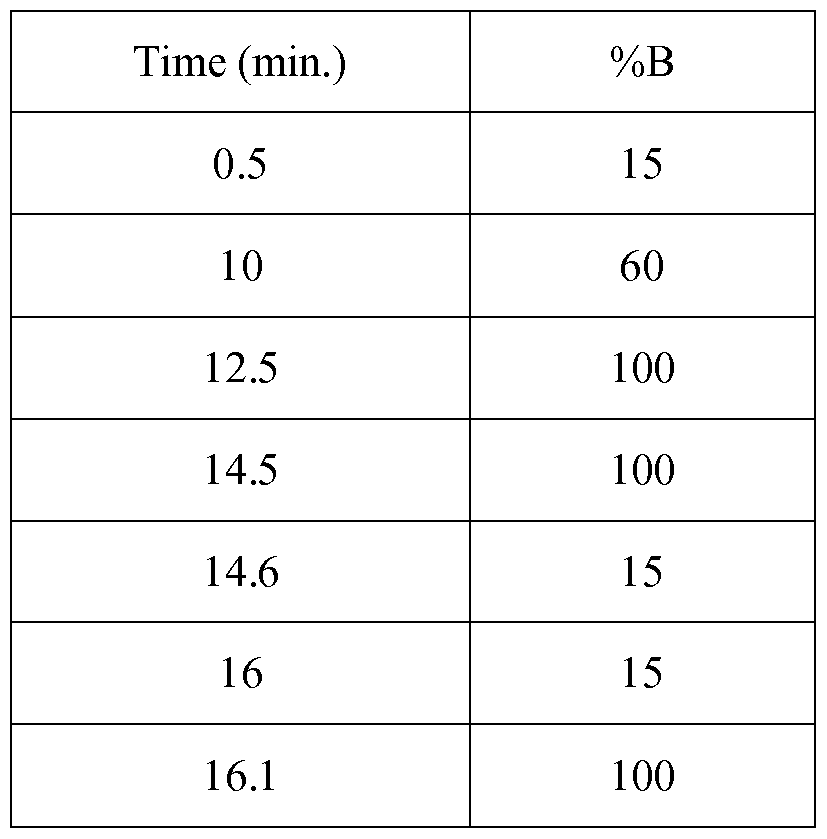


Claims
Priority Applications (8)
| Application Number | Priority Date | Filing Date | Title |
|---|---|---|---|
| CA2955685A CA2955685C (en) | 2014-08-04 | 2015-08-03 | Bispecific antibodies against cd3epsilon and bcma |
| SG11201700551TA SG11201700551TA (en) | 2014-08-04 | 2015-08-03 | Bispecific antibodies against cd3epsilon and bcma |
| MA39266A MA39266B1 (en) | 2014-08-04 | 2015-08-03 | Anti-cd3epsilon and bcma bispecific antibodies |
| US15/501,620 US10253104B2 (en) | 2014-08-04 | 2015-08-03 | Bispecific antibodies against CD3ϵ and BCMA |
| JP2017505793A JP6703520B2 (en) | 2014-08-04 | 2015-08-03 | Bispecific antibodies against CD3 epsilon and BCMA |
| NZ728534A NZ728534A (en) | 2014-08-04 | 2015-08-03 | Bispecific antibodies against cd3epsilon and bcma |
| AU2015299101A AU2015299101B2 (en) | 2014-08-04 | 2015-08-03 | Bispecific antibodies against CD3epsilon and BCMA |
| AU2020273347A AU2020273347A1 (en) | 2014-08-04 | 2020-11-20 | Bispecific antibodies against CD3epsilon and BCMA |
Applications Claiming Priority (2)
| Application Number | Priority Date | Filing Date | Title |
|---|---|---|---|
| EP14179705.0A EP2982692A1 (en) | 2014-08-04 | 2014-08-04 | Bispecific antibodies against CD3epsilon and BCMA |
| EP14179705.0 | 2014-08-04 |
Publications (1)
| Publication Number | Publication Date |
|---|---|
| WO2016020332A1 true WO2016020332A1 (en) | 2016-02-11 |
Family
ID=51260770
Family Applications (1)
| Application Number | Title | Priority Date | Filing Date |
|---|---|---|---|
| PCT/EP2015/067841 WO2016020332A1 (en) | 2014-08-04 | 2015-08-03 | Bispecific antibodies against cd3epsilon and bcma |
Country Status (22)
| Country | Link |
|---|---|
| US (1) | US10253104B2 (en) |
| EP (2) | EP2982692A1 (en) |
| JP (2) | JP6703520B2 (en) |
| AU (2) | AU2015299101B2 (en) |
| CA (1) | CA2955685C (en) |
| CY (1) | CY1118168T1 (en) |
| DK (1) | DK2982694T3 (en) |
| ES (1) | ES2602051T3 (en) |
| HK (1) | HK1218299A1 (en) |
| HR (1) | HRP20161211T1 (en) |
| HU (1) | HUE031076T2 (en) |
| LT (1) | LT2982694T (en) |
| MA (1) | MA39266B1 (en) |
| ME (1) | ME02533B (en) |
| NZ (1) | NZ728534A (en) |
| PL (1) | PL2982694T3 (en) |
| PT (1) | PT2982694T (en) |
| RS (1) | RS55410B1 (en) |
| SG (1) | SG11201700551TA (en) |
| SI (1) | SI2982694T1 (en) |
| SM (1) | SMT201600392B (en) |
| WO (1) | WO2016020332A1 (en) |
Cited By (51)
| Publication number | Priority date | Publication date | Assignee | Title |
|---|---|---|---|---|
| WO2018144535A1 (en) | 2017-01-31 | 2018-08-09 | Novartis Ag | Treatment of cancer using chimeric t cell receptor proteins having multiple specificities |
| US10087250B2 (en) | 2012-10-08 | 2018-10-02 | Roche Glycart Ag | Fc-free antibodies comprising two fab-fragments and methods of use |
| WO2018201051A1 (en) | 2017-04-28 | 2018-11-01 | Novartis Ag | Bcma-targeting agent, and combination therapy with a gamma secretase inhibitor |
| WO2018201056A1 (en) | 2017-04-28 | 2018-11-01 | Novartis Ag | Cells expressing a bcma-targeting chimeric antigen receptor, and combination therapy with a gamma secretase inhibitor |
| US10155815B2 (en) | 2013-02-26 | 2018-12-18 | Roche Glycart Ag | Bispecific T cell activating antigen binding molecules |
| WO2019053612A1 (en) | 2017-09-14 | 2019-03-21 | Glaxosmithkline Intellectual Property Development Limited | Combination treatment for cancer |
| WO2019053611A1 (en) | 2017-09-14 | 2019-03-21 | Glaxosmithkline Intellectual Property Development Limited | Combination treatment for cancer |
| WO2019053613A2 (en) | 2017-09-14 | 2019-03-21 | Glaxosmithkline Intellectual Property Development Limited | Combination treatment for cancer |
| WO2019079569A1 (en) | 2017-10-18 | 2019-04-25 | Novartis Ag | Compositions and methods for selective protein degradation |
| WO2019099639A1 (en) | 2017-11-15 | 2019-05-23 | Navartis Ag | Bcma-targeting chimeric antigen receptor, cd19-targeting chimeric antigen receptor, and combination therapies |
| WO2019108900A1 (en) | 2017-11-30 | 2019-06-06 | Novartis Ag | Bcma-targeting chimeric antigen receptor, and uses thereof |
| WO2019136432A1 (en) | 2018-01-08 | 2019-07-11 | Novartis Ag | Immune-enhancing rnas for combination with chimeric antigen receptor therapy |
| WO2019152660A1 (en) | 2018-01-31 | 2019-08-08 | Novartis Ag | Combination therapy using a chimeric antigen receptor |
| WO2019160956A1 (en) | 2018-02-13 | 2019-08-22 | Novartis Ag | Chimeric antigen receptor therapy in combination with il-15r and il15 |
| CN110167964A (en) * | 2016-11-02 | 2019-08-23 | 英格玛布有限责任公司 | Combine the bispecific antibody and immune drug for BCMA and CD3 for treating Huppert's disease |
| WO2020047449A2 (en) | 2018-08-31 | 2020-03-05 | Novartis Ag | Methods of making chimeric antigen receptor-expressing cells |
| WO2020047452A2 (en) | 2018-08-31 | 2020-03-05 | Novartis Ag | Methods of making chimeric antigen receptor-expressing cells |
| US10596257B2 (en) | 2016-01-08 | 2020-03-24 | Hoffmann-La Roche Inc. | Methods of treating CEA-positive cancers using PD-1 axis binding antagonists and anti-CEA/anti-CD3 bispecific antibodies |
| US10611840B2 (en) | 2014-08-04 | 2020-04-07 | Hoffman-La Roche Inc. | Bispecific T cell activating antigen binding molecules |
| WO2020089794A1 (en) | 2018-10-31 | 2020-05-07 | Glaxosmithkline Intellectual Property Development Limited | Methods of treating cancer |
| EP3204415B1 (en) * | 2014-10-09 | 2020-06-17 | EngMab Sàrl | Bispecific antibodies against cd3epsilon and ror1 |
| WO2020176397A1 (en) | 2019-02-25 | 2020-09-03 | Novartis Ag | Mesoporous silica particles compositions for viral delivery |
| US10766967B2 (en) | 2015-10-02 | 2020-09-08 | Hoffmann-La Roche Inc. | Bispecific T cell activating antigen binding molecules |
| US10781262B2 (en) | 2014-11-20 | 2020-09-22 | Hoffmann-La Roche Inc. | Combination therapy of T cell activating bispecific antigen binding molecules and PD-1 axis binding antagonists |
| WO2020191316A1 (en) | 2019-03-21 | 2020-09-24 | Novartis Ag | Car-t cell therapies with enhanced efficacy |
| WO2020191346A1 (en) | 2019-03-21 | 2020-09-24 | Regeneron Pharmaceuticals, Inc. | Combination of il-4/il-13 pathway inhibitors and plasma cell ablation for treating allergy |
| WO2020210678A1 (en) | 2019-04-12 | 2020-10-15 | Novartis Ag | Methods of making chimeric antigen receptor-expressing cells |
| WO2020219742A1 (en) | 2019-04-24 | 2020-10-29 | Novartis Ag | Compositions and methods for selective protein degradation |
| US10882918B2 (en) | 2016-09-30 | 2021-01-05 | Hoffmann-La Roche Inc. | Bispecific T cell activating antigen binding molecules |
| WO2021024133A2 (en) | 2019-08-06 | 2021-02-11 | Glaxosmithkline Intellectual Property Development Limited | Biopharmacuetical compositions and related methods |
| US11013801B2 (en) | 2015-12-09 | 2021-05-25 | Hoffmann-La Roche Inc. | Treatment method |
| WO2021108661A2 (en) | 2019-11-26 | 2021-06-03 | Novartis Ag | Chimeric antigen receptors and uses thereof |
| US11078291B2 (en) | 2016-02-17 | 2021-08-03 | Seagen Inc. | BCMA antibodies and use of same to treat cancer and immunological disorders |
| WO2021173985A2 (en) | 2020-02-27 | 2021-09-02 | Novartis Ag | Methods of making chimeric antigen receptor-expressing cells |
| WO2021173995A2 (en) | 2020-02-27 | 2021-09-02 | Novartis Ag | Methods of making chimeric antigen receptor-expressing cells |
| WO2021252920A1 (en) | 2020-06-11 | 2021-12-16 | Novartis Ag | Zbtb32 inhibitors and uses thereof |
| US11242390B2 (en) | 2016-03-22 | 2022-02-08 | Hoffmann-La Roche Inc. | Protease-activated T cell bispecific molecules |
| WO2022040586A2 (en) | 2020-08-21 | 2022-02-24 | Novartis Ag | Compositions and methods for in vivo generation of car expressing cells |
| WO2022135468A1 (en) * | 2020-12-23 | 2022-06-30 | 信达生物制药(苏州)有限公司 | Anti-bcma×cd3 bispecific antibody and use thereof |
| WO2022198335A1 (en) * | 2021-03-26 | 2022-09-29 | Zymeworks Inc. | Cysteine engineered antibody constructs, conjugates and methods of use |
| WO2022229853A1 (en) | 2021-04-27 | 2022-11-03 | Novartis Ag | Viral vector production system |
| WO2022248870A1 (en) | 2021-05-28 | 2022-12-01 | Glaxosmithkline Intellectual Property Development Limited | Combination therapies for treating cancer |
| WO2023012669A2 (en) | 2021-08-03 | 2023-02-09 | Glaxosmithkline Intellectual Property Development Limited | Biopharmaceutical compositions and stable isotope labeling peptide mapping method |
| WO2023021477A1 (en) | 2021-08-20 | 2023-02-23 | Novartis Ag | Methods of making chimeric antigen receptor–expressing cells |
| WO2023057893A1 (en) | 2021-10-05 | 2023-04-13 | Glaxosmithkline Intellectual Property Development Limited | Combination therapies for treating cancer |
| US11639397B2 (en) | 2011-08-23 | 2023-05-02 | Roche Glycart Ag | Bispecific antibodies specific for T-cell activating antigens and a tumor antigen and methods of use |
| WO2023144702A1 (en) | 2022-01-25 | 2023-08-03 | Glaxosmithkline Intellectual Property Development Limited | Combination therapy for cancer |
| US11866498B2 (en) | 2018-02-08 | 2024-01-09 | Genentech, Inc. | Bispecific antigen-binding molecules and methods of use |
| EP4065164A4 (en) * | 2019-11-26 | 2024-03-27 | Shanghai Epimab Biotherapeutics Co Ltd | Antibodies to cd3 and bcma, and bispecific binding proteins made therefrom |
| EP4097139A4 (en) * | 2020-01-28 | 2024-03-27 | Promab Biotechnologies Inc | Plap-cd3 epsilon bispecific antibodies |
| US11952424B2 (en) | 2019-03-29 | 2024-04-09 | Merus N.V. | Multivalent antibody |
Families Citing this family (35)
| Publication number | Priority date | Publication date | Assignee | Title |
|---|---|---|---|---|
| US20090162359A1 (en) | 2007-12-21 | 2009-06-25 | Christian Klein | Bivalent, bispecific antibodies |
| US9676845B2 (en) | 2009-06-16 | 2017-06-13 | Hoffmann-La Roche, Inc. | Bispecific antigen binding proteins |
| AR085403A1 (en) | 2011-02-28 | 2013-09-25 | Hoffmann La Roche | MONOVALENT PROTEINS THAT JOIN ANTIGENS |
| RU2607038C2 (en) | 2011-02-28 | 2017-01-10 | Ф. Хоффманн-Ля Рош Аг | Antigen-binding proteins |
| PL2953972T3 (en) * | 2013-02-05 | 2021-03-08 | Engmab Sàrl | Method for the selection of antibodies against bcma |
| GB201317928D0 (en) * | 2013-10-10 | 2013-11-27 | Ucl Business Plc | Molecule |
| JP6422956B2 (en) | 2013-10-11 | 2018-11-14 | エフ.ホフマン−ラ ロシュ アーゲーF. Hoffmann−La Roche Aktiengesellschaft | Multispecific domain exchange common variable light chain antibody |
| TWI712421B (en) | 2013-11-11 | 2020-12-11 | 日商中外製藥股份有限公司 | Antigen binding molecules containing altered antibody variable regions |
| US11154615B2 (en) | 2014-11-11 | 2021-10-26 | Chugai Seiyaku Kabushiki Kaisha | Library of antigen-binding molecules including modified antibody variable region |
| CN107001482B (en) | 2014-12-03 | 2021-06-15 | 豪夫迈·罗氏有限公司 | Multispecific antibodies |
| EP3466967A1 (en) | 2015-05-18 | 2019-04-10 | TCR2 Therapeutics Inc. | Compositions and methods for tcr reprogramming using fusion proteins |
| AR106188A1 (en) | 2015-10-01 | 2017-12-20 | Hoffmann La Roche | ANTI-CD19 HUMANIZED HUMAN ANTIBODIES AND METHODS OF USE |
| EP3150636A1 (en) * | 2015-10-02 | 2017-04-05 | F. Hoffmann-La Roche AG | Tetravalent multispecific antibodies |
| PT3433280T (en) * | 2016-03-22 | 2023-06-15 | Hoffmann La Roche | Protease-activated t cell bispecific molecules |
| WO2018026953A1 (en) | 2016-08-02 | 2018-02-08 | TCR2 Therapeutics Inc. | Compositions and methods for tcr reprogramming using fusion proteins |
| EP3445787B1 (en) | 2016-10-07 | 2020-12-02 | TCR2 Therapeutics Inc. | Compositions and methods for t-cell receptors reprogramming using fusion proteins |
| US11851491B2 (en) | 2016-11-22 | 2023-12-26 | TCR2 Therapeutics Inc. | Compositions and methods for TCR reprogramming using fusion proteins |
| WO2019035938A1 (en) | 2017-08-16 | 2019-02-21 | Elstar Therapeutics, Inc. | Multispecific molecules that bind to bcma and uses thereof |
| US11667713B2 (en) | 2017-12-28 | 2023-06-06 | Chugai Seiyaku Kabushiki Kaisha | Cytotoxicity-inducing therapeutic agent |
| US11492409B2 (en) | 2018-06-01 | 2022-11-08 | Novartis Ag | Binding molecules against BCMA and uses thereof |
| CA3100187A1 (en) * | 2018-06-26 | 2020-01-02 | Abl Bio, Inc. | Anti-bcma antibody and use thereof |
| EP3818083A2 (en) | 2018-07-03 | 2021-05-12 | Elstar Therapeutics, Inc. | Anti-tcr antibody molecules and uses thereof |
| CA3107126A1 (en) | 2018-07-19 | 2020-01-23 | Regeneron Pharmaceuticals, Inc. | Bispecific anti-bcma x anti-cd3 antibodies and uses thereof |
| WO2020053301A1 (en) * | 2018-09-11 | 2020-03-19 | Ichnos Sciences S.A. | Compositions comprising a bispecific antibody, bufffer and one or more stabilizing agents |
| KR102337683B1 (en) * | 2018-09-21 | 2021-12-13 | 주식회사 녹십자 | Highly efficient anti-TFPI antibody composition |
| EP3986925A1 (en) * | 2019-06-19 | 2022-04-27 | F. Hoffmann-La Roche AG | Method for the generation of a trivalent antibody expressing cell by targeted integration of multiple expression cassettes in a defined organization |
| CN110229232B (en) * | 2019-06-19 | 2020-05-19 | 北京智仁美博生物科技有限公司 | Bispecific antibodies and uses thereof |
| MX2022006714A (en) * | 2019-12-06 | 2022-08-08 | Regeneron Pharma | Methods of treating multiple myeloma with bispecific anti-bcma x anti-cd3 antibodies. |
| BR112022019498A2 (en) * | 2020-03-31 | 2022-11-16 | Chugai Pharmaceutical Co Ltd | DLL3 TARGETING MULTISPECIFIC ANTIGEN-BINDING MOLECULES AND THEIR USE |
| JP2023529981A (en) * | 2020-06-19 | 2023-07-12 | エフ・ホフマン-ラ・ロシュ・アクチェンゲゼルシャフト | Immunostimulatory Fc domain binding molecules |
| CR20220637A (en) | 2020-06-19 | 2023-01-31 | Hoffmann La Roche | Antibodies binding to cd3 and cd19 |
| AR125212A1 (en) * | 2021-03-24 | 2023-06-28 | Janssen Biotech Inc | PROTEINS INCLUDING CD3 ANTIGEN-BINDING DOMAINS AND THEIR USES |
| BR112023020832A2 (en) | 2021-04-08 | 2023-12-19 | Marengo Therapeutics Inc | TCR-BINDED MULTIFUNCTIONAL MOLECULES AND THEIR USES |
| CN115521381A (en) * | 2021-06-24 | 2022-12-27 | 益科思特(北京)医药科技发展有限公司 | Bispecific antibody combining BCMA and CD3, preparation method and application thereof |
| WO2024067222A1 (en) * | 2022-09-30 | 2024-04-04 | Nona Biosciences (Suzhou) Co., Ltd. | Cd3-targeting antibody and use thereof |
Citations (44)
| Publication number | Priority date | Publication date | Assignee | Title |
|---|---|---|---|---|
| EP0307434A1 (en) | 1987-03-18 | 1989-03-22 | Medical Res Council | Altered antibodies. |
| US5202238A (en) | 1987-10-27 | 1993-04-13 | Oncogen | Production of chimeric antibodies by homologous recombination |
| US5204244A (en) | 1987-10-27 | 1993-04-20 | Oncogen | Production of chimeric antibodies by homologous recombination |
| WO1996027011A1 (en) | 1995-03-01 | 1996-09-06 | Genentech, Inc. | A method for making heteromultimeric polypeptides |
| WO1998050431A2 (en) | 1997-05-02 | 1998-11-12 | Genentech, Inc. | A method for making multispecific antibodies having heteromultimeric and common components |
| WO1999037791A1 (en) | 1998-01-23 | 1999-07-29 | Vlaams Interuniversitair Instituut Voor Biotechnologie | Multipurpose antibody derivatives |
| WO2000041474A2 (en) | 1999-01-15 | 2000-07-20 | Novartis Ag | Anti-cd3 immunotoxins and therapeutic uses therefor |
| US6150090A (en) | 1986-01-09 | 2000-11-21 | Massachusetts Institute Of Technology | Nuclear factors associated with transcriptional regulation |
| WO2001024812A1 (en) | 1999-10-06 | 2001-04-12 | N.V. Nutricia | USE OF TRANSFORMING GROWTH FACTOR β AND GROWTH FACTORS IN THE TREATMENT AND PREVENTION OF DISEASES OF THE INTESTINAL MUCOSA |
| WO2001024811A1 (en) | 1999-10-06 | 2001-04-12 | Biogen, Inc. | April receptor (bcma) and uses thereof |
| US6331415B1 (en) | 1983-04-08 | 2001-12-18 | Genentech, Inc. | Methods of producing immunoglobulins, vectors and transformed host cells for use therein |
| WO2002066516A2 (en) | 2001-02-20 | 2002-08-29 | Zymogenetics, Inc. | Antibodies that bind both bcma and taci |
| WO2006093794A1 (en) | 2005-02-28 | 2006-09-08 | Centocor, Inc. | Heterodimeric protein binding compositions |
| WO2007110205A2 (en) | 2006-03-24 | 2007-10-04 | Merck Patent Gmbh | Engineered heterodimeric protein domains |
| WO2007117600A2 (en) | 2006-04-07 | 2007-10-18 | Macrogenics, Inc. | Combination therapy for treating autoimmune diseases |
| EP1870459A1 (en) | 2005-03-31 | 2007-12-26 | Chugai Seiyaku Kabushiki Kaisha | Methods for producing polypeptides by regulating polypeptide association |
| WO2007147901A1 (en) | 2006-06-22 | 2007-12-27 | Novo Nordisk A/S | Production of bispecific antibodies |
| WO2009080254A1 (en) | 2007-12-21 | 2009-07-02 | F. Hoffmann-La Roche Ag | Bivalent, bispecific antibodies |
| WO2009080253A1 (en) | 2007-12-21 | 2009-07-02 | F. Hoffmann-La Roche Ag | Bivalent, bispecific antibodies |
| WO2009080251A1 (en) | 2007-12-21 | 2009-07-02 | F. Hoffmann-La Roche Ag | Bivalent, bispecific antibodies |
| WO2009080252A1 (en) | 2007-12-21 | 2009-07-02 | F. Hoffmann-La Roche Ag | Bivalent, bispecific antibodies |
| WO2009089004A1 (en) | 2008-01-07 | 2009-07-16 | Amgen Inc. | Method for making antibody fc-heterodimeric molecules using electrostatic steering effects |
| WO2009132058A2 (en) | 2008-04-25 | 2009-10-29 | Zymogenetics, Inc. | Levels of bcma protein expression on b cells and use in diagnostic methods |
| WO2010104949A2 (en) | 2009-03-10 | 2010-09-16 | Biogen Idec Ma Inc. | Anti-bcma antibodies |
| WO2010129304A2 (en) | 2009-04-27 | 2010-11-11 | Oncomed Pharmaceuticals, Inc. | Method for making heteromultimeric molecules |
| WO2010145792A1 (en) | 2009-06-16 | 2010-12-23 | F. Hoffmann-La Roche Ag | Bispecific antigen binding proteins |
| WO2011090754A1 (en) | 2009-12-29 | 2011-07-28 | Emergent Product Development Seattle, Llc | Polypeptide heterodimers and uses thereof |
| WO2011143545A1 (en) | 2010-05-14 | 2011-11-17 | Rinat Neuroscience Corporation | Heterodimeric proteins and methods for producing and purifying them |
| WO2012020038A1 (en) | 2010-08-13 | 2012-02-16 | Roche Glycart Ag | Anti-tenascin-c a2 antibodies and methods of use |
| WO2012058768A1 (en) | 2010-11-05 | 2012-05-10 | Zymeworks Inc. | Stable heterodimeric antibody design with mutations in the fc domain |
| WO2012066058A1 (en) | 2010-11-16 | 2012-05-24 | Boehringer Ingelheim International Gmbh | Agents and methods for treating diseases that correlate with bcma expression |
| WO2012116927A1 (en) | 2011-02-28 | 2012-09-07 | F. Hoffmann-La Roche Ag | Monovalent antigen binding proteins |
| WO2012130831A1 (en) | 2011-03-29 | 2012-10-04 | Roche Glycart Ag | Antibody fc variants |
| WO2012131555A2 (en) | 2011-03-25 | 2012-10-04 | Glenmark Pharmaceuticals S.A. | Hetero-dimeric immunoglobulins |
| WO2012143498A1 (en) | 2011-04-21 | 2012-10-26 | Boehringer Ingelheim International Gmbh | Bcma-based stratification and therapy for multiple myeloma patients |
| WO2012163805A1 (en) | 2011-05-27 | 2012-12-06 | Glaxo Group Limited | Bcma (cd269/tnfrsf17) -binding proteins |
| WO2013002362A1 (en) | 2011-06-30 | 2013-01-03 | 中外製薬株式会社 | Heterodimerized polypeptide |
| WO2013012733A1 (en) | 2011-07-15 | 2013-01-24 | Biogen Idec Ma Inc. | Heterodimeric fc regions, binding molecules comprising same, and methods relating thereto |
| WO2013026833A1 (en) * | 2011-08-23 | 2013-02-28 | Roche Glycart Ag | Bispecific t cell activating antigen binding molecules |
| WO2013072406A1 (en) | 2011-11-15 | 2013-05-23 | Amgen Research (Munich) Gmbh | Binding molecules for bcma and cd3 |
| WO2013096291A2 (en) | 2011-12-20 | 2013-06-27 | Medimmune, Llc | Modified polypeptides for bispecific antibody scaffolds |
| EP2647707A1 (en) | 2010-11-30 | 2013-10-09 | Chugai Seiyaku Kabushiki Kaisha | Cytotoxicity-inducing therapeutic agent |
| WO2013154760A1 (en) | 2012-04-11 | 2013-10-17 | The United States Of America, As Represented By The Secretary, Department Of Health And Human Services | Chimeric antigen receptors targeting b-cell maturation antigen |
| WO2013157953A1 (en) | 2012-04-20 | 2013-10-24 | Merus B.V. | Methods and means for the production of ig-like molecules |
Family Cites Families (5)
| Publication number | Priority date | Publication date | Assignee | Title |
|---|---|---|---|---|
| WO2009080525A1 (en) | 2007-12-21 | 2009-07-02 | BSH Bosch und Siemens Hausgeräte GmbH | Spray device for iron |
| DE102011053039A1 (en) | 2011-06-03 | 2012-12-06 | Reinhold Hubner | Rail device, displacement device and umbrella stand |
| US20130058936A1 (en) * | 2011-08-23 | 2013-03-07 | Peter Bruenker | Bispecific antibodies specific for t-cell activating antigens and a tumor antigen and methods of use |
| UY35148A (en) * | 2012-11-21 | 2014-05-30 | Amgen Inc | HETERODIMERIC IMMUNOGLOBULINS |
| MX2017001556A (en) * | 2014-08-04 | 2017-05-15 | Hoffmann La Roche | Bispecific t cell activating antigen binding molecules. |
-
2014
- 2014-08-04 EP EP14179705.0A patent/EP2982692A1/en not_active Ceased
-
2015
- 2015-08-03 LT LTEP15179545.7T patent/LT2982694T/en unknown
- 2015-08-03 RS RS20160781A patent/RS55410B1/en unknown
- 2015-08-03 JP JP2017505793A patent/JP6703520B2/en active Active
- 2015-08-03 ES ES15179545.7T patent/ES2602051T3/en active Active
- 2015-08-03 US US15/501,620 patent/US10253104B2/en active Active
- 2015-08-03 ME MEP-2016-205A patent/ME02533B/en unknown
- 2015-08-03 SG SG11201700551TA patent/SG11201700551TA/en unknown
- 2015-08-03 PL PL15179545T patent/PL2982694T3/en unknown
- 2015-08-03 HU HUE15179545A patent/HUE031076T2/en unknown
- 2015-08-03 WO PCT/EP2015/067841 patent/WO2016020332A1/en active Application Filing
- 2015-08-03 EP EP15179545.7A patent/EP2982694B1/en active Active
- 2015-08-03 DK DK15179545.7T patent/DK2982694T3/en active
- 2015-08-03 SI SI201530004A patent/SI2982694T1/en unknown
- 2015-08-03 CA CA2955685A patent/CA2955685C/en active Active
- 2015-08-03 MA MA39266A patent/MA39266B1/en unknown
- 2015-08-03 AU AU2015299101A patent/AU2015299101B2/en active Active
- 2015-08-03 PT PT151795457T patent/PT2982694T/en unknown
- 2015-08-03 NZ NZ728534A patent/NZ728534A/en unknown
-
2016
- 2016-06-02 HK HK16106248.9A patent/HK1218299A1/en unknown
- 2016-09-15 CY CY20161100920T patent/CY1118168T1/en unknown
- 2016-09-21 HR HRP20161211TT patent/HRP20161211T1/en unknown
- 2016-10-31 SM SM201600392T patent/SMT201600392B/en unknown
-
2020
- 2020-05-08 JP JP2020082524A patent/JP2020122013A/en active Pending
- 2020-11-20 AU AU2020273347A patent/AU2020273347A1/en active Pending
Patent Citations (46)
| Publication number | Priority date | Publication date | Assignee | Title |
|---|---|---|---|---|
| US6331415B1 (en) | 1983-04-08 | 2001-12-18 | Genentech, Inc. | Methods of producing immunoglobulins, vectors and transformed host cells for use therein |
| US6150090A (en) | 1986-01-09 | 2000-11-21 | Massachusetts Institute Of Technology | Nuclear factors associated with transcriptional regulation |
| EP0307434A1 (en) | 1987-03-18 | 1989-03-22 | Medical Res Council | Altered antibodies. |
| US5202238A (en) | 1987-10-27 | 1993-04-13 | Oncogen | Production of chimeric antibodies by homologous recombination |
| US5204244A (en) | 1987-10-27 | 1993-04-20 | Oncogen | Production of chimeric antibodies by homologous recombination |
| WO1996027011A1 (en) | 1995-03-01 | 1996-09-06 | Genentech, Inc. | A method for making heteromultimeric polypeptides |
| WO1998050431A2 (en) | 1997-05-02 | 1998-11-12 | Genentech, Inc. | A method for making multispecific antibodies having heteromultimeric and common components |
| WO1999037791A1 (en) | 1998-01-23 | 1999-07-29 | Vlaams Interuniversitair Instituut Voor Biotechnologie | Multipurpose antibody derivatives |
| WO2000041474A2 (en) | 1999-01-15 | 2000-07-20 | Novartis Ag | Anti-cd3 immunotoxins and therapeutic uses therefor |
| WO2001024811A1 (en) | 1999-10-06 | 2001-04-12 | Biogen, Inc. | April receptor (bcma) and uses thereof |
| WO2001024812A1 (en) | 1999-10-06 | 2001-04-12 | N.V. Nutricia | USE OF TRANSFORMING GROWTH FACTOR β AND GROWTH FACTORS IN THE TREATMENT AND PREVENTION OF DISEASES OF THE INTESTINAL MUCOSA |
| WO2002066516A2 (en) | 2001-02-20 | 2002-08-29 | Zymogenetics, Inc. | Antibodies that bind both bcma and taci |
| WO2006093794A1 (en) | 2005-02-28 | 2006-09-08 | Centocor, Inc. | Heterodimeric protein binding compositions |
| EP1870459A1 (en) | 2005-03-31 | 2007-12-26 | Chugai Seiyaku Kabushiki Kaisha | Methods for producing polypeptides by regulating polypeptide association |
| WO2007110205A2 (en) | 2006-03-24 | 2007-10-04 | Merck Patent Gmbh | Engineered heterodimeric protein domains |
| WO2007117600A2 (en) | 2006-04-07 | 2007-10-18 | Macrogenics, Inc. | Combination therapy for treating autoimmune diseases |
| WO2007147901A1 (en) | 2006-06-22 | 2007-12-27 | Novo Nordisk A/S | Production of bispecific antibodies |
| WO2009080254A1 (en) | 2007-12-21 | 2009-07-02 | F. Hoffmann-La Roche Ag | Bivalent, bispecific antibodies |
| WO2009080253A1 (en) | 2007-12-21 | 2009-07-02 | F. Hoffmann-La Roche Ag | Bivalent, bispecific antibodies |
| WO2009080251A1 (en) | 2007-12-21 | 2009-07-02 | F. Hoffmann-La Roche Ag | Bivalent, bispecific antibodies |
| WO2009080252A1 (en) | 2007-12-21 | 2009-07-02 | F. Hoffmann-La Roche Ag | Bivalent, bispecific antibodies |
| WO2009089004A1 (en) | 2008-01-07 | 2009-07-16 | Amgen Inc. | Method for making antibody fc-heterodimeric molecules using electrostatic steering effects |
| WO2009132058A2 (en) | 2008-04-25 | 2009-10-29 | Zymogenetics, Inc. | Levels of bcma protein expression on b cells and use in diagnostic methods |
| WO2010104949A2 (en) | 2009-03-10 | 2010-09-16 | Biogen Idec Ma Inc. | Anti-bcma antibodies |
| WO2010129304A2 (en) | 2009-04-27 | 2010-11-11 | Oncomed Pharmaceuticals, Inc. | Method for making heteromultimeric molecules |
| WO2010145792A1 (en) | 2009-06-16 | 2010-12-23 | F. Hoffmann-La Roche Ag | Bispecific antigen binding proteins |
| WO2011090754A1 (en) | 2009-12-29 | 2011-07-28 | Emergent Product Development Seattle, Llc | Polypeptide heterodimers and uses thereof |
| WO2011143545A1 (en) | 2010-05-14 | 2011-11-17 | Rinat Neuroscience Corporation | Heterodimeric proteins and methods for producing and purifying them |
| WO2012020038A1 (en) | 2010-08-13 | 2012-02-16 | Roche Glycart Ag | Anti-tenascin-c a2 antibodies and methods of use |
| WO2012058768A1 (en) | 2010-11-05 | 2012-05-10 | Zymeworks Inc. | Stable heterodimeric antibody design with mutations in the fc domain |
| WO2012066058A1 (en) | 2010-11-16 | 2012-05-24 | Boehringer Ingelheim International Gmbh | Agents and methods for treating diseases that correlate with bcma expression |
| EP2647707A1 (en) | 2010-11-30 | 2013-10-09 | Chugai Seiyaku Kabushiki Kaisha | Cytotoxicity-inducing therapeutic agent |
| WO2012116927A1 (en) | 2011-02-28 | 2012-09-07 | F. Hoffmann-La Roche Ag | Monovalent antigen binding proteins |
| WO2012131555A2 (en) | 2011-03-25 | 2012-10-04 | Glenmark Pharmaceuticals S.A. | Hetero-dimeric immunoglobulins |
| WO2012130831A1 (en) | 2011-03-29 | 2012-10-04 | Roche Glycart Ag | Antibody fc variants |
| WO2012143498A1 (en) | 2011-04-21 | 2012-10-26 | Boehringer Ingelheim International Gmbh | Bcma-based stratification and therapy for multiple myeloma patients |
| WO2012163805A1 (en) | 2011-05-27 | 2012-12-06 | Glaxo Group Limited | Bcma (cd269/tnfrsf17) -binding proteins |
| WO2013002362A1 (en) | 2011-06-30 | 2013-01-03 | 中外製薬株式会社 | Heterodimerized polypeptide |
| WO2013012733A1 (en) | 2011-07-15 | 2013-01-24 | Biogen Idec Ma Inc. | Heterodimeric fc regions, binding molecules comprising same, and methods relating thereto |
| WO2013026833A1 (en) * | 2011-08-23 | 2013-02-28 | Roche Glycart Ag | Bispecific t cell activating antigen binding molecules |
| WO2013072406A1 (en) | 2011-11-15 | 2013-05-23 | Amgen Research (Munich) Gmbh | Binding molecules for bcma and cd3 |
| WO2013072415A1 (en) | 2011-11-15 | 2013-05-23 | Amgen Research (Munich) Gmbh | Binding molecules for bcma and cd3 |
| WO2013096291A2 (en) | 2011-12-20 | 2013-06-27 | Medimmune, Llc | Modified polypeptides for bispecific antibody scaffolds |
| WO2013154760A1 (en) | 2012-04-11 | 2013-10-17 | The United States Of America, As Represented By The Secretary, Department Of Health And Human Services | Chimeric antigen receptors targeting b-cell maturation antigen |
| WO2013157953A1 (en) | 2012-04-20 | 2013-10-24 | Merus B.V. | Methods and means for the production of ig-like molecules |
| WO2013157954A1 (en) | 2012-04-20 | 2013-10-24 | Merus B.V. | Methods and means for the production of ig-like molecules |
Non-Patent Citations (75)
| Title |
|---|
| AIWELL S; RIDGWAY JB; WELLS JA; CARTER P., J MOL BIOL, vol. 270, 1997, pages 26 - 35 |
| ANASETTI ET AL., TRANSPLANTATION, vol. 54, 1992, pages 844 |
| AUSUBEL, F., ET AL.,: "Current Protocols in Molecular Biology", 1987, GREENE PUBLISHING AND WILEY INTERSCIENCE |
| BARGOU ET AL., SCIENCE, vol. 321, no. 5891, 2008, pages 974 - 7 |
| BARNES, L.M. ET AL., BIOTECH. BIOENG., vol. 73, 2001, pages 261 - 270 |
| BARNES, L.M. ET AL., CYTOTECHNOLOGY, vol. 32, 2000, pages 109 - 123 |
| BOEMER, P. ET AL., J. IMMUNOL., vol. 147, 1991, pages 86 - 95 |
| BRUGGEMANN, M. ET AL., YEAR IMMUNOL., vol. 7, 1993, pages 33 - 40 |
| BRUNHOUSE, R; CEBRA, J.J., MOI. IMMUNOL., vol. 16, 1979, pages 907 - 917 |
| BURTON, D.R. ET AL., NATURE, vol. 288, 1980, pages 338 - 344 |
| CARTER, P. ET AL., PROC. NATL. ACAD. SCI. USA, vol. 89, 1992, pages 4285 - 4289 |
| COHEN SN ET AL., PNAS, vol. 69, no. 8, 1972, pages 2110 - 2114 |
| COLE ET AL.: "Monoclonal Antibodies and Cancer Therapy", 1985, ALAN R. LISS, pages: 77 |
| DEMCHENKO ET AL., BLOOD, vol. 115, no. 17, 2010, pages 3541 - 3552 |
| DESPLANCQ DK ET AL., PROTEIN ENG., vol. 7, no. 8, August 1994 (1994-08-01), pages 1027 - 33 |
| DIMOPULOS; TERPOS, ANN ONCOL, vol. 21, no. 7, 2010, pages VIIL43 - 150 |
| DREIER ET AL., INT J CANCER, 2002 |
| DUROCHER, Y. ET AL., NUCL. ACIDS. RES., vol. 30, 2002, pages E9 |
| GEISSE, S. ET AL., PROTEIN EXPR. PURIF., vol. 8, 1996, pages 271 - 282 |
| GOEBELER ET AL., ANN ONCOL, 2011 |
| GRAHAM; VAN DER EH, VIROLOGY, vol. 52, 1978, pages 546FF |
| GRAS M-P ET AL., INT IMMUNOL, vol. 7, 1995, pages 1093 - 1106 |
| HEZAREH, M. ET AL., J. VIROL., vol. 75, 2001, pages 12161 - 12168 |
| HOOGENBOOM, H.R; WINTER, G., J. MOL. BIOL., vol. 227, 1992, pages 381 - 388 |
| IDUSOGIE, E.E. ET AL., J. IMMUNOL., vol. 164, 2000, pages 4178 - 4184 |
| INAKI; LEE, NAT REV RHEUMATOL, vol. 6, 2010, pages 326 - 337 |
| ITO ET AL., CURR TOP MICROBIOL IMMUNOL, vol. 324, 2008, pages 53 - 76 |
| JAKOBOVITS, A. ET AL., NATURE, vol. 362, 1993, pages 255 - 258 |
| JAKOBOVITS, A. ET AL., PROC. NATL. ACAD. SCI. USA, vol. 90, 1993, pages 2551 - 2555 |
| JANEWAY CA, JR ET AL.: "Immunobiology. 5th ed.,", 2001, GARLAND PUBLISHING |
| KABAT ET AL.: "Sequences of Proteins of Immunological Interest, 5th ed.,", 1991, NATIONAL INSTITUTES OF HEALTH |
| KAUFMAN, RJ, MOI. BIOTECHNOL, vol. 16, 2000, pages 151 - 161 |
| KLINGER ET AL., BLOOD, 2012 |
| KONTERMANN RE, MABS, vol. 4, no. 2, 2012, pages 1 - 16 |
| KOYAMA ET AL., ANN RHEUM DIS, vol. 64, 2005, pages 1065 - 1067 |
| LUKAS, TJ. ET AL., J. IMMUNOL., vol. 127, 1981, pages 2555 - 2560 |
| MACK M. ET AL., PNAS, vol. 92, no. 15, 18 July 1995 (1995-07-18), pages 7021 - 7025 |
| MAKRIDES, S. C, PROTEIN EXPR. PURIF., vol. 17, 1999, pages 183 - 202 |
| MARKS, J.D. ET AL., J. MOL. BIOL., vol. 222, 1991, pages 581 - 597 |
| MATTHES ET AL., BLOOD, vol. 118, no. 7, 2011, pages 1838 - 1844 |
| MERCHANT A.M ET AL., NATURE BIOTECH, vol. 16, 1998, pages 677 - 681 |
| MERCHANT, A.M. ET AL., NAT. BIOTECHNOL., vol. 16, 1998, pages 677 - 681 |
| MOREAUX ET AL., BLOOD, vol. 103, no. 8, 2004, pages 3148 - 3157 |
| MORGAN, A. ET AL., IMMUNOLOGY, vol. 86, 1995, pages 319 - 324 |
| MORRISON, S.L. ET AL., J. IMMUNOL., vol. 160, 1998, pages 2802 - 2808 |
| MORRISON, S.L. ET AL., PROC. NATL. ACAD. SCI. USA, vol. 81, 1984, pages 6851 - 6855 |
| NEUBERGER, M.S. ET AL., NATURE, vol. 314, 1985, pages 268 - 270 |
| NORDERHAUG, L. ET AL., J. IMMUNOL. METHODS, vol. 204, 1997, pages 77 - 87 |
| ORLANDI, R. ET AL., PROC. NATL. ACAD. SCI. USA, vol. 86, 1989, pages 3833 - 3837 |
| PEREZ; NOLAN, NAT BIOTECHNOL, vol. 20, no. 2, 2002, pages 155 - 62 |
| RHOADES RA; PFLANZER RG: "Human Physiology, 4th ed.,", 2002, THOMSON LEARNING |
| RICKERT RC ET AL., IMMUNOL REV, vol. 244, no. 1, 2011, pages 115 - 133 |
| RIDGWAY JB; PRESTA LG; CARTER P, PROTEIN ENG., vol. 9, 1996, pages 617 - 621 |
| RIDGWAY, J.B. ET AL., PROTEIN ENG., vol. 9, 1996, pages 617 - 621 |
| RIECHMANN, L. ET AL., NATURE, vol. 332, 1988, pages 323 - 327 |
| RYAN, MC ET AL., MOL. CANCER THER., vol. 6, 2007, pages 3009 - 3018 |
| RYAN, MOL CANCER THER, vol. 6, no. 11, 2007, pages 3009 - 18 |
| SCHAEFER, W ET AL., PNAS, vol. 108, 2011, pages 11187 - 1191 |
| SCHAEFER, W. ET AL., PNAS, vol. 108, 2011, pages 11187 - 1191 |
| SCHLAEGER, E.- J.; CHRISTENSEN, K, CYTOTECHNOLOGY, vol. 30, 1999, pages 71 - 83 |
| SCHLAEGER, E.-J., J. IMMUNOL. METHODS, vol. 194, 1996, pages 191 - 199 |
| SCHLERETH ET AL., CANCER IMMUNOL IMMUNOTHER, vol. 55, 2006, pages 503 - 514 |
| SONDERMANN ET AL., NATURE, vol. 406, 20 July 2000 (2000-07-20), pages 267 - 273 |
| STUBENRAUCH ET AL., J PHARM BIOMED ANAL, 2009 |
| STUBENRAUCH ET AL., J PHARM BIOMED ANAL, 2013 |
| THOMMESEN, J.E. ET AL., MOI. IMMUNOL., vol. 37, 2000, pages 995 - 1004 |
| TOPP ET AL., J CLIN ONCO1, vol. 29, no. 18, 2011, pages 2493 - 8 |
| TOPP ET AL., J CLIN ONCOL, 2011 |
| TOPP ET AL., J CLIN ONCOL, vol. 29, no. 18, 2011, pages 2493 - 8 |
| VAN DIJK, M.A.; VAN DE WINKEL, J.G., CURR. OPIN. CHEM. BIOL., vol. 5, 2001, pages 368 - 374 |
| WERNER, R.G. ET AL., ARZNEIMITTELFORSCHUNG, vol. 48, 1998, pages 870 - 880 |
| WOOF J; BURTON D, NAT REV IMMUNOL, vol. 4, 2004, pages 89 - 99 |
| WORN; PLUCKTHUN, J MOL BIOL, vol. 305, 2001, pages 989 - 1010 |
| XIE, Z. ET AL., J IMMUNOL METHODS, vol. 286, 2005, pages 95 - 101 |
| YANG SJ, THE JOURNAL OF IMMUNOLOGY, vol. 137, 1986, pages 1097 - 1100 |
Cited By (61)
| Publication number | Priority date | Publication date | Assignee | Title |
|---|---|---|---|---|
| US11639397B2 (en) | 2011-08-23 | 2023-05-02 | Roche Glycart Ag | Bispecific antibodies specific for T-cell activating antigens and a tumor antigen and methods of use |
| US10087250B2 (en) | 2012-10-08 | 2018-10-02 | Roche Glycart Ag | Fc-free antibodies comprising two fab-fragments and methods of use |
| US10155815B2 (en) | 2013-02-26 | 2018-12-18 | Roche Glycart Ag | Bispecific T cell activating antigen binding molecules |
| US10781258B2 (en) | 2013-02-26 | 2020-09-22 | Roche Glycart Ag | Bispecific T cell activating antigen binding molecules |
| US10781257B2 (en) | 2013-02-26 | 2020-09-22 | Roche GlyeArt AG | Bispecific T cell activating antigen binding molecules |
| US10611840B2 (en) | 2014-08-04 | 2020-04-07 | Hoffman-La Roche Inc. | Bispecific T cell activating antigen binding molecules |
| US10611841B2 (en) | 2014-08-04 | 2020-04-07 | Hoffmann-La Roche Inc. | Bispecific T cell activating antigen binding molecules |
| US11117965B2 (en) | 2014-08-04 | 2021-09-14 | Hoffmann-La Roche Inc. | Bispecific T cell activating antigen binding molecules |
| US11952421B2 (en) | 2014-10-09 | 2024-04-09 | Bristol-Myers Squibb Company | Bispecific antibodies against CD3EPSILON and ROR1 |
| EP3204415B1 (en) * | 2014-10-09 | 2020-06-17 | EngMab Sàrl | Bispecific antibodies against cd3epsilon and ror1 |
| US11613587B2 (en) | 2014-11-20 | 2023-03-28 | Hoffmann-La Roche Inc. | Combination therapy of T cell activating bispecific antigen binding molecules and PD-1 axis binding antagonists |
| US10781262B2 (en) | 2014-11-20 | 2020-09-22 | Hoffmann-La Roche Inc. | Combination therapy of T cell activating bispecific antigen binding molecules and PD-1 axis binding antagonists |
| US10766967B2 (en) | 2015-10-02 | 2020-09-08 | Hoffmann-La Roche Inc. | Bispecific T cell activating antigen binding molecules |
| US11013801B2 (en) | 2015-12-09 | 2021-05-25 | Hoffmann-La Roche Inc. | Treatment method |
| US10596257B2 (en) | 2016-01-08 | 2020-03-24 | Hoffmann-La Roche Inc. | Methods of treating CEA-positive cancers using PD-1 axis binding antagonists and anti-CEA/anti-CD3 bispecific antibodies |
| US11078291B2 (en) | 2016-02-17 | 2021-08-03 | Seagen Inc. | BCMA antibodies and use of same to treat cancer and immunological disorders |
| US11767365B2 (en) | 2016-02-17 | 2023-09-26 | Seagen Inc. | BCMA antibodies and use of same to treat cancer and immunological disorders |
| US11242390B2 (en) | 2016-03-22 | 2022-02-08 | Hoffmann-La Roche Inc. | Protease-activated T cell bispecific molecules |
| US10882918B2 (en) | 2016-09-30 | 2021-01-05 | Hoffmann-La Roche Inc. | Bispecific T cell activating antigen binding molecules |
| CN110167964A (en) * | 2016-11-02 | 2019-08-23 | 英格玛布有限责任公司 | Combine the bispecific antibody and immune drug for BCMA and CD3 for treating Huppert's disease |
| JP2020500181A (en) * | 2016-11-02 | 2020-01-09 | エンクマフ エスアーエールエル | Bispecific antibodies to BCMA and CD3, and immunotherapeutics used in combination to treat multiple myeloma |
| CN110167964B (en) * | 2016-11-02 | 2023-12-01 | 百时美施贵宝公司 | Combination of bispecific antibodies and immunopharmaceuticals against BCMA and CD3 for the treatment of multiple myeloma |
| JP7267914B2 (en) | 2016-11-02 | 2023-05-02 | エンクマフ エスアーエールエル | Bispecific antibodies to BCMA and CD3 and immunotherapeutic agents used in combination to treat multiple myeloma |
| WO2018144535A1 (en) | 2017-01-31 | 2018-08-09 | Novartis Ag | Treatment of cancer using chimeric t cell receptor proteins having multiple specificities |
| WO2018201051A1 (en) | 2017-04-28 | 2018-11-01 | Novartis Ag | Bcma-targeting agent, and combination therapy with a gamma secretase inhibitor |
| WO2018201056A1 (en) | 2017-04-28 | 2018-11-01 | Novartis Ag | Cells expressing a bcma-targeting chimeric antigen receptor, and combination therapy with a gamma secretase inhibitor |
| WO2019053613A2 (en) | 2017-09-14 | 2019-03-21 | Glaxosmithkline Intellectual Property Development Limited | Combination treatment for cancer |
| WO2019053612A1 (en) | 2017-09-14 | 2019-03-21 | Glaxosmithkline Intellectual Property Development Limited | Combination treatment for cancer |
| WO2019053611A1 (en) | 2017-09-14 | 2019-03-21 | Glaxosmithkline Intellectual Property Development Limited | Combination treatment for cancer |
| WO2019079569A1 (en) | 2017-10-18 | 2019-04-25 | Novartis Ag | Compositions and methods for selective protein degradation |
| WO2019099639A1 (en) | 2017-11-15 | 2019-05-23 | Navartis Ag | Bcma-targeting chimeric antigen receptor, cd19-targeting chimeric antigen receptor, and combination therapies |
| WO2019108900A1 (en) | 2017-11-30 | 2019-06-06 | Novartis Ag | Bcma-targeting chimeric antigen receptor, and uses thereof |
| WO2019136432A1 (en) | 2018-01-08 | 2019-07-11 | Novartis Ag | Immune-enhancing rnas for combination with chimeric antigen receptor therapy |
| WO2019152660A1 (en) | 2018-01-31 | 2019-08-08 | Novartis Ag | Combination therapy using a chimeric antigen receptor |
| US11866498B2 (en) | 2018-02-08 | 2024-01-09 | Genentech, Inc. | Bispecific antigen-binding molecules and methods of use |
| WO2019160956A1 (en) | 2018-02-13 | 2019-08-22 | Novartis Ag | Chimeric antigen receptor therapy in combination with il-15r and il15 |
| WO2020047449A2 (en) | 2018-08-31 | 2020-03-05 | Novartis Ag | Methods of making chimeric antigen receptor-expressing cells |
| WO2020047452A2 (en) | 2018-08-31 | 2020-03-05 | Novartis Ag | Methods of making chimeric antigen receptor-expressing cells |
| WO2020089794A1 (en) | 2018-10-31 | 2020-05-07 | Glaxosmithkline Intellectual Property Development Limited | Methods of treating cancer |
| WO2020176397A1 (en) | 2019-02-25 | 2020-09-03 | Novartis Ag | Mesoporous silica particles compositions for viral delivery |
| WO2020191346A1 (en) | 2019-03-21 | 2020-09-24 | Regeneron Pharmaceuticals, Inc. | Combination of il-4/il-13 pathway inhibitors and plasma cell ablation for treating allergy |
| WO2020191316A1 (en) | 2019-03-21 | 2020-09-24 | Novartis Ag | Car-t cell therapies with enhanced efficacy |
| US11952424B2 (en) | 2019-03-29 | 2024-04-09 | Merus N.V. | Multivalent antibody |
| WO2020210678A1 (en) | 2019-04-12 | 2020-10-15 | Novartis Ag | Methods of making chimeric antigen receptor-expressing cells |
| WO2020219742A1 (en) | 2019-04-24 | 2020-10-29 | Novartis Ag | Compositions and methods for selective protein degradation |
| WO2021024133A2 (en) | 2019-08-06 | 2021-02-11 | Glaxosmithkline Intellectual Property Development Limited | Biopharmacuetical compositions and related methods |
| EP4065164A4 (en) * | 2019-11-26 | 2024-03-27 | Shanghai Epimab Biotherapeutics Co Ltd | Antibodies to cd3 and bcma, and bispecific binding proteins made therefrom |
| WO2021108661A2 (en) | 2019-11-26 | 2021-06-03 | Novartis Ag | Chimeric antigen receptors and uses thereof |
| EP4097139A4 (en) * | 2020-01-28 | 2024-03-27 | Promab Biotechnologies Inc | Plap-cd3 epsilon bispecific antibodies |
| WO2021173995A2 (en) | 2020-02-27 | 2021-09-02 | Novartis Ag | Methods of making chimeric antigen receptor-expressing cells |
| WO2021173985A2 (en) | 2020-02-27 | 2021-09-02 | Novartis Ag | Methods of making chimeric antigen receptor-expressing cells |
| WO2021252920A1 (en) | 2020-06-11 | 2021-12-16 | Novartis Ag | Zbtb32 inhibitors and uses thereof |
| WO2022040586A2 (en) | 2020-08-21 | 2022-02-24 | Novartis Ag | Compositions and methods for in vivo generation of car expressing cells |
| WO2022135468A1 (en) * | 2020-12-23 | 2022-06-30 | 信达生物制药(苏州)有限公司 | Anti-bcma×cd3 bispecific antibody and use thereof |
| WO2022198335A1 (en) * | 2021-03-26 | 2022-09-29 | Zymeworks Inc. | Cysteine engineered antibody constructs, conjugates and methods of use |
| WO2022229853A1 (en) | 2021-04-27 | 2022-11-03 | Novartis Ag | Viral vector production system |
| WO2022248870A1 (en) | 2021-05-28 | 2022-12-01 | Glaxosmithkline Intellectual Property Development Limited | Combination therapies for treating cancer |
| WO2023012669A2 (en) | 2021-08-03 | 2023-02-09 | Glaxosmithkline Intellectual Property Development Limited | Biopharmaceutical compositions and stable isotope labeling peptide mapping method |
| WO2023021477A1 (en) | 2021-08-20 | 2023-02-23 | Novartis Ag | Methods of making chimeric antigen receptor–expressing cells |
| WO2023057893A1 (en) | 2021-10-05 | 2023-04-13 | Glaxosmithkline Intellectual Property Development Limited | Combination therapies for treating cancer |
| WO2023144702A1 (en) | 2022-01-25 | 2023-08-03 | Glaxosmithkline Intellectual Property Development Limited | Combination therapy for cancer |
Also Published As
| Publication number | Publication date |
|---|---|
| SI2982694T1 (en) | 2017-01-31 |
| CA2955685C (en) | 2023-09-19 |
| PL2982694T3 (en) | 2017-02-28 |
| CY1118168T1 (en) | 2017-06-28 |
| PT2982694T (en) | 2016-09-29 |
| DK2982694T3 (en) | 2016-09-26 |
| LT2982694T (en) | 2016-11-10 |
| AU2015299101B2 (en) | 2020-08-27 |
| AU2020273347A1 (en) | 2020-12-17 |
| ES2602051T3 (en) | 2017-02-17 |
| HRP20161211T1 (en) | 2016-11-18 |
| RS55410B1 (en) | 2017-04-28 |
| SG11201700551TA (en) | 2017-02-27 |
| AU2015299101A1 (en) | 2017-02-16 |
| NZ728534A (en) | 2023-12-22 |
| US10253104B2 (en) | 2019-04-09 |
| SMT201600392B (en) | 2017-01-10 |
| US20170306036A1 (en) | 2017-10-26 |
| CA2955685A1 (en) | 2016-02-11 |
| EP2982694A1 (en) | 2016-02-10 |
| MA39266B1 (en) | 2017-01-31 |
| HUE031076T2 (en) | 2017-06-28 |
| JP6703520B2 (en) | 2020-06-03 |
| HK1218299A1 (en) | 2017-02-10 |
| JP2020122013A (en) | 2020-08-13 |
| JP2017532290A (en) | 2017-11-02 |
| EP2982692A1 (en) | 2016-02-10 |
| ME02533B (en) | 2017-02-20 |
| EP2982694B1 (en) | 2016-06-22 |
| MA39266A (en) | 2016-02-10 |
Similar Documents
| Publication | Publication Date | Title |
|---|---|---|
| AU2015299101B2 (en) | Bispecific antibodies against CD3epsilon and BCMA | |
| US20230322957A1 (en) | Monoclonal antibodies against bcma | |
| US11124577B2 (en) | Bispecific antibody against BCMA and CD3 and an immunological drug for combined use in treating multiple myeloma | |
| EP3204415B1 (en) | Bispecific antibodies against cd3epsilon and ror1 | |
| US20170327579A1 (en) | Bispecific antibodies against cd3epsilon and bcma | |
| EP2953974A1 (en) | Bispecific antibodies against cd3epsilon and bcma | |
| US11952421B2 (en) | Bispecific antibodies against CD3EPSILON and ROR1 |
Legal Events
| Date | Code | Title | Description |
|---|---|---|---|
| 121 | Ep: the epo has been informed by wipo that ep was designated in this application |
Ref document number: 15744597 Country of ref document: EP Kind code of ref document: A1 |
|
| ENP | Entry into the national phase |
Ref document number: 2955685 Country of ref document: CA |
|
| ENP | Entry into the national phase |
Ref document number: 2017505793 Country of ref document: JP Kind code of ref document: A |
|
| WWE | Wipo information: entry into national phase |
Ref document number: 15501620 Country of ref document: US |
|
| NENP | Non-entry into the national phase |
Ref country code: DE |
|
| ENP | Entry into the national phase |
Ref document number: 2015299101 Country of ref document: AU Date of ref document: 20150803 Kind code of ref document: A |
|
| REEP | Request for entry into the european phase |
Ref document number: 2015744597 Country of ref document: EP |
|
| WWE | Wipo information: entry into national phase |
Ref document number: 2015744597 Country of ref document: EP |
|
| 122 | Ep: pct application non-entry in european phase |
Ref document number: 15744597 Country of ref document: EP Kind code of ref document: A1 |