WO2017009712A1 - Hide1 compositions and methods - Google Patents
Hide1 compositions and methods Download PDFInfo
- Publication number
- WO2017009712A1 WO2017009712A1 PCT/IB2016/001079 IB2016001079W WO2017009712A1 WO 2017009712 A1 WO2017009712 A1 WO 2017009712A1 IB 2016001079 W IB2016001079 W IB 2016001079W WO 2017009712 A1 WO2017009712 A1 WO 2017009712A1
- Authority
- WO
- WIPO (PCT)
- Prior art keywords
- hide1
- cpa
- antibody
- cells
- patient
- Prior art date
Links
Classifications
-
- C—CHEMISTRY; METALLURGY
- C07—ORGANIC CHEMISTRY
- C07K—PEPTIDES
- C07K16/00—Immunoglobulins [IGs], e.g. monoclonal or polyclonal antibodies
- C07K16/18—Immunoglobulins [IGs], e.g. monoclonal or polyclonal antibodies against material from animals or humans
- C07K16/28—Immunoglobulins [IGs], e.g. monoclonal or polyclonal antibodies against material from animals or humans against receptors, cell surface antigens or cell surface determinants
- C07K16/2803—Immunoglobulins [IGs], e.g. monoclonal or polyclonal antibodies against material from animals or humans against receptors, cell surface antigens or cell surface determinants against the immunoglobulin superfamily
-
- A—HUMAN NECESSITIES
- A61—MEDICAL OR VETERINARY SCIENCE; HYGIENE
- A61K—PREPARATIONS FOR MEDICAL, DENTAL OR TOILETRY PURPOSES
- A61K31/00—Medicinal preparations containing organic active ingredients
- A61K31/33—Heterocyclic compounds
- A61K31/395—Heterocyclic compounds having nitrogen as a ring hetero atom, e.g. guanethidine or rifamycins
- A61K31/495—Heterocyclic compounds having nitrogen as a ring hetero atom, e.g. guanethidine or rifamycins having six-membered rings with two or more nitrogen atoms as the only ring heteroatoms, e.g. piperazine or tetrazines
- A61K31/505—Pyrimidines; Hydrogenated pyrimidines, e.g. trimethoprim
- A61K31/513—Pyrimidines; Hydrogenated pyrimidines, e.g. trimethoprim having oxo groups directly attached to the heterocyclic ring, e.g. cytosine
-
- A—HUMAN NECESSITIES
- A61—MEDICAL OR VETERINARY SCIENCE; HYGIENE
- A61K—PREPARATIONS FOR MEDICAL, DENTAL OR TOILETRY PURPOSES
- A61K39/00—Medicinal preparations containing antigens or antibodies
- A61K39/395—Antibodies; Immunoglobulins; Immune serum, e.g. antilymphocytic serum
- A61K39/39533—Antibodies; Immunoglobulins; Immune serum, e.g. antilymphocytic serum against materials from animals
- A61K39/3955—Antibodies; Immunoglobulins; Immune serum, e.g. antilymphocytic serum against materials from animals against proteinaceous materials, e.g. enzymes, hormones, lymphokines
-
- A—HUMAN NECESSITIES
- A61—MEDICAL OR VETERINARY SCIENCE; HYGIENE
- A61K—PREPARATIONS FOR MEDICAL, DENTAL OR TOILETRY PURPOSES
- A61K45/00—Medicinal preparations containing active ingredients not provided for in groups A61K31/00 - A61K41/00
- A61K45/06—Mixtures of active ingredients without chemical characterisation, e.g. antiphlogistics and cardiaca
-
- A—HUMAN NECESSITIES
- A61—MEDICAL OR VETERINARY SCIENCE; HYGIENE
- A61P—SPECIFIC THERAPEUTIC ACTIVITY OF CHEMICAL COMPOUNDS OR MEDICINAL PREPARATIONS
- A61P35/00—Antineoplastic agents
-
- C—CHEMISTRY; METALLURGY
- C07—ORGANIC CHEMISTRY
- C07K—PEPTIDES
- C07K14/00—Peptides having more than 20 amino acids; Gastrins; Somatostatins; Melanotropins; Derivatives thereof
- C07K14/435—Peptides having more than 20 amino acids; Gastrins; Somatostatins; Melanotropins; Derivatives thereof from animals; from humans
- C07K14/705—Receptors; Cell surface antigens; Cell surface determinants
- C07K14/70503—Immunoglobulin superfamily
-
- A—HUMAN NECESSITIES
- A61—MEDICAL OR VETERINARY SCIENCE; HYGIENE
- A61K—PREPARATIONS FOR MEDICAL, DENTAL OR TOILETRY PURPOSES
- A61K39/00—Medicinal preparations containing antigens or antibodies
- A61K2039/505—Medicinal preparations containing antigens or antibodies comprising antibodies
-
- A—HUMAN NECESSITIES
- A61—MEDICAL OR VETERINARY SCIENCE; HYGIENE
- A61K—PREPARATIONS FOR MEDICAL, DENTAL OR TOILETRY PURPOSES
- A61K39/00—Medicinal preparations containing antigens or antibodies
- A61K2039/57—Medicinal preparations containing antigens or antibodies characterised by the type of response, e.g. Th1, Th2
- A61K2039/572—Medicinal preparations containing antigens or antibodies characterised by the type of response, e.g. Th1, Th2 cytotoxic response
-
- C—CHEMISTRY; METALLURGY
- C07—ORGANIC CHEMISTRY
- C07K—PEPTIDES
- C07K2317/00—Immunoglobulins specific features
- C07K2317/50—Immunoglobulins specific features characterized by immunoglobulin fragments
- C07K2317/52—Constant or Fc region; Isotype
- C07K2317/524—CH2 domain
-
- C—CHEMISTRY; METALLURGY
- C07—ORGANIC CHEMISTRY
- C07K—PEPTIDES
- C07K2317/00—Immunoglobulins specific features
- C07K2317/50—Immunoglobulins specific features characterized by immunoglobulin fragments
- C07K2317/52—Constant or Fc region; Isotype
- C07K2317/526—CH3 domain
-
- C—CHEMISTRY; METALLURGY
- C07—ORGANIC CHEMISTRY
- C07K—PEPTIDES
- C07K2317/00—Immunoglobulins specific features
- C07K2317/50—Immunoglobulins specific features characterized by immunoglobulin fragments
- C07K2317/52—Constant or Fc region; Isotype
- C07K2317/53—Hinge
-
- C—CHEMISTRY; METALLURGY
- C07—ORGANIC CHEMISTRY
- C07K—PEPTIDES
- C07K2317/00—Immunoglobulins specific features
- C07K2317/50—Immunoglobulins specific features characterized by immunoglobulin fragments
- C07K2317/55—Fab or Fab'
-
- C—CHEMISTRY; METALLURGY
- C07—ORGANIC CHEMISTRY
- C07K—PEPTIDES
- C07K2317/00—Immunoglobulins specific features
- C07K2317/50—Immunoglobulins specific features characterized by immunoglobulin fragments
- C07K2317/56—Immunoglobulins specific features characterized by immunoglobulin fragments variable (Fv) region, i.e. VH and/or VL
- C07K2317/565—Complementarity determining region [CDR]
-
- C—CHEMISTRY; METALLURGY
- C07—ORGANIC CHEMISTRY
- C07K—PEPTIDES
- C07K2317/00—Immunoglobulins specific features
- C07K2317/60—Immunoglobulins specific features characterized by non-natural combinations of immunoglobulin fragments
- C07K2317/62—Immunoglobulins specific features characterized by non-natural combinations of immunoglobulin fragments comprising only variable region components
- C07K2317/622—Single chain antibody (scFv)
-
- C—CHEMISTRY; METALLURGY
- C07—ORGANIC CHEMISTRY
- C07K—PEPTIDES
- C07K2317/00—Immunoglobulins specific features
- C07K2317/70—Immunoglobulins specific features characterized by effect upon binding to a cell or to an antigen
- C07K2317/73—Inducing cell death, e.g. apoptosis, necrosis or inhibition of cell proliferation
- C07K2317/732—Antibody-dependent cellular cytotoxicity [ADCC]
-
- C—CHEMISTRY; METALLURGY
- C07—ORGANIC CHEMISTRY
- C07K—PEPTIDES
- C07K2317/00—Immunoglobulins specific features
- C07K2317/70—Immunoglobulins specific features characterized by effect upon binding to a cell or to an antigen
- C07K2317/73—Inducing cell death, e.g. apoptosis, necrosis or inhibition of cell proliferation
- C07K2317/734—Complement-dependent cytotoxicity [CDC]
-
- C—CHEMISTRY; METALLURGY
- C07—ORGANIC CHEMISTRY
- C07K—PEPTIDES
- C07K2317/00—Immunoglobulins specific features
- C07K2317/90—Immunoglobulins specific features characterized by (pharmaco)kinetic aspects or by stability of the immunoglobulin
- C07K2317/92—Affinity (KD), association rate (Ka), dissociation rate (Kd) or EC50 value
-
- C—CHEMISTRY; METALLURGY
- C07—ORGANIC CHEMISTRY
- C07K—PEPTIDES
- C07K2317/00—Immunoglobulins specific features
- C07K2317/90—Immunoglobulins specific features characterized by (pharmaco)kinetic aspects or by stability of the immunoglobulin
- C07K2317/94—Stability, e.g. half-life, pH, temperature or enzyme-resistance
-
- C—CHEMISTRY; METALLURGY
- C07—ORGANIC CHEMISTRY
- C07K—PEPTIDES
- C07K2319/00—Fusion polypeptide
- C07K2319/30—Non-immunoglobulin-derived peptide or protein having an immunoglobulin constant or Fc region, or a fragment thereof, attached thereto
Definitions
- HIDE1 COMPOSITIONS AND METHODS
- Na ⁇ ve T cells must receive two independent signals from antigen-presenting cells (APC) in order to become productively activated.
- the first, Signal 1 is antigen-specific and occurs when T cell antigen receptors encounter the appropriate antigen-MHC complex on the APC.
- the fate of the immune response is determined by a second, antigen-independent signal (Signal 2) which is delivered through a T cell costimulatory molecule that engages its APC- expressed ligand.
- This second signal could be either stimulatory (positive costimulation) or inhibitory (negative costimulation or coinhibition).
- T-cell activation In the absence of a costimulatory signal, or in the presence of a coinhibitory signal, T-cell activation is impaired or aborted, which may lead to a state of antigen-specific unresponsiveness (known as T-cell anergy), or may result in T-cell apoptotic death.
- T-cell anergy a state of antigen-specific unresponsiveness
- Costimulatory molecule pairs usually consist of ligands expressed on APCs and their cognate receptors expressed on T cells.
- the prototype ligand/receptor pairs of costimulatory molecules are B7/CD28 and CD40/CD40L.
- the B7 family consists of structurally related, cell-surface protein ligands, which may provide stimulatory or inhibitory input to an immune response.
- Members of the B7 family are structurally related, with the extracellular domain containing at least one variable or constant immunoglobulin domain.
- T cells become highly susceptible to induction of apoptosis.
- novel agents that are capable of modulating costimulatory signals, without compromising the immune system’s ability to defend against pathogens, are highly advantageous for treatment and prevention of such pathological conditions.
- TAAs tumor-associated antigens
- costimulatory pathways have been identified as immunologic checkpoints that attenuate T cell dependent immune responses, both at the level of initiation and effector function within tumor metastases. As engineered cancer vaccines continue to improve, it is becoming clear that such immunologic checkpoints are a major barrier to the vaccines’ ability to induce therapeutic anti-tumor responses.
- costimulatory molecules can serve as adjuvants for active (vaccination) and passive (antibody-mediated) cancer immunotherapy, providing strategies to thwart immune tolerance and stimulate the immune system.
- agents could be of use in other types of cancer immunotherapy, such as adoptive immunotherapy, in which tumor-specific T cell populations are expanded and directed to attack and kill tumor cells.
- Agents capable of augmenting such anti-tumor response have great therapeutic potential and may be of value in the attempt to overcome the obstacles to tumor immunotherapy.
- agents are also in clinical development for viral infections, for example the anti PD-1 Ab, MDX-1106, which is being tested for treatment of hepatitis C, and the anti-CTLA-4 Ab CP-675,206 (tremelimumab) which is in a clinical trial in hepatitis C virus-infected patients with hepatocellular carcinoma.
- iTregs target regulatory T cells
- inducible regulatory T cells iTregs
- Multiple immune-checkpoint receptors such as CTLA4, PD-1, TIM3 and LAG3, are expressed at high levels on the surface of iTregs and directly promote Treg cell-mediated suppression of effector immune responses.
- some immune-checkpoint antibodies may, in addition to increasing CTL immunity, further block the immunosuppressive activity of iTregs and thereby enhance anti-tumor immunity.
- CTLA4 blockade by ipilimumab both enhances effector T cell activity, and inhibits Treg immunosuppressive activity.
- B cells play a critical role in recognition of foreign antigens and they produce the antibodies necessary to provide protection against various types of infectious agents.
- T cell help to B cells is a pivotal process of adaptive immune responses.
- Follicular helper T (Tfh) cells are a subset of CD4 + T cells specialized in B cell help (reviewed by Crotty, Annu. Rev. Immunol.29: 621-663, 2011).
- Tfh cells express the B cell homing chemokine receptor, CXCR5, which drives Tfh cell migration into B cell follicles within lymph nodes in a CXCL13-dependent manner.
- Tfh cells for B cell help and T cell-dependent antibody responses indicates that this cell type is of great importance for protective immunity against various types of infectious agents, as well as for rational vaccine design.
- regulatory B cells (Bregs) have a role in impairing effective clearance of tumors.
- the mechanisms for Bregs effects in cancer are not well understood, however one proposed mechanism is through inhibition of cytotoxic CD8 + T cells.
- NK cells are effector lymphocytes of the innate immune system that are known to be involved in killing of pathological or diseased cells such as cancer and infected cells and pathogens. Natural killer cells have the capacity to kill cellular targets and produce cytokines without prior specific sensitization. NK cells are unique, as they have the ability to recognize stressed cells in the absence of antibodies and MHC, allowing for a much faster immune reaction. This role is especially important because harmful cells that are missing MHC 1 markers cannot be detected and destroyed by other immune cells, such as T cells.
- Induction of immune tolerance has long been considered the“holy grail” for autoimmune disease therapy.
- the immune system has the reciprocal tasks to protect the host against invading pathogens, but simultaneously to prevent damage resulting from unwanted reactions to self-antigens.
- the latter part is known as immune tolerance and performed by a complex set of interactive and complementary pathways, which regulate immune responses.
- T cells have the ability to react to a variety of antigens, both self and nonself. Therefore, there are many mechanisms that exist naturally to eliminate potentially self-reactive responses– this is known as natural tolerance.
- the main mechanism for eliminating potential auto- reactive T cells occurs in the thymus and is known as central tolerance.
- Some potentially autoreactive T cells escape central tolerance and, therefore, peripheral tolerance mechanisms also exist. Despite these mechanisms, some self-reactive T cells may ⁇ escape' and be present in the repertoire; it is believed that their activation may lead to autoimmune disease.
- CTLA-4 is the most extensively studied costimulatory molecule which down- regulates immune responses.
- the attributes of immunosuppressive qualities and capacity to induce tolerance have made its recognition as a potential immuno-therapeutic agent for autoimmune mediated inflammatory disorders.
- Abatacept (commercial name: Orencia) is a fusion protein composed of the ECD (extracellular domain) of CTLA-4 fused to the Fc fragment of hIgG1.
- Abatacept is believed to induce costimulation blockade, which has been approved for treating patients with rheumatoid arthritis, by effectively interfering with the inflammatory cascade.
- T helper type 1 (Th1) cells are induced by IL-12 and produce IFN- ⁇ ; while T helper type 2 (Th2) cells secrete IL-4, IL-5 and IL-13.
- Th1 cells can mediate proinflammatory or cell- mediated immune responses, whereas Th2 cells mainly promote certain types of humoral immunity.
- Some immune related diseases such as autoimmune reactions, inflammation, chronic infection and sepsis, are characterized by a dysregulation of the pro- versus anti- inflammatory tendencies of the immune system, as well as an imbalance in the Th1 versus Th2 cytokine balance.
- Th1 to Th2 protects the organism from systemic 'overshooting' with Th1/pro-inflammatory cytokines, by reducing the inflammatory tendencies of the immune system.
- Immunomodulatory therapies that are associated with a Th1 to Th2 immune shift have protective effects in Th1-mediated autoimmune diseases, such as multiple sclerosis and rheumatoid arthritis.
- Th1-mediated autoimmune diseases such as multiple sclerosis and rheumatoid arthritis.
- Laquinimod which has demonstrated efficacy in animal models of several autoimmune diseases including MS, shows immunomodulatory effects through Th1/Th2 shift, and does not lead to immunosuppression.
- Glatiramer acetate (Copaxone®) also induces Th1/Th2 shift with decreased secretion of proinflammatory cytokines, and increased secretion of antiinflammatory cytokines.
- glatiramer acetate -specific Th2 cells are able to migrate across the blood-brain barrier and cause in situ bystander suppression of autoaggressive Th1 T cells.
- Th1, Th17, Th2 and regulatory T cells play important roles in modulating autoimmunity and
- the present invention provides for a method of activating T cells of a patient comprising administering an anti-HIDE1 antibody to said patient, wherein a subset of the T cells of said patient are activated.
- the present invention provides for a method of activating cytotoxic T cells (CTLs) of a patient comprising administering an anti-HIDE1 antibody to said patient, wherein a subset of the CTLs of said patient are activated.
- CTLs cytotoxic T cells
- the present invention also provides for a method of activating NK cells of a patient comprising administering an anti-HIDE1 antibody to said patient, wherein a subset of the NK cells of said patient are activated.
- the present invention also provides for a method of activating ⁇ T cells of a patient comprising administering an anti-HIDE1 antibody to said patient, wherein a subset of the ⁇ T cells of said patient are activated.
- the present invention also provides for a method of activating Th1 cells of a patient comprising administering an anti-HIDE1 antibody to said patient, wherein a subset of the Th1 cells of said patient are activated.
- the present invention also provides for a method of decreasing or eliminating cell number and/or activity of at least one of regulatory T cells (Tregs) in a patient comprising administering an anti-HIDE1 antibody to said patient.
- Tregs regulatory T cells
- the present invention also provides for a method of increasing interferon- ⁇ production and/or pro-inflammatory cytokine secretion in a patient comprising administering an anti-HIDE1 antibody to said patient.
- the present invention also provides for a method of modulating myeloid cell polarization in a patient comprising administering an anti- HIDE1 antibody to said patient.
- the present invention also provides for a method of modulating myeloid cell shifting toward a pro-inflammatory response in a patient comprising administering an anti-HIDE1 antibody to said patient.
- the present invention also provides for a method of shifting myeloid from M2 toward M1 phenotype in a patient comprising administering an anti-HIDE1 antibody to said patient.
- the present invention also provides for a method of modulating myeloid cell in the TME to support anti-cancer immune response in a patient comprising administering an anti-HIDE1 antibody to said patient.
- the present invention also provides for a method of restricting the pro-tumorigenic effects of the myeloid cells in the tumor microenvironment in a patient comprising administering an anti-HIDE1 antibody to said patient.
- the present invention also provides for a method to elicit one or more of the following effects on immunity in a patient by
- an anti-HIDE1 antibody to said patient, wherein said effect is selected from the group consisting of: i) increases immune response, (ii) increases T cell activity, (iii) increases activation of ⁇ and/or ⁇ T cells, (iv) increases cytotoxic T cell activity, (v) increases NK and/or NKT cell activity, (vi) alleviates ⁇ and/or ⁇ T-cell suppression, (vii) increases pro- inflammatory cytokine secretion, (viii) increases IL-2 secretion; (ix) increases interferon- ⁇ production, (x) increases Th1 response, (xi) decrease Th2 response, (xii) decreases or eliminates cell number and/or activity of at least one of regulatory T cells (Tregs), myeloid derived suppressor cells (MDSCs), iMCs, mesenchymal stromal cells, TIE2-expressing monocytes, (xiii) reduces regulatory cell activity, and/or the activity of one or more of myeloid derived suppress
- the present invention also provides for a method of depleting myeloid cells or other circulating tumor cells expressing HIDE1 from a patient or patient sample, said method comprising: i) contacting said patient or said patient sample with an anti-HIDE1 antibody, wherein said anti-HIDE1 antibody binds to HIDE1 expressing cells, ii) identifying cells to which said anti-HIDE1 antibody has bound, and iii) removing said cells in step ii) from said patient or said patient sample.
- the present invention also provides for a method of treating cancer in a patient, comprising administering an anti-HIDE1 antibody to said patient, wherein said cancer is treated.
- said patient has cancer.
- the cancer is selected from the group consisting of Acute Myeloid Leukemia, Acute Myeloid Leukemia Induction Failure, Acute Lymphoblastic
- Leukemia Diffuse Large B ⁇ cell Lymphoma, Malignant Lymphoma, Non ⁇ Hodgkin Lymphoma, Diffuse Large B ⁇ Cell Lymphoma, Glioblastoma multiforme, Mesothelioma, Thymoma, Testicular Germ Cell Tumors, Kidney renal clear cell carcinoma, Sarcoma, Brain Lower Grade Glioma, Chronic Lymphocytic Leukemia, Non ⁇ Hodgkin Lymphoma ⁇ Follicular Lymphoma, Uterine Carcinosarcoma, Pediatric Brain Tumors, Lung
- the cancer is a cancer having high immune infiltrate of myeloid cells expressing HIDE1.
- the anti-HIDE1 antibody is selected from the group consisting of CPA.12.001 human IgG4, CPA.12.002 human IgG4, CPA.12.003 human IgG4,
- the anti-HIDE1 antibody is selected from the group consisting of CPA.12.006-H4, CPA.12.007-H4, and CPA.12.0012-H4. [0036] In some embodiments, the anti-HIDE1 antibody is selected from the group consisting of CPA.12.001, CPA.12.002, CPA.12.003, CPA.12.004, CPA.12.005, CPA.12.006,
- the present invention also provides for a method of diagnosing cancer comprising: a) contacting a tissue from a patient with an anti- HIDE1 antibody; and b) determining the presence of over-expression of HIDE1 in said tissue as an indication of the presence of cancer.
- the tissue is a blood sample.
- the tissue is a biopsy of a solid tumor.
- the anti-HIDE1 antibody is labeled.
- a second labeled antibody that binds to said anti-HIDE1 antibody is contacted with said sample.
- the present invention also provides for an anti-HIDE1 antigen-binding domain comprising: a) a heavy chain variable domain comprising a vhCDR1, vhCDR2, and vhCDR3 from an anti-HIDE1 antibody; and b) a light chain variable domain comprising a vlCDR1, vlCDR2, and vlCDR3 from said anti-HIDE1 antibody; wherein said anti-HIDE1 antibody is selected from the group consisting of
- the present invention also provides for an anti-HIDE1 antigen-binding domain comprising: a) a heavy chain variable domain comprising a vhCDR1, vhCDR2, and vhCDR3 from an anti-HIDE1 antibody; and b) a light chain variable domain comprising a vlCDR1, vlCDR2, and vlCDR3 from said anti-HIDE1 antibody; wherein said anti-HIDE1 antibody is selected from the group consisting of CPA.12.001, CPA.12.002, CPA.12.003, CPA.12.004, CPA.12.005, CPA.12.006,
- the anti-HIDE1 antigen binding domain of the antibody is a single chain Fv (scFv), wherein said heavy chain variable domain and said light chain variable domain are covalently attached via a scFv linker.
- the present invention also provides for an anti-HIDE1 antibody comprising: a) a heavy chain variable domain comprising a vhCDR1, vhCDR2, and vhCDR3 from an anti-HIDE1 antibody; and b) a light chain variable domain comprising a vlCDR1, vlCDR2, and vlCDR3 from said anti-HIDE1 antibody; wherein said anti-HIDE1 antibody is selected from the group consisting of CPA.12.001 human IgG4, CPA.12.002 human IgG4, CPA.12.003 human IgG4, CPA.12.004 human IgG4, CPA.12.005 human IgG4, CPA.12.006 human IgG4, CPA.12.007 human IgG4, CPA.12.008 human IgG4, CPA.12.009 human IgG4, CPA.12.011 human IgG4, CPA.12.012 human IgG4, CPA.12.013 human IgG4, CPA.12.013 human
- the present invention also provides for an anti-HIDE1 antibody comprising: a) a heavy chain variable domain comprising a vhCDR1, vhCDR2, and vhCDR3 from an anti-HIDE1 antibody; and b) a light chain variable domain comprising a vlCDR1, vlCDR2, and vlCDR3 from said anti-HIDE1 antibody; wherein said anti-HIDE1 antibody is selected from the group consisting of CPA.12.001, CPA.12.002, CPA.12.003, CPA.12.004, CPA.12.005, CPA.12.006, CPA.12.007, CPA.12.008,
- the present invention also provides for an anti-HIDE1 antibody that competes for binding with an antibody comprising: a) a heavy chain variable domain comprising a vhCDR1, vhCDR2, and vhCDR3 from an anti-HIDE1 antibody; and b) a light chain variable domain comprising a vlCDR1, a vlCDR2 and vlCDR3, from said anti-HIDE1 antibody; wherein said anti-HIDE1 antibody is selected from the group consisting of CPA.12.001 human IgG4, CPA.12.002 human IgG4, CPA.12.003 human IgG4, CPA.12.004 human IgG4, CPA.12.005 human IgG4, CPA.12.006 human IgG4, CPA.12.007 human IgG4, CPA.12.001 human IgG4, CPA.12.002 human IgG4, CPA.12.003 human IgG4, CPA.12.004 human IgG4, CPA.12.005 human Ig
- the present invention also provides for an anti-HIDE1 antibody that competes for binding with an antibody comprising: a) a heavy chain variable domain comprising a vhCDR1, vhCDR2, and vhCDR3 from an anti-HIDE1 antibody; and b) a light chain variable domain comprising a vlCDR1, a vlCDR2 and vlCDR3, from said anti-HIDE1 antibody; wherein said anti-HIDE1 antibody is selected from the group consisting of CPA.12.001, CPA.12.002, CPA.12.003, CPA.12.004, CPA.12.005,
- the present invention also provides for an anti-HIDE1 antibody that competes for binding with an antibody comprising: a) a heavy chain variable domain comprising a vhCDR1, vhCDR2, and vhCDR3 from an anti-HIDE1 antibody; and b) a light chain variable domain comprising a vlCDR1, a vlCDR2 and vlCDR3, from said anti-HIDE1 antibody; wherein said anti-HIDE1 antibody is selected from the group consisting of AB-506, AB-507, AB-508, AB-509, and AB-510.
- the present invention also provides for an anti-HIDE1 antibody that competes for binding with an antibody comprising: a) a heavy chain variable domain comprising a vhCDR1, vhCDR2, and vhCDR3 from an anti-HIDE1 antibody; and b) a light chain variable domain comprising a vlCDR1, a vlCDR2 and vlCDR3, from said anti-HIDE1 antibody; wherein said anti-HIDE1 antibody is selected from the group consisting of 33B4, 36C1, and 39A7.
- the present invention also provides for an anti-HIDE1 antibody that competes for functional activity with an antibody comprising: a) a heavy chain variable domain comprising a vhCDR1, vhCDR2, and vhCDR3 from an anti- HIDE1 antibody; and b) a light chain variable domain comprising a vlCDR1, a vlCDR2 and vlCDR3, from said anti-HIDE1 antibody; wherein said anti-HIDE1 antibody is selected from the group consisting of CPA.12.001 human IgG4, CPA.12.002 human IgG4, CPA.12.003 human IgG4, CPA.12.004 human IgG4, CPA.12.005 human IgG4, CPA.12.006 human IgG4, CPA.12.007 human IgG4, CPA.12.008 human IgG4, CPA.12.009 human IgG4, CPA.12.011 human IgG4, CPA.12.012 human human IgG4, CPA.12.012
- the present invention also provides for an anti-HIDE1 antibody that competes for functional activity with an antibody comprising: a) a heavy chain variable domain comprising a vhCDR1, vhCDR2, and vhCDR3 from an anti- HIDE1 antibody; and b) a light chain variable domain comprising a vlCDR1, a vlCDR2 and vlCDR3, from said anti-HIDE1 antibody; wherein said anti-HIDE1 antibody is selected from the group consisting of CPA.12.001, CPA.12.002, CPA.12.003, CPA.12.004, CPA.12.005, CPA.12.006, CPA.12.007, CPA.12.008, CPA.12.009, CPA.12.011, CPA.12.012,
- the present invention also provides for an anti-HIDE1 antibody that competes for functional activity with an antibody comprising: a) a heavy chain variable domain comprising a vhCDR1, vhCDR2, and vhCDR3 from an anti- HIDE1 antibody; and b) a light chain variable domain comprising a vlCDR1, a vlCDR2 and vlCDR3, from said anti-HIDE1 antibody; wherein said anti-HIDE1 antibody is selected from the group consisting of AB-506, AB-507, AB-508, AB-509, and AB-510.
- the present invention also provides for an anti-HIDE1 antibody that competes for functional activity with an antibody comprising: a) a heavy chain variable domain comprising a vhCDR1, vhCDR2, and vhCDR3 from an anti- HIDE1 antibody; and b) a light chain variable domain comprising a vlCDR1, a vlCDR2 and vlCDR3, from said anti-HIDE1 antibody; wherein said anti-HIDE1 antibody is selected from the group consisting of 33B4, 36C1, and 39A7.
- composition comprising an anti-HIDE1 antibody comprise an antibody selected from the group consisting of CPA.12.001 human IgG4, CPA.12.002 human IgG4, CPA.12.003 human IgG4, CPA.12.004 human IgG4, CPA.12.005 human IgG4, CPA.12.006 human IgG4, CPA.12.007 human IgG4, CPA.12.008 human IgG4, CPA.12.009 human IgG4, CPA.12.011 human IgG4, CPA.12.012 human IgG4, CPA.12.013 human IgG4, CPA.12.014 human IgG4, and CPA.12.015 human IgG4.
- CPA.12.001 human IgG4 CPA.12.002 human IgG4, CPA.12.003 human IgG4, CPA.12.004 human IgG4, CPA.12.005 human IgG4, CPA.12.006 human IgG4, CPA.12.007 human IgG4, CPA
- the antibody in the composition comprising an anti-HIDE1 antibody, is selected from the group consisting CPA.12.006-H4, CPA.12.007-H4, and CPA.12.0012-H4.
- the composition comprising an anti-HIDE1 antibody comprise an antibody selected from the group consisting of CPA.12.001, CPA.12.002, CPA.12.003, CPA.12.004, CPA.12.005, CPA.12.006, CPA.12.007, CPA.12.008,
- the antibody in the composition comprising an anti-HIDE1 antibody, is selected from the group consisting CPA.12.006-H4, CPA.12.007-H4, and CPA.12.0012-H4.
- the present invention also provides for a nucleic acid composition
- a nucleic acid composition comprising: a) a first nucleic acid encoding a heavy chain variable domain comprising a vhCDR1, vhCDR2, and vhCDR3 from an anti-HIDE1 antibody; and b) a second nucleic acid encoding a light chain variable domain comprising a vlCDR1, vlCDR2, and vlCDR3 from said anti-HIDE1 antibody; wherein said anti-HIDE1 antibody is selected from the group consisting of CPA.12.001 human IgG4, CPA.12.002 human IgG4,
- the present invention also provides for a nucleic acid composition
- a nucleic acid composition comprising: a) a first nucleic acid encoding a heavy chain variable domain comprising a vhCDR1, vhCDR2, and vhCDR3 from an anti-HIDE1 antibody; and b) a second nucleic acid encoding a light chain variable domain comprising a vlCDR1, vlCDR2, and vlCDR3 from said anti-HIDE1 antibody; wherein said anti-HIDE1 antibody is selected from the group consisting of CPA.12.001, CPA.12.002, CPA.12.003, CPA.12.004,
- the present invention also provides for an expression vector composition comprising: a) a first expression vector comprising a first nucleic acid as described above and herein; and b) a second expression vector comprising said second nucleic as described above and herein.
- the expression vector composition comprises an expression vector comprising the first nucleic acid of as described above and herein and the second nucleic acid of as described above and herein.
- the invention provides a host cell comprising the expression vector composition as described above and herein.
- the present invention also provides for a method of making an anti-HIDE1 antibody comprising: a) culturing the host cell as described above and herein under conditions wherein said antibody is expressed; and b) recovering said antibody.
- the present invention also provides for a method of activating cytotoxic T cells (CTLs) of a patient comprising administering an anti- HIDE1 antibody to said patient, wherein a subset of the CTLs of said patient are activated.
- CTLs cytotoxic T cells
- the antibody is optionally an antibody as described above and herein.
- the present invention also provides for a method of activating NK cells of a patient comprising administering an anti-HIDE1 antibody to said patient, wherein a subset of the NK cells of said patient are activated.
- the antibody is optionally an antibody as described above and herein.
- the present invention also provides for a method of activating ⁇ T cells of a patient comprising administering an anti-HIDE1 antibody to said patient, wherein a subset of the ⁇ T cells of said patient are activated.
- the antibody is optionally an antibody as described above and herein.
- the present invention also provides for a method of activating Th1 cells of a patient comprising administering an anti-HIDE1 antibody to said patient, wherein a subset of the Th1 cells of said patient are activated.
- the antibody is optionally an antibody as described above and herein.
- the present invention also provides for a method of decreasing or eliminating cell number and/or activity of at least one of regulatory T cells (Tregs) in a patient comprising administering an anti-HIDE1 antibody to said patient.
- the antibody is optionally an antibody as described above and herein.
- the present invention also provides for a method of increasing interferon- ⁇ production and/or pro-inflammatory cytokine secretion in a patient comprising administering an anti-HIDE1 antibody to said patient.
- the antibody is optionally an antibody as described above and herein.
- the present invention also provides for a method of activating monocytes of a patient comprising administering an anti-HIDE1 antibody to said patient, wherein a subset of the monocyte cells of said patient are activated.
- the antibody is optionally an antibody as described above and herein.
- the present invention also provides for a method of treating cancer in a patient comprising administering an anti-HIDE1 antibody to said patient.
- the antibody is optionally an antibody as described above and herein.
- the treatment is an increase in immune response.
- the treatment is an increase in activation of ⁇ and/or ⁇ T cells.
- the treatment is an increase in cytotoxic T cell activity.
- the treatment is an increase in natural killer (NK) and/or NKT cell activity.
- the treatment is an increase in ⁇ and/or ⁇ T-cell activity.
- the treatment is an increase in pro-inflammatory cytokine secretion. In some embodiments, the treatment is increase in IL-2 secretion. In some embodiments, the treatment is an increase in interferon- ⁇ production. In some embodiments, the treatment is an increase in Th1 response. In some embodiments, the treatment is a decrease in the cell number and/or activity of regulatory T cells. In some embodiments, the treatment decreases cell number and/or activity of at least one or more cells selected from the group consisting of regulatory T cells (Tregs), myeloid derived suppressor cells (MDSCs), iMCs, mesenchymal stromal cells, and TIE2-expressing monocytes.
- Tregs regulatory T cells
- MDSCs myeloid derived suppressor cells
- iMCs mesenchymal stromal cells
- TIE2-expressing monocytes TIE2-expressing monocytes.
- the treatment is decreases the cell activity, and/or the activity of one or more cells selected from the group consisting of myeloid derived suppressor cells (MDSCs), iMCs, mesenchymal stromal cells, and TIE2- expressing monocytes.
- MDSCs myeloid derived suppressor cells
- iMCs iMCs
- mesenchymal stromal cells e.g., IL-12 derived suppressor cells
- TIE2- expressing monocytes e.
- the said treatment is a decrease in M2 macrophages.
- the treatment is a decrease in M2 macrophage activity.
- the treatment is a decrease in N2 neutrophils.
- the treatment is a decrease in N2 neutrophils activity.
- the treatment is a decrease in inhibition of T cell activation.
- the treatment is a decrease in inhibition of CTL activation.
- the treatment is a decrease in inhibition of NK cell activation. In some embodiments, the treatment is a decrease in ⁇ and/or ⁇ T cell exhaustion. In some embodiments, the treatment is an increase in ⁇ and/or ⁇ T cell response. In some embodiments, the treatment is an increase in activity of cytotoxic cells. In some embodiments, the treatment is an induction of antigen-specific memory responses. In some embodiments, the treatment induces apoptosis or lysis of cells. In some embodiments, the treatment is an increase in cytotoxic or cytostatic effect on cells. In some embodiments, the treatment induces direct killing of cells. In some embodiments, the treatment is an increase in Th17 activity.
- the treatment induces complement dependent cytotoxicity and/or antibody dependent cell-mediated cytotoxicity.
- the present invention also provides for a method of treating an immune disorder, comprising administering to a patient a composition comprising an enhancer of HIDE1 associated immune suppression, to effect treatment.
- the treatment is a decrease in immune response.
- the treatment is a decrease in activation of ⁇ and/or ⁇ T cells.
- the treatment is a decrease in cytotoxic T cell activity.
- the treatment is a decrease in NK and/or NKT cell activity.
- the treatment is a decrease of ⁇ and/or ⁇ T-cell activity.
- the treatment is a decrease in pro- inflammatory cytokine secretion. In some embodiments, the treatment is a decrease in IL-2 secretion. In some embodiments, the treatment is a decrease in interferon- ⁇ production. In some embodiments, the treatment is a decrease in Th1 response. In some embodiments, the treatment is a decrease in Th2 response. In some embodiments, the treatment is an increase in inhibition of T cell activity. In some embodiments, the treatment is an increase in inhibition of CTL activity. In some embodiments, the treatment is an increase in inhibition of NK cell activity. In some embodiments, the treatment is an increase in ⁇ and/or ⁇ T cell exhaustion.
- the treatment is a decrease in ⁇ and/or ⁇ T cell response. In some embodiments, the treatment is a decrease in activity of cytotoxic cells. In some embodiments, the treatment is a reduction in antigen-specific memory responses. In some embodiments, the treatment is an inhibition of apoptosis or lysis of cells. In some embodiments, the treatment is a decrease in cytotoxic or cytostatic effect on cells. In some embodiments, the treatment is a reduction in direct killing of cells. In some embodiments, the treatment is a decrease in Th17 activity. In some embodiments, the treatment is a reduction of complement dependent cytotoxicity and/or antibody dependent cell-mediated cytotoxicity. [0069] According to at least some embodiments, the present invention also provides a method to elicit one or more of the following effects on immunity in a patient by
- HIDE1 peptide administered to said patient, wherein said effect is selected from the group consisting of: i) decreases immune response, (ii) decreases ⁇ and/or ⁇ T cell activation, (iii) decreases T cell activity, (iv) decreases cytotoxic T cell activity, (v) decreases natural killer (NK) and/or NKT cell activity, (vi) decreases ⁇ and/or ⁇ T-cell activity, (vii) decreases pro- inflammatory cytokine secretion, (viii) decreases IL-2 secretion; (ix) decreases interferon- ⁇ production, (x) decreases Th1 response, (xi) decreases Th2 response, (xii) increases cell number and/or activity of regulatory T cells, (xiii) increases regulatory cell activity and/or one or more of myeloid derived suppressor cells (MDSCs), iMCs, mesenchymal stromal cells, TIE2-expressing monocytes, (xiv) increases regulatory cell activity and/or
- the present invention also provides for a method of treating an immune disorder, comprising administering to a patient a composition comprising an enhancer of HIDE1 associated immune suppression to effect treatment.
- the enhancer is a HIDE1 peptide.
- the HIDE1 peptide is a HIDE1 ECD.
- the HIDE1 peptide is a HIDE1 polypeptide consisting of a HIDE1 polypeptide ECD domain having at least 95% identity to the ECD domain of an amino acid sequence selected from the group consisting of the sequences depicted in Figure 66.
- the present invention also provides for a method of activating cytotoxic T cells (CTLs) of a patient comprising administering the HIDE1 peptide as described above and herein to said patient, wherein a subset of the CTLs of said patient are inhibited.
- CTLs cytotoxic T cells
- the present invention also provides for a method of activating NK cells of a patient comprising administering the HIDE1 peptide as described above and herein to said patient, wherein a subset of the NK cells of said patient are inhibited.
- the present invention also provides for a method of activating ⁇ T cells of a patient comprising administering the HIDE1 peptide as described above and herein to said patient, wherein a subset of the ⁇ T cells of said patient are inhibited.
- the present invention also provides for a method of activating Th1 cells of a patient comprising administering the HIDE1 peptide as described above and herein to said patient, wherein a subset of the Th1 cells of said patient are inhibited.
- the present invention also provides for a method of increasing cell number and/or activity of at least one of regulatory T cells (Tregs) in a patient comprising administering the HIDE1 peptide as described above and herein to said patient.
- the present invention also provides for a method of decreasing interferon- ⁇ production and/or pro-inflammatory cytokine secretion in a patient comprising administering the HIDE1 peptide as described above and herein to said patient.
- the present invention also provides for a method of treating an autoimmune disease in a patient comprising administering the HIDE1 peptide as described above and herein to said patient.
- the patient has an immune disorder.
- the immune disorder is selected from the group consisting of an autoimmune disease, organ transplant rejection and inflammation.
- the autoimmune disease is selected from the group consisting of rheumatoid arthritis, lupus, Inflammatory bowel disease, psoriasis, multiple sclerosis and diabetes type I.
- the enhancer of HIDE1 is selected from the group consisting of a protein and a nucleic acid.
- the protein comprises an extracellular domain (ECD) of HIDE1.
- the protein is a fusion protein comprising said ECD and a fusion partner..
- the fusion partner is selected from the group consisting of a human IgG Fc domain and a human serum albumin (HSA).
- HSA human serum albumin
- the present invention also provides for a composition comprising an isolated HIDE1 polypeptide consisting of a HIDE1 polypeptide ECD domain having at least 95% identity to the ECD domain of an amino acid sequence selected from the group consisting of the sequences depicted in Figure 66.
- the isolated HIDE1 polypeptide has at least 99% identity to an amino acid sequence selected from the group consisting of the sequences depicted in Figure 66.
- the isolated HIDE1 polypeptide is selected from the group consisting of the sequences depicted in Figure 66.
- the present invention also provides for a composition comprising a HIDE1 fusion polypeptide comprising: a) an ECD from a HIDE1 polypeptide; and b) a covalently attached fusion partner moiety.
- the fusion partner moiety is selected from the group consisting of a human IgG Fc domain, a human serum albumin (HSA) and a polyethylene glycol (PEG).
- the ECD has an amino acid sequence selected from the group consisting of the sequences depicted in Figure 66.
- the HIDE1 polypeptide and said fusion partner moiety are directly covalently attached.
- the fusion partner moiety is a
- the HIDE1 polypeptide and said fusion partner moiety are covalently attached using an exogenous linker.
- the exogenous linker is selected from the group consisting of those depicted in Figure 66.
- the exogenous linker has the formula (GGGS)n, wherein n is from 1 to 5.
- the fusion partner moiety is a human serum albumin (HSA).
- HSA human serum albumin
- the fusion partner moiety is an Fc domain.
- the Fc domain is a human IgG Fc domain.
- the human IgG Fc domain is selected from the group consisting of the Fc domain of human IgG1, the Fc domain of human IgG2, the Fc domain of human IgG3, and the Fc domain of human IgG4.
- the Fc domain is a variant human Fc domain from IgG1 or IgG2.
- the composition comprises a
- the present invention also provides for a method of suppressing T cell activation of a patient comprising administering a composition as described above and herein to said patient such that said patient’s immune response is suppressed as a result of treatment.
- the patient has an immune disorder.
- the immune disorder is selected from the group consisting of an autoimmune disease, and organ transplant rejection.
- the autoimmune disease is selected from the group consisting of rheumatoid arthritis, lupus, Inflammatory bowel disease, psoriasis, multiple sclerosis, and Diabetes type I. BRIEF DESCRIPTION OF THE DRAWINGS
- Figure 1 Schematic presentation of elevation of endogenous expression of the immune checkpoint ligand (PDL-1) by induction of anti-tumor immunity.
- Figure 2 Shows induction of HIDE1 expression in DSS model of IBD.
- FIG. 3 Correlation of HIDE1 to CSF1R in human colorectal cancer (TCGA data).
- Figure 4 Shows induction of HIDE1 expression in DSS + AOM model of IBD.
- Figure 5 Correlation of HIDE1 to CD86 and CD68 in human colorectal cancer (TCGA data).
- Figure 6 Presents correlation of HIDE1 and CD11b in multiple autoimmune patient’s derived samples.
- Figure 7 Expression of HIDE1 in both monocytic and granulocytic MDSCs derived from mouse tumor model. Mean expression values for each group are shown as gray horizontal lines.
- Figure 8 Presents cancers with a strong myeloid infiltration, based on the CSF1R expression profile.
- FIG. 10 HIDE1 expression pattern in BioGPS.
- Figure 11 HIDE1 expression in cancer.
- FIG. 12 HIDE1 is upregulated in several tumor types. Examples from ovary cancer, melanoma and kidney cancers are shown.
- FIG. 13 FACS analysis of ectopically expressed HEK293 cells expressing human HIDE1 Flag pMSCV vector HEK293 cells expressing the human HIDE1 Flag were analyzed by FACS using rabbit anti Human HIDE1 (GenScript, light blue line). Rabbit IgG (Jackson, pink line) was used as isotype control. Detection was carried out using donkey anti-rabbit PE- conjugated secondary antibody and analysis by FACS.
- Figure 14 FACS analysis of ectopically expressed HEK293 cells expressing human HIDE1 Flag pMSCV vector using anti human HIDE1 mAbs.
- HEK293 cells expressing the human HIDE1 Flag or HEK293 pMSCV empty vector were analyzed by FACS using mouse anti Human HIDE1 mAbs (Biotem, A-C, green line or orange line respectively) or using mouse IgG isotype control (light blue or pink line respectively). Detection was carried out using goat anti mouse-PE-conjugated secondary Ab.
- Figure 15 FACS analysis of ectopically expressed SK-MEL-5 cells expressing human HIDE1 Flag pMSCV vector SK-MEL-5 cells expressing the human HIDE1 Flag were analyzed by FACS using Rabbit anti Human HIDE1 (GenScript, light blue line). Rabbit IgG (Jackson, pink line) was used as isotype control. Detection was carried out using donkey anti- rabbit PE-conjugated secondary antibody and analysis by FACS .
- Figure 16 Top 10 most enriched interactions, pathways and diseases for genes highly correlated with HIDE1 in a variety of cancers. Gradient black to gray scale reflects–log10(p- values). Rows are ranked order by the sum of each row.
- FIG. 17 FACS analysis of ectopically expressed HEK293 cells expressing mouse HIDE1 Flag pMSCV vector.
- HEK293 cells over expressing the mouse HIDE1 Flag or HEK293 cells transduced with an empty vector were analyzed by FACS using Rabbit polyclonal anti mouse HIDE1 (GenScript #488536_13).
- Rabbit IgG (Jackson) was used as an isotype control. Detection was carried out using Donkey Anti-Rabbit PE-conjugated secondary Ab.
- FIG. 18 FACS analysis of EL4 cells ectopically expressing mouse HIDE1 Flag pMSCV vector.
- EL4 cells over expressing the mouse HIDE1 Flag or EL4 cells transduced with an empty vector were analyzed by FACS using Rabbit (Rb) polyclonal anti mouse HIDE1 (GenScript #488536_13).
- Rabbit IgG (Jackson) was used as an isotype control. Detection was carried out using Donkey Anti-Rabbit PE-conjugated secondary Ab.
- FIG 19 WB analysis using anti Human or mouse HIDE1 pAbs on HEK293 cells expressing human or mouse HIDE1 protein.
- Whole cells extracts of HEK293 cells expressing the human HIDE1 flag (lane 2), HEK293 cells expressing the mouse HIDE1 flag (lane 3) or HEK293 transfected with an empty vector (lanes1) were analyzed using anti Flag antibody (A), a commercial antibody anti human HIDE1 (Sigma, D), pAb anti human-HIDE1 (GenScript, B) or with pAb anti mouse-HIDE1 (GenScript, C). Detection was carried out using goat anti rabbit-HRP (except anti flag which is already conjugated to HRP).
- Figure 20 presents WB analysis of ectopically expressed human HIDE1 Flag pCDNA3.1 vector.
- Whole cell extracts of HEK293 cell pools, transfected with expression construct encoding human HIDE1-flag (lane 2) or with empty vector (lane 1) were analyzed by WB using an anti-flag antibody (left panel) or anti-HIDE1 antibodies (right panel).
- Figure 21 FACS analysis using anti Human HIDE1 pAbs on HEK293 or SKMEL5 cells expressing Human HIDE1 protein.
- HEK293 cells expressing the human HIDE1 Flag (A) or SKMEL-5 expressing the human HIDE1 Flag (B) were analyzed by FACS using Rabbit anti Human HIDE1 (GenScript, light blue line).
- Rabbit IgG Rabbit IgG (Jackson, pink line) was used as isotype control. Detection was carried out using donkey anti-rabbit PE- conjugated secondary antibody and analysis by FACS.
- Figure 22 WB analysis using anti Human HIDE1 mAbs on HEK293 cells expressing human or mouse HIDE1 protein.
- Whole cells extracts of HEK293 cells expressing the human HIDE1 flag (lanes 2), HEK293 cells expressing the mouse HIDE1 flag (lanes 3) or HEK293 transfected with an empty vector (lanes1) were analyzed by WB using mAbs anti human HIDE1 (Biotem): 33B4-2F7(A), 36C1-2F6 (B) or 39A7-3A10-3G8 (C). Detection was carried out using Goat Anti Mouse-HRP (Anti flag is conjugated to HRP).
- Figure 23A & 23B FACS analysis using anti human HIDE1 Fab’s on HEK293 cells over-expressing human HIDE1 Flag protein.
- Figure 24A & 24B FACS analysis using anti-mouse HIDE1 Fab’s on HEK293 cells over-expressing mouse HIDE1 Flag protein.
- Figure 25 Schematic representation of the exon structure of Human (A) and Mouse (B) HIDE1 in the mRNA transcript (Introns are not represented in a real proportion). Primers that were used for the qRT-PCR are represented as a red arrows. TaqMan probes are indicated by different colors.
- Figure 26 Transcript expression of human HIDE1 in various Human cancer cell lines. Verification of the human transcript in several cell lines was performed by qRT- PCR using TaqMan probes. Column diagram represents data observed using TaqMan probe Hs01128131_m1. Ct values are detailed in the table. Analysis indicating high transcript in HL-60 KG-1 and U937 And lower levels in THP1, A704 and NCI-H28 cell lines.
- FIG. 27 Membrane expression of human HIDE1 protein in various human cancer cell lines. Human cell lines were stained with monoclonal anti-HIDE1 Abs BIOTEM 33B4-2F7 (Orange, upper panel) or 36C1-2F6 (Orange, lower panel) or with IgG1 isotype control antibody (light blue). Following cell washing, PE-Goat anti-mouse secondary conjugated Ab was added. Cells that were stained with the secondary antibody only are represented by a red color. Binding was evaluated by FACS. Histograms represent living cells gated using viability stain 450 (BD Bioscience).
- FIG. 28 Membrane expression of human HIDE1 protein in U937 human cancer cell line.
- U937 cell line was stained with various anti-human HIDE1 F(ab’)2, Serotec. Only 3 F(ab’)2 showed specific membrane staining of U937 cell line as shown by the black arrows in the Figure: Ab3295 (A. Purple line,), AbD333 (B. Olive line) and with Ab332 (B. Green line).
- PE-Goat anti-F(ab’)2 secondary conjugated Ab was added. Cells that were stained with the secondary antibody only are represented by a red color. Binding was evaluated by FACS. Histograms represent living cells gated using viability stain 450 (BD Bioscience).
- FIG. 29 qRT-PCR analysis of human HIDE1 transcript in HL-60 and U937 cell lines transfected with HIDE1 siRNA.
- HL-60 (A) or U973 (B) human cancer cell lines, untreated, transfected with human HIDE1 siRNA or with scrambled siRNA were analyzed by qRT-PCR using human HIDE1 TaqMan probe # Hs01128131_m1. Ct values are detailed in the table. Standard deviation of technical triplicates of the PCR reaction are indicated.
- Figure 30 Membrane expression of human HIDE1 protein in HL-60 and U937 human cell lines transfected with human HIDE1 siRNA.
- HL-60 or U937 Cells transfected with Human HIDE1 siRNA were stained with monoclonal anti-HIDE1 Abs BIOTEM, 33B4-2F7 (green) or with IgG1 isotype control antibody (blue).
- Cells transfected with Scrambled siRNA were stained with the same anti-HIDE1 (orange) or isotype control (red). Following cell washing, PE-Goat anti-mouse secondary conjugated Ab was added.
- Figure 31 Membrane expression of human HIDE1 protein in THP1 human cell line transfected with human HIDE1 siRNA.
- THP1 cells transfected with Human HIDE1 siRNA were stained with monoclonal anti-HIDE1 Abs BIOTEM (conjugated to AF647), Ab- 263 (green) or with IgG1 isotype control-AF647 antibody (blue). Cells transfected with Scrambled siRNA were stained with the same anti-HIDE1 (orange) or isotype control (red).
- FIG 32 Membrane expression of human HIDE1 protein in U937 human cell line transfected with human HIDE1 siRNA.
- U937 cells transfected with Human HIDE1 siRNA were stained with 3 different anti-HIDE1 F(ab’)2, Serotec (A. Ab329, B. Ab332 and C. Ab333- green line), or with non-relevant F(ab’)2, Ab 307 (blue).
- Cells transfected with Scrambled siRNA were stained with the same anti-HIDE1 (orange) or isotype control (red).
- Figure 33 Transcript expression of mouse HIDE1 in various mouse cell lines. Verification of the mouse transcript in several cell lines was performed by qRT-PCR using specific primers (A) or TaqMan probe (B). Ct values are indicated in the tables. Analysis indicating relatively high transcript level in J774A.1 EL4, YAC-1, A20 as well as in
- Figure 34 Membrane expression of mouse HIDE1 protein in various mouse cell lines. Mouse cell lines were stained with rabbit anti-HIDE1 pAb (Genescript, ID
- FIG. 35 Membrane expression of mouse HIDE1 protein in EL4 and J774A.2 mouse cancer cell lines.
- EL4 cell line was stained with various anti-mouse HIDE1 F(ab’)2, Serotec. Only 3 F(ab’)2 showed specific membrane staining of EL4 cell line (indicated with the black arrows in the figure): Ab345 (A. Purple line), Ab360 and Ab359 (B. yellow and blue lines respectively), whereas only 2 F(ab’)2 showed specific staining of J774A.2 cell line (indicated by the black arrows in the figure): Ab360 and Ab359 (C. yellow and blue lines respectively) only.
- Figure 36 qRT-PCR analysis of mouse HIDE1 transcript in EL4 mouse cell line EL4 cells: untreated (left column), transfected with mouse HIDE1 siRNA (right column) or with scrambled siRNA (middle column) were analyzed by qRT-PCR using mouse HIDE1 TaqMan custom probe # HOJEX12_CCAAZKY. Ct values are indicated in the table.
- FIG. 37 Membrane expression of mouse HIDE1 protein in EL4 mouse cell line transfected with mouse HIDE1 siRNA.
- EL4 mouse cell line transfected with mouse HIDE1 siRNA was stained (48 hours post transfection) with rabbit anti-HIDE1 pAbs (Genscript, ID.488536_13, green) or with Rabbit IgG isotype control antibody (blue).
- Cells transfected with Scrambled siRNA were stained with the same anti-HIDE1 (orange) or isotype control (red). Following cell washing, PE-Donkey anti-Rabbit secondary conjugated Ab was added.
- FIG. 38 HIDE1 Expression profile on fresh PBMCs. Freshly thawed PBMCs from 2 donors were thawed and stained with viability dye, washed and pre-blocked with Fc blocking solution followed by surface staining with surface markers in the presence of 2 anti-hHIDE1 antibodies or isotype control as described in materials and methods. HIDE1 surface expression levels were analyzed by flow cytometry.
- A Gating strategy for flow cytometry analysis gating on CD14+ and CD3+ cells is shown
- B Histograms plots represent the staining of anti-hHIDE1 Abs gating on CD14+ cells from 2 donors.
- C Histograms plots represent the staining of anti-hHIDE1 Abs gating on CD3+ cells. Values represent geometric mean fluorescent intensity (gMFI).
- FIG 39 HIDE1 expression profile on fresh PBMCs.
- PBMCs from 4 donors were pre-blocked with human Ig Fc fragments (200 ⁇ g/ml) in addition to Fc-blocking solution (Biolegend) to avoid non-specific binding via Fc receptors.
- Cells were then stained with anti- hHIDE1 antibodies or isotype control as described in materials and analyzed by flow cytometry. Histograms plots represent the staining of anti-hHIDE1 Abs gating on Monocytes (FSC-A, SSC-A). Values represent geometric mean fluorescent intensity ratio (MFIr) compare to isotype control.
- MFIr geometric mean fluorescent intensity ratio
- Figure 40 Transcript expression of mouse HIDE1 in subpopulation cells (CD11b+, CD11b-) and in whole tumor cells. Verification of the mouse transcript was performed by qRT-PCR using TaqMan probe. Column diagram represents data observed using TaqMan probe HOJEX12-CCAAZKY. Ct values are detailed in the table. Analysis indicating high transcript in CD11b+ subpopulation, and lower levels whole tumor cells and in CD11b- subpopulation.
- Figure 41 Transcript expression of mouse HIDE1 in subpopulation cells (A. CD11b+, CD11b-) and in whole tumor cells. Verification of the mouse transcript was performed by qRT-PCR using TaqMan probe. Column diagram represents data observed using TaqMan probe HOJEX12-CCAAZKY. Ct values are detailed in the table. Analysis indicating high transcript in CD11b+ subpopulation, and lower levels whole tumor cells and in CD11b- subpopulation.
- Figure 42 Transcript expression of mouse HIDE1 in spleen cells.
- Verification of the mouse transcript was performed by qRT-PCR using TaqMan probe.
- Column diagram represents data observed using TaqMan probe HOJEX12-CCAAZKY. Ct values are detailed in the table. Analysis indicating high transcript in spleen cells.
- Figure 43 Transcript expression of mouse HIDE1 in spleen cells.
- Verification of the mouse transcript was performed by qRT-PCR using TaqMan probe.
- Column diagram represents data observed using TaqMan probe HOJEX12-CCAAZKY. Ct values are detailed in the table. Analysis indicating high transcript in colon lamima propia and total colon cells compared to colon epithelia cells.
- Figure 44 Transcript expression of mouse HIDE1 in intestine cell populations. Verification of the mouse transcript was performed by qRT-PCR using TaqMan probe. Column diagram represents data observed using TaqMan probe HOJEX12- CCAAZKY. Ct values are detailed in the table. Analysis indicating high transcript in lymph nodes, spleen and peyer patches population, and lower levels in colon and small intestine cells population. [00128] Figure 45: Effect of various HIDE1-ECD-Ig (SEQ ID NO:18) on mouse CD4 T cell activation.
- Figure 46 Demonstrates inhibition of human T cell proliferation induced by anti-CD3 and anti-CD28 in the presence of irradiated autologous PBMCs by human HIDE1 ECD-Ig (SEQ ID NO:17).
- Figure 46A shows averages of three donors tested.
- Figures 46B- 46D show the individual data of each donor.
- the control Ig is Synagis.
- Figure 47 Schematic illustration of the experimental system.
- Figure 48 FACS analysis on HIDE1 transduced PBLs using an anti-FLAG antibody with intra-cellular staining. The percent of cells staining positive (relative to empty vector transduced) for the protein is provided
- Figure 49 FACS analysis performed on TCR transduced stimulated PBLs using a specific monoclonal antibody that recognizes the extra-cellular domain of the beta- chain from the transduced specific TCR. The percentage of cells staining positive is provided
- Figure 50 Expression of HIDE1 on the F4 expressing PBLs causes a reduction of IFN ⁇ secretion upon co-culture with SK-MEL23, MEL-624 and MEL-624.38 in comparison to expression of an empty vector
- Figure 51 Expression of HIDE1 on F4 expressing PBLs causes a reduction of secretion upon co-culture with SK-MEL23, MEL-624 and MEL-624.38.
- Figure 52 Expression of HIDE1 on F4 expressing PBLs causes a reduction in the expression of CD137 (4-1BB) upon co-culture with SK-MEL23, MEL-624 and MEL- 624.38.
- Figure 53 HIDE1 expression and H2Db expression on EL4 cells.
- A Mock cells and HIDE1 transuded EL4 cells (0.5x10 5 per sample) were stained with rabbit anti- HIDE1 (Genscript, 10 ⁇ g/ml) followed by PE donkey anti-rabbit IgG and analyzed by flow cytometry.
- B Mock and PDL1 transduced EL4 clone cells were stained with APC-anti- PDL1 and PE-anti-H2Db antibodies.
- FIG 54 Schematic illustration of pmel-1 experimental system.
- Primary stimulation Spleen cells were cultured in the presence of 1 ⁇ g/ml of gp100-peptide and 25ng/ml of IL-2 for 4 days.
- CD8+ cells were then isolated and cultured in the presence of 25ng/ml of IL-2 for overnight rest.
- Secondary stimulation pmel-1 cultures were washed and co-cultured overnight with gp-100 pulsed EL4 cells as described in material and methods.
- Surface CD137 and CD25 expression levels on gated pmel-1 CD8+ T cells were examined by flow cytometry. Cytokines secretion levels was tested by Th1/Th2/Th17 cytokine CBA kit.
- Figure 55 The effect of HIDE1 and PDL1 on pmel-1 CD8+ T cells activation upon co-culture with EL4 cells pulsed with gp-100.
- Pre-activated pmel-1 splenocytes were cultured alone (5x10 4 /well) or together with gp-100 (0.3 or 1ng/ml) pulsed EL4 cells (25x10 4 /well) at 2:1 E:T ratio.
- (B) Values of histograms represent CD137 expression (gMFI) levels of activated CD8+ pmel-1 cells co-cultured with PDL1- transduced EL4 as positive control treated with or without blocking anti-PDL1.
- (C) Values of histograms represent CD137 expression (gMFI) (left) or IFN ⁇ (right) levels of activated CD8+ pmel-1 cells co-cultured with HIDE1- transduced EL4 cells. Each bar is the mean ⁇ SEM of triplicate samples. One representative experiment out of three independent experiments performed is shown.
- Figure 56 Therapeutic effect of HIDE1-ECD-Ig (SEQ ID: 18) in the PLP139-151-induced R-EAE model in SJL mice.
- HIDE1-ECD-Ig (SEQ ID: 18) was administered in a therapeutic mode from the onset of disease remission, at 100 microg/mouse i.p.3 times per week for two weeks. Therapeutic effects on clinical symptoms are demonstrated as reduction in Mean Clinical Score.
- Figure 57 Presents HIDE1 tetramer binding to H9, Jurkat and U937 human cell lines. Cells were stained with viability dye, then incubated with HIDE1 tetramer (orange) and control EGFR and B7H4 tetramers (dark green and light green, respectively) at 3 ⁇ g/well (60 ⁇ g/ml) and evaluated by flow cytometry. The binding of HIDE1 is shown compared to control EGFR gated on live cells following singlet gating. Values of histograms represent the geometric mean fluorescent intensity (Geo Mean) of gated cells. Shown is one representative experiment out of three independent experiments performed.
- Figure 58A, 58B, & 58C Shows detection of CD137 and PD-1 surface expression.
- CD8+ T cells, CD4+ T cells and TILs were activated and monitored over time at 4 time-points as described in M&M. Resting or activated cells were first gated
- FSC-A lymphocytes
- FSC-H lymphocytes
- FSC-H singlets
- FSC-A CD4/CD8 positive cells
- CD137 and PD1 Shown is surface expression of PD-1 (left) and CD137 (right) on (A) CD8+ T cells
- B CD4+ T cells
- C TILs at different time-points normalized to isotype control over the time course of activation.
- Figure 59A, 59B, & 59C Shows HIDE1 binding to resting and activated CD4+ T and CD8+ T cells.
- CD4+ and CD8+ T cells were activated and monitored over time at 4 time-points as described in M&M. Cells were stained with viability dye, then incubated with HIDE1 tetramer and controls (3 ⁇ g/well), and evaluated by flow cytometry.
- A Binding of HIDE1 to CD4+ T cells. Binding of HIDE1 to live resting (time 0) and activated CD4+ cells following singlet gating for 24, 48, 72h and 144h compared to EGFR control.
- B Binding of HIDE1 to CD8+ T cells.
- Figure 60A, 60B, & 60C Shows HIDE1 binding to resting and activated TILs.
- TIL Mart1 and 209 were activated and monitored over time at 4 time-points as described in M&M. Cells were stained with viability dye, then incubated with HIDE1 tetramer and controls (3 ⁇ g/well), and evaluated by flow cytometry.
- A Binding of HIDE1 to TIL Mart1. Binding of HIDE1 to live resting (time 0) and activated TIL following singlet gating for 24, 48, 72h and 144h compared to EGFR control.
- B Binding of HIDE1 to TIL 209.
- Figure 61 Surface expression of HIDE1 by healthy PBLs was evaluated using whole blood taken from 3 different healthy donors using anti Hide1 mAbs 33B4- 2F7 as compared to staining with isotype control. Isotype: Red line. Anti HIDE1 mAb: Blue line
- Figure 62 Surface expression of HIDE1 by whole blood taken from 3 different AML patients using anti Hide1 mAbs 33B4-2F7 as compared to staining with isotype control. Isotype: Red line. Anti HIDE1 mAb: Blue line.
- Figure 63 Shows levels of HIDE1 RNA expression in patients treated with PD-1 inhibitor
- Figure 64A-N Anti-HIDE1 antibody sequences CPA.12.001 human IgG4, CPA.12.002 human IgG4, CPA.12.003 human IgG4, CPA.12.004 human IgG4, CPA.12.005 human IgG4, CPA.12.006 human IgG4, CPA.12.007 human IgG4, CPA.12.008 human IgG4, CPA.12.009 human IgG4, CPA.12.011 human IgG4, CPA.12.012 human IgG4, CPA.12.013 human IgG4, CPA.12.014 human IgG4, and CPA.12.015 human IgG4
- Figure 65 IgG1, IgG2, IgG3, and IgG4 sequences.
- Figure 66 HIDE1 ECD and peptide seqeunces.
- FIG. 67 Enhanced proliferation and cytokine secretion observed for HIDE1 siRNA knockdown
- A schematic representation of THP1-KD cells co-cultured with CD3 T cells polyclonal activated at 1ug/ml immobilized anti-CD3.
- B Histogram depicting levels of HIDE1, PDL1, CD86, HLA-I & HLA-II expression compared to isotype control. HIDE1 and PDL1 knockdown levels calculated were 71% and 61% respectively, as noted in histograms. Fold change of CD86, HLAI & HLA-II noted in histograms calculated showed no variations between HIDE1, PDL1 and SCR KD cells.
- Figure 68 Effect of anti-HIDE1 antibodies on IFN ⁇ and TNF ⁇ secretion in THP-1 Treated T cell poly activation assay.
- Graph depicts normalization of IFN ⁇ & TNF ⁇ levels, where each bar represents the effect of a specific ⁇ HIDE-1 hIgG1 or hIgG4 Ab on IFN ⁇ or TNF ⁇ secretion from A.
- Donor 18 B Donor 19.
- Figure 69 Effect of anti-HIDE1 antibodies on IFN ⁇ secretion in HIDE1 over- expression CHOS-OKT3 assay Graph depicts normalization of IFN ⁇ levels, where each bar represents the effect of a specific ⁇ HIDE-1 hIgG1 Ab on IFN ⁇ secretion from Donor 19.
- Figure 70 Effect of anti-HIDE1 antibodies on T cell proliferation in immature DC-MLR. Graph depicts normalization of proliferation, where each bar represents the effect of a specific ⁇ HIDE-1 hIgG1 Ab on CD4 or CD8 T cell proliferation from Donor 16 (A) and Donor 17 (B).
- Figure 71 hHIDE1/ hPD-L1 and HLA-A2 surface expression on Mel-526 and Mel-624 cells.
- Mock and hPDL1/HIDE1 transduced Mel-526 and Mel-624 cells (0.5x10 5 per sample) were stained with APC-anti-PDL1 (5 ⁇ g/ml) or AF647 anti HIDE1 (1ug/mL) and PE-anti-HLA-A2 (5 ⁇ g/ml) antibodies.
- gMFI levels of PE, APC and AF647 were compared between over expressing cells to mock transduced cells.
- HLA-A2 levels comparison is indicated by percentage.
- HIDE1 or PD-L1 expression is indicated by fold of expression.
- Figure 72 Effect of hPDL1 and hHIDE1 on TIL activation upon co-culture with Mel-526 and Mel-624 cells.
- A-B gp100 or MART-1 reactive TILs were co-cultured with hPDL1 (A,C) or hHIDE1 (B,D) overexpressing Mel-526 and Mel-624 cells at 1:1 effector to target ratio as described in material and methods.
- Values of histograms represent CD137 expression (gMFI) or IFN ⁇ secretion from TIL-209, TIL-154 and TIL-MART1. Each bar is the mean ⁇ SEM of triplicate samples.
- Figure 73 HIDE1 expression and HLA-A2 expression on Mel-526 and Mel- 624 cells.
- A Mock and hPDL1 transduced Mel-526 and Mel-624 cells (0.5x10 5 per sample) were stained with APC-anti-PDL1 (5 ⁇ g/ml) and PE-anti-HLA-A2 (5 ⁇ g/ml) antibodies.
- B Mock cells and hHIDE1 transuded Mel-526 and Mel-624 cells were stained with AF647 anti- HIDE1 (In-house, 1 ⁇ g/ml) and analyzed by flow cytometry.
- Figure 74 Effect of hPDL1 and hHIDE1 on TIL activation upon co-culture with Mel-526 and Mel-624 cells.
- A-B gp100 or MART-1 reactive TILs were co-cultured with hPDL1 (A) or hHIDE1 (B) overexpressing Mel-526 and Mel-624 cells at 1:1 effector to target ratio as described in material and methods.
- Values of histograms represent CD137 expression (gMFI) or IFN ⁇ secretion from TIL-209, TIL-154 and TIL-MART-1. Each bar is the mean ⁇ SEM of triplicate samples.
- FIG. 75 Experimental system and B7H4-Ig effect on T cell activity.
- A Schematic illustration of the experimental system. Plates were coated with anti-CD3 mAb (2ug/mL) in the presence of tested compound, as described in material and methods. The effect of the tested protein on T cell activation, manifested by activation markers and cytokine secretion, was analyzed.
- B Culture supernatants were collected 48 h post- stimulation and mouse IL-2 and IFN ⁇ levels were analyzed by ELISA. Results are shown as Mean ⁇ Standard errors of duplicate samples.
- Figure 76 Effect of HIDE1-Fc on mouse CD4 T cell activation.
- Figure 77 Anti-HIDE1 Fabs injected over HIDE1- HH-1 captured to a GLC chip (black lines). A 1:1 kinetic binding model (red line) provided rough estimates of the binding parameters. Panels where no fitting lines are shown indicate complex kinetics.
- Figure 78 Anti-HIDE1 Fabs injected over CD155 control fusion protein captured to a GLC chip (black lines). There were either no binding responses or the binding responses were minimal compared to the same HIDE1 Fabs injected over HIDE1- fusion protein ( Figure 77).
- Figure 79 Transcript expression of human HIDE1 in cells derived from three different areas (TILs, Invasive Front and Stroma) from MSI and MSS colorectal cancer patients. Expression of human transcript of HIDE1 was analyzed by qRT-PCR using 2 specific TaqMan probes Hs01128131_m1 (B) and Hs01128129_m1 (A). Analysis indicating higher transcript expression in TIL, Invasive front and Stroma areas in MSI patients (3/3) compared to MSS patients and higher expression in stroma and IF areas compared to TIL area.
- Figure 80 Serotec antibody sequences. Complementarity determining regions (CDRs) are underlined. CDR definition is according to standard definitions (Krebs, B, et al., J Immunol Methods 2001, 254:67-84). Constant domains CH1 and CL sequence are in italics, dimerization domain sequence (AP) is in green (ref.4), linker sequences are in bold, FLAG® tag (ref.5) is in pink and His6 tag (ref.6) is in blue.
- CDRs Complementarity determining regions
- Figure 81 Anti-HIDE1 antibody seqeunces 33B4, 36C1, and 39A7.
- Figure 82 Schematic representation of 33B4-Vlk and 33B4-Clk.
- Figure 83 Schematic representation of 33B4-VH and 33B4-CH.
- Figure 84 Schematic representation of 36C1-Vlk and 36C1-Clk.
- Figure 85 Schematic representation of 36C1-VH and 36C1-CH.
- Figure 86 Schematic representation of 39A7-Vlk and 39A7-Clk.
- Figure 87 Schematic representation of 39A7-VH and 39A7-CH.
- Figure 88 HIDE1 expression in blood cells and tissues with enriched blood cells (GTEx data).
- Figure 90 A-B FACS analysis using anti-human HIDE1 and anti-mouse HIDE1 Fab’s on HEK293 cells over-expressing cyno HIDE1 Flag protein.
- A HEK293 cells over-expressing the cyno HIDE1 (lower panel) or HEK293 transduced with empty vector (upper panel) were analyzed by FACS using Serotec anti-human HIDE1 Fab’s (1-16) and anti-mouse HIDE1 Fab’s (4,5). Detection was carried out using Goat Anti Human IgG F(ab')2-PE secondary Ab.
- Figure 91 Serotec summary for the epitope binning data for anti human HIDE1 antibodies, group 1.
- Figure 92 Serotec summary for the epitope binning data for anti human HIDE1 antibodies, group 2.
- Figure 93 Serotec summary for the epitope binning data for anti human HIDE1 antibodies, group 3.
- Figure 94 Serotec summary for the epitope binning data for anti mouse HIDE1 antibodies
- Figure 95 FACS analysis using anti human HIDE1 Reformatted Fab’s on HEK293 cells over-expressing human/mouse/cyno HIDE1 Flag protein.
- A HEK293 cells over-expressing the human HIDE1 Flag (orange line) or HEK293 cells over-expressing the mouse HIDE1 Flag (light green line) or HEK293 cells over-expressing the cyno HIDE1 (dark green line) or HEK293 transduced/transfected with empty vector (blue and red line respectively) were analyzed by FACS using Serotec anti-Human HIDE1 reformatted Fab’s (1-5). Detection was carried out using Goat Anti Human IgG-PE secondary Ab.
- Figure 96 Affinity measurements using FACS application for the anti-human HIDE1 reformatted Fab’s on CHO-S cells over-expressing human HIDE1 Flag protein.
- A CHO-S cells over-expressing the human HIDE1 Flag (circle dots) or HEK293 transduced with empty vector (square dots) were analyzed by FACS using Serotec anti-Human HIDE1 reformatted antibodies (1-5) in 7 concentrations- series dilution 1:3, 10-0.01 ⁇ g/ml . Detection was carried out using Goat Anti Human-PE secondary Ab.
- CHO-S OKT3 cells over expressing human HIDE1 Flag or CHO-S OKT3 cells transfected with an empty vector were analyzed by FACS using mouse monoclonal anti human HIDE1 (BIOTEM, 33B4-2F7- Alexa 647).
- Mouse IgG Biotem, Fl150528d-2695- Alexa 647) was used as an isotype control
- FIG. 98 FACS analysis of hek293 cells ectopically expressing cyno HIDE1 pcDNA3.1 vector.
- Hek293 cells over expressing cyno HIDE1 or HEK293 cells transfected with an empty vector were analyzed by FACS using mouse monoclonal anti human HIDE1 (BIOTEM, 33B4-2F7- Alexa 647).
- Mouse IgG Biotem, Fl150528d-2695- Alexa 647) was used as an isotype control.
- Cancer can be considered as an inability of the patient to recognize and eliminate cancerous cells.
- these transformed (e.g. cancerous) cells counteract immunosurveillance.
- Restoring the capacity of immune effector cells—especially T cells—to recognize and eliminate cancer is the goal of immunotherapy.
- the field of immuno-oncology sometimes referred to as“immunotherapy” is rapidly evolving, with several recent approvals of T cell checkpoint inhibitory antibodies such as Yervoy, Keytruda and Opdivo.
- checkpoint inhibitors are generally referred to as “checkpoint inhibitors” because they block normally negative regulators of T cell immunity. It is generally understood that a variety of immunomodulatory signals, both costimulatory and coinhibitory, can be used to orchestrate an optimal antigen-specific immune response. Generally, these antibodies bind to checkpoint inhibitor proteins such as CTLA-4 and PD-1, which under normal circumstances prevent or suppress activation of cytotoxic T cells (CTLs). By inhibiting the checkpoint protein, for example through the use of antibodies that bind these proteins, an increased T cell response against tumors can be achieved. That is, these cancer checkpoint proteins suppress the immune response; when the proteins are blocked, for example using antibodies to the checkpoint protein, the immune system is activated, leading to immune stimulation, resulting in treatment of conditions such as cancer and infectious disease.
- CTLs cytotoxic T cells
- HIDE1 is expressed on the cell surface of myeloid cells including but not limited to monocytes, dendritic cells, macrophages, M1/M2 tumor associated macrophages, neutrophils, Myeloid-derive suppressor cells (MDSC), and shares several similarities to other known immune checkpoints.
- HIDE1 blocking antibodies on myeloid cells including, for example, but not limited to monocytes, dendritic cells, macrophages, M1/M2 tumor associated macrophages, neutrophils, Myeloid-derive suppressor cells (MDSC), and/or on NK and T-cells can be assessed in vitro (and in some cases in vivo, as described more fully below) by measuring changes in the following parameters: proliferation, cytokine release and cell-surface makers.
- NK cells increases in cell proliferation, cytotoxicity (ability to kill target cells as measured by increases in CD107a, granzyme, and perforin expression, or by directly measuring target cells killing), cytokine production (e.g.
- IFN- ⁇ and TNF are indicative of immune modulation, e.g. enhanced killing of cancer cells.
- cell surface markers of activation e.g.,CD25, CD69, CD137, and PD1
- cytotoxicity e.g., IL-2, IL-4, IL-6, IFN ⁇ , TNF-a, IL-10, IL- 17A
- IL-2, IL-4, IL-6, IFN ⁇ , TNF-a, IL-10, IL- 17A are indicative of immune modulation, e.g. enhanced killing of cancer cells.
- myeloid cells effect on myeloid cells polarization, such as M2 to M1 shift, improvement of antigen presentation by myeloid cells including more efficient cross-presntation by professional as well as non-professional antigen-presenting cells, enhanced antigen uptake and processing by antigen-presenting cells. Additional effects can include relief of T cell suppression (i.e. indirect effect on T cell activation), effect on cell recruitment (i.e. influx of immune cells and shift to more“inflamed tumors”).
- T cell suppression i.e. indirect effect on T cell activation
- effect on cell recruitment i.e. influx of immune cells and shift to more“inflamed tumors”.
- the anti-HIDE1 antibody is a depleting HIDE1 antibody.
- a depleting anti-HIDE1 antibody binds to cell surface HIDE1.
- the anti HIDE1 depleting antibody preferably is able to deplete HIDE1 expressing cells including but not limited to monocytes, dendritic cells, macrophages, M1/M2 tumor associated macrophages, neutrophils, Myeloid-derive suppressor cells (MDSC) and as a result reduce the number of HIDE1 expressing cells in a patient treated with the anti HIDE1 depleting antibody.
- Such depletion may be achieved via various mechanisms such as antibody-dependent cell mediated cytotoxicity (ADCC) and/or complement dependent cytotoxicity (CDC), inhibition of HIDE1 expressing cells
- HIDE1+ cell death e.g. via apoptosis
- HIDE1 expressing cell depleting anti HIDE1 antibody might optionally be conjugated with or fused to a cytotoxic agent.
- “Complement dependent cytotoxicity” or“CDC” refers to the lysis of a target cell in the presence of complement. Activation of the classical complement pathway is initiated by the binding of the first component of the complement system to antibodies which are bound to their cognate antigen.
- a CDC assay e.g. as described in Gazzano-Santoro et al. (1997), or any other CDC assay known in the art, can be performed or employed with the anti-HIDE1 antibodies of the invention.
- ADCC antibody-dependent cell-mediated cytotoxicity
- FcRs Fc receptors
- cytotoxic cells e.g., Natural Killer (NK) cells, neutrophils, monocytes and macrophages
- NK Natural Killer
- antibodies including antigen binding domains, that bind to human HIDE1 and methods of activating T cells and/or NK cells and/or methods of activating myeloid cells which induce activatory, migratory, secretory effect enhancing T/NK cell function and migration to treat diseases such as cancer and infectious diseases, and other conditions where increased immune activity results in treatment.
- antibodies including antigen binding domains,that bind to human HIDE1 for use in methods of depleting myeloid cells, or other circulating tumor cells, in order to treat diseases such as cancer.
- HIDE1 shows potentiating effects on the following immune functions: induction or differentiation and proliferation of inducible T regulatory or suppressor cells (iTregs). These cells are known to be involved in eliciting tolerance to self-antigens and to suppress anti-tumor
- HIDE1 contributes to a non-functional phenotype of CD8 T cell from the tumor environment, also known as T cell exhaustion.
- the flip side of immuno-oncology is the suppression of T cell activation in conditions where the immune system is too active, or is launching an immune response to an auto-antigen, etc.
- HIDE1 proteins for example as fusion proteins, as discussed below
- treatment of immune conditions such as auto-immune disease, inflammation and allergic diseases can be treated.
- HIDE1 has an inhibitory effect on specific immune cells such as CD4 + T cells, CD8 + T cells or CTLs, and NK cells, which cells are known to be involved in the pathology of certain immune conditions such as autoimmune and inflammatory disorders, as well as eliciting a potentiating effect on Tregs or immuno-supressive myeloid cells
- HIDE1 polypeptides which potentiate or agonize the effects of HIDE1 on immunity may optionally be used for treating conditions wherein the suppression of T cell or NK mediated immunity and/or the induction of immune tolerance or prolonged suppression of antigen-specific immunity is therapeutically desirable, e.g., the treatment of autoimmune, inflammatory or allergic conditions, and/or the suppression of undesired immune responses such as to cell or gene therapy, adverse immune responses during pregnancy, and adverse immune responses to transplanted heterologous, allogeneic or xenogeneic cells, organs and tissues and for inhibiting or preventing the onset of graft versus host disease (GVHD)
- the present invention broadly relates to the development of novel“immunomodulatory proteins” wherein this includes HIDE1 polypeptides that antagonize or block the effects of HIDE1 on immunity and particularly the effects of HIDE1, on specific types of immune cells and cytokine production (i. e., immunostimulatory HIDE1 polypeptides or fusion proteins and/or immunostimulatory HIDE1 antibodies).
- HIDE1 polypeptides that antagonize or block the effects of HIDE1 on immunity and particularly the effects of HIDE1, on specific types of immune cells and cytokine production (i. e., immunostimulatory HIDE1 polypeptides or fusion proteins and/or immunostimulatory HIDE1 antibodies).
- the invention relates to HIDE1 polypeptides that agonize or mimic the effects of HIDE1 on immunity and particularly the effects of HIDE1 on specific types of immune cells and cytokine production (i.e.,“immunoinhibitory HIDE1 polypeptides or fusion proteins” or HIDE1 polypeptides that increase and/or enhance in immune suppression) , as well as immune suppressing polypeptides which mimic HIDE1 immune suppressive activity through binding to the HIDE1 binding/signalling partner and can be referred to as enhancers of HIDE1 associated immune suppression.
- HIDE1 polypeptides that agonize or mimic the effects of HIDE1 on immunity and particularly the effects of HIDE1 on specific types of immune cells and cytokine production (i.e.,“immunoinhibitory HIDE1 polypeptides or fusion proteins” or HIDE1 polypeptides that increase and/or enhance in immune suppression)
- immune suppressing polypeptides which mimic HIDE1 immune suppressive activity through binding to the HIDE1
- HIDE1 is an immune checkpoint protein, sometimes referred to as“an immuno-oncology protein”.
- immune checkpoint proteins can be exploited in several ways, to either immunopotentiate the immune system to increase immune activity, such as through the activation of T cells for treatments of diseases such as cancer and infectious disease, or through immunoinhibition, where immunosuppression is desired, for example in allergic reactions, autoimmune diseases and inflammation.
- HIDE1 as shown herein exhibits negative signaling on the immune system, by suppressing T cell activation and other pathways as outlined herein.
- HIDE1 by reducing the activity of HIDE1, for example by inhibiting its binding ability to its ligand (i.e., inhibiting binding to its binding partner or signaling partner), the suppression is decreased and the immune system can be activated or stimulated to treat cancer, for example.
- HIDE1 stimulating the activity with a“stimulator” and/or with an enhancer of HIDE1 associated immune suppression
- HIDE1 ECD that mimics HIDE1 immune suppressive activitiy through binding to HIDE1 counterpart (a“stimulator of HIDE1” or an enhancer of HIDE1 associated immune suppression; i.e., HIDE1 binding and/or signaling partner)
- HIDE1 binding and/or signaling partner a“stimulator of HIDE1” or an enhancer of HIDE1 associated immune suppression; i.e., HIDE1 binding and/or signaling partner
- HIDE1 tetramers have been shown to bind to activated T cells and TILs, activation status of T cells has been manifested by increased PD-1 and CD137 expression. Additionally, introduction of HIDE1 (for example, as an ECD Fc-fusion protein) was shown to inhibit the activation of T cells, as shown in the Examples. Accordingly, anti-HIDE1 antibodies can be used to treat conditions for which T cell or NK cell activation is desired such as cancer.
- the present invention is directed to compounds that either suppress the signaling pathway triggered by the binding interaction of HIDE1 and its binding and/or signaling partner (leading to increased T cell and NK cell activation, among other things, leading to treatment of diseases such as cancer and pathogen infection), or activate the signaling pathway triggered by the binding interaction of HIDE1 and its binding and/or signaling partner (leading to decreased T cell and NK cell activation, among other things), leading to treatment of diseases such as autoimmune diseases and inflammation.
- immunostimulatory actions of, for example, anti-HIDE1 antibodies that are useful for increasing immune function, for example for the treatment of cancer.
- these include, but are not limited to, (i) increases immune response, (ii) increases T cell activity, (iii) increases activation of ⁇ and/or ⁇ T cells, (iv) increases cytotoxic T cell activity, (v) increases NK and/or NKT cell activity, (vi) alleviates ⁇ and/or ⁇ T-cell suppression, (vii) increases pro-inflammatory cytokine secretion, (viii) increases IL-2 secretion; (ix) increases interferon- ⁇ production, (x) increases Th1 response, (xi) decrease Th2 response, (xii) decreases or eliminates cell number and/or activity of at least one of regulatory T cells (Tregs), myeloid derived suppressor cells (MDSCs), iMCs, mesenchymal stromal cells, TIE2-expressing monocytes
- Tregs regulatory T cells
- HIDE1 blocking antibodies on myeloid cells including but not limited to monocytes, dendritic cells, macrophages, M1/M2 tumor associated macrophages, neutrophils, Myeloid-derive suppressor cells (MDSC), and/or on NK and T- cells can be assessed in vitro (and in some cases in vivo, as described more fully below) by measuring changes in the following parameters: proliferation, cytokine release and cell- surface makers.
- proliferation proliferation
- cytokine release increases in cell proliferation, cytotoxicity (ability to kill target cells as measured by increases in CD107a, granzyme, and perforin expression, or by directly measuring target cells killing), cytokine production (e.g.
- IFN- ⁇ and TNF are indicative of immune modulation, e.g. enhanced killing of cancer cells.
- cell surface markers of activation e.g. CD25, CD69, CD137, and PD1
- cytotoxicity e.g. IL-2, IL-4, IL-6, IFN ⁇ , TNF-a, IL-10, IL-17A
- IL-2, IL-4, IL-6, IFN ⁇ , TNF-a, IL-10, IL-17A are indicative of immune modulation, e.g. enhanced killing of cancer cells.
- M1-M2 skuwing For monocytes, increases in monocyte differentiation, M1-M2 skuwing and vise versa detected by specific differentiating markers (M1- iNOS, IL-12p35, TNFa, IL-1b; M2 - IL-10, OH-1, CCL17, CCL22, Msr2, MRC1); Monocyte activation detected by cell surface markers of activation (e.g MHC, CD80/86, PDL1) and cytokine and chemokine secretion (CCL19/21).
- MHC cell surface markers of activation
- CD80/86, PDL1 e.g CD80/86, PDL1
- cytokine and chemokine secretion CCL19/21).
- the afforimentioned effects could be examined upon differentiating primary myeloid cells as well as cellslines towards macrophage-like cells using M1/M2 polarazing conditions.
- Indirect suppressive effect of HYDE1-expressing myeloid cells on effector immune cells including but not limited to CD4+ T cells, CD8+ T cells, NK cells, NKT cells, could be reversed with HIDE1-blocking Ab upon poly-clonal or antigen-specific activation of immune effector cells in the presence of HIDE1- expressing myeloid cells.
- the present invention provides antibodies, including antigen binding domains, that bind to human HIDE1 and methods of activating myeloid cells including but not limited to monocytes, dendritic cells, macrophages, M1/M2 tumor associated macrophages, neutrophils, Myeloid-derive suppressor cells (MDSC), and/or on, T cells and/or NK cells to treat diseases such as cancer and infectious diseases, and other conditions where increased immune activity results in treatment.
- the present invention also provides antibodies, including antigen binding domains, that bind to human HIDE1 for use in methods of depleting myeloid cells, or other circulating tumor cells, in order to treat diseases such as cancer.
- the present invention provides immunoinhibitory HIDE1 therapeutic agents (e.g., a compound, including but not limited to a HIDE1 peptide, that mimics HIDE1 immune suppressive activitiy through binding to HIDE1 counterpart, i.e., the HIDE1 binding and/or signalling partner; such compounds can also be referred to as enhancers of HIDE1 associated immune suppression), wherein said agents are used for treatment of immune related diseases and/or for reducing the undesirable immune activation that follows gene therapy, and wherein said agents mediate at least one of the following immune effects: (i) decreases immune response, (ii) decreases ⁇ and/or ⁇ T cell activation, (iii) decreases T cell activity, (iv) decreases cytotoxic T cell activity, (v) decreases natural killer (NK) and/or NKT cell activity, (vi) decreases ⁇ and/or ⁇ T-cell activity, (vii) decreases pro-inflammatory cytokine secretion, (i) decreases immune response, (ii)
- HIDE1 shows potentiating effects on the following immune functions: induction or differentiation and proliferation of inducible T regulatory or suppressor cells (iTregs). These cells are known to be involved in eliciting tolerance to self-antigens and to suppress anti-tumor immunity.
- iTregs inducible T regulatory or suppressor cells
- HIDE1 contributes to a non-functional phenotype of CD8 T cell from the tumor environment, also known as T cell exhaustion.
- the flip side of immuno-oncology is the suppression of T cell activation in conditions where the immune system is too active, or is launching an immune response to an auto-antigen, etc.
- HIDE1 proteins for example as fusion proteins, as discussed below
- treatment of immune conditions such as auto-immune disease
- HIDE1 has an inhibitory effect on specific immune cells such as CD4 + T cells, CD8 + T cells or CTLs, and NK cells, which cells are known to be involved in the pathology of certain immune conditions such as autoimmune and inflammatory disorders, as well as eliciting a potentiating effect on Tregs or immuno-supressive myeloid cells
- HIDE1 polypeptides which potentiate or agonize the effects of HIDE1 on immunity may optionally be used for treating conditions wherein the suppression of T cell or NK mediated immunity and/or the induction of immune tolerance or prolonged suppression of antigen-specific immunity is therapeutically desirable, e.g., the treatment of autoimmune, inflammatory or allergic conditions, and/or the suppression of undesired immune responses such as to cell or gene therapy, adverse immune responses during pregnancy, and adverse immune responses to transplanted heterologous, allogeneic or xenogeneic cells, organs and tissues and for inhibiting or preventing the onset of graft
- a HIDE1 ECD (for example, in the form of an Fc fusion, for example) binds to the HIDE1 binding and/or signaling and interrupts one or more inhibitory signals (via HIDE1) and thus acts in an immunoinhibitory manner (including increasing and/or enhancing immune suppression).
- the present invention broadly relates to the development of novel immunomodulatory proteins wherein this includes HIDE1
- polypeptides that antagonize or block the effects of HIDE1 on immunity and particularly the effects of HIDE1, on specific types of immune cells and cytokine production i. e., immunostimulatory HIDE1 polypeptides or fusion proteins and/or immunostimulatory HIDE1 antibodies.
- the invention relates to HIDE 1 polypeptides that agonize or mimic the effects of HIDE1 on immunity and particularly the effects of HIDE1 on specific types of immune cells and cytokine production (i.e., immunoinhibitory HIDE1 polypeptides or fusion proteins or HIDE1 polypeptides that increase and/or enhance in immune suppression) , as well as immune enhancing polypeptides which mimi HIDE1 immune suppressive activity through binding to the HIDE1 binding/signalling partner and can be referred to as enhancers of HIDE1 associated immune suppression.
- the present invention provides methods of screening for modulators of the interaction of HIDE1 and the binding and/or signaling partner for HIDE1, which either leads to immunostimulation or immunoinhibition, as outlined herein.
- compounds that inhibit the interaction of HIDE1 and the binding and/or signaling partner for HIDE1 which normally leads to the suppression of myeloid cells including but not limited to monocytes, dendritic cells, macrophages, tumor associated macrophages, neutrophils, Myeloid-derive suppressor cells (MDSC), and/or T cell and/or NK cell activation and migration and thus increase the immune response to allow for the ultimate administration to patients for the treatment of cancer and pathogen infections, for example.
- compounds that increase the signaling due to the interaction of HIDE1 and the binding and/or signaling partner for HIDE1 lead to increased suppression of myeloid cells including but not limited to monocytes, dendritic cells, macrophages, tumor associated macrophages, neutrophils, Myeloid-derive suppressor cells (MDSC), and/or T cell and/or NK cell activation and migration, thus resulting in decreased immune responses, to allow for the ultimate administration to patients for the treatment of diseases associated with increased immune function such as autoimmune diseases and inflammation.
- myeloid cells including but not limited to monocytes, dendritic cells, macrophages, tumor associated macrophages, neutrophils, Myeloid-derive suppressor cells (MDSC), and/or T cell and/or NK cell activation and migration
- stimulation of binding is termed“stimulation of binding”, which can be effected, for example, by adding a compound (a“stimulator”) such as the ECD of HIDE1, resulting in stimulated binding of the ECD to the endogenous binding and/or signaling partner for HIDE1 and triggering the signaling pathway.
- a“stimulator” such as the ECD of HIDE1
- the invention provides assays to screen for inhibitors of the binding association of HIDE1 and the binding and/or signaling partner for HIDE1.
- the methods of screening provide cells that comprise an exogenous recombinant nucleic acid encoding a human HIDE1 protein, generally the full length protein including the transmembrane domain, such that the HIDE1 protein is expressed in the correct orientation, resulting in the extracellular domain (ECD) being on the surface of the cell.
- exogenous in this context herein is meant that the gene (and any required expression vector sequences) is not endogenous (naturally occurring in the genome) to the cell.
- non-human cell lines to be used in the assays herein this means that the non-human cell line has a human gene transfected into the cell.
- the cells contain at least an additional, recombinant human gene, if not additional copies as well.
- cells expressing exogenous HIDE1 are contacted with candidate agent(s) as is more fully outlined below, and a labeled binding and/or signaling partner for HIDE1 (generally the ECD domain).
- a labeled binding and/or signaling partner for HIDE1 generally the ECD domain.
- the methods are reversed, and thus use cells that comprise an exogenous recombinant nucleic acid encoding a binding and/or signaling partner for HIDE1, generally the full length protein including the transmembrane domain, such that the binding and/or signaling partner for HIDE1 protein is expressed in the correct orientation, resulting in the extracellular domain (ECD) being on the surface of the cell.
- ECD extracellular domain
- cells expressing exogenous binding and/or signaling partner for HIDE1 are contacted with candidate agent(s) as is more fully outlined below, and a labeled binding and/or signaling partner for HIDE1 (generally the ECD domain).
- a labeled binding and/or signaling partner for HIDE1 generally the ECD domain.
- these assays can be done on surfaces such as in microtiter plates.
- the screening assay is a solid support assay, where one or the other of HIDE1 and/or the binding and/or signaling partner for HIDE1 is attached, for example to a microtiter plate.
- Candidate agents and labeled proteins, e.g. the“other” of the HIDE1 and/or the binding and/or signaling partner for HIDE1 is added. If the candidate agent blocks binding, this can be determined using the read out.
- HIDE1 is attached to the solid support, generally at discrete locations.
- a candidate agent and the binding and/or signaling partner for HIDE1 is added, for example that is either directly labeled (for example with a fluorophore as outlined below), or indirectly labeled (for example using a labeled anti-binding and/or signaling partner for HIDE1 antibody).
- the candidate agent blocks the interaction of HIDE1 and the binding and/or signaling partner for HIDE1, no signal will be seen. If the agent does not, the signal will be generated and bound to the support. Similarly, this can be done using attachment of the binding and/or signaling partner for HIDE1 to the solid support and adding labeled HIDE1 and candidate agents.
- binding/blocking agents can be run in assays that measure immuno-supressive function of myeloid cells including but not limited to monocytes, dendritic cells, macrophages, M1/M2 tumor associated macrophages, neutrophils, Myeloid-derive suppressor cells (MDSC), and/or T cell or NK cell activation and or migration, for example, to determine whether the blocking agents are immunostimulatory (increasing immune function such that diseases such as cancer can be treated) or
- the present invention also provides antibodies, including antigen binding domains, that bind to human HIDE1 for use in methods of depleting myeloid cells, or other circulating tumor cells, in order to treat diseases such as cancer.
- the assays below can also be used to assess treatment efficacy, as is more further outlined below.
- the functional assay uses CTLs.
- the CTLs express a T cell receptor (TCR) recognizing a specific antigen (Ag) presented on an MHC molecule.
- TCR Ag engagement CTLs undergo activation as manifested by cell proliferation, up- regulation of activation markers (e.g. CD25, CD137 etc.), and cytokine secretion (e.g.
- HIDE1 Upon contact with the binding and/or signaling partner for HIDE1 expressed on cancer cells or antigen presenting cells, HIDE1 mediates a negative signal to CTLs thereby causing down-regulation of CTL activation as manifested by the above readouts.
- contacting candidate agents that have shown binding and/or inhibition of receptor-ligand binding with CTLs will to interrupt the HIDE1 and/or the binding and/or signaling partner for HIDE1 interaction and thereby release the negative signal mediated by HIDE1 and enhances antigen specific CTL activation as manifested by cell proliferation, up-regulation of activation markers (e.g. CD25, CD137 etc.), and cytokine secretion (e.g. interferon gamma, IL2, TNF alfa etc.).
- activation markers e.g. CD25, CD137 etc.
- cytokine secretion e.g. interferon gamma, IL2, TNF alfa etc.
- the functional assay uses NK cells.
- the NK cells express various activating and inhibitory receptors. The execution of NK cytotoxic activity is determined by the balance between the activatory and inhibitory signals derived from these receptors.
- NK cells Upon engagement of NK cells with certain target cells, NK cells undergo activation as manifested by cell proliferation, cytokine secretion (e.g. interferon gamma, IL2, TNF alfa etc.) and cytotoxic activity.
- HIDE1 Upon contact with the HIDE1 and/or the binding and/or signaling partner expressed on cancer target cells, HIDE1 mediates a negative signal to NK cells thereby causing down-regulation of NK cell activation as manifested by the above readouts.
- NK cells Contacting of candidate agents with NK cells will interrupt the HIDE1 and/or the binding and/or signaling partner for HIDE1 interaction and thereby release the negative signal mediated by HIDE1 and enhances NK cell activation as manifested by cell proliferation, cytokine secretion (e.g. interferon gamma, IL2, TNF alfa etc.) and cytotoxic activity.
- cytokine secretion e.g. interferon gamma, IL2, TNF alfa etc.
- the signaling pathway assay measures increases or decreases in immune response as measured for an example by phosphorylation or de- phosphorylation of different factors, or by measuring other post translational modifications.
- An increase in activity indicates immunostimulatory activity and a decrease indicates immunoinhibitory activity (or an increase and/or enhancement in immune suppression). Appropriate increases or decreases in activity are outlined below.
- the signaling pathway assay measures increases or decreases in activation of ⁇ and/or ⁇ T cells as measured for an example by cytokine secretion or by proliferation or by changes in expression of activation markers like for an example CD137, CD107a, PD1, etc.
- An increase in activity indicates immunostimulatory activity and a decrease indicates immunoinhibitory activity (or an increase and/or enhancement in immune suppression). Appropriate increases or decreases in activity are outlined below.
- the signaling pathway assay measures increases or decreases in cytotoxic T cell activity as measured for an example by direct killing of target cells like for an example cancer cells or by cytokine secretion or by proliferation or by changes in expression of activation markers like for an example CD137, CD107a, PD1, etc.
- An increase in activity indicates immunostimulatory activity and a decrease indicates immunoinhibitory activity (or an increase and/or enhancement in immune suppression). Appropriate increases or decreases in activity are outlined below.
- the signaling pathway assay measures increases or decreases in NK and/or NKT cell activity as measured for an example by direct killing of target cells like for an example cancer cells or by cytokine secretion or by changes in expression of activation markers like for an example CD107a, etc.
- An increase in activity indicates immunostimulatory activity and a decrease indicates immunoinhibitory activity(or an increase and/or enhancement in immune suppression).
- Appropriate increases or decreases in activity are outlined below.
- the signaling pathway assay measures increases or decreases in ⁇ and/or ⁇ T-cell suppression. as measured for an example by cytokine secretion or by proliferation or by changes in expression of activation markers like for an example CD137, CD107a, PD1, etc.
- An increase in activity indicates
- immunostimulatory activity and a decrease indicates immunoinhibitory activity (or an increase and/or enhancement in immune suppression).
- Appropriate increases or decreases in activity are outlined below.
- the signaling pathway assay measures increases or decreases in pro-inflammatory cytokine secretion as measured for example by ELISA or by Luminex or by Multiplex bead based methods or by intracellular staining and FACS analysis or by Alispot etc.
- An increase in activity indicates immunostimulatory activity and a decrease indicates immunoinhibitory activity (or an increase and/or enhancement in immune suppression).
- Appropriate increases or decreases in activity are outlined below.
- the signaling pathway assay measures increases or decreases in IL-2 secretion as measured for example by ELISA or by Luminex or by Multiplex bead based methods or by intracellular staining and FACS analysis or by Alispot etc.
- An increase in activity indicates immunostimulatory activity and a decrease indicates immunoinhibitory activity (or an increase and/or enhancement in immune suppression).
- the signaling pathway assay measures increases or decreases in interferon- ⁇ production as measured for example by ELISA or by Luminex or by Multiplex bead based methods or by intracellular staining and FACS analysis or by Alispot etc.
- An increase in activity indicates immunostimulatory activity and a decrease indicates immunoinhibitory activity (or an increase and/or enhancement in immune suppression).
- Appropriate increases or decreases in activity are outlined below.
- the signaling pathway assay measures increases or decreases in Th1 response as measured for an example by cytokine secretion or by changes in expression of activation markers.
- An increase in response indicates immunostimulatory activity and a decrease indicates immunoinhibitory activity (or an increase and/or enhancement in immune suppression).
- Appropriate increases or decreases in activity are outlined below.
- the signaling pathway assay measures increases or decreases in Th2 response as measured for an example by cytokine secretion or by changes in expression of activation markers.
- An increase in response indicates immunostimulatory activity and a decrease indicates immunoinhibitory activity (or an increase and/or enhancement in immune suppression).
- Appropriate increases or decreases in activity are outlined below.
- the signaling pathway assay measures increases or decreases cell number and/or activity of at least one of regulatory T cells (Tregs), as measured for example by flow cytometry or by IHC.
- Tregs regulatory T cells
- a decrease in response indicates immunostimulatory activity and an increase indicates immunoinhibitory activity (or an increase and/or enhancement in immune suppression).
- Appropriate increases or decreases in activity are outlined below.
- the signaling pathway assay measures increases or decreases in M2 macrophages cell numbers, as measured for example by flow cytometry or by IHC.
- a decrease in response indicates immunostimulatory activity and an increase indicates immunoinhibitory activity (or an increase and/or enhancement in immune suppression).
- Appropriate increases or decreases in activity are outlined below.
- the signaling pathway assay measures increases or decreases in M2 macrophage pro-tumorigenic activity, as measured for an example by cytokine secretion or by changes in expression of activation markers. A decrease in response indicates immunostimulatory activity and an increase indicates immunoinhibitory activity (or an increase and/or enhancement in immune suppression).
- the signaling pathway assay measures increases or decreases in N2 neutrophils increase, as measured for example by flow cytometry or by IHC.
- a decrease in response indicates immunostimulatory activity and an increase indicates immunoinhibitory activity (or an increase and/or enhancement in immune suppression).
- Appropriate increases or decreases in activity are outlined below.
- the signaling pathway assay measures increases or decreases in N2 neutrophils pro-tumorigenic activity, as measured for an example by cytokine secretion or by changes in expression of activation markers.
- a decrease in response indicates immunostimulatory activity and an increase indicates immunoinhibitory activity (or an increase and/or enhancement in immune suppression).
- Appropriate increases or decreases in activity are outlined below.
- the signaling pathway assay measures increases or decreases in inhibition of T cell activation, as measured for an example by cytokine secretion or by proliferation or by changes in expression of activation markers like for an example CD137, CD107a, PD1, etc.
- An increase in response indicates immunostimulatory activity and a decrease indicates immunoinhibitory activity (or an increase and/or enhancement in immune suppression). Appropriate increases or decreases in activity are outlined below.
- the signaling pathway assay measures increases or decreases in inhibition of CTL activation as measured for an example by direct killing of target cells like for an example cancer cells or by cytokine secretion or by proliferation or by changes in expression of activation markers like for an example CD137, CD107a, PD1, etc.
- An increase in response indicates immunostimulatory activity and a decrease indicates immunoinhibitory activity (or an increase and/or enhancement in immune suppression).
- Appropriate increases or decreases in activity are outlined below.
- the signaling pathway assay measures increases or decreases in ⁇ and/or ⁇ T cell exhaustion as measured for an example by changes in expression of activation markers.
- a decrease in response indicates immunostimulatory activity and an increase indicates immunoinhibitory activity (or an increase and/or enhancement in immune suppression).
- Appropriate increases or decreases in activity are outlined below.
- the signaling pathway assay measures increases or decreases ⁇ and/or ⁇ T cell response as measured for an example by cytokine secretion or by proliferation or by changes in expression of activation markers like for an example CD137, CD107a, PD1, etc.
- An increase in activity indicates immunostimulatory activity and a decrease indicates immunoinhibitory activity (or an increase and/or enhancement in immune suppression). Appropriate increases or decreases in activity are outlined below.
- the signaling pathway assay measures increases or decreases in stimulation of antigen-specific memory responses as measured for an example by cytokine secretion or by proliferation or by changes in expression of activation markers like for an example CD45RA, CCR7 etc. An increase in activity indicates
- immunostimulatory activity and a decrease indicates immunoinhibitory activity (or an increase and/or enhancement in immune suppression).
- Appropriate increases or decreases in activity are outlined below.
- the signaling pathway assay measures increases or decreases in apoptosis or lysis of cancer cells as measured for an example by cytotoxicity assays such as for an example MTT, Cr release, Calcine AM, or by flow cytometry based assays like for an example CFSE dilution or propidium iodide staining etc.
- An increase in activity indicates immunostimulatory activity and a decrease indicates immunoinhibitory activity (or an increase and/or enhancement in immune suppression).
- Appropriate increases or decreases in activity are outlined below.
- the signaling pathway assay measures increases or decreases in stimulation of cytotoxic or cytostatic effect on cancer cells.
- cytotoxicity assays such as for an example MTT, Cr release, Calcine AM, or by flow cytometry based assays like for an example CFSE dilution or propidium iodide staining etc.
- An increase in activity indicates immunostimulatory activity and a decrease indicates immunoinhibitory activity (or an increase and/or enhancement in immune suppression). Appropriate increases or decreases in activity are outlined below.
- the signaling pathway assay measures increases or decreases direct killing of cancer cells as measured for an example by cytotoxicity assays such as for an example MTT, Cr release, Calcine AM, or by flow cytometry based assays like for an example CFSE dilution or propidium iodide staining etc.
- An increase in activity indicates immunostimulatory activity and a decrease indicates immunoinhibitory activity (or an increase and/or enhancement in immune suppression).
- Appropriate increases or decreases in activity are outlined below.
- the signaling pathway assay measures increases or decreases Th17 activity as measured for an example by cytokine secretion or by proliferation or by changes in expression of activation markers.
- An increase in activity indicates immunoinhibitory activity and a decrease indicates immunostimulatory activity (or an increase and/or enhancement in immune suppression).
- Appropriate increases or decreases in activity are outlined below.
- the signaling pathway assay measures increases or decreases in induction of complement dependent cytotoxicity and/or antibody dependent cell- mediated cytotoxicity, as measured for an example by cytotoxicity assays such as for an example MTT, Cr release, Calcine AM, or by flow cytometry based assays like for an example CFSE dilution or propidium iodide staining etc.
- An increase in activity indicates immunostimulatory activity and a decrease indicates immunoinhibitory activity (or an increase and/or enhancement in immune suppression).
- Appropriate increases or decreases in activity are outlined below.
- the assay measures increases or decreases in cell proliferation as a function of activation or inhibition, using well known methodologies such as thymidine incorporation and CFSE dilution.
- the assays are run by contacting candidate agents with the HIDE1 and the binding and/or signaling partner for HIDE1.
- candidate agents By“candidate agent”,“candidate bioactive agent” or“candidate drugs” or grammatical equivalents herein is meant any molecule, e.g. proteins (which herein includes proteins, polypeptides, and peptides), small organic or inorganic molecules, polysaccharides, polynucleotides, etc. which are to be tested for binding to HIDE1, inhibition of the HIDE1 and the binding and/or signaling partner for HIDE1 interaction, or activation of the binding and/or signaling partner for HIDE1.
- Candidate agents encompass numerous chemical classes.
- the candidate agents are organic molecules, particularly small organic molecules, comprising functional groups necessary for structural interaction with proteins, particularly hydrogen bonding, and typically include at least an amine, carbonyl, hydroxyl or carboxyl group, preferably at least two of the functional chemical groups.
- the candidate agents often comprise cyclical carbon or heterocyclic structures and/or aromatic or polyaromatic structures substituted with one or more chemical functional groups.
- Candidate agents are obtained from a wide variety of sources, as will be appreciated by those in the art, including libraries of synthetic or natural compounds. As will be appreciated by those in the art, in some embodiments the present invention provides a rapid and easy method for screening any library of candidate agents, including the wide variety of known combinatorial chemistry-type libraries.
- candidate agents are synthetic compounds. Any number of techniques are available for the random and directed synthesis of a wide variety of organic compounds and biomolecules, including expression of randomized oligonucleotides. Alternatively, a preferred embodiment utilizes libraries of natural compounds in the form of bacterial, fungal, plant and animal extracts that are available or readily produced.
- candidate bioactive agents include proteins, nucleic acids, and chemical moieties.
- the candidate bioactive agents are proteins.
- protein herein is meant at least two covalently attached amino acids, which includes proteins, polypeptides, oligopeptides and peptides.
- the protein may be made up of naturally occurring amino acids and peptide bonds, or synthetic peptidomimetic structures.
- amino acid or“peptide residue”, as used herein means both naturally occurring and synthetic amino acids.
- homo-phenylalanine, citrulline and noreleucine are considered amino acids for the purposes of the invention.
- “Amino acid” also includes imino acid residues such as proline and hydroxyproline.
- the side chains may be in either the (R) or the (S) configuration. In the preferred embodiment, the amino acids are in the (S) or L- configuration. If non-naturally occurring side chains are used, non-amino acid substituents may be used, for example to prevent or retard in vivo degradations.
- the candidate bioactive agents are naturally occurring proteins or fragments of naturally occurring proteins.
- cellular extracts containing proteins, or random or directed digests of proteinaceous cellular extracts may be attached to beads as is more fully described below.
- libraries of procaryotic and eucaryotic proteins may be made for screening against any number of targets.
- Particularly preferred in this embodiment are libraries of bacterial, fungal, viral, and mammalian proteins, with the latter being preferred, and human proteins being especially preferred.
- the candidate agents are antibodies to HIDE1, generated as is known in the art and outlined herein.
- the candidate agents are ECDs of HIDE1, including fusion proteins and variants, as is known in the art and more fully outlined herein.
- the candidate bioactive agents are peptides of from about 2 to about 50 amino acids, with from about 5 to about 30 amino acids being preferred, and from about 8 to about 20 being particularly preferred.
- the peptides may be digests of naturally occuring proteins as is outlined above, random peptides, or“biased” random peptides.
- “randomized” or grammatical equivalents herein is meant that each nucleic acid and peptide consists of essentially random nucleotides and amino acids, respectively. Since generally these random peptides (or nucleic acids, discussed below) are chemically synthesized, they may incorporate any nucleotide or amino acid at any position.
- the synthetic process can be designed to generate randomized proteins or nucleic acids, to allow the formation of all or most of the possible combinations over the length of the sequence, thus forming a library of randomized candidate bioactive proteinaceous agents.
- the library should provide a sufficiently structurally diverse population of randomized agents to effect a probabilistically sufficient range of diversity to allow binding to a particular target. Accordingly, an interaction library must be large enough so that at least one of its members will have a structure that gives it affinity for the target. Although it is difficult to gauge the required absolute size of an interaction library, nature provides a hint with the immune response: a diversity of 10 7 -10 8 different antibodies provides at least one combination with sufficient affinity to interact with most potential antigens faced by an organism. Published in vitro selection techniques have also shown that a library size of 10 7 to 10 8 is sufficient to find structures with affinity for the target.
- a library of all combinations of a peptide 7 to 20 amino acids in length has the potential to code for 20 7 (10 9 ) to 20 20 .
- the present methods allow a“working” subset of a theoretically complete interaction library for 7 amino acids, and a subset of shapes for the 20 20 library.
- at least 10.sup.6, preferably at least 10 7 , more preferably at least 10 8 and most preferably at least 10 9 different sequences are simultaneously analyzed in the subject methods. Preferred methods maximize library size and diversity.
- the library is fully randomized, with no sequence preferences or constants at any position.
- the library is biased. That is, some positions within the sequence are either held constant, or are selected from a limited number of possibilities.
- the nucleotides or amino acid residues are randomized within a defined class, for example, of hydrophobic amino acids, hydrophilic residues, sterically biased (either small or large) residues, towards the creation of cysteines, for cross-linking, prolines for SH-3 domains, serines, threonines, tyrosines or histidines for phosphorylation sites, etc., or to purines, etc.
- the present invention provides a method of screening for inhibitors of the binding association of HIDE1 polypeptide with a HIDE1 binding and/or signaling partner polypeptide, said method comprising: a) providing a surface comprising a first ligand protein comprising HIDE1 polypeptide or HIDE1 binding and/or signaling partner polypeptide; b) contacting said surface with a candidate agent under physiological conditions, wherein if said candidate agent binds to said first ligand protein it forms a first binding complex, c) contacting said surface with a second ligand protein comprising the other of HIDE1 polypeptide or HIDE1 binding and/or signaling partner polypeptide; and d) determining whether said HIDE1 polypeptide and said HIDE1 binding and/or signaling partner polypeptide are bound as an indication of whether said candidate agent inhibits said binding association.
- the present invention provides a method of screening for inhibitors of the binding association of HIDE1 polypeptide with HIDE1 binding and/or signaling partner polypeptide, said method comprising: a) providing a cell comprising an exogeneous recombinant nucleic acid encoding a human HIDE1 polypeptide, wherein said cell expresses said human HIDE1 polypeptide; b) contacting said cell with a candidate agent and a labeled HIDE1 binding and/or signaling partner polypeptide; c) determining whether said HIDE1 polypeptide binds to HIDE1 binding and/or signaling partner polypeptide as an indication of whether said candidate agent inhibits the binding of HIDE1 polypeptide with HIDE1 binding and/or signaling partner polypeptide.
- the present invention provides a method of screening for inhibitors of the binding association of HIDE1 polypeptide with HIDE1 binding and/or signaling partner polypeptide, said method comprising:a) providing a cell comprising an exogeneous recombinant nucleic acid encoding a human HIDE1 binding and/or signaling partner polypeptide, wherein said cell expresses said human HIDE1 binding and/or signaling partner polypeptide; b) contacting said cell with a candidate agent and a labeled HIDE1 polypeptide; c) determining whether said HIDE1 binding and/or signaling partner polypeptide binds to HIDE1 as an indication of whether said candidate agent inhibits the binding of HIDE1 polypeptide with HIDE1 binding and/or signaling partner polypeptide.
- the present invention provides a method of screening for inhibitors of the binding association of HIDE1 polypeptide with HIDE1 binding and/or signaling partner polypeptide, said method comprising:a) providing a test solution comprising: i) a HIDE1 polypeptide comprising a first FRET label; ii) a HIDE1 binding and/or signaling partner polypeptide comprising a second FRET label; c) providing a candidate agent; d) detecting a FRET signal between said first and second label, wherein a difference in said FRET signal in the presence or absence of said candidate agent indicates that the candidate agent inhibits said binding association.
- a plurality of candidate agents are tested.
- thecandidate agent is a protein.
- the protein is an anti- HIDE1 antibody.
- the protein comprises an extracellular domain (ECD) of HIDE1.
- the protein is a fusion protein comprising said ECD and a fusion partner.
- the fusion partner is selected from the group consisting of a human IgG Fc domain and a human serum albumin (HSA).
- HSA human serum albumin
- said the method further comprises: a) contacting said candidate agent with a population of cytotoxic T cells (CTLs) under conditions wherein said CTLs would normally be activated; and b) determining the effect of said agent on said activation.
- CTLs cytotoxic T cells
- the method further comprises: a) contacting said candidate agent with a population of cytotoxic T cells (CTLs); an b) determining the effect of said agent on IFN ⁇ production.
- CTLs cytotoxic T cells
- themethod further comprises: a) contacting said candidate agent with a population of ⁇ T cells under conditions wherein said ⁇ T cells would normally be activated; and b) determining the effect of said agent on said activation.
- the method further comprises:a) contacting said candidate agent with a population of Th1 cells under conditions wherein said Th1 cells would normally be activated; and b) determining the effect of said agent on said activation.
- the method further comprises: a) contacting said candidate agent with a population of regulatory T cells (Tregs) under conditions and determining the effect of said agent on Treg cell number or activity.
- Tregs regulatory T cells
- the determination is done by measuring the presence or absence of increased expression of a protein selected from the group consisting of IFNg, TNF ⁇ , GM-CSF, CD25, CD137, CD69, PD1, CD107A, HLA-DR, IL-2, IL-6, IL-4, IL-5, IL-10 and IL-13, wherein increased expression is an indication of activation.
- a protein selected from the group consisting of IFNg, TNF ⁇ , GM-CSF, CD25, CD137, CD69, PD1, CD107A, HLA-DR, IL-2, IL-6, IL-4, IL-5, IL-10 and IL-13, wherein increased expression is an indication of activation.
- HIDE1 or“HIDE1 protein” or“HIDE1 polypeptide” may optionally include any such protein, or variants, conjugates, or fragments thereof, including but not limited to known or wild type HIDE1, as described herein, including but not limited to SEQ ID NO:1, and also other HIDE1 variants, including but not limited to SEQ ID NOs: 7-11, as well as any soluble HIDE1 protein, including but not limited to any of SEQ ID NOs: 2,3,6,19-47, and/or variants thereof possessing at least 80% sequence identity, more preferably at least 90% sequence identity therewith and even more preferably at least 95, 96, 97, 98 or 99% sequence identity therewith, and/or fusions and or conjugates thereof, and/or polynucleotides encoding same.
- HIDE1 or“HIDE1 protein” or“HIDE1 polypeptide” may optionally include any such protein, or variants, conjugates, or fragments thereof, including but not limited to non-human HIDE1 orthologs, such as for example, mouse HIDE1 protein as set forth in SEQ ID NO: 12, and/or its corresponding exracellular domain, as set forth in any of SEQ ID NOs: 13-16.
- non-human HIDE1 orthologs such as for example, mouse HIDE1 protein as set forth in SEQ ID NO: 12, and/or its corresponding exracellular domain, as set forth in any of SEQ ID NOs: 13-16.
- HIDE1 soluble ectodomain
- ECD soluble ectodomain
- ectodomain fragment of HIDE1 polypeptides
- the HIDE1 ECD proteins and fragments thereof refer to any one of the polypeptide sequences listed in any of SEQ ID NOs: 2-6, 21-47, and/or variants thereof possessing at least 80% sequence identity, more preferably at least 90% sequence identity therewith and even more preferably at least 95, 96, 97, 98 or 99% sequence identity therewith, and/or mouse ortholog thereof listed in any one of SEQ ID NOs: 13-16, and/or fusions and or conjugates thereof, and/or polynucleotides encoding same.
- the fragment is of at least about 95 and so forth amino acids of the extracellular domain of HIDE1 protein, set forth in SEQ ID NO: 2, up to 115 amino acids of the HIDE1 protein extracellular domain, optionally including any integral value between 95 and 115 amino acids in length.
- the fragment is of at least about 101 and up to 109 amino acids of the HIDE1 protein extracellular domain, optionally including any integral value between 101 and 109 amino acids in length.
- the fragment is of at least about 103 up to 105 amino acids of the HIDE1 protein extracellular domain, optionally including any integral value between 103 and 105 amino acids in length. More preferably, the fragment is about 105 amino acids.
- the HIDE1 fragment protein may or may not include a signal peptide sequence, and may or may not include 1, 2, 3, 4, or 5 contiguous amino acids from the HIDE1 transmembrane domain.
- the fragments of the extracellular domain of HIDE1 can include any sequence corresponding to any portion of or comprising the Ig domain of the extracellular domain of HIDE1, having any sequence corresponding to residues of HIDE1 (SEQ ID NO:1) starting from any position between 17 and 21 and ending at any position between 109 and 113.
- the HIDE1 proteins contain an immunoglobulin domain within the extracellular domain.
- the Ig domain may optionally be responsible for receptor binding, by analogy to the other B7 family
- the Ig domain of the extracellular domain includes one disulfide bond formed between intra domain cysteine residues, as is typical for this fold and may optionally be important for structure-function.
- cysteines are located at residues 19 and 69.
- a soluble fragment of HIDE1 as described in greater detail below with regard to the section on fusion proteins, such a soluble fragment may optionally be described as a first fusion partner.
- Useful fragments are those that alone or when comprised in fusion proteins or multimerized retain the ability to bind to their natural molecular partner or partners, e.g., expressed on antigen presenting, T and NK cells, and/or which modulate T cell and/or NK cell activation.
- a HIDE1 polypeptide that is a fragment of full-length HIDE1 typically has at least 20 percent, 30 percent, 40 percent, 50 percent, 60 percent, 70 percent, 80 percent, 90 percent, 95 percent, 98 percent, 99 percent, 100 percent, or even more than 100 percent of the ability to bind its natural molecular partner(s) and/or of the modulation (preferably enhancing and/or agonizing of one or more of the functional effects of HIDE1 on immunity and on specific immune cells as compared to full-length HIDE1; optionally such a percentage may be any integral value between 20 and 100 percent.
- Soluble HIDE1 polypeptide fragments are fragments of HIDE1 polypeptides that may optionally be shed, secreted or otherwise extracted from the producing cells.
- the soluble fragments of HIDE1 polypeptides include fragments of the HIDE1 extracellular domain that retain HIDE1 biological activity, such as fragments that retain the ability to bind to their natural receptor or receptors and/or which modulate T or NK cell activation.
- the extracellular domain can include 1, 2, 3, 4, or 5 contiguous amino acids from the transmembrane domain, and/or 1, 2, 3, 4, or 5 contiguous amino acids from the signal sequence.
- the extracellular domain can have 1, 2, 3, 4, 5 or more amino acids removed from the C-terminus, N-terminus, or both.
- the HIDE1 extracellular domain polypeptide comprises the amino acid sequence of the Ig domain as set forth in any one of SEQ ID NO: 6, 24-47, or fragments or variants thereof. In other embodiments the HIDE1 extracellular domain polypeptide consists essentially of the amino acid sequence of the Ig domain as set forth in any one of SEQ ID NOs: 6, 24-47.
- the SEQ ID NOs:6, 24-47polypeptide fragments are expressed from nucleic acids that include sequences that encode a signal sequence.
- the signal sequence is generally cleaved from the immature polypeptide to produce the mature polypeptide lacking the signal sequence.
- the signal sequence of HIDE1 can be replaced by the signal sequence of another polypeptide using standard molecule biology techniques to affect the expression levels, secretion, solubility, or other property of the polypeptide.
- the signal peptide sequence that is used to replace the HIDE1 signal peptide sequence can be any known in the art.
- such“soluble ectodomain (ECD)” or“ectodomain” or “soluble” form of HIDE1 will modulate (preferably enhance and/or agonize) one or more of HIDE1’s effects on immunity and specific types of immune cells such as T helper, cytotoxic or effector T cells, Tregs NK cells and antigen presenting cells.
- the HIDE1 ECD fragments refer also to any one of the polypeptide sequences listed in any of SEQ ID NOs: 19-20, which are reasonably expected to comprise functional regions of the HIDE1 protein.
- This expectation is based on a systematic analysis of a set of protein complexes with solved 3D structures, which contained complexes of Ig proteins (for example PDB ID 1i85 which describe the complex of CTLA4 and CD86).
- the intermolecular contact residues from each co-structure were collected and projected on the sequence of HIDE1.
- HIDE1 extracellular domain polypeptides according to at least some embodiments of the present invention are expected to be useful for treatment of autoimmune diseases which have proved resistant to other drug treatments, because, without wishing to be limited by a single hypothesis, the mode of action of these HIDE1 extracellular domain polypeptides is expected to enable them to overcome such resistance. Methods of treatment are further described herein, below under the methods of using section.
- an isolated or recombinant HIDE1 new variant polypeptide or a fragment thereof consisting essentially of an amino acid sequence as set forth in any of SEQ ID NOs: 7-11, or variant thereof that possesses at least 90% or 95% , 96%, 97%, 98%, or 99% sequence identity therewith.
- an isolated or recombinant HIDE1 polypeptide comprising a HIDE1 ECD, consisting essentially of an amino acid sequence as set forth in any of SEQ ID NOs: 2-5, 21-23, or a fragment thereof or variant thereof that possesses at least 90% or 95%, 96%, 97%, 98%, or 99% sequence identity therewith, or mouse ortholog corresponding to amino acid sequence as set forth in any one of SEQ ID NOs: 13-16.
- an isolated or recombinant HIDE1 ECD fragment consisting essentially of an amino acid sequence as set forth in any of SEQ ID NOs: 6, 19-20, 24-47 or variant thereof that possesses at least 90% or 95%, 96%, 97%, 98%, or 99% sequence identity therewith.
- an isolated or recombinant HIDE1 polypeptide comprising a multimer or a fusion polypeptide comprising a HIDE1 ECD, consisting essentially of an amino acid sequence as set forth in any of SEQ ID NOs: 2-5, 21-23 or variant thereof that possesses at least 90% or 95%, 96%, 97%, 98%, or 99% sequence identity therewith, or mouse ortholog corresponding to amino acid sequence as set forth in any one of SEQ ID NOs: 13-16, or HIDE1 ECD fragment thereof, which fragment consists essentially of an amino acid sequence as set forth in any of SEQ ID NOs: 6, 19-20, 24-47 or variant thereof that possesses at least 90% or 95%, 96%, 97%, 98%, or 99% sequence identity therewith, wherein such multimer or fusion polypeptide may optionally comprise one or more of such HIDE1 ECD polypeptides or fragments thereof, e.g., 1-10 fragments, directly linked or attached to one
- a fusion protein comprising the polypeptide comprising a HIDE1 ECD, consisting essentially of an amino acid sequence as set forth in any of SEQ ID NOs:2-5, 21-23, or variant thereof that possesses at least 90% or 95%, 96%, 97%, 98%, or 99% sequence identity therewith, or mouse ortholog corresponding to amino acid sequence as set forth in any one of SEQ ID NOs: 13-16, or HIDE1 ECD fragment thereof, which fragment consists essentially of an amino acid sequence as set forth in any of SEQ ID NOs: 6, 19-20, 24-47 or variant thereof that possesses at least 90% or 95%, 96%, 97%, 98%, or 99% sequence identity therewith, joined to a second fusion partner composed of a heterologous sequence (i.e., a non- HIDE1 polypeptide), fused together directly or indirectly via a peptide linker sequence or a chemical linker.
- the heterologous sequence comprises at least
- immunoglobulin heavy chain constant region is derived from an immunoglobulin isotype selected from the group consisting of an IgG1, IgG2, IgG3, IgG4, IgM, IgE, IgA and IgD.
- the fusion protein has the amino acid sequence set forth in any one of SEQ ID NOs: 17, 18, and also optionally modulates immune cell response in vitro or in vivo.
- the subject invention provides isolated nucleic acid sequences encoding any one of the foregoing HIDE1 polypeptides comprising a HIDE1 ECD, consisting essentially of an amino acid sequence as set forth in any of SEQ ID NOs: 2-5, 21-23, or variant thereof that possesses at least 90% or 95%, 96%, 97%, 98%, or 99% sequence identity therewith, or mouse ortholog corresponding to amino acid sequence as set forth in any one of SEQ ID NOs: 13-16, or HIDE1 ECD fragment thereof, which fragment consists essentially of an amino acid sequence as set forth in any of SEQ ID NOs: 6, 19-20, 24-47 or variant thereof that possesses at least 90% or 95%, 96%, 97%, 98%, or 99% sequence identity therewith, or multimers or fusion proteins thereof.
- an expression vector or a virus containing at least one isolated nucleic acid sequence as described herein.
- a recombinant cell comprising an expression vector or a virus containing an isolated nucleic acid sequence as described herein, wherein the cell constitutively or inducibly expresses the polypeptide encoded by the DNA segment.
- a method of producing any one of the foregoing HIDE1 polypeptides comprising a HIDE1 ECD, consisting essentially of an amino acid sequence as set forth in any of SEQ ID NOs: 2-5, 21-23, or variant thereof that possesses at least 90% or 95%, 96%, 97%, 98%, or 99% sequence identity therewith, or mouse ortholog corresponding to amino acid sequence as set forth in any one of SEQ ID NOs: 13-16, or HIDE1 ECD fragment thereof, which fragment consists essentially of an amino acid sequence as set forth in any of SEQ ID NOs: 6, 19-20, 24-47, or variant thereof that possesses at least 90% or 95%, 96%, 97%, 98%, or 99% sequence identity therewith, or fusion proteins thereof, comprising culturing the recombinant cell as described herein, under conditions whereby the cell expresses the polypeptide encoded by the DNA segment or nucleic acid and recovering said polypeptide.
- the invention uses HIDE1 polypeptides in the form of fusion proteins, wherein the HIDE1 polypeptide (generally an ECD) is fused, recombinantly in frame to a fusion partner.
- HIDE1 polypeptide generally an ECD
- the HIDE1 polypeptide is fused to a“fusion partner” (also referred to herein as a“fusion partner moiety”), either directly or indirectly through the use of a linker as is more fully described below.
- a“fusion partner moiety” also referred to herein as a“fusion partner moiety”
- the fusion partner can be any moiety that is fused to the HIDE1 polypeptide for any number of biochemical and/or biological reasons.
- the fusion partner moiety increases the half life of the HIDE1 fusion protein as is described below.
- the fusion partner moiety adds an additional biologic or biochemical function to the HIDE1 polypeptide.
- the fusion partner is generally linked at either the N- terminus or the C-terminus of the HIDE1 polypeptide, optionally using a linker as described herein, such that the fusion protein has a formula selected from the group consisting of NH2- HIDE1 polypeptide-fusion partner-COOH, HIDE1 polypeptide, NH2-fusion partner-L- HIDE1 polypeptide-COOH, and NH2-fusion partner-HIDE1-polypeptide-COOH. 1. HSA
- the HIDE1 fusion partner is a human serum albumin (HSA), as is known in the art.
- HSA human serum albumin
- fusions to HSA are known to increase serum half life of the fusion protein, as compared to the protein itself.
- These can include standard flexible linkers such as described herein and shown in Figure 66.
- the HIDE1 polypeptide is fused to a fusion partner that is an Fc domain.
- Fc domain herein is meant the CH2-CH3 domains of an antibody, as is known in the art, optionally including some or all of the hinge region residues.
- the Fc domain is generally derived from a human IgG protein, generally IgG1, IgG2, IgG3 or IgG4, the sequences of which are shown in ( Figure 65 depicts the sequences of human IgG1, IgG2, IgG3 and IgG4).
- the choice of Fc domain may depend on whether effector function is desired (e.g., such that IgG1 is chosen), or not (IgG2 and IgG4, for example).
- amino acid substitutions there are a number of Fc domain variants that can be optionally and independently included as amino acid substitutions.
- amino acid substitution or “substitution” herein is meant the replacement of an amino acid at a particular position in a parent polypeptide sequence with a different amino acid.
- amino acid substitution or “substitution” herein is meant the replacement of an amino acid at a particular position in a parent polypeptide sequence with a different amino acid.
- the substitution is to an amino acid that is not naturally occurring at the particular position, either not naturally occurring within the organism or in any organism.
- the substitution E272Y refers to a variant polypeptide, in this case an Fc variant, in which the glutamic acid at position 272 is replaced with tyrosine.
- a protein which has been engineered to change the nucleic acid coding sequence but not change the starting amino acid is not an“amino acid substitution”; that is, despite the creation of a new gene encoding the same protein, if the protein has the same amino acid at the particular position that it started with, it is not an amino acid substitution.
- amino acid substitutions can be made in the Fc region, in general for altering binding to Fc ⁇ R receptors.
- Fc gamma receptor “Fc ⁇ R” or “FcgammaR” as used herein is meant any member of the family of proteins that bind the IgG antibody Fc region and is encoded by an Fc ⁇ R gene.
- this family includes but is not limited to Fc ⁇ RI (CD64), including isoforms Fc ⁇ RIa, Fc ⁇ RIb, and Fc ⁇ RIc; Fc ⁇ RII (CD32), including isoforms Fc ⁇ RIIa (including allotypes H131 and R131), Fc ⁇ RIIb (including Fc ⁇ RIIb-1 and Fc ⁇ RIIb-2), and Fc ⁇ RIIc; and Fc ⁇ RIII (CD16), including isoforms Fc ⁇ RIIIa (including allotypes V158 and F158) and Fc ⁇ RIIIb (including allotypes Fc ⁇ RIIIb-NA1 and Fc ⁇ RIIIb-NA2) (Jefferis et al., 2002, Immunol Lett 82:57-65, entirely incorporated by reference), as well as any undiscovered human Fc ⁇ Rs or Fc ⁇ R isoforms or allotypes.
- An Fc ⁇ R may be from any organism, including but not limited to humans, mice, rats, rabbits, and monkeys.
- Mouse Fc ⁇ Rs include but are not limited to Fc ⁇ RI (CD64), Fc ⁇ RII (CD32), Fc ⁇ RIII-1 (CD16), and Fc ⁇ RIII-2 (CD16-2), as well as any undiscovered mouse Fc ⁇ Rs or Fc ⁇ R isoforms or allotypes.
- Fc substitutions that can be made to alter binding to one or more of the Fc ⁇ R receptors.
- Substitutions that result in increased binding as well as decreased binding can be useful.
- ADCC antibody dependent cell-mediated cytotoxicity; the cell- mediated reaction wherein nonspecific cytotoxic cells that express Fc ⁇ Rs recognize bound antibody on a target cell and subsequently cause lysis of the target cell.
- Fc ⁇ RIIb an inhibitory receptor
- Amino acid substitutions that find use in the present invention include those listed in U.S. Ser.
- the antibodies of the invention are modified to increase its biological half-life.
- Various approaches are possible. For example, one or more of the following mutations can be introduced: T252L, T254S, T256F, as described in U.S. Pat. No. 6,277,375 to Ward.
- the antibody can be altered within the CH1 or CL region to contain a salvage receptor binding epitope taken from two loops of a CH2 domain of an Fc region of an IgG, as described in U.S. Pat. Nos.
- the Fc region is altered by replacing at least one amino acid residue with a different amino acid residue to alter the effector functions of the antibody.
- one or more amino acids selected from amino acid residues 234, 235, 236, 237, 297, 318, 320 and 322 can be replaced with a different amino acid residue such that the antibody has an altered affinity for an effector ligand but retains the antigen-binding ability of the parent antibody.
- the effector ligand to which affinity is altered can be, for example, an Fc receptor or the C1 component of complement. This approach is described in further detail in U.S. Pat. Nos.5,624,821 and 5,648,260, both by Winter et al.
- one or more amino acids selected from amino acid residues 329, 331 and 322 can be replaced with a different amino acid residue such that the antibody has altered C1q binding and/or reduced or abolished complement dependent cytotoxicity (CDC).
- CDC complement dependent cytotoxicity
- amino acid residues within amino acid positions 231 and 239 are altered to thereby alter the ability of the antibody to fix
- the Fc region is modified to increase the ability of the antibody to mediate antibody dependent cellular cytotoxicity (ADCC) and/or to increase the affinity of the antibody for an Fc ⁇ receptor by modifying one or more amino acids at the following positions: 238, 239, 248, 249, 252, 254, 255, 256, 258, 265, 267, 268, 269, 270, 272, 276, 278, 280, 283, 285, 286, 289, 290, 292, 293, 294, 295, 296, 298, 301, 303, 305, 307, 309, 312, 315, 320, 322, 324, 326, 327, 329, 330, 331, 333, 334, 335, 337, 338, 340, 360, 373, 376, 378, 382, 388, 389, 398, 414, 416, 419, 430, 434, 435, 437, 438 or 439.
- ADCC antibody dependent cellular cytotoxicity
- the glycosylation of an Fc domain is modified.
- an aglycosylated Fc domain can be made (i.e., the antibody lacks
- glycosylation can be altered to, for example, increase the affinity of the antibody for antigen or reduce effector function such as ADCC.
- Such carbohydrate modifications can be accomplished by, for example, altering one or more sites of
- glycosylation within the antibody sequence for example N297.
- one or more amino acid substitutions can be made that result in elimination of one or more variable region framework glycosylation sites to thereby eliminate glycosylation at that site.
- an Fc domain can be made that has an altered type of glycosylation, such as a hypofucosylated antibody having reduced amounts of fucosyl residues or an Fc domain having increased bisecting GlcNac structures.
- altered glycosylation patterns have been demonstrated to increase the ADCC ability of antibodies.
- carbohydrate modifications can be accomplished by, for example, expressing the antibody in a host cell with altered glycosylation machinery. Cells with altered glycosylation machinery have been described in the art and can be used as host cells in which to express recombinant antibodies according to at least some embodiments of the invention to thereby produce an antibody with altered glycosylation.
- the cell lines Ms704, Ms705, and Ms709 lack the fucosyltransferase gene, FUT8 ( ⁇ (1,6) fucosyltransferase), such that antibodies expressed in the Ms704, Ms705, and Ms709 cell lines lack fucose on their carbohydrates.
- the Ms704, Ms705, and Ms709 FUT8 cell lines are created by the targeted disruption of the FUT8 gene in CHO/DG44 cells using two replacement vectors (see U.S. Patent Publication No.20040110704 by Yamane et al. and Yamane-Ohnuki et al. (2004) Biotechnol Bioeng 87:614-22).
- a cell line with a functionally disrupted FUT8 gene which encodes a fucosyl transferase, such that antibodies expressed in such a cell line exhibit hypofucosylation by reducing or eliminating the ⁇ 1,6 bond-related enzyme.
- Hanai et al. also describe cell lines which have a low enzyme activity for adding fucose to the N-acetylglucosamine that binds to the Fc region of the antibody or does not have the enzyme activity, for example the rat myeloma cell line YB2/0 (ATCC CRL 1662).
- PCT Publication WO 03/035835 by Presta describes a variant CHO cell line, Lec13 cells, with reduced ability to attach fucose to Asn(297)-linked carbohydrates, also resulting in hypofucosylation of antibodies expressed in that host cell (see also Shields, R. L. et al. (2002) J. Biol. Chem.277:26733-26740).
- PCT Publication WO 99/54342 by Umana et al.
- glycoprotein-modifying glycosyl transferases e.g., ⁇ (1,4)-N-acetylglucosaminyltransferase III (GnTIII)
- GnTIII glycoprotein-modifying glycosyl transferases
- the fucose residues of the antibody may be cleaved off using a fucosidase enzyme.
- the fucosidase ⁇ -L-fucosidase removes fucosyl residues from antibodies (Tarentino, A. L. et al. (1975) Biochem.14:5516-23).
- the fusion partner moiety is one or more polyethylene glycol (PEG) moieties.
- PEG polyethylene glycol
- the modification of therapeutic protein drugs, such as erythropoietin, GM-CSF, interferon alpha and beta and human growth hormone is frequently done to increase to alter a number of pharmacological properties, including, but not limited to, increased solubility, extended serum half-life, decreased dosage frequency, increased stability, decreased immunogenicity and enhanced protection from proteases.
- PEG molecules are“loaded” onto each protein, depending on a number of factors, and the PEG molecules may be of varying length.
- the PEG moieties are covalently attached directly to the amino acid side chains of the HIDE1 polypeptide, using activated PEG derivatives as is well known in the art. That is, generally no additional linkers are used, e.g. there are no extra atoms between the PEG and the amino acid side chain. In other embodiments, linkers such as those outlined below are used. 4. Additional Heterologous Fusion Partners
- HIDE1 polypeptides can be fused (generally but optionally using linkers as outlined herein), with heterologous polypeptide that give additional biochemical functionalities to the HIDE1 polypeptides.
- heterologous fusion partner moieties including, but are not limited to, receptors, hormones, cytokines, antigens, B-cell targets, NK cell targets, T cell targets, TNF receptor superfamily members, Hedgehog family members, a receptor tyrosine kinases, a
- proteoglycan-related molecules a TGF- ⁇ superfamily members, Wnt-related molecules, receptor ligands, dendritic cell targets, myeloid cell targets, monocyte/macrophage cell targets or angiogenesis targets.
- HIDE1 polypeptides or fusion proteins according to the present invention will comprise a T cell target selected from the group consisting of
- EMMPRIN/CD147 TIM-1, EphB6, TIM-2, Fas/TNFRSF6, TIM-3, Fas Ligand/TNFSF6, TIM-4, Fc ⁇ RIII/CD16, TIM-6, GITR/TNFRSF18, TNF R1/TNFRSF1A, Granulysin, TNF R11/TNFRSF1B, HVEM/TNFRSF14, TRAIL R1/TNFRSF10A, ICAM-1/CD54, TRAIL R2/TNFRSF10B, ICAM-2/CD102, TRAIL R3/TNFRSF10C, IFN- ⁇ R1, TRAIL
- HIDE1 polypeptides or fusion proteins according to the present invention will comprise at least one monocyte/macrophage cell target selected from the group consisting of B7-1/CD80, ILT4/CD85d, B7-H1, ILT5/CD85a, Common ⁇ Chain, Integrin ⁇ 4/CD49d, BLAME/SLAMF8, Integrin ⁇ X/CD11c, CCL6/C10, Integrin ⁇ 2/CD18, CD155/PVR, Integrin ⁇ 3/CD61, CD31/PECAM-1, Latexin, CD36/SR-B3, Leukotriene B4 R1, CD40/TNFRSF5, LIMPII/SR-B2, CD43, LMIR1/CD300A, CD45, LMIR2/CD300c, CD68, LMIR3/CD300LF, CD84/SLAMF5, LMIR
- Aminopeptidase N/ANPEP ILT2/CD85j, Common ⁇ Chain, ILT3/CD85k, C1q R1/CD93, ILT4/CD85d, CCR1, ILT5/CD85a, CCR2, Integrin ⁇ 4/CD49d, CCR5, Integrin ⁇ M/CD11b, CCR8, Integrin ⁇ X/CD11c, CD155/PVR, Integrin ⁇ 2/CD18, CD14, Integrin ⁇ 3/CD61, CD36/SR-B3, LAIR1, CD43, LAIR2, CD45, Leukotriene B4 R1, CD68, LIMPII/SR-B2, CD84/SLAMF5, LMIR1/CD300A, CD97, LMIR2/CD300c, CD163, LMIR3/CD300LF, Coagulation Factor III/Tissue Factor, LMIR5/CD300LB, CX3CR1, CX3CL1,
- LMIR6/CD300LE CXCR4, LRP-1, CXCR6, M-CSF R, DEP-1/CD148, MD-1, DNAM-1, MD-2, EMMPRIN/CD147, MMR, Endoglin/CD105, NCAM-L1, Fc ⁇ R1/CD64, PSGL-1, Fc ⁇ RIII/CD16, RP105, G-CSF R, L-Selectin, GM-CSF R ⁇ , Siglec-3/CD33,
- HVEM/TNFRSF14 SLAM, ICAM-1/CD54, TCCR/WSX-1, ICAM-2/CD102, TREM-1, IL- 6 R, TREM-2, CXCR1/IL-8 RA, TREM-3 and TREML1/TLT-1.
- HIDE1 polypeptides or fusion proteins according to the present invention will comprise at least one Dendritic cell target is selected from the group consisting of CD36/SR-B3, LOX-1/SR-E1, CD68, MARCO, CD163, SR-AI/MSR, CD5L, SREC-I, CL-P1/COLEC12, SREC-II, LIMPII/SR-B2, RP105, TLR4, TLR1, TLR5, TLR2, TLR6, TLR3, TLR9, 4-1BB Ligand/TNFSF9, IL-12/IL-23 p40, 4-Amino-1,8-naphthalimide, ILT2/CD85j, CCL21/6Ckine, ILT3/CD85k, 8-oxo-dG, ILT4/CD85d, 8D6A, ILT5/CD85a, A2B5, Integrin ⁇ 4/CD49d, Aag, Integrin ⁇ 2/CD18, AMICA, Langer
- Osteoactivin/GPNMB Chem 23, PD-L2, CLEC-1, RP105, CLEC-2, Siglec-2/CD22, CRACC/SLAMF7, Siglec-3/CD33, DC-SIGN, Siglec-5, DC-SIGNR/CD299, Siglec-6, DCAR, Siglec-7, DCIR/CLEC4A, Siglec-9, DEC-205, Siglec-10, Dectin-1/CLEC7A, Siglec- F, Dectin-2/CLEC6A, SIGNR1/CD209, DEP-1/CD148, SIGNR4, DLEC, SLAM,
- EMMPRIN/CD147 TCCR/WSX-1, Fc ⁇ R1/CD64, TLR3, Fc ⁇ RIIB/CD32b, TREM-1, Fc ⁇ RIIC/CD32c, TREM-2, Fc ⁇ RIIA/CD32a, TREM-3, Fc ⁇ RIII/CD16, TREML1/TLT-1, ICAM-2/CD102 and Vanilloid R1.
- HIDE1 polypeptides or fusion proteins according to the present invention will comprise at least one TNF receptor superfamily member is selected from the group consisting of 4-1BB/TNFRSF9, NGF R/TNFRSF16, BAFF R/TNFRSF13C, Osteoprotegerin/TNFRSF11B, BCMA/TNFRSF17, OX40/TNFRSF4, CD27/TNFRSF7, RANK/TNFRSF11A, CD30/TNFRSF8, RELT/TNFRSF19L, CD40/TNFRSF5,
- TNF receptor superfamily member is selected from the group consisting of 4-1BB/TNFRSF9, NGF R/TNFRSF16, BAFF R/TNFRSF13C, Osteoprotegerin/TNFRSF11B, BCMA/TNFRSF17, OX40/TNFRSF4, CD27/TNFRSF7, RANK/TNFRSF11A, CD30/TNFRSF8, RELT/TNFRSF19L, CD40/TNFRSF5,
- TACI/TNFRSF13B DcR3/TNFRSF6B, TNF RI/TNFRSF1A, DcTRAIL R1/TNFRSF23, TNF RII/TNFRSF1B, DcTRAIL R2/TNFRSF22, TRAIL R1/TNFRSF10A,
- DR3/TNFRSF25 TRAIL R2/TNFRSF10B, DR6/TNFRSF21, TRAIL R3/TNFRSF10C, EDAR, TRAIL R4/TNFRSF10D, Fas/TNFRSF6, TROY/TNFRSF19, GITR/TNFRSF18, TWEAK R/TNFRSF12, HVEM/TNFRSF14, XEDAR, Lymphotoxin ⁇ R/TNFRSF3, 4-1BB Ligand/TNFSF9, Lymphotoxin, APRIL/TNFSF13, Lymphotoxin ⁇ /TNFSF3,
- BAFF/TNFSF13C OX40 Ligand/TNFSF4, CD27 Ligand/TNFSF7, TL1A/TNFSF15, CD30 Ligand/TNFSF8, TNF- ⁇ /TNFSF1A, CD40 Ligand/TNFSF5, TNF- ⁇ /TNFSF1B, EDA-A2, TRAIL/TNFSF10, Fas Ligand/TNFSF6, TRANCE/TNFSF11, GITR Ligand/TNFSF18, TWEAK/TNFSF12 and LIGHT/TNFSF14.
- HIDE1 polypeptides or fusion proteins according to the present invention will comprise at least one Hedgehog family member selected from the group consisting of Patched and Smoothened.
- HIDE1 polypeptides or fusion proteins according to the present invention will comprise at least one receptor tyrosine kinase selected from the group consisting of Ax1, FGF R4, C1q R1/CD93, FGF R5, DDR1, Flt-3, DDR2, HGF R, Dtk, IGF- I R, EGF R, IGF-II R, Eph, INSRR, EphA1, Insulin R/CD220, EphA2, M-CSF R, EphA3, Mer, EphA4, MSP R/Ron, EphA5, MuSK, EphA6, PDGF R ⁇ , EphA7, PDGF R ⁇ , EphA8, Ret, EphB1, ROR1, EphB2, ROR2, EphB3, SCF R/
- HIDE1 polypeptides or fusion proteins according to the present invention will comprise at least one Transforming Growth Factor (TGF)- ⁇ superfamily member selected from the group consisting of Activin RIA/ALK-2, GFR ⁇ -1, Activin RIB/ALK-4, GFR ⁇ 2, Activin RHA, GFR ⁇ -3, Activin RIIB, GFR ⁇ -4, ALK-1, MIS RII, ALK-7, Ret, BMPR-IA/ALK-3, TGF-bet ⁇ a R1/ALK-5, BMPR-IB/ALK-6, TGF- ⁇ RII, BMPR-II, TGF- ⁇ RIIb, Endoglin/CD 105 and TGF- ⁇ RIII.
- TGF Transforming Growth Factor
- HIDE1 polypeptides or fusion proteins according to the present invention will comprise at least one Wnt-related molecule selected from the group consisting of Frizzled-1, Frizzled-8, Frizzled-2, Frizzled-9, Frizzled-3, sFRP-1, Frizzled-4, sFRP-2, Frizzled-5, sFRP-3, Frizzled-6, sFRP-4, Frizzled-7, MFRP, LRP 5, LRP 6, Wnt-1, Wnt-8a, Wnt-3a, Wnt-10b, Wnt-4, Wnt-11, Wnt-5a, Wnt-9a and Wnt-7a.
- Wnt-related molecule selected from the group consisting of Frizzled-1, Frizzled-8, Frizzled-2, Frizzled-9, Frizzled-3, sFRP-1, Frizzled-4, sFRP-2, Frizzled-5, sFRP-3, Frizzled-6, sFRP-4, Frizzled-7, MFRP,
- HIDE1 polypeptides or fusion proteins according to the present invention will comprise at least one receptor ligand selected from the group consisting of 4-1BB Ligand/TNFSF9, Lymphotoxin, APRIL/TNFSF13, Lymphotoxin ⁇ /TNFSF3, BAFF/TNFSF13C, OX40 Ligand/TNFSF4, CD27 Ligand/TNFSF7,
- TL1A/TNFSF15 CD30 Ligand/TNFSF8, TNF- ⁇ /TNFSF1A, CD40 Ligand/TNFSF5, TNF- ⁇ /TNFSF1B, EDA-A2, TRAIL/TNFSF10, Fas Ligand/TNFSF6, TRANCE/TNFSF11, GITR Ligand/TNFSF18, TWEAK/TNFSF12, LIGHT/TNFSF14, Amphiregulin, NRG1 isoform GGF2, Betacellulin, NRG1 Isoform SMDF, EGF, NRG1- ⁇ /HRG1- ⁇ , Epigen, NRG1- ⁇ 1/HRG1- ⁇ 1, Epiregulin, TGF- ⁇ , HB-EGF, TMEFF1/Tomoregulin-1, Neuregulin-3, TMEFF2, IGF-I, IGF-II, Insulin, Activin A, Activin B, Activin AB, Activin C, BMP-2, BMP-7, BMP-3,
- HIDE1 polypeptides or fusion proteins will comprise at least one tumor antigen selected from the group consisting of Squamous Cell Carcinoma Antigen 1 (SCCA-1), (PROTEIN T4-A), Squamous Cell Carcinoma Antigen 2 (SCCA-2), Ovarian carcinoma antigen CA125 (1A1-3B; KIAA0049), MUCIN 1 (TUMOR-ASSOCIATED MUCIN; Carcinoma-Associated Mucin; Polymorphic Epithelial Mucin; PEM; PEMT; EPISIALIN; Tumor-Associated Epithelial Membrane Antigen; EMA; H23AG; Peanut-Reactive Urinary Mucin; PUM; and Breast Carcinoma- Associated Antigen DF3), CTCL tumor antigen se1-1, CTCL tumor antigen se14-3, CTCL tumor antigen se20-4, CTCL tumor antigen se20-9, CTCL tumor antigen se33-1, CTCL tumor antigen se37-2,
- SCCA-1 Squamous Cell Carcinom
- Hepatocellular carcinoma antigen gene 520 Tumor-Associated Antigen CO-029, Tumor- associated antigen MAGE-X2, Synovial sarcoma, X breakpoint 2, Squamous cell carcinoma antigen recognized by T cell, Serologically defined colon cancer antigen 1, Serologically defined breast cancer antigen NY-BR-15, Serologically defined breast cancer antigen NY- BR-16, Chromogranin A, parathyroid secretory protein 1, DUPAN-2, CA 19-9, CA 72-4, CA 195 and L6.
- HIDE1 polypeptides or fusion proteins according to the present invention will comprise at least one B cell target selected from the group consisting of CD10, CD19, CD20, CD21, CD22, CD23, CD24, CD37, CD38, CD39, CD40, CD72, CD73, CD74, CDw75, CDw76, CD77, CD78, CD79a/b, CD80, CD81, CD82, CD83, CD84, CD85, CD86, CD89, CD98, CD126, CD127, CDw130, CD138 and CDw150.
- B cell target selected from the group consisting of CD10, CD19, CD20, CD21, CD22, CD23, CD24, CD37, CD38, CD39, CD40, CD72, CD73, CD74, CDw75, CDw76, CD77, CD78, CD79a/b, CD80, CD81, CD82, CD83, CD84, CD85, CD86, CD89, CD98, CD126, CD127, CDw130, CD138 and CDw150
- HIDE1 polypeptides or fusion proteins according to the present invention will comprise at least one angiogenesis target is selected from the group consisting of Angiopoietin-1, Angiopoietin-like 2, Angiopoietin-2, Angiopoietin-like 3, Angiopoietin-3, Angiopoietin-like 7/CDT6, Angiopoietin-4, Tie-1, Angiopoietin-like 1, Tie- 2, Angiogenin, iNOS, Coagulation Factor III/Tissue Factor, nNOS, CTGF/CCN2,
- Thrombospondin-4, Nitric Oxide, VG5Q, eNOS EphA1, EphA5, EphA2, EphA6, EphA3, EphA7, EphA4, EphA8, EphB1, EphB4, EphB2, EphB6, EphB3, Ephrin-A1, Ephrin-A4, Ephrin-A2, Ephrin-A5, Ephrin-A3, Ephrin-B1, Ephrin-B3, Ephrin-B2, FGF acidic, FGF-12, FGF basic, FGF-13, FGF-3, FGF-16, FGF-4, FGF-17, FGF-5, FGF-19, FGF-6, FGF-20, FGF-8, FGF-21, FGF-9, FGF-23, FGF-10, KGF/FGF-7, FGF-11, FGF R1, FGF R4, FGF R2, FGF R5, FGF R3, Neuropilin-1, Neuropilin-2, Semaphorin 3A, Se
- EMMPRIN/CD147 Tie-2, Endoglin/CD105, TNF RI/TNFRSF1A, EPCR, TNF
- RII/TNFRSF1B Erythropoietin R
- TRAIL R1/TNFRSF10A Erythropoietin R
- ESAM TRAIL
- fusion proteins comprising a HIDE1 polypeptide and a fusion partner moiety
- optional flexible linkers are used to join the sequences in frame.
- a “flexible linker” herein refers to a peptide or polypeptide containing two or more amino acid residues joined by peptide bond(s) that provides increased rotational freedom for two polypeptides linked thereby than the two linked polypeptides would have in the absence of the flexible linker. Such rotational freedom allows each component of the fusion protein to interact with its intended target without hindrance.
- these linkers are mixtures of glycine and serine, such as–(GGGS)n-, where n is from 1, 2, 3, 4, or 5 (SEQ ID NO:1).
- Suitable peptide/polypeptide linker domains optionally include naturally occurring or non-naturally occurring peptides or polypeptides.
- Peptide linker sequences are at least 2 amino acids in length.
- the peptide or polypeptide domains are flexible peptides or polypeptides.
- Exemplary flexible peptides/polypeptides include, but are not limited to, the amino acid sequences Gly-Ser (SEQ ID NO:55), Gly-Ser-Gly-Ser (SEQ ID NO:56), Ala-Ser (SEQ ID NO:57), Gly-Gly-Gly-Ser (SEQ ID NO:58), Gly 4 -Ser (SEQ ID NO:59), (Gly 4 -Ser) 2 (SEQ ID NO:60), (Gly 4 -Ser) 3 (SEQ ID NO:61), (Gly 4 -Ser) 4 (SEQ ID NO: 62), [Gly 4 -Ser]2 Gly-Ala-Gly-Ser-Gly 4 -Ser (SEQ ID NO: 72), Gly-(Gly 4 -Ser) 2 (SEQ ID NO:73), Gly 4 -Ser-Gly (SEQ ID NO:74), Gly-Ser-Gly 2 (SEQ ID NO:75) and
- Suitable peptide linker domains optionally include the TEV linker ENLYFQG, a linear epitope recognized by the Tobacco Etch Virus protease.
- peptides/polypeptides include, but are not limited to, GSENLYFQGSG (SEQ ID NO:68).
- HIDE1 polypeptides or fusion proteins according to the present invention will comprise a HIDE1 polypeptide, and at least one heterologous polypeptide and/or binding moiety or HIDE1 polypeptides are linked to one another by an amino acid spacer.
- HIDE1 polypeptides or fusion proteins according to the present invention and at least one heterologous polypeptide and/or or binding moiety or HIDE1 polypeptides are linked to one another by an amino acid spacer of sufficient length of amino acid residues so that the different moieties can successfully bind to their individual targets.
- HIDE1 polypeptides or fusion proteins according to the present invention will comprise 2-10 of any of the HIDE1 ECD polypeptide fragments disclosed herein.
- HIDE1 polypeptides or fusion proteins according to the present invention will comprise one or more of HIDE1 polypeptide(s) and at least one heterologous polypeptide optionally intervened by a heterologous linker which optionally comprises a polypeptide that is not a fragment of a HIDE1 polypeptide.
- HIDE1 polypeptides or fusion proteins according to the present invention will comprise a linker which is a peptide comprising 5-50 amino acid residues, more preferably 5-25 amino acid residues.
- HIDE1 polypeptides or fusion proteins according to the present invention will comprise a linker which comprises, consists essentially of, glycine, serine, and/or alanine residues.
- HIDE1 polypeptides or fusion proteins according to the present invention will comprise a linker which comprises 5-50, 5-25, 5-15, 4-14, 4-12, or more amino acid residues, e.g., which may optionally include or consist of glycine, serine, and/or alanine residues.
- HIDE1 fragments are linked to each other (multimers) and/or one or more HIDE1 fragments, e.g., ECD fragments, are linked to a heterologous polypeptide such as an immunoglobulin or fragment thereof, especially an immunoglobulin heavy chain or fragment thereof by a peptide linker, preferably a“flexible linker” sequence.
- the linker sequence should allow effective positioning of the HIDE1 fragments and the heterologous polypeptide such as an immunoglobulin polypeptide or domains thereof to allow functional activity of both moieties and the domains thereof.
- Successful presentation of the polypeptide fusion can modulate the activity of a cell either to induce or to inhibit T-cell proliferation, or to initiate or inhibit an immune response to a particular site. This can be determined in appropriate assays such as disclosed herein below, including the in vitro assays that includes sequential steps of culturing T cells to proliferate same, and contacting the T cells with a fusion polypeptide according to the present invention or a cell expressing same and then evaluating whether the fusion polypeptide promotes or inhibits T cell proliferation.
- the phrase“effective positioning of the heterologous polypeptide and the HIDE1 polypeptide”, or other similar phrase, is intended to mean that the domains of these moieties are positioned so that HIDE1 domains and heterologous polypeptide domains are capable of interacting with immune or other target cells, e.g., HIDE1 expressing cells to initiate or inhibit an immune reaction, or to inhibit or stimulate cell development.
- the linker sequence also preferably permits effective positioning of the Fc domain and HIDE1 domains to allow functional activity of each domain.
- the Fc domains are effectively positioned to allow proper fusion protein complex formation and/or interactions with Fc receptors on immune cells or proteins of the complement system to stimulate Fc-mediated effects including opsonization, cell lysis, degranulation of mast cells, basophils, and eosinophils, and other Fc receptor-dependent processes; activation of the complement pathway; and enhanced in vivo half-life of the fusion protein complex.
- Linker sequences are discussed supra in connection with fusion proteins according to some embodiments of the present invention.
- Linker sequences can optionally be used to link two or more HIDE1 polypeptides of the biologically active polypeptide to generate a single-chain molecule with the desired functional activity.
- the linker sequence comprises from about 5 to 20 amino acids, more preferably from about 7 or 8 to about 16 amino acids.
- the linker sequence is preferably flexible so as not hold the HIDE1 polypeptide and moiety linked thereto, e.g., an effector molecule in a single undesired conformation.
- the linker sequence can be used, e.g., to space the recognition site from the fused molecule.
- the peptide linker sequence can be positioned between the biologically active HIDE1 polypeptide and the effector molecule, e.g., to chemically cross-link same and to provide molecular flexibility.
- the linker in some embodiments will predominantly comprise amino acids with small side chains, such as glycine, alanine and serine, to provide for flexibility.
- Preferably about 80 or 90 percent or greater of the linker sequence comprise glycine, alanine or serine residues, particularly glycine and serine residues.
- Other suitable linker sequences include flexible linker designs that have been used successfully to join antibody variable regions together, see Whitlow, M. et al., (1991) Methods: A Companion to Methods in Enzymology 2:97-105.
- the amino sequence of the linker should be capable of spanning a suitable distance from the C-terminal residue of the HIDE1 polypeptide to the N-terminal residue of the effector molecule.
- Suitable linker sequences can be readily identified empirically. Additionally, suitable size and sequences of linker sequences also can be determined by known computer modeling techniques based on the predicted size and shape of the fusion polypeptide. Other linker sequences are discussed supra in connection with fusion proteins according to some embodiments of the present invention.
- a polypeptide as described herein comprises 2-20 of any of the HIDE1 ECD polypeptide fragments linked together, such that the polypeptide has less than 95% homology to any of the HIDE1 sequence as described herein.
- the fragments are intervened by a heterologous linker which optionally comprises a polypeptide that is not a fragment of a HIDE1 polypeptide.
- the linker is a peptide comprising 5-50 amino acid residues, more preferably 5-25 amino acid residues.
- the linker comprises, consists essentially of, or consists of 4-12 glycine, serine, and/or alanine residues.
- the fusion proteins disclosed herein optionally contain a dimerization or multimerization or oligomerization domain that functions to dimerize, oligomerize or multimerize two or more fusion proteins, which may optionally be the same or different (heteromultimers or homomultimers).
- a HIDE1 fusion protein may optionally be attached to another HIDE1 fusion protein or another moiety, e.g. another costimulatory fusion protein.
- the domain that functions to dimerize or multimerize the fusion proteins can either be a separate domain, or alternatively can be contained within one of the other domains ( HIDE1 polypeptide, second polypeptide, or peptide/polypeptide linker domain) of the fusion protein.
- Dimerization or multimerization can occur between or among two or more fusion proteins through dimerization or multimerization domains.
- dimerization or multimerization of fusion proteins can occur by chemical crosslinking.
- the dimers or multimers that are formed can be homodimeric/homomultimeric or heterodimeric/ heteromultimeric.
- the second polypeptide“partner” in the HIDE1 fusion polypeptides may optionally be comprised of one or more other proteins, protein fragments or peptides as described herein, including but not limited to any immunoglobulin (Ig) protein or portion thereof, preferably the Fc region, or a portion of a biologically or chemically active protein such as the papillomavirus E7 gene product, melanoma-associated antigen p97), and HIV env protein (gp120).
- The“partner” is optionally selected to provide a soluble dimer/multimer and/or for one or more other biological activities as described herein.
- A“dimerization domain” is formed by the association of at least two amino acid residues or of at least two peptides or polypeptides (which may have the same, or different, amino acid sequences).
- the peptides or polypeptides may optionally interact with each other through covalent and/or non- covalent associations).
- Optional dimerization domains contain at least one cysteine that is capable of forming an intermolecular disulfide bond with a cysteine on the partner fusion protein.
- the dimerization domain can contain one or more cysteine residues such that disulfide bond(s) can form between the partner fusion proteins.
- dimerization domains contain one, two or three to about ten cysteine residues.
- the dimerization domain is the hinge region of an immunoglobulin.
- Additional exemplary dimerization domains can be any known in the art and include, but not limited to, coiled coils, acid patches, zinc fingers, calcium hands, a CH1-CL pair, an“interface” with an engineered“knob” and/or“protuberance” as described in U. S. Patent No.5,821,333, leucine zippers (e.g., from jun and/or fos) (U. S.
- Patent No.5,932,448) and/or the yeast transcriptional activator GCN4, SH2 (src homology 2), SH3 (src Homology 3) (Vidal, et al, Biochemistry, 43, 7336- 44 ((2004)), phosphotyrosine binding (PTB) (Zhou, et al., Nature, 378:584- 592 (1995)), WW (Sudol, Prog. Biochys.
- polypeptide pairs can be identified by methods known in the art, including yeast two hybrid screens. Yeast two hybrid screens are described in U. S. Pat. Nos.5,283,173 and 6,562,576. Affinities between a pair of interacting domains can be determined using methods known in the art, including as described in Katahira, et al., J. Biol Chem, 277, 9242-9246 (2002)). Alternatively, a library of peptide sequences can be screened for heterodimerization, for example, using the methods described in WO 01/00814. Useful methods for protein-protein interactions are also described in U. S. Patent No.6,790,624.
- A“multimerization domain” or“oligomerization domain” referred to herein is a domain that causes three or more peptides or polypeptides to interact with each other through covalent and/or non-covalent association(s). Suitable multimerization or
- oligomerization domains include, but are not limited to, coiled-coil domains.
- a coiled-coil is a peptide sequence with a contiguous pattern of mainly hydrophobic residues spaced 3 and 4 residues apart, usually in a sequence of seven amino acids (heptad repeat) or eleven amino acids (undecad repeat), which assembles (folds) to form a multimeric bundle of helices.
- Coiled-coils with sequences including some irregular distribution of the 3 and 4 residues spacing are also contemplated.
- Hydrophobic residues are in particular the hydrophobic amino acids Val, Ile, Leu, Met, Tyr, Phe and Trp.“Mainly hydrophobic” means that at least 50% of the residues must be selected from the mentioned hydrophobic amino acids.
- the coiled coil domain may optionally be derived from laminin.
- the heterotrimeric coiled coil protein laminin plays an important role in the formation of basement membranes. Hence, the multifunctional oligomeric structure is required for laminin function.
- Coiled coil domains may optionally also be derived from the thrombospondins in which three (TSP-I and TSP-2) or five (TSP-3, TSP-4 and TSP-5) chains are connected, or from COMP (COMPcc) (Guo, et at., EMBO J, 1998, 17: 5265-5272) which folds into a parallel five-stranded coiled coil (Malashkevich, et al., Science, 274: 761-765 (1996)).
- COMP COMP
- VASP vasodilator-stimulated phosphoprotein
- CMP matrilin-1
- viral fusion peptides soluble NSF (N-ethylmaleimide-sensitive factor) Attachment Protein receptor (SNARE) complexes
- leucine-rich repeats certain tRNA synthetases
- HIDE1 polypeptides, fusion proteins, or fragments thereof can be induced to form multimers by binding to a second multivalent polypeptide, such as an antibody.
- Antibodies suitable for use to multimerize HIDE1 polypeptides, fusion proteins, or fragments thereof include, but are not limited to, IgM antibodies and cross- linked, multivalent IgG, IgA, IgD, or IgE complexes.
- Dimerization or multimerization can occur between or among two or more fusion proteins through dimerization or multimerization domains, including those described above. Alternatively, dimerization or multimerization of fusion proteins can occur by chemical crosslinking. Fusion protein dimers can be homodimers or heterodimers. Fusion protein multimers can be homomultimers or heteromultimers. Fusion protein dimers as disclosed herein are of formula II:
- N-R4-R5-R6-C or, alternatively, are of formula III:
- peptide/polypeptide linker domain and“R6” is a HIDE1 polypeptide or a second
- “R6” is a HIDE1 polypeptide when“R4” is a second polypeptide
- “R6'“ is a second polypeptide when“R4” is a HIDE1 polypeptide is a HIDE1 polypeptide.
- “R1” is a HIDE1 polypeptide
- “R4” is also a HIDE1 polypeptide
- “R3” and“R6” are both second polypeptides.
- Fusion protein dimers are defined as heterodimers when these conditions are not met for any reason. For example, heterodimers may optionally contain domain orientations that meet these conditions (i.
- “R1” and“R4” are both HIDE1 polypeptides
- “R2” and“R5” are both peptide/polypeptide linker domains
- “R3” and“R6” are both second polypeptides
- the species of one or more of these domains is not identical.
- “R3” and“R6” may optionally both be HIDE1 polypeptides
- one polypeptide may optionally contain a wild-type HIDE1 amino acid sequence while the other polypeptide may optionally be a variant HIDE1 polypeptide.
- HIDE1 polypeptide is HIDE1, polypeptide that has been modified to have increased or decreased binding to a target cell, increased activity on immune cells, increased or decreased half-life or stability.
- Dimers of fusion proteins that contain either a C H1 or C L region of an immunoglobulin as part of the polypeptide linker domain preferably form heterodimers wherein one fusion protein of the dimer contains a C H1 region and the other fusion protein of the dimer contains a C L region.
- Fusion proteins can also be used to form multimers.
- multimers may optionally be parallel multimers, in which all fusion proteins of the multimer are aligned in the same orientation with respect to their N- and C- termini.
- Multimers may optionally be antiparallel multimers, in which the fusion proteins of the multimer are alternatively aligned in opposite orientations with respect to their N- and C-termini.
- Multimers can be either homomultimers or heteromultimers.
- the fusion protein is optionally produced in dimeric form; more preferably, the fusion is performed at the genetic level as described below, by joining polynucleotide sequences corresponding to the two (or more) proteins, portions of proteins and/or peptides, such that a joined or fused protein is produced by a cell according to the joined polynucleotide sequence.
- a description of preparation for such fusion proteins is described with regard to US Patent No.5,851,795 to Linsley et al, which is hereby incorporated by reference as if fully set forth herein as a non-limiting example only.
- the HIDE1 polypeptides and fusion proteins can contain a targeting domain to target the molecule to specific sites in the body.
- Optional targeting domains target the molecule to areas of inflammation.
- Exemplary targeting domains are antibodies, or antigen binding fragments thereof that are specific for inflamed tissue or to a proinflammatory cytokine including but not limited to IL17, IL-4, IL-6, IL-12, IL-21, IL-22, IL-23, MIF, TNF- ⁇ , and TNF- ⁇ and combinations thereof.
- the targeting domain may optionally target the molecule to the CNS or may optionally bind to VCAM-I on the vascular epithelium.
- Additional targeting domains can be peptide aptamers specific for a proinflammatory molecule.
- the HIDE1 fusion protein can include a binding partner specific for a polypeptide displayed on the surface of an immune cell, for example a T cell.
- the targeting domain specifically targets activated immune cells.
- Optional immune cells that are targeted include Th0, Th1, Th 17, Th2 and Th22 T cells, other cells that secrete, or cause other cells to secrete inflammatory molecules including, but not limited to, IL-1 ⁇ , TNF- ⁇ , TGF- ⁇ , IFN- ⁇ , IL-17, IL-6, IL-23, IL-22, IL-21, and MMPs, and Tregs.
- a targeting domain for Tregs may optionally bind specifically to CD25.
- HIDE1 polypeptides or fusion proteins are discussed supra in connection with the syntheseis of exemplary fusion proteins according to some embodiments of the present invention.
- a protein according to some embodiments of the present invention is a linear molecule
- various functional groups can be added to the termini of linear forms of the protein according to at least some embodiments of the present invention.
- the functional groups improve the activity of the protein with regard to one or more characteristics, including but not limited to, improvement in stability, penetration (through cellular membranes and/or tissue barriers), tissue localization, efficacy, decreased clearance, decreased toxicity, improved selectivity, improved resistance to expulsion by cellular pumps, and the like.
- the free N-terminus of one of the sequences contained in the compositions according to at least some embodiments of the present invention will be termed as the N-terminus of the composition, and the free C-terminal of the sequence will be considered as the C-terminus of the composition.
- Either the C-terminus or the N-terminus of the sequences, or both, can be linked to a carboxylic acid functional groups or an amine functional group, respectively.
- Non-limiting examples of suitable functional groups are described in Green and Wuts,“Protecting Groups in Organic Synthesis”, John Wiley and Sons, Chapters 5 and 7, (1991), the teachings of which are incorporated herein by reference.
- Preferred protecting groups are those that facilitate transport of the active ingredient attached thereto into a cell, for example, by reducing the hydrophilicity and increasing the lipophilicity of the active ingredient, these being an example for“a moiety for transport across cellular membranes”.
- These moieties can optionally and preferably be cleaved in vivo, either by hydrolysis or enzymatically, inside the cell. (Ditter et al., J.
- Hydroxyl protecting groups include esters, carbonates and carbamate protecting groups.
- Amine protecting groups include alkoxy and aryloxy carbonyl groups, as described above for N-terminal protecting groups.
- Carboxylic acid protecting groups include aliphatic, benzylic and aryl esters, as described above for C-terminal protecting groups.
- the carboxylic acid group in the side chain of one or more glutamic acid or aspartic acid residue in a composition accorind to some embodiments of the present invention is protected, preferably with a methyl, ethyl, benzyl or substituted benzyl ester, more preferably as a benzyl ester.
- N-terminal protecting groups include acyl groups (-CO-R1) and alkoxy carbonyl or aryloxy carbonyl groups (-CO-O-R1), wherein R1 is an aliphatic, substituted aliphatic, benzyl, substituted benzyl, aromatic or a substituted aromatic group.
- acyl groups include but are not limited to acetyl, (ethyl)-CO-, n-propyl-CO-, iso-propyl-CO-, n-butyl-CO-, sec-butyl-CO-, t-butyl-CO-, hexyl, lauroyl, palmitoyl, myristoyl, stearyl, oleoyl phenyl-CO-, substituted phenyl-CO-, benzyl-CO- and (substituted benzyl)-CO-.
- alkoxy carbonyl and aryloxy carbonyl groups include CH3-O-CO-, (ethyl)-O-CO-, n-propyl-O-CO-, iso-propyl-O-CO-, n-butyl-O-CO-, sec-butyl-O-CO-, t-butyl-O-CO-, phenyl-O- CO-, substituted
- phenyl-O-CO- and benzyl-O-CO- (substituted benzyl)- O-CO-, Adamantan, naphtalen, myristoleyl, toluen, biphenyl, cinnamoyl, nitrobenzoy, toluoyl, furoyl, benzoyl, cyclohexane, norbornane, or Z-caproic.
- one to four glycine residues can be present in the N-terminus of the molecule.
- the carboxyl group at the C-terminus of the compound can be protected, for example, by a group including but not limited to an amide (i. e., the hydroxyl group at the C-terminus is replaced with -NH 2 , -NHR 2 and -NR 2 R 3 ) or ester (i. e., the hydroxyl group at the C-terminus is replaced with -OR 2 ).
- R 2 and R 3 are optionally independently an aliphatic, substituted aliphatic, benzyl, substituted benzyl, aryl or a substituted aryl group.
- R 2 and R 3 can optionally form a C4 to C8 heterocyclic ring with from about 0-2 additional heteroatoms such as nitrogen, oxygen or sulfur.
- suitable heterocyclic rings include piperidinyl, pyrrolidinyl, morpholino, thiomorpholino or piperazinyl.
- C-terminal protecting groups include but are not limited to -NH 2 , -NHCH 3 , -N(CH 3 ) 2, -NH(ethyl), -N(ethyl) 2 , -N(methyl) (ethyl), -NH(benzyl), -N(C1-C4 alkyl)(benzyl), -NH(phenyl), -N(C1-C4 alkyl) (phenyl), -OCH 3,- O-(ethyl), -O-(n-propyl), -O-(n-butyl), -O-(iso-propyl), -O-(sec- butyl), -O-(t-butyl), -O-benz yl and -O-phenyl.
- A“peptidomimetic organic moiety” can optionally be substituted for amino acid residues in the composition of this invention both as conservative and as non- conservative substitutions. These moieties are also termed“non-natural amino acids” and may optionally replace amino acid residues, amino acids or act as spacer groups within the peptides in lieu of deleted amino acids.
- the peptidomimetic organic moieties optionally and preferably have steric, electronic or configurational properties similar to the replaced amino acid and such peptidomimetics are used to replace amino acids in the essential positions, and are considered conservative substitutions. However such similarities are not necessarily required. According to some embodiments of the present invention, one or more
- peptidomimetics are selected such that the composition at least substantially retains its physiological activity as compared to the native protein according to some embodiments of the present invention.
- Peptidomimetics may optionally be used to inhibit degradation of the peptides by enzymatic or other degradative processes.
- the peptidomimetics can optionally and preferably be produced by organic synthetic techniques.
- suitable peptidomimetics include D amino acids of the corresponding L amino acids, tetrazol (Zabrocki et al., J. Am. Chem.
- Exemplary, illustrative but non-limiting non-natural amino acids include ⁇ - amino acids ( ⁇ 3 and ⁇ 2), homo-amino acids, cyclic amino acids, aromatic amino acids, Pro and Pyr derivatives, 3-substituted Alanine derivatives, Glycine derivatives, ring-substituted Phe and Tyr Derivatives, linear core amino acids or diamino acids. They are available from a variety of suppliers, such as Sigma-Aldrich (USA).
- any part of a protein according to at least some embodiments of the present invention may optionally be chemically modified, i. e. changed by addition of functional groups.
- the side amino acid residues appearing in the native sequence may optionally be modified, although as described below alternatively other parts of the protein may optionally be modified, in addition to or in place of the side amino acid residues.
- the modification may optionally be performed during synthesis of the molecule if a chemical synthetic process is followed, for example by adding a chemically modified amino acid.
- chemical modification of an amino acid when it is already present in the molecule (“in situ” modification) is also possible.
- the amino acid of any of the sequence regions of the molecule can optionally be modified according to any one of the following exemplary types of modification (in the peptide conceptually viewed as“chemically modified”).
- Non-limiting exemplary types of modification include carboxymethylation, acylation, phosphorylation, glycosylation or fatty acylation.
- Ether bonds can optionally be used to join the serine or threonine hydroxyl to the hydroxyl of a sugar.
- Amide bonds can optionally be used to join the glutamate or aspartate carboxyl groups to an amino group on a sugar (Garg and Jeanloz, Advances in Carbohydrate Chemistry and Biochemistry, Vol.43, Academic Press (1985); Kunz, Ang. Chem. Int. Ed.
- Acetal and ketal bonds can also optionally be formed between amino acids and carbohydrates.
- Fatty acid acyl derivatives can optionally be made, for example, by acylation of a free amino group (e.g., lysine) (Toth et al., Peptides: Chemistry, Structure and Biology, Rivier and Marshal, eds., ESCOM Publ., Leiden, 1078-1079 (1990)).
- Examples of the numerous known modifications typically include, but are not limited to: acetylation, acylation, amidation, ADP-ribosylation, glycosylation, GPI anchor formation, covalent attachment of a lipid or lipid derivative, methylation, myristoylation, pegylation, prenylation, phosphorylation, ubiquitination, or any similar process.
- modifications optionally include the addition of a cycloalkane moiety to a biological molecule, such as a protein, as described in PCT Application No. WO 2006/050262, hereby incorporated by reference as if fully set forth herein. These moieties are designed for use with biomolecules and may optionally be used to impart various properties to proteins.
- any point on a protein may be modified.
- pegylation of a glycosylation moiety on a protein may optionally be performed, as described in PCT Application No. WO 2006/050247, hereby incorporated by reference as if fully set forth herein.
- One or more polyethylene glycol (PEG) groups may optionally be added to O- linked and/or N-linked glycosylation.
- the PEG group may optionally be branched or linear.
- any type of water-soluble polymer may be attached to a glycosylation site on a protein through a glycosyl linker.
- Proteins according to at least some embodiments of the present invention may optionally be modified to have an altered glycosylation pattern (i. e., altered from the original or native glycosylation pattern).
- altered means having one or more carbohydrate moieties deleted, and/or having at least one glycosylation site added to the original protein.
- Glycosylation of proteins is typically either N-linked or O-linked.
- N-linked refers to the attachment of the carbohydrate moiety to the side chain of an asparagine residue.
- the tripeptide sequences, asparagine-X-serine and asparagine-X-threonine, where X is any amino acid except proline, are the recognition sequences for enzymatic attachment of the carbohydrate moiety to the asparagine side chain.
- X is any amino acid except proline
- O-linked glycosylation refers to the attachment of one of the sugars N-acetylgalactosamine, galactose, or xylose to a hydroxyamino acid, most commonly serine or threonine, although 5- hydroxyproline or 5-hydroxylysine may optionally also be used.
- Addition of glycosylation sites to proteins according to at least some embodiments of the present invention is conveniently accomplished by altering the amino acid sequence of the protein such that it contains one or more of the above-described tripeptide sequences (for N-linked glycosylation sites).
- the alteration may optionally also be made by the addition of, or substitution by, one or more serine or threonine residues in the sequence of the original protein (for O-linked glycosylation sites).
- the protein's amino acid sequence may optionally also be altered by introducing changes at the DNA level.
- sugars may optionally be attached to (a) arginine and histidine, (b) free carboxyl groups, (c) free sulfhydryl groups such as those of cysteine, (d) free hydroxyl groups such as those of serine, threonine, or hydroxyproline, (e) aromatic residues such as those of phenylalanine, tyrosine, or tryptophan, or (f) the amide group of glutamine.
- arginine and histidine the sugars may optionally be attached to (a) arginine and histidine, (b) free carboxyl groups, (c) free sulfhydryl groups such as those of cysteine, (d) free hydroxyl groups such as those of serine, threonine, or hydroxyproline, (e) aromatic residues such as those of phenylalanine, tyrosine, or tryptophan, or (f) the amide group of glutamine.
- Removal of any carbohydrate moieties present on proteins according to at least some embodiments of the present invention may optionally be accomplished chemically or enzymatically. Chemical deglycosylation requires exposure of the protein to
- trifluoromethanesulfonic acid or an equivalent compound. This treatment results in the cleavage of most or all sugars except the linking sugar (N-acetylglucosamine or N- acetylgalactosamine), leaving the amino acid sequence intact.
- the disclosed HIDE1 fusion proteins optionally contain a peptide or polypeptide linker domain that separates the HIDE1 polypeptide from the second polypeptide (see, Figure 66).
- linker domain contains the hinge region of an immunoglobulin.
- the hinge region is derived from a human immunoglobulin. Suitable human immunoglobulins that the hinge can be derived from include IgG, IgD and IgA.
- the hinge region is derived from human IgG. Amino acid sequences of immunoglobulin hinge regions and other domains are well known in the art.
- HIDE1 fusion polypeptides contain the hinge, C H2 and C H3 regions of a human immunoglobulin C ⁇ 1 chain, optionally with the Cys at position 220 (according to full length human IgG1, position 5 in SEQ ID NO:48) replaced with a Ser having at least 85%, 90%, 95%, 99% or 100% sequence homology to amino acid sequence set forth in SEQ ID NO:49: SEQ ID NO:48
- the hinge can be further shortened to remove amino acids 1, 2, 3, 4, 5, or combinations thereof of any one of SEQ ID NOs: 48-50.
- amino acids 1-5 of any one of SEQ ID NOs:48-50 are deleted.
- Exemplary HIDE1 fusion polypeptides comprised of the hinge, CH2 and CH3 regions of a human immunoglobulin C ⁇ 1 chain with the Cys at position 220 replaced with a Ser are set forth in SEQ ID NO:17.
- the HIDE1 fusion polypeptides contain the CH2 and CH3 regions of human immunoglobulin C ⁇ 1 chain having N297A mutation (SEQ ID NO:50) or the human Fc carrying the C220S, C226 and C229S mutations (SEQ ID NO:86).
- HIDE1 fusion polypeptides contain the C H2 and C H3 regions of a human immunoglobulin C ⁇ 1 chain having at least 85%, 90%, 95%, 99% or 100% sequence homology to amino acid sequence set forth in SEQ ID NO:51:
- the HIDE1 fusion polypeptides contain the hinge, CH2 and CH3 regions of a murine immunoglobulin C ⁇ 2a chain at least 85%, 90%, 95%, 99% or 100% sequence homology to amino acid sequence set forth in SEQ ID NO: 52:
- the HIDE1 fusion polypeptides contain the CH2 and CH3 regions of a murine immunoglobulin C ⁇ 2a chain having N297A mutation (SEQ ID NO:53) or the murine Fc without the Hinge (SEQ ID NO:54).
- the linker domain optionally contains a hinge region of an immunoglobulin as described above, and further includes one or more additional immunoglobulin domains.
- the disclosed HIDE1 fusion proteins optionally contain a peptide or polypeptide linker domain that separates the HIDE1 polypeptide from the second polypeptide.
- linker domain contains the hinge region of an immunoglobulin.
- the hinge region is derived from a human immunoglobulin. Suitable human immunoglobulins that the hinge can be derived from include IgG, IgD and IgA.
- the hinge region is derived from human IgG. Amino acid sequences of immunoglobulin hinge regions and other domains are well known in the art.
- HIDE1 fusion polypeptides contain the hinge, CH2 and CH3 regions of a human immunoglobulin C ⁇ 1 chain, optionally with the Cys at position 220 (according to full length human IgG1, position 5 in SEQ ID NO:48) replaced with a Ser having at least 85%, 90%, 95%, 99% or 100% sequence homology to amino acid sequence set forth in SEQ ID NO:
- the hinge can be further shortened to remove amino acids 1, 2, 3, 4, 5, or combinations thereof of any one of SEQ ID NOs: 48-50.
- amino acids 1-5 of any one of SEQ ID NOs: 48-50 are deleted.
- Exemplary HIDE1 fusion polypeptides comprised of the hinge, CH2 and CH3 regions of a human immunoglobulin C ⁇ 1 chain with the Cys at position 220 replaced with a Ser are set forth in SEQ ID NO: 17.
- the HIDE1 fusion polypeptides contain the CH2 and CH3 regions of human immunoglobulin C ⁇ 1 chain having N297A mutation (SEQ ID NO:50) or the human Fc carrying the C220S, C226 and C229S mutations (SEQ ID NO:86).
- HIDE1 fusion polypeptides contain the CH2 and CH3 regions of a human immunoglobulin C ⁇ 1 chain having at least 85%, 90%, 95%, 99% or 100% sequence homology to amino acid sequence set forth in SEQ ID NO:51:
- the HIDE1 fusion polypeptides contain the hinge, CH2 and CH3 regions of a murine immunoglobulin C ⁇ 2a chain at least 85%, 90%, 95%, 99% or 100% sequence homology to amino acid sequence set forth in SEQ ID NO: 52:
- the HIDE1 fusion polypeptides contain the CH2 and CH3 regions of a murine immunoglobulin C ⁇ 2a chain having N297A mutation (SEQ ID NO:53) or the murine Fc without the Hinge (SEQ ID NO:54).
- the linker domain optionally contains a hinge region of an immunoglobulin as described above, and further includes one or more additional immunoglobulin domains.
- Nucleic acid compositions encoding the HIDE1 polypeptides of the invention are also provided, as well as expression vectors containing the nucleic acids and host cells transformed with the nucleic acid and/or expression vector compositions.
- nucleic acid compositions that encode the HIDE1 polypeptides are generally put into a single expression vectors is known in the art, transformed into host cells, where they are expressed to form the HIDE1 proteins (or fusion proteins) of the invention.
- the nucleic acids can be put into expression vectors that contain the appropriate
- transcriptional and translational control sequences including, but not limited to, signal and secretion sequences, regulatory sequences, promoters, origins of replication, selection genes, etc.
- DNAs can be obtained by standard molecular biology techniques (e.g., PCR amplification or gene synthesis) and the DNAs can be inserted into expression vectors such that the genes are operatively linked to
- transcriptional and translational control sequences are intended to mean that an antibody gene is ligated into a vector such that transcriptional and translational control sequences within the vector serve their intended function of regulating the transcription and translation of the antibody gene.
- the expression vector and expression control sequences are chosen to be compatible with the expression host cell used.
- the protein genes are inserted into the expression vector by standard methods (e.g., ligation of complementary restriction sites on the gene fragment and vector, or blunt end ligation if no restriction sites are present).
- the recombinant expression vector can encode a signal peptide that facilitates secretion of the protein
- the recombinant expression vectors carry regulatory sequences that control the expression of the genes in a host cell.
- the term“regulatory sequence” is intended to include promoters, enhancers and other expression control elements (e.g., polyadenylation signals) that control the transcription or translation of the genes.
- Preferred regulatory sequences for mammalian host cell expression include viral elements that direct high levels of protein expression in mammalian cells, such as promoters and/or enhancers derived from cytomegalovirus (CMV), Simian Virus 40 (SV40), adenovirus, (e.g., the adenovirus major late promoter (AdMLP) and polyoma.
- CMV cytomegalovirus
- SV40 Simian Virus 40
- AdMLP adenovirus major late promoter
- nonviral regulatory sequences may be used, such as the ubiquitin promoter or ⁇ -globin promoter.
- regulatory elements composed of sequences from different sources such as the SR ⁇ . promoter system, which contains sequences from the SV40 early promoter and the long terminal repeat of human T cell leukemia virus type 1 (Takebe, Y. et al. (1988) Mol. Cell. Biol.8:466-472).
- the recombinant expression vectors may carry additional sequences, such as sequences that regulate replication of the vector in host cells (e.g., origins of replication) and selectable marker genes.
- the selectable marker gene facilitates selection of host cells into which the vector has been introduced (see, e.g., U.S. Pat. Nos.4,399,216, 4,634,665 and 5,179,017, all by Axel et al.).
- the selectable marker gene confers resistance to drugs, such as G418, hygromycin or
- selectable marker genes include the dihydrofolate reductase (DHFR) gene (for use in dhfr- host cells with methotrexate selection/amplification) and the neo gene (for G418 selection).
- DHFR dihydrofolate reductase
- an expression vector encoding the protein is transfected into a host cell by standard techniques.
- the various forms of the term“transfection” are intended to encompass a wide variety of techniques commonly used for the introduction of exogenous DNA into a prokaryotic or eukaryotic host cell, e.g., electroporation, calcium-phosphate precipitation, DEAE-dextran transfection and the like.
- electroporation e.g., electroporation, calcium-phosphate precipitation, DEAE-dextran transfection and the like.
- it is theoretically possible to express the proteins according to at least some embodiments of the invention in either prokaryotic or eukaryotic host cells expression of antibodies in eukaryotic cells, and most preferably mammalian host cells, is the most preferred.
- Preferred mammalian host cells for expressing the recombinant proteins include Chinese Hamster Ovary (CHO cells) (including dhfr- CHO cells, described in Urlaub and Chasin, (1980) Proc. Natl. Acad. Sci. USA 77:4216-4220, used with a DHFR selectable marker, e.g., as described in R. J. Kaufman and P. A. Sharp (1982) Mol. Biol.159:601-621), NSO myeloma cells, COS cells and SP2 cells.
- Chinese Hamster Ovary CHO cells
- dhfr- CHO cells described in Urlaub and Chasin, (1980) Proc. Natl. Acad. Sci. USA 77:4216-4220
- a DHFR selectable marker e.g., as described in R. J. Kaufman and P. A. Sharp (1982) Mol. Biol.159:601-621
- NSO myeloma cells COS cells and SP2 cells
- another preferred expression system is the GS gene expression system disclosed in WO 87/04462, WO 89/01036 and EP 338,841.
- the proteins are produced by culturing the host cells for a period of time sufficient to allow for expression of the protein in the host cells or, more preferably, secretion of the protein into the culture medium in which the host cells are grown.
- Antibodies can be recovered from the culture medium using standard protein purification methods.
- HIDE1 protein coding sequences of interest include those encoded by native sequences, as well as nucleic acids that, by virtue of the degeneracy of the genetic code, are not identical in sequence to the disclosed nucleic acids, and variants thereof.
- Variant polypeptides can include amino acid substitutions as discussed herein. Techniques for in vitro mutagenesis of cloned genes are known. Also included in the subject invention are polypeptides that have been modified using ordinary molecular biological techniques so as to improve their resistance to proteolytic degradation or to optimize solubility properties or to render them more suitable as a therapeutic agent.
- the invention further provides nucleic acids which encode a HIDE1 protein according to the invention, or a fragment or conjugate thereof.
- the nucleic acids may be present in whole cells, in a cell lysate, or in a partially purified or substantially pure form.
- a nucleic acid is“isolated” or“rendered substantially pure” when purified away from other cellular components or other contaminants, e.g., other cellular nucleic acids or proteins, by standard techniques, including alkaline/SDS treatment, CsCl banding, column
- a nucleic acid according to at least some embodiments of the invention can be, for example, DNA or RNA and may or may not contain intronic sequences.
- Nucleic acid compositions encoding the anti-HIDE antibodies of the invention are also provided, as well as expression vectors containing the nucleic acids and host cells transformed with the nucleic acid and/or expression vector compositions.
- expression vectors containing the nucleic acids and host cells transformed with the nucleic acid and/or expression vector compositions are also provided, as well as expression vectors containing the nucleic acids and host cells transformed with the nucleic acid and/or expression vector compositions.
- the protein sequences depicted herein can be encoded by any number of possible nucleic acid sequences, due to the degeneracy of the genetic code.
- nucleic acid compositions that encode the HIDE1 antibodies will depend on the format of the antibody.
- tetrameric antibodies containing two heavy chains and two light chains are encoded by two different nucleic acids, one encoding the heavy chain and one encoding the light chain.
- These can be put into a single expression vector or two expression vectors, as is known in the art, transformed into host cells, where they are expressed to form the antibodies of the invention.
- a single nucleic acid encoding the variable heavy chain-linker-variable light chain is generally used, which can be inserted into an expression vector for transformation into host cells.
- the nucleic acids can be put into expression vectors that contain the appropriate transcriptional and translational control sequences, including, but not limited to, signal and secretion sequences, regulatory sequences, promoters, origins of replication, selection genes, etc.
- Preferred mammalian host cells for expressing the recombinant antibodies according to at least some embodiments of the invention include Chinese Hamster Ovary (CHO cells), PER.C6, HEK293 and others as is known in the art.
- the nucleic acids may be present in whole cells, in a cell lysate, or in a partially purified or substantially pure form.
- a nucleic acid is“isolated” or“rendered substantially pure” when purified away from other cellular components or other contaminants, e.g., other cellular nucleic acids or proteins, by standard techniques, including alkaline/SDS treatment, CsCl banding, column chromatography, agarose gel electrophoresis and others well known in the art.
- V H - and V L -encoding DNA fragments are operatively linked to another fragment encoding a flexible linker, e.g., encoding the amino acid sequence (Gly 4 -Ser) 3 (SEQ ID NO: 61), such that the V H and V L sequences can be expressed as a contiguous single-chain protein, with the V L and V H regions joined by the flexible linker (see e.g., Bird et al. (1988) Science 242:423-426; Huston et al. (1988) Proc. Natl. Acad. Sci. USA 85:5879-5883; McCafferty et al., (1990) Nature 348:552-554).
- a flexible linker e.g., encoding the amino acid sequence (Gly 4 -Ser) 3 (SEQ ID NO: 61)
- compositions used in the practice of the foregoing methods can be formulated into pharmaceutical compositions comprising a carrier suitable for the desired delivery method.
- Suitable carriers include any material that when combined with the therapeutic composition retains the anti-tumor function of the therapeutic composition and is generally non-reactive with the patient's immune system. Examples include, but are not limited to, any of a number of standard pharmaceutical carriers such as sterile phosphate buffered saline solutions, bacteriostatic water, and the like (see, generally, Remington's Pharmaceutical Sciences 16 th Edition, A. Osal., Ed., 1980).
- Acceptable carriers, excipients, or stabilizers are nontoxic to recipients at the dosages and concentrations employed, and include buffers such as phosphate, citrate, acetate, and other organic acids; antioxidants including ascorbic acid and methionine; preservatives (such as octadecyldimethylbenzyl ammonium chloride; hexamethonium chloride; benzalkonium chloride, benzethonium chloride; phenol, butyl orbenzyl alcohol; alkyl parabens such as methyl or propyl paraben; catechol; resorcinol; cyclohexanol; 3-pentanol; and m-cresol); low molecular weight (less than about 10 residues) polypeptides; proteins, such as serum albumin, gelatin, or immunoglobulins; hydrophilic polymers such as polyvinylpyrrolidone; amino acids such as glycine, glutamine, asparagine, hist
- coloring agents such as sodium
- metal complexes e.g. Zn-protein complexes
- non-ionic surfactants such as TWEEN TM , PLURONICS TM , or polyethylene glycol (PEG).
- the pharmaceutical composition that comprises the antibodies of the invention may be in a water-soluble form, such as being present as pharmaceutically acceptable salts, which is meant to include both acid and base addition salts.
- “Pharmaceutically acceptable acid addition salt” refers to those salts that retain the biological effectiveness of the free bases and that are not biologically or otherwise undesirable, formed with inorganic acids such as hydrochloric acid, hydrobromic acid, sulfuric acid, nitric acid, phosphoric acid and the like, and organic acids such as acetic acid, propionic acid, glycolic acid, pyruvic acid, oxalic acid, maleic acid, malonic acid, succinic acid, fumaric acid, tartaric acid, citric acid, benzoic acid, cinnamic acid, mandelic acid, methanesulfonic acid, ethanesulfonic acid, p-toluenesulfonic acid, salicylic acid and the like.
- “Pharmaceutically acceptable base addition salts” include those derived from inorganic bases such as sodium, potassium, lithium, ammonium, calcium, magnesium, iron, zinc, copper, manganese, aluminum salts and the like. Particularly preferred are the ammonium, potassium, sodium, calcium, and magnesium salts.
- Salts derived from pharmaceutically acceptable organic non-toxic bases include salts of primary, secondary, and tertiary amines, substituted amines including naturally occurring substituted amines, cyclic amines and basic ion exchange resins, such as isopropylamine, trimethylamine, diethylamine, triethylamine, tripropylamine, and ethanolamine.
- the formulations to be used for in vivo administration are preferrably sterile. This is readily accomplished by filtration through sterile filtration membranes or other methods.
- administration of the pharmaceutical composition comprising antibodies of the present invention may be done in a variety of ways, including, but not limited to subcutaneously and intravenously.
- Subcutaneous administration may be preferable in some circumstances because the patient may self-administer the pharmaceutical composition.
- Many protein therapeutics are not sufficiently potent to allow for formulation of a therapeutically effective dose in the maximum acceptable volume for subcutaneous administration. This problem may be addressed in part by the use of protein formulations comprising arginine-HCl, histidine, and polysorbate (see WO 04091658).
- Fc polypeptides of some embodiments of the present invention may be more amenable to subcutaneous administration due to, for example, increased potency, improved serum half-life, or enhanced solubility.
- protein therapeutics are often delivered by IV infusion or bolus.
- the antibodies of the present invention may also be delivered using such methods.
- administration may venious be by intravenous infusion with 0.9% sodium chloride as an infusion vehicle.
- any of a number of delivery systems are known in the art and may be used to administer the Fc variants of the present invention in various embodiments.
- Examples include, but are not limited to, encapsulation in liposomes, microparticles, microspheres (eg. PLA/PGA microspheres), and the like.
- an implant of a porous, non-porous, or gelatinous material, including membranes or fibers may be used.
- Sustained release systems may comprise a polymeric material or matrix such as polyesters, hydrogels, poly(vinylalcohol), polylactides, copolymers of L-glutamic acid and ethyl-L- gutamate, ethylene-vinyl acetate, lactic acid-glycolic acid copolymers such as the LUPRON DEPOT RTM , and poly-D-(-)-3-hydroxyburyric acid.
- a liposome is a small vesicle comprising various types of lipids, phospholipids and/or surfactant that is useful for delivery of a therapeutic agent to a mammal.
- Liposomes containing the antibody are prepared by methods known in the art, such as described in Epstein et al., 1985, Proc Natl Acad Sci USA, 82:3688; Hwang et al., 1980, Proc Natl Acad Sci USA, 77:4030; U.S. Pat. No.4,485,045; U.S. Pat. No.4,544,545; and PCT WO 97/38731. Liposomes with enhanced circulation time are disclosed in U.S.
- the components of the liposome are commonly arranged in a bilayer formation, similar to the lipid arrangement of biological membranes.
- Particularly useful liposomes can be generated by the reverse phase evaporation method with a lipid composition comprising phosphatidylcholine, cholesterol and PEG-derivatized phosphatidylethanolamine (PEG-PE).
- PEG-PE PEG-derivatized phosphatidylethanolamine
- Liposomes are extruded through filters of defined pore size to yield liposomes with the desired diameter.
- a chemotherapeutic agent or other therapeutically active agent is optionally contained within the liposome (Gabizon et al., 1989, J National Cancer Inst 81:1484).
- the antibodies may also be entrapped in microcapsules prepared by methods including but not limited to coacervation techniques, interfacial polymerization (for example using hydroxymethylcellulose or gelatin-microcapsules, or poly-(methylmethacylate) microcapsules), colloidal drug delivery systems (for example, liposomes, albumin microspheres, microemulsions, nano-particles and nanocapsules), and macroemulsions.
- coacervation techniques for example using hydroxymethylcellulose or gelatin-microcapsules, or poly-(methylmethacylate) microcapsules
- colloidal drug delivery systems for example, liposomes, albumin microspheres, microemulsions, nano-particles and nanocapsules
- macroemulsions for example, liposomes, albumin microspheres, microemulsions, nano-particles and nanocapsules
- Sustained-release preparations may be prepared.
- sustained-release preparations include semipermeable matrices of solid hydrophobic polymer, which matrices are in the form of shaped articles, e.g. films, or microcapsules.
- sustained-release matrices include polyesters, hydrogels (for example poly(2-hydroxyethyl-methacrylate), or poly(vinylalcohol)), polylactides (U.S. Pat. No.3,773,919), copolymers of L-glutamic acid and gamma ethyl-L-glutamate, non-degradable ethylene-vinyl acetate, degradable lactic acid- glycolic acid copolymers such as the LUPRON DEPOT.RTM.
- the dosing amounts and frequencies of administration are, in a preferred embodiment, selected to be therapeutically or prophylactically effective.
- adjustments for protein degradation, systemic versus localized delivery, and rate of new protease synthesis, as well as the age, body weight, general health, sex, diet, time of administration, drug interaction and the severity of the condition may be necessary, and will be ascertainable with routine experimentation by those skilled in the art.
- the concentration of the antibody in the formulation may vary from about 0.1 to 100 weight %. In a some embodiments, the concentration of the Fc variant is in the range of 0.003 to 1.0 molar.
- a therapeutically effective dose of the Fc variant of the present invention may be administered.
- therapeutically effective dose herein is meant a dose that produces the effects for which it is administered. The exact dose will depend on the purpose of the treatment, and will be ascertainable by one skilled in the art using known techniques. Dosages may range from 0.0001 to 100 mg/kg of body weight or greater, for example 0.1, 1, 10, or 50 mg/kg of body weight, with 1 to 10 mg/kg being preferred.
- the present invention provides a composition, e.g., a pharmaceutical composition, containing one or a combination of the HIDE1 therapeutic agent, according to at least some embodiments of the present invention.
- a pharmaceutical composition comprising a therapeutically effective amount of a therapeutic agent according to at least some embodiments of the present invention.
- composition according to at least some embodiments of the present invention is further preferably used for the treatment of immune related disorder.
- a composition is said to be a“pharmaceutically acceptable carrier” if its administration can be tolerated by a recipient patient.
- “pharmaceutically acceptable carrier” includes any and all solvents, dispersion media, coatings, antibacterial and antifungal agents, isotonic and absorption delaying agents, and the like that are
- the carrier is suitable for intravenous, intramuscular, subcutaneous, parenteral, spinal or epidermal administration (e.g., by injection or infusion).
- compositions include sterile water, buffered saline (e.g., Tris-HCl, acetate, phosphate), pH and ionic strength and optionally additives such as detergents and solubilizing agents (e.g., Polysorbate 20®, Polysorbate 80®), antioxidants (e. g, ascorbic acid, sodium metabisulfite), preservatives (e. g, Thimersol®, benzyl alcohol) and bulking substances (e.g., lactose, mannitol).
- buffered saline e.g., Tris-HCl, acetate, phosphate
- pH and ionic strength e.g., Tris-HCl, acetate, phosphate
- optionally additives such as detergents and solubilizing agents (e.g., Polysorbate 20®, Polysorbate 80®), antioxidants (e. g, ascorbic acid, sodium metabisulfite), preservatives (e. g
- aqueous and nonaqueous carriers examples include water, ethanol, polyols (such as glycerol, propylene glycol, polyethylene glycol, and the like), and suitable mixtures thereof, vegetable oils, such as olive oil, and injectable organic esters, such as ethyl oleate.
- polyols such as glycerol, propylene glycol, polyethylene glycol, and the like
- vegetable oils such as olive oil
- injectable organic esters such as ethyl oleate.
- Proper fluidity can be maintained, for example, by the use of coating materials, such as lecithin, by the maintenance of the required particle size in the case of dispersions, and by the use of surfactants.
- the active compound i.
- soluble polypeptide e., fusion protein or conjugate containing the ectodomain of the HIDE1 antigen
- the pharmaceutical compounds according to at least some embodiments of the present invention may optionally include one or more
- A“pharmaceutically acceptable salt” refers to a salt that retains the desired biological activity of the parent compound and does not impart any undesired toxicological effects (see e.g., Berge, S. M., et al. J. Pharm. Sci.66: 1-19. (1977)). Examples of such salts include acid addition salts and base addition salts.
- Acid addition salts include those derived from nontoxic inorganic acids, such as hydrochloric, nitric, phosphoric, sulfuric, hydrobromic, hydriodic, phosphorous and the like, as well as from nontoxic organic acids such as aliphatic mono- and dicarboxylic acids, phenyl-substituted alkanoic acids, hydroxy alkanoic acids, aromatic acids, aliphatic and aromatic sulfonic acids and the like.
- nontoxic inorganic acids such as hydrochloric, nitric, phosphoric, sulfuric, hydrobromic, hydriodic, phosphorous and the like
- nontoxic organic acids such as aliphatic mono- and dicarboxylic acids, phenyl-substituted alkanoic acids, hydroxy alkanoic acids, aromatic acids, aliphatic and aromatic sulfonic acids and the like.
- Base addition salts include those derived from alkaline earth metals, such as sodium, potassium, magnesium, calcium and the like, as well as from nontoxic organic amines, such as N,N'-dibenzylethylenediamine, N-methylglucamine, chloroprocaine, choline,
- a pharmaceutical composition according to at least some embodiments of the present invention also may optionally include a pharmaceutically acceptable anti-oxidant.
- pharmaceutically acceptable antioxidants include: (1) water soluble
- antioxidants such as ascorbic acid, cysteine hydrochloride, sodium bisulfate, sodium metabisulfite, sodium sulfite and the like; (2) oil-soluble antioxidants, such as ascorbyl palmitate, butylated hydroxyanisole (BHA), butylated hydroxytoluene (BHT), lecithin, propyl gallate, ⁇ -tocopherol, and the like; and (3) metal chelating agents, such as citric acid, ethylenediamine tetraacetic acid (EDTA), sorbitol, tartaric acid, phosphoric acid, and the like.
- EDTA ethylenediamine tetraacetic acid
- compositions may optionally also contain adjuvants such as preservatives, wetting agents, emulsifying agents and dispersing agents. Prevention of presence of microorganisms may optionally be ensured both by sterilization procedures, supra, and by the inclusion of various antibacterial and antifungal agents, for example, paraben, chlorobutanol, phenol sorbic acid, and the like. It may optionally also be desirable to include isotonic agents, such as sugars, sodium chloride, and the like into the compositions. In addition, prolonged absorption of the injectable pharmaceutical form may optionally be brought about by the inclusion of agents which delay absorption such as aluminum monostearate and gelatin.
- adjuvants such as preservatives, wetting agents, emulsifying agents and dispersing agents.
- Pharmaceutically acceptable carriers include sterile aqueous solutions or dispersions and sterile powders for the extemporaneous preparation of sterile injectable solutions or dispersion.
- sterile aqueous solutions or dispersions and sterile powders for the extemporaneous preparation of sterile injectable solutions or dispersion.
- the use of such media and agents for pharmaceutically active substances is known in the art. Except insofar as any conventional media or agent is incompatible with the active compound, use thereof in the pharmaceutical compositions according to at least some embodiments of the present invention is contemplated.
- Supplementary active compounds can also be incorporated into the compositions.
- compositions typically must be sterile and stable under the conditions of manufacture and storage.
- the composition can be formulated as a solution, microemulsion, liposome, or other ordered structure suitable to high drug concentration.
- the carrier can be a solvent or dispersion medium containing, for example, water, ethanol, polyol (for example, glycerol, propylene glycol, and liquid polyethylene glycol, and the like), and suitable mixtures thereof.
- the proper fluidity can be maintained, for example, by the use of a coating such as lecithin, by the maintenance of the required particle size in the case of dispersion and by the use of surfactants.
- isotonic agents for example, sugars, polyalcohols such as mannitol, sorbitol, or sodium chloride in the composition.
- Prolonged absorption of the injectable compositions can be brought about by including in the composition an agent that delays absorption, for example, monostearate salts and gelatin.
- Sterile injectable solutions can be prepared by incorporating the active compound in the required amount in an appropriate solvent with one or a combination of ingredients enumerated above, as required, followed by sterilization microfiltration.
- dispersions are prepared by incorporating the active compound into a sterile vehicle that contains a basic dispersion medium and the required other ingredients from those enumerated above.
- the preferred methods of preparation are vacuum drying and freeze-drying (lyophilization) that yield a powder of the active ingredient plus any additional desired ingredient from a previously sterile-filtered solution thereof.
- Sterile injectable solutions can be prepared by incorporating the active compound in the required amount in an appropriate solvent with one or a combination of ingredients enumerated above, as required, followed by sterilization microfiltration.
- dispersions are prepared by incorporating the active compound into a sterile vehicle that contains a basic dispersion medium and the required other ingredients from those enumerated above.
- a sterile vehicle that contains a basic dispersion medium and the required other ingredients from those enumerated above.
- the preferred methods of preparation are vacuum drying and freeze-drying (lyophilization) that yield a powder of the active ingredient plus any additional desired ingredient from a previously sterile-filtered solution thereof.
- the amount of active ingredient which can be combined with a carrier material to produce a single dosage form will vary depending upon the subject being treated, and the particular mode of administration.
- the amount of active ingredient which can be combined with a carrier material to produce a single dosage form will generally be that amount of the composition which produces a therapeutic effect. Generally, out of one hundred per cent, this amount will range from about 0.01 per cent to about ninety-nine percent of active ingredient, preferably from about 0.1 per cent to about 70 per cent, most preferably from about I per cent to about 30 per cent of active ingredient in combination with a pharmaceutically acceptable carrier.
- Dosage regimens are adjusted to provide the optimum desired response (e.g., a therapeutic response). For example, a single bolus may optionally be administered, several divided doses may optionally be administered over time or the dose may optionally be proportionally reduced or increased as indicated by the exigencies of the therapeutic situation. It is especially advantageous to formulate parenteral compositions in dosage unit form for ease of administration and uniformity of dosage.
- Dosage unit form as used herein refers to physically discrete units suited as unitary dosages for the subjects to be treated; each unit contains a predetermined quantity of active compound calculated to produce the desired therapeutic effect in association with the required pharmaceutical carrier.
- the fusion proteins may optionally be administered in an amount between 0.0001 to 100 mg/kg weight of the patient/day, preferably between 0.001 to 10.0 mg/kg/day, according to any suitable timing regimen.
- a therapeutic composition according to at least some embodiments of the present invention can be administered, for example, three times a day, twice a day, once a day, three times weekly, twice weekly or once weekly, once every two weeks or 3, 4, 5, 6, 7 or 8 weeks.
- the composition can be administered over a short or long period of time (e.g., 1 week, 1 month, 1 year, 5 years).
- therapeutic agent can be administered as a sustained release formulation, in which case less frequent administration is required. Dosage and frequency vary depending on the half-life of the therapeutic agent in the patient. The half-life for fusion proteins may vary widely. The dosage and frequency of administration can vary depending on whether the treatment is prophylactic or therapeutic. In prophylactic applications, a relatively low dosage is administered at relatively infrequent intervals over a long period of time. Some patients continue to receive treatment for the rest of their lives. In therapeutic applications, a relatively high dosage at relatively short intervals is sometimes required until progression of the disease is reduced or terminated, and preferably until the patient shows partial or complete amelioration of symptoms of disease. Thereafter, the patient can be administered a prophylactic regime.
- the actual dosage levels of the active ingredients in the pharmaceutical compositions of the present invention may be varied so as to obtain an amount of the active ingredient which is effective to achieve the desired therapeutic response for a particular patient, composition, and mode of administration, without being toxic to the patient.
- he selected dosage level will depend upon a variety of pharmacokinetic factors including the activity of the particular compositions of the present invention employed, or the ester, salt or amide thereof, the route of administration, the time of administration, the rate of excretion of the particular compound being employed, the duration of the treatment, other drugs, compounds and/or materials used in combination with the particular compositions employed, the age, sex, weight, condition, general health and prior medical history of the patient being treated, and like factors well known in the medical arts.
- a composition of the present invention can be administered via one or more routes of administration using one or more of a variety of methods known in the art.
- routes of administration for therapeutic agents include intravascular delivery (e.g., injection or infusion), intravenous, intramuscular, intradermal, intraperitoneal, subcutaneous, spinal, oral, enteral, rectal, pulmonary (e.g., inhalation), nasal, topical (including transdermal, buccal and sublingual), intravesical, intravitreal,
- intraperitoneal, vaginal, brain delivery e.g. intra-cerebroventricular, intra-cerebral, and convection enhanced diffusion
- CNS delivery e.g., intrathecal, perispinal, and intra-spinal
- parenteral including subcutaneous, intramuscular, intravenous and intradermal
- transmucosal e.g., sublingual administration
- administration or administration via an implant or other parenteral routes of administration, for example by injection or infusion, or other delivery routes and/or forms of administration known in the art.
- parenteral administration means modes of administration other than enteral and topical administration, usually by injection, and includes, without limitation, intravenous, intramuscular, intraarterial, intrathecal, intracapsular, intraorbital, intracardiac, intradermal, intraperitoneal, transtracheal, subcutaneous, subcuticular, intraarticular, subcapsular, subarachnoid, intraspinal, epidural and intrasternal injection and infusion.
- a protein, a therapeutic agent or a pharmaceutical composition according to at least some embodiments of the present invention can be administered intraperitoneally or intravenously.
- a HIDE1 therapeutic agent can be administered via a non- parenteral route, such as a topical, epidermal or mucosal route of administration, for example, intranasally, orally, vaginally, rectally, sublingually or topically.
- a non- parenteral route such as a topical, epidermal or mucosal route of administration, for example, intranasally, orally, vaginally, rectally, sublingually or topically.
- the active compounds can be prepared with carriers that will protect the compound against rapid release, such as a controlled release formulation, including implants, transdermal patches, and microencapsulated delivery systems.
- a controlled release formulation including implants, transdermal patches, and microencapsulated delivery systems.
- Biodegradable, biocompatible polymers can be used, such as ethylene vinyl acetate, polyanhydrides, polyglycolic acid, collagen, polyorthoesters, and polylactic acid. Many methods for the preparation of such formulations are patented or generally known to those skilled in the art. See, e.g., Sustained and Controlled Release Drug Delivery Systems, J. R. Robinson, ed., Marcel Dekker, Inc., New York, 1978.
- compositions can be administered with medical devices known in the art.
- a therapeutic composition according to at least some embodiments of the present invention can be administered with a needles hypodermic injection device, such as the devices disclosed in U. S. Pat. Nos.5,399,163; 5,383,851; 5,312,335; 5,064,413; 4,941,880; 4,790,824; or 4,596,556.
- a needles hypodermic injection device such as the devices disclosed in U. S. Pat. Nos.5,399,163; 5,383,851; 5,312,335; 5,064,413; 4,941,880; 4,790,824; or 4,596,556.
- Examples of well- known implants and modules useful in some embodiments of the present invention include: U. S. Pat. No.4,487,603, which discloses an implantable micro-infusion pump for dispensing medication at a controlled rate; U. S. Pat.
- HIDE1 soluble proteins, ectodomains, and/or fusion proteins can be formulated to ensure proper distribution in vivo.
- the blood- brain barrier excludes many highly hydrophilic compounds.
- the therapeutic compounds according to at least some embodiments of the present invention cross the BBB (if desired) they can be formulated, for example, in liposomes.
- liposomes For methods of manufacturing liposomes, see, e.g., U. S. Pat. Nos.4,522,811; 5,374,548; and 5,399,331.
- the liposomes may optionally comprise one or more moieties which are selectively transported into specific cells or organs, thus enhance targeted drug delivery (see, e.g., V.
- targeting moieties include folate or biotin (see, e.g., U. S. Pat. No.5,416,016 to Low et al. ); mannosides (Umezawa et al., Biochem. Biophys. Res. Commun.153:1038 (1988)); antibodies (P. G. Bloeman et al. FEBS Lett.357:140 (1995); M. Owais et al. Antimicrob. Agents Chemother.39:180 (1995)); surfactant protein A receptor (Briscoe et al. ) Am.
- compositions disclosed herein are administered in an aqueous solution, by parenteral injection.
- the formulation may optionally also be in the form of a suspension or emulsion.
- pharmaceutical compositions are provided including effective amounts of a peptide or polypeptide, and optionally include pharmaceutically acceptable diluents, preservatives, solubilizers, emulsifiers, adjuvants and/or carriers.
- compositions optionally include one or more for the following: diluents, sterile water, buffered saline of various buffer content (e.g., Tris-HCl, acetate, phosphate), pH and ionic strength; and additives such as detergents and solubilizing agents (e.g., TWEEN 20® (polysorbate-20), TWEEN 80® (polysorbate- 80)), anti-oxidants (e.g., water soluble antioxidants such as ascorbic acid, sodium
- metabisulfite cysteine hydrochloride, sodium bisulfate, sodium metabisulfite, sodium sulfite; oil-soluble antioxidants, such as ascorbyl palmitate, butylated hydroxyanisole (BHA), butylated hydroxytoluene (BHT), lecithin, propyl gallate, ⁇ -tocopherol; and metal chelating agents, such as citric acid, ethylenediamine tetraacetic acid (EDTA), sorbitol, tartaric acid, phosphoric acid), and preservatives (e.g., Thimersol, benzyl alcohol) and bulking substances (e.g., lactose, mannitol).
- oil-soluble antioxidants such as ascorbyl palmitate, butylated hydroxyanisole (BHA), butylated hydroxytoluene (BHT), lecithin, propyl gallate, ⁇ -tocopherol
- metal chelating agents such as citric acid
- non-aqueous solvents or vehicles examples include ethanol, propylene glycol, polyethylene glycol, vegetable oils, such as olive oil and corn oil, gelatin, and injectable organic esters such as ethyl oleate.
- the formulations may optionally be freeze dried (lyophilized) or vacuum dried and redissolved/resuspended immediately before use.
- the formulation may optionally be sterilized by, for example, filtration through a bacteria retaining filter, by incorporating sterilizing agents into the compositions, by irradiating the compositions, or by heating the compositions.
- HIDE1 polypeptides, fragments, fusion polypeptides, nucleic acids, and vectors disclosed herein can be applied topically. Topical administration does not work well for most peptide formulations, although it can be effective especially if applied to the lungs, nasal, oral (sublingual, buccal), vaginal, or rectal mucosa.
- compositions can be delivered to the lungs while inhaling and traverse across the lung epithelial lining to the blood stream when delivered either as an aerosol or spray dried particles having an aerodynamic diameter of less than about 5 microns.
- a wide range of mechanical devices designed for pulmonary delivery of therapeutic products can be used, including but not limited to nebulizers, metered dose inhalers, and powder inhalers, all of which are familiar to those skilled in the art.
- Some specific examples of commercially available devices are the Ultravent nebulizer (Mallinckrodt Inc., St. Louis, Mo. ); the Acorn II nebulizer (Marquest Medical Products, Englewood, Colo.
- Formulations for administration to the mucosa will typically be spray dried drug particles, which may optionally be incorporated into a tablet, gel, capsule, suspension or emulsion. Standard pharmaceutical excipients are available from any formulator. Oral formulations may optionally be in the form of chewing gum, gel strips, tablets or lozenges.
- Transdermal formulations may optionally also be prepared. These will typically be ointments, lotions, sprays, or patches, all of which can be prepared using standard technology. Transdermal formulations will require the inclusion of penetration enhancers.
- HIDE1 polypeptides, fragments, fusion polypeptides, nucleic acids, and vectors disclosed herein may optionally also be administered in controlled release formulations.
- Controlled release polymeric devices can be made for long term release systemically following implantation of a polymeric device (rod, cylinder, film, disk) or injection (microparticles).
- the matrix can be in the form of microparticles such as microspheres, where peptides are dispersed within a solid polymeric matrix or microcapsules, where the core is of a different material than the polymeric shell, and the peptide is dispersed or suspended in the core, which may optionally be liquid or solid in nature.
- microparticles, microspheres, and microcapsules are used interchangeably.
- the polymer may be cast as a thin slab or film, ranging from nanometers to four centimeters, a powder produced by grinding or other standard techniques, or even a gel such as a hydrogel.
- Either non-biodegradable or biodegradable matrices can be used for delivery of polypeptides or nucleic acids encoding the polypeptides, although biodegradable matrices are preferred.
- These may be natural or synthetic polymers, although synthetic polymers are preferred due to the better characterization of degradation and release profiles.
- the polymer is selected based on the period over which release is desired. In some cases linear release may be most useful, although in others a pulse release or“bulk release” may provide more effective results.
- the polymer may be in the form of a hydrogel (typically in absorbing up to about 90% by weight of water), and can optionally be crosslinked with multivalent ions or polymers.
- the matrices can be formed by solvent evaporation, spray drying, solvent extraction and other methods known to those skilled in the art.
- Bioerodible microspheres can be prepared using any of the methods developed for making microspheres for drug delivery, for example, as described by Mathiowitz and Langer, J. Controlled Release, 5:13-22 (1987); Mathiowitz, et al., Reactive Polymers, 6:275-283 (1987); and Mathiowitz, et al., J. Appl Polymer Sci, 35:755-774 (1988).
- the devices can be formulated for local release to treat the area of
- the invention provides anti-HIDE1 antibodies.
- the antibodies of the invention are specific for the HIDE1 extracellular domain as more fully outlined herein.
- the term“antibody” is used generally. Antibodies that find use in some embodiments of the present invention can take on a number of formats as described herein, including traditional antibodies as well as antibody derivatives, fragments and mimetics, described below. In general, the term“antibody” includes any polypeptide that includes at least one antigen binding domain, as more fully described below. Antibodies may be polyclonal, monoclonal, xenogeneic, allogeneic, syngeneic, or modified forms thereof, as described herein, with monoclonal antibodies finding particular use in many embodiments. In some embodiments, antibodies of the invention bind specifically or substantially specifically to HIDE1 molecules.
- monoclonal antibodies and “monoclonal antibody composition”, as used herein, refer to a population of antibody molecules that contain only one species of an antigen-binding site capable of immunoreacting with a particular epitope of an antigen
- polyclonal antibodies and “polyclonal antibody composition” refer to a population of antibody molecules that contain multiple species of antigen-binding sites capable of interacting with a particular antigen.
- a monoclonal antibody composition typically displays a single binding affinity for a particular antigen with which it immunoreacts.
- Traditional full length antibody structural units typically comprise a tetramer.
- Each tetramer is typically composed of two identical pairs of polypeptide chains, each pair having one“light” (typically having a molecular weight of about 25 kDa) and one“heavy” chain (typically having a molecular weight of about 50-70 kDa).
- Human light chains are classified as kappa and lambda light chains.
- the IgG class which has several subclasses, including, but not limited to IgG1, IgG2, IgG3, and IgG4.
- “isotype” as used herein is meant any of the subclasses of immunoglobulins defined by the chemical and antigenic characteristics of their constant regions. While the exemplary antibodies herein designated below and in Figure 64 are based on IgG4 and and in Figure 80 and 81 are based on IgG1 heavy constant regions, as shown in the figures, the anti-HIDE1 antibodies of the invention include those using IgG1, IgG2, IgG3 and IgG4 sequences, or combinations thereof. In some embodiments, the anti- HIDE1 antibodies of the invention include IgG4 sequences. For example, as is known in the art, different IgG isotypes have different effector functions which may or may not be desirable.
- the anti-HIDE1 antibodies of the invention can also swap out the IgG4 constant domains for IgG1, IgG2 or IgG3 constant domains or can swap out the IgG1 for IgG2, IgG3, or IgG4, with IgG2 and IgG4 finding particular use in a number of situations, for example for ease of manufacture or when reduced effector function is desired, the latter being desired in some situations.
- each chain includes a variable region of about 100 to 110 or more amino acids primarily responsible for antigen recognition, generally referred to in the art and herein as the“Fv domain” or“Fv region”.
- the variable region three loops are gathered for each of the V domains of the heavy chain and light chain to form an antigen-binding site.
- Each of the loops is referred to as a complementarity-determining region (hereinafter referred to as a“CDR”), in which the variation in the amino acid sequence is most significant.
- CDR complementarity-determining region
- “Variable” refers to the fact that certain segments of the variable region differ extensively in sequence among antibodies. Variability within the variable region is not evenly distributed. Instead, the V regions consist of relatively invariant stretches called framework regions (FRs) of 15-30 amino acids separated by shorter regions of extreme variability called“hypervariable regions”.
- Each V H and V L is composed of three hypervariable regions (“complementary determining regions,”“CDRs”) and four FRs, arranged from amino-terminus to carboxy- terminus in the following order: FR1-CDR1-FR2-CDR2-FR3-CDR3-FR4.
- the hypervariable region generally encompasses amino acid residues from about amino acid residues 24-34 (LCDR1;“L” denotes light chain), 50-56 (LCDR2) and 89- 97 (LCDR3) in the light chain variable region and around about 31-35B (HCDR1;“H” denotes heavy chain), 50-65 (HCDR2), and 95-102 (HCDR3) in the heavy chain variable region, although sometimes the numbering is shifted slightly as will be appreciated by those in the art; Kabat et al., SEQUENCES OF PROTEINS OF IMMUNOLOGICAL INTEREST, 5 th Ed. Public Health Service, National Institutes of Health, Bethesda, Md.
- each chain defines a constant region primarily responsible for effector function.
- Kabat et al. collected numerous primary sequences of the variable regions of heavy chains and light chains. Based on the degree of conservation of the sequences, they classified individual primary sequences into the CDR and the framework and made a list thereof (see SEQUENCES OF IMMUNOLOGICAL INTEREST, 5 th edition, NIH publication, No.91-3242, E. A. Kabat et al., entirely incorporated by reference).
- immunoglobulin domains there are several immunoglobulin domains in the heavy chain.
- immunoglobulin (Ig) domain herein is meant a region of an immunoglobulin having a distinct tertiary structure.
- the heavy chain domains including, the constant heavy (C H ) domains and the hinge domains.
- the IgG isotypes each have three CH regions.
- “C H ” domains in the context of IgG are as follows:“C H1 ” refers to positions 118-220 according to the EU index as in Kabat.“C H2 ” refers to positions 237-340 according to the EU index as in Kabat, and“C H3 ” refers to positions 341-447 according to the EU index as in Kabat.
- variable heavy domains variable light domains, heavy constant domains, light constant domains and Fc domains to be used as outlined herein.
- variable region as used herein is meant the region of an
- immunoglobulin that comprises one or more Ig domains substantially encoded by any of the V ⁇ or V ⁇ , and/or V H genes that make up the kappa, lambda, and heavy chain
- variable heavy domain comprises vhFR1-vhCDR1-vhFR2-vhCDR2-vhFR3-vhCDR3-vhFR4, and the variable light domain comprises vlFR1-vlCDR1-vlFR2-vlCDR2-vlFR3-vlCDR3-vlFR4.
- variable constant region herein is meant the C H1 -hinge-C H2 -C H3 portion of an antibody.
- Fc or “Fc region” or“Fc domain” as used herein is meant the polypeptide comprising the constant region of an antibody excluding the first constant region immunoglobulin domain and in some cases, part of the hinge.
- Fc refers to the last two constant region immunoglobulin domains of IgA, IgD, and IgG, the last three constant region immunoglobulin domains of IgE and IgM, and the flexible hinge N-terminal to these domains.
- Fc may include the J chain.
- the Fc domain comprises immunoglobulin domains C ⁇ 2 and C ⁇ 3 (C ⁇ 2 and C ⁇ 3) and the lower hinge region between C ⁇ 1 (C ⁇ 1) and C ⁇ 2 (C ⁇ 2).
- the human IgG heavy chain Fc region is usually defined to include residues C226 or P230 to its carboxyl-terminus, wherein the numbering is according to the EU index as in Kabat.
- amino acid modifications are made to the Fc region, for example to alter binding to one or more Fc ⁇ R receptors or to the FcRn receptor.
- “Fc variant” or“variant Fc” as used herein is meant a protein comprising an amino acid modification in an Fc domain.
- the Fc variants of the present invention are defined according to the amino acid modifications that compose them.
- N434S or 434S is an Fc variant with the substitution serine at position 434 relative to the parent Fc polypeptide, wherein the numbering is according to the EU index.
- M428L/N434S defines an Fc variant with the substitutions M428L and N434S relative to the parent Fc polypeptide.
- the identity of the WT amino acid may be unspecified, in which case the aforementioned variant is referred to as 428L/434S.
- substitutions are provided is arbitrary, that is to say that, for example, 428L/434S is the same Fc variant as M428L/N434S, and so on.
- amino acid position numbering is according to the EU index.
- Fab or“Fab region” as used herein is meant the polypeptide that comprises the V H , C H1 , V L , and C L immunoglobulin domains. Fab may refer to this region in isolation, or this region in the context of a full length antibody, antibody fragment or Fab fusion protein.
- Fv or“Fv fragment” or“Fv region” as used herein is meant a polypeptide that comprises the V L and V H domains of a single antibody. As will be appreciated by those in the art, these generally are made up of two chains.
- IMTG numbering system or the Kabat numbering system is generally used when referring to a residue in the variable domain (approximately, residues 1-107 of the light chain variable region and residues 1-113 of the heavy chain variable region) (e.g, Kabat et al., supra (1991)).
- EU numbering as in Kabat is generally used for constant domains and/or the Fc domains.
- the CDRs contribute to the formation of the antigen-binding, or more specifically, epitope binding site of antibodies.
- “Epitope” refers to a determinant that interacts with a specific antigen binding site in the variable region of an antibody molecule known as a paratope.
- Epitopes are groupings of molecules such as amino acids or sugar side chains and usually have specific structural characteristics, as well as specific charge characteristics.
- a single antigen may have more than one epitope.
- the epitope may comprise amino acid residues directly involved in the binding (also called immunodominant component of the epitope) and other amino acid residues, which are not directly involved in the binding, such as amino acid residues which are effectively blocked by the specifically antigen binding peptide; in other words, the amino acid residue is within the footprint of the specifically antigen binding peptide.
- Epitopes may be either conformational or linear.
- a conformational epitope is produced by spatially juxtaposed amino acids from different segments of the linear polypeptide chain.
- a linear epitope is one produced by adjacent amino acid residues in a polypeptide chain. Conformational and nonconformational epitopes may be distinguished in that the binding to the former but not the latter is lost in the presence of denaturing solvents.
- An epitope typically includes at least 3, and more usually, at least 5 or 8-10 amino acids in a unique spatial conformation. Antibodies that recognize the same epitope can be verified in a simple immunoassay showing the ability of one antibody to block the binding of another antibody to a target antigen, for example“binning”. Specific bins are described below.
- an“antigen-binding portion” of an antibody also used interchangeably with“antigen-binding fragment”,“antibody fragment” and“antibody derivative”). That is, for the purposes of the invention, an antibody of the invention has a minimum functional requirement that it bind to a HIDE1 antigen.
- antigen fragments and derivatives that retain the ability to bind an antigen and yet have alternative structures, including, but not limited to, (i) the Fab fragment consisting of V L , V H , C L and CH1 domains, (ii) the Fd fragment consisting of the V H and C H1 domains, (iii) F(ab')2 fragments, a bivalent fragment comprising two linked Fab fragments (vii) single chain Fv molecules (scFv), wherein a V H domain and a V L domain are linked by a peptide linker which allows the two domains to associate to form an antigen binding site (Bird et al., 1988, Science 242:423-426, Huston et al., 1988, Proc.
- scFv single chain Fv molecules
- an antibody or antigen-binding portion thereof may be part of a larger immunoadhesion molecules (sometimes also referred to as“fusion proteins”), formed by covalent or noncovalent association of the antibody or antibody portion with one or more other proteins or peptides.
- immunoadhesion molecules include use of the streptavidin core region to make a tetrameric scFv molecule and use of a cysteine residue, a marker peptide and a C-terminal polyhistidine tag to make bivalent and biotinylated scFv molecules.
- Antibody portions such as Fab and F(ab') 2 fragments, can be prepared from whole antibodies using conventional techniques, such as papain or pepsin digestion, respectively, of whole antibodies. Moreover, antibodies, antibody portions and immunoadhesion molecules can be obtained using standard recombinant DNA techniques, as described herein.
- the anti-HIDE1 antibodies of the invention are recombinant.
- Recombinant refers broadly with reference to a product, e.g., to a cell, or nucleic acid, protein, or vector, indicates that the cell, nucleic acid, protein or vector, has been modified by the introduction of a heterologous nucleic acid or protein or the alteration of a native nucleic acid or protein, or that the cell is derived from a cell so modified.
- recombinant cells express genes that are not found within the native (non- recombinant) form of the cell or express native genes that are otherwise abnormally expressed, under expressed or not expressed at all.
- recombinant antibody includes all antibodies that are prepared, expressed, created or isolated by recombinant means, such as (a) antibodies isolated from an animal (e.g., a mouse) that is transgenic or transchromosomal for human immunoglobulin genes or a hybridoma prepared therefrom (described further below), (b) antibodies isolated from a host cell transformed to express the human antibody, e.g., from a transfectoma, (c) antibodies isolated from a recombinant, combinatorial human antibody library, and (d) antibodies prepared, expressed, created or isolated by any other means that involve splicing of human immunoglobulin gene sequences to other DNA sequences.
- Such recombinant human antibodies have variable regions in which the framework and CDR regions are derived from human germline immunoglobulin sequences.
- such recombinant human antibodies can be subjected to in vitro mutagenesis (or, when an animal transgenic for human Ig sequences is used, in vivo somatic mutagenesis) and thus the amino acid sequences of the V H and V L regions of the recombinant antibodies are sequences that, while derived from and related to human germline V H and V L sequences, may not naturally exist within the human antibody germline repertoire in vivo.
- in vitro mutagenesis or, when an animal transgenic for human Ig sequences is used, in vivo somatic mutagenesis
- the antibodies of the invention can be modified, or engineered, to alter the amino acid sequences by amino acid substitutions.
- amino acid substitution or“substitution” herein is meant the replacement of an amino acid at a particular position in a parent polypeptide sequence with a different amino acid.
- the substitution is to an amino acid that is not naturally occurring at the particular position, either not naturally occurring within the organism or in any organism.
- the substitution E272Y refers to a variant polypeptide, in this case an Fc variant, in which the glutamic acid at position 272 is replaced with tyrosine.
- a protein which has been engineered to change the nucleic acid coding sequence but not change the starting amino acid is not an“amino acid substitution”; that is, despite the creation of a new gene encoding the same protein, if the protein has the same amino acid at the particular position that it started with, it is not an amino acid substitution.
- amino acid substitutions can be made to alter the affinity of the CDRs for the HIDE1 protein (including both increasing and decreasing binding, as is more fully outlined below), as well as to alter additional functional properties of the antibodies.
- the antibodies may be engineered to include modifications within the Fc region, typically to alter one or more functional properties of the antibody, such as serum half-life, complement fixation, Fc receptor binding, and/or antigen-dependent cellular cytotoxicity.
- an antibody according to at least some embodiments of the invention may be chemically modified (e.g., one or more chemical moieties can be attached to the antibody) or be modified to alter its glycosylation, again to alter one or more functional properties of the antibody. Such embodiments are described further below.
- the numbering of residues in the Fc region is that of the EU index of Kabat.
- the hinge region of C H1 is modified such that the number of cysteine residues in the hinge region is altered, e.g., increased or decreased.
- This approach is described further in U.S. Pat. No.5,677,425 by Bodmer et al.
- the number of cysteine residues in the hinge region of CH1 is altered to, for example, facilitate assembly of the light and heavy chains or to increase or decrease the stability of the antibody.
- the Fc hinge region of an antibody is mutated to decrease the biological half-life of the antibody. More specifically, one or more amino acid mutations are introduced into the CH2-CH3 domain interface region of the Fc-hinge fragment such that the antibody has impaired Staphylococcyl protein A (SpA) binding relative to native Fc-hinge domain SpA binding.
- SpA Staphylococcyl protein A
- amino acid substitutions can be made in the Fc region, in general for altering binding to Fc ⁇ R receptors.
- Fc gamma receptor “Fc ⁇ R” or “FcgammaR” as used herein is meant any member of the family of proteins that bind the IgG antibody Fc region and is encoded by an Fc ⁇ R gene.
- this family includes but is not limited to Fc ⁇ RI (CD64), including isoforms Fc ⁇ RIa, Fc ⁇ RIb, and Fc ⁇ RIc; Fc ⁇ RII (CD32), including isoforms Fc ⁇ RIIa (including allotypes H131 and R131), Fc ⁇ RIIb (including Fc ⁇ RIIb-1 and Fc ⁇ RIIb-2), and Fc ⁇ RIIc; and Fc ⁇ RIII (CD16), including isoforms Fc ⁇ RIIIa (including allotypes V158 and F158) and Fc ⁇ RIIIb (including allotypes Fc ⁇ RIIIb-NA1 and Fc ⁇ RIIIb-NA2) (Jefferis et al., 2002, Immunol Lett 82:57-65, entirely incorporated by reference), as well as any undiscovered human Fc ⁇ Rs or Fc ⁇ R isoforms or allotypes.
- An Fc ⁇ R may be from any organism, including but not limited to humans, mice, rats, rabbits, and monkeys.
- Mouse Fc ⁇ Rs include but are not limited to Fc ⁇ RI (CD64), Fc ⁇ RII (CD32), Fc ⁇ RIII-1 (CD16), and Fc ⁇ RIII-2 (CD16-2), as well as any undiscovered mouse Fc ⁇ Rs or Fc ⁇ R isoforms or allotypes.
- Fc substitutions that can be made to alter binding to one or more of the Fc ⁇ R receptors. Substitutions that result in increased binding as well as decreased binding can be useful. For example, it is known that increased binding to Fc ⁇ RIIIa generally results in increased ADCC (antibody dependent cell-mediated cytotoxicity; the cell- mediated reaction wherein nonspecific cytotoxic cells that express Fc ⁇ Rs recognize bound antibody on a target cell and subsequently cause lysis of the target cell. Similarly, decreased binding to Fc ⁇ RIIb (an inhibitory receptor) can be beneficial as well in some circumstances. Amino acid substitutions that find use in some embodiments of the present invention include those listed in U.S. Ser. Nos.11/124,620 (particularly FIG.41) and U.S. Patent No.
- the antibodies of the invention are modified to increase its biological half-life.
- Various approaches are possible. For example, one or more of the following mutations can be introduced: T252L, T254S, T256F, as described in U.S. Pat. No. 6,277,375 to Ward.
- the antibody can be altered within the C H1 or C L region to contain a salvage receptor binding epitope taken from two loops of a CH2 domain of an Fc region of an IgG, as described in U.S. Pat. Nos.
- the Fc region is altered by replacing at least one amino acid residue with a different amino acid residue to alter the effector functions of the antibody.
- one or more amino acids selected from amino acid residues 234, 235, 236, 237, 297, 318, 320 and 322 can be replaced with a different amino acid residue such that the antibody has an altered affinity for an effector ligand but retains the antigen-binding ability of the parent antibody.
- the effector ligand to which affinity is altered can be, for example, an Fc receptor or the C1 component of complement. This approach is described in further detail in U.S. Pat. Nos.5,624,821 and 5,648,260, both by Winter et al.
- one or more amino acids selected from amino acid residues 329, 331 and 322 can be replaced with a different amino acid residue such that the antibody has altered C1q binding and/or reduced or abolished complement dependent cytotoxicity (CDC).
- CDC complement dependent cytotoxicity
- amino acid residues within amino acid positions 231 and 239 are altered to thereby alter the ability of the antibody to fix
- the Fc region is modified to increase the ability of the antibody to mediate antibody dependent cellular cytotoxicity (ADCC) and/or to increase the affinity of the antibody for an Fc ⁇ receptor by modifying one or more amino acids at the following positions: 238, 239, 248, 249, 252, 254, 255, 256, 258, 265, 267, 268, 269, 270, 272, 276, 278, 280, 283, 285, 286, 289, 290, 292, 293, 294, 295, 296, 298, 301, 303, 305, 307, 309, 312, 315, 320, 322, 324, 326, 327, 329, 330, 331, 333, 334, 335, 337, 338, 340, 360, 373, 376, 378, 382, 388, 389, 398, 414, 416, 419, 430, 434, 435, 437, 438 or 439.
- ADCC antibody dependent cellular cytotoxicity
- the antibody can be modified to abrogate in vivo Fab arm exchange. Specifically, this process involves the exchange of IgG4 half-molecules (one heavy chain plus one light chain) between other IgG4 antibodies that effectively results in bispecific antibodies which are functionally monovalent. Mutations to the hinge region and constant domains of the heavy chain can abrogate this exchange (see Aalberse, RC, Schuurman J., 2002, Immunology 105:9-19).
- the glycosylation of an antibody is modified.
- an aglycosylated antibody can be made (i.e., the antibody lacks glycosylation).
- Glycosylation can be altered to, for example, increase the affinity of the antibody for antigen or reduce effector function such as ADCC.
- Such carbohydrate modifications can be accomplished by, for example, altering one or more sites of glycosylation within the antibody sequence, for example N297.
- one or more amino acid substitutions can be made that result in elimination of one or more variable region framework glycosylation sites to thereby eliminate glycosylation at that site.
- an antibody can be made that has an altered type of glycosylation, such as a hypofucosylated antibody having reduced amounts of fucosyl residues or an antibody having increased bisecting GlcNac structures.
- altered glycosylation patterns have been demonstrated to increase the ADCC ability of antibodies.
- carbohydrate modifications can be accomplished by, for example, expressing the antibody in a host cell with altered glycosylation machinery. Cells with altered glycosylation machinery have been described in the art and can be used as host cells in which to express recombinant antibodies according to at least some embodiments of the invention to thereby produce an antibody with altered glycosylation.
- the cell lines Ms704, Ms705, and Ms709 lack the fucosyltransferase gene, FUT8 ( ⁇ (1,6) fucosyltransferase), such that antibodies expressed in the Ms704, Ms705, and Ms709 cell lines lack fucose on their carbohydrates.
- the Ms704, Ms705, and Ms709 FUT8 cell lines are created by the targeted disruption of the FUT8 gene in CHO/DG44 cells using two replacement vectors (see U.S. Patent Publication No.20040110704 by Yamane et al. and Yamane-Ohnuki et al. (2004) Biotechnol Bioeng 87:614-22).
- a cell line with a functionally disrupted FUT8 gene which encodes a fucosyl transferase, such that antibodies expressed in such a cell line exhibit hypofucosylation by reducing or eliminating the ⁇ 1,6 bond-related enzyme.
- Hanai et al. also describe cell lines which have a low enzyme activity for adding fucose to the N-acetylglucosamine that binds to the Fc region of the antibody or does not have the enzyme activity, for example the rat myeloma cell line YB2/0 (ATCC CRL 1662).
- PCT Publication WO 03/035835 by Presta describes a variant CHO cell line, Lec13 cells, with reduced ability to attach fucose to Asn(297)-linked carbohydrates, also resulting in hypofucosylation of antibodies expressed in that host cell (see also Shields, R. L. et al. (2002) J. Biol. Chem.277:26733-26740).
- PCT Publication WO 99/54342 by Umana et al.
- glycoprotein-modifying glycosyl transferases e.g., ⁇ (1,4)-N-acetylglucosaminyltransferase III (GnTIII)
- GnTIII glycoprotein-modifying glycosyl transferases
- the fucose residues of the antibody may be cleaved off using a fucosidase enzyme.
- the fucosidase ⁇ -L-fucosidase removes fucosyl residues from antibodies (Tarentino, A. L. et al. (1975) Biochem.14:5516-23).
- Another modification of the antibodies herein that is contemplated by the invention is pegylation or the addition of other water soluble moieties, typically polymers, e.g., in order to enhance half-life.
- An antibody can be pegylated to, for example, increase the biological (e.g., serum) half-life of the antibody.
- the antibody, or fragment thereof typically is reacted with polyethylene glycol (PEG), such as a reactive ester or aldehyde derivative of PEG, under conditions in which one or more PEG groups become attached to the antibody or antibody fragment.
- PEG polyethylene glycol
- the pegylation is carried out via an acylation reaction or an alkylation reaction with a reactive PEG molecule (or an analogous reactive water-soluble polymer).
- a reactive PEG molecule or an analogous reactive water-soluble polymer.
- the term“polyethylene glycol” is intended to encompass any of the forms of PEG that have been used to derivatize other proteins, such as mono (C 1 -C 10 ) alkoxy- or aryloxy-polyethylene glycol or polyethylene glycol-maleimide.
- the antibody to be pegylated is an aglycosylated antibody. Methods for pegylating proteins are known in the art and can be applied to the antibodies according to at least some embodiments of the invention.
- affinity maturation is done. Amino acid modifications in the CDRs are sometimes referred to as“affinity maturation”.
- An“affinity matured” antibody is one having one or more alteration(s) in one or more CDRs which results in an improvement in the affinity of the antibody for antigen, compared to a parent antibody which does not possess those alteration(s).
- one or more amino acid modifications are made in one or more of the CDRs of the VISG1 antibodies of the invention.
- 1 or 2 or 3- amino acids are substituted in any single CDR, and generally no more than from 1, 2, 3.4, 5, 6, 7, 89 or 10 changes are made within a set of CDRs.
- any combination of no substitutions, 1, 2 or 3 substitutions in any CDR can be independently and optionally combined with any other substitution.
- Affinity maturation can be done to increase the binding affinity of the antibody for the HIDE1 antigen by at least about 10% to 50-100-150% or more, or from 1 to 5 fold as compared to the“parent” antibody.
- Preferred affinity matured antibodies will have nanomolar or even picomolar affinities for the HIDE1 antigen.
- Affinity matured antibodies are produced by known procedures. See, for example, Marks et al., 1992, Biotechnology 10:779-783 that describes affinity maturation by variable heavy chain (VH) and variable light chain (VL) domain shuffling. Random mutagenesis of CDR and/or framework residues is described in: Barbas, et al.1994, Proc. Nat. Acad.
- amino acid modifications can be made in one or more of the CDRs of the antibodies of the invention that are“silent”, e.g. that do not significantly alter the affinity of the antibody for the antigen. These can be made for a number of reasons, including optimizing expression (as can be done for the nucleic acids encoding the antibodies of the invention).
- variant CDRs and antibodies of the invention can include amino acid modifications in one or more of the CDRs of the enumerated antibodies of the invention.
- amino acid modifications can also independently and optionally be made in any region outside the CDRs, including framework and constant regions.
- HIDE1 Antibodies [00475] According to at least some embodiments of the present invention provides anti-HIDE1 antibodies. (For convenience,“anti-HIDE1 antibodies” and“HIDE1 antibodies” are used interchangeably).
- the anti-HIDE1 antibodies of the invention specifically bind to human HIDE1, and preferably the ECD of human HIDE1, as depicted in Figure 66.
- Specific binding for HIDE1 or a HIDE1 epitope can be exhibited, for example, by an antibody having a KD of at least about 10 -4 M, at least about 10 -5 M, at least about 10 -6 M, at least about 10 -7 M, at least about 10 -8 M, at least about 10 -9 M, alternatively at least about 10 -10 M, at least about 10 -11 M, at least about 10 -12 M, or greater, where KD refers to a dissociation rate of a particular antibody-antigen interaction.
- an antibody that specifically binds an antigen will have a KD that is 20-, 50-, 100-, 500-, 1000-, 5,000-, 10,000- or more times greater for a control molecule relative to the HIDE1 antigen or epitope
- the antibodies for optimal binding to HIDE1 expressed on the surface of myeloid cells, preferably have a KD less 50 nM and most preferably less than 1 nM, with less than 0.1 nM and less than 1 pM and 0.1 pM finding use in the methods of the invention.
- specific binding for a particular antigen or an epitope can be exhibited, for example, by an antibody having a KA or Ka for a HIDE1 antigen or epitope of at least 20-, 50-, 100-, 500-, 1000-, 5,000-, 10,000- or more times greater for the epitope relative to a control, where KA or Ka refers to an association rate of a particular antibody-antigen interaction.
- the anti-HIDE1 antibodies of the invention bind to human HIDE1 with a K D of 100 nM or less, 50 nM or less, 10 nM or less, or 1 nM or less (that is, higher binding affinity), or 1pM or less, wherein K D is determined by known methods, e.g. surface plasmon resonance (SPR, e.g. Biacore assays), ELISA, KinExA, and most typically SPR at 25 ⁇ or 37 ⁇ C.
- SPR surface plasmon resonance
- ELISA e.g. Biacore assays
- KinExA e.g. Biacore assays
- the invention provides antigen binding domains, including full length antibodies, which contain a number of specific, enumerated sets of 6 CDRs, including for use in the methods of the invention. See, Figures 64 and 80-81.
- the CDRs are those in Figure 64, as well as Tables 1-14.
- the CDRs are those in Figure 81, as well as Tables 15-17.
- these sets of CDRs may also be amino acid variants as described above.
- the framework regions of the variable heavy and variable light chains can be humanized as is known in the art (with occasional variants generated in the CDRs as needed), and thus humanized variants of the V H and V L chains of Figures 64 and 80-81 can be generated (see, also, Tables 1-17). Furthermore, the humanized variable heavy and light domains can then be fused with human constant regions, such as the constant regions from IgG1, IgG2, IgG3 and IgG4.
- murine V H and V L chains can be humanized as is known in the art, for example, using the IgBLAST program of the NCBI website, as outlined in Ye et al. Nucleic Acids Res.41:W34-W40 (2013), herein incorporated by reference in its entirety for the humanization methods.
- IgBLAST takes a murine VH and/or VL sequence and compares it to a library of known human germline sequences.
- the databases that can be used are IMGT human V H genes (F+ORF, 273 germline sequences) and IMGT human V L kappa genes (F+ORF, 74 germline sequences). CDRs were and will be defined according to the AbM definition (see, the World Wide Web at bioinfo.org.uk/abs).
- each humanized variable heavy (Humanized Heavy; HH) and variable light (Humanized Light, HL) sequence can be combined with the constant regions of human IgG1, IgG2, IgG3 and IgG4 in order to generate first, second , etc., humanized sequences.
- the anti-HIDE1 antibodies of the present invention include anti-HIDE1 antibodies wherein the V H and V L sequences of different anti-HIDE1 antibodies can be“mixed and matched” to create other anti-HIDE1 antibodies. HIDE1 binding of such“mixed and matched” antibodies can be tested using the binding assays described above. e.g., ELISAs).
- a V H sequence from a particular V H /V L pairing is replaced with a structurally similar V H sequence.
- a V L sequence from a particular V H /V L pairing is replaced with a structurally similar V L sequence.
- the V H and V L sequences of homologous antibodies are particularly amenable for mixing and matching.
- the antibodies of the invention comprise CDR amino acid sequences selected from the group consisting of (a) sequences as listed herein; (b) sequences that differ from those CDR amino acid sequences specified in (a) by 1, 2, 3, 4, 5, 6, 7, 8, 9, 10 or more amino acid substitutions; (c) amino acid sequences having 90% or greater, 95% or greater, 98% or greater, or 99% or greater sequence identity to the sequences specified in (a) or (b); (d) a polypeptide having an amino acid sequence encoded by a polynucleotide having a nucleic acid sequence encoding the amino acids as listed herein.
- an anti-HIDE1 antibody according to the invention comprises heavy and light chain variable regions comprising amino acid sequences that are homologous to isolated anti-HIDE1 amino acid sequences of preferred anti-HIDE1 immune molecules, respectively, wherein the antibodies retain the desired functional properties of the parent anti-HIDE1 antibodies.
- the comparison of sequences and determination of percent identity between two sequences can be accomplished using a mathematical algorithm, as described in the non-limiting examples below.
- the percent identity between two amino acid sequences can be determined using the algorithm of E. Meyers and W. Miller (Comput. Appl. Biosci., 4:11-17 (1988)) which has been incorporated into the ALIGN program (version 2.0), using a PAM120 weight residue table, a gap length penalty of 12 and a gap penalty of 4.
- the percent identity between two amino acid sequences can be determined using the Needleman and Wunsch (J. Mol.
- Biol.48:444-453 (1970)) algorithm which has been incorporated into the GAP program in the GCG software package (available commercially), using either a Blossum 62 matrix or a PAM250 matrix, and a gap weight of 16, 14, 12, 10, 8, 6, or 4 and a length weight of 1, 2, 3, 4, 5, or 6.
- the protein sequences of the present invention can further be used as a“query sequence” to perform a search against public databases to, for example, identify related sequences.
- Such searches can be performed using the XBLAST program (version 2.0) of Altschul, et al. (1990) J Mol. Biol.215:403-10.
- Gapped BLAST can be utilized as described in Altschul et al., (1997) Nucleic Acids Res.25(17):3389-3402.
- the default parameters of the respective programs e.g., XBLAST and NBLAST
- the percentage identity for comparison between HIDE1 antibodies is at least 75%, at least 80%, at least 90%, with at least about 95, 96, 97, 98 or 99% percent identity being preferred.
- the percentage identity may be along the whole amino acid sequence, for example the entire heavy or light chain or along a portion of the chains.
- included within the definition of the anti-HIDE1 antibodies of the invention are those that share identity along the entire variable region (for example, where the identity is 95 or 98% identical along the variable regions), or along the entire constant region, or along just the Fc domain.
- HIDE1 antibodies of the invention and/or for use in the invention include those with CDRs identical to those shown in Figure 64 and 80-81, as well as Tables 1-17, but whose identity along the variable region can be lower, for example 95 or 98% percent identical.
- the present invention provides not only the enumerated antibodies but additional antibodies that compete with the enumerated antibodies (the Figure 64 and 80-81 numbers enumerated herein that specifically bind to HIDE1) to specifically bind to the HIDE1 molecule. As is shown above and in Figures 64 and 80-81, as well as Tables 1-17.
- Additional antibodies to human HIDE1 can be done as is well known in the art, using well known methods such as those outlined in the examples.
- additional anti- HIDE1 antibodies can be generated by traditional methods such as immunizing mice (sometimes using DNA immunization, for example, such as is used by Aldevron), followed by screening against human HIDE1 protein and hybridoma generation, with antibody purification and recovery.
- Nucleic acid compositions encoding the anti-HIDE1 antibodies of the invention are also provided, as well as expression vectors containing the nucleic acids and host cells transformed with the nucleic acid and/or expression vector compositions.
- expression vectors containing the nucleic acids and host cells transformed with the nucleic acid and/or expression vector compositions are also provided, as well as expression vectors containing the nucleic acids and host cells transformed with the nucleic acid and/or expression vector compositions.
- the protein sequences depicted herein can be encoded by any number of possible nucleic acid sequences, due to the degeneracy of the genetic code.
- nucleic acid compositions that encode the HIDE1 antibodies will depend on the format of the antibody. For traditional, tetrameric antibodies containing two heavy chains and two light chains are encoded by two different nucleic acids, one encoding the heavy chain and one encoding the light chain. These can be put into a single expression vector or two expression vectors, as is known in the art, transformed into host cells, where they are expressed to form the antibodies of the invention. In some embodiments, for example when scFv constructs are used, a single nucleic acid encoding the variable heavy chain-linker-variable light chain is generally used, which can be inserted into an expression vector for transformation into host cells.
- the nucleic acids can be put into expression vectors that contain the appropriate transcriptional and translational control sequences, including, but not limited to, signal and secretion sequences, regulatory sequences, promoters, origins of replication, selection genes, etc.
- Preferred mammalian host cells for expressing the recombinant antibodies according to at least some embodiments of the invention include Chinese Hamster Ovary (CHO cells), PER.C6, HEK293 and others as is known in the art.
- the nucleic acids may be present in whole cells, in a cell lysate, or in a partially purified or substantially pure form.
- a nucleic acid is“isolated” or“rendered substantially pure” when purified away from other cellular components or other
- V H - and V L -encoding DNA fragments are operatively linked to another fragment encoding a flexible linker, e.g., encoding the amino acid sequence (Gly 4 -Ser) 3 (SEQ ID NO:61), such that the V H and V L sequences can be expressed as a contiguous single-chain protein, with the V L and V H regions joined by the flexible linker (see e.g., Bird et al. (1988) Science 242:423-426; Huston et al. (1988) Proc. Natl. Acad. Sci. USA 85:5879-5883; McCafferty et al., (1990) Nature 348:552-554).
- a flexible linker e.g., encoding the amino acid sequence (Gly 4 -Ser) 3 (SEQ ID NO:61)
- compositions used in the practice of the foregoing methods can be formulated into pharmaceutical compositions comprising a carrier suitable for the desired delivery method.
- Suitable carriers include any material that when combined with the therapeutic composition retains the anti-tumor function of the therapeutic composition and is generally non-reactive with the patient's immune system. Examples include, but are not limited to, any of a number of standard pharmaceutical carriers such as sterile phosphate buffered saline solutions, bacteriostatic water, and the like (see, generally, Remington's Pharmaceutical Sciences 16 th Edition, A. Osal., Ed., 1980).
- Acceptable carriers, excipients, or stabilizers are nontoxic to recipients at the dosages and concentrations employed, and include buffers such as phosphate, citrate, acetate, and other organic acids; antioxidants including ascorbic acid and methionine; preservatives (such as octadecyldimethylbenzyl ammonium chloride; hexamethonium chloride; benzalkonium chloride, benzethonium chloride; phenol, butyl orbenzyl alcohol; alkyl parabens such as methyl or propyl paraben; catechol; resorcinol; cyclohexanol; 3-pentanol; and m-cresol); low molecular weight (less than about 10 residues) polypeptides; proteins, such as serum albumin, gelatin, or immunoglobulins; hydrophilic polymers such as polyvinylpyrrolidone; amino acids such as glycine, glutamine, asparagine, hist
- coloring agents such as sodium
- salt-forming counter-ions such as sodium
- metal complexes e.g. Zn-protein complexes
- non-ionic surfactants such as TWEEN TM , PLURONICS TM or
- PEG polyethylene glycol
- the pharmaceutical composition that comprises the antibodies of the invention may be in a water-soluble form, such as being present as pharmaceutically acceptable salts, which is meant to include both acid and base addition salts.
- “Pharmaceutically acceptable acid addition salt” refers to those salts that retain the biological effectiveness of the free bases and that are not biologically or otherwise undesirable, formed with inorganic acids such as hydrochloric acid, hydrobromic acid, sulfuric acid, nitric acid, phosphoric acid and the like, and organic acids such as acetic acid, propionic acid, glycolic acid, pyruvic acid, oxalic acid, maleic acid, malonic acid, succinic acid, fumaric acid, tartaric acid, citric acid, benzoic acid, cinnamic acid, mandelic acid, methanesulfonic acid, ethanesulfonic acid, p-toluenesulfonic acid, salicylic acid and the like.
- “Pharmaceutically acceptable base addition salts” include those derived from inorganic bases such as sodium, potassium, lithium, ammonium, calcium, magnesium, iron, zinc, copper, manganese, aluminum salts and the like. Particularly preferred are the ammonium, potassium, sodium, calcium, and magnesium salts.
- Salts derived from pharmaceutically acceptable organic non-toxic bases include salts of primary, secondary, and tertiary amines, substituted amines including naturally occurring substituted amines, cyclic amines and basic ion exchange resins, such as isopropylamine, trimethylamine, diethylamine, triethylamine, tripropylamine, and ethanolamine.
- the formulations to be used for in vivo administration are preferrably sterile. This is readily accomplished by filtration through sterile filtration membranes or other methods.
- administration of the pharmaceutical composition comprising antibodies of the present invention may be done in a variety of ways, including, but not limited to subcutaneously and intravenously.
- Subcutaneous administration may be preferable in some circumstances because the patient may self-administer the pharmaceutical composition.
- Many protein therapeutics are not sufficiently potent to allow for formulation of a therapeutically effective dose in the maximum acceptable volume for subcutaneous administration. This problem may be addressed in part by the use of protein formulations comprising arginine-HCl, histidine, and polysorbate (see WO 04091658).
- Fc polypeptides of the present invention may be more amenable to subcutaneous administration due to, for example, increased potency, improved serum half-life, or enhanced solubility.
- protein therapeutics are often delivered by IV infusion or bolus.
- the antibodies of the present invention may also be delivered using such methods.
- administration may venious be by intravenous infusion with 0.9% sodium chloride as an infusion vehicle.
- any of a number of delivery systems are known in the art and may be used to administer the Fc variants of the various embodiments of the present invention. Examples include, but are not limited to, encapsulation in liposomes, microparticles, microspheres (eg. PLA/PGA microspheres), and the like.
- an implant of a porous, non-porous, or gelatinous material, including membranes or fibers, may be used.
- Sustained release systems may comprise a polymeric material or matrix such as polyesters, hydrogels, poly(vinylalcohol), polylactides, copolymers of L-glutamic acid and ethyl-L- gutamate, ethylene-vinyl acetate, lactic acid-glycolic acid copolymers such as the LUPRON DEPOT.RTM., and poly-D-(-)-3-hydroxyburyric acid.
- the antibodies disclosed herein may also be formulated as immunoliposomes.
- a liposome is a small vesicle comprising various types of lipids, phospholipids and/or surfactant that is useful for delivery of a therapeutic agent to a mammal.
- Liposomes containing the antibody are prepared by methods known in the art, such as described in Epstein et al., 1985, Proc Natl Acad Sci USA, 82:3688; Hwang et al., 1980, Proc Natl Acad Sci USA, 77:4030; U.S. Pat. No.4,485,045; U.S. Pat. No.
- Liposomes with enhanced circulation time are disclosed in U.S. Pat. No.5,013,556.
- the components of the liposome are commonly arranged in a bilayer formation, similar to the lipid arrangement of biological membranes.
- Particularly useful liposomes can be generated by the reverse phase evaporation method with a lipid composition comprising phosphatidylcholine, cholesterol and PEG-derivatized
- phosphatidylethanolamine PEG-PE
- Liposomes are extruded through filters of defined pore size to yield liposomes with the desired diameter.
- a chemotherapeutic agent or other therapeutically active agent is optionally contained within the liposome (Gabizon et al., 1989, J National Cancer Inst 81:1484).
- the antibodies may also be entrapped in microcapsules prepared by methods including but not limited to coacervation techniques, interfacial polymerization (for example using hydroxymethylcellulose or gelatin-microcapsules, or poly-(methylmethacylate) microcapsules), colloidal drug delivery systems (for example, liposomes, albumin
- sustained-release preparations may be prepared. Suitable examples of sustained-release preparations include semipermeable matrices of solid hydrophobic polymer, which matrices are in the form of shaped articles, e.g. films, or microcapsules. Examples of sustained-release matrices include polyesters, hydrogels (for example poly(2-hydroxyethyl-methacrylate), or poly(vinylalcohol)), polylactides (U.S. Pat.
- the dosing amounts and frequencies of administration are, in a preferred embodiment, selected to be therapeutically or prophylactically effective.
- adjustments for protein degradation, systemic versus localized delivery, and rate of new protease synthesis, as well as the age, body weight, general health, sex, diet, time of administration, drug interaction and the severity of the condition may be necessary, and will be ascertainable with routine experimentation by those skilled in the art.
- the concentration of the antibody in the formulation may vary from about 0.1 to 100 weight %. In a preferred embodiment, the concentration of the Fc variant is in the range of 0.003 to 1.0 molar. In order to treat a patient, in some embodiments a therapeutically effective dose of the Fc variant of the present invention may be administered.
- “therapeutically effective dose” herein is meant a dose that produces the effects for which it is administered. The exact dose will depend on the purpose of the treatment, and will be ascertainable by one skilled in the art using known techniques. Dosages may range from 0.0001 to 100 mg/kg of body weight or greater, for example 0.1, 1, 10, or 50 mg/kg of body weight, with 1 to 10 mg/kg being preferred.
- A“therapeutically effective dosage” of HIDE1 soluble protein or HIDE1 ectodomain or fusion protein containing same preferably results in a decrease in severity of disease symptoms, an increase in frequency and duration of disease symptom-free periods, an increase in lifespan, disease remission, or a prevention or reduction of impairment or disability due to the disease affliction.
- this property of a composition can be evaluated by examining the ability of the compound to inhibit, such inhibition in vitro by assays known to the skilled practitioner.
- a therapeutically effective amount of a therapeutic compound can decrease tumor size, or otherwise ameliorate symptoms in a subject.
- the anti-HIDE1 antibodies of the invention find use in treating patients, such as human subjects, generally with a condition associated with HIDE1.
- treatment refers to both therapeutic treatment and prophylactic or preventative measures, which in this example relates to treatment of cancer; however, also as described below, uses of antibodies and pharmaceutical compositions are also provided for treatment of infectious disease, sepsis, and/or autoimmune conditions, and/or for inhibiting an undesirable immune activation that follows gene therapy.
- Those in need of treatment include those already with cancer as well as those in which the cancer is to be prevented.
- the mammal to be treated herein may have been diagnosed as having the cancer or may be predisposed or susceptible to the cancer.
- the term“treating” refers to preventing, delaying the onset of, curing, reversing, attenuating, alleviating, minimizing, suppressing, halting the deleterious effects or stabilizing of discernible symptoms of the above-described cancerous diseases, disorders or conditions. It also includes managing the cancer as described above.
- “manage” it is meant reducing the severity of the disease, reducing the frequency of episodes of the disease, reducing the duration of such episodes, reducing the severity of such episodes, slowing/reducing cancer cell growth or proliferation, slowing progression of at least one symptom, amelioration of at least one measurable physical parameter and the like.
- immunostimulatory anti-HIDE1 immune molecules should promote myeloid cell, T cell or NK or cytokine immunity against target cells, e.g., cancer, infected or pathogen cells and thereby treat cancer or infectious diseases by depleting the cells involved in the disease condition.
- immunoinhibitory anti- HIDE1 immune molecules should reduce myeloid cell, T cell or NK activity and/or or the secretion of proinflammatory cytokines which are involved in the disease pathology of some immune disease such as autoimmune, inflammatory or allergic conditions and thereby treat or ameliorate the disease pathology and tissue destruction that may be associated with such conditions (e.g., joint destruction associated with rheumatoid arthritis conditions).
- the HIDE1 antibodies of the invention are provided in therapeutically effective dosages.
- A“therapeutically effective dosage” of an anti-HIDE1 immune molecule according to at least some embodiments of the present invention preferably results in a decrease in severity of disease symptoms, an increase in frequency and duration of disease symptom-free periods, an increase in lifespan, disease remission, or a prevention or reduction of impairment or disability due to the disease affliction.
- a“therapeutically effective dosage” preferably inhibits cell growth or tumor growth by at least about 20%, more preferably by at least about 40%, even more preferably by at least about 60%, and still more preferably by at least about 80% relative to untreated subjects.
- the ability of a compound to inhibit tumor growth can be evaluated in an animal model system predictive of efficacy in human tumors. Alternatively, this property of a composition can be evaluated by examining the ability of the compound to inhibit, such inhibition in vitro by assays known to the skilled practitioner.
- a therapeutically effective amount of a therapeutic compound can decrease tumor size, or otherwise ameliorate symptoms in a subject.
- the HIDE1 antibodies of the invention find particular use in the treatment of cancer.
- the antibodies of the invention are immunomodulatory, in that rather than directly attack cancerous cells, the anti-HIDE1 antibodies of the invention stimulate the immune system, generally by inhibiting the action of HIDE1.
- cancer immunotherapy is aimed to stimulate the patient’s own immune system to eliminate cancer cells, providing long-lived tumor destruction.
- Various approaches can be used in cancer immunotherapy, among them are therapeutic cancer vaccines to induce tumor-specific T cell responses, and
- the anti-HIDE1 antibodies of the invention are useful in treating cancer. Due to the nature of an immuno-oncology mechanism of action, HIDE1 does not necessarily need to be overexpressed on or correlated with a particular cancer type; that is, the goal is to have the anti-HIDE1 antibodies de-suppress myeloid, T cell and NK cell activation, such that the immune system will go after the cancers.
- Cancer refers broadly to any neoplastic disease (whether invasive or metastatic) characterized by abnormal and uncontrolled cell division causing malignant growth or tumor (e.g., unregulated cell growth.)
- the term“cancer” or“cancerous” as used herein should be understood to encompass any neoplastic disease (whether invasive, non-invasive or metastatic) which is characterized by abnormal and uncontrolled cell division causing malignant growth or tumor, non-limiting examples of which are described herein. This includes any physiological condition in mammals that is typically characterized by unregulated cell growth. Examples of cancer are exemplified in the working examples and also are described within the specification.
- Non-limiting examples of cancer that can be treated using anti-HIDE1 antibodies include, but are not limited to, carcinoma, lymphoma, blastoma, sarcoma, and leukemia. More particular examples of such cancers include squamous cell cancer, lung cancer (including small-cell lung cancer, non-small cell lung cancer, adenocarcinoma of the lung, and squamous carcinoma of the lung), cancer of the peritoneum, hepatocellular cancer, gastric or stomach cancer (including gastrointestinal cancer), pancreatic cancer, glioblastoma, cervical cancer, ovarian cancer, liver cancer, bladder cancer, hepatoma, breast cancer, colon cancer, colorectal cancer, endometrial or uterine carcinoma, salivary gland carcinoma, kidney or renal cancer, liver cancer, prostate cancer, vulval cancer, thyroid cancer, hepatic carcinoma and various types of head and neck cancer, as well as B-cell lymphoma (including low grade/follicular non-Hodgkin's lymph
- intermediate grade/follicular NHL intermediate grade diffuse NHL
- high grade follicular NHL intermediate grade diffuse NHL
- immunoblastic NHL high grade lymphoblastic NHL; high grade small non-cleaved cell NHL; bulky disease NHL; mantle cell lymphoma; AIDS-related lymphoma; and
- CLL chronic lymphocytic leukemia
- ALL acute lymphoblastic leukemia
- Hairy cell leukemia chronic myeloblastic leukemia
- PTLD post-transplant lymphoproliferative disorder
- cancers amenable for treatment by the present invention include, but are not limited to, carcinoma, lymphoma, blastoma, sarcoma, and leukemia or lymphoid malignancies. More particular examples of such cancers include colorectal, bladder, ovarian, melanoma, squamous cell cancer, lung cancer (including small- cell lung cancer, non-small cell lung cancer, adenocarcinoma of the lung, and squamous carcinoma of the lung), cancer of the peritoneum, hepatocellular cancer, gastric or stomach cancer (including gastrointestinal cancer), pancreatic cancer, glioblastoma, cervical cancer, ovarian cancer, liver cancer, bladder cancer, hepatoma, breast cancer, colon cancer, colorectal cancer, endometrial or uterine carcinoma, salivary gland carcinoma, kidney or renal cancer, liver cancer, prostate cancer, vulval cancer, thyroid cancer, hepatic carcinoma and various types of head and neck cancer, as well
- grade/follicular NHL intermediate grade diffuse NHL; high grade immunoblastic NHL; high grade lymphoblastic NHL; high grade small non-cleaved cell NHL; bulky disease NHL; mantle cell lymphoma; AIDS-related lymphoma; and Waldenström’s Macroglobulinemia); chronic lymphocytic leukemia (CLL); acute lymphoblastic leukemia (ALL); Hairy cell leukemia; chronic myeloblastic leukemia; and post-transplant lymphoproliferative disorder (PTLD), as well as abnormal vascular proliferation associated with phakomatoses, edema (such as that associated with brain tumors), and Meigs' syndrome.
- CLL chronic lymphocytic leukemia
- ALL acute lymphoblastic leukemia
- PTLD post-transplant lymphoproliferative disorder
- the cancer is selected from the group consisting of colorectal cancer, breast cancer, rectal cancer, non- small cell lung cancer, non-Hodgkin’s lymphoma (NHL), renal cell cancer, prostate cancer, liver cancer, pancreatic cancer, soft-tissue sarcoma, Kaposi’s sarcoma, carcinoid carcinoma, head and neck cancer, melanoma, ovarian cancer, mesothelioma, and multiple myeloma.
- the cancer is an early or advanced (including metastatic) bladder, ovarian or melanoma.
- the cancer is colorectal cancer.
- the cancerous conditions amenable for treatment of the present invention include cancers that express or do not express HIDE1 and further include non-metastatic or non-invasive as well as invasive or metastatic cancers wherein HIDE1 expression by immune, stromal or diseased cells suppress antitumor responses and anti-invasive immune responses.
- the method of the present invention is particularly suitable for the treatment of vascularized tumors.
- HIDE1 is over expressed and/or correlates with tumor leukocyte infiltration (as demonstrated by correlation to CSFR1,CD68, CD86, CD11b, IL-4, IL-10, IL-13 and IFN- ⁇ expression) in a number of different tumors of various origins, and thus is useful in treating any cancer, including but not limited to, prostate cancer, liver cancer (HCC), colorectal cancer, ovarian cancer, endometrial cancer, breast cancer, pancreatic cancer, stomach cancer, cervical cancer, head and neck cancer, thyroid cancer, testis cancer, urothelial cancer, lung cancer, melanoma, non melanoma skin cancer
- the tumors are myeloid or circulating tumor types.
- the cancer includes but is not limited to Acute Myeloid Leukemia, Acute Myeloid Leukemia Induction Failure, Acute Lymphoblastic Leukemia, Diffuse Large B ⁇ cell Lymphoma, Malignant Lymphoma, Non ⁇ Hodgkin Lymphoma, Diffuse Large B ⁇ Cell Lymphoma, Glioblastoma multiforme, Mesothelioma, Thymoma, Testicular Germ Cell Tumors, Kidney renal clear cell carcinoma, Sarcoma, Brain Lower Grade Glioma, Chronic Lymphocytic Leukemia, Non ⁇ Hodgkin Lymphoma ⁇ Follicular Lymphoma, Uterine Carcinosarcoma, Pediatric Brain Tumors, Lung adenocarcinoma, Cervical squamous cell carcinoma, endocervical adenocarcinoma,
- Pancreatic adenocarcinoma Skin Cutaneous Melanoma, Kidney renal papillary cell carcinoma, Liver hepatocellular carcinoma; Bladder Urothelial Carcinoma, Colon
- adenocarcinoma Head and Neck squamous cell carcinoma, Lung squamous cell carcinoma, Rectum adenocarcinoma, and Stomach adenocarcinoma.
- Cancer therapy herein refers to any method which prevents or treats cancer or ameliorates one or more of the symptoms of cancer.
- Such therapies will comprises administration of immunostimulatory anti-HIDE1 antibodies (including antigen- binding fragments) either alone or in combination with chemotherapy or radiotherapy or other biologics and for enhancing the activity thereof, i.e., in individuals wherein expression of HIDE1 suppresses antitumor responses and the efficacy of chemotherapy or radiotherapy or biologic efficacy.
- the cancer for treatment is a cancer having high immune infiltrate of myeloid cells expressing HIDE1 wherein said cancer could be treated by administering HIDE1 antibodies.
- said cancer is a myeloid cancer.
- the myeloid cells include but are not limited to monocytes, dendritic cells, macrophages, M1/M2 tumor associated macrophages, neutrophils, and myeloid-derive suppressor cells (MDSC).
- the anti-HIDE1 antibody for use in cancer treatment is a depleting HIDE1 antibody.
- a depleting anti-HIDE1 antibody binds to cell surface HIDE1.
- the anti HIDE1 depleting antibody preferably is able to deplete HIDE1 expressing cells including but not limited to monocytes, dendritic cells, macrophages, M1/M2 tumor associated macrophages, neutrophils, Myeloid-derive suppressor cells (MDSC) and as a result reduce the number of HIDE1 expressing cells in a patient treated with the anti HIDE1 depleting antibody.
- depletion may be achieved via various mechanisms such as antibody-dependent cell mediated cytotoxicity (ADCC) and/or complement dependent cytotoxicity (CDC), inhibition of HIDE1 expressing cells proliferation and/or induction of HIDE1+ cell death (e.g., via apoptosis).
- ADCC antibody-dependent cell mediated cytotoxicity
- CDC complement dependent cytotoxicity
- the anti-HIDE1 antibodies of the invention are administered to patients with cancer, and efficacy is assessed, in a number of ways as described herein.
- efficacy is assessed, in a number of ways as described herein.
- standard assays of efficacy can be run, such as cancer load, size of tumor, evaluation of presence or extent of metastasis, etc.
- immuno-oncology treatments can be assessed on the basis of immune status evaluations as well. This can be done in a number of ways, including both in vitro and in vivo assays. For example, evaluation of changes in immune status (e.g. presence of ICOS+ CD4+ T cells following ipi treatment) along with“old fashioned” measurements such as tumor burden, size, invasiveness, LN involvement, metastasis, etc. can be done.
- any or all of the following can be evaluated: the inhibitory effects of HIDE1 on CD4 + T cell activation or proliferation, CD8 + T (CTL) cell activation or proliferation, CD8 + T cell-mediated cytotoxic activity and/or CTL mediated cell depletion, NK cell activity and NK mediated cell depletion, the potentiating effects of HIDE1 on Treg cell
- HIDE1 Treg- or myeloid derived suppressor cell
- assessment of treatment is done by evaluating immune cell proliferation, using for example, CFSE dilution method, Ki67 intracellular staining of immune effector cells, and 3 H -Thymidine incorporation method,
- assessment of treatment is done by evaluating the increase in gene expression or increased protein levels of activation-associated markers, including one or more of: CD25, CD69, CD137, ICOS, PD1, GITR, OX40, and cell degranulation measured by surface expression of CD107A.
- assessment of treatment is done by assessing cytotoxic activity measured by target cell viability detection via estimating numerous cell parameters such as enzyme activity (including protease activity), cell membrane permeability, cell adherence, ATP production, co-enzyme production, and nucleotide uptake activity.
- enzyme activity including protease activity
- cell membrane permeability cell permeability
- cell adherence cell adherence
- ATP production co-enzyme production
- nucleotide uptake activity include, but are not limited to, Trypan Blue or PI staining, 51 Cr or 35 S release method, LDH activity, MTT and/or WST assays, Calcein-AM assay, Luminescent based assay, and others.
- assessment of treatment is done by assessing T cell activity measured by cytokine production, measure either intracellularly in culture supernatant using cytokines including, but not limited to, IFN ⁇ , TNF ⁇ , GM-CSF, IL2, IL6, IL4, IL5, IL10, IL13 using well known techniques.
- cytokines including, but not limited to, IFN ⁇ , TNF ⁇ , GM-CSF, IL2, IL6, IL4, IL5, IL10, IL13 using well known techniques.
- assessment of treatment can be done using assays that evaluate one or more of the following: (i) increases immune response, (ii) increases T cell activity, (iii) increases activation of ⁇ and/or ⁇ T cells, (iv) increases cytotoxic T cell activity, (v) increases NK and/or NKT cell activity, (vi) alleviates ⁇ and/or ⁇ T-cell suppression, (vii) increases pro-inflammatory cytokine secretion, (viii) increases IL-2 secretion; (vix) increases interferon- ⁇ production, (x) increases Th1 response, (xi) decrease Th2 response, (xii) decreases or eliminates cell number and/or activity of at least one of regulatory T cells (Tregs), myeloid derived suppressor cells (MDSCs), iMCs, mesenchymal stromal cells, TIE2-expressing monocytes, (xiii) reduces regulatory cell activity, and/or the activity of one or more of myeloid derived
- assessment of treatment can be done using assays that evaluate one or more of the following: (i) decreases immune response, (ii) decreases ⁇ and/or ⁇ T cell activation, (iii) decreases T cell activity, (iv) decreases cytotoxic T cell activity, (v) decreases natural killer (NK) and/or NKT cell activity, (vi) decreases ⁇ and/or ⁇ T-cell activity, (vii) decreases pro-inflammatory cytokine secretion, (viii) decreases IL-2 secretion; (ix) decreases interferon- ⁇ production, (x) decreases Th1 response, (xi) decreases Th2 response, (xii) increases cell number and/or activity of regulatory T cells, (xiii) increases regulatory cell activity and/or one or more of myeloid derived suppressor cells (MDSCs), iMCs, mesenchymal stromal cells, TIE2-expressing monocytes, (xiv) increases regulatory cell activity and/or the activity
- MDSCs my
- HIDE1 shows potentiating effects on the following immune functions: induction or differentiation and proliferation of inducible T regulatory or suppressor cells (iTregs). These cells are known to be involved in eliciting tolerance to self-antigens and to suppress anti-tumor immunity.
- iTregs inducible T regulatory or suppressor cells
- T cell activation is assessed using a Mixed
- Lymphocyte Reaction (MLR) assay as is described in Example 2.
- An increase in activity indicates immunostimulatory activity.
- Appropriate increases in activity are outlined below.
- the signaling pathway assay measures increases or decreases in immune response as measured for an example by phosphorylation or de- phosphorylation of different factors, or by measuring other post translational modifications.
- An increase in activity indicates immunostimulatory activity. Appropriate increases in activity are outlined below.
- the signaling pathway assay measures increases or decreases in activation of ⁇ and/or ⁇ T cells as measured for an example by cytokine secretion or by proliferation or by changes in expression of activation markers like for an example CD137, CD107a, PD1, etc.
- An increase in activity indicates immunostimulatory activity. Appropriate increases in activity are outlined below.
- the signaling pathway assay measures increases or decreases in cytotoxic T cell activity as measured for an example by direct killing of target cells like for an example cancer cells or by cytokine secretion or by proliferation or by changes in expression of activation markers like for an example CD137, CD107a, PD1, etc.
- An increase in activity indicates immunostimulatory activity. Appropriate increases in activity are outlined below.
- the signaling pathway assay measures increases or decreases in NK and/or NKT cell activity as measured for an example by direct killing of target cells like for an example cancer cells or by cytokine secretion or by changes in expression of activation markers like for an example CD107a, etc.
- An increase in activity indicates immunostimulatory activity. Appropriate increases in activity are outlined below.
- the signaling pathway assay measures increases or decreases in myeloid cell-mediated suppression of T or NK cell activity as measured for an example by direct killing of target cells like for an example cancer cells or by cytokine secretion or by changes in expression of activation markers like for an example CD107a, etc.
- An increase in activity indicates immunostimulatory activity. Appropriate increases in activity are outlined below
- the signaling pathway assay measures increases or decreases in myeloid cell-mediated suppression of T or NK cell migration as measured for an example by T or NK cell migration in two-chamber assay system. An increase in migration indicates immunostimulatory activity.
- the signaling pathway assay measures increases or decreases in pro-inflammatory cytokine secretion as measured for example by ELISA or by Luminex or by Multiplex bead based methods or by intracellular staining and FACS analysis or by Alispot etc. An increase in activity indicates immunostimulatory activity. Appropriate increases in activity are outlined below.
- the signaling pathway assay measures increases or decreases in IL-2 secretion as measured for example by ELISA or by Luminex or by Multiplex bead based methods or by intracellular staining and FACS analysis or by Alispot etc. An increase in activity indicates immunostimulatory activity. Appropriate increases in activity are outlined below.
- the signaling pathway assay measures increases or decreases in interferon- ⁇ production as measured for example by ELISA or by Luminex or by Multiplex bead based methods or by intracellular staining and FACS analysis or by Alispot etc.
- An increase in activity indicates immunostimulatory activity. Appropriate increases in activity are outlined below.
- the signaling pathway assay measures increases or decreases in Th1 response as measured for an example by cytokine secretion or by changes in expression of activation markers.
- An increase in activity indicates immunostimulatory activity. Appropriate increases in activity are outlined below.
- the signaling pathway assay measures increases or decreases in Th2 response as measured for an example by cytokine secretion or by changes in expression of activation markers.
- An increase in activity indicates immunostimulatory activity. Appropriate increases in activity are outlined below.
- the signaling pathway assay measures increases or decreases cell number and/or activity of at least one of regulatory T cells (Tregs), as measured for example by flow cytometry or by IHC. A decrease in response indicates immunostimulatory activity. Appropriate decreases are the same as for increases, outlined below.
- the signaling pathway assay measures increases or decreases in M2 macrophages cell numbers, as measured for example by flow cytometry or by IHC. A decrease in response indicates immunostimulatory activity. Appropriate decreases are the same as for increases, outlined below.
- the signaling pathway assay measures increases or decreases in M2 macrophage pro-tumorigenic activity, as measured for an example by cytokine secretion or by changes in expression of activation markers. A decrease in response indicates immunostimulatory activity. Appropriate decreases are the same as for increases, outlined below.
- the signaling pathway assay measures increases or decreases in N2 neutrophils increase, as measured for example by flow cytometry or by IHC. A decrease in response indicates immunostimulatory activity. Appropriate decreases are the same as for increases, outlined below.
- the signaling pathway assay measures increases or decreases in N2 neutrophils pro-tumorigenic activity, as measured for an example by cytokine secretion or by changes in expression of activation markers. A decrease in response indicates immunostimulatory activity. Appropriate decreases are the same as for increases, outlined below.
- the signaling pathway assay measures increases or decreases in inhibition of T cell activation, as measured for an example by cytokine secretion or by proliferation or by changes in expression of activation markers like for an example CD137, CD107a, PD1, etc.
- An increase in activity indicates immunostimulatory activity. Appropriate increases in activity are outlined below.
- the signaling pathway assay measures increases or decreases in inhibition of CTL activation as measured for an example by direct killing of target cells like for an example cancer cells or by cytokine secretion or by proliferation or by changes in expression of activation markers like for an example CD137, CD107a, PD1, etc.
- An increase in activity indicates immunostimulatory activity. Appropriate increases in activity are outlined below.
- the signaling pathway assay measures increases or decreases in ⁇ and/or ⁇ T cell exhaustion as measured for an example by changes in expression of activation markers. A decrease in response indicates immunostimulatory activity. Appropriate decreases are the same as for increases, outlined below.
- the signaling pathway assay measures increases or decreases ⁇ and/or ⁇ T cell response as measured for an example by cytokine secretion or by proliferation or by changes in expression of activation markers like for an example CD137, CD107a, PD1, etc.
- An increase in activity indicates immunostimulatory activity. Appropriate increases in activity are outlined below.
- the signaling pathway assay measures increases or decreases in stimulation of antigen-specific memory responses as measured for an example by cytokine secretion or by proliferation or by changes in expression of activation markers like for an example CD45RA, CCR7 etc.
- An increase in activity indicates immunostimulatory activity. Appropriate increases in activity are outlined below.
- the signaling pathway assay measures increases or decreases in apoptosis or lysis of cancer cells as measured for an example by cytotoxicity assays such as for an example MTT, Cr release, Calcine AM, or by flow cytometry based assays like for an example CFSE dilution or propidium iodide staining etc.
- cytotoxicity assays such as for an example MTT, Cr release, Calcine AM
- flow cytometry based assays like for an example CFSE dilution or propidium iodide staining etc.
- An increase in activity indicates immunostimulatory activity. Appropriate increases in activity are outlined below.
- the signaling pathway assay measures increases or decreases in stimulation of cytotoxic or cytostatic effect on cancer cells. as measured for an example by cytotoxicity assays such as for an example MTT, Cr release, Calcine AM, or by flow cytometry based assays like for an example CFSE dilution or propidium iodide staining etc. An increase in activity indicates immunostimulatory activity. Appropriate increases in activity are outlined below.
- the signaling pathway assay measures increases or decreases direct killing of cancer cells as measured for an example by cytotoxicity assays such as for an example MTT, Cr release, Calcine AM, or by flow cytometry based assays like for an example CFSE dilution or propidium iodide staining etc.
- cytotoxicity assays such as for an example MTT, Cr release, Calcine AM
- flow cytometry based assays like for an example CFSE dilution or propidium iodide staining etc.
- An increase in activity indicates immunostimulatory activity. Appropriate increases in activity are outlined below.
- the signaling pathway assay measures increases or decreases Th17 activity as measured for an example by cytokine secretion or by proliferation or by changes in expression of activation markers. An increase in activity indicates immunostimulatory activity. Appropriate increases in activity are outlined below.
- the signaling pathway assay measures increases or decreases in induction of complement dependent cytotoxicity and/or antibody dependent cell- mediated cytotoxicity, as measured for an example by cytotoxicity assays such as for an example MTT, Cr release, Calcine AM, or by flow cytometry based assays like for an example CFSE dilution or propidium iodide staining etc.
- cytotoxicity assays such as for an example MTT, Cr release, Calcine AM, or by flow cytometry based assays like for an example CFSE dilution or propidium iodide staining etc.
- An increase in activity indicates immunostimulatory activity.
- Appropriate increases in activity are outlined below.
- T cell activation is measured for an example by direct killing of target cells like for an example cancer cells or by cytokine secretion or by proliferation or by changes in expression of activation markers like for an example CD137, CD107a, PD1, etc.
- increases in proliferation cell surface markers of activation (e.g. CD25, CD69, CD137, PD1), cytotoxicity (ability to kill target cells), and cytokine production (e.g. IL-2, IL-4, IL-6, IFN ⁇ , TNF-a, IL-10, IL-17A) would be indicative of immune modulation that would be consistent with enhanced killing of cancer cells.
- NK cell activation is measured for example by direct killing of target cells like for an example cancer cells or by cytokine secretion or by changes in expression of activation markers like for an example CD107a, etc.
- increases in proliferation, cytotoxicity (ability to kill target cells and increases CD107a, granzyme, and perforin expression), cytokine production (e.g. IFN ⁇ and TNF ), and cell surface receptor expression (e.g. CD25) would be indicative of immune modulation that would be consistent with enhanced killing of cancer cells.
- ⁇ T cell activation is measured for example by cytokine secretion or by proliferation or by changes in expression of activation markers.
- Th1 cell activation is measured for example by cytokine secretion or by changes in expression of activation markers.
- combination therapies comprising a therapeutic antibody targeting an immunotherapy target and an additional therapeutic agent, specific for the disease condition, are showing great promise. For example, in the area of
- chemotherapeutic agent either a small molecule drug or an anti-tumor antibody
- immuno-oncology antibodies like anti-PD-1
- anti-HIDE1 antibodies outlined herein can be substituted in the same way.
- Any chemotherapeutic agent exhibiting anticancer activity can be used according to various embodiments of the present invention; various non- limiting examples are described in the specification.
- HIDE1-specific antibodies, antibody fragments, conjugates and compositions comprising same are used for treatment of all types of cancer in cancer immunotherapy in combination therapy.
- the terms“in combination with” and“co-administration” are not limited to the administration of said prophylactic or therapeutic agents at exactly the same time. Instead, it is meant that the anti-HIDE1 antibody and the other agent or agents are administered in a sequence and within a time interval such that they may act together to provide a benefit that is increased versus treatment with only either anti-HIDE1 antibody of the various
- the anti- HIDE1 antibody and the other agent or agents act additively, and especially preferred that they act synergistically.
- Such molecules are suitably present in combination in amounts that are effective for the purpose intended.
- the skilled medical practitioner can determine empirically, or by considering the pharmacokinetics and modes of action of the agents, the appropriate dose or doses of each therapeutic agent, as well as the appropriate timings and methods of administration.
- the antibodies of the present invention may be administered concomitantly with one or more other therapeutic regimens or agents.
- the additional therapeutic regimes or agents may be used to improve the efficacy or safety of the anti-HIDE1 antibody.
- the additional therapeutic regimes or agents may be used to treat the same disease or a comorbidity rather than to alter the action of the HIDE1 antibody.
- a HIDE1 antibody of the various embodiments of the present invention may be administered to the patient along with chemotherapy, radiation therapy, or both
- the HIDE1 antibodies of the various embodiments of the present invention may be administered in combination with one or more other prophylactic or therapeutic agents, including but not limited to cytotoxic agents, chemotherapeutic agents, cytokines, growth inhibitory agents, anti-hormonal agents, kinase inhibitors, anti-angiogenic agents, cardioprotectants, immunostimulatory agents, immunosuppressive agents, agents that promote proliferation of hematological cells, angiogenesis inhibitors, protein tyrosine kinase (PTK) inhibitors, or other therapeutic agents.
- cytotoxic agents including but not limited to cytotoxic agents, chemotherapeutic agents, cytokines, growth inhibitory agents, anti-hormonal agents, kinase inhibitors, anti-angiogenic agents, cardioprotectants, immunostimulatory agents, immunosuppressive agents, agents that promote proliferation of hematological cells, angiogenesis inhibitors, protein tyrosine kinase (PTK) inhibitors, or other therapeutic agents.
- the anti-HIDE1 immune molecules could be used in combination with any of the known in the art standard of care cancer treatment (as can be found, for example, on the World Wide Web at
- conventional/classical anti-cancer agents suitable for use with the present invention include but are not limited to platinum based compounds, antibiotics with anti-cancer activity, Anthracyclines, Anthracenediones, alkylating agents, antimetabolites, Antimitotic agents, Taxanes, Taxoids, microtubule inhibitors, Folate antagonists and/or folic acid analogs, Topoisomerase inhibitors, Aromatase inhibitors, GnRh analogs, inhibitors of 5 ⁇ -reductase, bisphosphonates; pyrimidine analogs, purine analogs and related inhibitors, vinca alkaloids, epipodophyllotoxins, antibiotics, L-Asparaginase, topoisomerase inhibitor, interferons, platinum coordination complexes, anthracenedione substituted urea, methyl hydrazine derivatives, adrenocortical suppressant,
- adrenocorticosteroids progestins, estrogens, antiestrogen, androgens, antiandrogen, and gonadotropin-releasing hormone analog.
- platinum based compounds such as oxaliplatin, cisplatin, carboplatin
- Antibiotics with anti- cancer activity such as dactinomycin, bleomycin, mitomycin-C, mithramycin and Anthracyclines, such as doxorubicin, daunorubicin, epirubicin, idarubicin; Anthracenediones, such as mitoxantrone
- Alkylating agents such as dacarbazine, melphalan, cyclophosphamide, temozolomide, chlorambucil, busulphan, nitrogen mustard, nitrosoureas
- Antimetabolites such as fluorouracil, raltitrexed, gemcitabine, cytosine arabinoside, hydroxyurea and Folate antagonists, such as methotrexate, trimethoprim, pyrimethamine, pemetrexed;
- the chemotherapeutic agent is selected from the group consisting of 5-fluorouracil (5-FU), leucovorin (LV), irenotecan, oxaliplatin, capecitabine, paclitaxel and doxetaxel.
- 5-fluorouracil 5-FU
- LV leucovorin
- irenotecan oxaliplatin
- capecitabine paclitaxel
- doxetaxel Two or more chemotherapeutic agents can be used in a cocktail to be administered in combination with administration of the anti-VEGF antibody.
- One preferred combination chemotherapy is fluorouracil-based, comprising 5-FU and one or more other chemotherapeutic agent(s).
- Suitable dosing regimens of combination chemotherapies are known in the art and described in, for example, Saltz et al. (1999) Proc ASCO 18:233a and Douillard et al.
- the biologic may optionally be another immune potentiators such as antibodies to PD-L1, PD-L2, CTLA-4, or VISTA as well as PD- L1, PD-L2, CTLA-4 or VISTA fusion proteins as well as cytokines, growth factor antagonists and agonists, hormones and anti-cytokine antibodies.
- immune potentiators such as antibodies to PD-L1, PD-L2, CTLA-4, or VISTA as well as PD- L1, PD-L2, CTLA-4 or VISTA fusion proteins as well as cytokines, growth factor antagonists and agonists, hormones and anti-cytokine antibodies.
- targeted therapies used as agents for combination with anti HIDE1 antibodies for treatment of cancer are selected from the group consisting of but not limited to: histone deacetylase (HDAC) inhibitors, such as vorinostat, romidepsin, panobinostat, belinostat, mocetinostat, abexinostat, entinostat, resminostat, givinostat, quisinostat, sodium butyrate; Proteasome inhibitors, such as bortezomib, carfilzomib, disulfiram; mTOR pathway inhibitors, such as temsirolimus, rapamycin, everolimus; PI3K inhibitors, such as perifosine, CAL101, PX-866, IPI-145, BAY 80-6946; B-raf inhibitors such as vemurafenib, sorafenib; JAK2 inhibitors,
- HDAC histone deacetylase
- trastuzumab pertuzumab, anti-CD20 mAbs such as rituximab, ofatumumab, veltuzumab and mAbs targeting other tumor antigens such as alemtuzumab, labetuzumab, adecatumumab, oregovomab, onartuzumab; TRAIL pathway agonists, such as dulanermin (soluble rhTRAIL), apomab, mapatumumab, lexatumumab, conatumumab, tigatuzumab; antibody fragments, bi-specific antibodies and bi-specific T-cell engagers (BiTEs), such as
- immunoconjugates such as ibritumomab triuxetan, tositumomab, brentuximab vedotin, gemtuzumab ozogamicin, clivatuzumab tetraxetan, pemtumomab, trastuzumab emtansine; anti-angiogenic therapy such as bevacizumab, etaracizumab, volociximab, ramucirumab, aflibercept, sorafenib, sunitinib, regorafenib, axitinib, nintedanib, motesanib, pazopanib, cediranib; metalloproteinase inhibitors such as marimastat; inhibitors of urokinase plasminogen activator receptor function; inhibitors of cathepsin activity.
- anti-angiogenic therapy such as bevacizumab,
- therapeutic antibodies which may optionally be used in combination with an immunostimulatory antibody according to at least some embodiments of the present invention include but are not limited to cetuximab, panitumumab, nimotuzumab,
- trastuzumab pertuzumab, rituximab, ofatumumab, veltuzumab, alemtuzumab, labetuzumab, adecatumumab, oregovomab, onartuzumab; apomab, mapatumumab, lexatumumab, conatumumab, tigatuzumab, catumaxomab, blinatumomab, ibritumomab triuxetan, tositumomab, brentuximab vedotin, gemtuzumab ozogamicin, clivatuzumab tetraxetan, pemtumomab, trastuzumab emtansine, bevacizumab, etaracizumab, volociximab, ramucirumab, aflibercept.
- Therapeutic agents targeting immunosuppressive cells such as Tregs and/or MDSCs which may optionally be used in combination with an immunostimulatory antibody according to at least some embodiments of the present invention include but are not limited to antimitotic drugs, cyclophosphamide, gemcitabine, mitoxantrone, fludarabine, thalidomide, thalidomide derivatives, COX-2 inhibitors, depleting or killing antibodies that directly target Tregs through recognition of Treg cell surface receptors, anti-CD25 daclizumab, basiliximab, ligand-directed toxins, denileukin diftitox (Ontak), a fusion protein of human IL-2 and diphtheria toxin, or LMB-2, a fusion between an scFv against CD25 and the pseudomonas exotoxin, antibodies targeting Treg cell surface receptors, TLR modulators, agents that interfere with the adenosinergic pathway, ectonucleot
- immunostimulatory antibodies which may according to at least some embodiments optionally be used in combination with an immunostimulatory antibody according to at least some embodiments of the present invention include but are not limited to agonistic or antagonistic antibodies targeting one or more of CTLA4, PD-1, PDL-1, LAG-3, TIM-3, BTLA, B7-H4, B7-H3, VISTA, and/or agonistic or antagonistic antibodies targeting one or more of CD40, CD137, OX40, GITR, CD27, CD28 or ICOS, or fusion proteins containing any of the foregoing or fragments thereof which function as immune agonists or antagonists.
- the HIDE1 protein apparently interacts with a receptor expressed by NK cells.
- the subject immunostimulatory antibody or immunostimulatory antigen-binding fragments may optionally be used on combination or coupled to an antibody or antigen-binding fragment thereof, or other moiety which specifically binds to an NK cell receptor.
- Such moieties which specifically bind to an NK cell receptor may optionally agonize or antagonize the effect of said NK cell receptor.
- Various non-limiting examples are given herein.
- NK receptors include those of unknown function, as well as those known to inhibit NK cell activity such as KIR2DL1, KIR2DL2/3, KIR2DL4, KIR2DL5A, KIR2DL5B, KIR3DL1, KIR3DL2, KIR3DL3, LILRB1, NKG2A, NKG2C, NKG2E and LILRB5 and those known to promote or activate NK cell activity such as NKp30, NKp44, NKp46, NKp46, NKG2D, KIR2DS4 CD2, CD16, CD69, DNAX accessory molecule-1 (DNAM-1), 2B4, NK1.1; a killer cells, KIR2DL2/3, KIR2DL4, KIR2DL5A, KIR2DL5B, KIR3DL1, KIR3DL2, KIR3DL3, LILRB1, NKG2A, NKG2C, NKG2E and LILRB5 and those known to promote or activate NK cell activity such as NKp30, NKp44, NKp
- immunoglobulin (Ig)-like activating receptors KAR
- ILTs/LIRs ILTs/LIRs
- NKRP-1 NKRP-1, CD69;
- CD94/NKG2C and CD94/NKG2E heterodimers NKG2D homodimer KIR2DS and KIR3DS.
- Therapeutic cancer vaccines may also optionally be used in combination with an immunostimulatory antibody or fragment thereof according to at least some embodiments of the present invention, including but not limited to exogenous cancer and infectious agent vaccines including proteins or peptides used to mount an immunogenic response to a tumor antigen or an infectious agent, recombinant virus and bacteria vectors encoding tumor antigens, DNA-based vaccines encoding tumor antigens, proteins targeted to dendritic cells, dendritic cell-based vaccines, whole tumor cell vaccines, gene modified tumor cells expressing GM-CSF, ICOS and/or Flt3-ligand, oncolytic virus vaccines.
- exogenous cancer and infectious agent vaccines including proteins or peptides used to mount an immunogenic response to a tumor antigen or an infectious agent, recombinant virus and bacteria vectors encoding tumor antigens, DNA-based vaccines encoding tumor antigens, proteins targeted to dendritic cells, dendritic cell-based vaccines, whole tumor cell vaccines, gene
- Cytokines which according to at least some embodiments may be used in combination with an immunostimulatory antibody according to at least some embodiments of the present invention include but are not limited to one or more cytokines such as interferons, interleukins, colony stimulating factors, and tumor necrosis factors such as IL-2, IL-7, IL-12, IL-15, IL-17, IL-18, IL-21, IL-23, IL-27, GM-CSF, IFN ⁇ (interferon ⁇ ), IFN ⁇ -2b, IFN ⁇ , IFN ⁇ , TNF- ⁇ , TNF- ⁇ and combinations thereof.
- cytokines such as interferons, interleukins, colony stimulating factors, and tumor necrosis factors such as IL-2, IL-7, IL-12, IL-15, IL-17, IL-18, IL-21, IL-23, IL-27, GM-CSF, IFN ⁇ (interferon ⁇ ), IFN ⁇ -2b, IFN ⁇ , IFN ⁇ , T
- Adoptive cell transfer therapy according to at least some embodiments that may optionally be used in combination with an immunostimulatory antibody according to at least some embodiments of the present invention include but are not limited to an ex vivo treatment selected from expansion of the patient autologous naturally occurring tumor specific T cells or genetic modification of T cells to confer specificity for tumor antigens. a. Treatment of Pathogen Infections
- anti-HIDE1 antibodies may optionally be used for treating infectious disease, for the same reasons that cancer can be treated: chronic infections are often characterized by varying degrees of functional impairment of virus-specific T-cell responses, and this defect is a principal reason for the inability of the host to eliminate the persisting pathogen.
- functional effector T cells are initially generated during the early stages of infection, they gradually lose function during the course of the chronic infection as a result of persistent exposure to foreign antigen, giving rise to T cell exhaustion.
- Exhausted T cells express high levels of multiple co-inhibitory receptors such as CTLA-4, PD-1, and LAG3 (Crawford et al., Curr Opin Immunol.
- T cell exhaustion can be reversed by blocking co-inhibitory pathways such as PD-1 or CTLA-4 (Rivas et al., J Immunol.2009 ;183:4284-91; Golden-Mason et al., J Virol. 2009;83:9122-30; Hofmeyer et al., J Biomed Biotechnol. Vol 2011, Art. ID 451694), thus allowing restoration of anti-viral immune function.
- co-inhibitory pathways such as PD-1 or CTLA-4 (Rivas et al., J Immunol.2009 ;183:4284-91; Golden-Mason et al., J Virol. 2009;83:9122-30; Hofmeyer et al., J Biomed Biotechnol. Vol 2011, Art. ID 451694)
- the therapeutic potential of co-inhibition blockade for treating viral infection was extensively studied by blocking the PD-1/PD-L1 pathway, which was shown to be efficacious in several animal models of infection including acute and chronic simian immunodeficiency virus (SIV) infection in rhesus macaques (Valu et al., Nature 2009;458:206–210) and in mouse models of chronic viral infection, such as lymphocytic choriomeningitis virus (LCMV) (Barber et al., Nature.2006;439:682-7), and Theiler’s murine encephalomyelitis virus (TMEV) model in SJL/J mice (Duncan and Miller PLoS One.2011;6:e18548).
- SIV simian immunodeficiency virus
- LCMV lymphocytic choriomeningitis virus
- TMEV murine encephalomyelitis virus
- PD-1/PD-L1 blockade improved anti-viral responses and promoted clearance of the persisting viruses.
- PD-1/PD-L1 blockade increased the humoral immunity manifested as elevated production of specific anti- virus antibodies in the plasma, which in combination with the improved cellular responses leads to decrease in plasma viral loads and increased survival.
- the term“infection” comprises the disorder, disease and/or condition as above, exhibiting clinically evident illness (i.e., characteristic medical signs and/or symptoms of disease) and/or which is asymtomatic for much or all of it course.
- the term“infection” also comprises disorder, disease and/or condition caused by persistence of foreign antigen that lead to exhaustion T cell phenotype characterized by impaired functionality which is manifested as reduced proliferation and cytokine production.
- the term “infectious disorder and/or disease” and/or“infection” further includes any of the below listed infectious disorders, diseases and/or conditions, caused by a bacterial infection, viral infection, fungal infection and /or parasite infection.
- Anti-HIDE1 antibodies can be administered alone or in combination with one or more additional therapeutic agents used for treatment of bacterial infections, viral infection, fungal infections, optionally as described herein.
- an infected subject is administered an anti-HIDE1 antibodies that antagonizes at least one HIDE1 mediated effect on immunity, e.g., its inhibitory effect on myeloid cell function, cytotoxic T cells or NK activity and/or its inhibitory effect on the production of proinflammatory cytokines, or inhibits the stimulatory effect of HIDE1 on TRegs thereby prompting the depletion or killing of the infected cells or the pathogen, and potentially resulting in disease remission based on enhanced killing of the pathogen or infected cells by the subject’s immune cells.
- HIDE1 mediated effect on immunity e.g., its inhibitory effect on myeloid cell function, cytotoxic T cells or NK activity and/or its inhibitory effect on the production of proinflammatory cytokines, or inhibits the stimulatory effect of HIDE1 on TRegs thereby prompting the depletion or killing of the infected cells or the pathogen, and potentially resulting in disease remission based on enhanced killing of the pathogen or inf
- the present invention provides at least on of the HIDE1-specific immunostimulatory antibody, antigen-binding fragment or conjugate or composition containing according to at least some embodiments of the present invention, pharmaceutical compositions, and/or uses thereof for treatment and/or diagnosis of infectious disease, wherein said infectious disease is e.g., a disease caused by bacterium, virus, fungus or yeast, mycoplasm or a parasite or sepsis associated therewith.
- infectious disease is e.g., a disease caused by bacterium, virus, fungus or yeast, mycoplasm or a parasite or sepsis associated therewith.
- Retroviridae e.g., human immunodeficiency viruses, such as HIV-1 or HIV-2, acquired immune deficiency (AIDS) also referred to as HTLV-III, LAV or HTLV-III/LAV, or HIV-III; and other isolates, such as HIV-LP;
- Picornaviridae e.g., polio viruses, hepatitis A virus; enteroviruses, human coxsackie viruses, rhinoviruses, echoviruses); Calciviridae (e.g., strains that cause gastroenteritis); Togaviridae (e.g., equine encephalitis viruses, rubella viruses); Flaviridae (e.g., dengue viruses, encephalitis viruses, yellow fever viruses); Coronaviridae (e.g., coronaviruses);
- Rhabdoviridae e.g., vesicular stomatitis viruses, rabies viruses
- Filoviridae e.g., ebola viruses
- Paramyxoviridae e.g., parainfluenza viruses, mumps virus, measles virus, respiratory syncytial virus
- Orthomyxoviridae e.g., influenza viruses
- Bungaviridae e.g., Hantaan viruses, bunga viruses, phleboviruses and Nairo viruses
- Reoviridae e.g., reoviruses, orbiviruses and rotaviruses
- Papovaviridae papilloma viruses, polyoma viruses
- Adenoviridae most adenoviruses
- Herpesviridae herpes simplex virus (HSV) 1 and 2, varicella zoster virus, cytomegalovirus (CMV), herpes viruses
- Poxviridae variola viruses, vaccinia viruses, pox viruses
- Iridoviridae e.g., African swine fever virus
- unclassified viruses e.g., the etiological agents of Spongiform encephalopathies, the agent of delta hepatitides (thought to be a defective satellite of hepatitis B virus), the agents of non-A, non-B hepatitis (class 1-- internally transmitted; class 2--parenterally transmitted (i.e., Hepatitis C); Norwalk and related viruses, and astroviruses) as well as Severe acute respiratory syndrome virus and respiratory syncytial virus (RS
- fungal infection comprises any infection caused by a fungus, optionally including but not limited to Cryptococcus neoformans, Histoplasma capsulatum, Coccidioides immitis, Blastomyces dermatitidis, Chlamydia trachomatis, and Candida albicans.
- parasite infection comprises any infection caused by a parasite, optionally including but not limited to protozoa, such as Amebae, Flagellates, Plasmodium falciparum, Toxoplasma gondii, Ciliates, Coccidia, Microsporidia, Sporozoa; helminthes, Nematodes (Roundworms), Cestodes (Tapeworms), Trematodes (Flukes), Arthropods, and aberrant proteins known as prions.
- protozoa such as Amebae, Flagellates, Plasmodium falciparum, Toxoplasma gondii, Ciliates, Coccidia, Microsporidia, Sporozoa
- helminthes Nematodes (Roundworms), Cestodes (Tapeworms), Trematodes (Flukes), Arthropods, and aberrant proteins known as prions.
- An infectious disorder and/or disease caused by bacteria may optionally comprise one or more of Sepsis, septic shock, sinusitis, skin infections, pneumonia, bronchitis, meningitis, Bacterial vaginosis, Urinary tract infection (UCI), Bacterial gastroenteritis, Impetigo and erysipelas, Erysipelas, Cellulitis, anthrax, whooping cough, lyme disease, Brucellosis, enteritis, acute enteritis, Tetanus, diphtheria, Pseudomembranous colitis, Gas gangrene, Acute food poisoning, Anaerobic cellulitis, Nosocomial infections, Diarrhea, Meningitis in infants, Traveller's diarrhea, Hemorrhagic colitis, Hemolytic-uremic syndrome, Tularemia, Peptic ulcer, Gastric and Duodenal ulcers, Legionnaire's Disease, Pontiac fever, Leptospi
- tuberculosis M. avium, M. Intracellulare, M. kansaii, M gordonae), Staphylococcus aureus, Neisseria gonorrhoeae, Neisseria meningitidis, Listeria monocytogenes, Streptococcus pyogenes (Group A Streptococcus), Streptococcus agalactiae (Group B Streptococcus), Streptococcus (viridans group), Streptococcus faecalis, Streptococcus bovis, Streptococcus (anaerobic sps.), Streptococcus pneumoniae, pathogenic Campylobacter sp., Enterococcus sp., Haemophilus influenzae, Bacillus anthracis, corynebacterium diphtheriae,
- corynebacterium sp. Erysipelothrix rhusiopathiae, Clostridium perfringens, Clostridium tetani, Enterobacter aerogenes, Klebsiella pneumoniae, Pasteurella multocida, Bacteroides sp., Fusobacterium nucleatum, Streptobacillus moniliformis, Treponema pallidum,
- Non limiting examples of infectious disorder and/or disease caused by virus is selected from the group consisting of but not limited to acquired immune deficiency (AIDS), West Nile encephalitis, coronavirus infection, rhinovirus infection, influenza, dengue, hemorrhagic fever; an otological infection; severe acute respiratory syndrome (SARS), acute febrile pharyngitis, pharyngoconjunctival fever, epidemic keratoconjunctivitis, infantile gastroenteritis, infectious mononucleosis, Burkitt lymphoma, acute hepatitis, chronic hepatitis, hepatic cirrhosis, hepatocellular carcinoma, primary HSV-1 infection,
- AIDS acquired immune deficiency
- SARS severe acute respiratory syndrome
- acute febrile pharyngitis pharyngoconjunctival fever
- epidemic keratoconjunctivitis infantile gastroenteritis
- infectious mononucleosis Burkitt lymphoma
- An infectious disorder and/or disease caused by fungi optionally includes but is not limited to allergic bronchopulmonary aspergillosis, Aspergilloma, Aspergillosis, Basidiobolomycosis, Blastomycosis, Candidiasis, Chronic pulmonary aspergillosis,
- Entomopathogenic fungus Epizootic lymphangitis, Epizootic ulcerative syndrome
- Non limiting examples of infectious disorder and/or disease caused by parasites is selected from the group consisting of but not limited to Acanthamoeba, Amoebiasis, Ascariasis, Ancylostomiasis, Anisakiasis, Babesiosis, Balantidiasis, Baylisascariasis, Blastocystosis, Candiru, Chagas disease, Clonorchiasis, Cochliomyia, Coccidia, Chinese Liver Fluke Cryptosporidiosis, Dientamoebiasis, Diphyllobothriasis, Dioctophyme renalis infection, Dracunculiasis, Echinococcosis, Elephantiasis, Enterobiasis, Fascioliasis, Fasciolopsiasis, Filariasis, Giardiasis, Gnathostomiasis, Hymenolepiasis, Halzoun Syndrome, I
- Toxoplasmosis Trichinosis, Trichomoniasis, Trichuriasis, Trypanosomiasis, and Tapeworm infection.
- infectious disease examples include a disease caused by any of hepatitis B, hepatitis C, infectious mononucleosis, EBV, cytomegalovirus, AIDS, HIV-1, HIV-2, tuberculosis, malaria and schistosomiasis.
- the therapeutic agents and/or a pharmaceutical composition comprising same, as recited herein, can be administered in combination with one or more additional therapeutic agents used for treatment of bacterial infections, including, but not limited to, antibiotics including Aminoglycosides, Carbapenems, Cephalosporins, Macrolides, Lincosamides, Nitrofurans, penicillins, Polypeptides, Quinolones, Sulfonamides, Tetracyclines, drugs against mycobacteria including but not limited to Clofazimine, Cycloserine, Cycloserine, Rifabutin, Rifapentine, Streptomycin and other antibacterial drugs such as Chloramphenicol, Fosfomycin, Metronidazole, Mupirocin, and Tinidazole.
- antibiotics including Aminoglycosides, Carbapenems, Cephalosporins, Macrolides, Lincosamides, Nitrofurans, penicillins, Polypeptides, Quinolones, Sul
- the therapeutic agents and/or a pharmaceutical composition comprising same, as recited herein, can be administered in combination with one or more additional therapeutic agents used for treatment of viral infections, including, but not limited to, antiviral drugs such as oseltamivir (brand name Tamiflu®) and zanamivir (brand name Relenza®) Arbidol® - adamantane derivatives (Amantadine®, Rimantadine®) - neuraminidase inhibitors
- antiviral drugs such as oseltamivir (brand name Tamiflu®) and zanamivir (brand name Relenza®) Arbidol® - adamantane derivatives (Amantadine®, Rimantadine®) - neuraminidase inhibitors
- nucleotide analog reverse transcriptase inhibitor including Purine analogue guanine (Aciclovir®/Valacyclovir®, Ganciclovir®/Valganciclovir®, Penciclovir®/Famciclovir®) and adenine (Vidarabine®), Pyrimidine analogue, uridine (Idoxuridine®, Trifluridine®, Edoxudine®), thymine
- nucleoside analogues/NARTIs Entecavir, Lamivudine®, Telbivudine®, Clevudine®
- nucleotide analogues/NtRTIs Adefovir®, Tenofovir
- nucleic acid inhibitors such as Cidofovir®; InterferonInterferon alfa-2b,
- Peginterferon ⁇ -2a Ribavirin®/Taribavirin®; antiretroviral drugs including zidovudine, lamivudine, abacavir, lopinavir, ritonavir, tenofovir/emtricitabine, efavirenz each of them alone or a various combinations, gp41 (Enfuvirtide), Raltegravir®, protease inhibitors such as Fosamprenavir®, Lopinavir® and Atazanavir®, Methisazone®, Docosanol®,
- Fomivirsen®,and Tromantadine® are Fomivirsen®,and Tromantadine®.
- the therapeutic agents and/or a pharmaceutical composition comprising same, as recited herein, can be administered in combination with one or more additional therapeutic agents used for treatment of fungal infections, including, but not limited to, antifungal drugs of the Polyene antifungals, Imidazole, triazole, and thiazole antifungals, Allylamines, Echinocandins or other anti-fungal drugs.
- additional therapeutic agents used for treatment of fungal infections including, but not limited to, antifungal drugs of the Polyene antifungals, Imidazole, triazole, and thiazole antifungals, Allylamines, Echinocandins or other anti-fungal drugs.
- anti-HIDE1 antibodies be used for treating sepsis.
- the term“sepsis” or“sepsis related condition” encompasses Sepsis, Severe sepsis, Septic shock, Systemic inflammatory response syndrome (SIRS), Bacteremia, Septicemia, Toxemia, Septic syndrome.
- SIRS Systemic inflammatory response syndrome
- Bacteremia Septicemia
- Toxemia Septic syndrome.
- Upregulation of inhibitory proteins has lately emerged as one of the critical mechanisms underlying the immunosuppression in sepsis.
- the PD-1/PDL-1 pathway appears to be a determining factor of the outcome of sepsis, regulating the delicate balance between effectiveness and damage by the antimicrobial immune response.
- the invention provides treatment of sepsis using anti-HIDE1 antibodies, either alone or in combination with known therapeutic agent effective for treating sepsis, such as those therapies that block the cytokine storm in the initial hyperinflammatory phase of sepsis, and/or with therapies that have immunostimulatory effect in order to overcome the sepsis-induced immunosuppression phase.
- the sepsis is selected from sepsis, severe sepsis, septic shock, systemic inflammatory response syndrome (SIRS), bacteremia, septicemia, toxemia, and septic syndrome.
- SIRS systemic inflammatory response syndrome
- bacteremia septicemia
- toxemia septic syndrome
- septic syndrome bacteremia
- septicemia toxemia
- septic syndrome bacteremia, septicemia, toxemia
- septic syndrome bacteremia
- the treatment is combined with another moiety useful for treating sepsis.
- a diagnostic method for determining whether to perform the use or to administer an antibody composition as described herein comprising performing the diagnostic method as described herein.
- hyperinflammatory phase of sepsis and/or with therapies that have immunostimulatory effect in order to overcome the sepsis-induced immunosuppression phase.
- Vasopressors example Norepinephrine, Dopamine, Epinephrine, vasopressin
- Steroids example: Hydrocortisone, Dexamethasone, or Fludrocortisone, intravenous or otherwise
- Inotropic therapy example Dobutamine for sepsis patients with myocardial dysfunction
- rhAPC Recombinant human activated protein C
- DrotAA drotrecogin alfa (activated)
- ⁇ -blockers additionally reduce local and systemic inflammation.
- Metabolic interventions such as pyruvate, succinate or high dose insulin substitutions.
- sPLA2-IIA selective inhibitors of sPLA2-IIA (such as LY315920NA/S-5920). Rationale: The Group IIA secretory phospholipase A2 (sPLA2-IIA), released during inflammation, is increased in severe sepsis, and plasma levels are inversely related to survival.
- Phospholipid emulsion such as GR270773
- Rationale Preclinical and ex vivo studies show that lipoproteins bind and neutralize endotoxin, and experimental animal studies demonstrate protection from septic death when lipoproteins are administered.
- Endotoxin neutralization correlates with the amount of phospholipid in the lipoprotein particles.
- anti-TNF- ⁇ antibody Rationale: Tumor necrosis factor- ⁇ (TNF- ⁇ ) induces many of the pathophysiological signs and symptoms observed in sepsis
- anti-CD14 antibody such as IC14
- Upstream recognition molecules like CD14, play key roles in the pathogenesis.
- Bacterial cell wall components bind to CD14 and co-receptors on myeloid cells, resulting in cellular activation and production of proinflammatory mediators.
- An anti-CD14 monoclonal antibody (IC14) has been shown to decrease lipopolysaccharide-induced responses in animal and human models of
- TLRs Toll-like receptors
- TLRs Toll-like receptors
- TLR4 Various drugs targeting TLR4 expression and pathway have a therapeutic potential in sepsis (Wittebole et al 2010 Mediators of Inflammation Vol 10 Article ID 568396).
- antibodies targeting TLR4 soluble TLR4, Statins (such as Rosuvastatin®, Simvastatin®), Ketamine, nicotinic analogues, eritoran (E5564), resatorvid (TAK242).
- antagonists of other TLRs such as chloroquine, inhibition of TLR-2 with a neutralizing antibody (anti-TLR-2).
- Talactoferrin or Recombinant Human Lactoferrin Rationale: Lactoferrin is a glycoprotein with anti-infective and anti-inflammatory properties found in secretions and immune cells.
- Talactoferrin alfa a recombinant form of human lactoferrin, has similar properties and plays an important role in maintaining the gastrointestinal mucosal barrier integrity.
- Talactoferrin showed efficacy in animal models of sepsis, and in clinical trials in patients with severe sepsis (Guntupalli et al Crit Care Med.2013;41(3):706-716).
- Milk fat globule EGF factor VIII (MFG-E8)– a bridging molecule between apoptotic cells and phagocytes, which promotes phagocytosis of apoptotic cells.
- Agonists of the‘cholinergic anti-inflammatory pathway’ such as nicotine and analogues. Rationale: Stimulating the vagus nerve reduces the production of cytokines, or immune system mediators, and blocks inflammation.
- This nerve“circuitry”, called the “inflammatory reflex”, is carried out through the specific action of acetylcholine, released from the nerve endings, on the ⁇ 7 subunit of the nicotinic acetylcholine receptor ( ⁇ 7nAChR) expressed on macrophages, a mechanism termed‘the cholinergic anti-inflammatory pathway’.
- vagus nerve stimulation or pharmacologic ⁇ 7 agonists prevents tissue injury in multiple models of systemic inflammation, shock, and sepsis (Matsuda et al 2012 J Nippon Med Sch.79:4-18; Huston 2012 Surg. Infect.13:187- 193).
- MSCs Mesenchymal stem cells exhibit multiple beneficial properties through their capacity to home to injured tissue, activate resident stem cells, secrete paracrine signals to limit systemic and local inflammatory response, beneficially modulate immune cells, promote tissue healing by decreasing apoptosis in threatened tissues and stimulating neoangiogenesis, and exhibit direct antimicrobial activity. These effects are associated with reduced organ dysfunction and improved survival in sepsis animal models, which have provided evidence that MSCs may optionally be useful therapeutic adjuncts (Wannemuehler et al 2012 J. Surg. Res.173:113- 26).
- anti-HIDE1 antibody Combination of anti-HIDE1 antibody with other immunomodulatory agents, such as immunostimulatory antibodies, cytokine therapy, immunomodulatory drugs.
- immunomodulatory agents bring about increased immune responsiveness, especially in situations in which immune defenses (whether innate and/or adaptive) have been degraded, such as in sepsis- induced hypoinflammatory and immunosuppressive condition.
- Reversal of sepsis-induced immunoparalysis by therapeutic agents that augments host immunity may optionally reduce the incidence of secondary infections and improve outcome in patients who have documented immune suppression (Hotchkiss et al 2013 Lancet Infect. Dis.13:260-268; Payen et al 2013 Crit Care.17:118).
- Immunostimulatory antibodies promote immune responses by directly modulating immune functions, i.e. blocking other inhibitory proteins or by enhancing costimulatory proteins.
- Experimental models of sepsis have shown that immunostimulation by antibody blockade of inhibitory proteins, such as PD-1, PDL-1 or CTLA-4 improved survival in sepsis (Brahmamdam et al 2010 J. Leukoc.
- Immunostimulatory antibodies include: 1) Antagonistic antibodies targeting inhibitory immune checkpoints include anti-CTLA4 mAbs (such as ipilimumab, tremelimumab), Anti-PD-1 (such as nivolumab BMS-936558/ MDX-1106/ONO-4538, AMP224, CT-011, lambrozilumab MK-3475), Anti-PDL-1 antagonists (such as BMS- 936559/ MDX-1105, MEDI4736, RG-7446/MPDL3280A); Anti-LAG-3 such as IMP-321), Anti-TIM-3, Anti-BTLA, Anti-B7-H4, Anti-B7-H3, anti-VISTA.2) Agonistic antibodies enhancing immunostimulatory proteins include Anti-CD40 mAbs (such as CP-870,893, lucatumumab, dacetuzumab), Anti-CD137 mAbs (such as BMS-663513 urelumab, PF-
- Cytokines which directly stimulate immune effector cells and enhance immune responses can be used in combination with anti-GEN antibody for sepsis therapy: IL-2, IL-7, IL-12, IL-15, IL-17, IL-18 and IL-21, IL-23, IL-27, GM-CSF, IFN ⁇ (interferon ⁇ ), IFN ⁇ , IFN ⁇ .
- Cytokine-based therapies embody a direct attempt to stimulate the patient’s own immune system.
- Experimental models of sepsis have shown administration of cytokines, such as IL-7 and IL-15, promote T cell viability and result in improved survival in sepsis (Unsinger et al 2010 J. Immunol.184:3768-3779; Inoue et al 2010 J. Immunol.
- Interferon- ⁇ reverses sepsis-induced immunoparalysis of monocytes in vitro.
- IFN ⁇ partially reverses immunoparalysis in vivo in humans.
- IFN ⁇ and granulocyte-macrophage colony–stimulating factor (GM-CSF) restore immune competence of ex vivo stimulated leukocytes of patients with sepsis (Mouktaroudi et al Crit Care.2010; 14: P17; Leentjens et al Am J Respir Crit Care Med Vol 186, pp 838–845, 2012).
- Immunomodulatory drugs such as thymosin ⁇ 1.
- Thymosin ⁇ 1 is a naturally occurring thymic peptide which acts as an endogenous regulator of both the innate and adaptive immune systems. It is used worldwide for treating diseases associated with immune dysfunction including viral infections such as hepatitis B and C, certain cancers, and for vaccine enhancement.
- recent development in immunomodulatory research has indicated the beneficial effect of Ta1 treatment in septic patients (Wu et al. Critical Care 2013, 17:R8).
- an immunostimulatory anti-HIDE1 antibody or antigen- binding fragment according to at least some embodiments of the present invention, which antibody antagonizes at least one HIDE1 mediated effect on immunity, e.g., its inhibitory effect on myeloid cell function, cytotoxic T cells or NK activity and/or its inhibitory effect on the production of proinflammatory cytokines, or inhibits the stimulatory effect of HIDE1 on TRegs thereby promoting the depletion or killing of the infected cells or the pathogen, and potentially resulting in disease remission based on enhanced killing of the pathogen or infected cells by the subject’s endogenous immune cells.
- an immunostimulatory anti-HIDE1 antibody or antigen- binding fragment according to at least some embodiments of the present invention, which antibody antagonizes at least one HIDE1 mediated effect on immunity, e.g., its inhibitory effect on myeloid cell function, cytotoxic T cells or NK activity and/or its inhibitory effect on the production of proinflammatory cytok
- anti-HIDE1 antibody fragments such as Fabs rather than intact antibodies as they may optionally reach the site of sepsis and infection quicker than intact antibodies.
- antibody half-life may optionally be of lesser concern due to the sometimes rapid morbidity of this disease.
- HIDE1 therapeutic agents and/or a pharmaceutical composition comprising same, as described herein, which function as HIDE1 agonizing therapeutic agents may optionally be used for treating an immune system related disease.
- the immune system related condition comprises an immune related condition, including but not limited to autoimmune, inflammatory or allergic diseases such as recited herein, transplant rejection and graft versus host disease.
- the immune condition is selected from autoimmune disease, inflammatory disease, allergic disease, transplant rejection, undesired gene or cell therapy immune responses, or graft versus host disease.
- the treatment is combined with another moiety useful for treating immune related condition.
- suitable for treating immune related condition include
- immunosuppressants such as corticosteroids, cyclosporin, cyclophosphamide, prednisone, azathioprine, methotrexate, rapamycin, tacrolimus, leflunomide or an analog thereof;
- mizoribine mycophenolic acid; mycophenolate mofetil; 15-deoxyspergualine or an analog thereof; biological agents such as TNF- ⁇ blockers or antagonists, or any other biological agent targeting any inflammatory cytokine, nonsteroidal antiinflammatory drugs/Cox-2 inhibitors, hydroxychloroquine, sulphasalazopryine, gold salts, etanercept, infliximab, mycophenolate mofetil, basiliximab, atacicept, rituximab, cytoxan, interferon ⁇ -1a, interferon ⁇ -1b, glatiramer acetate, mitoxantrone hydrochloride, anakinra and/or other biologics and/or intravenous immunoglobulin (IVIG), interferons such as IFN- ⁇ 1a (REBIF®. AVONEX® and CINNOVEX ®) and IFN- ⁇ 1b (BETASERON®); EXTAVIA®,
- cytotoxic agent e.g. Cyclosporin A or FK506
- calcineurin inhibitor e.g. Cyclosporin A or FK506
- an immunosuppressive macrolide e.g. Rapamycin or a derivative thereof
- a lymphocyte homing agent e.g. FTY720 or an analog thereof, corticosteroids
- cyclophosphamide azathioprene
- methotrexate leflunomide or an analog thereof; mizoribine; mycophenolic acid;
- mycophenolate mofetil 15-deoxyspergualine or an analog thereof; immunosuppressive monoclonal antibodies, e.g., monoclonal antibodies to leukocyte receptors, e.g., MHC, CD2, CD3, CD4, CD11a/CD18, CD7, CD25, CD27, B7, CD40, CD45, CD58, CD137, ICOS, CD150 (SLAM), OX40, 4-1BB or their ligands; or other immunomodulatory compounds, e.g. CTLA4-Ig (abatacept, ORENCIA®, belatacept), CD28-Ig, B7-H4-Ig, or other costimulatory agents, or adhesion molecule inhibitors, e.g. mAbs or low molecular weight inhibitors including LFA-1 antagonists, Selectin antagonists and VLA-4 antagonists, or another immunomodulatory agent.
- immunosuppressive monoclonal antibodies e.g., monoclonal antibodies to leukocyte receptor
- treatment of multiple sclerosis using HIDE1 immunoinhibitory proteins may optionally e.g., be combined with, any therapeutic agent or method suitable for treating multiple sclerosis.
- therapeutic agent or method for treating multiple sclerosis include interferon class, IFN- ⁇ -1a (REBIF®. AVONEX® and CINNOVEX ®) and IFN- ⁇ -1b (BETASERON®, EXTAVIA®, BETAFERON®, ZIFERON®); glatiramer acetate
- COPAXONE® a polypeptide
- TYSABRI® natalizumab
- IVIG immunoglobulin
- inosine Ocrelizumab (R1594), Mylinax (Caldribine®), alemtuzumab (Campath®), daclizumab (Zenapax®), Panaclar/ dimethyl fumarate (BG-12), Teriflunomide (HMR1726), fingolimod (FTY720), laquinimod (ABR216062), as well as Hematopoietic stem cell transplantation, NeuroVax®, Rituximab (Rituxan®) BCG vaccine, low dose naltrexone, helminthic therapy, angioplasty, venous stents, and alternative therapy, such as vitamin D, polyunsaturated fats, medical marijuana.
- treatment of rheumatoid arthritis, using HIDE1 immunoinhibitory proteins may optionally be combined with, for example, any therapeutic agent or method suitable for treating rheumatoid arthritis.
- Non-limiting examples of such known therapeutic agents or methods for treating rheumatoid arthritis include glucocorticoids, nonsteroidal anti-inflammatory drug (NSAID) such as salicylates, or cyclooxygenase-2 inhibitors, ibuprofen and naproxen, diclofenac, indomethacin, etodolac Disease-modifying antirheumatic drugs (DMARDs)- Oral DMARDs: Auranofin (Ridaura®), Azathioprine (Imuran®), Cyclosporine (Sandimmune®, Gengraf, Neoral, generic), D-Penicillamine (Cuprimine), Hydroxychloroquine (Plaquenil®), IM gold Gold sodium thiomalate (Myochrysine®) Aurothioglucose (Solganal®), Leflunomide (Arava®), Methotrexate (Rheumatrex®), Minocycline (Minocin®), St
- TNF- ⁇ blockers including Adalimumab (Humira®) Etanercept (Enbrel®), Infliximab (Remicade®), golimumab (Simponi®), certolizumab pegol (Cimzia®), and other biological DMARDs, such as Anakinra (Kineret®), Rituximab (Rituxan®), Tocilizumab (Actemra®), CD28 inhibitor including Abatacept (Orencia®) and Belatacept.
- treatment of IBD using the agents according to at least some embodiments of the present invention may optionally be combined with, for example, any known therapeutic agent or method for treating IBD.
- known therapeutic agents or methods for treating IBD include immunosuppression to control the symptom, such as prednisone, Mesalazine (including Asacol®, Pentasa®, Lialda®, Aspiro®, azathioprine (Imuran®), methotrexate, or 6-mercaptopurine, steroids, Ondansetron®, TNF- ⁇ blockers (including infliximab, adalimumab golimumab, certolizumab pegol), Orencia® (abatacept), ustekinumab (Stelara®), Briakinumab (ABT-874), Certolizumab pegol
- TNF- ⁇ blockers including infliximab, adalimumab golimumab, certolizumab pegol), Orencia®
- treatment of psoriasis using the agents according to at least some embodiments of the present invention may optionally be combined with, for example, any known therapeutic agent or method for treating psoriasis.
- known therapeutics for treating psoriasis include topical agents, typically used for mild disease, phototherapy for moderate disease, and systemic agents for severe disease.
- Non-limiting examples of topical agents bath solutions and moisturizers, mineral oil, and petroleum jelly; ointment and creams containing coal tar, dithranol (anthralin), corticosteroids like desoximetasone (Topicort), Betamethasone, fluocinonide, vitamin D3 analogues (for example, calcipotriol), and retinoids.
- Non-limiting examples of phototherapy sunlight;
- Non-limiting examples of systemic agents biologics, such as interleukin antagonists, TNF- ⁇ blockers including antibodies such as infliximab (Remicade®), adalimumab (Humira®), golimumab, certolizumab pegol, and recombinant TNF- ⁇ decoy receptor, etanercept (Enbrel®); drugs that target T cells, such as efalizumab (Xannelim®/Raptiva®), alefacept (Ameviv®), dendritic cells such Efalizumab; monoclonal antibodies (MAbs) targeting cytokines, including anti- IL- 12/IL-23 (ustekinumab (Stelara®)) and anti-Interleukin-17; Briakinumab® (ABT-874); small molecules, including but not limited to ISA247; immunosuppressants,
- treatment of type 1 diabetes using the agents according to at least some embodiments of the present invention may optionally be combined with, for example, any known therapeutic agent or method for treating type 1diabetes.
- known therapeutics for treating type 1 diabetes include insulin, insulin analogs, islet transplantation, stem cell therapy including PROCHYMAL®, non-insulin therapies such as il-1 ⁇ inhibitors including Anakinra (Kineret®), Abatacept (Orencia®), Diamyd, alefacept (Ameviv®), Otelixizumab, DiaPep277 (Hsp60 derived peptide), ⁇ 1-Antitrypsin, Prednisone, azathioprine, and Cyclosporin, E1-INT (an injectable islet neogenesis therapy comprising an epidermal growth factor analog and a gastrin analog), statins including Zocor®, Simlup®, Simcard®, Simvacor®, and Sitagliptin® (
- treatment of uveitis using the agents according to at least some embodiments of the present invention may optionally be combined with, for example, any known therapeutic agent or method for treating uveitis.
- known therapeutics for treating uveitis include corticosteroids, topical cycloplegics, such as atropine or homatropine, or injection of PSTTA (posterior subtenon triamcinolone acetate), antimetabolite medications, such as methotrexate, TNF- ⁇ blockers (including infliximab, adalimumab, etanercept, golimumab, and certolizumab pegol).
- treatment for Sjögren’s syndrome using the agents according to at least some embodiments of the present invention may optionally be combined with, for example, any known therapeutic agent or method for treating for Sjögren’s syndrome.
- known therapeutics for treating for Sjögren’s syndrome include
- Cyclosporine pilocarpine (Salagen®) and cevimeline (Evoxac®), Hydroxychloroquine (Plaquenil), cortisone (prednisone and others) and/or azathioprine (Imuran®) or
- cyclophosphamide (Cytoxan®), Dexamethasone, Thalidomide, Dehydroepiandrosterone, NGX267, Rebamipide®, FID 114657, Etanercept®, Raptiva®, Belimumab,
- MabThera® (rituximab); Anakinra®, intravenous immune globulin (IVIG), Allogeneic Mesenchymal Stem Cells (AlloMSC®), and Automatic neuro-electrostimulation by“Saliwell Crown”.
- treatment for systemic lupus erythematosus using the agents according to at least some embodiments of the present invention may optionally be combined with, for example, any known therapeutic agent or method for treating for systemic lupus erythematosus.
- known therapeutics for treating for systemic lupus erythematosus include corticosteroids and Disease-modifying antirheumatic drugs (DMARDs), commonly anti-malarial drugs such as plaquenil and immunosuppressants (e.g. methotrexate and azathioprine) Hydroxychloroquine, cytotoxic drugs (e.g.,
- Dehydroepiandrosterone Levothyroxine, abetimus sodium (LJP 394), Memantine®, Opiates, Rapamycin®, renal transplantation, stem cell transplantation and combinations of any of the foregoing.
- the immunoinhibitory HIDE1 therapeutic agents and/or a pharmaceutical composition comprising same, as recited herein, according to at least some embodiments of the present invention, may optionally be administered as the sole active ingredient or together with other drugs in immunomodulating regimens or other anti-inflammatory agents e.g. for the treatment or prevention of allo- or xenograft acute or chronic rejection or inflammatory or autoimmune disorders, or to induce tolerance.
- One of the cardinal features of some models of tolerance is that once the tolerance state has been established, it can be perpetuated to naive recipients by the adoptive transfer of donor-specific regulatory cells. Such adoptive transfer studies have also addressed the capacity of T-cell subpopulations and non-T cells to transfer tolerance. Such tolerance can be induced by blocking costimulation or upon engagement of a co-inhibitory B7 with its counter receptor. This approach, that has been successfully applied in animals and is evaluated in clinical trials in humans, (Scalapino KJ and Daikh DI. PLoS One, 4(6):e6031 (2009); Riley et al., Immunity 30(5): 656–665 (2009)) provides a promising treatment option for autoimmune disorders and transplantation.
- HIDE1 secreted or soluble form or ECD and/or variants, and/or orthologs, and/or fusions and/or conjugates thereof are used for adoptive immunotherapy.
- the present invention provides methods for in vivo or ex vivo tolerance induction, comprising administering effective amount of HIDE1 secreted or soluble form or ECD and/or variants, and/or orthologs, and/or fusions and/or conjugates thereof, to a patient or to leukocytes isolated from the patient, in order to induce
- a method of inhibiting immune responses involves isolating immune cells from a patient, transfecting them with a nucleic acid molecule encoding a form of HIDE1, such that the cells express all or a portion of the HIDE1 polypeptide according to various embodiments of the present invention on their surface, and reintroducing the transfected cells into the patient.
- the transfected cells have the capacity toinhibitimmune responses in the patient.
- anti-HIDE1 antibodies also find use in the in vitro or in vivo diagnosis, including imaging, of tumors that over-express HIDE1. It should be noted, however, that as discussed herein, HIDE1, as an immuno-oncology target protein, is not necessarily overexpressed on cancer cells rather within the immune infiltrates in the cancer. In some instances it is; rather, the mechanism of action, activation of immune cells such as T cells and NK cells, that results in cancer diagnosis. Accordingly, anti-HIDE1 antibodies can be used to diagnose cancer.
- immune cells infiltrating the tumors that over express HIDE1, and thus can be diagnosed by anti-HIDE1 antibodies include, but are not limited to, squamous cell cancer, lung cancer (including small-cell lung cancer, non-small cell lung cancer, adenocarcinoma of the lung, and squamous carcinoma of the lung), cancer of the peritoneum, hepatocellular cancer, gastric or stomach cancer (including gastrointestinal cancer), pancreatic cancer, glioblastoma, cervical cancer, ovarian cancer, liver cancer, bladder cancer, hepatoma, breast cancer, colon cancer, colorectal cancer, endometrial or uterine carcinoma, salivary gland carcinoma, kidney or renal cancer, liver cancer, prostate cancer, vulval cancer, thyroid cancer, hepatic carcinoma and various types of head and neck cancer, as well as B-cell lymphoma (including low grade/follicular non-Hodgkin's lymphoma (NHL); small lymphocytic (SL) NHL
- a tissue from a patient such as a biopsy sample
- a HIDE1 antibody generally labeled, such that the antibody binds to the endogenous HIDE1.
- the level of signal is compared to that of normal non-cancerous tissue either from the same patient or a reference sample, to determine the presence or absence of cancer.
- the biopsy sample can be from a solid tumor, a blood sample (for lymphomas and leukemias such as ALL, T cell lymphoma, etc).
- the anti-HIDE1 is labeled, for example with a fluorophore or other optical label, that is detected using a fluorometer or other optical detection system as is well known in the art.
- a secondary labeled antibody is contacted with the sample, for example using an anti-human IgG antibody from a different mammal (mouse, rat, rabbit, goat, etc.) to form a sandwich assay as is known in the art.
- the anti-HIDE1 mAb could be directly labeled (i.e. biotin) and detection can be done by a secondary Ab directed to the labeling agent in the art.
- the anti-HIDE1 antibody (including antibody fragments) is injected into the patient and imaging is done.
- the antibody is generally labeled with an optical label or an MRI label, such as a gadolinium chelate, radioactive labeling of mAb (including fragments).
- the antibodies described herein are used for both diagnosis and treatment, or for diagnosis alone. When anti-HIDE1 antibodies are used for both diagnosis and treatment, some embodiments rely on two different anti-HIDE1 antibodies to two different epitopes, such that the diagnostic antibody does not compete for binding with the therapeutic antibody, although in some cases the same antibody can be used for both.
- compositions comprising a diagnostic antibody and a therapeutic antibody, and in some embodiments, the diagnostic antibody is labeled as described herein.
- composition of therapeutic and diagnostic antibodies can also be co-administered with other drugs as outlined herein.
- Particularly useful antibodies for use in diagnosis include, but are not limited to these enumerated antibodies, or antibodies that utilize the CDRs with variant sequences
- a diagnostic antibody is labeled.
- labeled herein is meant that the antibodies disclosed herein have one or more elements, isotopes, or chemical compounds attached to enable the detection in a screen or diagnostic procedure.
- labels fall into several classes: a) immune labels, which may be an epitope incorporated as a fusion partner that is recognized by an antibody, b) isotopic labels, which may be radioactive or heavy isotopes, c) small molecule labels, which may include fluorescent and colorimetric dyes, or molecules such as biotin that enable other labeling methods, and d) labels such as particles (including bubbles for ultrasound labeling) or paramagnetic labels that allow body imagining. Labels may be incorporated into the antibodies at any position and may be incorporated in vitro or in vivo during protein expression, as is known in the art.
- Diagnosis can be done either in vivo, by administration of a diagnostic antibody that allows whole body imaging as described below, or in vitro, on samples removed from a patient.“Sample” in this context includes any number of things, including, but not limited to, bodily fluids (including, but not limited to, blood, urine, serum, lymph, saliva, anal and vaginal secretions, perspiration and semen), as well as tissue samples such as result from biopsies of relevant tissues.
- in vivo imaging is done, including but not limited to ultrasound, CT scans, X-rays, MRI and PET scans, as well as optical techniques, such as those using optical labels for tumors near the surface of the body.
- In vivo imaging of diseases associated with HIDE1 may be performed by any suitable technique.
- 99 Tc-labeling or labeling with another .beta.-ray emitting isotope may be used to label anti-HIDE1 antibodies.
- Variations on this technique may include the use of magnetic resonance imaging (MRI) to improve imaging over gamma camera techniques.
- MRI magnetic resonance imaging
- the present invention provides an in vivo imaging method wherein an anti-HIDE1 antibody is conjugated to a detection-promoting agent, the conjugated antibody is administered to a host, such as by injection into the bloodstream, and the presence and location of the labeled antibody in the host is assayed.
- the present invention provides a method for screening for the presence of disease-related cells in a human patient or a biological sample taken from a human patient.
- radioisotopes may be bound to an anti-HIDE1 antibody either directly, or indirectly by using an intermediary functional group.
- intermediary functional groups include chelators, such as ethylenediaminetetraacetic acid and diethylenetriaminepentaacetic acid (see for instance U.S. Pat. No.5,057,313), in such diagnostic assays involving radioisotope-conjugated anti-HIDE1 antibodies, the dosage of conjugated anti-HIDE1 antibody delivered to the patient typically is maintained at as low a level as possible through the choice of isotope for the best combination of minimum half-life, minimum retention in the body, and minimum quantity of isotope, which will permit detection and accurate measurement.
- diagnostic methods may be performed using anti-HIDE1 antibodies that are conjugated to dyes (such as with the biotin-streptavidin complex), contrast agents, fluorescent compounds or molecules and enhancing agents (e.g. paramagnetic ions) for magnetic resonance imaging (MRI) (see, e.g., U.S. Pat. No.6,331,175, which describes MRI techniques and the preparation of antibodies conjugated to a MRI enhancing agent).
- dyes such as with the biotin-streptavidin complex
- contrast agents e.g. paramagnetic ions
- fluorescent compounds or molecules e.g. paramagnetic ions
- enhancing agents e.g. paramagnetic ions
- Such diagnostic/detection agents may be selected from agents for use in magnetic resonance imaging, and fluorescent compounds.
- a reagent having a long tail to which are attached a multiplicity of chelating groups for binding the ions.
- a tail may be a polymer such as a polylysine, polysaccharide, or other derivatized or derivatizable chain having pendant groups to which can be bound chelating groups such as, e.g., porphyrins, polyamines, crown ethers, bisthiosemicarbazones, polyoximes, and like groups known to be useful for this purpose.
- Chelates may be coupled to anti-HIDE1 antibodies using standard chemistries.
- a chelate is normally linked to an anti-HIDE1 antibody by a group that enables formation of a bond to the molecule with minimal loss of immunoreactivity and minimal aggregation and/or internal cross-linking.
- Examples of potentially useful metal-chelate combinations include 2-benzyl- DTPA and its monomethyl and cyclohexyl analogs, used with diagnostic isotopes in the general energy range of 60 to 4,000 keV, such as 125 I, 123 I, 124 I, 62 Cu, 64 Cu, 18 F, 111 In, 67 Ga, 99 Tc, 94 Tc, 11 C, 13 N, 5 O, and 76 Br, for radio-imaging.
- Labels include a radionuclide, a radiological contrast agent, a paramagnetic ion, a metal, a fluorescent label, a chemiluminescent label, an ultrasound contrast agent and a photoactive agent.
- diagnostic agents are well known and any such known diagnostic agent may be used.
- Non-limiting examples of diagnostic agents may include a radionuclide such as 110 In, 111 In, 177 Lu, 18 F, 52 Fe, 62 Cu, 64 Cu, 67 Cu, 67 Ga, 68 Ga, 86 Y, 90 Y, 89 Zr, 94 mTc, 94 Tc, 99 mTc, 120 I, 123 I, 124 I, 125 I, 131 I, 154-158 Gd, 32 P, 11 C, 13 N, 15 O, 186 Re, 188 Re, 51 Mn, 52 mMn, 55 Co, 72 As, 75 Br, 76 Br, 82 mRb, 83 Sr, or other .gamma.-, .beta.-, or positron- emitters.
- a radionuclide such as 110 In, 111 In, 177 Lu, 18 F, 52 Fe, 62 Cu, 64 Cu, 67 Cu, 67 Ga, 68 Ga, 86 Y, 90 Y, 89 Zr,
- Paramagnetic ions of use may include chromium (III), manganese (II), iron (III), iron (II), cobalt (II), nickel (III), copper (III), neodymium (III), samarium (III), ytterbium (III), gadolinium (III), vanadium (II), terbium (III), dysprosium (III), holmium (III) or erbium (III), Metal contrast agents may include lanthanum (III), gold (III), lead (II) or bismuth (III).
- Ultrasound contrast agents may comprise liposomes, such as gas filled liposomes.
- Radiopaque diagnostic agents may be selected from compounds, barium compounds, gallium compounds, and thallium compounds.
- chelates when complexed with non-radioactive metals, such as manganese, iron, and gadolinium may be useful for MRI diagnostic methods in connection with anti-HIDE1 antibodies.
- Macrocyclic chelates such as NOTA, DOTA, and TETA are of use with a variety of metals and radiometals, most particularly with
- radionuclides of gallium, yttrium, and copper may be made very stable by tailoring the ring size to the metal of interest.
- Other ring-type chelates such as macrocyclic polyethers, which are of interest for stably binding nuclides, such as 223 Ra may also be suitable in diagnostic methods.
- the present invention provides diagnostic anti- HIDE1 antibody conjugates, wherein the anti-HIDE1 antibody conjugate is conjugated to a contrast agent (such as for magnetic resonance imaging, computed tomography, or ultrasound contrast-enhancing agent) or a radionuclide that may be, for example, a .gamma.-, .beta.-, .alpha.-, Auger electron-, or positron-emitting isotope.
- a contrast agent such as for magnetic resonance imaging, computed tomography, or ultrasound contrast-enhancing agent
- a radionuclide that may be, for example, a .gamma.-, .beta.-, .alpha.-, Auger electron-, or positron-emitting isotope.
- Anti-HIDE1 antibodies may also be useful in, for example, detecting expression of an antigen of interest in specific cells, tissues, or serum.
- the antibody typically will be labeled with a detectable moiety for in vitro assays.
- suitable labels for use in in vitro testing.
- Suitable dyes for use in this aspect of the invention include, but are not limited to, fluorescent lanthanide complexes, including those of Europium and Terbium, fluorescein, rhodamine, tetramethylrhodamine, eosin, erythrosin, coumarin, methyl- coumarins, quantum dots (also referred to as“nanocrystals”; see U.S. Ser.
Abstract
Description


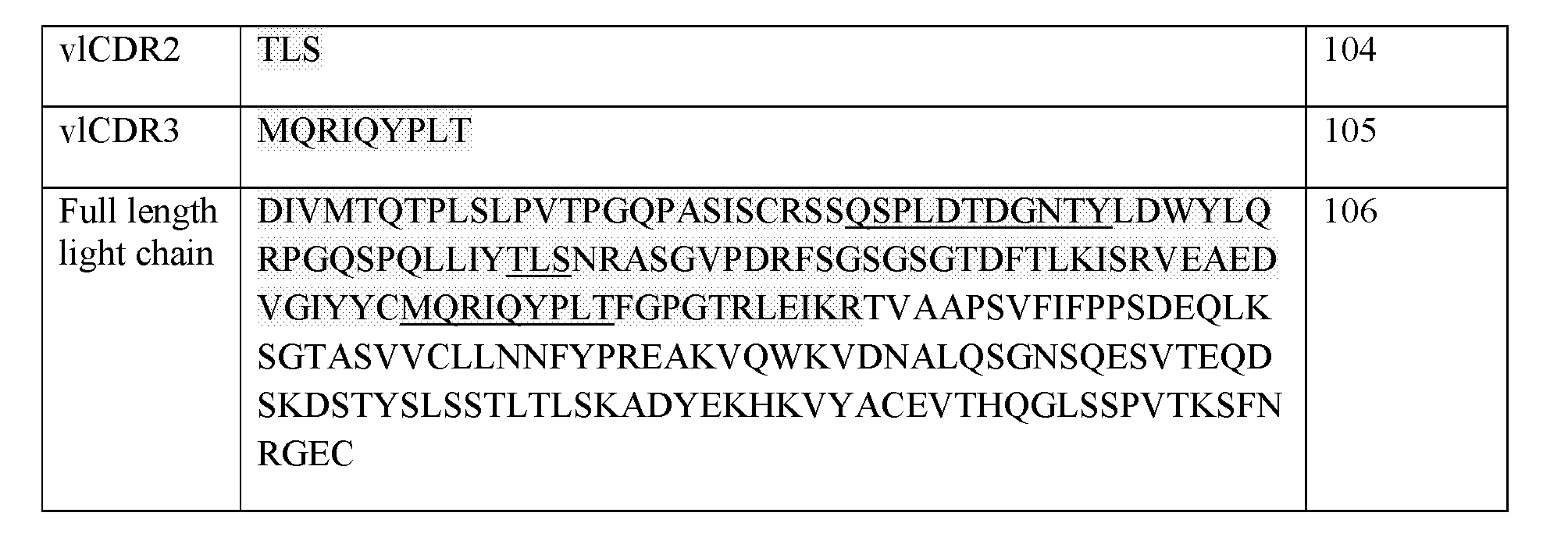


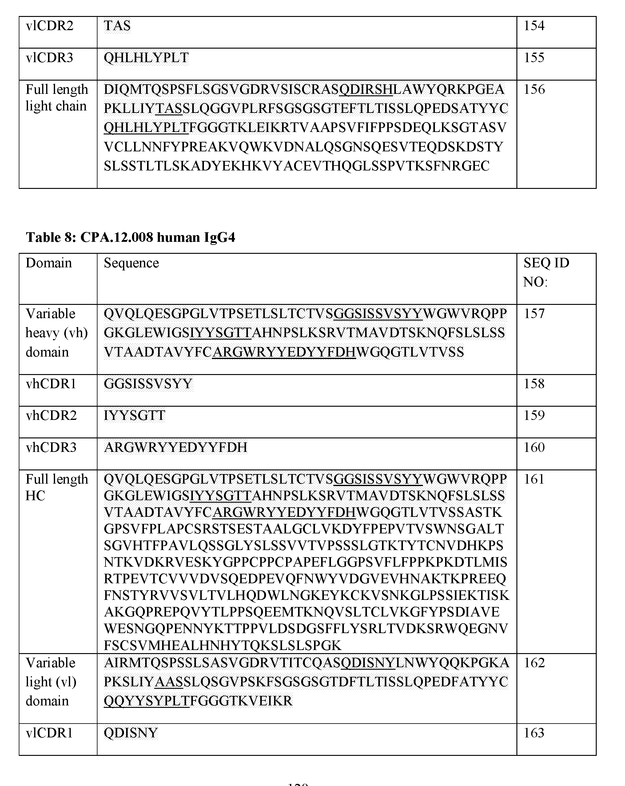


Claims
Priority Applications (8)
| Application Number | Priority Date | Filing Date | Title |
|---|---|---|---|
| EP16751633.5A EP3322733B1 (en) | 2015-07-13 | 2016-07-13 | Hide1 compositions and methods |
| ES16751633T ES2896055T3 (en) | 2015-07-13 | 2016-07-13 | HIDE1 compositions and methods |
| AU2016291846A AU2016291846B2 (en) | 2015-07-13 | 2016-07-13 | HIDE1 Compositions and Methods |
| CA2991980A CA2991980A1 (en) | 2015-07-13 | 2016-07-13 | Hide1 compositions and methods |
| EP21188908.4A EP3971211A1 (en) | 2015-07-13 | 2016-07-13 | Hide1 compositions and methods |
| DK16751633.5T DK3322733T3 (en) | 2015-07-13 | 2016-07-13 | HIDE1 COMPOSITIONS AND METHODS |
| US15/580,723 US20180305451A1 (en) | 2015-07-13 | 2016-07-13 | Hide1 compositions and methods |
| IL256261A IL256261B (en) | 2015-07-13 | 2017-12-11 | Hide1 compositions and methods |
Applications Claiming Priority (4)
| Application Number | Priority Date | Filing Date | Title |
|---|---|---|---|
| US201562191804P | 2015-07-13 | 2015-07-13 | |
| US201562191775P | 2015-07-13 | 2015-07-13 | |
| US62/191,804 | 2015-07-13 | ||
| US62/191,775 | 2015-07-13 |
Publications (1)
| Publication Number | Publication Date |
|---|---|
| WO2017009712A1 true WO2017009712A1 (en) | 2017-01-19 |
Family
ID=56686842
Family Applications (1)
| Application Number | Title | Priority Date | Filing Date |
|---|---|---|---|
| PCT/IB2016/001079 WO2017009712A1 (en) | 2015-07-13 | 2016-07-13 | Hide1 compositions and methods |
Country Status (7)
| Country | Link |
|---|---|
| US (1) | US20180305451A1 (en) |
| EP (2) | EP3971211A1 (en) |
| AU (1) | AU2016291846B2 (en) |
| CA (1) | CA2991980A1 (en) |
| DK (1) | DK3322733T3 (en) |
| ES (1) | ES2896055T3 (en) |
| WO (1) | WO2017009712A1 (en) |
Citations (67)
| Publication number | Priority date | Publication date | Assignee | Title |
|---|---|---|---|---|
| US3773919A (en) | 1969-10-23 | 1973-11-20 | Du Pont | Polylactide-drug mixtures |
| US4399216A (en) | 1980-02-25 | 1983-08-16 | The Trustees Of Columbia University | Processes for inserting DNA into eucaryotic cells and for producing proteinaceous materials |
| US4439196A (en) | 1982-03-18 | 1984-03-27 | Merck & Co., Inc. | Osmotic drug delivery system |
| US4447224A (en) | 1982-09-20 | 1984-05-08 | Infusaid Corporation | Variable flow implantable infusion apparatus |
| US4447233A (en) | 1981-04-10 | 1984-05-08 | Parker-Hannifin Corporation | Medication infusion pump |
| US4475196A (en) | 1981-03-06 | 1984-10-02 | Zor Clair G | Instrument for locating faults in aircraft passenger reading light and attendant call control system |
| US4485045A (en) | 1981-07-06 | 1984-11-27 | Research Corporation | Synthetic phosphatidyl cholines useful in forming liposomes |
| US4486194A (en) | 1983-06-08 | 1984-12-04 | James Ferrara | Therapeutic device for administering medicaments through the skin |
| US4487603A (en) | 1982-11-26 | 1984-12-11 | Cordis Corporation | Implantable microinfusion pump system |
| US4522811A (en) | 1982-07-08 | 1985-06-11 | Syntex (U.S.A.) Inc. | Serial injection of muramyldipeptides and liposomes enhances the anti-infective activity of muramyldipeptides |
| EP0154316A2 (en) | 1984-03-06 | 1985-09-11 | Takeda Chemical Industries, Ltd. | Chemically modified lymphokine and production thereof |
| US4544545A (en) | 1983-06-20 | 1985-10-01 | Trustees University Of Massachusetts | Liposomes containing modified cholesterol for organ targeting |
| US4596556A (en) | 1985-03-25 | 1986-06-24 | Bioject, Inc. | Hypodermic injection apparatus |
| US4634665A (en) | 1980-02-25 | 1987-01-06 | The Trustees Of Columbia University In The City Of New York | Processes for inserting DNA into eucaryotic cells and for producing proteinaceous materials |
| WO1987004462A1 (en) | 1986-01-23 | 1987-07-30 | Celltech Limited | Recombinant dna sequences, vectors containing them and method for the use thereof |
| WO1987005330A1 (en) | 1986-03-07 | 1987-09-11 | Michel Louis Eugene Bergh | Method for enhancing glycoprotein stability |
| US4790824A (en) | 1987-06-19 | 1988-12-13 | Bioject, Inc. | Non-invasive hypodermic injection device |
| WO1989001036A1 (en) | 1987-07-23 | 1989-02-09 | Celltech Limited | Recombinant dna expression vectors |
| EP0338841A1 (en) | 1988-04-18 | 1989-10-25 | Celltech Limited | Recombinant DNA methods, vectors and host cells |
| US4941880A (en) | 1987-06-19 | 1990-07-17 | Bioject, Inc. | Pre-filled ampule and non-invasive hypodermic injection device assembly |
| EP0401384A1 (en) | 1988-12-22 | 1990-12-12 | Kirin-Amgen, Inc. | Chemically modified granulocyte colony stimulating factor |
| US5013556A (en) | 1989-10-20 | 1991-05-07 | Liposome Technology, Inc. | Liposomes with enhanced circulation time |
| US5057313A (en) | 1986-02-25 | 1991-10-15 | The Center For Molecular Medicine And Immunology | Diagnostic and therapeutic antibody conjugates |
| US5064413A (en) | 1989-11-09 | 1991-11-12 | Bioject, Inc. | Needleless hypodermic injection device |
| US5179017A (en) | 1980-02-25 | 1993-01-12 | The Trustees Of Columbia University In The City Of New York | Processes for inserting DNA into eucaryotic cells and for producing proteinaceous materials |
| US5283173A (en) | 1990-01-24 | 1994-02-01 | The Research Foundation Of State University Of New York | System to detect protein-protein interactions |
| US5312335A (en) | 1989-11-09 | 1994-05-17 | Bioject Inc. | Needleless hypodermic injection device |
| WO1994013804A1 (en) | 1992-12-04 | 1994-06-23 | Medical Research Council | Multivalent and multispecific binding proteins, their manufacture and use |
| US5374548A (en) | 1986-05-02 | 1994-12-20 | Genentech, Inc. | Methods and compositions for the attachment of proteins to liposomes using a glycophospholipid anchor |
| WO1994029351A2 (en) | 1993-06-16 | 1994-12-22 | Celltech Limited | Antibodies |
| US5383851A (en) | 1992-07-24 | 1995-01-24 | Bioject Inc. | Needleless hypodermic injection device |
| US5399331A (en) | 1985-06-26 | 1995-03-21 | The Liposome Company, Inc. | Method for protein-liposome coupling |
| US5416016A (en) | 1989-04-03 | 1995-05-16 | Purdue Research Foundation | Method for enhancing transmembrane transport of exogenous molecules |
| US5624821A (en) | 1987-03-18 | 1997-04-29 | Scotgen Biopharmaceuticals Incorporated | Antibodies with altered effector functions |
| US5677425A (en) | 1987-09-04 | 1997-10-14 | Celltech Therapeutics Limited | Recombinant antibody |
| WO1997038731A1 (en) | 1996-04-18 | 1997-10-23 | The Regents Of The University Of California | Immunoliposomes that optimize internalization into target cells |
| US5821333A (en) | 1995-03-01 | 1998-10-13 | Genetech, Inc. | Method for making heteromultimeric polypeptides |
| US5851795A (en) | 1991-06-27 | 1998-12-22 | Bristol-Myers Squibb Company | Soluble CTLA4 molecules and uses thereof |
| US5869046A (en) | 1995-04-14 | 1999-02-09 | Genentech, Inc. | Altered polypeptides with increased half-life |
| US5932448A (en) | 1991-11-29 | 1999-08-03 | Protein Design Labs., Inc. | Bispecific antibody heterodimers |
| WO1999054342A1 (en) | 1998-04-20 | 1999-10-28 | Pablo Umana | Glycosylation engineering of antibodies for improving antibody-dependent cellular cytotoxicity |
| WO2000029004A1 (en) | 1998-11-18 | 2000-05-25 | Peptor Ltd. | Small functional units of antibody heavy chain variable regions |
| WO2000042072A2 (en) | 1999-01-15 | 2000-07-20 | Genentech, Inc. | Polypeptide variants with altered effector function |
| US6121022A (en) | 1995-04-14 | 2000-09-19 | Genentech, Inc. | Altered polypeptides with increased half-life |
| US6165745A (en) | 1992-04-24 | 2000-12-26 | Board Of Regents, The University Of Texas System | Recombinant production of immunoglobulin-like domains in prokaryotic cells |
| WO2001000814A2 (en) | 1999-06-25 | 2001-01-04 | Universität Zürich | Hetero-associating coiled-coil peptides and screenign method therefor |
| WO2001009162A2 (en) * | 1999-07-30 | 2001-02-08 | Millennium Pharmaceuticals, Inc. | Secreted proteins and uses thereof |
| US6194551B1 (en) | 1998-04-02 | 2001-02-27 | Genentech, Inc. | Polypeptide variants |
| US6277375B1 (en) | 1997-03-03 | 2001-08-21 | Board Of Regents, The University Of Texas System | Immunoglobulin-like domains with increased half-lives |
| US6331175B1 (en) | 1985-07-05 | 2001-12-18 | Immunomedics, Inc. | Method and kit for imaging and treating organs and tissues |
| EP1176195A1 (en) | 1999-04-09 | 2002-01-30 | Kyowa Hakko Kogyo Co., Ltd. | Method for controlling the activity of immunologically functional molecule |
| US6403374B1 (en) | 1996-08-16 | 2002-06-11 | The Regents Of The University Of California | Long wavelength engineered fluorescent proteins |
| WO2002066600A2 (en) * | 2000-12-29 | 2002-08-29 | Hyseq, Inc. | Methods and materials relating to leukocyte immunoglobulin receptor-like (lir-like) polypeptides and polynucleotides |
| WO2003035835A2 (en) | 2001-10-25 | 2003-05-01 | Genentech, Inc. | Glycoprotein compositions |
| US6562576B2 (en) | 2001-01-04 | 2003-05-13 | Myriad Genetics, Inc. | Yeast two-hybrid system and use thereof |
| US6737056B1 (en) | 1999-01-15 | 2004-05-18 | Genentech, Inc. | Polypeptide variants with altered effector function |
| US20040110704A1 (en) | 2002-04-09 | 2004-06-10 | Kyowa Hakko Kogyo Co., Ltd. | Cells of which genome is modified |
| US6790624B2 (en) | 1999-06-30 | 2004-09-14 | The University Of Connecticut | Coiled-coil mediated heterodimerization functional interaction trap |
| WO2004080148A2 (en) * | 2002-10-02 | 2004-09-23 | Nuvelo, Inc. | Novel nucleic acids and polypeptides |
| US6800733B2 (en) | 1994-11-10 | 2004-10-05 | The Regents Of The University Of California | Modified green fluorescent proteins |
| WO2004091658A1 (en) | 2003-04-04 | 2004-10-28 | Genentech, Inc. | High concentration antibody and protein formulations |
| WO2006050247A2 (en) | 2004-10-29 | 2006-05-11 | Neose Technologies, Inc. | Remodeling and glycopegylation of fibroblast growth factor (fgf) |
| WO2006050262A2 (en) | 2004-11-01 | 2006-05-11 | The Regents Of The University Of California | Compositions and methods for modification of biomolecules |
| EP1712563A1 (en) * | 2004-01-27 | 2006-10-18 | Medical and Biological Laboratories Co., Ltd. | Method of isolating monocytes |
| US7157566B2 (en) | 2001-02-26 | 2007-01-02 | The Regents Of The University Of California | Monomeric and dimeric fluorescent protein variants and methods for making same |
| US8883973B2 (en) | 2004-11-12 | 2014-11-11 | Xencor, Inc. | Fc variants with altered binding to FcRn |
| US9315584B2 (en) | 2009-03-25 | 2016-04-19 | Tohoku University | LH-type bispecific antibody |
-
2016
- 2016-07-13 DK DK16751633.5T patent/DK3322733T3/en active
- 2016-07-13 EP EP21188908.4A patent/EP3971211A1/en active Pending
- 2016-07-13 US US15/580,723 patent/US20180305451A1/en not_active Abandoned
- 2016-07-13 EP EP16751633.5A patent/EP3322733B1/en active Active
- 2016-07-13 CA CA2991980A patent/CA2991980A1/en active Pending
- 2016-07-13 AU AU2016291846A patent/AU2016291846B2/en active Active
- 2016-07-13 WO PCT/IB2016/001079 patent/WO2017009712A1/en active Application Filing
- 2016-07-13 ES ES16751633T patent/ES2896055T3/en active Active
Patent Citations (70)
| Publication number | Priority date | Publication date | Assignee | Title |
|---|---|---|---|---|
| US3773919A (en) | 1969-10-23 | 1973-11-20 | Du Pont | Polylactide-drug mixtures |
| US4399216A (en) | 1980-02-25 | 1983-08-16 | The Trustees Of Columbia University | Processes for inserting DNA into eucaryotic cells and for producing proteinaceous materials |
| US4634665A (en) | 1980-02-25 | 1987-01-06 | The Trustees Of Columbia University In The City Of New York | Processes for inserting DNA into eucaryotic cells and for producing proteinaceous materials |
| US5179017A (en) | 1980-02-25 | 1993-01-12 | The Trustees Of Columbia University In The City Of New York | Processes for inserting DNA into eucaryotic cells and for producing proteinaceous materials |
| US4475196A (en) | 1981-03-06 | 1984-10-02 | Zor Clair G | Instrument for locating faults in aircraft passenger reading light and attendant call control system |
| US4447233A (en) | 1981-04-10 | 1984-05-08 | Parker-Hannifin Corporation | Medication infusion pump |
| US4485045A (en) | 1981-07-06 | 1984-11-27 | Research Corporation | Synthetic phosphatidyl cholines useful in forming liposomes |
| US4439196A (en) | 1982-03-18 | 1984-03-27 | Merck & Co., Inc. | Osmotic drug delivery system |
| US4522811A (en) | 1982-07-08 | 1985-06-11 | Syntex (U.S.A.) Inc. | Serial injection of muramyldipeptides and liposomes enhances the anti-infective activity of muramyldipeptides |
| US4447224A (en) | 1982-09-20 | 1984-05-08 | Infusaid Corporation | Variable flow implantable infusion apparatus |
| US4487603A (en) | 1982-11-26 | 1984-12-11 | Cordis Corporation | Implantable microinfusion pump system |
| US4486194A (en) | 1983-06-08 | 1984-12-04 | James Ferrara | Therapeutic device for administering medicaments through the skin |
| US4544545A (en) | 1983-06-20 | 1985-10-01 | Trustees University Of Massachusetts | Liposomes containing modified cholesterol for organ targeting |
| EP0154316A2 (en) | 1984-03-06 | 1985-09-11 | Takeda Chemical Industries, Ltd. | Chemically modified lymphokine and production thereof |
| US4596556A (en) | 1985-03-25 | 1986-06-24 | Bioject, Inc. | Hypodermic injection apparatus |
| US5399331A (en) | 1985-06-26 | 1995-03-21 | The Liposome Company, Inc. | Method for protein-liposome coupling |
| US6331175B1 (en) | 1985-07-05 | 2001-12-18 | Immunomedics, Inc. | Method and kit for imaging and treating organs and tissues |
| WO1987004462A1 (en) | 1986-01-23 | 1987-07-30 | Celltech Limited | Recombinant dna sequences, vectors containing them and method for the use thereof |
| US5057313A (en) | 1986-02-25 | 1991-10-15 | The Center For Molecular Medicine And Immunology | Diagnostic and therapeutic antibody conjugates |
| WO1987005330A1 (en) | 1986-03-07 | 1987-09-11 | Michel Louis Eugene Bergh | Method for enhancing glycoprotein stability |
| US5374548A (en) | 1986-05-02 | 1994-12-20 | Genentech, Inc. | Methods and compositions for the attachment of proteins to liposomes using a glycophospholipid anchor |
| US5648260A (en) | 1987-03-18 | 1997-07-15 | Scotgen Biopharmaceuticals Incorporated | DNA encoding antibodies with altered effector functions |
| US5624821A (en) | 1987-03-18 | 1997-04-29 | Scotgen Biopharmaceuticals Incorporated | Antibodies with altered effector functions |
| US4790824A (en) | 1987-06-19 | 1988-12-13 | Bioject, Inc. | Non-invasive hypodermic injection device |
| US4941880A (en) | 1987-06-19 | 1990-07-17 | Bioject, Inc. | Pre-filled ampule and non-invasive hypodermic injection device assembly |
| WO1989001036A1 (en) | 1987-07-23 | 1989-02-09 | Celltech Limited | Recombinant dna expression vectors |
| US5677425A (en) | 1987-09-04 | 1997-10-14 | Celltech Therapeutics Limited | Recombinant antibody |
| EP0338841A1 (en) | 1988-04-18 | 1989-10-25 | Celltech Limited | Recombinant DNA methods, vectors and host cells |
| EP0401384A1 (en) | 1988-12-22 | 1990-12-12 | Kirin-Amgen, Inc. | Chemically modified granulocyte colony stimulating factor |
| US5416016A (en) | 1989-04-03 | 1995-05-16 | Purdue Research Foundation | Method for enhancing transmembrane transport of exogenous molecules |
| US5013556A (en) | 1989-10-20 | 1991-05-07 | Liposome Technology, Inc. | Liposomes with enhanced circulation time |
| US5312335A (en) | 1989-11-09 | 1994-05-17 | Bioject Inc. | Needleless hypodermic injection device |
| US5064413A (en) | 1989-11-09 | 1991-11-12 | Bioject, Inc. | Needleless hypodermic injection device |
| US5283173A (en) | 1990-01-24 | 1994-02-01 | The Research Foundation Of State University Of New York | System to detect protein-protein interactions |
| US5851795A (en) | 1991-06-27 | 1998-12-22 | Bristol-Myers Squibb Company | Soluble CTLA4 molecules and uses thereof |
| US5932448A (en) | 1991-11-29 | 1999-08-03 | Protein Design Labs., Inc. | Bispecific antibody heterodimers |
| US6165745A (en) | 1992-04-24 | 2000-12-26 | Board Of Regents, The University Of Texas System | Recombinant production of immunoglobulin-like domains in prokaryotic cells |
| US5383851A (en) | 1992-07-24 | 1995-01-24 | Bioject Inc. | Needleless hypodermic injection device |
| US5399163A (en) | 1992-07-24 | 1995-03-21 | Bioject Inc. | Needleless hypodermic injection methods and device |
| WO1994013804A1 (en) | 1992-12-04 | 1994-06-23 | Medical Research Council | Multivalent and multispecific binding proteins, their manufacture and use |
| WO1994029351A2 (en) | 1993-06-16 | 1994-12-22 | Celltech Limited | Antibodies |
| US6800733B2 (en) | 1994-11-10 | 2004-10-05 | The Regents Of The University Of California | Modified green fluorescent proteins |
| US5821333A (en) | 1995-03-01 | 1998-10-13 | Genetech, Inc. | Method for making heteromultimeric polypeptides |
| US5869046A (en) | 1995-04-14 | 1999-02-09 | Genentech, Inc. | Altered polypeptides with increased half-life |
| US6121022A (en) | 1995-04-14 | 2000-09-19 | Genentech, Inc. | Altered polypeptides with increased half-life |
| WO1997038731A1 (en) | 1996-04-18 | 1997-10-23 | The Regents Of The University Of California | Immunoliposomes that optimize internalization into target cells |
| US6403374B1 (en) | 1996-08-16 | 2002-06-11 | The Regents Of The University Of California | Long wavelength engineered fluorescent proteins |
| US6277375B1 (en) | 1997-03-03 | 2001-08-21 | Board Of Regents, The University Of Texas System | Immunoglobulin-like domains with increased half-lives |
| US6194551B1 (en) | 1998-04-02 | 2001-02-27 | Genentech, Inc. | Polypeptide variants |
| WO1999054342A1 (en) | 1998-04-20 | 1999-10-28 | Pablo Umana | Glycosylation engineering of antibodies for improving antibody-dependent cellular cytotoxicity |
| WO2000029004A1 (en) | 1998-11-18 | 2000-05-25 | Peptor Ltd. | Small functional units of antibody heavy chain variable regions |
| WO2000042072A2 (en) | 1999-01-15 | 2000-07-20 | Genentech, Inc. | Polypeptide variants with altered effector function |
| US7371826B2 (en) | 1999-01-15 | 2008-05-13 | Genentech, Inc. | Polypeptide variants with altered effector function |
| US6737056B1 (en) | 1999-01-15 | 2004-05-18 | Genentech, Inc. | Polypeptide variants with altered effector function |
| EP1176195A1 (en) | 1999-04-09 | 2002-01-30 | Kyowa Hakko Kogyo Co., Ltd. | Method for controlling the activity of immunologically functional molecule |
| WO2001000814A2 (en) | 1999-06-25 | 2001-01-04 | Universität Zürich | Hetero-associating coiled-coil peptides and screenign method therefor |
| US6790624B2 (en) | 1999-06-30 | 2004-09-14 | The University Of Connecticut | Coiled-coil mediated heterodimerization functional interaction trap |
| WO2001009162A2 (en) * | 1999-07-30 | 2001-02-08 | Millennium Pharmaceuticals, Inc. | Secreted proteins and uses thereof |
| WO2002066600A2 (en) * | 2000-12-29 | 2002-08-29 | Hyseq, Inc. | Methods and materials relating to leukocyte immunoglobulin receptor-like (lir-like) polypeptides and polynucleotides |
| US6562576B2 (en) | 2001-01-04 | 2003-05-13 | Myriad Genetics, Inc. | Yeast two-hybrid system and use thereof |
| US7157566B2 (en) | 2001-02-26 | 2007-01-02 | The Regents Of The University Of California | Monomeric and dimeric fluorescent protein variants and methods for making same |
| WO2003035835A2 (en) | 2001-10-25 | 2003-05-01 | Genentech, Inc. | Glycoprotein compositions |
| US20040110704A1 (en) | 2002-04-09 | 2004-06-10 | Kyowa Hakko Kogyo Co., Ltd. | Cells of which genome is modified |
| WO2004080148A2 (en) * | 2002-10-02 | 2004-09-23 | Nuvelo, Inc. | Novel nucleic acids and polypeptides |
| WO2004091658A1 (en) | 2003-04-04 | 2004-10-28 | Genentech, Inc. | High concentration antibody and protein formulations |
| EP1712563A1 (en) * | 2004-01-27 | 2006-10-18 | Medical and Biological Laboratories Co., Ltd. | Method of isolating monocytes |
| WO2006050247A2 (en) | 2004-10-29 | 2006-05-11 | Neose Technologies, Inc. | Remodeling and glycopegylation of fibroblast growth factor (fgf) |
| WO2006050262A2 (en) | 2004-11-01 | 2006-05-11 | The Regents Of The University Of California | Compositions and methods for modification of biomolecules |
| US8883973B2 (en) | 2004-11-12 | 2014-11-11 | Xencor, Inc. | Fc variants with altered binding to FcRn |
| US9315584B2 (en) | 2009-03-25 | 2016-04-19 | Tohoku University | LH-type bispecific antibody |
Non-Patent Citations (161)
| Title |
|---|
| ALTSCHUL ET AL., J MOL. BIOL., vol. 215, 1990, pages 403 - 10 |
| ALTSCHUL ET AL., NUCLEIC ACIDS RES., vol. 25, no. 17, 1997, pages 3389 - 3402 |
| ANDERSON ET AL., ARCH. BIOCHEM. BIOPHYS., vol. 239, 1985, pages 538 |
| ANDERSSON-ENGELS ET AL., PHYS. MED. BIOL.,, vol. 42, 1997, pages 815 - 824 |
| APLIN; WRISTON, CRC CRIT. REV. BIOCHEM., vol. 22, 1981, pages 259 - 306 |
| ARAKAWA ET AL., JBIOL. CHEM., vol. 269, no. 45, 1994, pages 27833 - 27839 |
| BARBAS ET AL., PROC. NAT. ACAD. SCI, USA, vol. 91, 1994, pages 3809 - 3813 |
| BARBER ET AL., NATURE, vol. 439, 2006, pages 682 - 7 |
| BECERK ET AL., MUTAT. RES, vol. 722, no. 2, 17 June 2011 (2011-06-17), pages 171 - 182 |
| BERGE, S. M. ET AL., J. PHARM. SCI., vol. 66, 1977, pages 1 - 19 |
| BHADRA ET AL., PROC NATL. ACAD SCI., vol. 108, no. 22, 2011, pages 9196 - 201 |
| BIRD ET AL., SCIENCE, vol. 242, 1988, pages 423 - 426 |
| BRAHMAMDAM ET AL., J. LEUKOC. BIOL., vol. 88, 2010, pages 233 - 240 |
| BRISCOE ET AL., AM. J PHYSIOL., vol. 1233, 1995, pages 134 |
| CAMPO ET AL., NOD. PATHOL., vol. 26, no. 1, January 2013 (2013-01-01), pages S97 - S 110 |
| CANCER RES., vol. 73, no. 9, 1 May 2013 (2013-05-01), pages 2782 - 94 |
| CHAN CA; CARTER PJ, NATURE REV IMMUNOL, vol. 10, 2010, pages 301 - 316 |
| CHANG ET AL., CRITICAL CARE, vol. 17, 2013, pages R85 |
| CHOTHIA; LESK, J. MOL. BIOL., vol. 196, 1987, pages 901 - 917 |
| CINEL; OPAL, CRIT. CARE MED., vol. 37, 2009, pages 291 - 304 |
| CRAMERI ET AL., NAT. BIOTECHNOL., vol. 14, 1996, pages 315 - 319 |
| CRAWFORD ET AL., CURR OPIN IMMUNOL, vol. 21, 2009, pages 179 - 186 |
| CRAWFORD ET AL., CURR OPIN IMMUNOL., vol. 21, 2009, pages 179 - 186 |
| CROTTY, ANNU. REV. IMMUNOL., vol. 29, 2011, pages 621 - 663 |
| DELLINGER ET AL., INTENSIVE CARE MED, vol. 39, 2013, pages 165 - 228 |
| DI MAIO ET AL., J. CHEM. SOC. PERKIN TRANS., 1985, pages 1687 |
| DITTER ET AL., J. PHARM SCI., vol. 57, 1968, pages 783 |
| DITTER ET AL., J. PHARM. SCI., vol. 57, 1968, pages 828 |
| DITTER ET AL., J. PHARM. SCI., vol. 58, 1969, pages 557 |
| DOAN ET AL., MOL. MICROBIOL.,, vol. 55, 2005, pages 1767 - 1781 |
| DOUILLARD ET AL., LANCET, vol. 355, 2000, pages 1041 - 7 |
| DUNCAN; MILLER, PLOS ONE., vol. 6, 2011, pages E18548 |
| E. A. KABAT: "SEQUENCES OF IMMUNOLOGICAL INTEREST, 5th ed.", NIH PUBLICATION, NO. 91-3242 |
| E. MEYERS; W. MILLER, COMPUT. APPL. BIOSCI., vol. 4, 1988, pages 11 - 17 |
| EDGE ET AL., ANAL. BIOCHEM., vol. 118, 1981, pages 131 |
| EPSTEIN ET AL., PROC NATL ACAD SCI USA, vol. 82, 1985, pages 3688 |
| F. AUSUBEL, ET AL.: "Current Protocols in Molecular Biology", 1987, GREENE PUBLISHING AND WILEY INTERSCIENCE |
| FISCHER ET AL., FEBSLETT., vol. 577, 2004, pages 227 - 232 |
| FISCHER ET AL., FEBSLETT., vol. 580, 2006, pages 2495 - 2502 |
| FRANGIONI, CURR. OPIN. CHEM. BIOL., vol. 7, 2003, pages 626 - 634 |
| GABIZON ET AL., J NATIONAL CANCER INST, vol. 81, 1989, pages 1484 |
| GARG; JEANLOZ: "Advances in Carbohydrate Chemistry and Biochemistry", vol. 43, 1985, ACADEMIC PRESS |
| GARVEY ET AL., J. ORG. CHEM., vol. 56, 1990, pages 436 |
| GOEDDEL: "Methods in Enzymology", vol. 185, 1990, ACADEMIC PRESS, article "Gene Expression Technology" |
| GOLDEN-MASON ET AL., J VIROL., vol. 83, 2009, pages 9122 - 30 |
| GOODKIND ET AL., COMPUTERS AND CHEM. ENG., vol. 29, no. 3, 2005, pages 589 |
| GOYERT; SILVER, J EUK. BIOL., vol. 88, no. 2, 2010, pages 225 - 226 |
| GOYERT; SILVER, J EUK. BIOL.,, vol. 88, no. 2, 2010, pages 225 - 226 |
| GREEN; WUTS: "Protecting Groups in Organic Synthesis", 1991, JOHN WILEY AND SONS, article "chapters 5 and 7" |
| GREENWALD ET AL.: "The B7 Family Revisited", ANN. REV. IMMUNOL., vol. 23, 2005, pages 515 - 48, XP002481408, DOI: doi:10.1146/annurev.immunol.23.021704.115611 |
| GUNTUPALLI ET AL., CRIT CARE MED., vol. 41, no. 3, 2013, pages 706 - 716 |
| GUO, EMBO J, vol. 17, 1998, pages 5265 - 5272 |
| HAKIMUDDIN ET AL., ARCH. BIOCHEM. BIOPHYS., vol. 259, 1987, pages 52 |
| HAN ET AL., BIOINFORM. BIOL. INSIGHTS, vol. 9, no. 1, 15 November 2015 (2015-11-15), pages 29 - 46 |
| HAWKINS ET AL., J. MOL. BIOL., vol. 226, 1992, pages 889 - 896 |
| HOFMEYER ET AL., J BIOMED BIOTECHNOL., vol. 2011 |
| HOLLIGER ET AL., PROC. NATL. ACAD. SCI. USA, vol. 90, 1993, pages 6444 - 6448 |
| HOTCHKISS ET AL., LANCET INFECT. DIS., vol. 13, 2013, pages 260 - 268 |
| HU5 ET AL., JBIOL CHEM., vol. 273, 1998, pages 33489 - 33494 |
| HUANG ET AL., PNAS, vol. 106, 2009, pages 6303 - 6308 |
| HUGO W; ZARETSKY JM; SUN L; SONG C ET AL.: "Genomic and Transcriptomic Features of Response to Anti-PD-1 Therapy in Metastatic Melanoma", CELL, vol. 165, no. 1, 24 March 2016 (2016-03-24), pages 35 - 44, XP029473850, DOI: doi:10.1016/j.cell.2016.02.065 |
| HUSTON ET AL., PROC. NATL. ACAD. SCI. USA, vol. 85, 1988, pages 5879 - 5883 |
| HUSTON, SURG. INFECT., vol. 13, 2012, pages 187 - 193 |
| HWANG ET AL., PROC NATL ACAD SCI USA, vol. 77, 1980, pages 4030 |
| INOUE ET AL., J. IMMUNOL., vol. 184, 2010, pages 1401 - 1409 |
| INOUE ET AL., SHOCK, vol. 36, 2011, pages 38 - 44 |
| INTEST RES., vol. 13, no. 2, April 2015 (2015-04-01), pages 105 - 111 |
| JACKSON ET AL., J. IMMUNOL., vol. 154, no. 7, 1995, pages 3310 - 9 |
| JEFFERIS ET AL., IMMUNOL LETT, vol. 82, 2002, pages 57 - 65 |
| JONES ET AL., TETRAHEDRON LETT., vol. 29, 1988, pages 3853 - 3856 |
| K. KEINANEN; M. L. LAUKKANEN, FEBS LETT., vol. 346, 1994, pages 123 |
| KABAT ET AL.: "SEQUENCES OF PROTEINS OF IMMUNOLOGICAL INTEREST, 5th ed.", 1991, NATIONAL INSTITUTES OF HEALTH |
| KAHN ET AL., TETRAHEDRON LETT., vol. 30, 1989, pages 2317 |
| KATAHIRA ET AL., J. BIOL CHEM, vol. 277, 2002, pages 9242 - 9246 |
| KAUFMANN ET AL., J IMMUNOL, vol. 182, 2009, pages 5891 - 5897 |
| KAZMIERSKI ET AL., J. AM. CHEM. SOC., vol. 133, 1991, pages 2275 - 2283 |
| KAZMIERSKI; HRUBY, TETRAHEDRON LETT., 1991 |
| KEMP ET AL., J. ORG. CHEM., vol. 50, 1985, pages 5834 - 5838 |
| KEMP ET AL., J. ORG. CHEM., vol. 54, 1987, pages 109 - 115 |
| KEMP ET AL., TETRAHEDRON LETT., vol. 29, 1988, pages 4935 - 4938 |
| KEMP ET AL., TETRAHEDRON LETT., vol. 29, 1988, pages 5057 - 5060 |
| KEMP ET AL., TETRAHEDRON LETT., vol. 29, 1988, pages 5081 - 5082 |
| KILLION; FIDLER, IMMUNOMETHODS, vol. 4, 1994, pages 273 |
| KIM ET AL., NATURE, vol. 378, 1995, pages 85 - 88 |
| KING ET AL., BIOCHEMISTRY, vol. 26, 1987, pages 2294 |
| KOMAU ET AL., SCIENCE, vol. 269, 1995, pages 1737 - 1740 |
| KREBS, B ET AL., J IMMUNOL METHODS, vol. 254, 2001, pages 67 - 84 |
| KUNZ, ANG. CHEM. INT. ED. ENGLISH, vol. 26, 1987, pages 294 - 308 |
| LEENTJENS ET AL., AM JRESPIR CRIT CARE MED, vol. 186, 2012, pages 838 - 845 |
| LINDBERG ET AL., DRUG METABOLISM AND DISPOSITION, vol. 17, 1989, pages 311 |
| M. OWAIS; 1995 ET AL., ANTIMICROB. AGENTS CHEMOTHER., vol. 39, pages 180 |
| MALASHKEVICH ET AL., SCIENCE, vol. 274, pages 761 - 765 |
| MARKS ET AL., BIOTECHNOLOGY, vol. 10, 1992, pages 779 - 783 |
| MATHIOWITZ ET AL., J. APPL POLYMER SCI, vol. 35, 1988, pages 755 - 774 |
| MATHIOWITZ ET AL., REACTIVE POLYMERS, vol. 6, 1987, pages 275 - 283 |
| MATHIOWITZ; LANGER, J. CONTROLLED RELEASE, vol. 5, 1987, pages 13 - 22 |
| MATSUDA ET AL., J NIPPON MED SCH., vol. 79, 2012, pages 4 - 18 |
| MATSUDA ET AL., JNIPPON MED SCH., vol. 79, 2012, pages 4 - 18 |
| MCCAFFERTY ET AL., NATURE, vol. 348, 1990, pages 552 - 554 |
| MIYAKE ET AL., J. TAKEDA RES. LABS, vol. 43, 1989, pages 53 - 76 |
| MOUKTAROUDI ET AL., CRIT CARE., vol. 14, 2010, pages 17 |
| NAGAI; SATO, TETRAHEDRON LETT., vol. 26, 1985, pages 647 - 650 |
| NAGAL ET AL., NAT. BIOTECHNOL., vol. 20, 2002, pages 87 - 90 |
| NEEDLEMAN; WUNSCH, J. MOL. BIOL., vol. 48, 1970, pages 444 - 453 |
| OLSON ET AL., J. AM. CHEM. SOC., vol. 112, 1990, pages 323 - 333 |
| OSOL, A.: "Remington's Pharmaceutical Sciences, 16th ed.", 1980 |
| P. G. BLOEMAN ET AL., FEBSLETT., vol. 357, 1995, pages 140 |
| PAYEN ET AL., CRIT CARE., vol. 17, 2013, pages 118 |
| R. J. KAUFMAN; P. A. SHARP, MOL. BIOL., vol. 159, 1982, pages 601 - 621 |
| RADZIEJEWSKI ET AL., BIOCHEM., vol. 32, no. 48, 1993, pages 1350 |
| RAMANUJAM ET AL., IEEE TRANSACTIONS ON BIOMEDICAL ENGINEERING, vol. 48, 2001, pages 1034 - 1041 |
| REMINGTON'S PHARMACEUTICAL SCIENCES, 1980 |
| RILEY ET AL., IMMUNITY, vol. 30, no. 5, 2009, pages 656 - 665 |
| RIVAS ET AL., J IMMUNOL., vol. 183, 2009, pages 4284 - 91 |
| RIZZO ET AL., NAT. BIOTECHNOL, vol. 22, 2004, pages 445 |
| SADUM ET AL.: "Immune Signatures of Murine and Human Cancers Reveal Unique Mechanisms of Tumor Escape and New Targets for Cancer Immunotherapy", CLIN. CANC. RES., vol. 13, no. 13, 2007, pages 4016 - 4025, XP055248327, DOI: doi:10.1158/1078-0432.CCR-07-0016 |
| SALTZ ET AL., PROCASCO, vol. 18, 1999, pages 233A |
| SCALAPINO KJ; DAIKH DI, PLOS ONE, vol. 4, no. 6, 2009, pages E6031 |
| SCHREIER ET AL., J. BIOL. CHEM., vol. 269, 1994, pages 9090 |
| SCHUURMAN J., IMMUNOLOGY, vol. 105, 2002, pages 9 - 19 |
| SHANER ET AL., NAT. BIOTECHNOL., vol. 22, 2004, pages 1567 - 1572 |
| SHARPE ET AL., NAT IMMITNOL, vol. 8, 2007, pages 239 - 245 |
| SHARPE ET AL., NAT IMMUNOL, vol. 8, 2007, pages 239 - 245 |
| SHIELDS, R. L ET AL., J. BIOL. CHEM., vol. 276, 2001, pages 6591 - 6604 |
| SHIELDS, R. L. ET AL., J. BIOL. CHEM., vol. 276, 2001, pages 6591 - 6604 |
| SHIELDS, R. L. ET AL., J. BIOL. CHEM., vol. 277, 2002, pages 26733 - 26740 |
| SHIER ET AL., GENE, vol. 169, 1995, pages 147 - 155 |
| SINGHAL ET AL., FASEB J., vol. 1, 1987, pages 220 |
| SUDOL, PROG. BIOCHYS. MOL BIOL., vol. 65, 1996, pages 113 - 132 |
| TAKEBE, Y ET AL., MOL. CELL. BIOL., vol. 8, 1988, pages 466 - 472 |
| TARENTINO, A. L. ET AL., BIOCHEM., vol. 14, 1975, pages 5516 - 23 |
| TAYLOR ET AL., BIOMED RES., vol. 2014, pages 8 |
| THOTAKURA ET AL., METH. ENZYMOL., vol. 138, 1987, pages 350 |
| TOMLINSON, METHODS ENZYMOL., vol. 326, 2000, pages 461 - 479 |
| TOTH ET AL.: "Peptides: Chemistry, Structure and Biology", 1990, ESCOM PUBL., pages: 1078 - 1079 |
| TSIEN, ANNU. REV. BIOCHEM., vol. 67, 1998, pages 509 |
| TUNEK ET AL., BIOCHEM. PHARM, vol. 37, 1988, pages 3867 |
| UMANA ET AL., NAT. BIOTECH, vol. 17, 1999, pages 176 - 180 |
| UMANA ET AL., NAT. BIOTECH., vol. 17, 1999, pages 176 - 180 |
| UMEZAWA ET AL., BIOCHEM. BIOPHYS. RES. COMMUN, vol. 153, 1988, pages 1038 |
| UNSINGER ET AL., J. IMMUNOL., vol. 184, 2010, pages 3768 - 3779 |
| URLAUB; CHASIN, PROC. NATL. ACAD. SCI. USA, vol. 77, 1980, pages 4216 - 4220 |
| VALU ET AL., NATURE, vol. 458, 2009, pages 206 - 210 |
| VIDAL ET AL., BIOCHEMISTRY, vol. 43, 2004, pages 7336 - 44 |
| WANG ET AL., PROC. NATL. ACAD. SCI. U. S. A., vol. 101, 2004, pages 16745 - 16749 |
| WANG ET AL.: "Recent Advances in Capillary Electrophoresis-Based Proteomic Techniques for Biomarker Discovery", METHODS. MOL. BIOL., vol. 984, 2013, pages 1 - 12 |
| WANG: "Immune Suppression by Tumor Specific CD4+ Regulatory T cells", CANCER. SEMIN. CANCER. BIOL., vol. 16, 2006, pages 73 - 79, XP024908039, DOI: doi:10.1016/j.semcancer.2005.07.009 |
| WANNEMUEHLER ET AL., J. SURG. RES., vol. 173, 2012, pages 113 - 26 |
| WATTS: "TNF/TNFR Family Members in Co-stimulation of T Cell Responses", ANN. REV. IMMUNOL., vol. 23, 2005, pages 23 - 68, XP008051583, DOI: doi:10.1146/annurev.immunol.23.021704.115839 |
| WHITLOW, M. ET AL., METHODS: A COMPANION TO METHODS IN ENZYMOLOGY, vol. 2, 1991, pages 97 - 105 |
| WITTEBOLE ET AL., MEDIATORS OF INFLAMMATION, vol. 10, 2010 |
| WORLD J IMMUNOL, vol. 4, no. 1, 27 March 2014 (2014-03-27), pages 26 - 33 |
| WU ET AL., CRITICAL CARE, vol. 17, 2013, pages R8 |
| YAMANE-OHNUKI ET AL., BIOTECHNOL BIOENG, vol. 87, 2004, pages 614 - 22 |
| YE ET AL., NUCLEIC ACIDS RES., vol. 41, 2013, pages W34 - W40 |
| YELTON ET AL., J. IMMUNOL., vol. 155, 1995, pages 1994 - 2004 |
| ZABROCKI ET AL., J. AM. CHEM. SOC., vol. 110, 1988, pages 5875 - 5880 |
| ZECHEL ET AL., INT. J. PEP. PROTEIN RES., vol. 43, 1991 |
| ZHANG ET AL., CRIT. CARE, vol. 15, 2011, pages R70 |
| ZHANG ET AL., CRITICAL CARE, vol. 14, 2010, pages R220 |
| ZHOU ET AL., NATURE, vol. 378, 1995, pages 584 - 592 |
Also Published As
| Publication number | Publication date |
|---|---|
| US20180305451A1 (en) | 2018-10-25 |
| EP3322733B1 (en) | 2021-09-15 |
| DK3322733T3 (en) | 2021-10-18 |
| CA2991980A1 (en) | 2017-01-19 |
| EP3971211A1 (en) | 2022-03-23 |
| ES2896055T3 (en) | 2022-02-23 |
| AU2016291846B2 (en) | 2022-05-26 |
| AU2016291846A1 (en) | 2018-01-25 |
| EP3322733A1 (en) | 2018-05-23 |
Similar Documents
| Publication | Publication Date | Title |
|---|---|---|
| US11795209B2 (en) | PVRIG polypeptides and methods of treatment | |
| US11649285B2 (en) | Identification of VSIG3/VISTA as a novel immune checkpoint and use thereof for immunotherapy | |
| US20160347814A1 (en) | Vstm5 polypeptides and uses thereof as a drug for treatment of cancer, infectious diseases and immune related diseases | |
| AU2015357463B2 (en) | Identification of VSIG8 as the putative vista receptor and its use thereof to produce vista/VSIG8 modulators | |
| US11919960B2 (en) | Anti-CD25 antibody agents | |
| EP3295951A1 (en) | Anti-pvrig antibodies and methods of use | |
| US20160272707A1 (en) | Vstm5 antibodies, and uses thereof for treatment of cancer, infectious diseases and immune related diseases | |
| AU2013278843A1 (en) | LSR antibodies, and uses thereof for treatment of cancer | |
| AU2016291846B2 (en) | HIDE1 Compositions and Methods | |
| IL256261A (en) | Hide1 compositions and methods |
Legal Events
| Date | Code | Title | Description |
|---|---|---|---|
| 121 | Ep: the epo has been informed by wipo that ep was designated in this application |
Ref document number: 16751633 Country of ref document: EP Kind code of ref document: A1 |
|
| WWE | Wipo information: entry into national phase |
Ref document number: 15580723 Country of ref document: US |
|
| ENP | Entry into the national phase |
Ref document number: 2991980 Country of ref document: CA |
|
| NENP | Non-entry into the national phase |
Ref country code: DE |
|
| ENP | Entry into the national phase |
Ref document number: 2016291846 Country of ref document: AU Date of ref document: 20160713 Kind code of ref document: A |
|
| WWE | Wipo information: entry into national phase |
Ref document number: 2016751633 Country of ref document: EP |