WO2017064675A1 - Hindered disulfide drug conjugates - Google Patents
Hindered disulfide drug conjugates Download PDFInfo
- Publication number
- WO2017064675A1 WO2017064675A1 PCT/IB2016/056186 IB2016056186W WO2017064675A1 WO 2017064675 A1 WO2017064675 A1 WO 2017064675A1 IB 2016056186 W IB2016056186 W IB 2016056186W WO 2017064675 A1 WO2017064675 A1 WO 2017064675A1
- Authority
- WO
- WIPO (PCT)
- Prior art keywords
- antibody
- drug
- linker
- leaving group
- stereoisomer
- Prior art date
Links
- 239000003814 drug Substances 0.000 title claims abstract description 253
- 229940079593 drug Drugs 0.000 title claims abstract description 238
- BWGNESOTFCXPMA-UHFFFAOYSA-N Dihydrogen disulfide Chemical compound SS BWGNESOTFCXPMA-UHFFFAOYSA-N 0.000 title claims abstract description 72
- 238000000034 method Methods 0.000 claims abstract description 120
- 229910052799 carbon Inorganic materials 0.000 claims abstract description 57
- 229910052717 sulfur Inorganic materials 0.000 claims abstract description 51
- 230000021615 conjugation Effects 0.000 claims abstract description 45
- XUJNEKJLAYXESH-UHFFFAOYSA-N cysteine Natural products SCC(N)C(O)=O XUJNEKJLAYXESH-UHFFFAOYSA-N 0.000 claims abstract description 45
- 235000018417 cysteine Nutrition 0.000 claims abstract description 45
- 125000001183 hydrocarbyl group Chemical group 0.000 claims abstract description 15
- 150000001875 compounds Chemical class 0.000 claims description 201
- 239000000203 mixture Substances 0.000 claims description 160
- 239000000562 conjugate Substances 0.000 claims description 150
- YMWUJEATGCHHMB-UHFFFAOYSA-N Dichloromethane Chemical compound ClCCl YMWUJEATGCHHMB-UHFFFAOYSA-N 0.000 claims description 135
- 239000000427 antigen Substances 0.000 claims description 111
- 108091007433 antigens Proteins 0.000 claims description 104
- 102000036639 antigens Human genes 0.000 claims description 104
- 206010028980 Neoplasm Diseases 0.000 claims description 99
- 125000005647 linker group Chemical group 0.000 claims description 90
- ZMXDDKWLCZADIW-UHFFFAOYSA-N dimethylformamide Substances CN(C)C=O ZMXDDKWLCZADIW-UHFFFAOYSA-N 0.000 claims description 88
- -1 cyclic amine Chemical class 0.000 claims description 83
- 239000011541 reaction mixture Substances 0.000 claims description 83
- 238000004895 liquid chromatography mass spectrometry Methods 0.000 claims description 77
- 108090000765 processed proteins & peptides Proteins 0.000 claims description 69
- 201000011510 cancer Diseases 0.000 claims description 62
- 108090000623 proteins and genes Proteins 0.000 claims description 62
- 229940049595 antibody-drug conjugate Drugs 0.000 claims description 59
- XLYOFNOQVPJJNP-UHFFFAOYSA-N water Substances O XLYOFNOQVPJJNP-UHFFFAOYSA-N 0.000 claims description 59
- 239000000611 antibody drug conjugate Substances 0.000 claims description 57
- 238000006243 chemical reaction Methods 0.000 claims description 55
- XEKOWRVHYACXOJ-UHFFFAOYSA-N Ethyl acetate Chemical compound CCOC(C)=O XEKOWRVHYACXOJ-UHFFFAOYSA-N 0.000 claims description 54
- 102000004196 processed proteins & peptides Human genes 0.000 claims description 52
- 235000018102 proteins Nutrition 0.000 claims description 44
- 102000004169 proteins and genes Human genes 0.000 claims description 44
- 239000002904 solvent Substances 0.000 claims description 43
- WYURNTSHIVDZCO-UHFFFAOYSA-N Tetrahydrofuran Chemical compound C1CCOC1 WYURNTSHIVDZCO-UHFFFAOYSA-N 0.000 claims description 38
- WEVYAHXRMPXWCK-UHFFFAOYSA-N Acetonitrile Chemical compound CC#N WEVYAHXRMPXWCK-UHFFFAOYSA-N 0.000 claims description 36
- 125000002496 methyl group Chemical group [H]C([H])([H])* 0.000 claims description 34
- 229910052757 nitrogen Inorganic materials 0.000 claims description 32
- 102100038080 B-cell receptor CD22 Human genes 0.000 claims description 31
- 239000000047 product Substances 0.000 claims description 31
- RTZKZFJDLAIYFH-UHFFFAOYSA-N Diethyl ether Chemical compound CCOCC RTZKZFJDLAIYFH-UHFFFAOYSA-N 0.000 claims description 30
- YUOCYTRGANSSRY-UHFFFAOYSA-N pyrrolo[2,3-i][1,2]benzodiazepine Chemical compound C1=CN=NC2=C3C=CN=C3C=CC2=C1 YUOCYTRGANSSRY-UHFFFAOYSA-N 0.000 claims description 30
- 101000884305 Homo sapiens B-cell receptor CD22 Proteins 0.000 claims description 29
- 125000004434 sulfur atom Chemical group 0.000 claims description 26
- 125000004432 carbon atom Chemical group C* 0.000 claims description 25
- 108060003951 Immunoglobulin Proteins 0.000 claims description 22
- 125000000623 heterocyclic group Chemical group 0.000 claims description 22
- 102000018358 immunoglobulin Human genes 0.000 claims description 22
- 238000011282 treatment Methods 0.000 claims description 22
- 239000000539 dimer Substances 0.000 claims description 21
- 241000894007 species Species 0.000 claims description 20
- 125000004429 atom Chemical group 0.000 claims description 19
- 239000012634 fragment Substances 0.000 claims description 19
- 229910052760 oxygen Inorganic materials 0.000 claims description 19
- 239000007983 Tris buffer Substances 0.000 claims description 18
- 238000004519 manufacturing process Methods 0.000 claims description 18
- 125000006850 spacer group Chemical group 0.000 claims description 17
- 102100033423 GDNF family receptor alpha-1 Human genes 0.000 claims description 16
- 101000579425 Homo sapiens Proto-oncogene tyrosine-protein kinase receptor Ret Proteins 0.000 claims description 16
- 102100026711 Metalloreductase STEAP2 Human genes 0.000 claims description 16
- 102100028286 Proto-oncogene tyrosine-protein kinase receptor Ret Human genes 0.000 claims description 16
- 239000008194 pharmaceutical composition Substances 0.000 claims description 15
- 150000003512 tertiary amines Chemical class 0.000 claims description 15
- 101000628535 Homo sapiens Metalloreductase STEAP2 Proteins 0.000 claims description 14
- 125000001995 cyclobutyl group Chemical group [H]C1([H])C([H])([H])C([H])(*)C1([H])[H] 0.000 claims description 13
- 239000000178 monomer Substances 0.000 claims description 13
- CSCPPACGZOOCGX-UHFFFAOYSA-N Acetone Chemical compound CC(C)=O CSCPPACGZOOCGX-UHFFFAOYSA-N 0.000 claims description 12
- LYCAIKOWRPUZTN-UHFFFAOYSA-N Ethylene glycol Chemical compound OCCO LYCAIKOWRPUZTN-UHFFFAOYSA-N 0.000 claims description 12
- 102100034845 KiSS-1 receptor Human genes 0.000 claims description 12
- 102100022430 Melanocyte protein PMEL Human genes 0.000 claims description 12
- DNIAPMSPPWPWGF-UHFFFAOYSA-N Propylene glycol Chemical compound CC(O)CO DNIAPMSPPWPWGF-UHFFFAOYSA-N 0.000 claims description 12
- 102100032780 Semaphorin-5B Human genes 0.000 claims description 12
- 108010044540 auristatin Proteins 0.000 claims description 12
- 239000006227 byproduct Substances 0.000 claims description 12
- 101000997961 Homo sapiens GDNF family receptor alpha-1 Proteins 0.000 claims description 11
- 125000001559 cyclopropyl group Chemical group [H]C1([H])C([H])([H])C1([H])* 0.000 claims description 11
- 101000654679 Homo sapiens Semaphorin-5B Proteins 0.000 claims description 10
- 101000713169 Homo sapiens Solute carrier family 52, riboflavin transporter, member 2 Proteins 0.000 claims description 10
- 102100032129 Lymphocyte antigen 6K Human genes 0.000 claims description 10
- 102100036862 Solute carrier family 52, riboflavin transporter, member 2 Human genes 0.000 claims description 10
- 102000005962 receptors Human genes 0.000 claims description 10
- 108020003175 receptors Proteins 0.000 claims description 10
- 101000620359 Homo sapiens Melanocyte protein PMEL Proteins 0.000 claims description 9
- 102100026159 Tomoregulin-1 Human genes 0.000 claims description 9
- 125000002015 acyclic group Chemical group 0.000 claims description 9
- 125000000449 nitro group Chemical group [O-][N+](*)=O 0.000 claims description 9
- YLQBMQCUIZJEEH-UHFFFAOYSA-N tetrahydrofuran Natural products C=1C=COC=1 YLQBMQCUIZJEEH-UHFFFAOYSA-N 0.000 claims description 9
- 108010023729 Complement 3d Receptors Proteins 0.000 claims description 8
- 101001091205 Homo sapiens KiSS-1 receptor Proteins 0.000 claims description 8
- 101001065550 Homo sapiens Lymphocyte antigen 6K Proteins 0.000 claims description 8
- 101000835745 Homo sapiens Teratocarcinoma-derived growth factor 1 Proteins 0.000 claims description 8
- 102100031036 Leucine-rich repeat-containing G-protein coupled receptor 5 Human genes 0.000 claims description 8
- 102100026404 Teratocarcinoma-derived growth factor 1 Human genes 0.000 claims description 8
- 102100029690 Tumor necrosis factor receptor superfamily member 13C Human genes 0.000 claims description 8
- 102100039094 Tyrosinase Human genes 0.000 claims description 8
- QVGXLLKOCUKJST-UHFFFAOYSA-N atomic oxygen Chemical group [O] QVGXLLKOCUKJST-UHFFFAOYSA-N 0.000 claims description 8
- HXCHCVDVKSCDHU-LULTVBGHSA-N calicheamicin Chemical compound C1[C@H](OC)[C@@H](NCC)CO[C@H]1O[C@H]1[C@H](O[C@@H]2C\3=C(NC(=O)OC)C(=O)C[C@](C/3=C/CSSSC)(O)C#C\C=C/C#C2)O[C@H](C)[C@@H](NO[C@@H]2O[C@H](C)[C@@H](SC(=O)C=3C(=C(OC)C(O[C@H]4[C@@H]([C@H](OC)[C@@H](O)[C@H](C)O4)O)=C(I)C=3C)OC)[C@@H](O)C2)[C@@H]1O HXCHCVDVKSCDHU-LULTVBGHSA-N 0.000 claims description 8
- 229930195731 calicheamicin Natural products 0.000 claims description 8
- 125000000113 cyclohexyl group Chemical group [H]C1([H])C([H])([H])C([H])([H])C([H])(*)C([H])([H])C1([H])[H] 0.000 claims description 8
- 125000001511 cyclopentyl group Chemical group [H]C1([H])C([H])([H])C([H])([H])C([H])(*)C1([H])[H] 0.000 claims description 8
- 229930188854 dolastatin Natural products 0.000 claims description 8
- 125000001495 ethyl group Chemical group [H]C([H])([H])C([H])([H])* 0.000 claims description 8
- 125000006273 (C1-C3) alkyl group Chemical group 0.000 claims description 7
- 231100000729 Amatoxin Toxicity 0.000 claims description 7
- 102000011412 Complement 3d Receptors Human genes 0.000 claims description 7
- 101000853730 Homo sapiens RING finger and transmembrane domain-containing protein 2 Proteins 0.000 claims description 7
- 101000834937 Homo sapiens Tomoregulin-1 Proteins 0.000 claims description 7
- OUYCCCASQSFEME-QMMMGPOBSA-N L-tyrosine Chemical compound OC(=O)[C@@H](N)CC1=CC=C(O)C=C1 OUYCCCASQSFEME-QMMMGPOBSA-N 0.000 claims description 7
- 102100032131 Lymphocyte antigen 6E Human genes 0.000 claims description 7
- 102100035928 RING finger and transmembrane domain-containing protein 2 Human genes 0.000 claims description 7
- DHXVGJBLRPWPCS-UHFFFAOYSA-N Tetrahydropyran Chemical compound C1CCOCC1 DHXVGJBLRPWPCS-UHFFFAOYSA-N 0.000 claims description 7
- 125000003178 carboxy group Chemical group [H]OC(*)=O 0.000 claims description 7
- 239000011734 sodium Substances 0.000 claims description 7
- 102100027203 B-cell antigen receptor complex-associated protein beta chain Human genes 0.000 claims description 6
- 102100025218 B-cell differentiation antigen CD72 Human genes 0.000 claims description 6
- 102100026094 C-type lectin domain family 12 member A Human genes 0.000 claims description 6
- 102100024220 CD180 antigen Human genes 0.000 claims description 6
- 108010001445 CD79 Antigens Proteins 0.000 claims description 6
- 102000000796 CD79 Antigens Human genes 0.000 claims description 6
- 102100031511 Fc receptor-like protein 2 Human genes 0.000 claims description 6
- 101000980829 Homo sapiens CD180 antigen Proteins 0.000 claims description 6
- 101001063456 Homo sapiens Leucine-rich repeat-containing G-protein coupled receptor 5 Proteins 0.000 claims description 6
- 101000825475 Homo sapiens Protein shisa-2 homolog Proteins 0.000 claims description 6
- 101000834948 Homo sapiens Tomoregulin-2 Proteins 0.000 claims description 6
- 101000844504 Homo sapiens Transient receptor potential cation channel subfamily M member 4 Proteins 0.000 claims description 6
- 102100038210 Lymphocyte antigen 6 complex locus protein G6d Human genes 0.000 claims description 6
- 241000124008 Mammalia Species 0.000 claims description 6
- 102100022938 Protein shisa-2 homolog Human genes 0.000 claims description 6
- 102100038437 Sodium-dependent phosphate transport protein 2B Human genes 0.000 claims description 6
- 102100026160 Tomoregulin-2 Human genes 0.000 claims description 6
- AMRJKAQTDDKMCE-UHFFFAOYSA-N dolastatin Chemical compound CC(C)C(N(C)C)C(=O)NC(C(C)C)C(=O)N(C)C(C(C)C)C(OC)CC(=O)N1CCCC1C(OC)C(C)C(=O)NC(C=1SC=CN=1)CC1=CC=CC=C1 AMRJKAQTDDKMCE-UHFFFAOYSA-N 0.000 claims description 6
- 125000004433 nitrogen atom Chemical group N* 0.000 claims description 6
- 150000003335 secondary amines Chemical class 0.000 claims description 6
- 125000004178 (C1-C4) alkyl group Chemical group 0.000 claims description 5
- WVHGJJRMKGDTEC-WCIJHFMNSA-N 2-[(1R,4S,8R,10S,13S,16S,27R,34S)-34-[(2S)-butan-2-yl]-8,22-dihydroxy-13-[(2R,3S)-3-hydroxybutan-2-yl]-2,5,11,14,27,30,33,36,39-nonaoxo-27lambda4-thia-3,6,12,15,25,29,32,35,38-nonazapentacyclo[14.12.11.06,10.018,26.019,24]nonatriaconta-18(26),19(24),20,22-tetraen-4-yl]acetamide Chemical compound CC[C@H](C)[C@@H]1NC(=O)CNC(=O)[C@@H]2Cc3c([nH]c4cc(O)ccc34)[S@](=O)C[C@H](NC(=O)CNC1=O)C(=O)N[C@@H](CC(N)=O)C(=O)N1C[C@H](O)C[C@H]1C(=O)N[C@@H]([C@@H](C)[C@H](C)O)C(=O)N2 WVHGJJRMKGDTEC-WCIJHFMNSA-N 0.000 claims description 5
- 102100032768 Complement receptor type 2 Human genes 0.000 claims description 5
- 101000914491 Homo sapiens B-cell antigen receptor complex-associated protein beta chain Proteins 0.000 claims description 5
- 101001012157 Homo sapiens Receptor tyrosine-protein kinase erbB-2 Proteins 0.000 claims description 5
- 102000052922 Large Neutral Amino Acid-Transporter 1 Human genes 0.000 claims description 5
- FXHOOIRPVKKKFG-UHFFFAOYSA-N N,N-Dimethylacetamide Chemical compound CN(C)C(C)=O FXHOOIRPVKKKFG-UHFFFAOYSA-N 0.000 claims description 5
- 102100036735 Prostate stem cell antigen Human genes 0.000 claims description 5
- 108091006232 SLC7A5 Proteins 0.000 claims description 5
- 108010014709 amatoxin Proteins 0.000 claims description 5
- WKPWGQKGSOKKOO-RSFHAFMBSA-N maytansine Chemical compound CO[C@@H]([C@@]1(O)C[C@](OC(=O)N1)([C@H]([C@@H]1O[C@@]1(C)[C@@H](OC(=O)[C@H](C)N(C)C(C)=O)CC(=O)N1C)C)[H])\C=C\C=C(C)\CC2=CC(OC)=C(Cl)C1=C2 WKPWGQKGSOKKOO-RSFHAFMBSA-N 0.000 claims description 5
- 125000000956 methoxy group Chemical group [H]C([H])([H])O* 0.000 claims description 5
- 210000002307 prostate Anatomy 0.000 claims description 5
- 239000003053 toxin Substances 0.000 claims description 5
- 231100000765 toxin Toxicity 0.000 claims description 5
- 108700012359 toxins Proteins 0.000 claims description 5
- JKMHFZQWWAIEOD-UHFFFAOYSA-N 2-[4-(2-hydroxyethyl)piperazin-1-yl]ethanesulfonic acid Chemical compound OCC[NH+]1CCN(CCS([O-])(=O)=O)CC1 JKMHFZQWWAIEOD-UHFFFAOYSA-N 0.000 claims description 4
- 108091008875 B cell receptors Proteins 0.000 claims description 4
- 101710129514 B-cell differentiation antigen CD72 Proteins 0.000 claims description 4
- 102100032312 Brevican core protein Human genes 0.000 claims description 4
- 101710188619 C-type lectin domain family 12 member A Proteins 0.000 claims description 4
- 102000000844 Cell Surface Receptors Human genes 0.000 claims description 4
- 108010001857 Cell Surface Receptors Proteins 0.000 claims description 4
- 102000020313 Cell-Penetrating Peptides Human genes 0.000 claims description 4
- 108010051109 Cell-Penetrating Peptides Proteins 0.000 claims description 4
- LVZWSLJZHVFIQJ-UHFFFAOYSA-N Cyclopropane Chemical compound C1CC1 LVZWSLJZHVFIQJ-UHFFFAOYSA-N 0.000 claims description 4
- IAZDPXIOMUYVGZ-UHFFFAOYSA-N Dimethylsulphoxide Chemical compound CS(C)=O IAZDPXIOMUYVGZ-UHFFFAOYSA-N 0.000 claims description 4
- JRZJKWGQFNTSRN-UHFFFAOYSA-N Geldanamycin Natural products C1C(C)CC(OC)C(O)C(C)C=C(C)C(OC(N)=O)C(OC)CCC=C(C)C(=O)NC2=CC(=O)C(OC)=C1C2=O JRZJKWGQFNTSRN-UHFFFAOYSA-N 0.000 claims description 4
- 101000846911 Homo sapiens Fc receptor-like protein 2 Proteins 0.000 claims description 4
- 101000958332 Homo sapiens Lymphocyte antigen 6 complex locus protein G6d Proteins 0.000 claims description 4
- 101001065568 Homo sapiens Lymphocyte antigen 6E Proteins 0.000 claims description 4
- 101000623901 Homo sapiens Mucin-16 Proteins 0.000 claims description 4
- 101000829779 Homo sapiens Probable G-protein coupled receptor 19 Proteins 0.000 claims description 4
- 101000604039 Homo sapiens Sodium-dependent phosphate transport protein 2B Proteins 0.000 claims description 4
- 101000606090 Homo sapiens Tyrosinase Proteins 0.000 claims description 4
- 108010076800 Kisspeptin-1 Receptors Proteins 0.000 claims description 4
- 108010006444 Leucine-Rich Repeat Proteins Proteins 0.000 claims description 4
- 229930126263 Maytansine Natural products 0.000 claims description 4
- 102000003735 Mesothelin Human genes 0.000 claims description 4
- 108090000015 Mesothelin Proteins 0.000 claims description 4
- 102100023123 Mucin-16 Human genes 0.000 claims description 4
- 102100037603 P2X purinoceptor 5 Human genes 0.000 claims description 4
- 102100023417 Probable G-protein coupled receptor 19 Human genes 0.000 claims description 4
- 108010029485 Protein Isoforms Proteins 0.000 claims description 4
- 102000001708 Protein Isoforms Human genes 0.000 claims description 4
- 102100030086 Receptor tyrosine-protein kinase erbB-2 Human genes 0.000 claims description 4
- 102100031228 Transient receptor potential cation channel subfamily M member 4 Human genes 0.000 claims description 4
- 108060008724 Tyrosinase Proteins 0.000 claims description 4
- 150000001412 amines Chemical group 0.000 claims description 4
- 239000002246 antineoplastic agent Substances 0.000 claims description 4
- OWMVSZAMULFTJU-UHFFFAOYSA-N bis-tris Chemical compound OCCN(CCO)C(CO)(CO)CO OWMVSZAMULFTJU-UHFFFAOYSA-N 0.000 claims description 4
- 239000002299 complementary DNA Substances 0.000 claims description 4
- 229940127089 cytotoxic agent Drugs 0.000 claims description 4
- 229940113088 dimethylacetamide Drugs 0.000 claims description 4
- 125000002147 dimethylamino group Chemical group [H]C([H])([H])N(*)C([H])([H])[H] 0.000 claims description 4
- QTQAWLPCGQOSGP-GBTDJJJQSA-N geldanamycin Chemical compound N1C(=O)\C(C)=C/C=C\[C@@H](OC)[C@H](OC(N)=O)\C(C)=C/[C@@H](C)[C@@H](O)[C@H](OC)C[C@@H](C)CC2=C(OC)C(=O)C=C1C2=O QTQAWLPCGQOSGP-GBTDJJJQSA-N 0.000 claims description 4
- 210000004901 leucine-rich repeat Anatomy 0.000 claims description 4
- 238000011068 loading method Methods 0.000 claims description 4
- 239000003880 polar aprotic solvent Substances 0.000 claims description 4
- 230000005945 translocation Effects 0.000 claims description 4
- 102100020998 Aspartate beta-hydroxylase domain-containing protein 1 Human genes 0.000 claims description 3
- 108010046304 B-Cell Activation Factor Receptor Proteins 0.000 claims description 3
- 102100025473 Carcinoembryonic antigen-related cell adhesion molecule 6 Human genes 0.000 claims description 3
- 102100031517 Fc receptor-like protein 1 Human genes 0.000 claims description 3
- 101710120224 Fc receptor-like protein 1 Proteins 0.000 claims description 3
- 102100031507 Fc receptor-like protein 5 Human genes 0.000 claims description 3
- 101000783987 Homo sapiens Aspartate beta-hydroxylase domain-containing protein 1 Proteins 0.000 claims description 3
- 101000914326 Homo sapiens Carcinoembryonic antigen-related cell adhesion molecule 6 Proteins 0.000 claims description 3
- 101000846908 Homo sapiens Fc receptor-like protein 5 Proteins 0.000 claims description 3
- 101000934338 Homo sapiens Myeloid cell surface antigen CD33 Proteins 0.000 claims description 3
- 102000018697 Membrane Proteins Human genes 0.000 claims description 3
- 108010052285 Membrane Proteins Proteins 0.000 claims description 3
- 102100025243 Myeloid cell surface antigen CD33 Human genes 0.000 claims description 3
- 101710178300 Tumor necrosis factor receptor superfamily member 13C Proteins 0.000 claims description 3
- 230000001419 dependent effect Effects 0.000 claims description 3
- 239000003085 diluting agent Substances 0.000 claims description 3
- 239000000546 pharmaceutical excipient Substances 0.000 claims description 3
- JBEHUBNEABQXHR-UHFFFAOYSA-N 2-cyclopropyl-1h-benzo[g]indole Chemical compound C1CC1C1=CC2=CC=C(C=CC=C3)C3=C2N1 JBEHUBNEABQXHR-UHFFFAOYSA-N 0.000 claims description 2
- 102000009027 Albumins Human genes 0.000 claims description 2
- 108010088751 Albumins Proteins 0.000 claims description 2
- 108700031308 Antennapedia Homeodomain Proteins 0.000 claims description 2
- 102100027205 B-cell antigen receptor complex-associated protein alpha chain Human genes 0.000 claims description 2
- 102000007350 Bone Morphogenetic Proteins Human genes 0.000 claims description 2
- 108010007726 Bone Morphogenetic Proteins Proteins 0.000 claims description 2
- 108010085074 Brevican Proteins 0.000 claims description 2
- 208000011691 Burkitt lymphomas Diseases 0.000 claims description 2
- 102100031658 C-X-C chemokine receptor type 5 Human genes 0.000 claims description 2
- 101100004180 Chironomus tentans BR3 gene Proteins 0.000 claims description 2
- 102100030886 Complement receptor type 1 Human genes 0.000 claims description 2
- 102100026245 E3 ubiquitin-protein ligase RNF43 Human genes 0.000 claims description 2
- 102000002045 Endothelin Human genes 0.000 claims description 2
- 108050009340 Endothelin Proteins 0.000 claims description 2
- 108090000722 Glial cell line-derived neurotrophic factor receptors Proteins 0.000 claims description 2
- 239000007995 HEPES buffer Substances 0.000 claims description 2
- 102100031546 HLA class II histocompatibility antigen, DO beta chain Human genes 0.000 claims description 2
- 108010027412 Histocompatibility Antigens Class II Proteins 0.000 claims description 2
- 102000018713 Histocompatibility Antigens Class II Human genes 0.000 claims description 2
- 101000914489 Homo sapiens B-cell antigen receptor complex-associated protein alpha chain Proteins 0.000 claims description 2
- 101000922405 Homo sapiens C-X-C chemokine receptor type 5 Proteins 0.000 claims description 2
- 101000727061 Homo sapiens Complement receptor type 1 Proteins 0.000 claims description 2
- 101000941929 Homo sapiens Complement receptor type 2 Proteins 0.000 claims description 2
- 101000692702 Homo sapiens E3 ubiquitin-protein ligase RNF43 Proteins 0.000 claims description 2
- 101100119857 Homo sapiens FCRL2 gene Proteins 0.000 claims description 2
- 101000866281 Homo sapiens HLA class II histocompatibility antigen, DO beta chain Proteins 0.000 claims description 2
- 101000576802 Homo sapiens Mesothelin Proteins 0.000 claims description 2
- 101001024605 Homo sapiens Next to BRCA1 gene 1 protein Proteins 0.000 claims description 2
- 101000883798 Homo sapiens Probable ATP-dependent RNA helicase DDX53 Proteins 0.000 claims description 2
- 101100425948 Homo sapiens TNFRSF13C gene Proteins 0.000 claims description 2
- DGAQECJNVWCQMB-PUAWFVPOSA-M Ilexoside XXIX Chemical compound C[C@@H]1CC[C@@]2(CC[C@@]3(C(=CC[C@H]4[C@]3(CC[C@@H]5[C@@]4(CC[C@@H](C5(C)C)OS(=O)(=O)[O-])C)C)[C@@H]2[C@]1(C)O)C)C(=O)O[C@H]6[C@@H]([C@H]([C@@H]([C@H](O6)CO)O)O)O.[Na+] DGAQECJNVWCQMB-PUAWFVPOSA-M 0.000 claims description 2
- 101710174256 Leucine-rich repeat-containing G-protein coupled receptor 5 Proteins 0.000 claims description 2
- 102000004086 Ligand-Gated Ion Channels Human genes 0.000 claims description 2
- 108090000543 Ligand-Gated Ion Channels Proteins 0.000 claims description 2
- 101710160635 Lymphocyte antigen 6 complex locus protein G6d Proteins 0.000 claims description 2
- 101710157879 Lymphocyte antigen 6E Proteins 0.000 claims description 2
- 101710158212 Lymphocyte antigen 6K Proteins 0.000 claims description 2
- 102100025096 Mesothelin Human genes 0.000 claims description 2
- 101710147242 Metalloreductase STEAP2 Proteins 0.000 claims description 2
- 101100182721 Mus musculus Ly6e gene Proteins 0.000 claims description 2
- 101000623899 Mus musculus Mucin-13 Proteins 0.000 claims description 2
- 101100042271 Mus musculus Sema3b gene Proteins 0.000 claims description 2
- 108010092528 Phosphate Transport Proteins Proteins 0.000 claims description 2
- 102000016462 Phosphate Transport Proteins Human genes 0.000 claims description 2
- 102000004160 Phosphoric Monoester Hydrolases Human genes 0.000 claims description 2
- 108090000608 Phosphoric Monoester Hydrolases Proteins 0.000 claims description 2
- 102100038236 Probable ATP-dependent RNA helicase DDX53 Human genes 0.000 claims description 2
- 206010060862 Prostate cancer Diseases 0.000 claims description 2
- 208000000236 Prostatic Neoplasms Diseases 0.000 claims description 2
- 108010067787 Proteoglycans Proteins 0.000 claims description 2
- 102000016611 Proteoglycans Human genes 0.000 claims description 2
- 108010080192 Purinergic Receptors Proteins 0.000 claims description 2
- 102000014400 SH2 domains Human genes 0.000 claims description 2
- 108050003452 SH2 domains Proteins 0.000 claims description 2
- 108091058557 SILV Proteins 0.000 claims description 2
- 108091006576 SLC34A2 Proteins 0.000 claims description 2
- 101710199399 Semaphorin-5B Proteins 0.000 claims description 2
- BQCADISMDOOEFD-UHFFFAOYSA-N Silver Chemical compound [Ag] BQCADISMDOOEFD-UHFFFAOYSA-N 0.000 claims description 2
- 101100523267 Staphylococcus aureus qacC gene Proteins 0.000 claims description 2
- 102000003618 TRPM4 Human genes 0.000 claims description 2
- 101710098080 Teratocarcinoma-derived growth factor Proteins 0.000 claims description 2
- 101710175559 Tomoregulin-1 Proteins 0.000 claims description 2
- 229940112869 bone morphogenetic protein Drugs 0.000 claims description 2
- 125000002091 cationic group Chemical group 0.000 claims description 2
- ZUBDGKVDJUIMQQ-UBFCDGJISA-N endothelin-1 Chemical compound C([C@@H](C(=O)N[C@@H](CC(C)C)C(=O)N[C@@H](CC(O)=O)C(=O)N[C@@H]([C@@H](C)CC)C(=O)N[C@@H]([C@@H](C)CC)C(=O)N[C@@H](CC=1C2=CC=CC=C2NC=1)C(O)=O)NC(=O)[C@H]1NC(=O)[C@H](CC=2C=CC=CC=2)NC(=O)[C@@H](CC=2C=CC(O)=CC=2)NC(=O)[C@H](C(C)C)NC(=O)[C@H]2CSSC[C@@H](C(N[C@H](CO)C(=O)N[C@@H](CO)C(=O)N[C@H](CC(C)C)C(=O)N[C@@H](CCSC)C(=O)N[C@H](CC(O)=O)C(=O)N[C@@H](CCCCN)C(=O)N[C@@H](CCC(O)=O)C(=O)N2)=O)NC(=O)[C@@H](CO)NC(=O)[C@H](N)CSSC1)C1=CNC=N1 ZUBDGKVDJUIMQQ-UBFCDGJISA-N 0.000 claims description 2
- 229910052709 silver Inorganic materials 0.000 claims description 2
- 239000004332 silver Substances 0.000 claims description 2
- 229910052708 sodium Inorganic materials 0.000 claims description 2
- 208000035782 susceptibility to 1 Hirschsprung disease Diseases 0.000 claims description 2
- VILAVOFMIJHSJA-UHFFFAOYSA-N dicarbon monoxide Chemical group [C]=C=O VILAVOFMIJHSJA-UHFFFAOYSA-N 0.000 claims 5
- QUNOQBDEVTWCTA-UHFFFAOYSA-N 2-[2-[3-[2-(1,3-dioxobenzo[de]isoquinolin-2-yl)ethylamino]propylamino]ethyl]benzo[de]isoquinoline-1,3-dione Chemical compound C1=CC(C(=O)N(CCNCCCNCCN2C(C=3C=CC=C4C=CC=C(C=34)C2=O)=O)C2=O)=C3C2=CC=CC3=C1 QUNOQBDEVTWCTA-UHFFFAOYSA-N 0.000 claims 3
- 229950004438 elinafide Drugs 0.000 claims 3
- 101001136592 Homo sapiens Prostate stem cell antigen Proteins 0.000 claims 2
- 239000006177 biological buffer Substances 0.000 claims 2
- 101150089783 Gfral gene Proteins 0.000 claims 1
- 230000035484 reaction time Effects 0.000 claims 1
- 230000008685 targeting Effects 0.000 abstract description 21
- 150000001721 carbon Chemical group 0.000 abstract description 12
- NINIDFKCEFEMDL-UHFFFAOYSA-N Sulfur Chemical group [S] NINIDFKCEFEMDL-UHFFFAOYSA-N 0.000 abstract description 4
- 239000011593 sulfur Substances 0.000 abstract description 4
- 241000282414 Homo sapiens Species 0.000 description 94
- 210000004027 cell Anatomy 0.000 description 90
- 239000000243 solution Substances 0.000 description 80
- OKKJLVBELUTLKV-UHFFFAOYSA-N Methanol Chemical compound OC OKKJLVBELUTLKV-UHFFFAOYSA-N 0.000 description 77
- 230000027455 binding Effects 0.000 description 66
- 239000007787 solid Substances 0.000 description 53
- 238000004811 liquid chromatography Methods 0.000 description 45
- 230000035772 mutation Effects 0.000 description 42
- 229920001184 polypeptide Polymers 0.000 description 38
- 239000000523 sample Substances 0.000 description 38
- 229940125782 compound 2 Drugs 0.000 description 37
- OMRPLUKQNWNZAV-CONSDPRKSA-N (6as)-3-[3-[[(6as)-2-methoxy-8-(4-methoxyphenyl)-11-oxo-6a,7-dihydropyrrolo[2,1-c][1,4]benzodiazepin-3-yl]oxy]propoxy]-8-(4-aminophenyl)-2-methoxy-6a,7-dihydropyrrolo[2,1-c][1,4]benzodiazepin-11-one Chemical compound C1=CC(OC)=CC=C1C1=CN2C(=O)C3=CC(OC)=C(OCCCOC=4C(=CC=5C(=O)N6C=C(C[C@H]6C=NC=5C=4)C=4C=CC(N)=CC=4)OC)C=C3N=C[C@@H]2C1 OMRPLUKQNWNZAV-CONSDPRKSA-N 0.000 description 35
- 125000003275 alpha amino acid group Chemical group 0.000 description 32
- 125000003118 aryl group Chemical group 0.000 description 32
- VHYFNPMBLIVWCW-UHFFFAOYSA-N 4-Dimethylaminopyridine Chemical compound CN(C)C1=CC=NC=C1 VHYFNPMBLIVWCW-UHFFFAOYSA-N 0.000 description 30
- JUJWROOIHBZHMG-UHFFFAOYSA-N Pyridine Chemical compound C1=CC=NC=C1 JUJWROOIHBZHMG-UHFFFAOYSA-N 0.000 description 30
- QGJOPFRUJISHPQ-UHFFFAOYSA-N Carbon disulfide Chemical compound S=C=S QGJOPFRUJISHPQ-UHFFFAOYSA-N 0.000 description 29
- HPALAKNZSZLMCH-UHFFFAOYSA-M sodium;chloride;hydrate Chemical compound O.[Na+].[Cl-] HPALAKNZSZLMCH-UHFFFAOYSA-M 0.000 description 29
- XUJNEKJLAYXESH-REOHCLBHSA-N L-Cysteine Chemical compound SC[C@H](N)C(O)=O XUJNEKJLAYXESH-REOHCLBHSA-N 0.000 description 28
- 239000000872 buffer Substances 0.000 description 26
- NUJOXMJBOLGQSY-UHFFFAOYSA-N manganese dioxide Chemical compound O=[Mn]=O NUJOXMJBOLGQSY-UHFFFAOYSA-N 0.000 description 26
- 125000000539 amino acid group Chemical group 0.000 description 25
- 235000019439 ethyl acetate Nutrition 0.000 description 25
- RWSXRVCMGQZWBV-WDSKDSINSA-N glutathione Chemical compound OC(=O)[C@@H](N)CCC(=O)N[C@@H](CS)C(=O)NCC(O)=O RWSXRVCMGQZWBV-WDSKDSINSA-N 0.000 description 25
- IEDXPSOJFSVCKU-HOKPPMCLSA-N [4-[[(2S)-5-(carbamoylamino)-2-[[(2S)-2-[6-(2,5-dioxopyrrolidin-1-yl)hexanoylamino]-3-methylbutanoyl]amino]pentanoyl]amino]phenyl]methyl N-[(2S)-1-[[(2S)-1-[[(3R,4S,5S)-1-[(2S)-2-[(1R,2R)-3-[[(1S,2R)-1-hydroxy-1-phenylpropan-2-yl]amino]-1-methoxy-2-methyl-3-oxopropyl]pyrrolidin-1-yl]-3-methoxy-5-methyl-1-oxoheptan-4-yl]-methylamino]-3-methyl-1-oxobutan-2-yl]amino]-3-methyl-1-oxobutan-2-yl]-N-methylcarbamate Chemical group CC[C@H](C)[C@@H]([C@@H](CC(=O)N1CCC[C@H]1[C@H](OC)[C@@H](C)C(=O)N[C@H](C)[C@@H](O)c1ccccc1)OC)N(C)C(=O)[C@@H](NC(=O)[C@H](C(C)C)N(C)C(=O)OCc1ccc(NC(=O)[C@H](CCCNC(N)=O)NC(=O)[C@@H](NC(=O)CCCCCN2C(=O)CCC2=O)C(C)C)cc1)C(C)C IEDXPSOJFSVCKU-HOKPPMCLSA-N 0.000 description 24
- 208000037265 diseases, disorders, signs and symptoms Diseases 0.000 description 24
- UIIMBOGNXHQVGW-UHFFFAOYSA-M Sodium bicarbonate Chemical compound [Na+].OC([O-])=O UIIMBOGNXHQVGW-UHFFFAOYSA-M 0.000 description 23
- 238000003556 assay Methods 0.000 description 22
- FANCTJAFZSYTIS-IQUVVAJASA-N (1r,3s,5z)-5-[(2e)-2-[(1r,3as,7ar)-7a-methyl-1-[(2r)-4-(phenylsulfonimidoyl)butan-2-yl]-2,3,3a,5,6,7-hexahydro-1h-inden-4-ylidene]ethylidene]-4-methylidenecyclohexane-1,3-diol Chemical compound C([C@@H](C)[C@@H]1[C@]2(CCCC(/[C@@H]2CC1)=C\C=C\1C([C@@H](O)C[C@H](O)C/1)=C)C)CS(=N)(=O)C1=CC=CC=C1 FANCTJAFZSYTIS-IQUVVAJASA-N 0.000 description 21
- 239000007795 chemical reaction product Substances 0.000 description 21
- 238000006467 substitution reaction Methods 0.000 description 21
- LFQSCWFLJHTTHZ-UHFFFAOYSA-N Ethanol Chemical compound CCO LFQSCWFLJHTTHZ-UHFFFAOYSA-N 0.000 description 20
- 108010021625 Immunoglobulin Fragments Proteins 0.000 description 20
- 102000008394 Immunoglobulin Fragments Human genes 0.000 description 20
- 238000005160 1H NMR spectroscopy Methods 0.000 description 18
- HEDRZPFGACZZDS-MICDWDOJSA-N Trichloro(2H)methane Chemical compound [2H]C(Cl)(Cl)Cl HEDRZPFGACZZDS-MICDWDOJSA-N 0.000 description 18
- 229940125904 compound 1 Drugs 0.000 description 18
- 125000000151 cysteine group Chemical group N[C@@H](CS)C(=O)* 0.000 description 18
- 239000000543 intermediate Substances 0.000 description 18
- DJQYYYCQOZMCRC-UHFFFAOYSA-N 2-aminopropane-1,3-dithiol Chemical group SCC(N)CS DJQYYYCQOZMCRC-UHFFFAOYSA-N 0.000 description 17
- 239000007832 Na2SO4 Substances 0.000 description 17
- PMZURENOXWZQFD-UHFFFAOYSA-L Sodium Sulfate Chemical compound [Na+].[Na+].[O-]S([O-])(=O)=O PMZURENOXWZQFD-UHFFFAOYSA-L 0.000 description 17
- 238000010494 dissociation reaction Methods 0.000 description 17
- 230000005593 dissociations Effects 0.000 description 17
- 150000007523 nucleic acids Chemical class 0.000 description 17
- 239000002953 phosphate buffered saline Substances 0.000 description 17
- 229910052938 sodium sulfate Inorganic materials 0.000 description 17
- 125000004209 (C1-C8) alkyl group Chemical group 0.000 description 16
- 102000039446 nucleic acids Human genes 0.000 description 16
- 108020004707 nucleic acids Proteins 0.000 description 16
- LENZDBCJOHFCAS-UHFFFAOYSA-N tris Chemical compound OCC(N)(CO)CO LENZDBCJOHFCAS-UHFFFAOYSA-N 0.000 description 16
- 235000001014 amino acid Nutrition 0.000 description 15
- 238000004458 analytical method Methods 0.000 description 15
- 239000000706 filtrate Substances 0.000 description 15
- 239000012044 organic layer Substances 0.000 description 15
- UMJSCPRVCHMLSP-UHFFFAOYSA-N pyridine Natural products COC1=CC=CN=C1 UMJSCPRVCHMLSP-UHFFFAOYSA-N 0.000 description 15
- 230000002829 reductive effect Effects 0.000 description 15
- 229960000549 4-dimethylaminophenol Drugs 0.000 description 14
- IAZDPXIOMUYVGZ-WFGJKAKNSA-N Dimethyl sulfoxide Chemical compound [2H]C([2H])([2H])S(=O)C([2H])([2H])[2H] IAZDPXIOMUYVGZ-WFGJKAKNSA-N 0.000 description 14
- CSNNHWWHGAXBCP-UHFFFAOYSA-L Magnesium sulfate Chemical compound [Mg+2].[O-][S+2]([O-])([O-])[O-] CSNNHWWHGAXBCP-UHFFFAOYSA-L 0.000 description 14
- 125000000217 alkyl group Chemical group 0.000 description 14
- 210000004369 blood Anatomy 0.000 description 14
- 239000008280 blood Substances 0.000 description 14
- 239000012267 brine Substances 0.000 description 14
- 208000035475 disorder Diseases 0.000 description 14
- 230000006870 function Effects 0.000 description 14
- 238000001727 in vivo Methods 0.000 description 14
- 230000003834 intracellular effect Effects 0.000 description 14
- 230000010056 antibody-dependent cellular cytotoxicity Effects 0.000 description 13
- 229940126214 compound 3 Drugs 0.000 description 13
- 238000005516 engineering process Methods 0.000 description 13
- 238000003818 flash chromatography Methods 0.000 description 13
- 238000002953 preparative HPLC Methods 0.000 description 13
- 230000004044 response Effects 0.000 description 13
- QTBSBXVTEAMEQO-UHFFFAOYSA-N Acetic acid Chemical compound CC(O)=O QTBSBXVTEAMEQO-UHFFFAOYSA-N 0.000 description 12
- 108010024636 Glutathione Proteins 0.000 description 12
- 230000015572 biosynthetic process Effects 0.000 description 12
- 229960003180 glutathione Drugs 0.000 description 12
- 230000001976 improved effect Effects 0.000 description 12
- 238000002347 injection Methods 0.000 description 12
- 239000007924 injection Substances 0.000 description 12
- 230000000694 effects Effects 0.000 description 11
- 238000011156 evaluation Methods 0.000 description 11
- 229910000030 sodium bicarbonate Inorganic materials 0.000 description 11
- 229940124597 therapeutic agent Drugs 0.000 description 11
- HZAXFHJVJLSVMW-UHFFFAOYSA-N 2-Aminoethan-1-ol Chemical compound NCCO HZAXFHJVJLSVMW-UHFFFAOYSA-N 0.000 description 10
- FPQQSJJWHUJYPU-UHFFFAOYSA-N 3-(dimethylamino)propyliminomethylidene-ethylazanium;chloride Chemical compound Cl.CCN=C=NCCCN(C)C FPQQSJJWHUJYPU-UHFFFAOYSA-N 0.000 description 10
- 229940045799 anthracyclines and related substance Drugs 0.000 description 10
- 210000000349 chromosome Anatomy 0.000 description 10
- 201000010099 disease Diseases 0.000 description 10
- 239000003102 growth factor Substances 0.000 description 10
- 229940088597 hormone Drugs 0.000 description 10
- 239000005556 hormone Substances 0.000 description 10
- 239000000463 material Substances 0.000 description 10
- 239000003208 petroleum Substances 0.000 description 10
- 239000000256 polyoxyethylene sorbitan monolaurate Substances 0.000 description 10
- 238000002198 surface plasmon resonance spectroscopy Methods 0.000 description 10
- 125000004169 (C1-C6) alkyl group Chemical group 0.000 description 9
- AOJJSUZBOXZQNB-TZSSRYMLSA-N Doxorubicin Chemical compound O([C@H]1C[C@@](O)(CC=2C(O)=C3C(=O)C=4C=CC=C(C=4C(=O)C3=C(O)C=21)OC)C(=O)CO)[C@H]1C[C@H](N)[C@H](O)[C@H](C)O1 AOJJSUZBOXZQNB-TZSSRYMLSA-N 0.000 description 9
- 229910004373 HOAc Inorganic materials 0.000 description 9
- 229920001213 Polysorbate 20 Polymers 0.000 description 9
- HEMHJVSKTPXQMS-UHFFFAOYSA-M Sodium hydroxide Chemical compound [OH-].[Na+] HEMHJVSKTPXQMS-UHFFFAOYSA-M 0.000 description 9
- 230000004071 biological effect Effects 0.000 description 9
- 239000012636 effector Substances 0.000 description 9
- 238000004128 high performance liquid chromatography Methods 0.000 description 9
- 229940127121 immunoconjugate Drugs 0.000 description 9
- 230000001965 increasing effect Effects 0.000 description 9
- 235000010486 polyoxyethylene sorbitan monolaurate Nutrition 0.000 description 9
- 238000002360 preparation method Methods 0.000 description 9
- QWPXBEHQFHACTK-KZVYIGENSA-N (10e,12e)-86-chloro-12,14,4-trihydroxy-85,14-dimethoxy-33,2,7,10-tetramethyl-15,16-dihydro-14h-7-aza-1(6,4)-oxazina-3(2,3)-oxirana-8(1,3)-benzenacyclotetradecaphane-10,12-dien-6-one Chemical compound CN1C(=O)CC(O)C2(C)OC2C(C)C(OC(=O)N2)CC2(O)C(OC)\C=C\C=C(C)\CC2=CC(OC)=C(Cl)C1=C2 QWPXBEHQFHACTK-KZVYIGENSA-N 0.000 description 8
- 241001465754 Metazoa Species 0.000 description 8
- CDBYLPFSWZWCQE-UHFFFAOYSA-L Sodium Carbonate Chemical compound [Na+].[Na+].[O-]C([O-])=O CDBYLPFSWZWCQE-UHFFFAOYSA-L 0.000 description 8
- FAPWRFPIFSIZLT-UHFFFAOYSA-M Sodium chloride Chemical compound [Na+].[Cl-] FAPWRFPIFSIZLT-UHFFFAOYSA-M 0.000 description 8
- PZBFGYYEXUXCOF-UHFFFAOYSA-N TCEP Chemical compound OC(=O)CCP(CCC(O)=O)CCC(O)=O PZBFGYYEXUXCOF-UHFFFAOYSA-N 0.000 description 8
- ZMANZCXQSJIPKH-UHFFFAOYSA-N Triethylamine Chemical compound CCN(CC)CC ZMANZCXQSJIPKH-UHFFFAOYSA-N 0.000 description 8
- YLEIFZAVNWDOBM-ZTNXSLBXSA-N ac1l9hc7 Chemical compound C([C@H]12)C[C@@H](C([C@@H](O)CC3)(C)C)[C@@]43C[C@@]14CC[C@@]1(C)[C@@]2(C)C[C@@H]2O[C@]3(O)[C@H](O)C(C)(C)O[C@@H]3[C@@H](C)[C@H]12 YLEIFZAVNWDOBM-ZTNXSLBXSA-N 0.000 description 8
- 239000003795 chemical substances by application Substances 0.000 description 8
- 230000004540 complement-dependent cytotoxicity Effects 0.000 description 8
- 229910052739 hydrogen Inorganic materials 0.000 description 8
- 230000008676 import Effects 0.000 description 8
- 238000011534 incubation Methods 0.000 description 8
- 208000032839 leukemia Diseases 0.000 description 8
- 238000012986 modification Methods 0.000 description 8
- 239000012071 phase Substances 0.000 description 8
- 125000001997 phenyl group Chemical group [H]C1=C([H])C([H])=C(*)C([H])=C1[H] 0.000 description 8
- 238000000746 purification Methods 0.000 description 8
- 238000013207 serial dilution Methods 0.000 description 8
- XOAAWQZATWQOTB-UHFFFAOYSA-N taurine Chemical compound NCCS(O)(=O)=O XOAAWQZATWQOTB-UHFFFAOYSA-N 0.000 description 8
- 230000001225 therapeutic effect Effects 0.000 description 8
- INUSQTPGSHFGHM-UHFFFAOYSA-N 2,4-dichloro-5-nitropyrimidine Chemical compound [O-][N+](=O)C1=CN=C(Cl)N=C1Cl INUSQTPGSHFGHM-UHFFFAOYSA-N 0.000 description 7
- 108020004414 DNA Proteins 0.000 description 7
- 108091007491 NSP3 Papain-like protease domains Proteins 0.000 description 7
- 229940024606 amino acid Drugs 0.000 description 7
- 150000001413 amino acids Chemical class 0.000 description 7
- 239000006260 foam Substances 0.000 description 7
- 229940022353 herceptin Drugs 0.000 description 7
- 210000004408 hybridoma Anatomy 0.000 description 7
- 238000000338 in vitro Methods 0.000 description 7
- 230000004048 modification Effects 0.000 description 7
- 238000011160 research Methods 0.000 description 7
- KDYFGRWQOYBRFD-UHFFFAOYSA-L succinate(2-) Chemical compound [O-]C(=O)CCC([O-])=O KDYFGRWQOYBRFD-UHFFFAOYSA-L 0.000 description 7
- 108010087819 Fc receptors Proteins 0.000 description 6
- 102000009109 Fc receptors Human genes 0.000 description 6
- QWPXBEHQFHACTK-UHFFFAOYSA-N Maytansinol Natural products CN1C(=O)CC(O)C2(C)OC2C(C)C(OC(=O)N2)CC2(O)C(OC)C=CC=C(C)CC2=CC(OC)=C(Cl)C1=C2 QWPXBEHQFHACTK-UHFFFAOYSA-N 0.000 description 6
- 238000005481 NMR spectroscopy Methods 0.000 description 6
- 108700020796 Oncogene Proteins 0.000 description 6
- VMHLLURERBWHNL-UHFFFAOYSA-M Sodium acetate Chemical compound [Na+].CC([O-])=O VMHLLURERBWHNL-UHFFFAOYSA-M 0.000 description 6
- 125000002947 alkylene group Chemical group 0.000 description 6
- 125000000304 alkynyl group Chemical group 0.000 description 6
- 210000003719 b-lymphocyte Anatomy 0.000 description 6
- 239000012043 crude product Substances 0.000 description 6
- 230000001472 cytotoxic effect Effects 0.000 description 6
- 239000003107 drug analog Substances 0.000 description 6
- 125000005843 halogen group Chemical group 0.000 description 6
- 239000001257 hydrogen Substances 0.000 description 6
- 125000002887 hydroxy group Chemical group [H]O* 0.000 description 6
- 125000006239 protecting group Chemical group 0.000 description 6
- 229920006395 saturated elastomer Polymers 0.000 description 6
- 239000001632 sodium acetate Substances 0.000 description 6
- 235000017281 sodium acetate Nutrition 0.000 description 6
- 238000003756 stirring Methods 0.000 description 6
- UCPYLLCMEDAXFR-UHFFFAOYSA-N triphosgene Chemical compound ClC(Cl)(Cl)OC(=O)OC(Cl)(Cl)Cl UCPYLLCMEDAXFR-UHFFFAOYSA-N 0.000 description 6
- NFGXHKASABOEEW-UHFFFAOYSA-N 1-methylethyl 11-methoxy-3,7,11-trimethyl-2,4-dodecadienoate Chemical compound COC(C)(C)CCCC(C)CC=CC(C)=CC(=O)OC(C)C NFGXHKASABOEEW-UHFFFAOYSA-N 0.000 description 5
- 102000004127 Cytokines Human genes 0.000 description 5
- 108090000695 Cytokines Proteins 0.000 description 5
- 102100026120 IgG receptor FcRn large subunit p51 Human genes 0.000 description 5
- NQTADLQHYWFPDB-UHFFFAOYSA-N N-Hydroxysuccinimide Chemical compound ON1C(=O)CCC1=O NQTADLQHYWFPDB-UHFFFAOYSA-N 0.000 description 5
- 101100434411 Saccharomyces cerevisiae (strain ATCC 204508 / S288c) ADH1 gene Proteins 0.000 description 5
- 101150102866 adc1 gene Proteins 0.000 description 5
- 125000003342 alkenyl group Chemical group 0.000 description 5
- 229910052796 boron Inorganic materials 0.000 description 5
- 239000000969 carrier Substances 0.000 description 5
- 231100000433 cytotoxic Toxicity 0.000 description 5
- 230000007423 decrease Effects 0.000 description 5
- 238000011161 development Methods 0.000 description 5
- 230000018109 developmental process Effects 0.000 description 5
- 125000002228 disulfide group Chemical group 0.000 description 5
- 229960004679 doxorubicin Drugs 0.000 description 5
- 150000002148 esters Chemical class 0.000 description 5
- 230000005284 excitation Effects 0.000 description 5
- 125000001072 heteroaryl group Chemical group 0.000 description 5
- NPZTUJOABDZTLV-UHFFFAOYSA-N hydroxybenzotriazole Substances O=C1C=CC=C2NNN=C12 NPZTUJOABDZTLV-UHFFFAOYSA-N 0.000 description 5
- 230000002401 inhibitory effect Effects 0.000 description 5
- 125000002183 isoquinolinyl group Chemical group C1(=NC=CC2=CC=CC=C12)* 0.000 description 5
- 238000005259 measurement Methods 0.000 description 5
- 239000003921 oil Substances 0.000 description 5
- 125000002924 primary amino group Chemical group [H]N([H])* 0.000 description 5
- 229940002612 prodrug Drugs 0.000 description 5
- 239000000651 prodrug Substances 0.000 description 5
- 238000000159 protein binding assay Methods 0.000 description 5
- 238000010791 quenching Methods 0.000 description 5
- 230000000171 quenching effect Effects 0.000 description 5
- 125000002943 quinolinyl group Chemical group N1=C(C=CC2=CC=CC=C12)* 0.000 description 5
- 238000012552 review Methods 0.000 description 5
- 239000000725 suspension Substances 0.000 description 5
- 238000002560 therapeutic procedure Methods 0.000 description 5
- 125000003396 thiol group Chemical group [H]S* 0.000 description 5
- 239000013598 vector Substances 0.000 description 5
- QLILRKBRWXALIE-UHFFFAOYSA-N 3-nitropyridine Chemical compound [O-][N+](=O)C1=CC=CN=C1 QLILRKBRWXALIE-UHFFFAOYSA-N 0.000 description 4
- IJGRMHOSHXDMSA-UHFFFAOYSA-N Atomic nitrogen Chemical compound N#N IJGRMHOSHXDMSA-UHFFFAOYSA-N 0.000 description 4
- 206010006187 Breast cancer Diseases 0.000 description 4
- OKTJSMMVPCPJKN-UHFFFAOYSA-N Carbon Chemical compound [C] OKTJSMMVPCPJKN-UHFFFAOYSA-N 0.000 description 4
- 102100031968 Ephrin type-B receptor 2 Human genes 0.000 description 4
- DHMQDGOQFOQNFH-UHFFFAOYSA-N Glycine Chemical compound NCC(O)=O DHMQDGOQFOQNFH-UHFFFAOYSA-N 0.000 description 4
- UFHFLCQGNIYNRP-UHFFFAOYSA-N Hydrogen Chemical compound [H][H] UFHFLCQGNIYNRP-UHFFFAOYSA-N 0.000 description 4
- 101710177940 IgG receptor FcRn large subunit p51 Proteins 0.000 description 4
- QNAYBMKLOCPYGJ-REOHCLBHSA-N L-alanine Chemical compound C[C@H](N)C(O)=O QNAYBMKLOCPYGJ-REOHCLBHSA-N 0.000 description 4
- 241000699670 Mus sp. Species 0.000 description 4
- JGFZNNIVVJXRND-UHFFFAOYSA-N N,N-Diisopropylethylamine (DIPEA) Chemical compound CCN(C(C)C)C(C)C JGFZNNIVVJXRND-UHFFFAOYSA-N 0.000 description 4
- SEQKRHFRPICQDD-UHFFFAOYSA-N N-tris(hydroxymethyl)methylglycine Chemical compound OCC(CO)(CO)[NH2+]CC([O-])=O SEQKRHFRPICQDD-UHFFFAOYSA-N 0.000 description 4
- 108091005461 Nucleic proteins Chemical group 0.000 description 4
- 206010035226 Plasma cell myeloma Diseases 0.000 description 4
- 235000004279 alanine Nutrition 0.000 description 4
- 125000004450 alkenylene group Chemical group 0.000 description 4
- 125000002877 alkyl aryl group Chemical group 0.000 description 4
- 125000002619 bicyclic group Chemical group 0.000 description 4
- 230000001413 cellular effect Effects 0.000 description 4
- KRKNYBCHXYNGOX-UHFFFAOYSA-N citric acid Chemical class OC(=O)CC(O)(C(O)=O)CC(O)=O KRKNYBCHXYNGOX-UHFFFAOYSA-N 0.000 description 4
- 230000000295 complement effect Effects 0.000 description 4
- 229940125898 compound 5 Drugs 0.000 description 4
- 125000004122 cyclic group Chemical group 0.000 description 4
- 230000006378 damage Effects 0.000 description 4
- NKLCNNUWBJBICK-UHFFFAOYSA-N dess–martin periodinane Chemical compound C1=CC=C2I(OC(=O)C)(OC(C)=O)(OC(C)=O)OC(=O)C2=C1 NKLCNNUWBJBICK-UHFFFAOYSA-N 0.000 description 4
- 238000003745 diagnosis Methods 0.000 description 4
- ZUOUZKKEUPVFJK-UHFFFAOYSA-N diphenyl Chemical compound C1=CC=CC=C1C1=CC=CC=C1 ZUOUZKKEUPVFJK-UHFFFAOYSA-N 0.000 description 4
- 125000005842 heteroatom Chemical group 0.000 description 4
- 230000001900 immune effect Effects 0.000 description 4
- 230000002998 immunogenetic effect Effects 0.000 description 4
- 229940072221 immunoglobulins Drugs 0.000 description 4
- 125000001449 isopropyl group Chemical group [H]C([H])([H])C([H])(*)C([H])([H])[H] 0.000 description 4
- 230000000670 limiting effect Effects 0.000 description 4
- 238000011275 oncology therapy Methods 0.000 description 4
- 210000000056 organ Anatomy 0.000 description 4
- 238000010647 peptide synthesis reaction Methods 0.000 description 4
- 238000002823 phage display Methods 0.000 description 4
- COLNVLDHVKWLRT-UHFFFAOYSA-N phenylalanine Natural products OC(=O)C(N)CC1=CC=CC=C1 COLNVLDHVKWLRT-UHFFFAOYSA-N 0.000 description 4
- 238000010992 reflux Methods 0.000 description 4
- 238000012216 screening Methods 0.000 description 4
- 229910000029 sodium carbonate Inorganic materials 0.000 description 4
- 239000011780 sodium chloride Substances 0.000 description 4
- GEHJYWRUCIMESM-UHFFFAOYSA-L sodium sulfite Chemical compound [Na+].[Na+].[O-]S([O-])=O GEHJYWRUCIMESM-UHFFFAOYSA-L 0.000 description 4
- 238000003786 synthesis reaction Methods 0.000 description 4
- 229960003080 taurine Drugs 0.000 description 4
- 238000004448 titration Methods 0.000 description 4
- OUYCCCASQSFEME-UHFFFAOYSA-N tyrosine Natural products OC(=O)C(N)CC1=CC=C(O)C=C1 OUYCCCASQSFEME-UHFFFAOYSA-N 0.000 description 4
- 125000004400 (C1-C12) alkyl group Chemical group 0.000 description 3
- UXFQFBNBSPQBJW-UHFFFAOYSA-N 2-amino-2-methylpropane-1,3-diol Chemical compound OCC(N)(C)CO UXFQFBNBSPQBJW-UHFFFAOYSA-N 0.000 description 3
- STQGQHZAVUOBTE-UHFFFAOYSA-N 7-Cyan-hept-2t-en-4,6-diinsaeure Natural products C1=2C(O)=C3C(=O)C=4C(OC)=CC=CC=4C(=O)C3=C(O)C=2CC(O)(C(C)=O)CC1OC1CC(N)C(O)C(C)O1 STQGQHZAVUOBTE-UHFFFAOYSA-N 0.000 description 3
- 239000004475 Arginine Substances 0.000 description 3
- UHOVQNZJYSORNB-UHFFFAOYSA-N Benzene Chemical compound C1=CC=CC=C1 UHOVQNZJYSORNB-UHFFFAOYSA-N 0.000 description 3
- 108091003079 Bovine Serum Albumin Proteins 0.000 description 3
- 102000002086 C-type lectin-like Human genes 0.000 description 3
- 108050009406 C-type lectin-like Proteins 0.000 description 3
- 241000282693 Cercopithecidae Species 0.000 description 3
- SBJKKFFYIZUCET-JLAZNSOCSA-N Dehydro-L-ascorbic acid Chemical compound OC[C@H](O)[C@H]1OC(=O)C(=O)C1=O SBJKKFFYIZUCET-JLAZNSOCSA-N 0.000 description 3
- SBJKKFFYIZUCET-UHFFFAOYSA-N Dehydroascorbic acid Natural products OCC(O)C1OC(=O)C(=O)C1=O SBJKKFFYIZUCET-UHFFFAOYSA-N 0.000 description 3
- 230000010777 Disulfide Reduction Effects 0.000 description 3
- KCXVZYZYPLLWCC-UHFFFAOYSA-N EDTA Chemical compound OC(=O)CN(CC(O)=O)CCN(CC(O)=O)CC(O)=O KCXVZYZYPLLWCC-UHFFFAOYSA-N 0.000 description 3
- 239000004471 Glycine Substances 0.000 description 3
- 101000869050 Homo sapiens Caveolae-associated protein 2 Proteins 0.000 description 3
- 101001064462 Homo sapiens Ephrin type-B receptor 2 Proteins 0.000 description 3
- COLNVLDHVKWLRT-QMMMGPOBSA-N L-phenylalanine Chemical compound OC(=O)[C@@H](N)CC1=CC=CC=C1 COLNVLDHVKWLRT-QMMMGPOBSA-N 0.000 description 3
- AYFVYJQAPQTCCC-GBXIJSLDSA-N L-threonine Chemical compound C[C@@H](O)[C@H](N)C(O)=O AYFVYJQAPQTCCC-GBXIJSLDSA-N 0.000 description 3
- KDXKERNSBIXSRK-UHFFFAOYSA-N Lysine Natural products NCCCCC(N)C(O)=O KDXKERNSBIXSRK-UHFFFAOYSA-N 0.000 description 3
- 239000004472 Lysine Substances 0.000 description 3
- 241001529936 Murinae Species 0.000 description 3
- 241000699666 Mus <mouse, genus> Species 0.000 description 3
- 102000007399 Nuclear hormone receptor Human genes 0.000 description 3
- 241000283973 Oryctolagus cuniculus Species 0.000 description 3
- 241000609499 Palicourea Species 0.000 description 3
- MTCFGRXMJLQNBG-UHFFFAOYSA-N Serine Natural products OCC(N)C(O)=O MTCFGRXMJLQNBG-UHFFFAOYSA-N 0.000 description 3
- 108010003723 Single-Domain Antibodies Proteins 0.000 description 3
- 241000187747 Streptomyces Species 0.000 description 3
- AYFVYJQAPQTCCC-UHFFFAOYSA-N Threonine Natural products CC(O)C(N)C(O)=O AYFVYJQAPQTCCC-UHFFFAOYSA-N 0.000 description 3
- 239000004473 Threonine Substances 0.000 description 3
- YXFVVABEGXRONW-UHFFFAOYSA-N Toluene Chemical compound CC1=CC=CC=C1 YXFVVABEGXRONW-UHFFFAOYSA-N 0.000 description 3
- QIVBCDIJIAJPQS-UHFFFAOYSA-N Tryptophan Natural products C1=CC=C2C(CC(N)C(O)=O)=CNC2=C1 QIVBCDIJIAJPQS-UHFFFAOYSA-N 0.000 description 3
- 108010073929 Vascular Endothelial Growth Factor A Proteins 0.000 description 3
- 102000005789 Vascular Endothelial Growth Factors Human genes 0.000 description 3
- 108010019530 Vascular Endothelial Growth Factors Proteins 0.000 description 3
- 125000004423 acyloxy group Chemical group 0.000 description 3
- 101150042711 adc2 gene Proteins 0.000 description 3
- 125000001931 aliphatic group Chemical group 0.000 description 3
- 125000003545 alkoxy group Chemical group 0.000 description 3
- 125000003368 amide group Chemical group 0.000 description 3
- 230000001093 anti-cancer Effects 0.000 description 3
- ODKSFYDXXFIFQN-UHFFFAOYSA-N arginine Natural products OC(=O)C(N)CCCNC(N)=N ODKSFYDXXFIFQN-UHFFFAOYSA-N 0.000 description 3
- 239000012300 argon atmosphere Substances 0.000 description 3
- 125000001797 benzyl group Chemical group [H]C1=C([H])C([H])=C(C([H])=C1[H])C([H])([H])* 0.000 description 3
- 229940098773 bovine serum albumin Drugs 0.000 description 3
- 230000021164 cell adhesion Effects 0.000 description 3
- 210000000170 cell membrane Anatomy 0.000 description 3
- 239000003153 chemical reaction reagent Substances 0.000 description 3
- 238000012875 competitive assay Methods 0.000 description 3
- 238000004590 computer program Methods 0.000 description 3
- 230000001268 conjugating effect Effects 0.000 description 3
- 238000010168 coupling process Methods 0.000 description 3
- 125000004093 cyano group Chemical group *C#N 0.000 description 3
- 230000003013 cytotoxicity Effects 0.000 description 3
- 231100000135 cytotoxicity Toxicity 0.000 description 3
- 238000002784 cytotoxicity assay Methods 0.000 description 3
- 231100000263 cytotoxicity test Toxicity 0.000 description 3
- STQGQHZAVUOBTE-VGBVRHCVSA-N daunorubicin Chemical compound O([C@H]1C[C@@](O)(CC=2C(O)=C3C(=O)C=4C=CC=C(C=4C(=O)C3=C(O)C=21)OC)C(C)=O)[C@H]1C[C@H](N)[C@H](O)[C@H](C)O1 STQGQHZAVUOBTE-VGBVRHCVSA-N 0.000 description 3
- 235000020960 dehydroascorbic acid Nutrition 0.000 description 3
- 239000011615 dehydroascorbic acid Substances 0.000 description 3
- 230000009977 dual effect Effects 0.000 description 3
- 238000010828 elution Methods 0.000 description 3
- 238000001704 evaporation Methods 0.000 description 3
- 230000008020 evaporation Effects 0.000 description 3
- 238000002474 experimental method Methods 0.000 description 3
- 238000001914 filtration Methods 0.000 description 3
- 238000009472 formulation Methods 0.000 description 3
- 125000000524 functional group Chemical group 0.000 description 3
- 230000004927 fusion Effects 0.000 description 3
- 230000014509 gene expression Effects 0.000 description 3
- 229910052736 halogen Inorganic materials 0.000 description 3
- 150000002367 halogens Chemical class 0.000 description 3
- RAXXELZNTBOGNW-UHFFFAOYSA-N imidazole Natural products C1=CNC=N1 RAXXELZNTBOGNW-UHFFFAOYSA-N 0.000 description 3
- 238000001802 infusion Methods 0.000 description 3
- 239000003112 inhibitor Substances 0.000 description 3
- 230000003993 interaction Effects 0.000 description 3
- 238000004255 ion exchange chromatography Methods 0.000 description 3
- 210000004698 lymphocyte Anatomy 0.000 description 3
- 230000007246 mechanism Effects 0.000 description 3
- 230000001404 mediated effect Effects 0.000 description 3
- 210000001616 monocyte Anatomy 0.000 description 3
- 201000000050 myeloid neoplasm Diseases 0.000 description 3
- CTMCWCONSULRHO-UHQPFXKFSA-N nemorubicin Chemical compound C1CO[C@H](OC)CN1[C@@H]1[C@H](O)[C@H](C)O[C@@H](O[C@@H]2C3=C(O)C=4C(=O)C5=C(OC)C=CC=C5C(=O)C=4C(O)=C3C[C@](O)(C2)C(=O)CO)C1 CTMCWCONSULRHO-UHQPFXKFSA-N 0.000 description 3
- 229950010159 nemorubicin Drugs 0.000 description 3
- 239000007800 oxidant agent Substances 0.000 description 3
- 238000003752 polymerase chain reaction Methods 0.000 description 3
- 230000003389 potentiating effect Effects 0.000 description 3
- 239000002244 precipitate Substances 0.000 description 3
- 230000035755 proliferation Effects 0.000 description 3
- 125000004076 pyridyl group Chemical group 0.000 description 3
- 150000003254 radicals Chemical class 0.000 description 3
- 229910052705 radium Inorganic materials 0.000 description 3
- 230000009257 reactivity Effects 0.000 description 3
- 230000009467 reduction Effects 0.000 description 3
- 238000010405 reoxidation reaction Methods 0.000 description 3
- 229910052701 rubidium Inorganic materials 0.000 description 3
- 150000003839 salts Chemical class 0.000 description 3
- 125000002914 sec-butyl group Chemical group [H]C([H])([H])C([H])([H])C([H])(*)C([H])([H])[H] 0.000 description 3
- 238000001542 size-exclusion chromatography Methods 0.000 description 3
- 238000000935 solvent evaporation Methods 0.000 description 3
- 125000001424 substituent group Chemical group 0.000 description 3
- 208000024891 symptom Diseases 0.000 description 3
- 150000003573 thiols Chemical class 0.000 description 3
- 210000001519 tissue Anatomy 0.000 description 3
- 230000009261 transgenic effect Effects 0.000 description 3
- MFRNYXJJRJQHNW-DEMKXPNLSA-N (2s)-2-[[(2r,3r)-3-methoxy-3-[(2s)-1-[(3r,4s,5s)-3-methoxy-5-methyl-4-[methyl-[(2s)-3-methyl-2-[[(2s)-3-methyl-2-(methylamino)butanoyl]amino]butanoyl]amino]heptanoyl]pyrrolidin-2-yl]-2-methylpropanoyl]amino]-3-phenylpropanoic acid Chemical compound CN[C@@H](C(C)C)C(=O)N[C@@H](C(C)C)C(=O)N(C)[C@@H]([C@@H](C)CC)[C@H](OC)CC(=O)N1CCC[C@H]1[C@H](OC)[C@@H](C)C(=O)N[C@H](C(O)=O)CC1=CC=CC=C1 MFRNYXJJRJQHNW-DEMKXPNLSA-N 0.000 description 2
- QFLWZFQWSBQYPS-AWRAUJHKSA-N (3S)-3-[[(2S)-2-[[(2S)-2-[5-[(3aS,6aR)-2-oxo-1,3,3a,4,6,6a-hexahydrothieno[3,4-d]imidazol-4-yl]pentanoylamino]-3-methylbutanoyl]amino]-3-(4-hydroxyphenyl)propanoyl]amino]-4-[1-bis(4-chlorophenoxy)phosphorylbutylamino]-4-oxobutanoic acid Chemical compound CCCC(NC(=O)[C@H](CC(O)=O)NC(=O)[C@H](Cc1ccc(O)cc1)NC(=O)[C@@H](NC(=O)CCCCC1SC[C@@H]2NC(=O)N[C@H]12)C(C)C)P(=O)(Oc1ccc(Cl)cc1)Oc1ccc(Cl)cc1 QFLWZFQWSBQYPS-AWRAUJHKSA-N 0.000 description 2
- SLURUCSFDHKXFR-WWMWMSKMSA-N (7s,9s)-7-[[(1s,3r,4as,9s,9ar,10as)-9-methoxy-1-methyl-3,4,4a,6,7,9,9a,10a-octahydro-1h-pyrano[1,2][1,3]oxazolo[3,4-b][1,4]oxazin-3-yl]oxy]-6,9,11-trihydroxy-9-(2-hydroxyacetyl)-4-methoxy-8,10-dihydro-7h-tetracene-5,12-dione Chemical compound O=C1C2=CC=CC(OC)=C2C(=O)C(C(O)=C23)=C1C(O)=C3C[C@@](O)(C(=O)CO)C[C@@H]2O[C@H]1C[C@@H]2N3CCO[C@H](OC)[C@H]3O[C@@H]2[C@H](C)O1 SLURUCSFDHKXFR-WWMWMSKMSA-N 0.000 description 2
- 125000003161 (C1-C6) alkylene group Chemical group 0.000 description 2
- DNIAPMSPPWPWGF-GSVOUGTGSA-N (R)-(-)-Propylene glycol Chemical compound C[C@@H](O)CO DNIAPMSPPWPWGF-GSVOUGTGSA-N 0.000 description 2
- RHKRZQHJRZVTBP-UHFFFAOYSA-N 1-(chloromethyl)-2,3-dihydro-1h-benzo[e]indole Chemical compound C1=CC=CC2=C3C(CCl)CNC3=CC=C21 RHKRZQHJRZVTBP-UHFFFAOYSA-N 0.000 description 2
- QZTKDVCDBIDYMD-UHFFFAOYSA-N 2,2'-[(2-amino-2-oxoethyl)imino]diacetic acid Chemical compound NC(=O)CN(CC(O)=O)CC(O)=O QZTKDVCDBIDYMD-UHFFFAOYSA-N 0.000 description 2
- IHPYMWDTONKSCO-UHFFFAOYSA-N 2,2'-piperazine-1,4-diylbisethanesulfonic acid Chemical compound OS(=O)(=O)CCN1CCN(CCS(O)(=O)=O)CC1 IHPYMWDTONKSCO-UHFFFAOYSA-N 0.000 description 2
- AJTVSSFTXWNIRG-UHFFFAOYSA-N 2-[bis(2-hydroxyethyl)amino]ethanesulfonic acid Chemical compound OCC[NH+](CCO)CCS([O-])(=O)=O AJTVSSFTXWNIRG-UHFFFAOYSA-N 0.000 description 2
- ACERFIHBIWMFOR-UHFFFAOYSA-N 2-hydroxy-3-[(1-hydroxy-2-methylpropan-2-yl)azaniumyl]propane-1-sulfonate Chemical compound OCC(C)(C)NCC(O)CS(O)(=O)=O ACERFIHBIWMFOR-UHFFFAOYSA-N 0.000 description 2
- LVQFQZZGTZFUNF-UHFFFAOYSA-N 2-hydroxy-3-[4-(2-hydroxy-3-sulfonatopropyl)piperazine-1,4-diium-1-yl]propane-1-sulfonate Chemical compound OS(=O)(=O)CC(O)CN1CCN(CC(O)CS(O)(=O)=O)CC1 LVQFQZZGTZFUNF-UHFFFAOYSA-N 0.000 description 2
- XMTQQYYKAHVGBJ-UHFFFAOYSA-N 3-(3,4-DICHLOROPHENYL)-1,1-DIMETHYLUREA Chemical compound CN(C)C(=O)NC1=CC=C(Cl)C(Cl)=C1 XMTQQYYKAHVGBJ-UHFFFAOYSA-N 0.000 description 2
- VPFUWHKTPYPNGT-UHFFFAOYSA-N 3-(3,4-dihydroxyphenyl)-1-(5-hydroxy-2,2-dimethylchromen-6-yl)propan-1-one Chemical compound OC1=C2C=CC(C)(C)OC2=CC=C1C(=O)CCC1=CC=C(O)C(O)=C1 VPFUWHKTPYPNGT-UHFFFAOYSA-N 0.000 description 2
- DVLFYONBTKHTER-UHFFFAOYSA-N 3-(N-morpholino)propanesulfonic acid Chemical compound OS(=O)(=O)CCCN1CCOCC1 DVLFYONBTKHTER-UHFFFAOYSA-N 0.000 description 2
- INEWUCPYEUEQTN-UHFFFAOYSA-N 3-(cyclohexylamino)-2-hydroxy-1-propanesulfonic acid Chemical compound OS(=O)(=O)CC(O)CNC1CCCCC1 INEWUCPYEUEQTN-UHFFFAOYSA-N 0.000 description 2
- NUFBIAUZAMHTSP-UHFFFAOYSA-N 3-(n-morpholino)-2-hydroxypropanesulfonic acid Chemical compound OS(=O)(=O)CC(O)CN1CCOCC1 NUFBIAUZAMHTSP-UHFFFAOYSA-N 0.000 description 2
- RZQXOGQSPBYUKH-UHFFFAOYSA-N 3-[[1,3-dihydroxy-2-(hydroxymethyl)propan-2-yl]azaniumyl]-2-hydroxypropane-1-sulfonate Chemical compound OCC(CO)(CO)NCC(O)CS(O)(=O)=O RZQXOGQSPBYUKH-UHFFFAOYSA-N 0.000 description 2
- XCBLFURAFHFFJF-UHFFFAOYSA-N 3-[bis(2-hydroxyethyl)azaniumyl]-2-hydroxypropane-1-sulfonate Chemical compound OCCN(CCO)CC(O)CS(O)(=O)=O XCBLFURAFHFFJF-UHFFFAOYSA-N 0.000 description 2
- 125000001963 4 membered heterocyclic group Chemical group 0.000 description 2
- 125000002373 5 membered heterocyclic group Chemical group 0.000 description 2
- BHQUBONFIYNJDA-UHFFFAOYSA-N 5-nitro-1h-pyridine-2-thione Chemical compound [O-][N+](=O)C1=CC=C(S)N=C1 BHQUBONFIYNJDA-UHFFFAOYSA-N 0.000 description 2
- ROUFCTKIILEETD-UHFFFAOYSA-N 5-nitro-2-[(5-nitropyridin-2-yl)disulfanyl]pyridine Chemical compound N1=CC([N+](=O)[O-])=CC=C1SSC1=CC=C([N+]([O-])=O)C=N1 ROUFCTKIILEETD-UHFFFAOYSA-N 0.000 description 2
- 125000004070 6 membered heterocyclic group Chemical group 0.000 description 2
- 125000003341 7 membered heterocyclic group Chemical group 0.000 description 2
- KDCGOANMDULRCW-UHFFFAOYSA-N 7H-purine Chemical compound N1=CNC2=NC=NC2=C1 KDCGOANMDULRCW-UHFFFAOYSA-N 0.000 description 2
- 208000031261 Acute myeloid leukaemia Diseases 0.000 description 2
- 208000036762 Acute promyelocytic leukaemia Diseases 0.000 description 2
- VHUUQVKOLVNVRT-UHFFFAOYSA-N Ammonium hydroxide Chemical compound [NH4+].[OH-] VHUUQVKOLVNVRT-UHFFFAOYSA-N 0.000 description 2
- 108010028006 B-Cell Activating Factor Proteins 0.000 description 2
- 230000003844 B-cell-activation Effects 0.000 description 2
- 102100027052 Bone morphogenetic protein receptor type-1B Human genes 0.000 description 2
- 125000004648 C2-C8 alkenyl group Chemical group 0.000 description 2
- 125000004649 C2-C8 alkynyl group Chemical group 0.000 description 2
- BVKZGUZCCUSVTD-UHFFFAOYSA-L Carbonate Chemical compound [O-]C([O-])=O BVKZGUZCCUSVTD-UHFFFAOYSA-L 0.000 description 2
- 229940126062 Compound A Drugs 0.000 description 2
- 230000004568 DNA-binding Effects 0.000 description 2
- ROSDSFDQCJNGOL-UHFFFAOYSA-N Dimethylamine Chemical compound CNC ROSDSFDQCJNGOL-UHFFFAOYSA-N 0.000 description 2
- 102100020743 Dipeptidase 1 Human genes 0.000 description 2
- 206010061818 Disease progression Diseases 0.000 description 2
- 102000004190 Enzymes Human genes 0.000 description 2
- 108090000790 Enzymes Proteins 0.000 description 2
- 102000003886 Glycoproteins Human genes 0.000 description 2
- 108090000288 Glycoproteins Proteins 0.000 description 2
- 108010017213 Granulocyte-Macrophage Colony-Stimulating Factor Proteins 0.000 description 2
- 102100039620 Granulocyte-macrophage colony-stimulating factor Human genes 0.000 description 2
- OWXMKDGYPWMGEB-UHFFFAOYSA-N HEPPS Chemical compound OCCN1CCN(CCCS(O)(=O)=O)CC1 OWXMKDGYPWMGEB-UHFFFAOYSA-N 0.000 description 2
- NLDMNSXOCDLTTB-UHFFFAOYSA-N Heterophylliin A Natural products O1C2COC(=O)C3=CC(O)=C(O)C(O)=C3C3=C(O)C(O)=C(O)C=C3C(=O)OC2C(OC(=O)C=2C=C(O)C(O)=C(O)C=2)C(O)C1OC(=O)C1=CC(O)=C(O)C(O)=C1 NLDMNSXOCDLTTB-UHFFFAOYSA-N 0.000 description 2
- 241000282412 Homo Species 0.000 description 2
- 206010021143 Hypoxia Diseases 0.000 description 2
- XDXDZDZNSLXDNA-TZNDIEGXSA-N Idarubicin Chemical compound C1[C@H](N)[C@H](O)[C@H](C)O[C@H]1O[C@@H]1C2=C(O)C(C(=O)C3=CC=CC=C3C3=O)=C3C(O)=C2C[C@@](O)(C(C)=O)C1 XDXDZDZNSLXDNA-TZNDIEGXSA-N 0.000 description 2
- XDXDZDZNSLXDNA-UHFFFAOYSA-N Idarubicin Natural products C1C(N)C(O)C(C)OC1OC1C2=C(O)C(C(=O)C3=CC=CC=C3C3=O)=C3C(O)=C2CC(O)(C(C)=O)C1 XDXDZDZNSLXDNA-UHFFFAOYSA-N 0.000 description 2
- 102000018071 Immunoglobulin Fc Fragments Human genes 0.000 description 2
- 108010091135 Immunoglobulin Fc Fragments Proteins 0.000 description 2
- SIKJAQJRHWYJAI-UHFFFAOYSA-N Indole Chemical compound C1=CC=C2NC=CC2=C1 SIKJAQJRHWYJAI-UHFFFAOYSA-N 0.000 description 2
- 108010002352 Interleukin-1 Proteins 0.000 description 2
- 102000000589 Interleukin-1 Human genes 0.000 description 2
- 102100030703 Interleukin-22 Human genes 0.000 description 2
- 229930194542 Keto Natural products 0.000 description 2
- ODKSFYDXXFIFQN-BYPYZUCNSA-P L-argininium(2+) Chemical compound NC(=[NH2+])NCCC[C@H]([NH3+])C(O)=O ODKSFYDXXFIFQN-BYPYZUCNSA-P 0.000 description 2
- QIVBCDIJIAJPQS-VIFPVBQESA-N L-tryptophane Chemical compound C1=CC=C2C(C[C@H](N)C(O)=O)=CNC2=C1 QIVBCDIJIAJPQS-VIFPVBQESA-N 0.000 description 2
- KZSNJWFQEVHDMF-BYPYZUCNSA-N L-valine Chemical compound CC(C)[C@H](N)C(O)=O KZSNJWFQEVHDMF-BYPYZUCNSA-N 0.000 description 2
- 108090000581 Leukemia inhibitory factor Proteins 0.000 description 2
- 102000004058 Leukemia inhibitory factor Human genes 0.000 description 2
- 206010025323 Lymphomas Diseases 0.000 description 2
- AFVFQIVMOAPDHO-UHFFFAOYSA-M Methanesulfonate Chemical compound CS([O-])(=O)=O AFVFQIVMOAPDHO-UHFFFAOYSA-M 0.000 description 2
- 101100327295 Mus musculus Cd22 gene Proteins 0.000 description 2
- 201000003793 Myelodysplastic syndrome Diseases 0.000 description 2
- 102100031789 Myeloid-derived growth factor Human genes 0.000 description 2
- PCLIMKBDDGJMGD-UHFFFAOYSA-N N-bromosuccinimide Chemical compound BrN1C(=O)CCC1=O PCLIMKBDDGJMGD-UHFFFAOYSA-N 0.000 description 2
- AKCRVYNORCOYQT-YFKPBYRVSA-N N-methyl-L-valine Chemical class CN[C@@H](C(C)C)C(O)=O AKCRVYNORCOYQT-YFKPBYRVSA-N 0.000 description 2
- JOCBASBOOFNAJA-UHFFFAOYSA-N N-tris(hydroxymethyl)methyl-2-aminoethanesulfonic acid Chemical compound OCC(CO)(CO)NCCS(O)(=O)=O JOCBASBOOFNAJA-UHFFFAOYSA-N 0.000 description 2
- 229910003827 NRaRb Inorganic materials 0.000 description 2
- 102400000058 Neuregulin-1 Human genes 0.000 description 2
- 108090000556 Neuregulin-1 Proteins 0.000 description 2
- 101710160107 Outer membrane protein A Proteins 0.000 description 2
- 206010057249 Phagocytosis Diseases 0.000 description 2
- YGYAWVDWMABLBF-UHFFFAOYSA-N Phosgene Chemical compound ClC(Cl)=O YGYAWVDWMABLBF-UHFFFAOYSA-N 0.000 description 2
- WCUXLLCKKVVCTQ-UHFFFAOYSA-M Potassium chloride Chemical compound [Cl-].[K+] WCUXLLCKKVVCTQ-UHFFFAOYSA-M 0.000 description 2
- 208000017414 Precursor T-cell acute lymphoblastic leukemia Diseases 0.000 description 2
- RJKFOVLPORLFTN-LEKSSAKUSA-N Progesterone Chemical compound C1CC2=CC(=O)CC[C@]2(C)[C@@H]2[C@@H]1[C@@H]1CC[C@H](C(=O)C)[C@@]1(C)CC2 RJKFOVLPORLFTN-LEKSSAKUSA-N 0.000 description 2
- 208000033826 Promyelocytic Acute Leukemia Diseases 0.000 description 2
- KYQCOXFCLRTKLS-UHFFFAOYSA-N Pyrazine Chemical compound C1=CN=CC=N1 KYQCOXFCLRTKLS-UHFFFAOYSA-N 0.000 description 2
- KAESVJOAVNADME-UHFFFAOYSA-N Pyrrole Chemical compound C=1C=CNC=1 KAESVJOAVNADME-UHFFFAOYSA-N 0.000 description 2
- 241000700159 Rattus Species 0.000 description 2
- 229910006069 SO3H Inorganic materials 0.000 description 2
- PXIPVTKHYLBLMZ-UHFFFAOYSA-N Sodium azide Chemical compound [Na+].[N-]=[N+]=[N-] PXIPVTKHYLBLMZ-UHFFFAOYSA-N 0.000 description 2
- 108010021119 Trichosanthin Proteins 0.000 description 2
- 102100036922 Tumor necrosis factor ligand superfamily member 13B Human genes 0.000 description 2
- KZSNJWFQEVHDMF-UHFFFAOYSA-N Valine Natural products CC(C)C(N)C(O)=O KZSNJWFQEVHDMF-UHFFFAOYSA-N 0.000 description 2
- 238000002835 absorbance Methods 0.000 description 2
- 150000001241 acetals Chemical class 0.000 description 2
- 239000004480 active ingredient Substances 0.000 description 2
- 125000002252 acyl group Chemical group 0.000 description 2
- UCTWMZQNUQWSLP-UHFFFAOYSA-N adrenaline Chemical compound CNCC(O)C1=CC=C(O)C(O)=C1 UCTWMZQNUQWSLP-UHFFFAOYSA-N 0.000 description 2
- 230000002776 aggregation Effects 0.000 description 2
- 238000004220 aggregation Methods 0.000 description 2
- 125000004419 alkynylene group Chemical group 0.000 description 2
- 238000010171 animal model Methods 0.000 description 2
- RDOXTESZEPMUJZ-UHFFFAOYSA-N anisole Chemical compound COC1=CC=CC=C1 RDOXTESZEPMUJZ-UHFFFAOYSA-N 0.000 description 2
- 239000003242 anti bacterial agent Substances 0.000 description 2
- 229940088710 antibiotic agent Drugs 0.000 description 2
- 229940125644 antibody drug Drugs 0.000 description 2
- 238000011230 antibody-based therapy Methods 0.000 description 2
- 230000000890 antigenic effect Effects 0.000 description 2
- 229940041181 antineoplastic drug Drugs 0.000 description 2
- 239000008346 aqueous phase Substances 0.000 description 2
- 125000004104 aryloxy group Chemical group 0.000 description 2
- 239000012298 atmosphere Substances 0.000 description 2
- 230000001363 autoimmune Effects 0.000 description 2
- 239000008228 bacteriostatic water for injection Substances 0.000 description 2
- 230000008901 benefit Effects 0.000 description 2
- HTZCNXWZYVXIMZ-UHFFFAOYSA-M benzyl(triethyl)azanium;chloride Chemical compound [Cl-].CC[N+](CC)(CC)CC1=CC=CC=C1 HTZCNXWZYVXIMZ-UHFFFAOYSA-M 0.000 description 2
- 239000004305 biphenyl Substances 0.000 description 2
- 235000010290 biphenyl Nutrition 0.000 description 2
- HHKZCCWKTZRCCL-UHFFFAOYSA-N bis-tris propane Chemical compound OCC(CO)(CO)NCCCNC(CO)(CO)CO HHKZCCWKTZRCCL-UHFFFAOYSA-N 0.000 description 2
- 210000004556 brain Anatomy 0.000 description 2
- 229910052794 bromium Inorganic materials 0.000 description 2
- 210000004899 c-terminal region Anatomy 0.000 description 2
- 244000309466 calf Species 0.000 description 2
- 125000002837 carbocyclic group Chemical group 0.000 description 2
- 125000002915 carbonyl group Chemical group [*:2]C([*:1])=O 0.000 description 2
- 238000012054 celltiter-glo Methods 0.000 description 2
- 230000008859 change Effects 0.000 description 2
- 238000012512 characterization method Methods 0.000 description 2
- 238000004587 chromatography analysis Methods 0.000 description 2
- 238000003776 cleavage reaction Methods 0.000 description 2
- 239000012230 colorless oil Substances 0.000 description 2
- 238000004440 column chromatography Methods 0.000 description 2
- 238000002648 combination therapy Methods 0.000 description 2
- ARUVKPQLZAKDPS-UHFFFAOYSA-L copper(II) sulfate Chemical compound [Cu+2].[O-][S+2]([O-])([O-])[O-] ARUVKPQLZAKDPS-UHFFFAOYSA-L 0.000 description 2
- 210000004748 cultured cell Anatomy 0.000 description 2
- 125000000753 cycloalkyl group Chemical group 0.000 description 2
- 210000005220 cytoplasmic tail Anatomy 0.000 description 2
- 239000002254 cytotoxic agent Substances 0.000 description 2
- 231100000599 cytotoxic agent Toxicity 0.000 description 2
- 229960000975 daunorubicin Drugs 0.000 description 2
- 230000017858 demethylation Effects 0.000 description 2
- 238000010520 demethylation reaction Methods 0.000 description 2
- 238000001212 derivatisation Methods 0.000 description 2
- 238000013461 design Methods 0.000 description 2
- 238000000502 dialysis Methods 0.000 description 2
- 230000004069 differentiation Effects 0.000 description 2
- 125000000118 dimethyl group Chemical group [H]C([H])([H])* 0.000 description 2
- OGGXGZAMXPVRFZ-UHFFFAOYSA-N dimethylarsinic acid Chemical compound C[As](C)(O)=O OGGXGZAMXPVRFZ-UHFFFAOYSA-N 0.000 description 2
- 230000003292 diminished effect Effects 0.000 description 2
- HCUYBXPSSCRKRF-UHFFFAOYSA-N diphosgene Chemical compound ClC(=O)OC(Cl)(Cl)Cl HCUYBXPSSCRKRF-UHFFFAOYSA-N 0.000 description 2
- 230000005750 disease progression Effects 0.000 description 2
- 238000006073 displacement reaction Methods 0.000 description 2
- VHJLVAABSRFDPM-QWWZWVQMSA-N dithiothreitol Chemical compound SC[C@@H](O)[C@H](O)CS VHJLVAABSRFDPM-QWWZWVQMSA-N 0.000 description 2
- VYFYYTLLBUKUHU-UHFFFAOYSA-N dopamine Chemical compound NCCC1=CC=C(O)C(O)=C1 VYFYYTLLBUKUHU-UHFFFAOYSA-N 0.000 description 2
- 230000003828 downregulation Effects 0.000 description 2
- 239000003937 drug carrier Substances 0.000 description 2
- 150000002170 ethers Chemical class 0.000 description 2
- 239000013604 expression vector Substances 0.000 description 2
- 238000000855 fermentation Methods 0.000 description 2
- 230000004151 fermentation Effects 0.000 description 2
- 239000012065 filter cake Substances 0.000 description 2
- 229910052731 fluorine Inorganic materials 0.000 description 2
- FVIZARNDLVOMSU-UHFFFAOYSA-N ginsenoside K Natural products C1CC(C2(CCC3C(C)(C)C(O)CCC3(C)C2CC2O)C)(C)C2C1C(C)(CCC=C(C)C)OC1OC(CO)C(O)C(O)C1O FVIZARNDLVOMSU-UHFFFAOYSA-N 0.000 description 2
- ZTQSADJAYQOCDD-UHFFFAOYSA-N ginsenoside-Rd2 Natural products C1CC(C2(CCC3C(C)(C)C(OC4C(C(O)C(O)C(CO)O4)O)CCC3(C)C2CC2O)C)(C)C2C1C(C)(CCC=C(C)C)OC(C(C(O)C1O)O)OC1COC1OCC(O)C(O)C1O ZTQSADJAYQOCDD-UHFFFAOYSA-N 0.000 description 2
- 125000003630 glycyl group Chemical group [H]N([H])C([H])([H])C(*)=O 0.000 description 2
- 210000003714 granulocyte Anatomy 0.000 description 2
- 230000012010 growth Effects 0.000 description 2
- 230000036541 health Effects 0.000 description 2
- 238000013537 high throughput screening Methods 0.000 description 2
- 125000004356 hydroxy functional group Chemical group O* 0.000 description 2
- 230000007954 hypoxia Effects 0.000 description 2
- 239000005457 ice water Substances 0.000 description 2
- 229960000908 idarubicin Drugs 0.000 description 2
- 150000002466 imines Chemical class 0.000 description 2
- 230000002163 immunogen Effects 0.000 description 2
- 230000005764 inhibitory process Effects 0.000 description 2
- NOESYZHRGYRDHS-UHFFFAOYSA-N insulin Chemical compound N1C(=O)C(NC(=O)C(CCC(N)=O)NC(=O)C(CCC(O)=O)NC(=O)C(C(C)C)NC(=O)C(NC(=O)CN)C(C)CC)CSSCC(C(NC(CO)C(=O)NC(CC(C)C)C(=O)NC(CC=2C=CC(O)=CC=2)C(=O)NC(CCC(N)=O)C(=O)NC(CC(C)C)C(=O)NC(CCC(O)=O)C(=O)NC(CC(N)=O)C(=O)NC(CC=2C=CC(O)=CC=2)C(=O)NC(CSSCC(NC(=O)C(C(C)C)NC(=O)C(CC(C)C)NC(=O)C(CC=2C=CC(O)=CC=2)NC(=O)C(CC(C)C)NC(=O)C(C)NC(=O)C(CCC(O)=O)NC(=O)C(C(C)C)NC(=O)C(CC(C)C)NC(=O)C(CC=2NC=NC=2)NC(=O)C(CO)NC(=O)CNC2=O)C(=O)NCC(=O)NC(CCC(O)=O)C(=O)NC(CCCNC(N)=N)C(=O)NCC(=O)NC(CC=3C=CC=CC=3)C(=O)NC(CC=3C=CC=CC=3)C(=O)NC(CC=3C=CC(O)=CC=3)C(=O)NC(C(C)O)C(=O)N3C(CCC3)C(=O)NC(CCCCN)C(=O)NC(C)C(O)=O)C(=O)NC(CC(N)=O)C(O)=O)=O)NC(=O)C(C(C)CC)NC(=O)C(CO)NC(=O)C(C(C)O)NC(=O)C1CSSCC2NC(=O)C(CC(C)C)NC(=O)C(NC(=O)C(CCC(N)=O)NC(=O)C(CC(N)=O)NC(=O)C(NC(=O)C(N)CC=1C=CC=CC=1)C(C)C)CC1=CN=CN1 NOESYZHRGYRDHS-UHFFFAOYSA-N 0.000 description 2
- 102000006495 integrins Human genes 0.000 description 2
- 108010044426 integrins Proteins 0.000 description 2
- 238000001990 intravenous administration Methods 0.000 description 2
- 229910052740 iodine Inorganic materials 0.000 description 2
- 238000001155 isoelectric focusing Methods 0.000 description 2
- 125000004551 isoquinolin-3-yl group Chemical group C1=NC(=CC2=CC=CC=C12)* 0.000 description 2
- 125000000468 ketone group Chemical group 0.000 description 2
- 210000003734 kidney Anatomy 0.000 description 2
- 239000003446 ligand Substances 0.000 description 2
- 210000004072 lung Anatomy 0.000 description 2
- 229910052943 magnesium sulfate Inorganic materials 0.000 description 2
- 230000036210 malignancy Effects 0.000 description 2
- 238000004949 mass spectrometry Methods 0.000 description 2
- XMYQHJDBLRZMLW-UHFFFAOYSA-N methanolamine Chemical compound NCO XMYQHJDBLRZMLW-UHFFFAOYSA-N 0.000 description 2
- 125000002950 monocyclic group Chemical group 0.000 description 2
- 208000025113 myeloid leukemia Diseases 0.000 description 2
- 125000001624 naphthyl group Chemical group 0.000 description 2
- QJGQUHMNIGDVPM-UHFFFAOYSA-N nitrogen group Chemical group [N] QJGQUHMNIGDVPM-UHFFFAOYSA-N 0.000 description 2
- 239000012074 organic phase Substances 0.000 description 2
- 230000002018 overexpression Effects 0.000 description 2
- 238000007254 oxidation reaction Methods 0.000 description 2
- 230000001590 oxidative effect Effects 0.000 description 2
- 230000036961 partial effect Effects 0.000 description 2
- 210000003819 peripheral blood mononuclear cell Anatomy 0.000 description 2
- 230000008782 phagocytosis Effects 0.000 description 2
- 229910052698 phosphorus Inorganic materials 0.000 description 2
- 229940068977 polysorbate 20 Drugs 0.000 description 2
- 239000002243 precursor Substances 0.000 description 2
- 230000002265 prevention Effects 0.000 description 2
- 150000003141 primary amines Chemical class 0.000 description 2
- 230000000069 prophylactic effect Effects 0.000 description 2
- 125000001436 propyl group Chemical group [H]C([*])([H])C([H])([H])C([H])([H])[H] 0.000 description 2
- LVTJOONKWUXEFR-FZRMHRINSA-N protoneodioscin Natural products O(C[C@@H](CC[C@]1(O)[C@H](C)[C@@H]2[C@]3(C)[C@H]([C@H]4[C@@H]([C@]5(C)C(=CC4)C[C@@H](O[C@@H]4[C@H](O[C@H]6[C@@H](O)[C@@H](O)[C@@H](O)[C@H](C)O6)[C@@H](O)[C@H](O[C@H]6[C@@H](O)[C@@H](O)[C@@H](O)[C@H](C)O6)[C@H](CO)O4)CC5)CC3)C[C@@H]2O1)C)[C@H]1[C@H](O)[C@H](O)[C@H](O)[C@@H](CO)O1 LVTJOONKWUXEFR-FZRMHRINSA-N 0.000 description 2
- 230000005180 public health Effects 0.000 description 2
- 125000004548 quinolin-3-yl group Chemical group N1=CC(=CC2=CC=CC=C12)* 0.000 description 2
- 125000004550 quinolin-6-yl group Chemical group N1=CC=CC2=CC(=CC=C12)* 0.000 description 2
- 230000002285 radioactive effect Effects 0.000 description 2
- 230000002441 reversible effect Effects 0.000 description 2
- 239000012047 saturated solution Substances 0.000 description 2
- 230000007017 scission Effects 0.000 description 2
- 210000003491 skin Anatomy 0.000 description 2
- 150000003384 small molecules Chemical class 0.000 description 2
- 239000007790 solid phase Substances 0.000 description 2
- 239000012453 solvate Substances 0.000 description 2
- 238000010561 standard procedure Methods 0.000 description 2
- 239000007858 starting material Substances 0.000 description 2
- 239000012258 stirred mixture Substances 0.000 description 2
- 238000003860 storage Methods 0.000 description 2
- KDYFGRWQOYBRFD-UHFFFAOYSA-N succinic acid Chemical compound OC(=O)CCC(O)=O KDYFGRWQOYBRFD-UHFFFAOYSA-N 0.000 description 2
- YBBRCQOCSYXUOC-UHFFFAOYSA-N sulfuryl dichloride Chemical compound ClS(Cl)(=O)=O YBBRCQOCSYXUOC-UHFFFAOYSA-N 0.000 description 2
- 239000004094 surface-active agent Substances 0.000 description 2
- 230000004083 survival effect Effects 0.000 description 2
- VZGDMQKNWNREIO-UHFFFAOYSA-N tetrachloromethane Chemical compound ClC(Cl)(Cl)Cl VZGDMQKNWNREIO-UHFFFAOYSA-N 0.000 description 2
- 125000001544 thienyl group Chemical group 0.000 description 2
- FYSNRJHAOHDILO-UHFFFAOYSA-N thionyl chloride Chemical compound ClS(Cl)=O FYSNRJHAOHDILO-UHFFFAOYSA-N 0.000 description 2
- 238000013518 transcription Methods 0.000 description 2
- 230000035897 transcription Effects 0.000 description 2
- 238000011830 transgenic mouse model Methods 0.000 description 2
- LWIHDJKSTIGBAC-UHFFFAOYSA-K tripotassium phosphate Chemical compound [K+].[K+].[K+].[O-]P([O-])([O-])=O LWIHDJKSTIGBAC-UHFFFAOYSA-K 0.000 description 2
- 230000004614 tumor growth Effects 0.000 description 2
- 239000004474 valine Substances 0.000 description 2
- 210000005166 vasculature Anatomy 0.000 description 2
- 239000003643 water by type Substances 0.000 description 2
- SFLSHLFXELFNJZ-QMMMGPOBSA-N (-)-norepinephrine Chemical compound NC[C@H](O)C1=CC=C(O)C(O)=C1 SFLSHLFXELFNJZ-QMMMGPOBSA-N 0.000 description 1
- ASQOQJYHIYYTEJ-GBESFXJTSA-N (1r,7s,9as)-7-decyl-2,3,4,6,7,8,9,9a-octahydro-1h-quinolizin-1-ol Chemical compound O[C@@H]1CCCN2C[C@@H](CCCCCCCCCC)CC[C@H]21 ASQOQJYHIYYTEJ-GBESFXJTSA-N 0.000 description 1
- MTCFGRXMJLQNBG-REOHCLBHSA-N (2S)-2-Amino-3-hydroxypropansäure Chemical compound OC[C@H](N)C(O)=O MTCFGRXMJLQNBG-REOHCLBHSA-N 0.000 description 1
- QNNVICQPXUUBSN-GSVOUGTGSA-N (2r)-2-sulfanylpropan-1-ol Chemical compound C[C@@H](S)CO QNNVICQPXUUBSN-GSVOUGTGSA-N 0.000 description 1
- ORFNVPGICPYLJV-YTVPMEHESA-N (2s)-2-[[(2r,3r)-3-[(2s)-1-[(3r,4s,5s)-4-[[(2s)-2-[[(2s)-2-[6-(2,5-dioxopyrrol-1-yl)hexanoyl-methylamino]-3-methylbutanoyl]amino]-3-methylbutanoyl]-methylamino]-3-methoxy-5-methylheptanoyl]pyrrolidin-2-yl]-3-methoxy-2-methylpropanoyl]amino]-3-phenylpropan Chemical compound C([C@H](NC(=O)[C@H](C)[C@@H](OC)[C@@H]1CCCN1C(=O)C[C@H]([C@H]([C@@H](C)CC)N(C)C(=O)[C@@H](NC(=O)[C@H](C(C)C)N(C)C(=O)CCCCCN1C(C=CC1=O)=O)C(C)C)OC)C(O)=O)C1=CC=CC=C1 ORFNVPGICPYLJV-YTVPMEHESA-N 0.000 description 1
- AGGWFDNPHKLBBV-YUMQZZPRSA-N (2s)-2-[[(2s)-2-amino-3-methylbutanoyl]amino]-5-(carbamoylamino)pentanoic acid Chemical compound CC(C)[C@H](N)C(=O)N[C@H](C(O)=O)CCCNC(N)=O AGGWFDNPHKLBBV-YUMQZZPRSA-N 0.000 description 1
- 125000003837 (C1-C20) alkyl group Chemical group 0.000 description 1
- 125000003088 (fluoren-9-ylmethoxy)carbonyl group Chemical group 0.000 description 1
- KOFLVDBWRHFSAB-UHFFFAOYSA-N 1,2,4,5-tetrahydro-1-(phenylmethyl)-5,9b(1',2')-benzeno-9bh-benz(g)indol-3(3ah)-one Chemical compound C1C(C=2C3=CC=CC=2)C2=CC=CC=C2C23C1C(=O)CN2CC1=CC=CC=C1 KOFLVDBWRHFSAB-UHFFFAOYSA-N 0.000 description 1
- IDPURXSQCKYKIJ-UHFFFAOYSA-N 1-(4-methoxyphenyl)methanamine Chemical compound COC1=CC=C(CN)C=C1 IDPURXSQCKYKIJ-UHFFFAOYSA-N 0.000 description 1
- QRUQHUGSKJTJOR-UHFFFAOYSA-N 1-(chloromethyl)-2,3-dihydro-1h-benzo[e]indol-5-amine Chemical compound C12=CC=CC=C2C(N)=CC2=C1C(CCl)CN2 QRUQHUGSKJTJOR-UHFFFAOYSA-N 0.000 description 1
- UNILWMWFPHPYOR-KXEYIPSPSA-M 1-[6-[2-[3-[3-[3-[2-[2-[3-[[2-[2-[[(2r)-1-[[2-[[(2r)-1-[3-[2-[2-[3-[[2-(2-amino-2-oxoethoxy)acetyl]amino]propoxy]ethoxy]ethoxy]propylamino]-3-hydroxy-1-oxopropan-2-yl]amino]-2-oxoethyl]amino]-3-[(2r)-2,3-di(hexadecanoyloxy)propyl]sulfanyl-1-oxopropan-2-yl Chemical compound O=C1C(SCCC(=O)NCCCOCCOCCOCCCNC(=O)COCC(=O)N[C@@H](CSC[C@@H](COC(=O)CCCCCCCCCCCCCCC)OC(=O)CCCCCCCCCCCCCCC)C(=O)NCC(=O)N[C@H](CO)C(=O)NCCCOCCOCCOCCCNC(=O)COCC(N)=O)CC(=O)N1CCNC(=O)CCCCCN\1C2=CC=C(S([O-])(=O)=O)C=C2CC/1=C/C=C/C=C/C1=[N+](CC)C2=CC=C(S([O-])(=O)=O)C=C2C1 UNILWMWFPHPYOR-KXEYIPSPSA-M 0.000 description 1
- UDZWHXWBCXFBSF-UHFFFAOYSA-N 1-amino-1-cyclohexylethanesulfonic acid Chemical compound OS(=O)(=O)C(N)(C)C1CCCCC1 UDZWHXWBCXFBSF-UHFFFAOYSA-N 0.000 description 1
- XLUXAKJPHJEFKL-UHFFFAOYSA-N 1-ethylcyclobutane-1-carboxylic acid Chemical compound CCC1(C(O)=O)CCC1 XLUXAKJPHJEFKL-UHFFFAOYSA-N 0.000 description 1
- RUFPHBVGCFYCNW-UHFFFAOYSA-N 1-naphthylamine Chemical compound C1=CC=C2C(N)=CC=CC2=C1 RUFPHBVGCFYCNW-UHFFFAOYSA-N 0.000 description 1
- MQLACMBJVPINKE-UHFFFAOYSA-N 10-[(3-hydroxy-4-methoxyphenyl)methylidene]anthracen-9-one Chemical compound C1=C(O)C(OC)=CC=C1C=C1C2=CC=CC=C2C(=O)C2=CC=CC=C21 MQLACMBJVPINKE-UHFFFAOYSA-N 0.000 description 1
- WJFKNYWRSNBZNX-UHFFFAOYSA-N 10H-phenothiazine Chemical compound C1=CC=C2NC3=CC=CC=C3SC2=C1 WJFKNYWRSNBZNX-UHFFFAOYSA-N 0.000 description 1
- HYZJCKYKOHLVJF-UHFFFAOYSA-N 1H-benzimidazole Chemical compound C1=CC=C2NC=NC2=C1 HYZJCKYKOHLVJF-UHFFFAOYSA-N 0.000 description 1
- JAAIPIWKKXCNOC-UHFFFAOYSA-N 1h-tetrazol-1-ium-5-thiolate Chemical compound SC1=NN=NN1 JAAIPIWKKXCNOC-UHFFFAOYSA-N 0.000 description 1
- WYZLYWUZERABRL-UHFFFAOYSA-N 2,4-dibromobutanoyl chloride Chemical compound ClC(=O)C(Br)CCBr WYZLYWUZERABRL-UHFFFAOYSA-N 0.000 description 1
- OZAIFHULBGXAKX-UHFFFAOYSA-N 2-(2-cyanopropan-2-yldiazenyl)-2-methylpropanenitrile Chemical compound N#CC(C)(C)N=NC(C)(C)C#N OZAIFHULBGXAKX-UHFFFAOYSA-N 0.000 description 1
- SXGZJKUKBWWHRA-UHFFFAOYSA-N 2-(N-morpholiniumyl)ethanesulfonate Chemical compound [O-]S(=O)(=O)CC[NH+]1CCOCC1 SXGZJKUKBWWHRA-UHFFFAOYSA-N 0.000 description 1
- VGAYLSQATFJLJZ-UHFFFAOYSA-N 2-(propan-2-yldisulfanyl)pyridine Chemical compound CC(C)SSC1=CC=CC=N1 VGAYLSQATFJLJZ-UHFFFAOYSA-N 0.000 description 1
- HTFNVAVTYILUCF-UHFFFAOYSA-N 2-[2-ethoxy-4-[4-(4-methylpiperazin-1-yl)piperidine-1-carbonyl]anilino]-5-methyl-11-methylsulfonylpyrimido[4,5-b][1,4]benzodiazepin-6-one Chemical compound CCOc1cc(ccc1Nc1ncc2N(C)C(=O)c3ccccc3N(c2n1)S(C)(=O)=O)C(=O)N1CCC(CC1)N1CCN(C)CC1 HTFNVAVTYILUCF-UHFFFAOYSA-N 0.000 description 1
- KISWVXRQTGLFGD-UHFFFAOYSA-N 2-[[2-[[6-amino-2-[[2-[[2-[[5-amino-2-[[2-[[1-[2-[[6-amino-2-[(2,5-diamino-5-oxopentanoyl)amino]hexanoyl]amino]-5-(diaminomethylideneamino)pentanoyl]pyrrolidine-2-carbonyl]amino]-3-hydroxypropanoyl]amino]-5-oxopentanoyl]amino]-5-(diaminomethylideneamino)p Chemical compound C1CCN(C(=O)C(CCCN=C(N)N)NC(=O)C(CCCCN)NC(=O)C(N)CCC(N)=O)C1C(=O)NC(CO)C(=O)NC(CCC(N)=O)C(=O)NC(CCCN=C(N)N)C(=O)NC(CO)C(=O)NC(CCCCN)C(=O)NC(C(=O)NC(CC(C)C)C(O)=O)CC1=CC=C(O)C=C1 KISWVXRQTGLFGD-UHFFFAOYSA-N 0.000 description 1
- UOXJNGFFPMOZDM-UHFFFAOYSA-N 2-[di(propan-2-yl)amino]ethylsulfanyl-methylphosphinic acid Chemical compound CC(C)N(C(C)C)CCSP(C)(O)=O UOXJNGFFPMOZDM-UHFFFAOYSA-N 0.000 description 1
- FBUTXZSKZCQABC-UHFFFAOYSA-N 2-amino-1-methyl-7h-purine-6-thione Chemical compound S=C1N(C)C(N)=NC2=C1NC=N2 FBUTXZSKZCQABC-UHFFFAOYSA-N 0.000 description 1
- 229940058020 2-amino-2-methyl-1-propanol Drugs 0.000 description 1
- VZZMNSJSZXVCCW-UHFFFAOYSA-N 2-amino-n-(4-nitrophenyl)acetamide Chemical compound NCC(=O)NC1=CC=C([N+]([O-])=O)C=C1 VZZMNSJSZXVCCW-UHFFFAOYSA-N 0.000 description 1
- 101710111653 2-methylisocitrate lyase Proteins 0.000 description 1
- 125000003903 2-propenyl group Chemical group [H]C([*])([H])C([H])=C([H])[H] 0.000 description 1
- 125000000175 2-thienyl group Chemical group S1C([*])=C([H])C([H])=C1[H] 0.000 description 1
- RFSIFTKIXZLPHR-UHFFFAOYSA-N 3,5-dinitropyridine Chemical compound [O-][N+](=O)C1=CN=CC([N+]([O-])=O)=C1 RFSIFTKIXZLPHR-UHFFFAOYSA-N 0.000 description 1
- PBVAJRFEEOIAGW-UHFFFAOYSA-N 3-[bis(2-carboxyethyl)phosphanyl]propanoic acid;hydrochloride Chemical compound Cl.OC(=O)CCP(CCC(O)=O)CCC(O)=O PBVAJRFEEOIAGW-UHFFFAOYSA-N 0.000 description 1
- 125000001541 3-thienyl group Chemical group S1C([H])=C([*])C([H])=C1[H] 0.000 description 1
- AOJJSUZBOXZQNB-VTZDEGQISA-N 4'-epidoxorubicin Chemical compound O([C@H]1C[C@@](O)(CC=2C(O)=C3C(=O)C=4C=CC=C(C=4C(=O)C3=C(O)C=21)OC)C(=O)CO)[C@H]1C[C@H](N)[C@@H](O)[C@H](C)O1 AOJJSUZBOXZQNB-VTZDEGQISA-N 0.000 description 1
- 108010082808 4-1BB Ligand Proteins 0.000 description 1
- IMRGVWZLCZERSQ-UHFFFAOYSA-N 4-chloropyridine-3-carboxylic acid Chemical compound OC(=O)C1=CN=CC=C1Cl IMRGVWZLCZERSQ-UHFFFAOYSA-N 0.000 description 1
- 102100033400 4F2 cell-surface antigen heavy chain Human genes 0.000 description 1
- NMUSYJAQQFHJEW-UHFFFAOYSA-N 5-Azacytidine Natural products O=C1N=C(N)N=CN1C1C(O)C(O)C(CO)O1 NMUSYJAQQFHJEW-UHFFFAOYSA-N 0.000 description 1
- KUZSBKJSGSKPJH-VXGBXAGGSA-N 5-[(9R)-6-[(3R)-3-methylmorpholin-4-yl]-11-oxa-1,3,5-triazatricyclo[7.4.0.02,7]trideca-2,4,6-trien-4-yl]pyrazin-2-amine Chemical compound C[C@@H]1COCCN1c1nc(nc2N3CCOC[C@H]3Cc12)-c1cnc(N)cn1 KUZSBKJSGSKPJH-VXGBXAGGSA-N 0.000 description 1
- XAUDJQYHKZQPEU-KVQBGUIXSA-N 5-aza-2'-deoxycytidine Chemical compound O=C1N=C(N)N=CN1[C@@H]1O[C@H](CO)[C@@H](O)C1 XAUDJQYHKZQPEU-KVQBGUIXSA-N 0.000 description 1
- NMUSYJAQQFHJEW-KVTDHHQDSA-N 5-azacytidine Chemical group O=C1N=C(N)N=CN1[C@H]1[C@H](O)[C@H](O)[C@@H](CO)O1 NMUSYJAQQFHJEW-KVTDHHQDSA-N 0.000 description 1
- SFHYNDMGZXWXBU-LIMNOBDPSA-N 6-amino-2-[[(e)-(3-formylphenyl)methylideneamino]carbamoylamino]-1,3-dioxobenzo[de]isoquinoline-5,8-disulfonic acid Chemical compound O=C1C(C2=3)=CC(S(O)(=O)=O)=CC=3C(N)=C(S(O)(=O)=O)C=C2C(=O)N1NC(=O)N\N=C\C1=CC=CC(C=O)=C1 SFHYNDMGZXWXBU-LIMNOBDPSA-N 0.000 description 1
- ZCYVEMRRCGMTRW-UHFFFAOYSA-N 7553-56-2 Chemical compound [I] ZCYVEMRRCGMTRW-UHFFFAOYSA-N 0.000 description 1
- MITGKKFYIJJQGL-UHFFFAOYSA-N 9-(4-chlorobenzoyl)-6-methylsulfonyl-2,3-dihydro-1H-carbazol-4-one Chemical compound ClC1=CC=C(C(=O)N2C3=CC=C(C=C3C=3C(CCCC2=3)=O)S(=O)(=O)C)C=C1 MITGKKFYIJJQGL-UHFFFAOYSA-N 0.000 description 1
- MSNVESLISHTIRS-UHFFFAOYSA-N 9h-pyrrolo[2,1-c][1,4]benzodiazepine Chemical compound N1=C2C=CC=CC2=CN2CC=CC2=C1 MSNVESLISHTIRS-UHFFFAOYSA-N 0.000 description 1
- 239000007991 ACES buffer Substances 0.000 description 1
- OZAIFHULBGXAKX-VAWYXSNFSA-N AIBN Substances N#CC(C)(C)\N=N\C(C)(C)C#N OZAIFHULBGXAKX-VAWYXSNFSA-N 0.000 description 1
- 208000030507 AIDS Diseases 0.000 description 1
- 101150035093 AMPD gene Proteins 0.000 description 1
- 108010066676 Abrin Proteins 0.000 description 1
- QTBSBXVTEAMEQO-UHFFFAOYSA-M Acetate Chemical compound CC([O-])=O QTBSBXVTEAMEQO-UHFFFAOYSA-M 0.000 description 1
- 241000186046 Actinomyces Species 0.000 description 1
- 208000036832 Adenocarcinoma of ovary Diseases 0.000 description 1
- 239000000275 Adrenocorticotropic Hormone Substances 0.000 description 1
- 235000001674 Agaricus brunnescens Nutrition 0.000 description 1
- OMNVYXHOSHNURL-WPRPVWTQSA-N Ala-Phe Chemical compound C[C@H](N)C(=O)N[C@H](C(O)=O)CC1=CC=CC=C1 OMNVYXHOSHNURL-WPRPVWTQSA-N 0.000 description 1
- 102100033312 Alpha-2-macroglobulin Human genes 0.000 description 1
- 101800002638 Alpha-amanitin Proteins 0.000 description 1
- 241000948470 Amanita phalloides Species 0.000 description 1
- 102000004881 Angiotensinogen Human genes 0.000 description 1
- 108090001067 Angiotensinogen Proteins 0.000 description 1
- 101710095342 Apolipoprotein B Proteins 0.000 description 1
- 102100040202 Apolipoprotein B-100 Human genes 0.000 description 1
- 102100029470 Apolipoprotein E Human genes 0.000 description 1
- 101710095339 Apolipoprotein E Proteins 0.000 description 1
- 108700016232 Arg(2)-Sar(4)- dermorphin (1-4) Proteins 0.000 description 1
- DCXYFEDJOCDNAF-UHFFFAOYSA-N Asparagine Natural products OC(=O)C(N)CC(N)=O DCXYFEDJOCDNAF-UHFFFAOYSA-N 0.000 description 1
- 101000669426 Aspergillus restrictus Ribonuclease mitogillin Proteins 0.000 description 1
- 101800001288 Atrial natriuretic factor Proteins 0.000 description 1
- 102400001282 Atrial natriuretic peptide Human genes 0.000 description 1
- 101800001890 Atrial natriuretic peptide Proteins 0.000 description 1
- 208000023275 Autoimmune disease Diseases 0.000 description 1
- 230000024704 B cell apoptotic process Effects 0.000 description 1
- 108020003591 B-Form DNA Proteins 0.000 description 1
- 101710166261 B-cell antigen receptor complex-associated protein beta chain Proteins 0.000 description 1
- 208000003950 B-cell lymphoma Diseases 0.000 description 1
- 101710187595 B-cell receptor CD22 Proteins 0.000 description 1
- 208000032791 BCR-ABL1 positive chronic myelogenous leukemia Diseases 0.000 description 1
- 239000007989 BIS-Tris Propane buffer Substances 0.000 description 1
- 208000031648 Body Weight Changes Diseases 0.000 description 1
- ZOXJGFHDIHLPTG-UHFFFAOYSA-N Boron Chemical compound [B] ZOXJGFHDIHLPTG-UHFFFAOYSA-N 0.000 description 1
- 241000283690 Bos taurus Species 0.000 description 1
- 208000026310 Breast neoplasm Diseases 0.000 description 1
- 101710140080 Brevican core protein Proteins 0.000 description 1
- WKBOTKDWSSQWDR-UHFFFAOYSA-N Bromine atom Chemical compound [Br] WKBOTKDWSSQWDR-UHFFFAOYSA-N 0.000 description 1
- SXYQOVDPGKJOCA-ZCFIWIBFSA-N C(OC[C@@H](C)SSC1=NC=C(C=C1)[N+](=O)[O-])(=O)Cl Chemical compound C(OC[C@@H](C)SSC1=NC=C(C=C1)[N+](=O)[O-])(=O)Cl SXYQOVDPGKJOCA-ZCFIWIBFSA-N 0.000 description 1
- 102000007499 CD27 Ligand Human genes 0.000 description 1
- 108010046080 CD27 Ligand Proteins 0.000 description 1
- 108010017987 CD30 Ligand Proteins 0.000 description 1
- 108010029697 CD40 Ligand Proteins 0.000 description 1
- 102100032937 CD40 ligand Human genes 0.000 description 1
- 102100032912 CD44 antigen Human genes 0.000 description 1
- 239000008000 CHES buffer Substances 0.000 description 1
- 102000000905 Cadherin Human genes 0.000 description 1
- 108050007957 Cadherin Proteins 0.000 description 1
- 101100381481 Caenorhabditis elegans baz-2 gene Proteins 0.000 description 1
- 102400000113 Calcitonin Human genes 0.000 description 1
- 108060001064 Calcitonin Proteins 0.000 description 1
- OYPRJOBELJOOCE-UHFFFAOYSA-N Calcium Chemical compound [Ca] OYPRJOBELJOOCE-UHFFFAOYSA-N 0.000 description 1
- 241000282472 Canis lupus familiaris Species 0.000 description 1
- 101710158575 Cap-specific mRNA (nucleoside-2'-O-)-methyltransferase Proteins 0.000 description 1
- 201000009030 Carcinoma Diseases 0.000 description 1
- 108010075016 Ceruloplasmin Proteins 0.000 description 1
- 102100023321 Ceruloplasmin Human genes 0.000 description 1
- 108010008955 Chemokine CXCL13 Proteins 0.000 description 1
- 102000006574 Chemokine CXCL13 Human genes 0.000 description 1
- ZAMOUSCENKQFHK-UHFFFAOYSA-N Chlorine atom Chemical compound [Cl] ZAMOUSCENKQFHK-UHFFFAOYSA-N 0.000 description 1
- 102100025841 Cholecystokinin Human genes 0.000 description 1
- 101800001982 Cholecystokinin Proteins 0.000 description 1
- 108010062540 Chorionic Gonadotropin Proteins 0.000 description 1
- 102000011022 Chorionic Gonadotropin Human genes 0.000 description 1
- PTOAARAWEBMLNO-KVQBGUIXSA-N Cladribine Chemical group C1=NC=2C(N)=NC(Cl)=NC=2N1[C@H]1C[C@H](O)[C@@H](CO)O1 PTOAARAWEBMLNO-KVQBGUIXSA-N 0.000 description 1
- 108091026890 Coding region Proteins 0.000 description 1
- 102000008186 Collagen Human genes 0.000 description 1
- 108010035532 Collagen Proteins 0.000 description 1
- 108010047041 Complementarity Determining Regions Proteins 0.000 description 1
- 108091035707 Consensus sequence Proteins 0.000 description 1
- 102400000739 Corticotropin Human genes 0.000 description 1
- 101800000414 Corticotropin Proteins 0.000 description 1
- 108010022152 Corticotropin-Releasing Hormone Proteins 0.000 description 1
- 239000000055 Corticotropin-Releasing Hormone Substances 0.000 description 1
- 102000012289 Corticotropin-Releasing Hormone Human genes 0.000 description 1
- 241000699800 Cricetinae Species 0.000 description 1
- 108700032819 Croton tiglium crotin II Proteins 0.000 description 1
- 108010069514 Cyclic Peptides Proteins 0.000 description 1
- 102000001189 Cyclic Peptides Human genes 0.000 description 1
- UHDGCWIWMRVCDJ-CCXZUQQUSA-N Cytarabine Chemical group O=C1N=C(N)C=CN1[C@H]1[C@@H](O)[C@H](O)[C@@H](CO)O1 UHDGCWIWMRVCDJ-CCXZUQQUSA-N 0.000 description 1
- XUIIKFGFIJCVMT-GFCCVEGCSA-N D-thyroxine Chemical compound IC1=CC(C[C@@H](N)C(O)=O)=CC(I)=C1OC1=CC(I)=C(O)C(I)=C1 XUIIKFGFIJCVMT-GFCCVEGCSA-N 0.000 description 1
- WEAHRLBPCANXCN-UHFFFAOYSA-N Daunomycin Natural products CCC1(O)CC(OC2CC(N)C(O)C(C)O2)c3cc4C(=O)c5c(OC)cccc5C(=O)c4c(O)c3C1 WEAHRLBPCANXCN-UHFFFAOYSA-N 0.000 description 1
- 102000007260 Deoxyribonuclease I Human genes 0.000 description 1
- 108010008532 Deoxyribonuclease I Proteins 0.000 description 1
- OKKJLVBELUTLKV-MZCSYVLQSA-N Deuterated methanol Chemical compound [2H]OC([2H])([2H])[2H] OKKJLVBELUTLKV-MZCSYVLQSA-N 0.000 description 1
- 102000016607 Diphtheria Toxin Human genes 0.000 description 1
- 108010053187 Diphtheria Toxin Proteins 0.000 description 1
- OFDNQWIFNXBECV-UHFFFAOYSA-N Dolastatin 10 Natural products CC(C)C(N(C)C)C(=O)NC(C(C)C)C(=O)N(C)C(C(C)CC)C(OC)CC(=O)N1CCCC1C(OC)C(C)C(=O)NC(C=1SC=CN=1)CC1=CC=CC=C1 OFDNQWIFNXBECV-UHFFFAOYSA-N 0.000 description 1
- 241000255601 Drosophila melanogaster Species 0.000 description 1
- 108010066486 EGF Family of Proteins Proteins 0.000 description 1
- 102000018386 EGF Family of Proteins Human genes 0.000 description 1
- 238000002965 ELISA Methods 0.000 description 1
- 101150029707 ERBB2 gene Proteins 0.000 description 1
- 108010059378 Endopeptidases Proteins 0.000 description 1
- 102000005593 Endopeptidases Human genes 0.000 description 1
- HTIJFSOGRVMCQR-UHFFFAOYSA-N Epirubicin Natural products COc1cccc2C(=O)c3c(O)c4CC(O)(CC(OC5CC(N)C(=O)C(C)O5)c4c(O)c3C(=O)c12)C(=O)CO HTIJFSOGRVMCQR-UHFFFAOYSA-N 0.000 description 1
- 241000283086 Equidae Species 0.000 description 1
- 102000003951 Erythropoietin Human genes 0.000 description 1
- 108090000394 Erythropoietin Proteins 0.000 description 1
- 241000588724 Escherichia coli Species 0.000 description 1
- 101710082714 Exotoxin A Proteins 0.000 description 1
- 108010007457 Extracellular Signal-Regulated MAP Kinases Proteins 0.000 description 1
- 238000011771 FVB mouse Methods 0.000 description 1
- 241000282326 Felis catus Species 0.000 description 1
- 102000018233 Fibroblast Growth Factor Human genes 0.000 description 1
- 108050007372 Fibroblast Growth Factor Proteins 0.000 description 1
- 108010067306 Fibronectins Proteins 0.000 description 1
- 102000016359 Fibronectins Human genes 0.000 description 1
- PXGOKWXKJXAPGV-UHFFFAOYSA-N Fluorine Chemical compound FF PXGOKWXKJXAPGV-UHFFFAOYSA-N 0.000 description 1
- 108010079345 Follicle Stimulating Hormone Proteins 0.000 description 1
- 102000012673 Follicle Stimulating Hormone Human genes 0.000 description 1
- 108010014612 Follistatin Proteins 0.000 description 1
- 102000016970 Follistatin Human genes 0.000 description 1
- 102000003688 G-Protein-Coupled Receptors Human genes 0.000 description 1
- 108090000045 G-Protein-Coupled Receptors Proteins 0.000 description 1
- 101710105157 GDNF family receptor alpha-1 Proteins 0.000 description 1
- 102400000921 Gastrin Human genes 0.000 description 1
- 108010052343 Gastrins Proteins 0.000 description 1
- 108700004714 Gelonium multiflorum GEL Proteins 0.000 description 1
- 101800001586 Ghrelin Proteins 0.000 description 1
- 102400000442 Ghrelin-28 Human genes 0.000 description 1
- 102400000321 Glucagon Human genes 0.000 description 1
- 108060003199 Glucagon Proteins 0.000 description 1
- WQZGKKKJIJFFOK-GASJEMHNSA-N Glucose Chemical compound OC[C@H]1OC(O)[C@H](O)[C@@H](O)[C@@H]1O WQZGKKKJIJFFOK-GASJEMHNSA-N 0.000 description 1
- 239000000579 Gonadotropin-Releasing Hormone Substances 0.000 description 1
- 108010017080 Granulocyte Colony-Stimulating Factor Proteins 0.000 description 1
- 102000004269 Granulocyte Colony-Stimulating Factor Human genes 0.000 description 1
- 235000005612 Grewia tenax Nutrition 0.000 description 1
- 244000041633 Grewia tenax Species 0.000 description 1
- 102000009465 Growth Factor Receptors Human genes 0.000 description 1
- 108010009202 Growth Factor Receptors Proteins 0.000 description 1
- 108010051696 Growth Hormone Proteins 0.000 description 1
- 239000000095 Growth Hormone-Releasing Hormone Substances 0.000 description 1
- 239000007996 HEPPS buffer Substances 0.000 description 1
- GIZQLVPDAOBAFN-UHFFFAOYSA-N HEPPSO Chemical compound OCCN1CCN(CC(O)CS(O)(=O)=O)CC1 GIZQLVPDAOBAFN-UHFFFAOYSA-N 0.000 description 1
- 208000031886 HIV Infections Diseases 0.000 description 1
- 102100028999 High mobility group protein HMGI-C Human genes 0.000 description 1
- 208000017604 Hodgkin disease Diseases 0.000 description 1
- 208000021519 Hodgkin lymphoma Diseases 0.000 description 1
- 208000010747 Hodgkins lymphoma Diseases 0.000 description 1
- 101000800023 Homo sapiens 4F2 cell-surface antigen heavy chain Proteins 0.000 description 1
- 101000984546 Homo sapiens Bone morphogenetic protein receptor type-1B Proteins 0.000 description 1
- 101000731086 Homo sapiens Brevican core protein Proteins 0.000 description 1
- 101000912622 Homo sapiens C-type lectin domain family 12 member A Proteins 0.000 description 1
- 101000868273 Homo sapiens CD44 antigen Proteins 0.000 description 1
- 101100496086 Homo sapiens CLEC12A gene Proteins 0.000 description 1
- 101000932213 Homo sapiens Dipeptidase 1 Proteins 0.000 description 1
- 101000935587 Homo sapiens Flavin reductase (NADPH) Proteins 0.000 description 1
- 101000986379 Homo sapiens High mobility group protein HMGI-C Proteins 0.000 description 1
- 101000599951 Homo sapiens Insulin-like growth factor I Proteins 0.000 description 1
- 101001002470 Homo sapiens Interferon lambda-1 Proteins 0.000 description 1
- 101000960954 Homo sapiens Interleukin-18 Proteins 0.000 description 1
- 101001044893 Homo sapiens Interleukin-20 receptor subunit alpha Proteins 0.000 description 1
- 101000853002 Homo sapiens Interleukin-25 Proteins 0.000 description 1
- 101000853000 Homo sapiens Interleukin-26 Proteins 0.000 description 1
- 101000998139 Homo sapiens Interleukin-32 Proteins 0.000 description 1
- 101000878605 Homo sapiens Low affinity immunoglobulin epsilon Fc receptor Proteins 0.000 description 1
- 101000917826 Homo sapiens Low affinity immunoglobulin gamma Fc region receptor II-a Proteins 0.000 description 1
- 101000917858 Homo sapiens Low affinity immunoglobulin gamma Fc region receptor III-A Proteins 0.000 description 1
- 101000628547 Homo sapiens Metalloreductase STEAP1 Proteins 0.000 description 1
- 101001128431 Homo sapiens Myeloid-derived growth factor Proteins 0.000 description 1
- 241000713772 Human immunodeficiency virus 1 Species 0.000 description 1
- 206010020853 Hypertonic bladder Diseases 0.000 description 1
- 108010073807 IgG Receptors Proteins 0.000 description 1
- 102000009490 IgG Receptors Human genes 0.000 description 1
- 108010054477 Immunoglobulin Fab Fragments Proteins 0.000 description 1
- 102000001706 Immunoglobulin Fab Fragments Human genes 0.000 description 1
- 102000006496 Immunoglobulin Heavy Chains Human genes 0.000 description 1
- 108010019476 Immunoglobulin Heavy Chains Proteins 0.000 description 1
- 108010067060 Immunoglobulin Variable Region Proteins 0.000 description 1
- 102000017727 Immunoglobulin Variable Region Human genes 0.000 description 1
- 206010061218 Inflammation Diseases 0.000 description 1
- 102000004877 Insulin Human genes 0.000 description 1
- 108090001061 Insulin Proteins 0.000 description 1
- 108090000723 Insulin-Like Growth Factor I Proteins 0.000 description 1
- 102100037852 Insulin-like growth factor I Human genes 0.000 description 1
- 102100026720 Interferon beta Human genes 0.000 description 1
- 102100037850 Interferon gamma Human genes 0.000 description 1
- 108010047761 Interferon-alpha Proteins 0.000 description 1
- 102000006992 Interferon-alpha Human genes 0.000 description 1
- 108090000467 Interferon-beta Proteins 0.000 description 1
- 108010074328 Interferon-gamma Proteins 0.000 description 1
- 102000003777 Interleukin-1 beta Human genes 0.000 description 1
- 108090000193 Interleukin-1 beta Proteins 0.000 description 1
- 108090000174 Interleukin-10 Proteins 0.000 description 1
- 102000003814 Interleukin-10 Human genes 0.000 description 1
- 108010065805 Interleukin-12 Proteins 0.000 description 1
- 108090000176 Interleukin-13 Proteins 0.000 description 1
- 108090000172 Interleukin-15 Proteins 0.000 description 1
- 102000003812 Interleukin-15 Human genes 0.000 description 1
- 101800003050 Interleukin-16 Proteins 0.000 description 1
- 102000049772 Interleukin-16 Human genes 0.000 description 1
- 108050003558 Interleukin-17 Proteins 0.000 description 1
- 102000013691 Interleukin-17 Human genes 0.000 description 1
- 102000003810 Interleukin-18 Human genes 0.000 description 1
- 102100039898 Interleukin-18 Human genes 0.000 description 1
- 108090000171 Interleukin-18 Proteins 0.000 description 1
- 102100039879 Interleukin-19 Human genes 0.000 description 1
- 108050009288 Interleukin-19 Proteins 0.000 description 1
- 108010002350 Interleukin-2 Proteins 0.000 description 1
- 102100022706 Interleukin-20 receptor subunit alpha Human genes 0.000 description 1
- 108010065637 Interleukin-23 Proteins 0.000 description 1
- 102000013264 Interleukin-23 Human genes 0.000 description 1
- 102100036679 Interleukin-26 Human genes 0.000 description 1
- 108010066979 Interleukin-27 Proteins 0.000 description 1
- 108010002386 Interleukin-3 Proteins 0.000 description 1
- 101710181613 Interleukin-31 Proteins 0.000 description 1
- 108010067003 Interleukin-33 Proteins 0.000 description 1
- 101710181549 Interleukin-34 Proteins 0.000 description 1
- 108091007973 Interleukin-36 Proteins 0.000 description 1
- 108090000978 Interleukin-4 Proteins 0.000 description 1
- 108010002616 Interleukin-5 Proteins 0.000 description 1
- 108090001005 Interleukin-6 Proteins 0.000 description 1
- 108010002586 Interleukin-7 Proteins 0.000 description 1
- 108090001007 Interleukin-8 Proteins 0.000 description 1
- 108010002335 Interleukin-9 Proteins 0.000 description 1
- 102000000304 Intracellular lipid binding proteins Human genes 0.000 description 1
- 108050008756 Intracellular lipid binding proteins Proteins 0.000 description 1
- 102000004310 Ion Channels Human genes 0.000 description 1
- 108090000862 Ion Channels Proteins 0.000 description 1
- 102000036770 Islet Amyloid Polypeptide Human genes 0.000 description 1
- 108010041872 Islet Amyloid Polypeptide Proteins 0.000 description 1
- ONIBWKKTOPOVIA-BYPYZUCNSA-N L-Proline Chemical compound OC(=O)[C@@H]1CCCN1 ONIBWKKTOPOVIA-BYPYZUCNSA-N 0.000 description 1
- DCXYFEDJOCDNAF-REOHCLBHSA-N L-asparagine Chemical compound OC(=O)[C@@H](N)CC(N)=O DCXYFEDJOCDNAF-REOHCLBHSA-N 0.000 description 1
- CKLJMWTZIZZHCS-REOHCLBHSA-N L-aspartic acid Chemical compound OC(=O)[C@@H](N)CC(O)=O CKLJMWTZIZZHCS-REOHCLBHSA-N 0.000 description 1
- ROHFNLRQFUQHCH-YFKPBYRVSA-N L-leucine Chemical compound CC(C)C[C@H](N)C(O)=O ROHFNLRQFUQHCH-YFKPBYRVSA-N 0.000 description 1
- XNSAINXGIQZQOO-UHFFFAOYSA-N L-pyroglutamyl-L-histidyl-L-proline amide Natural products NC(=O)C1CCCN1C(=O)C(NC(=O)C1NC(=O)CC1)CC1=CN=CN1 XNSAINXGIQZQOO-UHFFFAOYSA-N 0.000 description 1
- 125000000510 L-tryptophano group Chemical group [H]C1=C([H])C([H])=C2N([H])C([H])=C(C([H])([H])[C@@]([H])(C(O[H])=O)N([H])[*])C2=C1[H] 0.000 description 1
- 102000007330 LDL Lipoproteins Human genes 0.000 description 1
- 108010007622 LDL Lipoproteins Proteins 0.000 description 1
- 102000007547 Laminin Human genes 0.000 description 1
- 108010085895 Laminin Proteins 0.000 description 1
- 108090001090 Lectins Proteins 0.000 description 1
- 102000004856 Lectins Human genes 0.000 description 1
- 108010092277 Leptin Proteins 0.000 description 1
- 102000016267 Leptin Human genes 0.000 description 1
- ROHFNLRQFUQHCH-UHFFFAOYSA-N Leucine Natural products CC(C)CC(N)C(O)=O ROHFNLRQFUQHCH-UHFFFAOYSA-N 0.000 description 1
- 206010024305 Leukaemia monocytic Diseases 0.000 description 1
- 206010024612 Lipoma Diseases 0.000 description 1
- 108010013563 Lipoprotein Lipase Proteins 0.000 description 1
- 102100022119 Lipoprotein lipase Human genes 0.000 description 1
- 102100038007 Low affinity immunoglobulin epsilon Fc receptor Human genes 0.000 description 1
- 102100029204 Low affinity immunoglobulin gamma Fc region receptor II-a Human genes 0.000 description 1
- 108060001084 Luciferase Proteins 0.000 description 1
- 239000005089 Luciferase Substances 0.000 description 1
- 206010058467 Lung neoplasm malignant Diseases 0.000 description 1
- 102000009151 Luteinizing Hormone Human genes 0.000 description 1
- 108010073521 Luteinizing Hormone Proteins 0.000 description 1
- 208000028018 Lymphocytic leukaemia Diseases 0.000 description 1
- 102100026894 Lymphotoxin-beta Human genes 0.000 description 1
- 108090000362 Lymphotoxin-beta Proteins 0.000 description 1
- WBSCNDJQPKSPII-KKUMJFAQSA-N Lys-Lys-Lys Chemical group NCCCC[C@H](N)C(=O)N[C@@H](CCCCN)C(=O)N[C@@H](CCCCN)C(O)=O WBSCNDJQPKSPII-KKUMJFAQSA-N 0.000 description 1
- 239000007993 MOPS buffer Substances 0.000 description 1
- 102000007651 Macrophage Colony-Stimulating Factor Human genes 0.000 description 1
- 108010046938 Macrophage Colony-Stimulating Factor Proteins 0.000 description 1
- 208000035268 Mast Cell Activation disease Diseases 0.000 description 1
- 241001441512 Maytenus serrata Species 0.000 description 1
- 101710151321 Melanostatin Proteins 0.000 description 1
- YJPIGAIKUZMOQA-UHFFFAOYSA-N Melatonin Natural products COC1=CC=C2N(C(C)=O)C=C(CCN)C2=C1 YJPIGAIKUZMOQA-UHFFFAOYSA-N 0.000 description 1
- 102000003939 Membrane transport proteins Human genes 0.000 description 1
- 108090000301 Membrane transport proteins Proteins 0.000 description 1
- 102100026712 Metalloreductase STEAP1 Human genes 0.000 description 1
- 206010027476 Metastases Diseases 0.000 description 1
- 241000237852 Mollusca Species 0.000 description 1
- 244000302512 Momordica charantia Species 0.000 description 1
- 235000009811 Momordica charantia Nutrition 0.000 description 1
- 208000034578 Multiple myelomas Diseases 0.000 description 1
- 241000699660 Mus musculus Species 0.000 description 1
- 102000047918 Myelin Basic Human genes 0.000 description 1
- 101710107068 Myelin basic protein Proteins 0.000 description 1
- 208000033833 Myelomonocytic Chronic Leukemia Diseases 0.000 description 1
- 201000007224 Myeloproliferative neoplasm Diseases 0.000 description 1
- FSVCELGFZIQNCK-UHFFFAOYSA-N N,N-bis(2-hydroxyethyl)glycine Chemical compound OCCN(CCO)CC(O)=O FSVCELGFZIQNCK-UHFFFAOYSA-N 0.000 description 1
- YNLCVAQJIKOXER-UHFFFAOYSA-N N-[tris(hydroxymethyl)methyl]-3-aminopropanesulfonic acid Chemical compound OCC(CO)(CO)NCCCS(O)(=O)=O YNLCVAQJIKOXER-UHFFFAOYSA-N 0.000 description 1
- MKWKNSIESPFAQN-UHFFFAOYSA-N N-cyclohexyl-2-aminoethanesulfonic acid Chemical compound OS(=O)(=O)CCNC1CCCCC1 MKWKNSIESPFAQN-UHFFFAOYSA-N 0.000 description 1
- 102400000064 Neuropeptide Y Human genes 0.000 description 1
- 241000187654 Nocardia Species 0.000 description 1
- 208000015914 Non-Hodgkin lymphomas Diseases 0.000 description 1
- 108091028043 Nucleic acid sequence Proteins 0.000 description 1
- 241000238413 Octopus Species 0.000 description 1
- 102000004140 Oncostatin M Human genes 0.000 description 1
- 108090000630 Oncostatin M Proteins 0.000 description 1
- 206010061328 Ovarian epithelial cancer Diseases 0.000 description 1
- 102400000050 Oxytocin Human genes 0.000 description 1
- 101800000989 Oxytocin Proteins 0.000 description 1
- XNOPRXBHLZRZKH-UHFFFAOYSA-N Oxytocin Natural products N1C(=O)C(N)CSSCC(C(=O)N2C(CCC2)C(=O)NC(CC(C)C)C(=O)NCC(N)=O)NC(=O)C(CC(N)=O)NC(=O)C(CCC(N)=O)NC(=O)C(C(C)CC)NC(=O)C1CC1=CC=C(O)C=C1 XNOPRXBHLZRZKH-UHFFFAOYSA-N 0.000 description 1
- 101710189969 P2X purinoceptor 5 Proteins 0.000 description 1
- 101150044441 PECAM1 gene Proteins 0.000 description 1
- 239000007990 PIPES buffer Substances 0.000 description 1
- 102000003982 Parathyroid hormone Human genes 0.000 description 1
- 108090000445 Parathyroid hormone Proteins 0.000 description 1
- 241001494479 Pecora Species 0.000 description 1
- 108091005804 Peptidases Proteins 0.000 description 1
- 102000007079 Peptide Fragments Human genes 0.000 description 1
- 108010033276 Peptide Fragments Proteins 0.000 description 1
- 108010067902 Peptide Library Proteins 0.000 description 1
- 102000000447 Peptide-N4-(N-acetyl-beta-glucosaminyl) Asparagine Amidase Human genes 0.000 description 1
- 108010055817 Peptide-N4-(N-acetyl-beta-glucosaminyl) Asparagine Amidase Proteins 0.000 description 1
- 108010030544 Peptidyl-Lys metalloendopeptidase Proteins 0.000 description 1
- PCNDJXKNXGMECE-UHFFFAOYSA-N Phenazine Natural products C1=CC=CC2=NC3=CC=CC=C3N=C21 PCNDJXKNXGMECE-UHFFFAOYSA-N 0.000 description 1
- 101100413173 Phytolacca americana PAP2 gene Proteins 0.000 description 1
- 239000004793 Polystyrene Substances 0.000 description 1
- 208000008691 Precursor B-Cell Lymphoblastic Leukemia-Lymphoma Diseases 0.000 description 1
- 108010015078 Pregnancy-Associated alpha 2-Macroglobulins Proteins 0.000 description 1
- 241000288906 Primates Species 0.000 description 1
- 108010057464 Prolactin Proteins 0.000 description 1
- 102000003946 Prolactin Human genes 0.000 description 1
- ONIBWKKTOPOVIA-UHFFFAOYSA-N Proline Natural products OC(=O)C1CCCN1 ONIBWKKTOPOVIA-UHFFFAOYSA-N 0.000 description 1
- 101710120463 Prostate stem cell antigen Proteins 0.000 description 1
- 239000004365 Protease Substances 0.000 description 1
- 101710149951 Protein Tat Proteins 0.000 description 1
- 241000589517 Pseudomonas aeruginosa Species 0.000 description 1
- WTKZEGDFNFYCGP-UHFFFAOYSA-N Pyrazole Chemical compound C=1C=NNC=1 WTKZEGDFNFYCGP-UHFFFAOYSA-N 0.000 description 1
- CZPWVGJYEJSRLH-UHFFFAOYSA-N Pyrimidine Chemical compound C1=CN=CN=C1 CZPWVGJYEJSRLH-UHFFFAOYSA-N 0.000 description 1
- 102000009572 RNA Polymerase II Human genes 0.000 description 1
- 108010009460 RNA Polymerase II Proteins 0.000 description 1
- 101100372762 Rattus norvegicus Flt1 gene Proteins 0.000 description 1
- 108020004511 Recombinant DNA Proteins 0.000 description 1
- 102100037486 Reverse transcriptase/ribonuclease H Human genes 0.000 description 1
- 108010083644 Ribonucleases Proteins 0.000 description 1
- 102000006382 Ribonucleases Human genes 0.000 description 1
- 108010039491 Ricin Proteins 0.000 description 1
- 241000283984 Rodentia Species 0.000 description 1
- RXGJTYFDKOHJHK-UHFFFAOYSA-N S-deoxo-amaninamide Natural products CCC(C)C1NC(=O)CNC(=O)C2Cc3c(SCC(NC(=O)CNC1=O)C(=O)NC(CC(=O)N)C(=O)N4CC(O)CC4C(=O)NC(C(C)C(O)CO)C(=O)N2)[nH]c5ccccc35 RXGJTYFDKOHJHK-UHFFFAOYSA-N 0.000 description 1
- 206010039491 Sarcoma Diseases 0.000 description 1
- 108010086019 Secretin Proteins 0.000 description 1
- 102100037505 Secretin Human genes 0.000 description 1
- 108090000184 Selectins Proteins 0.000 description 1
- 102000003800 Selectins Human genes 0.000 description 1
- 108010029157 Sialic Acid Binding Ig-like Lectin 2 Proteins 0.000 description 1
- XUIMIQQOPSSXEZ-UHFFFAOYSA-N Silicon Chemical compound [Si] XUIMIQQOPSSXEZ-UHFFFAOYSA-N 0.000 description 1
- 101710142969 Somatoliberin Proteins 0.000 description 1
- 102100022831 Somatoliberin Human genes 0.000 description 1
- 102000013275 Somatomedins Human genes 0.000 description 1
- 108010056088 Somatostatin Proteins 0.000 description 1
- 102000005157 Somatostatin Human genes 0.000 description 1
- 102100038803 Somatotropin Human genes 0.000 description 1
- 108010019965 Spectrin Proteins 0.000 description 1
- 102000005890 Spectrin Human genes 0.000 description 1
- 101000857870 Squalus acanthias Gonadoliberin Proteins 0.000 description 1
- 229930006000 Sucrose Natural products 0.000 description 1
- CZMRCDWAGMRECN-UGDNZRGBSA-N Sucrose Chemical compound O[C@H]1[C@H](O)[C@@H](CO)O[C@@]1(CO)O[C@@H]1[C@H](O)[C@@H](O)[C@H](O)[C@@H](CO)O1 CZMRCDWAGMRECN-UGDNZRGBSA-N 0.000 description 1
- UZMAPBJVXOGOFT-UHFFFAOYSA-N Syringetin Natural products COC1=C(O)C(OC)=CC(C2=C(C(=O)C3=C(O)C=C(O)C=C3O2)O)=C1 UZMAPBJVXOGOFT-UHFFFAOYSA-N 0.000 description 1
- 210000001744 T-lymphocyte Anatomy 0.000 description 1
- 102000036693 Thrombopoietin Human genes 0.000 description 1
- 108010041111 Thrombopoietin Proteins 0.000 description 1
- 102000002938 Thrombospondin Human genes 0.000 description 1
- 108060008245 Thrombospondin Proteins 0.000 description 1
- 108010061174 Thyrotropin Proteins 0.000 description 1
- 102000011923 Thyrotropin Human genes 0.000 description 1
- 239000000627 Thyrotropin-Releasing Hormone Substances 0.000 description 1
- 102400000336 Thyrotropin-releasing hormone Human genes 0.000 description 1
- 101800004623 Thyrotropin-releasing hormone Proteins 0.000 description 1
- 102000004338 Transferrin Human genes 0.000 description 1
- 108090000901 Transferrin Proteins 0.000 description 1
- 102000004887 Transforming Growth Factor beta Human genes 0.000 description 1
- 108090001012 Transforming Growth Factor beta Proteins 0.000 description 1
- 108010009583 Transforming Growth Factors Proteins 0.000 description 1
- 102000009618 Transforming Growth Factors Human genes 0.000 description 1
- 239000007997 Tricine buffer Substances 0.000 description 1
- GSEJCLTVZPLZKY-UHFFFAOYSA-N Triethanolamine Chemical compound OCCN(CCO)CCO GSEJCLTVZPLZKY-UHFFFAOYSA-N 0.000 description 1
- OKJPEAGHQZHRQV-UHFFFAOYSA-N Triiodomethane Natural products IC(I)I OKJPEAGHQZHRQV-UHFFFAOYSA-N 0.000 description 1
- 102100031988 Tumor necrosis factor ligand superfamily member 6 Human genes 0.000 description 1
- 108050002568 Tumor necrosis factor ligand superfamily member 6 Proteins 0.000 description 1
- 102100032100 Tumor necrosis factor ligand superfamily member 8 Human genes 0.000 description 1
- 102100032101 Tumor necrosis factor ligand superfamily member 9 Human genes 0.000 description 1
- 208000034953 Twin anemia-polycythemia sequence Diseases 0.000 description 1
- 108010051765 Type I Bone Morphogenetic Protein Receptors Proteins 0.000 description 1
- VGQOVCHZGQWAOI-UHFFFAOYSA-N UNPD55612 Natural products N1C(O)C2CC(C=CC(N)=O)=CN2C(=O)C2=CC=C(C)C(O)=C12 VGQOVCHZGQWAOI-UHFFFAOYSA-N 0.000 description 1
- 101150117115 V gene Proteins 0.000 description 1
- 108010053099 Vascular Endothelial Growth Factor Receptor-2 Proteins 0.000 description 1
- 102100033177 Vascular endothelial growth factor receptor 2 Human genes 0.000 description 1
- 108010004977 Vasopressins Proteins 0.000 description 1
- 102000002852 Vasopressins Human genes 0.000 description 1
- 240000001866 Vernicia fordii Species 0.000 description 1
- 241000700605 Viruses Species 0.000 description 1
- MECHNRXZTMCUDQ-UHFFFAOYSA-N Vitamin D2 Natural products C1CCC2(C)C(C(C)C=CC(C)C(C)C)CCC2C1=CC=C1CC(O)CCC1=C MECHNRXZTMCUDQ-UHFFFAOYSA-N 0.000 description 1
- 238000012452 Xenomouse strains Methods 0.000 description 1
- ABRVLXLNVJHDRQ-UHFFFAOYSA-N [2-pyridin-3-yl-6-(trifluoromethyl)pyridin-4-yl]methanamine Chemical compound FC(C1=CC(=CC(=N1)C=1C=NC=CC=1)CN)(F)F ABRVLXLNVJHDRQ-UHFFFAOYSA-N 0.000 description 1
- NLMBVBUNULOTNS-HOKPPMCLSA-N [4-[[(2s)-5-(carbamoylamino)-2-[[(2s)-2-[6-(2,5-dioxopyrrol-1-yl)hexanoylamino]-3-methylbutanoyl]amino]pentanoyl]amino]phenyl]methyl n-[(2s)-1-[[(2s)-1-[[(3r,4s,5s)-1-[(2s)-2-[(1r,2r)-3-[[(1s,2r)-1-hydroxy-1-phenylpropan-2-yl]amino]-1-methoxy-2-methyl-3-o Chemical compound C1([C@H](O)[C@@H](C)NC(=O)[C@H](C)[C@@H](OC)[C@@H]2CCCN2C(=O)C[C@H]([C@H]([C@@H](C)CC)N(C)C(=O)[C@@H](NC(=O)[C@H](C(C)C)N(C)C(=O)OCC=2C=CC(NC(=O)[C@H](CCCNC(N)=O)NC(=O)[C@@H](NC(=O)CCCCCN3C(C=CC3=O)=O)C(C)C)=CC=2)C(C)C)OC)=CC=CC=C1 NLMBVBUNULOTNS-HOKPPMCLSA-N 0.000 description 1
- UANZFOLGBWXAMM-ZCFIWIBFSA-N [N+](=O)([O-])C=1C=CC(=NC=1)SS[C@@H](CO)C Chemical compound [N+](=O)([O-])C=1C=CC(=NC=1)SS[C@@H](CO)C UANZFOLGBWXAMM-ZCFIWIBFSA-N 0.000 description 1
- SKUQSGHOPITFNA-UHFFFAOYSA-N [N+](=O)([O-])S(=S)(O)C#N Chemical compound [N+](=O)([O-])S(=S)(O)C#N SKUQSGHOPITFNA-UHFFFAOYSA-N 0.000 description 1
- WYBZFSGKJOYRQH-UHFFFAOYSA-N [nitro(phenyl)methyl] carbamate Chemical class NC(=O)OC([N+]([O-])=O)C1=CC=CC=C1 WYBZFSGKJOYRQH-UHFFFAOYSA-N 0.000 description 1
- DHKHKXVYLBGOIT-UHFFFAOYSA-N acetaldehyde Diethyl Acetal Natural products CCOC(C)OCC DHKHKXVYLBGOIT-UHFFFAOYSA-N 0.000 description 1
- 159000000021 acetate salts Chemical class 0.000 description 1
- 125000002777 acetyl group Chemical group [H]C([H])([H])C(*)=O 0.000 description 1
- 230000009471 action Effects 0.000 description 1
- 239000013543 active substance Substances 0.000 description 1
- 150000001263 acyl chlorides Chemical class 0.000 description 1
- 230000010933 acylation Effects 0.000 description 1
- 238000005917 acylation reaction Methods 0.000 description 1
- 125000005042 acyloxymethyl group Chemical group 0.000 description 1
- 239000002671 adjuvant Substances 0.000 description 1
- 239000003463 adsorbent Substances 0.000 description 1
- 108010011559 alanylphenylalanine Proteins 0.000 description 1
- 125000003158 alcohol group Chemical group 0.000 description 1
- HAXFWIACAGNFHA-UHFFFAOYSA-N aldrithiol Chemical group C=1C=CC=NC=1SSC1=CC=CC=N1 HAXFWIACAGNFHA-UHFFFAOYSA-N 0.000 description 1
- 239000002168 alkylating agent Substances 0.000 description 1
- 108010050122 alpha 1-Antitrypsin Proteins 0.000 description 1
- 102000015395 alpha 1-Antitrypsin Human genes 0.000 description 1
- 229940024142 alpha 1-antitrypsin Drugs 0.000 description 1
- 239000004007 alpha amanitin Substances 0.000 description 1
- SRHNADOZAAWYLV-XLMUYGLTSA-N alpha-L-Fucp-(1->2)-beta-D-Galp-(1->4)-[alpha-L-Fucp-(1->3)]-beta-D-GlcpNAc Chemical compound O[C@H]1[C@H](O)[C@H](O)[C@H](C)O[C@H]1O[C@H]1[C@H](O[C@H]2[C@@H]([C@@H](NC(C)=O)[C@H](O)O[C@@H]2CO)O[C@H]2[C@H]([C@H](O)[C@H](O)[C@H](C)O2)O)O[C@H](CO)[C@H](O)[C@@H]1O SRHNADOZAAWYLV-XLMUYGLTSA-N 0.000 description 1
- HSFWRNGVRCDJHI-UHFFFAOYSA-N alpha-acetylene Natural products C#C HSFWRNGVRCDJHI-UHFFFAOYSA-N 0.000 description 1
- CIORWBWIBBPXCG-SXZCQOKQSA-N alpha-amanitin Chemical compound O=C1N[C@@H](CC(N)=O)C(=O)N2C[C@H](O)C[C@H]2C(=O)N[C@@H]([C@@H](C)[C@@H](O)CO)C(=O)N[C@@H](C2)C(=O)NCC(=O)N[C@@H]([C@@H](C)CC)C(=O)NCC(=O)N[C@H]1C[S@@](=O)C1=C2C2=CC=C(O)C=C2N1 CIORWBWIBBPXCG-SXZCQOKQSA-N 0.000 description 1
- CIORWBWIBBPXCG-UHFFFAOYSA-N alpha-amanitin Natural products O=C1NC(CC(N)=O)C(=O)N2CC(O)CC2C(=O)NC(C(C)C(O)CO)C(=O)NC(C2)C(=O)NCC(=O)NC(C(C)CC)C(=O)NCC(=O)NC1CS(=O)C1=C2C2=CC=C(O)C=C2N1 CIORWBWIBBPXCG-UHFFFAOYSA-N 0.000 description 1
- 108010001818 alpha-sarcin Proteins 0.000 description 1
- 230000004075 alteration Effects 0.000 description 1
- 239000012080 ambient air Substances 0.000 description 1
- 125000003277 amino group Chemical group 0.000 description 1
- CBTVGIZVANVGBH-UHFFFAOYSA-N aminomethyl propanol Chemical compound CC(C)(N)CO CBTVGIZVANVGBH-UHFFFAOYSA-N 0.000 description 1
- 239000000908 ammonium hydroxide Substances 0.000 description 1
- 230000019552 anatomical structure morphogenesis Effects 0.000 description 1
- 239000003098 androgen Substances 0.000 description 1
- 230000002491 angiogenic effect Effects 0.000 description 1
- 239000003817 anthracycline antibiotic agent Substances 0.000 description 1
- VGQOVCHZGQWAOI-HYUHUPJXSA-N anthramycin Chemical compound N1[C@@H](O)[C@@H]2CC(\C=C\C(N)=O)=CN2C(=O)C2=CC=C(C)C(O)=C12 VGQOVCHZGQWAOI-HYUHUPJXSA-N 0.000 description 1
- 230000000843 anti-fungal effect Effects 0.000 description 1
- 230000000259 anti-tumor effect Effects 0.000 description 1
- 230000005888 antibody-dependent cellular phagocytosis Effects 0.000 description 1
- 238000013459 approach Methods 0.000 description 1
- 239000012062 aqueous buffer Substances 0.000 description 1
- 239000007864 aqueous solution Substances 0.000 description 1
- KBZOIRJILGZLEJ-LGYYRGKSSA-N argipressin Chemical compound C([C@H]1C(=O)N[C@@H](CCC(N)=O)C(=O)N[C@@H](CC(N)=O)C(=O)N[C@@H](CSSC[C@@H](C(N[C@@H](CC=2C=CC(O)=CC=2)C(=O)N1)=O)N)C(=O)N1[C@@H](CCC1)C(=O)N[C@@H](CCCN=C(N)N)C(=O)NCC(N)=O)C1=CC=CC=C1 KBZOIRJILGZLEJ-LGYYRGKSSA-N 0.000 description 1
- 235000009582 asparagine Nutrition 0.000 description 1
- 229960001230 asparagine Drugs 0.000 description 1
- 235000003704 aspartic acid Nutrition 0.000 description 1
- 229960002756 azacitidine Drugs 0.000 description 1
- 230000009286 beneficial effect Effects 0.000 description 1
- 125000001584 benzyloxycarbonyl group Chemical group C(=O)(OCC1=CC=CC=C1)* 0.000 description 1
- SQVRNKJHWKZAKO-UHFFFAOYSA-N beta-N-Acetyl-D-neuraminic acid Natural products CC(=O)NC1C(O)CC(O)(C(O)=O)OC1C(O)C(O)CO SQVRNKJHWKZAKO-UHFFFAOYSA-N 0.000 description 1
- OQFSQFPPLPISGP-UHFFFAOYSA-N beta-carboxyaspartic acid Natural products OC(=O)C(N)C(C(O)=O)C(O)=O OQFSQFPPLPISGP-UHFFFAOYSA-N 0.000 description 1
- 239000007998 bicine buffer Substances 0.000 description 1
- GPRLTFBKWDERLU-UHFFFAOYSA-N bicyclo[2.2.2]octane Chemical group C1CC2CCC1CC2 GPRLTFBKWDERLU-UHFFFAOYSA-N 0.000 description 1
- GNTFBMAGLFYMMZ-UHFFFAOYSA-N bicyclo[3.2.2]nonane Chemical group C1CC2CCC1CCC2 GNTFBMAGLFYMMZ-UHFFFAOYSA-N 0.000 description 1
- 230000001588 bifunctional effect Effects 0.000 description 1
- 238000013357 binding ELISA Methods 0.000 description 1
- 230000003115 biocidal effect Effects 0.000 description 1
- 229960000074 biopharmaceutical Drugs 0.000 description 1
- 210000003443 bladder cell Anatomy 0.000 description 1
- 201000000053 blastoma Diseases 0.000 description 1
- 230000004579 body weight change Effects 0.000 description 1
- 210000000988 bone and bone Anatomy 0.000 description 1
- 210000000481 breast Anatomy 0.000 description 1
- 201000008275 breast carcinoma Diseases 0.000 description 1
- GDTBXPJZTBHREO-UHFFFAOYSA-N bromine Substances BrBr GDTBXPJZTBHREO-UHFFFAOYSA-N 0.000 description 1
- OSVHLUXLWQLPIY-KBAYOESNSA-N butyl 2-[(6aR,9R,10aR)-1-hydroxy-9-(hydroxymethyl)-6,6-dimethyl-6a,7,8,9,10,10a-hexahydrobenzo[c]chromen-3-yl]-2-methylpropanoate Chemical compound C(CCC)OC(C(C)(C)C1=CC(=C2[C@H]3[C@H](C(OC2=C1)(C)C)CC[C@H](C3)CO)O)=O OSVHLUXLWQLPIY-KBAYOESNSA-N 0.000 description 1
- 125000004744 butyloxycarbonyl group Chemical group 0.000 description 1
- 125000000480 butynyl group Chemical group [*]C#CC([H])([H])C([H])([H])[H] 0.000 description 1
- 229950004243 cacodylic acid Drugs 0.000 description 1
- 229960004015 calcitonin Drugs 0.000 description 1
- BBBFJLBPOGFECG-VJVYQDLKSA-N calcitonin Chemical compound N([C@H](C(=O)N[C@@H](CC(C)C)C(=O)NCC(=O)N[C@@H](CCCCN)C(=O)N[C@@H](CC(C)C)C(=O)N[C@@H](CO)C(=O)N[C@@H](CCC(N)=O)C(=O)N[C@@H](CCC(O)=O)C(=O)N[C@@H](CC(C)C)C(=O)N[C@@H](CC=1NC=NC=1)C(=O)N[C@@H](CCCCN)C(=O)N[C@@H](CC(C)C)C(=O)N[C@@H](CCC(N)=O)C(=O)N[C@@H]([C@@H](C)O)C(=O)N[C@@H](CC=1C=CC(O)=CC=1)C(=O)N1[C@@H](CCC1)C(=O)N[C@@H](CCCNC(N)=N)C(=O)N[C@@H]([C@@H](C)O)C(=O)N[C@@H](CC(N)=O)C(=O)N[C@@H]([C@@H](C)O)C(=O)NCC(=O)N[C@@H](CO)C(=O)NCC(=O)N[C@@H]([C@@H](C)O)C(=O)N1[C@@H](CCC1)C(N)=O)C(C)C)C(=O)[C@@H]1CSSC[C@H](N)C(=O)N[C@@H](CO)C(=O)N[C@@H](CC(N)=O)C(=O)N[C@@H](CC(C)C)C(=O)N[C@@H](CO)C(=O)N[C@@H]([C@@H](C)O)C(=O)N1 BBBFJLBPOGFECG-VJVYQDLKSA-N 0.000 description 1
- GMRQFYUYWCNGIN-NKMMMXOESA-N calcitriol Chemical compound C1(/[C@@H]2CC[C@@H]([C@]2(CCC1)C)[C@@H](CCCC(C)(C)O)C)=C\C=C1\C[C@@H](O)C[C@H](O)C1=C GMRQFYUYWCNGIN-NKMMMXOESA-N 0.000 description 1
- 229960005084 calcitriol Drugs 0.000 description 1
- 235000020964 calcitriol Nutrition 0.000 description 1
- 239000011612 calcitriol Substances 0.000 description 1
- 239000011575 calcium Substances 0.000 description 1
- 229910052791 calcium Inorganic materials 0.000 description 1
- 238000005251 capillar electrophoresis Methods 0.000 description 1
- 125000003917 carbamoyl group Chemical group [H]N([H])C(*)=O 0.000 description 1
- 150000001720 carbohydrates Chemical class 0.000 description 1
- 235000014633 carbohydrates Nutrition 0.000 description 1
- NSQLIUXCMFBZME-MPVJKSABSA-N carperitide Chemical compound C([C@H]1C(=O)NCC(=O)NCC(=O)N[C@@H](CCCNC(N)=N)C(=O)N[C@@H](CCSC)C(=O)N[C@@H](CC(O)=O)C(=O)N[C@@H](CCCNC(N)=N)C(=O)N[C@H](C(NCC(=O)N[C@@H](C)C(=O)N[C@@H](CCC(N)=O)C(=O)N[C@@H](CO)C(=O)NCC(=O)N[C@@H](CC(C)C)C(=O)NCC(=O)N[C@@H](CSSC[C@@H](C(=O)N1)NC(=O)[C@H](CO)NC(=O)[C@H](CO)NC(=O)[C@H](CCCNC(N)=N)NC(=O)[C@H](CCCNC(N)=N)NC(=O)[C@H](CC(C)C)NC(=O)[C@@H](N)CO)C(=O)N[C@@H](CC(N)=O)C(=O)N[C@@H](CO)C(=O)N[C@@H](CC=1C=CC=CC=1)C(=O)N[C@@H](CCCNC(N)=N)C(=O)N[C@@H](CC=1C=CC(O)=CC=1)C(O)=O)=O)[C@@H](C)CC)C1=CC=CC=C1 NSQLIUXCMFBZME-MPVJKSABSA-N 0.000 description 1
- 238000005341 cation exchange Methods 0.000 description 1
- 238000005277 cation exchange chromatography Methods 0.000 description 1
- 150000001768 cations Chemical class 0.000 description 1
- 238000004113 cell culture Methods 0.000 description 1
- 230000011712 cell development Effects 0.000 description 1
- 230000010261 cell growth Effects 0.000 description 1
- 230000006037 cell lysis Effects 0.000 description 1
- 230000004663 cell proliferation Effects 0.000 description 1
- 230000003833 cell viability Effects 0.000 description 1
- 238000003570 cell viability assay Methods 0.000 description 1
- 230000023549 cell-cell signaling Effects 0.000 description 1
- 230000004700 cellular uptake Effects 0.000 description 1
- 238000005119 centrifugation Methods 0.000 description 1
- AOXOCDRNSPFDPE-UKEONUMOSA-N chembl413654 Chemical compound C([C@H](C(=O)NCC(=O)N[C@H](CC=1C2=CC=CC=C2NC=1)C(=O)N[C@H](CCSC)C(=O)N[C@H](CC(O)=O)C(=O)N[C@H](CC=1C=CC=CC=1)C(N)=O)NC(=O)[C@@H](C)NC(=O)[C@@H](CCC(O)=O)NC(=O)[C@@H](CCC(O)=O)NC(=O)[C@@H](CCC(O)=O)NC(=O)[C@H](CCC(O)=O)NC(=O)[C@H](CCC(O)=O)NC(=O)[C@H](CC(C)C)NC(=O)[C@H](CC=1C2=CC=CC=C2NC=1)NC(=O)[C@H]1N(CCC1)C(=O)CNC(=O)[C@@H](N)CCC(O)=O)C1=CC=C(O)C=C1 AOXOCDRNSPFDPE-UKEONUMOSA-N 0.000 description 1
- 238000007385 chemical modification Methods 0.000 description 1
- 239000003638 chemical reducing agent Substances 0.000 description 1
- 230000000973 chemotherapeutic effect Effects 0.000 description 1
- 210000004978 chinese hamster ovary cell Anatomy 0.000 description 1
- 239000000460 chlorine Substances 0.000 description 1
- 229910052801 chlorine Inorganic materials 0.000 description 1
- 229940107137 cholecystokinin Drugs 0.000 description 1
- 230000001684 chronic effect Effects 0.000 description 1
- 201000010902 chronic myelomonocytic leukemia Diseases 0.000 description 1
- 229960002173 citrulline Drugs 0.000 description 1
- 229960002436 cladribine Drugs 0.000 description 1
- 238000010367 cloning Methods 0.000 description 1
- 230000004186 co-expression Effects 0.000 description 1
- 229920001436 collagen Polymers 0.000 description 1
- 210000001072 colon Anatomy 0.000 description 1
- 230000001447 compensatory effect Effects 0.000 description 1
- 230000024203 complement activation Effects 0.000 description 1
- JNGZXGGOCLZBFB-IVCQMTBJSA-N compound E Chemical compound N([C@@H](C)C(=O)N[C@@H]1C(N(C)C2=CC=CC=C2C(C=2C=CC=CC=2)=N1)=O)C(=O)CC1=CC(F)=CC(F)=C1 JNGZXGGOCLZBFB-IVCQMTBJSA-N 0.000 description 1
- 239000000470 constituent Substances 0.000 description 1
- 238000007796 conventional method Methods 0.000 description 1
- 238000001816 cooling Methods 0.000 description 1
- 229910000365 copper sulfate Inorganic materials 0.000 description 1
- 229910000366 copper(II) sulfate Inorganic materials 0.000 description 1
- IDLFZVILOHSSID-OVLDLUHVSA-N corticotropin Chemical compound C([C@@H](C(=O)N[C@@H](CO)C(=O)N[C@@H](CCSC)C(=O)N[C@@H](CCC(O)=O)C(=O)N[C@@H](CC=1NC=NC=1)C(=O)N[C@@H](CC=1C=CC=CC=1)C(=O)N[C@@H](CCCNC(N)=N)C(=O)N[C@@H](CC=1C2=CC=CC=C2NC=1)C(=O)NCC(=O)N[C@@H](CCCCN)C(=O)N1[C@@H](CCC1)C(=O)N[C@@H](C(C)C)C(=O)NCC(=O)N[C@@H](CCCCN)C(=O)N[C@@H](CCCCN)C(=O)N[C@@H](CCCNC(N)=N)C(=O)N[C@@H](CCCNC(N)=N)C(=O)N1[C@@H](CCC1)C(=O)N[C@@H](C(C)C)C(=O)N[C@@H](CCCCN)C(=O)N[C@@H](C(C)C)C(=O)N[C@@H](CC=1C=CC(O)=CC=1)C(=O)N1[C@@H](CCC1)C(=O)N[C@@H](CC(N)=O)C(=O)NCC(=O)N[C@@H](C)C(=O)N[C@@H](CCC(O)=O)C(=O)N[C@@H](CC(O)=O)C(=O)N[C@@H](CCC(O)=O)C(=O)N[C@@H](CO)C(=O)N[C@@H](C)C(=O)N[C@@H](CCC(O)=O)C(=O)N[C@@H](C)C(=O)N[C@@H](CC=1C=CC=CC=1)C(=O)N1[C@@H](CCC1)C(=O)N[C@@H](CC(C)C)C(=O)N[C@@H](CCC(O)=O)C(=O)N[C@@H](CC=1C=CC=CC=1)C(O)=O)NC(=O)[C@@H](N)CO)C1=CC=C(O)C=C1 IDLFZVILOHSSID-OVLDLUHVSA-N 0.000 description 1
- 229960000258 corticotropin Drugs 0.000 description 1
- 229940041967 corticotropin-releasing hormone Drugs 0.000 description 1
- KLVRDXBAMSPYKH-RKYZNNDCSA-N corticotropin-releasing hormone (human) Chemical compound C([C@@H](C(=O)N[C@@H](CO)C(=O)N[C@@H](CC(N)=O)C(=O)N[C@@H](CCCNC(N)=N)C(=O)N[C@@H](CCCCN)C(=O)N[C@@H](CC(C)C)C(=O)N[C@@H](CCSC)C(=O)N[C@@H](CCC(O)=O)C(=O)N[C@H](C(=O)N[C@@H]([C@@H](C)CC)C(N)=O)[C@@H](C)CC)NC(=O)[C@H](C)NC(=O)[C@H](CCC(N)=O)NC(=O)[C@H](CCC(N)=O)NC(=O)[C@H](C)NC(=O)[C@H](CC(C)C)NC(=O)[C@H](CCC(N)=O)NC(=O)[C@H](CCC(O)=O)NC(=O)[C@H](C)NC(=O)[C@H](CCCNC(N)=N)NC(=O)[C@H](C)NC(=O)[C@H](CCSC)NC(=O)[C@H](CCC(O)=O)NC(=O)[C@H](CC(C)C)NC(=O)[C@@H](NC(=O)[C@H](CCC(O)=O)NC(=O)[C@H](CCCNC(N)=N)NC(=O)[C@H](CC(C)C)NC(=O)[C@H](CC(C)C)NC(=O)[C@H](CC=1N=CNC=1)NC(=O)[C@H](CC=1C=CC=CC=1)NC(=O)[C@@H](NC(=O)[C@H](CC(C)C)NC(=O)[C@H](CC(O)=O)NC(=O)[C@H](CC(C)C)NC(=O)[C@H](CO)NC(=O)[C@@H](NC(=O)[C@H]1N(CCC1)C(=O)[C@H]1N(CCC1)C(=O)[C@H](CCC(O)=O)NC(=O)[C@H](CCC(O)=O)NC(=O)[C@@H](N)CO)[C@@H](C)CC)C(C)C)C(C)C)C1=CNC=N1 KLVRDXBAMSPYKH-RKYZNNDCSA-N 0.000 description 1
- 238000004132 cross linking Methods 0.000 description 1
- 239000012045 crude solution Substances 0.000 description 1
- 238000011461 current therapy Methods 0.000 description 1
- 125000000582 cycloheptyl group Chemical group [H]C1([H])C([H])([H])C([H])([H])C([H])([H])C([H])(*)C([H])([H])C1([H])[H] 0.000 description 1
- 125000003678 cyclohexadienyl group Chemical group C1(=CC=CCC1)* 0.000 description 1
- 125000006547 cyclononyl group Chemical group [H]C1([H])C([H])([H])C([H])([H])C([H])([H])C([H])([H])C([H])(*)C([H])([H])C([H])([H])C1([H])[H] 0.000 description 1
- 125000000640 cyclooctyl group Chemical group [H]C1([H])C([H])([H])C([H])([H])C([H])([H])C([H])(*)C([H])([H])C([H])([H])C1([H])[H] 0.000 description 1
- NQUFBBVYXNYYDX-UHFFFAOYSA-N cyclopropanethiol Chemical group SC1CC1 NQUFBBVYXNYYDX-UHFFFAOYSA-N 0.000 description 1
- 229960000684 cytarabine Drugs 0.000 description 1
- 239000000824 cytostatic agent Substances 0.000 description 1
- 230000001085 cytostatic effect Effects 0.000 description 1
- 231100000050 cytotoxic potential Toxicity 0.000 description 1
- 239000002619 cytotoxin Substances 0.000 description 1
- 238000006298 dechlorination reaction Methods 0.000 description 1
- 229960003603 decitabine Drugs 0.000 description 1
- 230000007123 defense Effects 0.000 description 1
- 230000007812 deficiency Effects 0.000 description 1
- 230000002950 deficient Effects 0.000 description 1
- 230000002939 deleterious effect Effects 0.000 description 1
- 230000003831 deregulation Effects 0.000 description 1
- 238000001514 detection method Methods 0.000 description 1
- 229930191339 dianthin Natural products 0.000 description 1
- 235000014113 dietary fatty acids Nutrition 0.000 description 1
- ZBCBWPMODOFKDW-UHFFFAOYSA-N diethanolamine Chemical compound OCCNCCO ZBCBWPMODOFKDW-UHFFFAOYSA-N 0.000 description 1
- 230000029087 digestion Effects 0.000 description 1
- KCFYHBSOLOXZIF-UHFFFAOYSA-N dihydrochrysin Natural products COC1=C(O)C(OC)=CC(C2OC3=CC(O)=CC(O)=C3C(=O)C2)=C1 KCFYHBSOLOXZIF-UHFFFAOYSA-N 0.000 description 1
- OGGXGZAMXPVRFZ-UHFFFAOYSA-M dimethylarsinate Chemical compound C[As](C)([O-])=O OGGXGZAMXPVRFZ-UHFFFAOYSA-M 0.000 description 1
- 230000003467 diminishing effect Effects 0.000 description 1
- 206010013023 diphtheria Diseases 0.000 description 1
- LOKCTEFSRHRXRJ-UHFFFAOYSA-I dipotassium trisodium dihydrogen phosphate hydrogen phosphate dichloride Chemical compound P(=O)(O)(O)[O-].[K+].P(=O)(O)([O-])[O-].[Na+].[Na+].[Cl-].[K+].[Cl-].[Na+] LOKCTEFSRHRXRJ-UHFFFAOYSA-I 0.000 description 1
- 230000009266 disease activity Effects 0.000 description 1
- 238000009826 distribution Methods 0.000 description 1
- 150000002019 disulfides Chemical class 0.000 description 1
- OFDNQWIFNXBECV-VFSYNPLYSA-N dolastatin 10 Chemical compound CC(C)[C@H](N(C)C)C(=O)N[C@@H](C(C)C)C(=O)N(C)[C@@H]([C@@H](C)CC)[C@H](OC)CC(=O)N1CCC[C@H]1[C@H](OC)[C@@H](C)C(=O)N[C@H](C=1SC=CN=1)CC1=CC=CC=C1 OFDNQWIFNXBECV-VFSYNPLYSA-N 0.000 description 1
- 108010045524 dolastatin 10 Proteins 0.000 description 1
- 229960003638 dopamine Drugs 0.000 description 1
- 238000012377 drug delivery Methods 0.000 description 1
- 238000009510 drug design Methods 0.000 description 1
- 238000001962 electrophoresis Methods 0.000 description 1
- 238000000132 electrospray ionisation Methods 0.000 description 1
- 239000003480 eluent Substances 0.000 description 1
- 201000008184 embryoma Diseases 0.000 description 1
- 230000012202 endocytosis Effects 0.000 description 1
- 230000002708 enhancing effect Effects 0.000 description 1
- 108010028531 enomycin Proteins 0.000 description 1
- 229960001904 epirubicin Drugs 0.000 description 1
- 210000000981 epithelium Anatomy 0.000 description 1
- 229960002061 ergocalciferol Drugs 0.000 description 1
- 210000003013 erythroid precursor cell Anatomy 0.000 description 1
- 229940105423 erythropoietin Drugs 0.000 description 1
- 229940011871 estrogen Drugs 0.000 description 1
- 239000000262 estrogen Substances 0.000 description 1
- 125000002534 ethynyl group Chemical group [H]C#C* 0.000 description 1
- 239000000284 extract Substances 0.000 description 1
- 238000000605 extraction Methods 0.000 description 1
- 229930195729 fatty acid Natural products 0.000 description 1
- 239000000194 fatty acid Substances 0.000 description 1
- 150000004665 fatty acids Chemical class 0.000 description 1
- 210000003754 fetus Anatomy 0.000 description 1
- 238000000684 flow cytometry Methods 0.000 description 1
- 229960000390 fludarabine Drugs 0.000 description 1
- GIUYCYHIANZCFB-FJFJXFQQSA-N fludarabine phosphate Chemical group C1=NC=2C(N)=NC(F)=NC=2N1[C@@H]1O[C@H](COP(O)(O)=O)[C@@H](O)[C@@H]1O GIUYCYHIANZCFB-FJFJXFQQSA-N 0.000 description 1
- 239000011737 fluorine Substances 0.000 description 1
- 125000001153 fluoro group Chemical group F* 0.000 description 1
- 229940028334 follicle stimulating hormone Drugs 0.000 description 1
- 125000002485 formyl group Chemical group [H]C(*)=O 0.000 description 1
- 230000005714 functional activity Effects 0.000 description 1
- 125000002541 furyl group Chemical group 0.000 description 1
- 238000001641 gel filtration chromatography Methods 0.000 description 1
- 238000002523 gelfiltration Methods 0.000 description 1
- 238000001415 gene therapy Methods 0.000 description 1
- 238000007429 general method Methods 0.000 description 1
- 238000010353 genetic engineering Methods 0.000 description 1
- 210000004602 germ cell Anatomy 0.000 description 1
- GNKDKYIHGQKHHM-RJKLHVOGSA-N ghrelin Chemical compound C([C@H](NC(=O)[C@@H](NC(=O)[C@H](CO)NC(=O)CN)COC(=O)CCCCCCC)C(=O)N[C@@H](CC(C)C)C(=O)N[C@@H](CO)C(=O)N1[C@@H](CCC1)C(=O)N[C@@H](CCC(O)=O)C(=O)N[C@@H](CC=1N=CNC=1)C(=O)N[C@@H](CCC(N)=O)C(=O)N[C@@H](CCCNC(N)=N)C(=O)N[C@@H](C(C)C)C(=O)N[C@@H](CCC(N)=O)C(=O)N[C@@H](CCC(N)=O)C(=O)N[C@@H](CCCNC(N)=N)C(=O)N[C@@H](CCCCN)C(=O)N[C@@H](CCC(O)=O)C(=O)N[C@@H](CO)C(=O)N[C@@H](CCCCN)C(=O)N[C@@H](CCCCN)C(=O)N1[C@@H](CCC1)C(=O)N1[C@@H](CCC1)C(=O)N[C@@H](C)C(=O)N[C@@H](CCCCN)C(=O)N[C@@H](CC(C)C)C(=O)N[C@@H](CCC(N)=O)C(=O)N1[C@@H](CCC1)C(=O)N[C@@H](CCCNC(N)=N)C(O)=O)C1=CC=CC=C1 GNKDKYIHGQKHHM-RJKLHVOGSA-N 0.000 description 1
- 230000000762 glandular Effects 0.000 description 1
- 239000011521 glass Substances 0.000 description 1
- 230000002518 glial effect Effects 0.000 description 1
- MASNOZXLGMXCHN-ZLPAWPGGSA-N glucagon Chemical compound C([C@@H](C(=O)N[C@H](C(=O)N[C@@H](CCC(N)=O)C(=O)N[C@@H](CC=1C2=CC=CC=C2NC=1)C(=O)N[C@@H](CC(C)C)C(=O)N[C@@H](CCSC)C(=O)N[C@@H](CC(N)=O)C(=O)N[C@@H]([C@@H](C)O)C(O)=O)C(C)C)NC(=O)[C@H](CC(O)=O)NC(=O)[C@H](CCC(N)=O)NC(=O)[C@H](C)NC(=O)[C@H](CCCNC(N)=N)NC(=O)[C@H](CCCNC(N)=N)NC(=O)[C@H](CO)NC(=O)[C@H](CC(O)=O)NC(=O)[C@H](CC(C)C)NC(=O)[C@H](CC=1C=CC(O)=CC=1)NC(=O)[C@H](CCCCN)NC(=O)[C@H](CO)NC(=O)[C@H](CC=1C=CC(O)=CC=1)NC(=O)[C@H](CC(O)=O)NC(=O)[C@H](CO)NC(=O)[C@@H](NC(=O)[C@H](CC=1C=CC=CC=1)NC(=O)[C@@H](NC(=O)CNC(=O)[C@H](CCC(N)=O)NC(=O)[C@H](CO)NC(=O)[C@@H](N)CC=1NC=NC=1)[C@@H](C)O)[C@@H](C)O)C1=CC=CC=C1 MASNOZXLGMXCHN-ZLPAWPGGSA-N 0.000 description 1
- 229960004666 glucagon Drugs 0.000 description 1
- 239000003862 glucocorticoid Substances 0.000 description 1
- 229930182470 glycoside Natural products 0.000 description 1
- XLXSAKCOAKORKW-AQJXLSMYSA-N gonadorelin Chemical compound C([C@@H](C(=O)NCC(=O)N[C@@H](CC(C)C)C(=O)N[C@@H](CCCNC(N)=N)C(=O)N1[C@@H](CCC1)C(=O)NCC(N)=O)NC(=O)[C@H](CO)NC(=O)[C@H](CC=1C2=CC=CC=C2NC=1)NC(=O)[C@H](CC=1N=CNC=1)NC(=O)[C@H]1NC(=O)CC1)C1=CC=C(O)C=C1 XLXSAKCOAKORKW-AQJXLSMYSA-N 0.000 description 1
- 229940035638 gonadotropin-releasing hormone Drugs 0.000 description 1
- 239000000122 growth hormone Substances 0.000 description 1
- 208000014951 hematologic disease Diseases 0.000 description 1
- 210000003958 hematopoietic stem cell Anatomy 0.000 description 1
- 206010073071 hepatocellular carcinoma Diseases 0.000 description 1
- 231100000844 hepatocellular carcinoma Toxicity 0.000 description 1
- 150000002390 heteroarenes Chemical class 0.000 description 1
- 239000000833 heterodimer Substances 0.000 description 1
- 238000005734 heterodimerization reaction Methods 0.000 description 1
- 125000004051 hexyl group Chemical group [H]C([H])([H])C([H])([H])C([H])([H])C([H])([H])C([H])([H])C([H])([H])* 0.000 description 1
- 125000005980 hexynyl group Chemical group 0.000 description 1
- 210000005260 human cell Anatomy 0.000 description 1
- 229940084986 human chorionic gonadotropin Drugs 0.000 description 1
- OAKJQQAXSVQMHS-UHFFFAOYSA-N hydrazine Substances NN OAKJQQAXSVQMHS-UHFFFAOYSA-N 0.000 description 1
- BHEPBYXIRTUNPN-UHFFFAOYSA-N hydridophosphorus(.) (triplet) Chemical compound [PH] BHEPBYXIRTUNPN-UHFFFAOYSA-N 0.000 description 1
- 150000002431 hydrogen Chemical group 0.000 description 1
- 125000004435 hydrogen atom Chemical group [H]* 0.000 description 1
- 230000007062 hydrolysis Effects 0.000 description 1
- 238000006460 hydrolysis reaction Methods 0.000 description 1
- 230000002209 hydrophobic effect Effects 0.000 description 1
- 125000001165 hydrophobic group Chemical group 0.000 description 1
- 238000004191 hydrophobic interaction chromatography Methods 0.000 description 1
- VDEGQTCMQUFPFH-UHFFFAOYSA-N hydroxy-dimethyl-arsine Natural products C[As](C)O VDEGQTCMQUFPFH-UHFFFAOYSA-N 0.000 description 1
- TUJKJAMUKRIRHC-UHFFFAOYSA-N hydroxyl Chemical compound [OH] TUJKJAMUKRIRHC-UHFFFAOYSA-N 0.000 description 1
- CBOIHMRHGLHBPB-UHFFFAOYSA-N hydroxymethyl Chemical compound O[CH2] CBOIHMRHGLHBPB-UHFFFAOYSA-N 0.000 description 1
- 125000004029 hydroxymethyl group Chemical group [H]OC([H])([H])* 0.000 description 1
- 230000003463 hyperproliferative effect Effects 0.000 description 1
- 230000002267 hypothalamic effect Effects 0.000 description 1
- 238000003384 imaging method Methods 0.000 description 1
- 125000000879 imine group Chemical group 0.000 description 1
- 230000028993 immune response Effects 0.000 description 1
- 210000000987 immune system Anatomy 0.000 description 1
- 230000003053 immunization Effects 0.000 description 1
- 238000002649 immunization Methods 0.000 description 1
- 230000005847 immunogenicity Effects 0.000 description 1
- 230000016784 immunoglobulin production Effects 0.000 description 1
- 238000000099 in vitro assay Methods 0.000 description 1
- PZOUSPYUWWUPPK-UHFFFAOYSA-N indole Natural products CC1=CC=CC2=C1C=CN2 PZOUSPYUWWUPPK-UHFFFAOYSA-N 0.000 description 1
- RKJUIXBNRJVNHR-UHFFFAOYSA-N indolenine Natural products C1=CC=C2CC=NC2=C1 RKJUIXBNRJVNHR-UHFFFAOYSA-N 0.000 description 1
- 230000001939 inductive effect Effects 0.000 description 1
- 230000008595 infiltration Effects 0.000 description 1
- 238000001764 infiltration Methods 0.000 description 1
- 230000002757 inflammatory effect Effects 0.000 description 1
- 230000004054 inflammatory process Effects 0.000 description 1
- 239000004615 ingredient Substances 0.000 description 1
- 108091008042 inhibitory receptors Proteins 0.000 description 1
- 229940125396 insulin Drugs 0.000 description 1
- 230000010354 integration Effects 0.000 description 1
- 238000009830 intercalation Methods 0.000 description 1
- 230000002687 intercalation Effects 0.000 description 1
- 108090000681 interleukin 20 Proteins 0.000 description 1
- 102000004114 interleukin 20 Human genes 0.000 description 1
- 108010074108 interleukin-21 Proteins 0.000 description 1
- 108010074109 interleukin-22 Proteins 0.000 description 1
- 108090000237 interleukin-24 Proteins 0.000 description 1
- 102000003898 interleukin-24 Human genes 0.000 description 1
- 238000001361 intraarterial administration Methods 0.000 description 1
- 238000007918 intramuscular administration Methods 0.000 description 1
- 238000007912 intraperitoneal administration Methods 0.000 description 1
- 238000010253 intravenous injection Methods 0.000 description 1
- 239000011630 iodine Substances 0.000 description 1
- INQOMBQAUSQDDS-UHFFFAOYSA-N iodomethane Chemical compound IC INQOMBQAUSQDDS-UHFFFAOYSA-N 0.000 description 1
- JMMWKPVZQRWMSS-UHFFFAOYSA-N isopropanol acetate Natural products CC(C)OC(C)=O JMMWKPVZQRWMSS-UHFFFAOYSA-N 0.000 description 1
- 229940011051 isopropyl acetate Drugs 0.000 description 1
- 125000004254 isoquinolin-1-yl group Chemical group [H]C1=C([H])C2=C([H])C([H])=C([H])C([H])=C2C(*)=N1 0.000 description 1
- 125000004552 isoquinolin-4-yl group Chemical group C1=NC=C(C2=CC=CC=C12)* 0.000 description 1
- GWYFCOCPABKNJV-UHFFFAOYSA-M isovalerate Chemical compound CC(C)CC([O-])=O GWYFCOCPABKNJV-UHFFFAOYSA-M 0.000 description 1
- 239000010410 layer Substances 0.000 description 1
- 239000002523 lectin Substances 0.000 description 1
- 229940039781 leptin Drugs 0.000 description 1
- NRYBAZVQPHGZNS-ZSOCWYAHSA-N leptin Chemical compound O=C([C@H](CO)NC(=O)[C@H](CC(C)C)NC(=O)[C@H](CC(O)=O)NC(=O)[C@H](CC(C)C)NC(=O)[C@H](CCC(N)=O)NC(=O)[C@H](CC=1C2=CC=CC=C2NC=1)NC(=O)[C@H](CC(C)C)NC(=O)[C@@H](NC(=O)[C@H](CC(O)=O)NC(=O)[C@H](CCC(N)=O)NC(=O)[C@H](CC(C)C)NC(=O)[C@H](CO)NC(=O)CNC(=O)[C@H](CCC(N)=O)NC(=O)[C@@H](N)CC(C)C)CCSC)N1CCC[C@H]1C(=O)NCC(=O)N[C@@H](CS)C(O)=O NRYBAZVQPHGZNS-ZSOCWYAHSA-N 0.000 description 1
- 102000019758 lipid binding proteins Human genes 0.000 description 1
- 108091016323 lipid binding proteins Proteins 0.000 description 1
- 239000007788 liquid Substances 0.000 description 1
- 238000002514 liquid chromatography mass spectrum Methods 0.000 description 1
- 239000007791 liquid phase Substances 0.000 description 1
- 239000012280 lithium aluminium hydride Substances 0.000 description 1
- GLXDVVHUTZTUQK-UHFFFAOYSA-M lithium;hydroxide;hydrate Chemical compound [Li+].O.[OH-] GLXDVVHUTZTUQK-UHFFFAOYSA-M 0.000 description 1
- 210000004185 liver Anatomy 0.000 description 1
- 238000004020 luminiscence type Methods 0.000 description 1
- 201000005296 lung carcinoma Diseases 0.000 description 1
- 229940040129 luteinizing hormone Drugs 0.000 description 1
- 208000003747 lymphoid leukemia Diseases 0.000 description 1
- 125000003588 lysine group Chemical group [H]N([H])C([H])([H])C([H])([H])C([H])([H])C([H])([H])C([H])(N([H])[H])C(*)=O 0.000 description 1
- 229920002521 macromolecule Polymers 0.000 description 1
- 230000014759 maintenance of location Effects 0.000 description 1
- 210000004962 mammalian cell Anatomy 0.000 description 1
- 208000000516 mast-cell leukemia Diseases 0.000 description 1
- 201000008749 mast-cell sarcoma Diseases 0.000 description 1
- 230000008774 maternal effect Effects 0.000 description 1
- 229960003987 melatonin Drugs 0.000 description 1
- DRLFMBDRBRZALE-UHFFFAOYSA-N melatonin Chemical compound COC1=CC=C2NC=C(CCNC(C)=O)C2=C1 DRLFMBDRBRZALE-UHFFFAOYSA-N 0.000 description 1
- 230000009061 membrane transport Effects 0.000 description 1
- 239000002207 metabolite Substances 0.000 description 1
- 229910052751 metal Inorganic materials 0.000 description 1
- 239000002184 metal Substances 0.000 description 1
- 230000009401 metastasis Effects 0.000 description 1
- TWXDDNPPQUTEOV-FVGYRXGTSA-N methamphetamine hydrochloride Chemical compound Cl.CN[C@@H](C)CC1=CC=CC=C1 TWXDDNPPQUTEOV-FVGYRXGTSA-N 0.000 description 1
- UZKWTJUDCOPSNM-UHFFFAOYSA-N methoxybenzene Substances CCCCOC=C UZKWTJUDCOPSNM-UHFFFAOYSA-N 0.000 description 1
- 125000000250 methylamino group Chemical group [H]N(*)C([H])([H])[H] 0.000 description 1
- 230000000813 microbial effect Effects 0.000 description 1
- 230000008880 microtubule cytoskeleton organization Effects 0.000 description 1
- 230000005012 migration Effects 0.000 description 1
- 238000013508 migration Methods 0.000 description 1
- 239000002395 mineralocorticoid Substances 0.000 description 1
- 108010010621 modeccin Proteins 0.000 description 1
- 239000003607 modifier Substances 0.000 description 1
- 125000001620 monocyclic carbocycle group Chemical group 0.000 description 1
- 201000006894 monocytic leukemia Diseases 0.000 description 1
- 108010093470 monomethyl auristatin E Proteins 0.000 description 1
- 238000010172 mouse model Methods 0.000 description 1
- 201000005987 myeloid sarcoma Diseases 0.000 description 1
- OHDXDNUPVVYWOV-UHFFFAOYSA-N n-methyl-1-(2-naphthalen-1-ylsulfanylphenyl)methanamine Chemical compound CNCC1=CC=CC=C1SC1=CC=CC2=CC=CC=C12 OHDXDNUPVVYWOV-UHFFFAOYSA-N 0.000 description 1
- 210000000822 natural killer cell Anatomy 0.000 description 1
- 229930014626 natural product Natural products 0.000 description 1
- 230000035407 negative regulation of cell proliferation Effects 0.000 description 1
- 108010068617 neonatal Fc receptor Proteins 0.000 description 1
- 230000001537 neural effect Effects 0.000 description 1
- 230000004766 neurogenesis Effects 0.000 description 1
- 239000002547 new drug Substances 0.000 description 1
- 210000004967 non-hematopoietic stem cell Anatomy 0.000 description 1
- 208000002154 non-small cell lung carcinoma Diseases 0.000 description 1
- 231100000252 nontoxic Toxicity 0.000 description 1
- 230000003000 nontoxic effect Effects 0.000 description 1
- UMRZSTCPUPJPOJ-KNVOCYPGSA-N norbornane Chemical group C1C[C@H]2CC[C@@H]1C2 UMRZSTCPUPJPOJ-KNVOCYPGSA-N 0.000 description 1
- 229960002748 norepinephrine Drugs 0.000 description 1
- SFLSHLFXELFNJZ-UHFFFAOYSA-N norepinephrine Natural products NCC(O)C1=CC=C(O)C(O)=C1 SFLSHLFXELFNJZ-UHFFFAOYSA-N 0.000 description 1
- 238000001668 nucleic acid synthesis Methods 0.000 description 1
- 230000001293 nucleolytic effect Effects 0.000 description 1
- URPYMXQQVHTUDU-OFGSCBOVSA-N nucleopeptide y Chemical compound C([C@@H](C(=O)N[C@@H]([C@@H](C)CC)C(=O)N[C@@H](CC(N)=O)C(=O)N[C@@H](CC(C)C)C(=O)N[C@@H]([C@@H](C)CC)C(=O)N[C@@H]([C@@H](C)O)C(=O)N[C@@H](CCCNC(N)=N)C(=O)N[C@@H](CCC(N)=O)C(=O)N[C@@H](CCCNC(N)=N)C(=O)N[C@@H](CC=1C=CC(O)=CC=1)C(N)=O)NC(=O)[C@H](CC=1NC=NC=1)NC(=O)[C@H](CCCNC(N)=N)NC(=O)[C@H](CC(C)C)NC(=O)[C@H](C)NC(=O)[C@H](CO)NC(=O)[C@H](CC=1C=CC(O)=CC=1)NC(=O)[C@H](CC=1C=CC(O)=CC=1)NC(=O)[C@H](CCCNC(N)=N)NC(=O)[C@H](C)NC(=O)[C@H](CC(C)C)NC(=O)[C@H](CC(O)=O)NC(=O)[C@H](CCC(O)=O)NC(=O)[C@H](C)NC(=O)[C@H]1N(CCC1)C(=O)[C@H](C)NC(=O)[C@H](CC(O)=O)NC(=O)[C@H](CCC(O)=O)NC(=O)CNC(=O)[C@H]1N(CCC1)C(=O)[C@H](CC(N)=O)NC(=O)[C@H](CC(O)=O)NC(=O)[C@H]1N(CCC1)C(=O)[C@H](CCCCN)NC(=O)[C@H](CO)NC(=O)[C@H]1N(CCC1)C(=O)[C@@H](N)CC=1C=CC(O)=CC=1)C1=CC=C(O)C=C1 URPYMXQQVHTUDU-OFGSCBOVSA-N 0.000 description 1
- 230000000269 nucleophilic effect Effects 0.000 description 1
- 230000035764 nutrition Effects 0.000 description 1
- 235000016709 nutrition Nutrition 0.000 description 1
- 238000005457 optimization Methods 0.000 description 1
- 150000002894 organic compounds Chemical class 0.000 description 1
- 230000002611 ovarian Effects 0.000 description 1
- 208000013371 ovarian adenocarcinoma Diseases 0.000 description 1
- 208000011932 ovarian sarcoma Diseases 0.000 description 1
- 201000006588 ovary adenocarcinoma Diseases 0.000 description 1
- 230000003647 oxidation Effects 0.000 description 1
- 239000001301 oxygen Substances 0.000 description 1
- 125000004430 oxygen atom Chemical group O* 0.000 description 1
- XNOPRXBHLZRZKH-DSZYJQQASA-N oxytocin Chemical compound C([C@H]1C(=O)N[C@H](C(N[C@@H](CCC(N)=O)C(=O)N[C@@H](CC(N)=O)C(=O)N[C@@H](CSSC[C@H](N)C(=O)N1)C(=O)N1[C@@H](CCC1)C(=O)N[C@@H](CC(C)C)C(=O)NCC(N)=O)=O)[C@@H](C)CC)C1=CC=C(O)C=C1 XNOPRXBHLZRZKH-DSZYJQQASA-N 0.000 description 1
- 229960001723 oxytocin Drugs 0.000 description 1
- 210000000496 pancreas Anatomy 0.000 description 1
- KJKJUXGEMYCCJN-UHFFFAOYSA-N parathiazine Chemical compound C12=CC=CC=C2SC2=CC=CC=C2N1CCN1CCCC1 KJKJUXGEMYCCJN-UHFFFAOYSA-N 0.000 description 1
- 229950011293 parathiazine Drugs 0.000 description 1
- 229960001319 parathyroid hormone Drugs 0.000 description 1
- 239000000199 parathyroid hormone Substances 0.000 description 1
- 230000007310 pathophysiology Effects 0.000 description 1
- 229940124633 peptidic drug Drugs 0.000 description 1
- 230000002093 peripheral effect Effects 0.000 description 1
- 230000003094 perturbing effect Effects 0.000 description 1
- 108010076042 phenomycin Proteins 0.000 description 1
- 229950000688 phenothiazine Drugs 0.000 description 1
- UYWQUFXKFGHYNT-UHFFFAOYSA-N phenylmethyl ester of formic acid Natural products O=COCC1=CC=CC=C1 UYWQUFXKFGHYNT-UHFFFAOYSA-N 0.000 description 1
- 230000004962 physiological condition Effects 0.000 description 1
- 239000000902 placebo Substances 0.000 description 1
- 229940068196 placebo Drugs 0.000 description 1
- 229940037129 plain mineralocorticoids for systemic use Drugs 0.000 description 1
- 239000004033 plastic Substances 0.000 description 1
- 210000001778 pluripotent stem cell Anatomy 0.000 description 1
- 229920002857 polybutadiene Polymers 0.000 description 1
- 229920000642 polymer Polymers 0.000 description 1
- 229920002223 polystyrene Polymers 0.000 description 1
- 239000001103 potassium chloride Substances 0.000 description 1
- 235000011164 potassium chloride Nutrition 0.000 description 1
- 229910000160 potassium phosphate Inorganic materials 0.000 description 1
- 235000011009 potassium phosphates Nutrition 0.000 description 1
- ZNNZYHKDIALBAK-UHFFFAOYSA-M potassium thiocyanate Chemical compound [K+].[S-]C#N ZNNZYHKDIALBAK-UHFFFAOYSA-M 0.000 description 1
- OXCMYAYHXIHQOA-UHFFFAOYSA-N potassium;[2-butyl-5-chloro-3-[[4-[2-(1,2,4-triaza-3-azanidacyclopenta-1,4-dien-5-yl)phenyl]phenyl]methyl]imidazol-4-yl]methanol Chemical compound [K+].CCCCC1=NC(Cl)=C(CO)N1CC1=CC=C(C=2C(=CC=CC=2)C2=N[N-]N=N2)C=C1 OXCMYAYHXIHQOA-UHFFFAOYSA-N 0.000 description 1
- 208000017426 precursor B-cell acute lymphoblastic leukemia Diseases 0.000 description 1
- 239000003755 preservative agent Substances 0.000 description 1
- 230000002335 preservative effect Effects 0.000 description 1
- 230000003449 preventive effect Effects 0.000 description 1
- 229960003387 progesterone Drugs 0.000 description 1
- 239000000186 progesterone Substances 0.000 description 1
- 229940097325 prolactin Drugs 0.000 description 1
- 230000000644 propagated effect Effects 0.000 description 1
- 230000001902 propagating effect Effects 0.000 description 1
- 125000002568 propynyl group Chemical group [*]C#CC([H])([H])[H] 0.000 description 1
- 230000002797 proteolythic effect Effects 0.000 description 1
- XNSAINXGIQZQOO-SRVKXCTJSA-N protirelin Chemical compound NC(=O)[C@@H]1CCCN1C(=O)[C@@H](NC(=O)[C@H]1NC(=O)CC1)CC1=CN=CN1 XNSAINXGIQZQOO-SRVKXCTJSA-N 0.000 description 1
- CAFSXVAFGILCCI-UHFFFAOYSA-N pyrazine-2,3-diamine Chemical compound NC1=NC=CN=C1N CAFSXVAFGILCCI-UHFFFAOYSA-N 0.000 description 1
- LISFMEBWQUVKPJ-UHFFFAOYSA-N quinolin-2-ol Chemical compound C1=CC=C2NC(=O)C=CC2=C1 LISFMEBWQUVKPJ-UHFFFAOYSA-N 0.000 description 1
- 125000004159 quinolin-2-yl group Chemical group [H]C1=C([H])C([H])=C2C([H])=C([H])C(*)=NC2=C1[H] 0.000 description 1
- 125000004549 quinolin-4-yl group Chemical group N1=CC=C(C2=CC=CC=C12)* 0.000 description 1
- 238000007420 radioactive assay Methods 0.000 description 1
- 238000001959 radiotherapy Methods 0.000 description 1
- 230000008707 rearrangement Effects 0.000 description 1
- 238000006894 reductive elimination reaction Methods 0.000 description 1
- 230000022532 regulation of transcription, DNA-dependent Effects 0.000 description 1
- RWWYLEGWBNMMLJ-YSOARWBDSA-N remdesivir Chemical compound NC1=NC=NN2C1=CC=C2[C@]1([C@@H]([C@@H]([C@H](O1)CO[P@](=O)(OC1=CC=CC=C1)N[C@H](C(=O)OCC(CC)CC)C)O)O)C#N RWWYLEGWBNMMLJ-YSOARWBDSA-N 0.000 description 1
- 230000003362 replicative effect Effects 0.000 description 1
- 238000004366 reverse phase liquid chromatography Methods 0.000 description 1
- 238000004007 reversed phase HPLC Methods 0.000 description 1
- 125000006413 ring segment Chemical group 0.000 description 1
- 102220005400 rs34324664 Human genes 0.000 description 1
- 229960002101 secretin Drugs 0.000 description 1
- OWMZNFCDEHGFEP-NFBCVYDUSA-N secretin human Chemical compound C([C@@H](C(=O)N[C@H](C(=O)N[C@@H](CO)C(=O)N[C@@H](CCC(O)=O)C(=O)N[C@@H](CC(C)C)C(=O)N[C@@H](CO)C(=O)N[C@@H](CCCNC(N)=N)C(=O)N[C@@H](CC(C)C)C(=O)N[C@@H](CCCNC(N)=N)C(=O)N[C@@H](CCC(O)=O)C(=O)NCC(=O)N[C@@H](C)C(=O)N[C@@H](CCCNC(N)=N)C(=O)N[C@@H](CC(C)C)C(=O)N[C@@H](CCC(N)=O)C(=O)N[C@@H](CCCNC(N)=N)C(=O)N[C@@H](CC(C)C)C(=O)N[C@@H](CC(C)C)C(=O)N[C@@H](CCC(N)=O)C(=O)NCC(=O)N[C@@H](CC(C)C)C(=O)N[C@@H](C(C)C)C(N)=O)[C@@H](C)O)NC(=O)[C@@H](NC(=O)CNC(=O)[C@H](CC(O)=O)NC(=O)[C@H](CO)NC(=O)[C@@H](N)CC=1NC=NC=1)[C@@H](C)O)C1=CC=CC=C1 OWMZNFCDEHGFEP-NFBCVYDUSA-N 0.000 description 1
- 238000012163 sequencing technique Methods 0.000 description 1
- 125000003607 serino group Chemical group [H]N([H])[C@]([H])(C(=O)[*])C(O[H])([H])[H] 0.000 description 1
- SQVRNKJHWKZAKO-OQPLDHBCSA-N sialic acid Chemical compound CC(=O)N[C@@H]1[C@@H](O)C[C@@](O)(C(O)=O)OC1[C@H](O)[C@H](O)CO SQVRNKJHWKZAKO-OQPLDHBCSA-N 0.000 description 1
- 229910052710 silicon Inorganic materials 0.000 description 1
- 239000010703 silicon Substances 0.000 description 1
- IZTQOLKUZKXIRV-YRVFCXMDSA-N sincalide Chemical compound C([C@@H](C(=O)N[C@@H](CCSC)C(=O)NCC(=O)N[C@@H](CC=1C2=CC=CC=C2NC=1)C(=O)N[C@@H](CCSC)C(=O)N[C@@H](CC(O)=O)C(=O)N[C@@H](CC=1C=CC=CC=1)C(N)=O)NC(=O)[C@@H](N)CC(O)=O)C1=CC=C(OS(O)(=O)=O)C=C1 IZTQOLKUZKXIRV-YRVFCXMDSA-N 0.000 description 1
- 230000003007 single stranded DNA break Effects 0.000 description 1
- 238000002415 sodium dodecyl sulfate polyacrylamide gel electrophoresis Methods 0.000 description 1
- 229940074404 sodium succinate Drugs 0.000 description 1
- ZDQYSKICYIVCPN-UHFFFAOYSA-L sodium succinate (anhydrous) Chemical compound [Na+].[Na+].[O-]C(=O)CCC([O-])=O ZDQYSKICYIVCPN-UHFFFAOYSA-L 0.000 description 1
- AKHNMLFCWUSKQB-UHFFFAOYSA-L sodium thiosulfate Chemical compound [Na+].[Na+].[O-]S([O-])(=O)=S AKHNMLFCWUSKQB-UHFFFAOYSA-L 0.000 description 1
- NHXLMOGPVYXJNR-ATOGVRKGSA-N somatostatin Chemical compound C([C@H]1C(=O)N[C@H](C(N[C@@H](CO)C(=O)N[C@@H](CSSC[C@@H](C(=O)N[C@@H](CCCCN)C(=O)N[C@@H](CC(N)=O)C(=O)N[C@@H](CC=2C=CC=CC=2)C(=O)N[C@@H](CC=2C=CC=CC=2)C(=O)N[C@@H](CC=2C3=CC=CC=C3NC=2)C(=O)N[C@@H](CCCCN)C(=O)N[C@H](C(=O)N1)[C@@H](C)O)NC(=O)CNC(=O)[C@H](C)N)C(O)=O)=O)[C@H](O)C)C1=CC=CC=C1 NHXLMOGPVYXJNR-ATOGVRKGSA-N 0.000 description 1
- 229960000553 somatostatin Drugs 0.000 description 1
- 238000001228 spectrum Methods 0.000 description 1
- 210000000952 spleen Anatomy 0.000 description 1
- 230000002269 spontaneous effect Effects 0.000 description 1
- 239000003381 stabilizer Substances 0.000 description 1
- 210000000130 stem cell Anatomy 0.000 description 1
- 238000007920 subcutaneous administration Methods 0.000 description 1
- 238000010254 subcutaneous injection Methods 0.000 description 1
- 239000007929 subcutaneous injection Substances 0.000 description 1
- 125000000547 substituted alkyl group Chemical group 0.000 description 1
- 125000003107 substituted aryl group Chemical group 0.000 description 1
- 239000008362 succinate buffer Substances 0.000 description 1
- 239000001384 succinic acid Substances 0.000 description 1
- 239000005720 sucrose Substances 0.000 description 1
- 125000000446 sulfanediyl group Chemical group *S* 0.000 description 1
- 229940124530 sulfonamide Drugs 0.000 description 1
- 150000003456 sulfonamides Chemical class 0.000 description 1
- 230000001629 suppression Effects 0.000 description 1
- 230000002459 sustained effect Effects 0.000 description 1
- 230000005062 synaptic transmission Effects 0.000 description 1
- 238000001308 synthesis method Methods 0.000 description 1
- 238000010189 synthetic method Methods 0.000 description 1
- 229940037128 systemic glucocorticoids Drugs 0.000 description 1
- 201000000596 systemic lupus erythematosus Diseases 0.000 description 1
- 125000000999 tert-butyl group Chemical group [H]C([H])([H])C(*)(C([H])([H])[H])C([H])([H])[H] 0.000 description 1
- 125000001302 tertiary amino group Chemical group 0.000 description 1
- 230000002381 testicular Effects 0.000 description 1
- ZRKFYGHZFMAOKI-QMGMOQQFSA-N tgfbeta Chemical compound C([C@H](NC(=O)[C@H](C(C)C)NC(=O)CNC(=O)[C@H](CCC(O)=O)NC(=O)[C@H](CCCNC(N)=N)NC(=O)[C@H](CC(N)=O)NC(=O)[C@H](CC(C)C)NC(=O)[C@H]([C@@H](C)O)NC(=O)[C@H](CCC(O)=O)NC(=O)[C@H]([C@@H](C)O)NC(=O)[C@H](CC(C)C)NC(=O)CNC(=O)[C@H](C)NC(=O)[C@H](CO)NC(=O)[C@H](CCC(N)=O)NC(=O)[C@@H](NC(=O)[C@H](C)NC(=O)[C@H](C)NC(=O)[C@@H](NC(=O)[C@H](CC(C)C)NC(=O)[C@@H](N)CCSC)C(C)C)[C@@H](C)CC)C(=O)N[C@@H]([C@@H](C)O)C(=O)N[C@@H](C(C)C)C(=O)N[C@@H](CC=1C=CC=CC=1)C(=O)N[C@@H](C)C(=O)N1[C@@H](CCC1)C(=O)N[C@@H]([C@@H](C)O)C(=O)N[C@@H](CC(N)=O)C(=O)N[C@@H](CCC(O)=O)C(=O)N[C@@H](C)C(=O)N[C@@H](CC=1C=CC=CC=1)C(=O)N[C@@H](CCCNC(N)=N)C(=O)N[C@@H](C)C(=O)N[C@@H](CC(C)C)C(=O)N1[C@@H](CCC1)C(=O)N1[C@@H](CCC1)C(=O)N[C@@H](CCCNC(N)=N)C(=O)N[C@@H](CCC(O)=O)C(=O)N[C@@H](CCCNC(N)=N)C(=O)N[C@@H](CO)C(=O)N[C@@H](CCCNC(N)=N)C(=O)N[C@@H](CC(C)C)C(=O)N[C@@H](CC(C)C)C(O)=O)C1=CC=C(O)C=C1 ZRKFYGHZFMAOKI-QMGMOQQFSA-N 0.000 description 1
- 229940126585 therapeutic drug Drugs 0.000 description 1
- 230000002446 thrombocytic effect Effects 0.000 description 1
- 108010037277 thymic shared antigen-1 Proteins 0.000 description 1
- 229940034199 thyrotropin-releasing hormone Drugs 0.000 description 1
- 229940034208 thyroxine Drugs 0.000 description 1
- XUIIKFGFIJCVMT-UHFFFAOYSA-N thyroxine-binding globulin Natural products IC1=CC(CC([NH3+])C([O-])=O)=CC(I)=C1OC1=CC(I)=C(O)C(I)=C1 XUIIKFGFIJCVMT-UHFFFAOYSA-N 0.000 description 1
- YONPGGFAJWQGJC-UHFFFAOYSA-K titanium(iii) chloride Chemical compound Cl[Ti](Cl)Cl YONPGGFAJWQGJC-UHFFFAOYSA-K 0.000 description 1
- UCFGDBYHRUNTLO-QHCPKHFHSA-N topotecan Chemical compound C1=C(O)C(CN(C)C)=C2C=C(CN3C4=CC5=C(C3=O)COC(=O)[C@]5(O)CC)C4=NC2=C1 UCFGDBYHRUNTLO-QHCPKHFHSA-N 0.000 description 1
- 229960000303 topotecan Drugs 0.000 description 1
- 231100000331 toxic Toxicity 0.000 description 1
- 230000002588 toxic effect Effects 0.000 description 1
- 230000001988 toxicity Effects 0.000 description 1
- 231100000419 toxicity Toxicity 0.000 description 1
- 238000012546 transfer Methods 0.000 description 1
- 239000012581 transferrin Substances 0.000 description 1
- 230000014616 translation Effects 0.000 description 1
- 102000035160 transmembrane proteins Human genes 0.000 description 1
- 108091005703 transmembrane proteins Proteins 0.000 description 1
- 102000027257 transmembrane receptors Human genes 0.000 description 1
- 108091008578 transmembrane receptors Proteins 0.000 description 1
- 238000002054 transplantation Methods 0.000 description 1
- 230000032258 transport Effects 0.000 description 1
- 125000004044 trifluoroacetyl group Chemical group FC(C(=O)*)(F)F 0.000 description 1
- 125000000430 tryptophan group Chemical group [H]N([H])C(C(=O)O*)C([H])([H])C1=C([H])N([H])C2=C([H])C([H])=C([H])C([H])=C12 0.000 description 1
- 210000004881 tumor cell Anatomy 0.000 description 1
- 208000029729 tumor suppressor gene on chromosome 11 Diseases 0.000 description 1
- 230000007306 turnover Effects 0.000 description 1
- 125000001493 tyrosinyl group Chemical group [H]OC1=C([H])C([H])=C(C([H])=C1[H])C([H])([H])C([H])(N([H])[H])C(*)=O 0.000 description 1
- 238000000108 ultra-filtration Methods 0.000 description 1
- 229960003726 vasopressin Drugs 0.000 description 1
- 239000003981 vehicle Substances 0.000 description 1
- 210000002845 virion Anatomy 0.000 description 1
- 230000003612 virological effect Effects 0.000 description 1
- MECHNRXZTMCUDQ-RKHKHRCZSA-N vitamin D2 Chemical compound C1(/[C@@H]2CC[C@@H]([C@]2(CCC1)C)[C@H](C)/C=C/[C@H](C)C(C)C)=C\C=C1\C[C@@H](O)CCC1=C MECHNRXZTMCUDQ-RKHKHRCZSA-N 0.000 description 1
- 235000001892 vitamin D2 Nutrition 0.000 description 1
- 239000011653 vitamin D2 Substances 0.000 description 1
- 238000005406 washing Methods 0.000 description 1
- 238000002424 x-ray crystallography Methods 0.000 description 1
- 229960005502 α-amanitin Drugs 0.000 description 1
Classifications
-
- A—HUMAN NECESSITIES
- A61—MEDICAL OR VETERINARY SCIENCE; HYGIENE
- A61K—PREPARATIONS FOR MEDICAL, DENTAL OR TOILETRY PURPOSES
- A61K38/00—Medicinal preparations containing peptides
- A61K38/04—Peptides having up to 20 amino acids in a fully defined sequence; Derivatives thereof
- A61K38/05—Dipeptides
-
- A—HUMAN NECESSITIES
- A61—MEDICAL OR VETERINARY SCIENCE; HYGIENE
- A61K—PREPARATIONS FOR MEDICAL, DENTAL OR TOILETRY PURPOSES
- A61K31/00—Medicinal preparations containing organic active ingredients
- A61K31/33—Heterocyclic compounds
- A61K31/395—Heterocyclic compounds having nitrogen as a ring hetero atom, e.g. guanethidine or rifamycins
- A61K31/535—Heterocyclic compounds having nitrogen as a ring hetero atom, e.g. guanethidine or rifamycins having six-membered rings with at least one nitrogen and one oxygen as the ring hetero atoms, e.g. 1,2-oxazines
- A61K31/5365—Heterocyclic compounds having nitrogen as a ring hetero atom, e.g. guanethidine or rifamycins having six-membered rings with at least one nitrogen and one oxygen as the ring hetero atoms, e.g. 1,2-oxazines ortho- or peri-condensed with heterocyclic ring systems
-
- A—HUMAN NECESSITIES
- A61—MEDICAL OR VETERINARY SCIENCE; HYGIENE
- A61K—PREPARATIONS FOR MEDICAL, DENTAL OR TOILETRY PURPOSES
- A61K31/00—Medicinal preparations containing organic active ingredients
- A61K31/33—Heterocyclic compounds
- A61K31/395—Heterocyclic compounds having nitrogen as a ring hetero atom, e.g. guanethidine or rifamycins
- A61K31/55—Heterocyclic compounds having nitrogen as a ring hetero atom, e.g. guanethidine or rifamycins having seven-membered rings, e.g. azelastine, pentylenetetrazole
- A61K31/551—Heterocyclic compounds having nitrogen as a ring hetero atom, e.g. guanethidine or rifamycins having seven-membered rings, e.g. azelastine, pentylenetetrazole having two nitrogen atoms, e.g. dilazep
- A61K31/5513—1,4-Benzodiazepines, e.g. diazepam or clozapine
- A61K31/5517—1,4-Benzodiazepines, e.g. diazepam or clozapine condensed with five-membered rings having nitrogen as a ring hetero atom, e.g. imidazobenzodiazepines, triazolam
-
- A—HUMAN NECESSITIES
- A61—MEDICAL OR VETERINARY SCIENCE; HYGIENE
- A61K—PREPARATIONS FOR MEDICAL, DENTAL OR TOILETRY PURPOSES
- A61K47/00—Medicinal preparations characterised by the non-active ingredients used, e.g. carriers or inert additives; Targeting or modifying agents chemically bound to the active ingredient
- A61K47/50—Medicinal preparations characterised by the non-active ingredients used, e.g. carriers or inert additives; Targeting or modifying agents chemically bound to the active ingredient the non-active ingredient being chemically bound to the active ingredient, e.g. polymer-drug conjugates
- A61K47/51—Medicinal preparations characterised by the non-active ingredients used, e.g. carriers or inert additives; Targeting or modifying agents chemically bound to the active ingredient the non-active ingredient being chemically bound to the active ingredient, e.g. polymer-drug conjugates the non-active ingredient being a modifying agent
- A61K47/54—Medicinal preparations characterised by the non-active ingredients used, e.g. carriers or inert additives; Targeting or modifying agents chemically bound to the active ingredient the non-active ingredient being chemically bound to the active ingredient, e.g. polymer-drug conjugates the non-active ingredient being a modifying agent the modifying agent being an organic compound
- A61K47/542—Carboxylic acids, e.g. a fatty acid or an amino acid
-
- A—HUMAN NECESSITIES
- A61—MEDICAL OR VETERINARY SCIENCE; HYGIENE
- A61K—PREPARATIONS FOR MEDICAL, DENTAL OR TOILETRY PURPOSES
- A61K47/00—Medicinal preparations characterised by the non-active ingredients used, e.g. carriers or inert additives; Targeting or modifying agents chemically bound to the active ingredient
- A61K47/50—Medicinal preparations characterised by the non-active ingredients used, e.g. carriers or inert additives; Targeting or modifying agents chemically bound to the active ingredient the non-active ingredient being chemically bound to the active ingredient, e.g. polymer-drug conjugates
- A61K47/51—Medicinal preparations characterised by the non-active ingredients used, e.g. carriers or inert additives; Targeting or modifying agents chemically bound to the active ingredient the non-active ingredient being chemically bound to the active ingredient, e.g. polymer-drug conjugates the non-active ingredient being a modifying agent
- A61K47/54—Medicinal preparations characterised by the non-active ingredients used, e.g. carriers or inert additives; Targeting or modifying agents chemically bound to the active ingredient the non-active ingredient being chemically bound to the active ingredient, e.g. polymer-drug conjugates the non-active ingredient being a modifying agent the modifying agent being an organic compound
- A61K47/545—Heterocyclic compounds
-
- A—HUMAN NECESSITIES
- A61—MEDICAL OR VETERINARY SCIENCE; HYGIENE
- A61K—PREPARATIONS FOR MEDICAL, DENTAL OR TOILETRY PURPOSES
- A61K47/00—Medicinal preparations characterised by the non-active ingredients used, e.g. carriers or inert additives; Targeting or modifying agents chemically bound to the active ingredient
- A61K47/50—Medicinal preparations characterised by the non-active ingredients used, e.g. carriers or inert additives; Targeting or modifying agents chemically bound to the active ingredient the non-active ingredient being chemically bound to the active ingredient, e.g. polymer-drug conjugates
- A61K47/51—Medicinal preparations characterised by the non-active ingredients used, e.g. carriers or inert additives; Targeting or modifying agents chemically bound to the active ingredient the non-active ingredient being chemically bound to the active ingredient, e.g. polymer-drug conjugates the non-active ingredient being a modifying agent
- A61K47/54—Medicinal preparations characterised by the non-active ingredients used, e.g. carriers or inert additives; Targeting or modifying agents chemically bound to the active ingredient the non-active ingredient being chemically bound to the active ingredient, e.g. polymer-drug conjugates the non-active ingredient being a modifying agent the modifying agent being an organic compound
- A61K47/55—Medicinal preparations characterised by the non-active ingredients used, e.g. carriers or inert additives; Targeting or modifying agents chemically bound to the active ingredient the non-active ingredient being chemically bound to the active ingredient, e.g. polymer-drug conjugates the non-active ingredient being a modifying agent the modifying agent being an organic compound the modifying agent being also a pharmacologically or therapeutically active agent, i.e. the entire conjugate being a codrug, i.e. a dimer, oligomer or polymer of pharmacologically or therapeutically active compounds
-
- A—HUMAN NECESSITIES
- A61—MEDICAL OR VETERINARY SCIENCE; HYGIENE
- A61K—PREPARATIONS FOR MEDICAL, DENTAL OR TOILETRY PURPOSES
- A61K47/00—Medicinal preparations characterised by the non-active ingredients used, e.g. carriers or inert additives; Targeting or modifying agents chemically bound to the active ingredient
- A61K47/50—Medicinal preparations characterised by the non-active ingredients used, e.g. carriers or inert additives; Targeting or modifying agents chemically bound to the active ingredient the non-active ingredient being chemically bound to the active ingredient, e.g. polymer-drug conjugates
- A61K47/51—Medicinal preparations characterised by the non-active ingredients used, e.g. carriers or inert additives; Targeting or modifying agents chemically bound to the active ingredient the non-active ingredient being chemically bound to the active ingredient, e.g. polymer-drug conjugates the non-active ingredient being a modifying agent
- A61K47/62—Medicinal preparations characterised by the non-active ingredients used, e.g. carriers or inert additives; Targeting or modifying agents chemically bound to the active ingredient the non-active ingredient being chemically bound to the active ingredient, e.g. polymer-drug conjugates the non-active ingredient being a modifying agent the modifying agent being a protein, peptide or polyamino acid
- A61K47/64—Drug-peptide, drug-protein or drug-polyamino acid conjugates, i.e. the modifying agent being a peptide, protein or polyamino acid which is covalently bonded or complexed to a therapeutically active agent
- A61K47/643—Albumins, e.g. HSA, BSA, ovalbumin or a Keyhole Limpet Hemocyanin [KHL]
-
- A—HUMAN NECESSITIES
- A61—MEDICAL OR VETERINARY SCIENCE; HYGIENE
- A61K—PREPARATIONS FOR MEDICAL, DENTAL OR TOILETRY PURPOSES
- A61K47/00—Medicinal preparations characterised by the non-active ingredients used, e.g. carriers or inert additives; Targeting or modifying agents chemically bound to the active ingredient
- A61K47/50—Medicinal preparations characterised by the non-active ingredients used, e.g. carriers or inert additives; Targeting or modifying agents chemically bound to the active ingredient the non-active ingredient being chemically bound to the active ingredient, e.g. polymer-drug conjugates
- A61K47/51—Medicinal preparations characterised by the non-active ingredients used, e.g. carriers or inert additives; Targeting or modifying agents chemically bound to the active ingredient the non-active ingredient being chemically bound to the active ingredient, e.g. polymer-drug conjugates the non-active ingredient being a modifying agent
- A61K47/68—Medicinal preparations characterised by the non-active ingredients used, e.g. carriers or inert additives; Targeting or modifying agents chemically bound to the active ingredient the non-active ingredient being chemically bound to the active ingredient, e.g. polymer-drug conjugates the non-active ingredient being a modifying agent the modifying agent being an antibody, an immunoglobulin or a fragment thereof, e.g. an Fc-fragment
- A61K47/6801—Drug-antibody or immunoglobulin conjugates defined by the pharmacologically or therapeutically active agent
- A61K47/6803—Drugs conjugated to an antibody or immunoglobulin, e.g. cisplatin-antibody conjugates
- A61K47/68031—Drugs conjugated to an antibody or immunoglobulin, e.g. cisplatin-antibody conjugates the drug being an auristatin
-
- A—HUMAN NECESSITIES
- A61—MEDICAL OR VETERINARY SCIENCE; HYGIENE
- A61K—PREPARATIONS FOR MEDICAL, DENTAL OR TOILETRY PURPOSES
- A61K47/00—Medicinal preparations characterised by the non-active ingredients used, e.g. carriers or inert additives; Targeting or modifying agents chemically bound to the active ingredient
- A61K47/50—Medicinal preparations characterised by the non-active ingredients used, e.g. carriers or inert additives; Targeting or modifying agents chemically bound to the active ingredient the non-active ingredient being chemically bound to the active ingredient, e.g. polymer-drug conjugates
- A61K47/51—Medicinal preparations characterised by the non-active ingredients used, e.g. carriers or inert additives; Targeting or modifying agents chemically bound to the active ingredient the non-active ingredient being chemically bound to the active ingredient, e.g. polymer-drug conjugates the non-active ingredient being a modifying agent
- A61K47/68—Medicinal preparations characterised by the non-active ingredients used, e.g. carriers or inert additives; Targeting or modifying agents chemically bound to the active ingredient the non-active ingredient being chemically bound to the active ingredient, e.g. polymer-drug conjugates the non-active ingredient being a modifying agent the modifying agent being an antibody, an immunoglobulin or a fragment thereof, e.g. an Fc-fragment
- A61K47/6801—Drug-antibody or immunoglobulin conjugates defined by the pharmacologically or therapeutically active agent
- A61K47/6803—Drugs conjugated to an antibody or immunoglobulin, e.g. cisplatin-antibody conjugates
- A61K47/68033—Drugs conjugated to an antibody or immunoglobulin, e.g. cisplatin-antibody conjugates the drug being a maytansine
-
- A—HUMAN NECESSITIES
- A61—MEDICAL OR VETERINARY SCIENCE; HYGIENE
- A61K—PREPARATIONS FOR MEDICAL, DENTAL OR TOILETRY PURPOSES
- A61K47/00—Medicinal preparations characterised by the non-active ingredients used, e.g. carriers or inert additives; Targeting or modifying agents chemically bound to the active ingredient
- A61K47/50—Medicinal preparations characterised by the non-active ingredients used, e.g. carriers or inert additives; Targeting or modifying agents chemically bound to the active ingredient the non-active ingredient being chemically bound to the active ingredient, e.g. polymer-drug conjugates
- A61K47/51—Medicinal preparations characterised by the non-active ingredients used, e.g. carriers or inert additives; Targeting or modifying agents chemically bound to the active ingredient the non-active ingredient being chemically bound to the active ingredient, e.g. polymer-drug conjugates the non-active ingredient being a modifying agent
- A61K47/68—Medicinal preparations characterised by the non-active ingredients used, e.g. carriers or inert additives; Targeting or modifying agents chemically bound to the active ingredient the non-active ingredient being chemically bound to the active ingredient, e.g. polymer-drug conjugates the non-active ingredient being a modifying agent the modifying agent being an antibody, an immunoglobulin or a fragment thereof, e.g. an Fc-fragment
- A61K47/6801—Drug-antibody or immunoglobulin conjugates defined by the pharmacologically or therapeutically active agent
- A61K47/6803—Drugs conjugated to an antibody or immunoglobulin, e.g. cisplatin-antibody conjugates
- A61K47/68035—Drugs conjugated to an antibody or immunoglobulin, e.g. cisplatin-antibody conjugates the drug being a pyrrolobenzodiazepine
-
- A—HUMAN NECESSITIES
- A61—MEDICAL OR VETERINARY SCIENCE; HYGIENE
- A61K—PREPARATIONS FOR MEDICAL, DENTAL OR TOILETRY PURPOSES
- A61K47/00—Medicinal preparations characterised by the non-active ingredients used, e.g. carriers or inert additives; Targeting or modifying agents chemically bound to the active ingredient
- A61K47/50—Medicinal preparations characterised by the non-active ingredients used, e.g. carriers or inert additives; Targeting or modifying agents chemically bound to the active ingredient the non-active ingredient being chemically bound to the active ingredient, e.g. polymer-drug conjugates
- A61K47/51—Medicinal preparations characterised by the non-active ingredients used, e.g. carriers or inert additives; Targeting or modifying agents chemically bound to the active ingredient the non-active ingredient being chemically bound to the active ingredient, e.g. polymer-drug conjugates the non-active ingredient being a modifying agent
- A61K47/68—Medicinal preparations characterised by the non-active ingredients used, e.g. carriers or inert additives; Targeting or modifying agents chemically bound to the active ingredient the non-active ingredient being chemically bound to the active ingredient, e.g. polymer-drug conjugates the non-active ingredient being a modifying agent the modifying agent being an antibody, an immunoglobulin or a fragment thereof, e.g. an Fc-fragment
- A61K47/6835—Medicinal preparations characterised by the non-active ingredients used, e.g. carriers or inert additives; Targeting or modifying agents chemically bound to the active ingredient the non-active ingredient being chemically bound to the active ingredient, e.g. polymer-drug conjugates the non-active ingredient being a modifying agent the modifying agent being an antibody, an immunoglobulin or a fragment thereof, e.g. an Fc-fragment the modifying agent being an antibody or an immunoglobulin bearing at least one antigen-binding site
- A61K47/6849—Medicinal preparations characterised by the non-active ingredients used, e.g. carriers or inert additives; Targeting or modifying agents chemically bound to the active ingredient the non-active ingredient being chemically bound to the active ingredient, e.g. polymer-drug conjugates the non-active ingredient being a modifying agent the modifying agent being an antibody, an immunoglobulin or a fragment thereof, e.g. an Fc-fragment the modifying agent being an antibody or an immunoglobulin bearing at least one antigen-binding site the antibody targeting a receptor, a cell surface antigen or a cell surface determinant
-
- A—HUMAN NECESSITIES
- A61—MEDICAL OR VETERINARY SCIENCE; HYGIENE
- A61K—PREPARATIONS FOR MEDICAL, DENTAL OR TOILETRY PURPOSES
- A61K47/00—Medicinal preparations characterised by the non-active ingredients used, e.g. carriers or inert additives; Targeting or modifying agents chemically bound to the active ingredient
- A61K47/50—Medicinal preparations characterised by the non-active ingredients used, e.g. carriers or inert additives; Targeting or modifying agents chemically bound to the active ingredient the non-active ingredient being chemically bound to the active ingredient, e.g. polymer-drug conjugates
- A61K47/51—Medicinal preparations characterised by the non-active ingredients used, e.g. carriers or inert additives; Targeting or modifying agents chemically bound to the active ingredient the non-active ingredient being chemically bound to the active ingredient, e.g. polymer-drug conjugates the non-active ingredient being a modifying agent
- A61K47/68—Medicinal preparations characterised by the non-active ingredients used, e.g. carriers or inert additives; Targeting or modifying agents chemically bound to the active ingredient the non-active ingredient being chemically bound to the active ingredient, e.g. polymer-drug conjugates the non-active ingredient being a modifying agent the modifying agent being an antibody, an immunoglobulin or a fragment thereof, e.g. an Fc-fragment
- A61K47/6835—Medicinal preparations characterised by the non-active ingredients used, e.g. carriers or inert additives; Targeting or modifying agents chemically bound to the active ingredient the non-active ingredient being chemically bound to the active ingredient, e.g. polymer-drug conjugates the non-active ingredient being a modifying agent the modifying agent being an antibody, an immunoglobulin or a fragment thereof, e.g. an Fc-fragment the modifying agent being an antibody or an immunoglobulin bearing at least one antigen-binding site
- A61K47/6851—Medicinal preparations characterised by the non-active ingredients used, e.g. carriers or inert additives; Targeting or modifying agents chemically bound to the active ingredient the non-active ingredient being chemically bound to the active ingredient, e.g. polymer-drug conjugates the non-active ingredient being a modifying agent the modifying agent being an antibody, an immunoglobulin or a fragment thereof, e.g. an Fc-fragment the modifying agent being an antibody or an immunoglobulin bearing at least one antigen-binding site the antibody targeting a determinant of a tumour cell
- A61K47/6855—Medicinal preparations characterised by the non-active ingredients used, e.g. carriers or inert additives; Targeting or modifying agents chemically bound to the active ingredient the non-active ingredient being chemically bound to the active ingredient, e.g. polymer-drug conjugates the non-active ingredient being a modifying agent the modifying agent being an antibody, an immunoglobulin or a fragment thereof, e.g. an Fc-fragment the modifying agent being an antibody or an immunoglobulin bearing at least one antigen-binding site the antibody targeting a determinant of a tumour cell the tumour determinant being from breast cancer cell
-
- A—HUMAN NECESSITIES
- A61—MEDICAL OR VETERINARY SCIENCE; HYGIENE
- A61K—PREPARATIONS FOR MEDICAL, DENTAL OR TOILETRY PURPOSES
- A61K47/00—Medicinal preparations characterised by the non-active ingredients used, e.g. carriers or inert additives; Targeting or modifying agents chemically bound to the active ingredient
- A61K47/50—Medicinal preparations characterised by the non-active ingredients used, e.g. carriers or inert additives; Targeting or modifying agents chemically bound to the active ingredient the non-active ingredient being chemically bound to the active ingredient, e.g. polymer-drug conjugates
- A61K47/51—Medicinal preparations characterised by the non-active ingredients used, e.g. carriers or inert additives; Targeting or modifying agents chemically bound to the active ingredient the non-active ingredient being chemically bound to the active ingredient, e.g. polymer-drug conjugates the non-active ingredient being a modifying agent
- A61K47/68—Medicinal preparations characterised by the non-active ingredients used, e.g. carriers or inert additives; Targeting or modifying agents chemically bound to the active ingredient the non-active ingredient being chemically bound to the active ingredient, e.g. polymer-drug conjugates the non-active ingredient being a modifying agent the modifying agent being an antibody, an immunoglobulin or a fragment thereof, e.g. an Fc-fragment
- A61K47/6835—Medicinal preparations characterised by the non-active ingredients used, e.g. carriers or inert additives; Targeting or modifying agents chemically bound to the active ingredient the non-active ingredient being chemically bound to the active ingredient, e.g. polymer-drug conjugates the non-active ingredient being a modifying agent the modifying agent being an antibody, an immunoglobulin or a fragment thereof, e.g. an Fc-fragment the modifying agent being an antibody or an immunoglobulin bearing at least one antigen-binding site
- A61K47/6851—Medicinal preparations characterised by the non-active ingredients used, e.g. carriers or inert additives; Targeting or modifying agents chemically bound to the active ingredient the non-active ingredient being chemically bound to the active ingredient, e.g. polymer-drug conjugates the non-active ingredient being a modifying agent the modifying agent being an antibody, an immunoglobulin or a fragment thereof, e.g. an Fc-fragment the modifying agent being an antibody or an immunoglobulin bearing at least one antigen-binding site the antibody targeting a determinant of a tumour cell
- A61K47/6867—Medicinal preparations characterised by the non-active ingredients used, e.g. carriers or inert additives; Targeting or modifying agents chemically bound to the active ingredient the non-active ingredient being chemically bound to the active ingredient, e.g. polymer-drug conjugates the non-active ingredient being a modifying agent the modifying agent being an antibody, an immunoglobulin or a fragment thereof, e.g. an Fc-fragment the modifying agent being an antibody or an immunoglobulin bearing at least one antigen-binding site the antibody targeting a determinant of a tumour cell the tumour determinant being from a cell of a blood cancer
-
- A—HUMAN NECESSITIES
- A61—MEDICAL OR VETERINARY SCIENCE; HYGIENE
- A61P—SPECIFIC THERAPEUTIC ACTIVITY OF CHEMICAL COMPOUNDS OR MEDICINAL PREPARATIONS
- A61P35/00—Antineoplastic agents
-
- C—CHEMISTRY; METALLURGY
- C07—ORGANIC CHEMISTRY
- C07D—HETEROCYCLIC COMPOUNDS
- C07D213/00—Heterocyclic compounds containing six-membered rings, not condensed with other rings, with one nitrogen atom as the only ring hetero atom and three or more double bonds between ring members or between ring members and non-ring members
- C07D213/02—Heterocyclic compounds containing six-membered rings, not condensed with other rings, with one nitrogen atom as the only ring hetero atom and three or more double bonds between ring members or between ring members and non-ring members having three double bonds between ring members or between ring members and non-ring members
- C07D213/04—Heterocyclic compounds containing six-membered rings, not condensed with other rings, with one nitrogen atom as the only ring hetero atom and three or more double bonds between ring members or between ring members and non-ring members having three double bonds between ring members or between ring members and non-ring members having no bond between the ring nitrogen atom and a non-ring member or having only hydrogen or carbon atoms directly attached to the ring nitrogen atom
- C07D213/60—Heterocyclic compounds containing six-membered rings, not condensed with other rings, with one nitrogen atom as the only ring hetero atom and three or more double bonds between ring members or between ring members and non-ring members having three double bonds between ring members or between ring members and non-ring members having no bond between the ring nitrogen atom and a non-ring member or having only hydrogen or carbon atoms directly attached to the ring nitrogen atom with hetero atoms or with carbon atoms having three bonds to hetero atoms with at the most one bond to halogen, e.g. ester or nitrile radicals, directly attached to ring carbon atoms
- C07D213/62—Oxygen or sulfur atoms
- C07D213/70—Sulfur atoms
- C07D213/71—Sulfur atoms to which a second hetero atom is attached
-
- C—CHEMISTRY; METALLURGY
- C07—ORGANIC CHEMISTRY
- C07D—HETEROCYCLIC COMPOUNDS
- C07D233/00—Heterocyclic compounds containing 1,3-diazole or hydrogenated 1,3-diazole rings, not condensed with other rings
- C07D233/54—Heterocyclic compounds containing 1,3-diazole or hydrogenated 1,3-diazole rings, not condensed with other rings having two double bonds between ring members or between ring members and non-ring members
- C07D233/66—Heterocyclic compounds containing 1,3-diazole or hydrogenated 1,3-diazole rings, not condensed with other rings having two double bonds between ring members or between ring members and non-ring members with hetero atoms or with carbon atoms having three bonds to hetero atoms with at the most one bond to halogen, e.g. ester or nitrile radicals, directly attached to ring carbon atoms
- C07D233/84—Sulfur atoms
-
- C—CHEMISTRY; METALLURGY
- C07—ORGANIC CHEMISTRY
- C07D—HETEROCYCLIC COMPOUNDS
- C07D235/00—Heterocyclic compounds containing 1,3-diazole or hydrogenated 1,3-diazole rings, condensed with other rings
- C07D235/02—Heterocyclic compounds containing 1,3-diazole or hydrogenated 1,3-diazole rings, condensed with other rings condensed with carbocyclic rings or ring systems
- C07D235/04—Benzimidazoles; Hydrogenated benzimidazoles
- C07D235/24—Benzimidazoles; Hydrogenated benzimidazoles with hetero atoms or with carbon atoms having three bonds to hetero atoms with at the most one bond to halogen, e.g. ester or nitrile radicals, directly attached in position 2
- C07D235/28—Sulfur atoms
-
- C—CHEMISTRY; METALLURGY
- C07—ORGANIC CHEMISTRY
- C07D—HETEROCYCLIC COMPOUNDS
- C07D249/00—Heterocyclic compounds containing five-membered rings having three nitrogen atoms as the only ring hetero atoms
- C07D249/02—Heterocyclic compounds containing five-membered rings having three nitrogen atoms as the only ring hetero atoms not condensed with other rings
- C07D249/08—1,2,4-Triazoles; Hydrogenated 1,2,4-triazoles
- C07D249/10—1,2,4-Triazoles; Hydrogenated 1,2,4-triazoles with hetero atoms or with carbon atoms having three bonds to hetero atoms with at the most one bond to halogen, e.g. ester or nitrile radicals, directly attached to ring carbon atoms
-
- C—CHEMISTRY; METALLURGY
- C07—ORGANIC CHEMISTRY
- C07D—HETEROCYCLIC COMPOUNDS
- C07D257/00—Heterocyclic compounds containing rings having four nitrogen atoms as the only ring hetero atoms
- C07D257/02—Heterocyclic compounds containing rings having four nitrogen atoms as the only ring hetero atoms not condensed with other rings
- C07D257/04—Five-membered rings
-
- C—CHEMISTRY; METALLURGY
- C07—ORGANIC CHEMISTRY
- C07D—HETEROCYCLIC COMPOUNDS
- C07D263/00—Heterocyclic compounds containing 1,3-oxazole or hydrogenated 1,3-oxazole rings
- C07D263/52—Heterocyclic compounds containing 1,3-oxazole or hydrogenated 1,3-oxazole rings condensed with carbocyclic rings or ring systems
- C07D263/54—Benzoxazoles; Hydrogenated benzoxazoles
- C07D263/58—Benzoxazoles; Hydrogenated benzoxazoles with hetero atoms or with carbon atoms having three bonds to hetero atoms with at the most one bond to halogen, e.g. ester or nitrile radicals, directly attached in position 2
-
- C—CHEMISTRY; METALLURGY
- C07—ORGANIC CHEMISTRY
- C07D—HETEROCYCLIC COMPOUNDS
- C07D277/00—Heterocyclic compounds containing 1,3-thiazole or hydrogenated 1,3-thiazole rings
- C07D277/60—Heterocyclic compounds containing 1,3-thiazole or hydrogenated 1,3-thiazole rings condensed with carbocyclic rings or ring systems
- C07D277/62—Benzothiazoles
- C07D277/68—Benzothiazoles with hetero atoms or with carbon atoms having three bonds to hetero atoms with at the most one bond to halogen, e.g. ester or nitrile radicals, directly attached in position 2
- C07D277/70—Sulfur atoms
- C07D277/76—Sulfur atoms attached to a second hetero atom
- C07D277/78—Sulfur atoms attached to a second hetero atom to a second sulphur atom
-
- C—CHEMISTRY; METALLURGY
- C07—ORGANIC CHEMISTRY
- C07D—HETEROCYCLIC COMPOUNDS
- C07D471/00—Heterocyclic compounds containing nitrogen atoms as the only ring hetero atoms in the condensed system, at least one ring being a six-membered ring with one nitrogen atom, not provided for by groups C07D451/00 - C07D463/00
- C07D471/02—Heterocyclic compounds containing nitrogen atoms as the only ring hetero atoms in the condensed system, at least one ring being a six-membered ring with one nitrogen atom, not provided for by groups C07D451/00 - C07D463/00 in which the condensed system contains two hetero rings
- C07D471/04—Ortho-condensed systems
-
- C—CHEMISTRY; METALLURGY
- C07—ORGANIC CHEMISTRY
- C07D—HETEROCYCLIC COMPOUNDS
- C07D473/00—Heterocyclic compounds containing purine ring systems
- C07D473/26—Heterocyclic compounds containing purine ring systems with an oxygen, sulphur, or nitrogen atom directly attached in position 2 or 6, but not in both
- C07D473/32—Nitrogen atom
- C07D473/34—Nitrogen atom attached in position 6, e.g. adenine
-
- C—CHEMISTRY; METALLURGY
- C07—ORGANIC CHEMISTRY
- C07D—HETEROCYCLIC COMPOUNDS
- C07D498/00—Heterocyclic compounds containing in the condensed system at least one hetero ring having nitrogen and oxygen atoms as the only ring hetero atoms
- C07D498/12—Heterocyclic compounds containing in the condensed system at least one hetero ring having nitrogen and oxygen atoms as the only ring hetero atoms in which the condensed system contains three hetero rings
- C07D498/18—Bridged systems
-
- C—CHEMISTRY; METALLURGY
- C07—ORGANIC CHEMISTRY
- C07D—HETEROCYCLIC COMPOUNDS
- C07D521/00—Heterocyclic compounds containing unspecified hetero rings
Definitions
- the field of the disclosure relates generally to methods of preparing conjugates comprising a selective carrier, a hindered linker, and a drug moiety wherein the linker is conjugated to the selective carrier by a disulfide bond.
- Conjugates comprising a selective carrier-linker-drug structure (e.g., an antibody-drug conjugate, or ADC), are attractive selective chemo-therapeutic molecules, as they combine ideal properties of selectivity to a target cell and cytotoxic drugs.
- Selective carriers include proteins, peptides and antibodies.
- effective carrier-drug conjugates are characterized by at least one or more of the following: (i) a carrier-drug conjugate formation method wherein the antibody retains sufficient specificity to a target and wherein the drug efficacy is maintained; (ii) carrier-drug conjugate stability sufficient to limit drug release in the blood; (iii) sufficient cell membrane transport efficiency (endocytosis) to achieve a therapeutic intracellular carrier-drug conjugate concentration; (iv) sufficient intracellular drug release from the carrier-drug conjugate sufficient to achieve a therapeutic drug concentration; and (v) drug cytotoxicity in nanomolar or sub- nanomolar amounts.
- the disclosure relates to a linker-drug conjugate compound of structure (I):
- X is a leaving group.
- the leaving group, X, and the sulfur atom, S, are bound via a disulfide bond.
- R 1 and R 2 are independently selected from H and C 1-3 alkyl, wherein only one of R 1 and R 2 can be H, or R 1 and R 2 together with the carbon atom to which they are bound form a three- to six-membered ring optionally comprising an oxygen heteroatom.
- Sp is an optional immolating spacer wherein n is 0 or 1 and D is a drug moiety.
- the disclosure relates to a disulfide conjugate compound of structure (II):
- the carrier comprises a cysteine sulfur atom, S 2
- R 1 and R are independently selected from H and C 1-3 alkyl, wherein only one of R 1 and R 2 can be H, or R 1 and R 2 together with the carbon atom to which they are bound form a three- to six-membered ring optionally comprising an oxygen heteroatom.
- Sp is an optional spacer wherein n is 0 or 1 and D is a drug moiety.
- the disclosure relates to a method of preparing a disulfide conjugate compound of structure (II):
- the method comprises forming a reaction mixture comprising (i) a solvent system comprising water, (ii) a carrier comprising at least one cysteine having a sulfhydryl moiety having a sulfur atom S A capable of bonding to a linker sulfur atom to form the disulfide bond, and (iii) a stoichiometric excess of a linker-drug conjugate compound of structure (I):
- X is a leaving group; the leaving group and the linker are bound via disulfide bond; each R 1 and R 2 are independently selected from H and C 1-3 alkyl, wherein only one of R 1 and R 2 can be H, or R 1 and R 2 together with the carbon atom to which they are bound form a three- to six-membered ring optionally comprising an oxygen heteroatom; Sp is an optional spacer wherein n is 0 or 1; and D is a drug moiety.
- the reaction mixture is reacted to form a product mixture comprising the disulfide conjugate compound of structure (II) wherein p is 1, 2, 3, 4, 5, 6, 7 or 8, and wherein the average value of p for a plurality of formed disulfide conjugate compounds in the product mixture is from about 1 to about 5, from about 1.5 to about 3, from about 1.5 to about 2.5, from about 1.7 to about 2.3, from about 1.8 to about 2.2, or about 2.
- the disclosure relates to a pharmaceutical composition
- a pharmaceutical composition comprising the disulfide conjugate compound as described elsewhere herein and a pharmaceutically acceptable diluent, carrier or excipient.
- the disclosure relates to a method of treating cancer comprising administering to a patient the pharmaceutical composition.
- the disclosure relates to an antibody-drug conjugate compound as described elsewhere herein for use in a method for treating cancer. In some other embodiments, the disclosure relates to a method of making an antibody- drug conjugate compound as described elsewhere herein, the method comprising reacting an antibody with a linker-drug compound of Formula (I) as described elsewhere herein.
- the disclosure relates to an article of manufacture comprising a pharmaceutical composition as described elsewhere herein, a container, and a package insert or label indicating that the pharmaceutical composition can be used to treat cancer.
- Figure 1 is a LC/MS ESI scan depicting the purity by area percent for a reaction product mixture comprising an antibody-linker-drug conjugate having the antibody bound to a linker by a disulfide bond and formed by reacting one or more Herceptin 4D5 HC A118C antibody cysteine thiol groups with a leaving group-S-linker-drug component wherein the leaving group was 5-nitropyridine, the linker carbon atom bearing the sulfur atom was hindered with cyclopropyl, and the drug was a pyrrolobenzodiazepine (as further discussed in example 4, below).
- Figure 2 is a LC/MS ESI scan depicting the purity by area percent for a reaction product mixture comprising an antibody-linker-drug conjugate having the antibody bound to a linker by a disulfide bond and formed by reacting one or more Herceptin 4D5 HC A118C antibody cysteine thiol groups with a leaving group-S-linker-drug component wherein the leaving group was 5-nitropyridine and the linker carbon atom bearing the sulfur atom was hindered with cyclobutyl, and the drug was a pyrrolobenzodiazepine (as further discussed in example 4, below).
- Figures 3A and 3B are LC/MS ESI scans depicting the purity by area percent for a reaction product mixture comprising an antibody-linker-drug conjugate having the antibody bound to a linker by a disulfide bond and formed by reacting one or more CD22 LC K149C antibody cysteine thiol groups with a leaving group-S-linker-drug component wherein the leaving group was methylsulfonate.
- Figure 3A depicts the results for Linker 1
- Figure 3B depicts the results for Linker 2 (as further discussed in example 5, below).
- Figures 4A to 4D are LC/MS ESI scans depicting the purity by area percent for a reaction product mixture formed at varying pH and comprising an antibody-linker-probe conjugate having the antibody bound to a linker by a disulfide bond and formed by reacting one or more CD22 LC K149C antibody cysteine thiol groups with a leaving group-S-linker-drug component wherein the leaving group was 5-nitropyridine and the linker carbon atom bearing the sulfur atom was hindered with two methyl groups.
- Figures 4A to 4D depict the LC/MS ESI results for the antibody probe conjugates formed in pH 5.0, 6.3, 7.5 and 8.5 buffers, respectively (as further discussed in example 6, below).
- Figures 5A to 5D are LC/MS ESI scans depicting the purity by area percent for a reaction product mixture formed at varying pH and comprising an antibody-linker-probe conjugate having the antibody bound to a linker by a disulfide bond and formed by reacting one or more CD22 LC K149C antibody cysteine thiol groups with a leaving group-S-linker-probe component wherein the leaving group was 3,5-dinitropyridine and the linker carbon atom bearing the sulfur atom was hindered with two methyl groups.
- Figures 5A to 5D depict the LC/MS ESI results for antibody-probe conjugates formed in pH 5.0, 6.3, 7.5 and 8.5 buffers, respectively (as further discussed in example 7, below).
- Figures 6A to 6D are LC/MS ESI scans depicting the purity by area percent for a reaction product mixture formed at varying pH and comprising an antibody-linker-probe conjugate having the antibody bound to a linker by a disulfide bond and formed by reacting one or more CD22 LC K149C antibody cysteine thiol groups with a leaving group-S-linker-probe component wherein the leaving group was and the linker carbon atom bearing the sulfur atom was hindered with two methyl groups.
- Figures 6A to 6D depict the LC/MS ESI results for antibody-probe conjugates formed in pH 5.0, 6.3, 7.5 and 8.5 buffers, respectively (as further discussed in example 8, below).
- Figures 7A to 7D are LC/MS ESI scans depicting the purity by area percent for a reaction product mixture formed at varying pH and comprising an antibody-linker-probe conjugate having the antibody bound to a linker by a disulfide bond and formed by reacting one or more CD22 LC K149C antibody cysteine thiol groups with a leaving group-S-linker-probe component wherein the leaving group was: and the linker carbon atom bearing the sulfur atom was hindered with two methyl groups.
- Figures 7A to 7D depict the LC/MS ESI results for antibody-probe conjugates formed in pH 5.0, 6.3, 7.5 and 8.5 buffers, respectively (as further discussed in example 9, below).
- Figures 8A to 8D are LC/MS ESI scans depicting the purity by area percent for four reaction product mixtures formed at pH 8.5 and comprising an antibody-linker-probe conjugate having the antibody bound to a linker by a disulfide bond and formed by reacting one or more CD22 LC K149C antibody cysteine thiol groups with different leaving group-S-linker-probe component wherein the leaving groups were as follows and the carbon atom bearing the sulfur atom for each linker was hindered with two methyl groups (as further discussed in example 10, below).
- Figures 9A to 9D are LC/MS ESI scans depicting the purity by area percent for four reaction product mixtures formed at varying pH and comprising an ADC having the antibody bound to a linker by a disulfide bond and formed by reacting one or more CD22 LC K149C antibody cysteine thiol groups with a leaving group-S-linker-drug component wherein the leaving group was, 5-nitropyridine, the linker carbon atom bearing the sulfur atom was hindered with two methyl groups, and the drug was a maytansinoid.
- Figures 9A to 9D depict the LC/MS ESI results for antibody-linker-probe conjugates formed in pH 5.0, 6.3, 7.5 and 8.5 buffers, respectively (as further discussed in example 11, below).
- Figures 10A and 10B are LC/MS ESI scans depicting the purity by area percent for a reaction product mixture formed at pH 5 and pH 8, respectively, and comprising an antibody- probe conjugate having the antibody bound to a linker by a disulfide bond and formed by reacting one or more anti-HER2 4D5 HC A118C antibody cysteine thiol groups with a leaving group-S-linker-probe component wherein the leaving group was pyridine and the linker carbon atom bearing the sulfur atom was hindered with a methyl group (as further discussed in example 13, below).
- Figures 11A to 11F depict % Cell Viability versus Log Concentration [ ⁇ M] for compounds designated as“19-1”,“19-2”,“20-1”,“20-2”,“21-1” and“21-2”.
- Compound 19-1 was PBD dimer anti-CD22 ADC comprising a methyl-hindered disulfide linker and was evaluated against CD22-expressing BJAB and WSU-DLCL2 cell lines (Figs. 11A and 11B).
- Compound 19-2 was a PBD dimer anti-NaPi ADC comprising a methyl-hindered disulfide linker and was evaluated against CD22-expressing BJAB and WSU-DLCL2 cell lines (Figs. 11A and 11B).
- Compound 20-1 was a PBD dimer anti-CD22 ADC comprising a cyclopropyl- hindered disulfide linker and was evaluated against CD22-expressing BJAB and WSU-DLCL2 cell lines (Figs.11C and 11D).
- Compound 20-2 was a PBD dimer anti-NaPi ADC comprising a cyclopropyl-hindered disulfide linker and was evaluated against CD22-expressing BJAB and WSU-DLCL2 cell lines (Figs.11C and 11D).
- Compound 21-1 was PBD dimer anti-CD22 ADC comprising a cyclobutyl-hindered disulfide linker and was evaluated against CD22-expressing BJAB and WSU-DLCL2 cell lines (Figs. 11E and 11F).
- Compound 21-2 was a PBD dimer anti-NaPi ADC comprising a cyclobutyl-hindered disulfide linker and was evaluated against CD22-expressing BJAB and WSU-DLCL2 cell lines (Figs.11E and 11F).
- Figures 12A1 to 12D3 depict relative abundance of disulfide cleavage and immolation products over time. Products were monitored by LC/MS from incubations of drug analog 1 (a PBD dimer having a methyl-hindered disulfide linker and designated as“1” in Figs. A1, B1, C1 and D1), drug analog 2 (a PBD dimer having a cyclopropyl-hindered disulfide linker and designated as“2” in Figs. A2, B2, C2 and D2), and drug analog 11 (a PBD dimer having a cyclobutyl-hindered disulfide linker and designated as“11” in Figs. A3, B3, C3 and D3).
- drug analog 1 a PBD dimer having a methyl-hindered disulfide linker and designated as“1” in Figs. A1, B1, C1 and D1
- drug analog 2 a PBD dimer having a cyclopropyl-hin
- compounds 3a and 3b (designated as “3a” and“3b”, respectively) refer to drug analog 1 (having a methyl-hindered disulfide) conjugated to cysteine (compound 3a) and glutathione (compound 3b) via a disulfide bond.
- compounds 4a and 4b (designated as“4a” and“4b”, respectively) refer to drug analog 2 (having a cyclopropyl-hindered disulfide) conjugated to cysteine (compound 4a) and glutathione (compound 4b) via a disulfide bond.
- compounds 12a and 12b refer to drug analog 11 (having a cyclobutyl-hindered disulfide) conjugated to cysteine (compound 12a) and glutathione (compound 12b) (cyclobutyl) via a disulfide bond.
- compound 6 designated as“6”
- compound 7 refers to a disulfide reduction/immolation product of compounds 4a and 4b.
- compound 13 designated as“13” refers to a disulfide reduction/immolation product of compounds 12a and 12b.
- Figure 13 depicts the anti-tumor effects in mean tumor volume (mm 3 ) versus post-dose time (days) of disulfide-linked PBD ADCs in mice bearing human B-cell lymphoma WSU- DLCL2 xenografts for compounds designated as“1-1”,“2-1”,“3-1”,“2-2” and“3-2”.
- Compound 1-1 was an anti-CD22 PBD dimer ADC having a disulfide linker hindered with a methyl moiety.
- Compound 2-1 was an anti-CD22 PBD dimer ADC having a disulfide linker hindered with a cyclopropyl moiety.
- Compound 3-1 was an anti-CD22 PBD dimer ADC having a disulfide linker hindered with a cyclobutyl moiety.
- Compound 2-2 was a non-target control anti-NaPi PBD dimer ADC having a disulfide linker hindered with a cyclopropyl moiety.
- Compound 3-2 was a non-target control anti-NaPi PBD dimer ADC having a disulfide linker hindered with a cyclobutyl moiety.
- Figure 14 depicts the tolerability of mice in mean % body weight change versus time (days) of compounds 1-1, 2-1, 3-1, 2-2 and 3-2 of Figure 13.
- Figure 15 depicts the in vivo stability of compounds 1-1, 2-1 and 3-1 of Figures 13 and 14 in terms of normalized average drug to antibody ratio (DAR) % versus time (days) post-dose.
- DAR drug to antibody ratio
- Figures 16A and 16B depict the DNA binding potential of cyclopropyl thiol compound 7 (a PBD having a cyclopropyl thiol moiety of the structure -C(O)-O-CH 2 -C(cyclopropyl)-SH at one N10 position) incubated in buffer (16A) or 1 mg/mL calf DNA at 37°C for 1 h (16B).
- Figures 16C and 16D depict the DNA binding potential of PBD dimer 8 incubated in buffer (Fig. 16C) or 1 mg/mL calf DNA at 37°C for 1 h (Fig.16D).
- the present disclosure is generally directed to a method of preparing a disulfide conjugate, as well as the disulfide conjugate itself, that enables the preparation of a conjugate having improved bloodstream stability and intracellular drug release. More particularly, the present disclosure is directed to such a disulfide conjugate comprising a selective carrier-S-S-hindered linker-drug conjugate, the method of preparation comprising forming a reaction mixture comprising a solvent comprising water or predominantly comprising water, a selective carrier having at least one cysteine sulfhydryl moiety and a leaving group-S-S- hindered linker-drug compound. The reaction mixture is reacted to form a reaction product mixture comprising the selective carrier-S-S-hindered linker-drug conjugate of structure (II).
- the present disclosure is further generally directed to linker-drug conjugate of the general structure (I); that is, a conjugate having the structure: leaving group-S- S-hindered linker-drug.
- the leaving groups of the present disclosure provide for sufficient reactivity to enable bio-conjugation of the hindered linker-drug to a cysteine thiol of a selective carrier in a solvent system comprising water or predominantly comprising water.
- the present disclosure is further generally directed to a disulfide conjugate having the general structure: selective carrier-S-S-hindered linker-drug (i.e., the structure (II)).
- the conjugates provide for stability in the bloodstream and also provide for effective intracellular drug release.
- compositions comprising disulfide conjugates of the present disclosure
- methods of treating cancer by administering pharmaceutical compositions comprising the disulfide conjugates; use of the disulfide conjugates in the manufacture of a medicament for the treatment of cancer; a disulfide conjugate compound of the present disclosure for use in a method for treating cancer; a method of making a disulfide conjugate compound of the present disclosure, the method comprising reacting a carrier with a linker-drug compound of the present disclosure; and packaged disulfide conjugate pharmaceutical compositions of the present disclosure for use in the treatment of cancer.
- A“Linker” is a bifunctional or multifunctional moiety that can be used to link one or more drug moieties (D) to a carrier such as an antibody (Ab) to form an carrier-linker-drug conjugate of the general formula
- carrier-D conjugates can be prepared using a linker having reactive functionalities for covalently attaching to the drug and to the carrier.
- the cysteine thiol of a cysteine-engineered antibody (Ab) can form a bond with a reactive functional group of a linker or a drug-linker intermediate to make an ADC.
- a linker has a functionality that is capable of reacting with a free cysteine present on an antibody to form a covalent disulfide bond (See, e.g., the conjugation method at page 766 of Klussman, et al (2004), Bioconjugate Chemistry 15(4):765-773, and the Examples herein, incorporated herein by reference in its entirety).
- a linker may comprise one or more“spacer” units between the disulfide group and the drug moiety.
- Exemplary spacer components include valine-citrulline (“val-cit” or“vc”), alanine- phenylalanine (“ala-phe”), and p-aminobenzyloxycarbonyl (a“PABC”).
- val-cit valine-citrulline
- ala-phe alanine- phenylalanine
- a“PABC” p-aminobenzyloxycarbonyl
- “Immolating” refers to a moiety, such as a linker or spacer, that is cleavable in vitro and/or in vivo such as by an enzyme (e.g. a protease), glutathione and/or pH change.
- an enzyme e.g. a protease
- glutathione e.g. glutathione
- “Hindered linker” refers to a linker having a carbon atom bearing a sulfur capable of forming a disulfide bond wherein the carbon atom is substituted with at least one substituent other than H, and more particularly is substituted with a hydrocarbyl or a substituted hydrocarbyl moiety as further detailed herein below.
- Bind refers to the strength of the sum total of noncovalent interactions between a single binding site of a molecule (e.g., an antibody) and its binding partner (e.g., an antigen).
- binding affinity refers to intrinsic binding affinity which reflects a 1:1 interaction between members of a binding pair (e.g., antibody and antigen).
- the affinity of a molecule X for its partner Y can generally be represented by the dissociation constant (Kd). Affinity can be measured by common methods known in the art, including those described herein. Specific illustrative and exemplary embodiments for measuring binding affinity are known in the art, any of which can be used for purposes of the present invention.
- a "Kd” or "Kd value” may be measured by a radiolabeled antigen binding assay (RIA) performed with the Fab version of an antibody of interest and its antigen as described by the following assay that measures solution binding affinity of Fabs for antigen by equilibrating Fab with a minimal concentration of (125I)- labeled antigen in the presence of a titration series of unlabeled antigen, then capturing bound antigen with an anti-Fab antibody-coated plate (Chen, et al., (1999) J. Mol Biol 293:865-881).
- RIA radiolabeled antigen binding assay
- microtiter plates may be coated overnight with 5 ug/ml of a capturing anti-Fab antibody (Cappel Labs) in 50 mM sodium carbonate (pH 9.6), and subsequently blocked with 2% (w/v) bovine serum albumin in PBS for two to five hours at room temperature (approximately 23°C.).
- a capturing anti-Fab antibody Cappel Labs
- bovine serum albumin bovine serum albumin in PBS for two to five hours at room temperature (approximately 23°C.).
- 100 pM or 26 pM [125I]-antigen may be mixed with serial dilutions of a Fab of interest (e.g., consistent with assessment of an anti-VEGF antibody, Fab-12, in Presta et al., (1997) Cancer Res.
- the Fab of interest may be then incubated overnight; however, the incubation may continue for a longer period (e.g., 65 hours) to insure that equilibrium is reached. Thereafter, the mixtures may be transferred to the capture plate for incubation at room temperature (e.g., for one hour). The solution may be then removed and the plate washed eight times with 0.1% Tween-20 in PBS. When the plates have dried, 150 ul/well of scintillant (MicroScint-20; Packard) may be added, and the plates may be counted on a Topcount gamma counter (Packard) for ten minutes.
- a Topcount gamma counter Packard
- Concentrations of each Fab that give less than or equal to 20% of maximal binding may be chosen for use in competitive binding assays.
- the Kd or Kd value may be measured by using surface plasmon resonance assays using a BIAcore TM -2000 or a BIAcore TM -3000 (BIAcore, Inc., Piscataway, N.J.) at 25°C with immobilized antigen CM5 chips at about10 response units (RU).
- carboxymethylated dextran biosensor chips may be activated with N-ethyl-N'-(3- dimethylaminopropyl)-carbodiimide hydrochloride (EDC) and N-hydroxysuccinimide (NHS) according to the supplier's instructions.
- EDC N-ethyl-N'-(3- dimethylaminopropyl)-carbodiimide hydrochloride
- NHS N-hydroxysuccinimide
- Antigen may be diluted with 10 mM sodium acetate, pH 4.8, into 5 ug/ml (about 0.2 uM) before injection at a flow rate of 5 ul/minute to achieve approximately 10 response units (RU) of coupled protein.
- 1M ethanolamine may be injected to block unreacted groups.
- two- fold serial dilutions of Fab (0.78 nM to 500 nM) may be injected in PBS with 0.05% Tween 20 (PBST) at 25°C. at a flow rate of approximately 25 ul/min.
- Association rates (kon) and dissociation rates (koff) may be calculated using a simple one-to-one Langmuir binding model (BIAcore Evaluation Software version 3.2) by simultaneous fitting the association and dissociation sensorgram.
- the equilibrium dissociation constant (Kd) may be calculated as the ratio koff/kon. See, e.g., Chen, Y., et al., (1999) J. Mol Biol 293:865-881.
- CM5 chips carboxymethylated dextran biosensor chips (CM5, BIAcore Inc.) may be activated with N-ethyl-N'-(3- dimethylaminopropyl)-carbodiimide hydrochloride (EDC) and N-hydroxysuccinimide (NHS) according to the supplier's instructions.
- EDC N-ethyl-N'-(3- dimethylaminopropyl)-carbodiimide hydrochloride
- NHS N-hydroxysuccinimide
- Antigen may be diluted with 10 mM sodium acetate, pH 4.8, into 5 ug/ml (about 0.2 uM) before injection at a flow rate of 5 ul/minute to achieve approximately 10 response units (RU) of coupled protein. Following the injection of 1M ethanolamine to block unreacted groups. For kinetics measurements, two-fold serial dilutions of Fab (0.78 nM to 500 nM) may be injected in PBS with 0.05% Tween 20 (PBST) at 25°C at a flow rate of approximately 25 ul/min.
- PBST Tween 20
- association rates (kon) and dissociation rates (koff) may be calculated using a simple one-to-one Langmuir binding model (BIAcore Evaluation Software version 3.2) by simultaneous fitting the association and dissociation sensorgram.
- the equilibrium dissociation constant (Kd) may be calculated as the ratio koff/kon. See, e.g., Chen, Y., et al., (1999) J. Mol Biol 293:865-881.
- the "Kd" or “Kd value” may be measured by a radiolabeled antigen binding assay (RIA) performed with the Fab version of the antibody and antigen molecule as described by the following assay that measures solution binding affinity of Fabs for antigen by equilibrating Fab with a minimal concentration of (125I)-labeled antigen in the presence of a titration series of unlabeled antigen, then capturing bound antigen with an anti-Fab antibody-coated plate (Chen, et al., (1999) J. Mol Biol 293:865-881).
- RIA radiolabeled antigen binding assay
- microtiter plates may be coated overnight with 5 ug/ml of a capturing anti-Fab antibody (Cappel Labs) in 50 mM sodium carbonate (pH 9.6), and subsequently blocked with 2% (w/v) bovine serum albumin in PBS for two to five hours at room temperature (approximately 23°C).
- a non-adsorbant plate (Nunc #269620)
- 100 pM or 26 pM [125I]- antigen may be mixed with serial dilutions of a Fab of interest (consistent with assessment of an anti-VEGF antibody, Fab-12, in Presta et al., (1997) Cancer Res. 57:4593-4599).
- the Fab of interest may be then incubated overnight; however, the incubation may continue for a longer period (e.g., 65 hours) to insure that equilibrium is reached. Thereafter, the mixtures may be transferred to the capture plate for incubation at room temperature for one hour. The solution may be then removed and the plate washed eight times with 0.1% Tween-20 in PBS. When the plates have dried, 150 ul/well of scintillant (MicroScint-20; Packard) may be added, and the plates may be counted on a Topcount gamma counter (Packard) for ten minutes. Concentrations of each Fab that give less than or equal to 20% of maximal binding may be chosen for use in competitive binding assays.
- a longer period e.g. 65 hours
- the Kd or Kd value may be measured by using surface plasmon resonance assays using a BIAcore TM -2000 or a BIAcore TM - 3000 (BIAcore, Inc., Piscataway, N.J.) at 25°C with immobilized antigen CM5 chips at about10 response units (RU).
- CM5 carboxymethylated dextran biosensor chips
- EDC N-ethyl-N'-(3-dimethylaminopropyl)-carbodiimide hydrochloride
- NHS N-hydroxysuccinimide
- Antigen may be diluted with 10 mM sodium acetate, pH 4.8, into 5 ug/ml (about 0.2 uM) before injection at a flow rate of 5 ul/minute to achieve approximately 10 response units (RU) of coupled protein. Following the injection of antigen, 1M ethanolamine may be injected to block unreacted groups. For kinetics measurements, two-fold serial dilutions of Fab (0.78 nM to 500 nM) may be injected in PBS with 0.05% Tween 20 (PBST) at 25°C at a flow rate of approximately 25 ul/min.
- PBST Tween 20
- association rates (kon) and dissociation rates (koff) may be calculated using a simple one-to-one Langmuir binding model (BIAcore Evaluation Software version 3.2) by simultaneous fitting the association and dissociation sensorgram.
- the equilibrium dissociation constant (Kd) may be calculated as the ratio koff/kon. See, e.g., Chen, Y., et al., (1999) J. Mol Biol 293:865-881.
- CM5 chips carboxymethylated dextran biosensor chips (CM5, BIAcore Inc.) may be activated with N-ethyl-N'-(3-dimethylaminopropyl)-carbodiimide hydrochloride (EDC) and N-hydroxysuccinimide (NHS) according to the supplier's instructions.
- CM5 carboxymethylated dextran biosensor chips
- EDC N-ethyl-N'-(3-dimethylaminopropyl)-carbodiimide hydrochloride
- NHS N-hydroxysuccinimide
- Antigen may be diluted with 10 mM sodium acetate, pH 4.8, into 5 ug/ml (about 0.2 uM) before injection at a flow rate of 5 ul/minute to achieve approximately 10 response units (RU) of coupled protein. Following the injection of 1M ethanolamine to block unreacted groups. For kinetics measurements, two-fold serial dilutions of Fab (0.78 nM to 500 nM) may be injected in PBS with 0.05% Tween 20 (PBST) at 25°C at a flow rate of approximately 25 ul/min.
- PBST Tween 20
- association rates (kon) and dissociation rates (koff) may be calculated using a simple one-to- one Langmuir binding model (BIAcore Evaluation Software version 3.2) by simultaneous fitting the association and dissociation sensorgram.
- the equilibrium dissociation constant (Kd) may be calculated as the ratio koff/kon. See, e.g., Chen, Y., et al., (1999) J. Mol Biol 293:865-881.
- Carrier “Carrier”,“Selective Carrier” and“Cell Targeting Moiety” refer to a biological carrier such as an antibody, a peptide, a polypeptide, a hormone, a growth factor, a small molecule, and a protein.
- the carrier has binding affinity for a target expressing an antigen.
- Predominantly Comprises refers to at least 50%, at least 75%, at least 90%, at least 95% or at least 99% of a referenced component on a recited basis, such as for instance and without limitation, w/w%, v/v%, w/v%, mole% or equivalent% basis. “Consisting essentially of” generally limits a feature, composition or method to the recited elements and/or steps, but does not exclude the possibility of additional elements and/or steps that do not materially affect the function, composition and/or characteristics of the recited feature, composition or method.
- antibody herein is used in the broadest sense and specifically covers monoclonal antibodies, polyclonal antibodies, dimers, multimers, multispecific antibodies (e.g., bispecific antibodies), and antibody fragments, so long as they exhibit the desired biological activity (Miller et al. (2003) Jour. of Immunology 170:4854-4861). Antibodies may be murine, human, humanized, chimeric, or derived from other species. An antibody is a protein generated by the immune system that is capable of recognizing and binding to a specific antigen. (Janeway, C., Travers, P., Walport, M., Shlomchik (2001) Immuno Biology, 5th Ed., Garland Publishing, New York).
- a target antigen generally has numerous binding sites, also called epitopes, recognized by CDRs on multiple antibodies. Each antibody that specifically binds to a different epitope has a different structure. Thus, one antigen may have more than one corresponding antibody.
- An antibody includes a full-length immunoglobulin molecule or an immunologically active portion of a full-length immunoglobulin molecule, i.e., a molecule that contains an antigen binding site that immunospecifically binds an antigen of a target of interest or part thereof, such targets including but not limited to, cancer cell or cells that produce autoimmune antibodies associated with an autoimmune disease.
- the immunoglobulin disclosed herein can be of any type (e.g., IgG, IgE, IgM, IgD, and IgA), class (e.g., IgG1, IgG2, IgG3, IgG4, IgA1 and IgA2) or subclass of immunoglobulin molecule.
- the immunoglobulins can be derived from any species. In one aspect, however, the immunoglobulin is of human, murine, or rabbit origin.
- Antibody fragments comprise a portion of a full length antibody, generally the antigen binding or variable region thereof.
- Examples of antibody fragments include Fab, Fab', F(ab')2, and Fv fragments; diabodies; linear antibodies; minibodies (Olafsen et al. (2004) Protein Eng. Design & Sel. 17(4):315-323), fragments produced by a Fab expression library, anti-idiotypic (anti-Id) antibodies, CDR (complementary determining region), and epitope-binding fragments of any described herein which immunospecifically bind to cancer cell antigens, viral antigens or microbial antigens, single-chain antibody molecules; and multispecific antibodies formed from antibody fragments.
- the term“monoclonal antibody” as used herein refers to an antibody obtained from a population of substantially homogeneous antibodies, i.e., the individual antibodies comprising the population are identical except for possible naturally occurring mutations that may be present in minor amounts. Monoclonal antibodies are highly specific, being directed against a single antigenic site. Furthermore, in contrast to polyclonal antibody preparations which include different antibodies directed against different determinants (epitopes), each monoclonal antibody is directed against a single determinant on the antigen. In addition to their specificity, the monoclonal antibodies are advantageous in that they may be synthesized uncontaminated by other antibodies.
- the modifier“monoclonal” indicates the character of the antibody as being obtained from a substantially homogeneous population of antibodies, and is not to be construed as requiring production of the antibody by any particular method.
- the monoclonal antibodies to be used in accordance with the present disclosure may be made by the hybridoma method first described by Kohler et al. (1975) Nature 256:495, or may be made by recombinant DNA methods (see for example: US 4816567; US 5807715).
- the monoclonal antibodies may also be isolated from phage antibody libraries using the techniques described in Clackson et al. (1991) Nature, 352:624-628; Marks et al. (1991) J. Mol. Biol., 222:581-597; for example.
- the monoclonal antibodies herein specifically include“chimeric” antibodies in which a portion of the heavy and/or light chain is identical with or homologous to corresponding sequences in antibodies derived from a particular species or belonging to a particular antibody class or subclass, while the remainder of the chain(s) is identical with or homologous to corresponding sequences in antibodies derived from another species or belonging to another antibody class or subclass, as well as fragments of such antibodies, so long as they exhibit the desired biological activity (US 4816567; and Morrison et al. (1984) Proc. Natl. Acad. Sci. USA, 81:6851-6855).
- Chimeric antibodies of interest herein include“primatized” antibodies comprising variable domain antigen-binding sequences derived from a non-human primate (e.g., Old World Monkey, Ape, etc.) and human constant region sequences.
- An“intact antibody” herein is one comprising a VL and VH domains, as well as a light chain constant domain (CL) and heavy chain constant domains, CH1, CH2 and CH3.
- the constant domains may be native sequence constant domains (e.g., human native sequence constant domains) or amino acid sequence variant thereof.
- the intact antibody may have one or more“effector functions” which refer to those biological activities attributable to the Fc constant region (a native sequence Fc region or amino acid sequence variant Fc region) of an antibody. Examples of antibody effector functions include C1q binding; complement dependent cytotoxicity; Fc receptor binding; antibody-dependent cell-mediated cytotoxicity (ADCC); phagocytosis; and down regulation of cell surface receptors such as B cell receptor and BCR.
- immunoglobulin antibodies can be assigned to different“classes.” There are five major classes of intact immunoglobulin antibodies: IgA, IgD, IgE, IgG, and IgM, and several of these may be further divided into“subclasses” (isotypes), e.g., IgG1, IgG2, IgG3, IgG4, IgA, and IgA2.
- the heavy- chain constant domains that correspond to the different classes of antibodies are called ⁇ , ⁇ , ⁇ , ⁇ , and ⁇ , respectively.
- the subunit structures and three-dimensional configurations of different classes of immunoglobulins are well known.
- Ig forms include hinge-modifications or hingeless forms (Roux et al. (1998) J. Immunol. 161:4083-4090; Lund et al. (2000) Eur. J. Biochem. 267:7246-7256; US 2005/0048572; US 2004/0229310).
- A“cysteine engineered antibody” or“cysteine engineered antibody variant” is an antibody in which one or more residues of an antibody are substituted with a cysteine residue.
- the thiol group(s) of the cysteine engineered antibodies can be conjugated to a small molecule to form a THIOMAB TM antibody drug conjugate.
- the substituted residues occur at accessible sites of the antibody.
- reactive thiol groups are thereby positioned at accessible sites of the antibody and may be used to conjugate the antibody to the drug moiety to create an immunoconjugate, as described further herein.
- a THIOMAB TM antibody may be an antibody with a single mutation of a non-cysteine native residue to a cysteine in the light chain (e.g., G64C, K149C, S121C or R142C according to Kabat numbering) or in the heavy chain (e.g., D101C or V184C or T205C according to Kabat numbering).
- a THIOMAB TM antibody has a single cysteine mutation in the heavy or light chain such that each full-length antibody (i.e., an antibody with two heavy chains and two light chains) has two engineered cysteine residues. Cysteine engineered antibodies and preparatory methods are disclosed by US 2012/0121615 A1 (incorporated by reference herein in its entirety).
- cancer and“cancerous” refer to or describe the physiological condition in mammals that is typically characterized by unregulated cell growth/proliferation.
- cancer include, but are not limited to, carcinoma, lymphoma (e.g., Hodgkin’s and non- Hodgkin’s lymphoma), blastoma, sarcoma, and leukemia.
- cancers include acute myeloid leukemia (AML), myelodysplastic syndrome (MDS), chronic myelogenous leukemia (CML), chronic myelomonocytic leukemia, acute promyelocytic leukemia (APL), chronic myeloproliferative disorder, thrombocytic leukemia, precursor B-cell acute lymphoblastic leukemia (pre-B-ALL), precursor T-cell acute lymphoblastic leukemia (preT-ALL), multiple myeloma (MM), mast cell disease, mast cell leukemia, mast cell sarcoma, myeloid sarcomas, lymphoid leukemia, and undifferentiated leukemia.
- the cancer is myeloid leukemia.
- the cancer is acute myeloid leukemia (AML).
- chimeric antibody refers to an antibody in which a portion of the heavy and/or light chain is derived from a particular source or species, while the remainder of the heavy and/or light chain is derived from a different source or species.
- The“class” of an antibody refers to the type of constant domain or constant region possessed by its heavy chain.
- the heavy chain constant domains that correspond to the different classes of immunoglobulins are called ⁇ , ⁇ , ⁇ , ⁇ , and ⁇ , respectively.
- “Effector functions” refer to those biological activities attributable to the Fc region of an antibody, which vary with the antibody isotype.
- antibody effector functions include: C1q binding and complement dependent cytotoxicity (CDC); Fc receptor binding; antibody-dependent cell-mediated cytotoxicity (ADCC); phagocytosis; down regulation of cell surface receptors (e.g. B cell receptor); and B cell activation.
- an“effective amount” of an agent refers to an amount effective, at dosages and for periods of time necessary, to achieve the desired therapeutic or prophylactic result.
- epitopope refers to the particular site on an antigen molecule to which an antibody binds.
- the particular site on an antigen molecule to which an antibody binds is determined by hydroxyl radical footprinting.
- Fc region herein is used to define a C-terminal region of an immunoglobulin heavy chain that contains at least a portion of the constant region.
- the term includes native sequence Fc regions and variant Fc regions.
- a human IgG heavy chain Fc region extends from Cys226, or from Pro230, to the carboxyl-terminus of the heavy chain.
- the C-terminal lysine (Lys447) of the Fc region may or may not be present.
- numbering of amino acid residues in the Fc region or constant region is according to the EU numbering system, also called the EU index, as described in Kabat et al., Sequences of Proteins of Immunological Interest, 5th Ed. Public Health Service, National Institutes of Health, Bethesda, MD, 1991.
- “Framework” or“FR” refers to variable domain residues other than hypervariable region (HVR) residues.
- the FR of a variable domain generally consists of four FR domains: FR1, FR2, FR3, and FR4. Accordingly, the HVR and FR sequences generally appear in the following sequence in VH (or VL): FR1-H1(L1)-FR2-H2(L2)-FR3-H3(L3)-FR4.
- full length antibody “intact antibody,” and“whole antibody” are used herein interchangeably to refer to an antibody having a structure substantially similar to a native antibody structure or having heavy chains that contain an Fc region as defined herein.
- host cell “host cell line,” and“host cell culture” are used interchangeably and refer to cells into which exogenous nucleic acid has been introduced, including the progeny of such cells.
- Host cells include “transformants” and “transformed cells,” which include the primary transformed cell and progeny derived therefrom without regard to the number of passages. Progeny may not be completely identical in nucleic acid content to a parent cell, but may contain mutations. Mutant progeny that have the same function or biological activity as screened or selected for in the originally transformed cell are included herein.
- A“human antibody” is one which possesses an amino acid sequence which corresponds to that of an antibody produced by a human or a human cell or derived from a non-human source that utilizes human antibody repertoires or other human antibody-encoding sequences. This definition of a human antibody specifically excludes a humanized antibody comprising non-human antigen-binding residues.
- A“human consensus framework” is a framework which represents the most commonly occurring amino acid residues in a selection of human immunoglobulin VL or VH framework sequences.
- the selection of human immunoglobulin VL or VH sequences is from a subgroup of variable domain sequences.
- the subgroup of sequences is a subgroup as in Kabat et al., Sequences of Proteins of Immunological Interest, Fifth Edition, NIH Publication 91-3242, Bethesda MD (1991), vols. 1-3.
- the subgroup is subgroup kappa I as in Kabat et al., supra.
- the subgroup is subgroup III as in Kabat et al., supra.
- A“humanized” antibody refers to a chimeric antibody comprising amino acid residues from non-human HVRs and amino acid residues from human FRs.
- a humanized antibody will comprise substantially all of at least one, and typically two, variable domains, in which all or substantially all of the HVRs (e.g., CDRs) correspond to those of a non-human antibody, and all or substantially all of the FRs correspond to those of a human antibody.
- a humanized antibody optionally may comprise at least a portion of an antibody constant region derived from a human antibody.
- A“humanized form” of an antibody, e.g., a non-human antibody refers to an antibody that has undergone humanization.
- variable region or“variable domain” refers to the domain of an antibody heavy or light chain that is involved in binding the antibody to antigen.
- the variable domains of the heavy chain and light chain (VH and VL, respectively) of a native antibody generally have similar structures, with each domain comprising four conserved framework regions (FRs) and three hypervariable regions (HVRs).
- FRs conserved framework regions
- HVRs hypervariable regions
- antibodies that bind a particular antigen may be isolated using a VH or VL domain from an antibody that binds the antigen to screen a library of complementary VL or VH domains, respectively. See, e.g., Portolano et al., J. Immunol. 150:880-887 (1993); Clarkson et al., Nature 352:624-628 (1991).
- vector refers to a nucleic acid molecule capable of propagating another nucleic acid to which it is linked.
- the term includes the vector as a self- replicating nucleic acid structure as well as the vector incorporated into the genome of a host cell into which it has been introduced.
- Certain vectors are capable of directing the expression of nucleic acids to which they are operatively linked. Such vectors are referred to herein as “expression vectors.”
- hypervariable region refers to each of the regions of an antibody variable domain which are hypervariable in sequence and/or form structurally defined loops (“hypervariable loops”).
- native four-chain antibodies comprise six HVRs; three in the VH (H1, H2, H3), and three in the VL (L1, L2, L3).
- HVRs generally comprise amino acid residues from the hypervariable loops and/or from the“complementarity determining regions” (CDRs), the latter being of highest sequence variability and/or involved in antigen recognition.
- Exemplary hypervariable loops occur at amino acid residues 26-32 (L1), 50-52 (L2), 91-96 (L3), 26-32 (H1), 53-55 (H2), and 96-101 (H3).
- Exemplary CDRs CDR-L1, CDR-L2, CDR-L3, CDR-H1, CDR-H2, and CDR-H3 occur at amino acid residues 24-34 of L1, 50-56 of L2, 89-97 of L3, 31-35B of H1, 50-65 of H2, and 95-102 of H3.
- CDRs generally comprise the amino acid residues that form the hypervariable loops.
- CDRs also comprise“specificity determining residues,” or“SDRs,” which are residues that contact antigen. SDRs are contained within regions of the CDRs called abbreviated-CDRs, or a-CDRs.
- Exemplary a-CDRs (a-CDR-L1, a-CDR-L2, a-CDR-L3, a-CDR- H1, a-CDR-H2, and a-CDR-H3) occur at amino acid residues 31-34 of L1, 50-55 of L2, 89-96 of L3, 31-35B of H1, 50-58 of H2, and 95-102 of H3.
- HVR residues and other residues in the variable domain are numbered herein according to Kabat et al., supra.
- An“individual” or“subject” is a mammal. Mammals include, but are not limited to, domesticated animals (e.g., cows, sheep, cats, dogs, and horses), primates (e.g., humans and non-human primates such as monkeys), rabbits, and rodents (e.g., mice and rats). In certain embodiments, the individual or subject is a human.
- an“isolated antibody” is one which has been separated from a component of its natural environment.
- an antibody is purified to greater than 95% or 99% purity as determined by, for example, electrophoretic (e.g., SDS-PAGE, isoelectric focusing (IEF), capillary electrophoresis) or chromatographic (e.g., ion exchange or reverse phase HPLC).
- electrophoretic e.g., SDS-PAGE, isoelectric focusing (IEF), capillary electrophoresis
- chromatographic e.g., ion exchange or reverse phase HPLC
- native IgG antibodies are heterotetrameric glycoproteins of about 150,000 daltons, composed of two identical light chains and two identical heavy chains that are disulfide-bonded. From N- to C-terminus, each heavy chain has a variable region (VH), also called a variable heavy domain or a heavy chain variable domain, followed by three constant domains (CH1, CH2, and CH3). Similarly, from N- to C-terminus, each light chain has a variable region (VL), also called a variable light domain or a light chain variable domain, followed by a constant light (CL) domain.
- VH variable heavy domain
- VL variable region
- the light chain of an antibody may be assigned to one of two types, called kappa ( ⁇ ) and lambda ( ⁇ ), based on the amino acid sequence of its constant domain.
- package insert is used to refer to instructions customarily included in commercial packages of therapeutic products, that contain information about the indications, usage, dosage, administration, combination therapy, contraindications and/or warnings concerning the use of such therapeutic products.
- Percent (%) amino acid sequence identity with respect to a reference polypeptide sequence is defined as the percentage of amino acid residues in a candidate sequence that are identical with the amino acid residues in the reference polypeptide sequence, after aligning the sequences and introducing gaps, if necessary, to achieve the maximum percent sequence identity, and not considering any conservative substitutions as part of the sequence identity. Alignment for purposes of determining percent amino acid sequence identity can be achieved in various ways that are within the skill in the art, for instance, using publicly available computer software such as BLAST, BLAST-2, ALIGN or Megalign (DNASTAR) software. Those skilled in the art can determine appropriate parameters for aligning sequences, including any algorithms needed to achieve maximal alignment over the full length of the sequences being compared.
- % amino acid sequence identity values are generated using the sequence comparison computer program ALIGN-2.
- the ALIGN-2 sequence comparison computer program was authored by Genentech, Inc., and the source code has been filed with user documentation in the U.S. Copyright Office, Washington D.C., 20559, where it is registered under U.S. Copyright Registration No. TXU510087.
- the ALIGN-2 program is publicly available from Genentech, Inc., South San Francisco, California, or may be compiled from the source code.
- the ALIGN-2 program should be compiled for use on a UNIX operating system, including digital UNIX V4.0D. All sequence comparison parameters are set by the ALIGN-2 program and do not vary.
- the % amino acid sequence identity of a given amino acid sequence A to, with, or against a given amino acid sequence B is calculated as follows: 100 times the fraction X/Y where X is the number of amino acid residues scored as identical matches by the sequence alignment program ALIGN-2 in that program’s alignment of A and B, and where Y is the total number of amino acid residues in B.
- pharmaceutical formulation refers to a preparation which is in such form as to permit the biological activity of an active ingredient contained therein to be effective, and which contains no additional components which are unacceptably toxic to a subject to which the formulation would be administered.
- A“pharmaceutically acceptable carrier” refers to an ingredient in a pharmaceutical formulation, other than an active ingredient, which is nontoxic to a subject.
- a pharmaceutically acceptable carrier includes, but is not limited to, a buffer, excipient, stabilizer, or preservative.
- beneficial or desired clinical results include, but are not limited to, alleviation of symptoms, diminishment of extent of disease, stabilized (i.e., not worsening) state of disease, delay or slowing of disease progression, amelioration or palliation of the disease state, and remission (whether partial or total), whether detectable or undetectable.
- Treatment can also mean prolonging survival as compared to expected survival if not receiving treatment.
- Those in need of treatment include those already with the condition or disorder as well as those prone to have the condition or disorder or those in which the condition or disorder is to be prevented.
- the term“therapeutically effective amount” refers to an amount of a drug effective to treat a disease or disorder in a mammal.
- the therapeutically effective amount of the drug may reduce the number of cancer cells; reduce the tumor size; inhibit (i.e., slow to some extent and preferably stop) cancer cell infiltration into peripheral organs; inhibit (i.e., slow to some extent and preferably stop) tumor metastasis; inhibit, to some extent, tumor growth; and/or relieve to some extent one or more of the symptoms associated with the cancer.
- the drug may prevent growth and/or kill existing cancer cells, it may be cytostatic and/or cytotoxic.
- efficacy can, for example, be measured by assessing the time to disease progression (TTP) and/or determining the response rate (RR).
- leaving group refers to a moiety that is displaced by the sulfur atom on the antibody Cys residue (or“leaves”) in the course of a chemical reaction involving the groups as described herein.
- hydrocarbyl as used herein describes organic compounds or radicals consisting exclusively of the elements carbon and hydrogen. These moieties include, without limitation, alkyl, alkenyl, alkynyl, and aryl moieties. These moieties also include alkyl, alkenyl, alkynyl, and aryl moieties substituted with other aliphatic or cyclic hydrocarbon groups, such as alkaryl, alkenaryl and alkynaryl. Unless otherwise indicated, these moieties preferably comprise 1 to 20 carbon atoms, 1 to 10 carbon atoms or 1 to 6 carbon atoms.
- alkyl groups described herein are preferably lower alkyl containing from one to eight carbon atoms in the principal chain. They may be straight or branched chain or cyclic including, but not limited to, methyl, ethyl, propyl, isopropyl, allyl, benzyl, hexyl and the like.
- alkynyl groups described herein are preferably lower alkynyl containing from two to eight carbon atoms in the principal chain and up to 20 carbon atoms. They may be straight or branched chain including, but not limited to, ethynyl, propynyl, butynyl, isobutynyl, hexynyl, and the like.
- aryl as used herein alone or as part of another group denotes optionally substituted homocyclic aromatic groups, preferably monocyclic or bicyclic groups containing from 5 to 20 carbons, from 5 to 10 carbons, or from 5 to 6 carbons in the ring portion, including, but not limited to, phenyl, biphenyl, naphthyl, substituted phenyl, substituted biphenyl or substituted naphthyl.
- the aryl moieties may optionally comprise one or more hetero atoms selected from O, S and N.
- Such heteroaromatics may comprise 1 or 2 nitrogen atoms, 1 or 2 sulfur atoms, 1 or 2 oxygen atoms, and combinations thereof, in the ring, wherein the each hetero atom is bonded to the remainder of the molecule through a carbon.
- Non limiting exemplary groups include pyridine, pyrazine, pyrimidine, pyrazole, pyrrole, imidazole, thiopene, thiopyrrilium, parathiazine, indole, purine, benzimidazole, quinolone, phenothiazine.
- Non-limiting exemplary substituents include one or more of the following groups: hydrocarbyl, substituted hydrocarbyl, alkyl, alkoxy, acyl, acyloxy, alkenyl, alkenoxy, aryl, aryloxy, amino, amido, acetal, carbamyl, carbocyclo, cyano, ester, ether, halogen, heterocyclo, hydroxy, keto, ketal, phospho, nitro, and thio.
- substituted moieties described herein are moieties such as hydrocarbyl, alkyl and aryl which are substituted with at least one atom other than carbon, including moieties in which a carbon chain atom is substituted with a hetero atom such as nitrogen, oxygen, silicon, phosphorous, boron, sulfur, or a halogen atom.
- substituents include, but are not limited to, halogen, heterocyclo, alkoxy, alkenoxy, alkynoxy, aryloxy, hydroxy, keto, acyl, acyloxy, nitro, tertiary amino, amido, nitro, cyano, thio, sulfinate, sulfonamide, ketals, acetals, esters and ethers.
- “carbocycle”,“carbocyclic ring” and“cycloalkyl” refer to a monovalent non- aromatic, saturated or partially unsaturated ring having 3 to 12 carbon atoms (C 3-12 ) as a monocyclic ring or 7 to 12 carbon atoms as a bicyclic ring.
- Bicyclic carbocycles having 7 to 12 atoms can be arranged, e.g., as a bicyclo [4,5], [5,5], [5,6] or [6,6] system, and bicyclic carbocycles having 9 or 10 ring atoms can be arranged as a bicyclo [5,6] or [6,6] system, or as bridged systems such as bicyclo[2.2.1]heptane, bicyclo[2.2.2]octane and bicyclo[3.2.2]nonane.
- monocyclic carbocycles include, but are not limited to, cyclopropyl, cyclobutyl, cyclopentyl, 1-cyclopent-1-enyl, 1-cyclopent-2-enyl, 1-cyclopent-3-enyl, cyclohexyl, 1- cyclohex-1-enyl, 1-cyclohex-2-enyl, 1-cyclohex-3-enyl, cyclohexadienyl, cycloheptyl, cyclooctyl, cyclononyl, cyclodecyl, cycloundecyl, cyclododecyl, and the like.
- halogen as used herein alone or as part of another group refer to chlorine, bromine, fluorine, and iodine.
- the hindered linkers of the present disclosure generally conform to the following structure wherein the carbon atom bearing the S atom is substituted, such that at least one of R 1 and R 2 is not hydrogen:
- the wavy bond at the sulfur atom indicates the point of attachment to a thiol leaving group. In some other aspects, the wavy bond at the sulfur atom indicates the point of attachment to a carrier cysteine sulfur atom. In any such aspect, the point of attachment at the carbon atom bearing the carbonyl moiety indicates a direct or indirect point of attachment to a drug or to a spacer-drug.
- R 1 and R 2 are independently selected from H and C 1-3 alkyl, wherein only one of R 1 and R 2 may be H. In some particular such aspects, R 1 and R 2 are independently selected from H, -CH 3 and -CH 2 CH 3 , wherein only one of R 1 and R 2 is H.
- R 1 and R 2 together with the carbon atom to which they are bound form a three- to six-membered ring optionally comprising an oxygen heteroatom.
- R 1 and R 2 together with the carbon atom to which they are bound form a ring selected from cyclopropyl, cyclobutyl, cyclopentyl, tetrahydrofuran, cyclohexyl and tetrahydropyran.
- hindered linkers are as follows where the wavy line at the sulfur atom refers to the point of attachment to a leaving group as defined elsewhere herein and wherein the wavy line at the carbonyl moiety refers to the point of attachment to a moiety comprising a drug:
- the hindered linking group component of the conjugates of the present disclosure are intracellularly immolative. Without being bound to any particular theory, the intracellular release mechanism is believed to proceed according to the following mechanism wherein linker immolation and drug release is mediated by glutathione:
- the hindered linker component of the conjugates of the present disclosure improves drug stability and release and provides for improved bloodstream stability and intracellular drug release (immolation) as compared to conjugates not comprising a hindered linking group.
- the leaving groups of the present disclosure function to direct reaction of the sulfur atom bound to the hindered carbon with a cysteine thiol and thereby provide for improved specificity and reactivity, such that the carrier-linker conjugation may be carried out in solvent systems comprising water, predominantly comprising water, or consisting essentially of water, as further described herein.
- the leaving group may suitably be selected from the following:
- X 1 , X 2 , X 3 , X 4 and X 5 are independently C, N, S or O, provided at least one of X 1 to X 5 is N, the dashed lines represent optional double bonds, and A denotes a six-membered ring.
- Y 2 Y 3 and Y 4 are independently C, N, S or O, provided at least one of Y 1 to Y 4 is N, the dashed lines represent optional double bonds, and B denotes a five-membered ring.
- Z 1 , Z 2 , Z 3 , Z 4 , Z 5 and Z 6 are independently C, N, S or O, provided at least one of Z 1 and Z 2 is N, the dashed lines represent optional double bonds, E denotes a six-membered ring, and D denotes a fused five-membered ring.
- Each R 3 is independently selected from -NO 2 , -NH 2 , -C(O)OH, -S(O)(O)R 5 , -OR 5’ , -C(O)N(R 5 )(R 5 ), -Cl, - F, -CN and–Br.
- Each R 5 is independently selected from H, optionally substituted C 1-6 hydrocarbyl, optionally substituted C 5-6 carbocycle, and optionally substituted C 5-6 heterocycle.
- R 5’ is C 1-4 alkyl.
- q is 0, 1, 2 or 3.
- Each carbon atom in the ring structure of leaving group 1, leaving group 2, leaving group 3 and/or leaving group 4 is optionally substituted with R 5 .
- Each nitrogen atom in the ring structure of leaving group 1, leaving group 2, leaving group 3 and/or leaving group 4 is optionally substituted with R 5 to form a tertiary amine or a quaternary amine.
- X 1 , X 2 , X 3 , X 4 and X 5 are independently C or N, no more than two of X 1 to X 5 are N, and ring A is unsaturated.
- Y 1 , Y 2 Y 3 and Y 4 are independently C or N, and B ring is unsaturated.
- Z 1 is N
- Z 2 is selected from N
- S and O
- Z 3 to Z 6 are selected from C and N, no more than two of Z 3 to Z 6 are N
- ring E is unsaturated.
- Each R 3 is independently selected from -NO 2 , -NH 2 , -C(O)OH, H 3 CS(O)(O)- and -C(O)N(CH 3 ) 2 .
- the leaving groups of the present disclosure when present as a component in the leaving group-linker-drug conjugates and the carrier-leaving group conjugates of the present disclosure, provide for improved leaving group displacement. Without being bound to any particular theory, it is believed that electron-deficient leaving groups improve conjugation regioselectivity and that leaving groups with lower pKa values are more effectively displaced. In some aspects, the pKa is from about -4 to about 12 or from about 2 to about 12.
- Improved leaving group displacement allows for direct conjugation of a carrier cysteine thiol to a linker of the present disclosure by a disulfide bond in aqueous reaction systems and at mild reaction conditions of temperature and pH with high selectivity and yield. Such mild reaction conditions are typically required to maintain the selectivity and targeting characteristics of labile biologic carriers.
- Some embodiments of the disclosure are directed to activated hindered linkers of structure (I):
- X, R 1 , and R 2 are as defined elsewhere herein, n is 0 or 1, and Sp is a spacer.
- the spacer may comprise p-aminobenzyloxycarbonyl of the following structure wherein the wavy line indicates the point of attachment to the hindered linker carbonyl carbon atom and wherein R 30 is selected from H, optionally substituted C 1-6 hydrocarbyl, optionally substituted C 5-6 carbocycle, and optionally substituted C 5-6 heterocycle:
- R 30 is H.
- the bond between the hindered linker carbonyl carbon atom and the drug moiety may suitably be an ether or may be a tertiary amine formed from an acyclic or cyclic amine drug atom.
- the linkage of the spacer carbonyl carbon atom to the drug moiety may suitably be a secondary amine formed from an aliphatic (acyclic) primary amine drug atom, may suitably be a tertiary amine formed from an aliphatic secondary amine drug atom, or may suitably be a tertiary amine formed from a cyclic amine drug atom.
- activated hindered linking groups suitable for conjugation to a drug may be suitably prepared according to the following reaction scheme (1)
- a stoichiometric excess of sulfuryl chloride is added to a stirred suspension or solution of a leaving group, X, in a dry solvent.
- the reaction product may optionally be isolated, such as for instance by solvent evaporation.
- the leaving group reaction product is combined with a linker precursor solution in a dry solvent and reacted with stirring to form a reaction product mixture comprising the activated linker intermediate.
- Suitable solvents and reaction conditions are derivable by those skilled in the art.
- a solvent is dichloromethane (“DCM”)
- the leaving group concentration may be from about 0.1 to about 1 mol/L
- the linker group concentration may be from about 0.1 to about 1 mol/L
- the temperature for each reaction may be from about -10°C to about 30°C
- each reaction may be done under an inert atmosphere.
- each reaction is initiated at from about -10°C to about 5°C and then warmed to room temperature and held for a period of time sufficient to complete the reaction. Reaction completion may be determined by analytical methods known to those skilled in the art, such as LC/MS.
- the activated linker intermediate may be isolated from the reaction product mixture by methods known in the art.
- precipitate in the reaction product mixture may be removed by solid-liquid methods known to those skilled in the art, such as filtration or centrifugation.
- the activated linker intermediate may then be isolated from the filtrate by solvent evaporation.
- the isolated activated linker intermediate may optionally be purified.
- the isolated activated linker intermediate may be: (1) treated with basified water; (2) extracted into a solvent (e.g., DCM); (3) washed with water and/or brine; (4) dried (e.g., with MgSO 4 ); (5) filtered and isolated as a solid by solvent removal (e.g., evaporation); and, optionally, (6) purified by a chromatographic method known the art, such as reverse phase high pressure liquid chromatography, ion exchange chromatography or flash chromatography.
- a solvent e.g., DCM
- MgSO 4 e.g., MgSO 4
- an equivalent excess of triphosgene (or diphosgene or phosgene) is added to a stirred solution of the activated linker intermediate in dry solvent (e.g., DCM) and reacted under an inert atmosphere until completion to form the activated linker.
- dry solvent e.g., DCM
- the activated linker intermediate concentration may be from about 0.05 to about 0.5 mol/L.
- the activated linker may be isolated as a solid by solvent removal or may be directly conjugated with a drug as described elsewhere herein.
- the carriers of the present disclosure are any cell-targeting biologic compound comprising at least one cysteine sulfhydryl moiety.
- Cell-targeting compounds include antibodies, peptides and polypeptides, hormones, growth factors and proteins.
- a cell targeting moiety may bind to and be internalized by a lung, breast, brain, prostate, spleen, pancreatic, cervical, ovarian, head and neck, esophageal, liver, skin, kidney, leukemia, bone, testicular, colon or bladder cell.
- the target cells are cancer cells that express tumor-associated antigens (TAA).
- TAA tumor-associated antigens
- Tumor-associated antigens are known in the art, and can be prepared for use in generating antibodies using methods and information which are well known in the art.
- TAA tumor-associated antigens
- researchers have sought to identify transmembrane or otherwise tumor-associated polypeptides that are specifically expressed on the surface of one or more particular type(s) of cancer cell as compared to on one or more normal non-cancerous cell(s).
- tumor-associated polypeptides are more abundantly expressed on the surface of the cancer cells as compared to on the surface of the non-cancerous cells.
- the identification of such tumor-associated cell surface antigen polypeptides has given rise to the ability to specifically target cancer cells for destruction via antibody-based therapies.
- TAA tumor-associated antigens
- TAA (1)- (53) listed herein.
- information relating to these antigens, all of which are known in the art, is listed herein and includes names, alternative names, Genbank accession numbers and primary reference(s), following nucleic acid and protein sequence identification conventions of the National Center for Biotechnology Information (NCBI).
- NCBI National Center for Biotechnology Information
- Nucleic acid and protein sequences corresponding to TAA (1)-(53) are available in public databases such as GenBank.
- Tumor-associated antigens targeted by antibodies include all amino acid sequence variants and isoforms possessing at least about 70%, 80%, 85%, 90%, or 95% sequence identity relative to the sequences identified in the cited references, or which exhibit substantially the same biological properties or characteristics as a TAA having a sequence found in the cited references.
- a TAA having a variant sequence generally is able to bind specifically to an antibody that binds specifically to the TAA with the corresponding sequence listed.
- BMPR1B bone morphogenetic protein receptor-type IB, Genbank accession no. NM_001203
- BMPR1B bone morphogenetic protein receptor-type IB, Genbank accession no. NM_001203
- WO2004063362 Claim 2
- WO2003042661 Claim 12
- US2003134790-A1 Page 38-39
- WO2002102235 Claim 13; Page 296
- WO2003055443 Page 91-92
- WO200299122 Example 2; Page 528-530
- WO2003029421 (Claim 6); WO2003024392 (Claim 2; Fig 112); WO200298358 (Claim 1; Page 183); WO200254940 (Page 100-101); WO200259377(Page 349-350); WO200230268 (Claim 27; Page 376); WO200148204 (Example
- MPF MPF
- MSLN MSLN
- SMR megakaryocyte potentiating factor
- mesothelin Genbank accession no. NM_005823
- Yamaguchi N., et al. Biol. Chem. 269 (2), 805-808 (1994), Proc. Natl. Acad. Sci. U.S.A.96 (20):11531-11536 (1999), Proc. Natl. Acad. Sci. U.S.A.93 (1):136- 140 (1996), J. Biol. Chem.
- Napi3b (NAPI-3B, NPTIIb, SLC34A2, solute carrier family 34 (sodium phosphate), member 2, type II sodium-dependent phosphate transporter 3b,Genbank accession no. NM_006424) J. Biol. Chem. 277 (22):19665-19672 (2002), Genomics 62 (2):281-284 (1999), Feild, J.A., et al. (1999) Biochem. Biophys. Res. Commun.
- Sema 5b (FLJ10372, KIAA1445, Mm.42015, SEMA5B, SEMAG, Semaphorin 5b Hlog, sema domain, seven thrombospondin repeats (type 1 and type 1-like), transmembrane domain (TM) and short cytoplasmic domain, (semaphorin) 5B, Genbank accession no. AB040878) Nagase T., et al. (2000) DNA Res.
- PSCA hlg (2700050C12Rik, C530008O16Rik, RIKEN cDNA 2700050C12, RIKEN cDNA 2700050C12 gene, Genbank accession no. AY358628); Ross et al. (2002) Cancer Res.
- ETBR Endothelin type B receptor, Genbank accession no. AY275463
- Nakamuta M. et al. Biochem. Biophys. Res. Commun. 177, 34-39, 1991
- Ogawa Y. et al. Biochem. Biophys. Res. Commun. 178, 248-255, 1991
- Arai H. et al. Jpn. Circ. J. 56, 1303-1307, 1992
- Arai H. et al. J. Biol. Chem. 268, 3463-3470, 1993
- Sakamoto A. Yanagisawa M., et al. Biochem. Biophys. Res.
- MSG783 (RNF124, hypothetical protein FLJ20315, Genbank accession no. NM_017763); WO2003104275 (Claim 1); WO2004046342 (Example 2); WO2003042661 (Claim 12); WO2003083074 (Claim 14; Page 61); WO2003018621 (Claim 1); WO2003024392 (Claim 2; Fig 93); WO200166689 (Example 6); Cross-references: LocusID:54894; NP_060233.2; NM_017763_1.
- STEAP2 (HGNC_8639, IPCA-1, PCANAP1, STAMP1, STEAP2, STMP, prostate cancer associated gene 1, prostate cancer associated protein 1, six transmembrane epithelial antigen of prostate 2, six transmembrane prostate protein, Genbank accession no. AF455138) Lab. Invest.
- TrpM4 (BR22450, FLJ20041, TRPM4, TRPM4B, transient receptor potential cation channel, subfamily M, member 4, Genbank accession no. NM_017636) Xu, X.Z., et al. Proc. Natl. Acad. Sci. U.S.A. 98 (19):10692-10697 (2001), Cell 109 (3):397-407 (2002), J. Biol. Chem.
- CRIPTO (CR, CR1, CRGF, CRIPTO, TDGF1, teratocarcinoma-derived growth factor, Genbank accession no. NP_003203 or NM_003212) Ciccodicola, A., et al. EMBO J. 8 (7):1987-1991 (1989), Am. J. Hum. Genet.
- CD21 (CR2 (Complement receptor 2) or C3DR (C3d/Epstein Barr virus receptor) or Hs.73792 Genbank accession no. M26004) Fujisaku et al. (1989) J. Biol. Chem. 264 (4):2118- 2125); Weis J.J., et al. J. Exp. Med. 167, 1047-1066, 1988; Moore M., et al. Proc. Natl. Acad. Sci. U.S.A. 84, 9194-9198, 1987; Barel M., et al. Mol. Immunol. 35, 1025-1031, 1998; Weis J.J., et al. Proc. Natl. Acad. Sci.
- CD79b (CD79B, CD79 ⁇ , IGb (immunoglobulin-associated beta), B29, Genbank accession no. NM_000626 or 11038674) Proc. Natl. Acad. Sci. U.S.A. (2003) 100 (7):4126- 4131, Blood (2002) 100 (9):3068-3076, Muller et al. (1992) Eur. J. Immunol.
- FcRH2 (IFGP4, IRTA4, SPAP1A (SH2 domain containing phosphatase anchor protein 1a), SPAP1B, SPAP1C, Genbank accession no. NM_030764, AY358130) Genome Res. 13 (10):2265-2270 (2003), Immunogenetics 54 (2):87-95 (2002), Blood 99 (8):2662-2669 (2002), Proc. Natl. Acad. Sci. U.S.A. 98 (17):9772-9777 (2001), Xu, M.J., et al. (2001) Biochem. Biophys. Res. Commun.
- HER2 ErbB2, Genbank accession no. M11730) Coussens L., et al. Science (1985) 230(4730):1132-1139); Yamamoto T., et al. Nature 319, 230-234, 1986; Semba K., et al. Proc. Natl. Acad. Sci. U.S.A. 82, 6497-6501, 1985; Swiercz J.M., et al. J. Cell Biol. 165, 869-880, 2004; Kuhns J.J., et al. J. Biol. Chem. 274, 36422-36427, 1999; Cho H.-S., et al.
- NCA CEACAM6, Genbank accession no. M18728
- Barnett T. et al. Genomics 3, 59-66, 1988
- Tawaragi Y. et al. Biochem. Biophys. Res. Commun. 150, 89-96, 1988
- Strausberg R.L. et al. Proc. Natl. Acad. Sci. U.S.A.
- MDP DPEP1, Genbank accession no. BC017023
- WO2003016475 (Claim 1); WO200264798 (Claim 33; Page 85-87); JP05003790 (Fig 6-8); WO9946284 (Fig 9); Cross-references: MIM:179780; AAH17023.1; BC017023_1.
- IL20R ⁇ (IL20Ra, ZCYTOR7, Genbank accession no. AF184971); Clark H.F., et al. Genome Res. 13, 2265-2270, 2003; Mungall A.J., et al. Nature 425, 805-811, 2003; Blumberg H., et al. Cell 104, 9-19, 2001; Dumoutier L., et al. J. Immunol.167, 3545-3549, 2001; Parrish- Novak J., et al. J. Biol. Chem. 277, 47517-47523, 2002; Pletnev S., et al. (2003) Biochemistry 42:12617-12624; Sheikh F., et al.
- EphB2R (DRT, ERK, Hek5, EPHT3, Tyro5, Genbank accession no. NM_004442) Chan, J. and Watt, V.M., Oncogene 6 (6), 1057-1061 (1991) Oncogene 10 (5):897-905 (1995), Annu. Rev. Neurosci. 21:309-345 (1998), Int. Rev. Cytol.
- PSCA Prostate stem cell antigen precursor, Genbank accession no. AJ297436
- Reiter R.E. et al. Proc. Natl. Acad. Sci. U.S.A.95, 1735-1740, 1998; Gu Z., et al. Oncogene 19, 1288-1296, 2000; Biochem. Biophys. Res. Commun.
- BAFF-R B cell -activating factor receptor, BLyS receptor 3, BR3, Genbank accession No. AF116456
- BAFF receptor /pid NP_443177.1 - Homo sapiens Thompson, J.S., et al.
- CD22 B-cell receptor CD22-B isoform, BL-CAM, Lyb-8, Lyb8, SIGLEC-2, FLJ22814, Genbank accession No. AK026467); Wilson et al. (1991) J. Exp. Med.173:137-146; WO2003072036 (Claim 1; Fig 1); Cross-references: MIM:107266; NP_001762.1; NM_001771_1.
- CD79a (CD79A, CD79 ⁇ , immunoglobulin-associated alpha, a B cell-specific protein that covalently interacts with Ig beta (CD79B) and forms a complex on the surface with Ig M molecules, transduces a signal involved in B-cell differentiation), pI: 4.84, MW: 25028 TM: 2 [P] Gene Chromosome: 19q13.2, Genbank accession No. NP_001774.10) WO2003088808, US20030228319; WO2003062401 (claim 9); US2002150573 (claim 4, pages 13-14); WO9958658 (claim 13, Fig 16); WO9207574 (Fig 1); US5644033; Ha et al. (1992) J.
- CXCR5 Bokitt's lymphoma receptor 1, a G protein-coupled receptor that is activated by the CXCL13 chemokine, functions in lymphocyte migration and humoral defense, plays a role in HIV-2 infection and perhaps development of AIDS, lymphoma, myeloma, and leukemia); 372 aa, pI: 8.54 MW: 41959 TM: 7 [P] Gene Chromosome: 11q23.3, Genbank accession No.
- WO2004040000 WO2004015426; US2003105292 (Example 2); US6555339 (Example 2); WO200261087 (Fig 1); WO200157188 (Claim 20, page 269); WO200172830 (pages 12-13); WO200022129 (Example 1, pages 152-153, Example 2, pages 254-256); WO9928468 (claim 1, page 38); US5440021 (Example 2, col 49-52); WO9428931 (pages 56-58); WO9217497 (claim 7, Fig 5); Dobner et al. (1992) Eur. J. Immunol. 22:2795- 2799; Barella et al. (1995) Biochem. J.309:773-779.
- HLA-DOB Beta subunit of MHC class II molecule (Ia antigen) that binds peptides and presents them to CD4+ T lymphocytes); 273 aa, pI: 6.56 MW: 30820 TM: 1 [P] Gene Chromosome: 6p21.3, Genbank accession No. NP_002111.1) Tonnelle et al. (1985) EMBO J. 4(11):2839-2847; Jonsson et al. (1989) Immunogenetics 29(6):411-413; Beck et al. (1992) J. Mol. Biol. 228:433-441; Strausberg et al. (2002) Proc. Natl. Acad.
- P2X5 Purinergic receptor P2X ligand-gated ion channel 5, an ion channel gated by extracellular ATP, may be involved in synaptic transmission and neurogenesis, deficiency may contribute to the pathophysiology of idiopathic detrusor instability
- 422 aa pI: 7.63, MW: 47206 TM: 1
- CD72 B-cell differentiation antigen CD72, Lyb-2) PROTEIN SEQUENCE Full maeaity...tafrfpd (1..359; 359 aa), pI: 8.66, MW: 40225 TM: 1 [P] Gene Chromosome: 9p13.3, Genbank accession No. NP_001773.1) WO2004042346 (claim 65); WO2003026493 (pages 51- 52, 57-58); WO200075655 (pages 105-106); Von Hoegen et al. (1990) J. Immunol. 144(12):4870-4877; Strausberg et al. (2002) Proc. Natl. Acad. Sci USA 99:16899-16903.
- LY64 Lymphocyte antigen 64 (RP105), type I membrane protein of the leucine rich repeat (LRR) family, regulates B-cell activation and apoptosis, loss of function is associated with increased disease activity in patients with systemic lupus erythematosis); 661 aa, pI: 6.20, MW: 74147 TM: 1 [P] Gene Chromosome: 5q12, Genbank accession No. NP_005573.1) US2002193567; WO9707198 (claim 11, pages 39-42); Miura et al. (1996) Genomics 38(3):299- 304; Miura et al. (1998) Blood 92:2815-2822; WO2003083047; WO9744452 (claim 8, pages 57-61); WO200012130 (pages 24-26).
- RP105 type I membrane protein of the leucine rich repeat
- FcRH1 Fc receptor-like protein 1, a putative receptor for the immunoglobulin Fc domain that contains C2 type Ig-like and ITAM domains, may have a role in B-lymphocyte differentiation
- IRTA2 Immunoglobulin superfamily receptor translocation associated 2, a putative immunoreceptor with possible roles in B cell development and lymphomagenesis; deregulation of the gene by translocation occurs in some B cell malignancies
- Gene Chromosome 1q21, Genbank accession No. Human:AF343662, AF343663, AF343664, AF343665, AF369794, AF397453, AK090423, AK090475, AL834187, AY358085; Mouse:AK089756, AY158090, AY506558; NP_112571.1.
- WO2003024392 (claim 2, Fig 97); Nakayama et al. (2000) Biochem. Biophys. Res. Commun. 277(1):124-127; WO2003077836; WO200138490 (claim 3, Fig 18B-1-18B-2).
- TENB2 (TMEFF2, tomoregulin, TPEF, HPP1, TR, putative transmembrane proteoglycan, related to the EGF/heregulin family of growth factors and follistatin); 374 aa, NCBI Accession: AAD55776, AAF91397, AAG49451, NCBI RefSeq: NP_057276; NCBI Gene: 23671; OMIM: 605734; SwissProt Q9UIK5; Genbank accession No.
- PMEL17 (silver homolog; SILV; D12S53E; PMEL17; SI; SIL); ME20; gp100) BC001414; BT007202; M32295; M77348; NM_006928; McGlinchey, R.P. et al. (2009) Proc. Natl. Acad. Sci. U.S.A.106 (33), 13731-13736; Kummer, M.P. et al. (2009) J. Biol. Chem.284 (4), 2296-2306.
- TMEFF1 transmembrane protein with EGF-like and two follistatin-like domains 1; Tomoregulin-1); H7365; C9orf2; C9ORF2; U19878; X83961; NM_080655; NM_003692; Harms, P.W. (2003) Genes Dev. 17 (21), 2624-2629; Gery, S. et al. (2003) Oncogene 22 (18):2723-2727.
- GDNF-Ra1 GDNF family receptor alpha 1; GFRA1; GDNFR; GDNFRA; RETL1; TRNR1; RET1L; GDNFR-alpha1; GFR-ALPHA-1
- Ly6E lymphocyte antigen 6 complex, locus E; Ly67,RIG-E,SCA-2,TSA-1
- NP_002337.1 NP_002346.2
- de Nooij-van Dalen A.G. et al. (2003) Int. J. Cancer 103 (6), 768-774
- TMEM46 shisa homolog 2 (Xenopus laevis); SHISA2
- NP_001007539.1 NM_001007538.1
- Furushima K. et al. (2007) Dev. Biol. 306 (2), 480-492
- Clark H.F. et al. (2003) Genome Res.13 (10):2265-2270.
- Ly6G6D lymphocyte antigen 6 complex, locus G6D; Ly6-D, MEGT1; NP_067079.2; NM_021246.2; Mallya, M. et al. (2002) Genomics 80 (1):113-123; Ribas, G. et al. (1999) J. Immunol.163 (1):278-287.
- LGR5 leucine-rich repeat-containing G protein-coupled receptor 5; GPR49, GPR67
- NP_003658.1 NM_003667.2
- Salanti G. et al. (2009) Am. J. Epidemiol.170 (5):537- 545; Yamamoto, Y. et al. (2003) Hepatology 37 (3):528-533.
- RET ret proto-oncogene
- MEN2A proto-oncogene
- HSCR1 HSCR1
- MEN2B MTC1
- PTC PCT
- CDHF12 Hs.168114
- RET51 RET-ELE1
- NP_066124.1 NM_020975.4
- Tsukamoto H. et al. (2009) Cancer Sci.100 (10):1895-1901
- LY6K lymphocyte antigen 6 complex, locus K; LY6K; HSJ001348; FLJ35226; NP_059997.3; NM_017527.3; Ishikawa, N. et al. (2007) Cancer Res. 67 (24):11601-11611; de Nooij-van Dalen, A.G. et al. (2003) Int. J. Cancer 103 (6):768-774.
- GPR19 G protein-coupled receptor 19; Mm.4787
- NP_006134.1 NM_006143.2
- Montpetit A. and Spett, D. (1999) Hum. Genet. 105 (1-2):162-164; O'Dowd, B.F. et al. (1996) FEBS Lett.394 (3):325-329.
- GPR54 (KISS1 receptor; KISS1R; GPR54; HOT7T175; AXOR12); NP_115940.2; NM_032551.4; Navenot, J.M. et al. (2009) Mol. Pharmacol. 75 (6):1300-1306; Hata, K. et al. (2009) Anticancer Res.29 (2):617-623.
- ASPHD1 aspartate beta-hydroxylase domain containing 1; LOC253982; NP_859069.2; NM_181718.3; Gerhard, D.S. et al. (2004) Genome Res.14 (10B):2121-2127.
- Tyrosinase (TYR; OCAIA; OCA1A; tyrosinase; SHEP3); NP_000363.1; NM_000372.4; Bishop, D.T. et al. (2009) Nat. Genet.41 (8):920-925; Nan, H. et al. (2009) Int. J. Cancer 125 (4):909-917.
- TMEM118 ring finger protein, transmembrane 2; RNFT2; FLJ14627
- NP_001103373.1 NM_001109903.1
- GPR172A G protein-coupled receptor 172A; GPCR41; FLJ11856; D15Ertd747e); NP_078807.1; NM_024531.3; Ericsson, T.A. et al. (2003) Proc. Natl. Acad. Sci. U.S.A. 100 (11):6759-6764; Takeda, S. et al. (2002) FEBS Lett.520 (1-3):97-101.
- CD33 a member of the sialic acid binding, immunoglobulin-like lectin family, is a 67-kDa glycosylated transmembrane protein. CD33is expressed on most myeloid and monocytic leukemia cells in addition to committed myelomonocytic and erythroid progenitor cells. It is not seen on the earliest pluripotent stem cells, mature granulocytes, lymphoid cells, or nonhematopoietic cells (Sabbath et al., (1985) J. Clin. Invest.75:756-56; Andrews et al., (1986) Blood 68:1030-5). CD33 contains two tyrosine residues on its cytoplasmic tail, each of which is followed by hydrophobic residues similar to the immunoreceptor tyrosine-based inhibitory motif (ITIM) seen in many inhibitory receptors.
- ITIM immunoreceptor tyrosine-based inhibitory motif
- CLL-1 (CLEC12A, MICL, and DCAL2)
- CTL/CTLD C-type lectin/C- type lectin-like domain
- CLL-1 has been shown to be a type II transmembrane receptor comprising a single C-type lectin-like domain (which is not predicted to bind either calcium or sugar), a stalk region, a transmembrane domain and a short cytoplasmic tail containing an ITIM motif.
- an antibody is humanized.
- an antibody comprises HVRs as in any of the embodiments of the disclosure, and further comprises a human acceptor framework, e.g. a human immunoglobulin framework or a human consensus framework.
- the human acceptor framework is the human VL kappa I consensus (VLKI) framework and/or the VH framework VH1.
- the human acceptor framework is the human VL kappa I consensus (VLKI) framework and/or the VH framework VH1 comprising any one of the following mutations.
- the antibody comprises a VH as in any of the embodiments provided herein, and a VL as in any of the embodiments provided herein.
- an antibody according to any of the embodiments herein is a monoclonal antibody, including a human antibody.
- an antibody is an antibody fragment, e.g., a Fv, Fab, Fab’, scFv, diabody, or F(ab’)2 fragment.
- the antibody is a substantially full length antibody, e.g., an IgG1 antibody, IgG2a antibody or other antibody class or isotype as defined herein.
- an antibody according to any of the embodiments herein may incorporate any of the features, singly or in combination, as described herein.
- an antibody provided herein has a dissociation constant (Kd) of ⁇ 1 ⁇ M, ⁇ 100 nM, ⁇ 50 nM, ⁇ 10 nM, ⁇ 5 nM, ⁇ 1 nM, ⁇ 0.1 nM, ⁇ 0.01 nM, or ⁇ 0.001 nM, and optionally is ⁇ 10 -13 M. (e.g.10 -8 M or less, e.g. from 10 -8 M to 10 -13 M, e.g., from 10 -9 M to 10 -13 M).
- Kd dissociation constant
- Kd is measured by a radiolabeled antigen binding assay (RIA) performed with the Fab version of an antibody of interest and its antigen as described by the following assay.
- Solution binding affinity of Fabs for antigen is measured by equilibrating Fab with a minimal concentration of ( 125 I)-labeled antigen in the presence of a titration series of unlabeled antigen, then capturing bound antigen with an anti-Fab antibody-coated plate (see, e.g., Chen et al., J. Mol. Biol. 293:865-881(1999)).
- MICROTITER ® multi-well plates (Thermo Scientific) are coated overnight with 5 ⁇ g/ml of a capturing anti-Fab antibody (Cappel Labs) in 50 mM sodium carbonate (pH 9.6), and subsequently blocked with 2% (w/v) bovine serum albumin in PBS for two to five hours at room temperature (approximately 23°C).
- a non-adsorbent plate (Nunc #269620) 100 pM or 26 pM [ 125 I]-antigen are mixed with serial dilutions of a Fab of interest (e.g., consistent with assessment of the anti-VEGF antibody, Fab-12, in Presta et al., Cancer Res.57:4593-4599 (1997)).
- the Fab of interest is then incubated overnight; however, the incubation may continue for a longer period (e.g., about 65 hours) to ensure that equilibrium is reached. Thereafter, the mixtures are transferred to the capture plate for incubation at room temperature (e.g., for one hour).
- Kd is measured using surface plasmon resonance assays using a BIACORE ® -2000, BAICORE ® -T200 or a BIACORE ® -3000 (BIAcore, Inc., Piscataway, NJ) at 25°C with immobilized antigen CM5 chips at ⁇ 10 response units (RU).
- CM5 carboxymethylated dextran biosensor chips
- EDC N-ethyl-N’- (3-dimethylaminopropyl)-carbodiimide hydrochloride
- NHS N- hydroxysuccinimide
- Antigen is diluted with 10 mM sodium acetate, pH 4.8, to 5 ⁇ g/ml ( ⁇ 0.2 ⁇ M) and/or HBS-P (0.01 M Hepes pH7.4, 0.15M NaCl, 0.005% Surfactant P20) before injection at a flow rate of 5 ⁇ l/minute and/or 30 ⁇ l/minute to achieve approximately 10 response units (RU) of coupled protein.
- 1 M ethanolamine is injected to block unreacted groups.
- an antibody provided herein is an antibody fragment.
- Antibody fragments include, but are not limited to, Fab, Fab’, Fab’-SH, F(ab’)2, Fv, and scFv fragments, and other fragments described herein.
- Fab, Fab’, Fab’-SH, F(ab’)2, Fv, and scFv fragments and other fragments described herein.
- Fab, Fab’, Fab’-SH, F(ab’)2, Fv, and scFv fragments and other fragments described herein.
- Patent Nos.5,571,894 and 5,587,458 For discussion of Fab and F(ab')2 fragments comprising salvage receptor binding epitope residues and having increased in vivo half-life, see U.S. Patent No.5,869,046.
- Diabodies are antibody fragments with two antigen-binding sites that may be bivalent or bispecific. See, for example, EP 404,097; WO 1993/01161; Hudson et al., Nat. Med.9:129-134 (2003); and Hollinger et al., Proc. Natl. Acad. Sci. USA 90: 6444-6448 (1993). Triabodies and tetrabodies are also described in Hudson et al., Nat. Med.9:129-134 (2003).
- Single-domain antibodies are antibody fragments comprising all or a portion of the heavy chain variable domain or all or a portion of the light chain variable domain of an antibody.
- a single-domain antibody is a human single-domain antibody (Domantis, Inc., Waltham, MA; see, e.g., U.S. Patent No.6,248,516 B1).
- Antibody fragments can be made by various techniques, including but not limited to proteolytic digestion of an intact antibody as well as production by recombinant host cells (e.g. E. coli or phage), as described herein.
- recombinant host cells e.g. E. coli or phage
- an antibody provided herein is a chimeric antibody.
- Certain chimeric antibodies are described, e.g., in U.S. Patent No.4,816,567; and Morrison et al., Proc. Natl. Acad. Sci. USA, 81:6851-6855 (1984)).
- a chimeric antibody comprises a non-human variable region (e.g., a variable region derived from a mouse, rat, hamster, rabbit, or non-human primate, such as a monkey) and a human constant region.
- a chimeric antibody is a“class switched” antibody in which the class or subclass has been changed from that of the parent antibody. Chimeric antibodies include antigen-binding fragments thereof.
- a chimeric antibody is a humanized antibody.
- a non- human antibody is humanized to reduce immunogenicity to humans, while retaining the specificity and affinity of the parental non-human antibody.
- a humanized antibody comprises one or more variable domains in which HVRs, e.g., CDRs, (or portions thereof) are derived from a non-human antibody, and FRs (or portions thereof) are derived from human antibody sequences.
- HVRs e.g., CDRs, (or portions thereof) are derived from a non-human antibody
- FRs or portions thereof
- a humanized antibody optionally will also comprise at least a portion of a human constant region.
- some FR residues in a humanized antibody are substituted with corresponding residues from a non-human antibody (e.g., the antibody from which the HVR residues are derived), e.g., to restore or improve antibody specificity or affinity.
- a non-human antibody e.g., the antibody from which the HVR residues are derived
- Human framework regions that may be used for humanization include but are not limited to: framework regions selected using the "best-fit" method (see, e.g., Sims et al. J. Immunol. 151:2296 (1993)); framework regions derived from the consensus sequence of human antibodies of a particular subgroup of light or heavy chain variable regions (see, e.g., Carter et al. Proc. Natl. Acad. Sci. USA, 89:4285 (1992); and Presta et al. J. Immunol., 151:2623 (1993)); human mature (somatically mutated) framework regions or human germline framework regions (see, e.g., Almagro and Fransson, Front. Biosci.
- an antibody provided herein is a human antibody.
- Human antibodies can be produced using various techniques known in the art. Human antibodies are described generally in van Dijk and van de Winkel, Curr. Opin. Pharmacol. 5: 368-74 (2001) and Lonberg, Curr. Opin. Immunol.20:450-459 (2008). Human antibodies may be prepared by administering an immunogen to a transgenic animal that has been modified to produce intact human antibodies or intact antibodies with human variable regions in response to antigenic challenge. Such animals typically contain all or a portion of the human immunoglobulin loci, which replace the endogenous immunoglobulin loci, or which are present extrachromosomally or integrated randomly into the animal’s chromosomes.
- Human antibodies can also be made by hybridoma-based methods. Human myeloma and mouse-human heteromyeloma cell lines for the production of human monoclonal antibodies have been described. (See, e.g., Kozbor J. Immunol., 133: 3001 (1984); Brodeur et al., Monoclonal Antibody Production Techniques and Applications, pp.51-63 (Marcel Dekker, Inc., New York, 1987); and Boerner et al., J. Immunol., 147: 86 (1991).) Human antibodies generated via human B-cell hybridoma technology are also described in Li et al., Proc. Natl. Acad. Sci. USA, 103:3557-3562 (2006).
- Additional methods include those described, for example, in U.S. Patent No. 7,189,826 (describing production of monoclonal human IgM antibodies from hybridoma cell lines) and Ni, Xiandai Mianyixue, 26(4):265-268 (2006) (describing human- human hybridomas).
- Human hybridoma technology Trioma technology
- Vollmers and Brandlein, Histology and Histopathology, 20(3):927-937 (2005) and Vollmers and Brandlein, Methods and Findings in Experimental and Clinical Pharmacology, 27(3):185-91 (2005).
- Human antibodies may also be generated by isolating Fv clone variable domain sequences selected from human-derived phage display libraries. Such variable domain sequences may then be combined with a desired human constant domain. Techniques for selecting human antibodies from antibody libraries are described herein.
- Antibodies of the disclosure may be isolated by screening combinatorial libraries for antibodies with the desired activity or activities. For example, a variety of methods are known in the art for generating phage display libraries and screening such libraries for antibodies possessing the desired binding characteristics. Such methods are reviewed, e.g., in Hoogenboom et al. in Methods in Molecular Biology 178:1-37 (O’Brien et al., ed., Human Press, Totowa, NJ, 2001) and further described, e.g., in the McCafferty et al., Nature 348:552-554; Clackson et al., Nature 352: 624-628 (1991); Marks et al., J. Mol. Biol.
- repertoires of VH and VL genes are separately cloned by polymerase chain reaction (PCR) and recombined randomly in phage libraries, which can then be screened for antigen-binding phage as described in Winter et al., Ann. Rev. Immunol., 12: 433-455 (1994).
- Phage typically display antibody fragments, either as single-chain Fv (scFv) fragments or as Fab fragments.
- scFv single-chain Fv
- Libraries from immunized sources provide high-affinity antibodies to the immunogen without the requirement of constructing hybridomas.
- naive repertoire can be cloned (e.g., from human) to provide a single source of antibodies to a wide range of non-self and also self antigens without any immunization as described by Griffiths et al., EMBO J, 12: 725-734 (1993).
- naive libraries can also be made synthetically by cloning unrearranged V-gene segments from stem cells, and using PCR primers containing random sequence to encode the highly variable CDR3 regions and to accomplish rearrangement in vitro, as described by Hoogenboom and Winter, J. Mol. Biol., 227: 381-388 (1992).
- Patent publications describing human antibody phage libraries include, for example: US Patent No. 5,750,373, and US Patent Publication Nos. 2005/0079574, 2005/0119455, 2005/0266000, 2007/0117126, 2007/0160598, 2007/0237764, 2007/0292936, and 2009/0002360.
- Antibodies or antibody fragments isolated from human antibody libraries are considered human antibodies or human antibody fragments herein.
- an antibody provided herein is a multispecific antibody, e.g. a bispecific antibody.
- the term“multispecific antibody” is used in the broadest sense and specifically covers an antibody comprising an antigen-binding domain that has polyepitopic specificity (i.e., is capable of specifically binding to two, or more, different epitopes on one biological molecule or is capable of specifically binding to epitopes on two, or more, different biological molecules).
- multispecific antibodies are monoclonal antibodies that have binding specificities for at least two different sites.
- an antigen-binding domain of a multispecific antibody (such as a bispecific antibody) comprises two VH/VL units, wherein a first VH/VL unit specifically binds to a first epitope and a second VH/VL unit specifically binds to a second epitope, wherein each VH/VL unit comprises a heavy chain variable domain (VH) and a light chain variable domain (VL).
- Such multispecific antibodies include, but are not limited to, full length antibodies, antibodies having two or more VL and VH domains, antibody fragments such as Fab, Fv, dsFv, scFv, diabodies, bispecific diabodies and triabodies, antibody fragments that have been linked covalently or non-covalently.
- a VH/VL unit that further comprises at least a portion of a heavy chain variable region and/or at least a portion of a light chain variable region may also be referred to as an“arm” or“hemimer” or“half antibody.”
- a hemimer comprises a sufficient portion of a heavy chain variable region to allow intramolecular disulfide bonds to be formed with a second hemimer.
- a hemimer comprises a knob mutation or a hole mutation, for example, to allow heterodimerization with a second hemimer or half antibody that comprises a complementary hole mutation or knob mutation. Knob mutations and hole mutations are discussed further herein.
- a multispecific antibody provided herein may be a bispecific antibody.
- the term“bispecific antibody” is used in the broadest sense and covers a multispecific antibody comprising an antigen-binding domain that is capable of specifically binding to two different epitopes on one biological molecule or is capable of specifically binding to epitopes on two different biological molecules.
- a bispecific antibody may also be referred to herein as having“dual specificity” or as being“dual specific.” Bispecific antibodies can be prepared as full length antibodies or antibody fragments.
- biparatopic antibody refers to a bispecific antibody where a first antigen-binding domain and a second antigen-binding domain bind to two different epitopes on the same antigen molecule or it may bind to epitopes on two different antigen molecules.
- the first antigen-binding domain and the second antigen-binding domain of the biparatopic antibody may bind the two epitopes within one and the same antigen molecule (intramolecular binding).
- the first antigen-binding domain and the second antigen-binding domain of the biparatopic antibody may bind to two different epitopes on the same antibody molecule.
- the two different epitopes that a biparatopic antibody binds are epitopes that are not normally bound at the same time by one monospecific antibody, such as e.g. a conventional antibody or one immunoglobulin single variable domain.
- the first antigen-binding domain and the second antigen-binding domain of the biparatopic antibody may bind epitopes located within two distinct antigen molecules.
- Techniques for making multispecific antibodies include, but are not limited to, recombinant co-expression of two immunoglobulin heavy chain-light chain pairs having different specificities (see Milstein and Cuello, Nature 305: 537 (1983)), WO 93/08829, and Traunecker et al., EMBO J. 10: 3655 (1991)), and“knob-in-hole” engineering (see, e.g., U.S. Patent No.5,731,168), WO2009/089004, US2009/0182127, US2011/0287009, Marvin and Zhu, Acta Pharmacol. Sin. (2005) 26(6):649-658, and Kontermann (2005) Acta Pharmacol. Sin., 26:1-9).
- knock-into-hole or“KnH” technology as used herein refers to the technology directing the pairing of two polypeptides together in vitro or in vivo by introducing a protuberance (knob) into one polypeptide and a cavity (hole) into the other polypeptide at an interface in which they interact.
- KnHs have been introduced in the Fc:Fc binding interfaces, CL:CH1 interfaces or VH/VL interfaces of antibodies (see, e.g., US 2011/0287009, US2007/0178552, WO 96/027011, WO 98/050431, and Zhu et al., 1997, Protein Science 6:781- 788).
- KnHs drive the pairing of two different heavy chains together during the manufacture of multispecific antibodies.
- multispecific antibodies having KnH in their Fc regions can further comprise single variable domains linked to each Fc region, or further comprise different heavy chain variable domains that pair with similar or different light chain variable domains.
- KnH technology can be also be used to pair two different receptor extracellular domains together or any other polypeptide sequences that comprises different target recognition sequences (e.g., including affibodies, peptibodies and other Fc fusions).
- knock mutation refers to a mutation that introduces a protuberance (knob) into a polypeptide at an interface in which the polypeptide interacts with another polypeptide.
- the other polypeptide has a hole mutation.
- A“protuberance” refers to at least one amino acid side chain which projects from the interface of a first polypeptide and is therefore positionable in a compensatory cavity in the adjacent interface (i.e. the interface of a second polypeptide) so as to stabilize the heteromultimer, and thereby favor heteromultimer formation over homomultimer formation, for example.
- the protuberance may exist in the original interface or may be introduced synthetically (e.g. by altering nucleic acid encoding the interface). In some embodiments, nucleic acid encoding the interface of the first polypeptide is altered to encode the protuberance.
- nucleic acid encoding at least one“original” amino acid residue in the interface of the first polypeptide is replaced with nucleic acid encoding at least one“import” amino acid residue which has a larger side chain volume than the original amino acid residue. It will be appreciated that there can be more than one original and corresponding import residue.
- the side chain volumes of the various amino residues are shown, for example, in Table 1 of US2011/0287009. A mutation to introduce a“protuberance” may be referred to as a“knob mutation.”
- import residues for the formation of a protuberance are naturally occurring amino acid residues selected from arginine (R), phenylalanine (F), tyrosine (Y) and tryptophan (W).
- an import residue is tryptophan or tyrosine.
- the original residue for the formation of the protuberance has a small side chain volume, such as alanine, asparagine, aspartic acid, glycine, serine, threonine or valine.
- A“cavity” refers to at least one amino acid side chain which is recessed from the interface of a second polypeptide and therefore accommodates a corresponding protuberance on the adjacent interface of a first polypeptide.
- the cavity may exist in the original interface or may be introduced synthetically (e.g. by altering nucleic acid encoding the interface).
- nucleic acid encoding the interface of the second polypeptide is altered to encode the cavity.
- the nucleic acid encoding at least one“original” amino acid residue in the interface of the second polypeptide is replaced with DNA encoding at least one“import” amino acid residue which has a smaller side chain volume than the original amino acid residue. It will be appreciated that there can be more than one original and corresponding import residue.
- import residues for the formation of a cavity are naturally occurring amino acid residues selected from alanine (A), serine (S), threonine (T) and valine (V).
- an import residue is serine, alanine or threonine.
- the original residue for the formation of the cavity has a large side chain volume, such as tyrosine, arginine, phenylalanine or tryptophan.
- a mutation to introduce a“cavity” may be referred to as a“hole mutation.”
- the protuberance is“positionable” in the cavity which means that the spatial location of the protuberance and cavity on the interface of a first polypeptide and second polypeptide respectively and the sizes of the protuberance and cavity are such that the protuberance can be located in the cavity without significantly perturbing the normal association of the first and second polypeptides at the interface.
- protuberances such as Tyr, Phe and Trp do not typically extend perpendicularly from the axis of the interface and have preferred conformations
- the alignment of a protuberance with a corresponding cavity may, in some instances, rely on modeling the protuberance/cavity pair based upon a three-dimensional structure such as that obtained by X-ray crystallography or nuclear magnetic resonance (NMR). This can be achieved using widely accepted techniques in the art.
- a knob mutation in an IgG1 constant region is T366W.
- a hole mutation in an IgG1 constant region comprises one or more mutations selected from T366S, L368A and Y407V.
- a hole mutation in an IgG1 constant region comprises T366S, L368A and Y407V.
- a knob mutation in an IgG4 constant region is T366W.
- a hole mutation in an IgG4 constant region comprises one or more mutations selected from T366S, L368A, and Y407V.
- a hole mutation in an IgG4 constant region comprises T366S, L368A, and Y407V.
- Multi-specific antibodies may also be made by engineering electrostatic steering effects for making antibody Fc-heterodimeric molecules (WO 2009/089004A1); cross-linking two or more antibodies or fragments (see, e.g., US 4676980, and Brennan et al., Science, 229: 81 (1985)); using leucine zippers to produce bi-specific antibodies (see, e.g., Kostelny et al., J. Immunol., 148(5):1547-1553 (1992)); using "diabody” technology for making bispecific antibody fragments (Hollinger et al., Proc. Natl. Acad. Sci.
- one or more amino acid modifications may be introduced into the Fc region of an antibody provided herein, thereby generating an Fc region variant.
- the Fc region variant may comprise a human Fc region sequence (e.g., a human IgG1, IgG2, IgG3 or IgG4 Fc region) comprising an amino acid modification (e.g. a substitution) at one or more amino acid positions.
- the disclosure contemplates an antibody variant that possesses some but not all effector functions, which make it a desirable candidate for applications in which the half life of the antibody in vivo is important yet certain effector functions (such as complement and ADCC) are unnecessary or deleterious.
- In vitro and/or in vivo cytotoxicity assays can be conducted to confirm the reduction/depletion of CDC and/or ADCC activities.
- Fc receptor (FcR) binding assays can be conducted to ensure that the antibody lacks Fc ⁇ R binding (hence likely lacking ADCC activity), but retains FcRn binding ability.
- NK cells express Fc(RIII only, whereas monocytes express Fc(RI, Fc(RII and Fc(RIII.
- FcR expression on hematopoietic cells is summarized in Table 3 on page 464 of Ravetch and Kinet, Annu. Rev. Immunol.9:457-492 (1991).
- Non-limiting examples of in vitro assays to assess ADCC activity of a molecule of interest is described in US 5500362 (see, e.g. Hellstrom, I. et al. Proc. Nat’l Acad. Sci. USA 83:7059-7063 (1986)) and Hellstrom, I et al., Proc. Nat’l Acad. Sci.
- non-radioactive assays methods may be employed (see, for example, ACTITM non-radioactive cytotoxicity assay for flow cytometry (CellTechnology, Inc. Mountain View, CA; and CytoTox 96 ® non-radioactive cytotoxicity assay (Promega, Madison, WI).
- Useful effector cells for such assays include peripheral blood mononuclear cells (PBMC) and Natural Killer (NK) cells.
- ADCC activity of the molecule of interest may be assessed in vivo, e.g., in an animal model such as that disclosed in Clynes et al. Proc. Nat’l Acad. Sci. USA 95:652-656 (1998).
- C1q binding assays may also be carried out to confirm that the antibody is unable to bind C1q and hence lacks CDC activity. See, e.g., C1q and C3c binding ELISA in WO 2006/029879 and WO 2005/100402.
- a CDC assay may be performed (see, for example, Gazzano- Santoro et al., J. Immunol.
- FcRn binding and in vivo clearance/half life determinations can also be performed using methods known in the art (Petkova, S.B. et al., Int’l. Immunol.18(12):1759-1769 (2006)).
- Antibodies with reduced effector function include those with substitution of one or more of Fc region residues 238, 265, 269, 270, 297, 327 and 329 (U.S. Patent No. 6,737,056).
- Fc mutants include Fc mutants with substitutions at two or more of amino acid positions 265, 269, 270, 297 and 327, including the so-called“DANA” Fc mutant with substitution of residues 265 and 297 to alanine (US 7332581).
- Pro329 of a wild-type human Fc region is substituted with glycine or arginine or an amino acid residue large enough to destroy the proline sandwich within the Fc/Fc ⁇ receptor interface, that is formed between the proline329 of the Fc and tryptophan residues Trp 87 and Trp 110 of FcgRIII (Sondermann et al.: Nature 406, 267-273 (20 July 2000)).
- At least one further amino acid substitution in the Fc variant is S228P, E233P, L234A, L235A, L235E, N297A, N297D, or P331S and still in another embodiment said at least one further amino acid substitution is L234A and L235A of the human IgG1 Fc region or S228P and L235E of the human IgG4 Fc region (US 8969526 which is incorporated by reference in its entirety).
- a polypeptide comprises the Fc variant of a wild-type human IgG Fc region wherein the polypeptide has Pro329 of the human IgG Fc region substituted with glycine and wherein the Fc variant comprises at least two further amino acid substitutions at L234A and L235A of the human IgG1 Fc region or S228P and L235E of the human IgG4 Fc region, and wherein the residues are numbered according to the EU index of Kabat (U.S. Patent No. 8,969,526 which is incorporated by reference in its entirety).
- the polypeptide comprising the P329G, L234A and L235A substitutions exhibit a reduced affinity to the human Fc ⁇ RIIIA and Fc ⁇ RIIA, for down-modulation of ADCC to at least 20% of the ADCC induced by the polypeptide comprising the wild type human IgG Fc region, and/or for down- modulation of ADCP (U.S. Patent No. 8,969,526 which is incorporated by reference in its entirety).
- polypeptide comprising an Fc variant of a wild type human Fc polypeptide comprises a triple mutation: an amino acid substitution at position Pro329, a L234A and a L235A mutation (P329 / LALA) (U.S. Patent No.8,969,526 which is incorporated by reference in its entirety).
- the polypeptide comprises the following amino acid substitutions: P329G, L234A, and L235A.
- an antibody variant comprises an Fc region with one or more amino acid substitutions which improve ADCC, e.g., substitutions at positions 298, 333, and/or 334 of the Fc region (EU numbering of residues).
- alterations are made in the Fc region that result in altered (i.e., either improved or diminished) C1q binding and/or Complement Dependent Cytotoxicity (CDC), e.g., as described in US Patent No. 6,194,551, WO 99/51642, and Idusogie et al., J. Immunol.164: 4178-4184 (2000).
- CDC Complement Dependent Cytotoxicity
- Antibodies with increased half-lives and improved binding to the neonatal Fc receptor (FcRn), which is responsible for the transfer of maternal IgGs to the fetus are described in US2005/0014934A1 (Hinton et al.). Those antibodies comprise an Fc region with one or more substitutions therein which improve binding of the Fc region to FcRn.
- Such Fc variants include those with substitutions at one or more of Fc region residues: 238, 256, 265, 272, 286, 303, 305, 307, 311, 312, 317, 340, 356, 360, 362, 376, 378, 380, 382, 413, 424 or 434, e.g., substitution of Fc region residue 434 (US Patent No.7,371,826).
- cysteine engineered antibodies e.g.,“THIOMABTM antibody”
- one or more residues of an antibody are substituted with cysteine residues.
- the substituted residues occur at accessible sites of the antibody.
- reactive thiol groups are thereby positioned at accessible sites of the antibody and may be used to conjugate the antibody to the drug moiety to create an immunoconjugate, as described further herein.
- any one or more of the following residues may be substituted with cysteine: V205 (Kabat numbering) of the light chain; K149 (Kabat numbering) of the light chain; A118 (EU numbering) of the heavy chain; and S400 (EU numbering) of the heavy chain Fc region.
- Cysteine engineered antibodies may be generated as described, e.g., in U.S. Patent No. 7,521,541.
- a THIOMABTM antibody comprises one of the heavy or light chain cysteine substitutions listed in Table A below.
- a THIOMABTM antibody comprises one of the heavy chain cysteine substitutions listed in Table B.
- a THIOMABTM antibody comprises one of the light chain cysteine substitutions listed in Table C.
- a THIOMABTM antibody comprises one of the heavy or light chain cysteine substitutions listed in Table D.
- a THIOMAB TM antibody comprises a pair of heavy or light chain cysteine substitutions listed in any of Table A to D.
- Non-limiting examples of such pairs include: (i) LC K149C and LC S121C; (ii) HC A118C and LC S121C, and (iii) LC K149C and LC V205C.
- Cysteine engineered antibodies which may be useful in the antibody-drug conjugates of the disclosure in the treatment of cancer include, but are not limited to, antibodies against cell surface receptors and tumor-associated antigens (TAA). Tumor-associated antigens are known in the art, and can be prepared for use in generating antibodies using methods and information which are well known in the art. In attempts to discover effective cellular targets for cancer diagnosis and therapy, researchers have sought to identify transmembrane or otherwise tumor- associated polypeptides that are specifically expressed on the surface of one or more particular type(s) of cancer cell as compared to on one or more normal non-cancerous cell(s).
- TAA tumor-associated antigens
- tumor-associated polypeptides are more abundantly expressed on the surface of the cancer cells as compared to on the surface of the non-cancerous cells.
- the identification of such tumor-associated cell surface antigen polypeptides has given rise to the ability to specifically target cancer cells for destruction via antibody-based therapies.
- TAA tumor-associated antigens
- TAA (1)- (53) listed herein.
- information relating to these antigens, all of which are known in the art, is listed herein and includes names, alternative names, Genbank accession numbers and primary reference(s), following nucleic acid and protein sequence identification conventions of the National Center for Biotechnology Information (NCBI).
- NCBI National Center for Biotechnology Information
- Nucleic acid and protein sequences corresponding to TAA (1)-(53) are available in public databases such as GenBank.
- Tumor-associated antigens targeted by antibodies include all amino acid sequence variants and isoforms possessing at least about 70%, 80%, 85%, 90%, or 95% sequence identity relative to the sequences identified in the cited references, or which exhibit substantially the same biological properties or characteristics as a TAA having a sequence found in the cited references.
- a TAA having a variant sequence generally is able to bind specifically to an antibody that binds specifically to the TAA with the corresponding sequence listed.
- the carrier may be a cell-penetrating peptide (“CPP”).
- CPPs are small peptides, typically consisting of less than 30 amino acids, that exhibit tumor-penetration and internalization ability.
- CPP drug conjugates are capable of translocation and internalization and therefore provide delivery of the drug payload.
- CPPs are known in the art and examples include the Tat protein from the HIV-1 virus and the Drosophila melanogaster Antennapedia homeodomain.
- CPPs further include engineered cationic peptides such as See.
- the linker may be conjugated to a CPP cysteine sulfhydryl moiety by way of a disulfide bond.
- the CPP comprises at least one cysteine residue per equivalent.
- a cysteine residue may be inserted at one or both ends of a linear CPP.
- the carrier may be a cell targeting peptide or polypeptide (“CTP”) having a least one cysteine thiol moiety per equivalent.
- CTP cell targeting peptide or polypeptide
- MimoDB is a publically available mimotope database of over 20,000 peptide sequences and over 1400 targets wherein CTP-target affinity values are specifically provided.
- TumorHoPe is a database of over 700 peptide sequences that references the target tumor and affinity.
- CTPs may be identified using in vivo phase display technology (biopanning) methods known in the art wherein peptide libraries may be screened against one or more cancer cell lines to identify peptides that may be suitable for use as a carrier having sufficient specificity for the cancer cell.
- a random peptide is expressed on the phage surface by fusion of the corresponding coding sequence to a gene encoding one of the phage surface proteins.
- the desired phages are selected on the basis of their binding to the target such as isolated organ fragments (ex vivo procedure) or cultured cells (in vitro procedure).
- Identification of targeting peptides can also be done by an in vivo procedure that is achieved by injecting phage libraries into mice and subsequently rescuing the bound phages from the targeted organs. Selected peptides are identified by sequencing the genome phage region encoding the displayed peptide. In vivo organ screening can be successfully applied to isolate peptide sequences that conferred selective phage homing to the brain and kidney (Pasqualini et al., Nature 380 (1996) 364-366, incorporated by reference herein in its entirety), to the vasculature of lung, skin and pancreas (Rajotte et al., J. Clin. Invest.
- the displaying phage may then be screened against one or more cell types of interest, and phages that interact with an antigen expressed by the cell may be amplified and isolated.
- the targeting peptide may then be sequenced and identified.
- CTPs may be isolated from natural sources using standard techniques (see, for instance, U.S. published app. no. 2003/0232013, incorporated by reference herein in its entirety).
- CTPs may be synthesized using standard techniques. For instance, CTPs may be synthesized using the solid-phase synthetic techniques (see, for instance, Merrifield, RB, “Solid Phase Peptide Synthesis. I. Synthesis of a Tetrapeptide”, J. Am. Chem. Soc.1963, 85(14), 2149-2154, incorporated by reference herein in its entirety).
- the peptide may be tetramerized on a trilysine core (see McGuire MJ, et al.). As disclosed by McGuire, tetramerization enhances affinity by providing for multivalent bonding to a cell, and by maintaining the peptide valency and orientation displayed on the phage.
- targeting peptide monomers may be oligomerized on a tetracysteine core.
- the linker may be conjugated to a CTP cysteine sulfhydryl moiety by way of a disulfide bond.
- the CTP comprises at least one cysteine residue per equivalent.
- a cysteine residue may be inserted at one or both ends of a linear CTP.
- a core bearing one or more CTPs may comprise at least one cysteine residue, or at least one cysteine residue may be derivatized to such a core.
- the carrier may be a growth factor, a hormone, a cytokine or a protein having at least one cysteine thiol moiety per equivalent.
- the cell targeting moiety may be a growth factor (see, for instance, U.S. published app. no. 2014/0349944, incorporated by reference herein in its entirety).
- transforming growth factors epidermal growth factors, insulin-like growth factor, fibroblast growth factors (1-23), B lymphocyte stimulator (BLyS), heregulin, platelet-derived growth factors, vascular endothelial growth factor (VEGF), or hypoxia inducible factor may be used as a cell targeting moiety according to the disclosure.
- growth factors enable the targeting of constructs to cells that express the cognate growth factor receptors.
- VEGF can be used to target cells that express FLK-1 and/or Flt-1.
- the cell targeting moiety may be a hormone (see, for instance, U.S. 2014/0349944).
- hormones for use in the disclosure include, but are not limited to, human chorionic gonadotropin, gonadotropin releasing hormone, an androgen, an estrogen, thyroid-stimulating hormone, follicle-stimulating hormone, luteinizing hormone, prolactin, growth hormone, adrenocorticotropic hormone, antidiuretic hormone, oxytocin, thyrotropin-releasing hormone, growth hormone releasing hormone, corticotropin- releasing hormone, somatostatin, dopamine, melatonin, thyroxine, calcitonin, parathyroid hormone, glucocorticoids, mineralocorticoids, adrenaline, noradrenaline, progesterone, insulin, glucagon, amylin, erythropoitin, calcitriol, calciferol, atrial-natri
- cell targeting moieties may be cytokines, such as, granulocyte-colony stimulating factor, macrophage-colony stimulating factor, granulocyte- macrophage colony stimulating factor, leukemia inhibitory factor, erythropoietin, granulocyte macrophage colony stimulating factor, oncostatin M, leukemia inhibitory factor, IFN- ⁇ , IFN- ⁇ , IFN- ⁇ , LT- ⁇ , CD40 ligand, Fas ligand, CD27 ligand, CD30 ligand, 4-1BBL, TGF- ⁇ , IL 1 ⁇ , IL-1 ⁇ , IL-1 RA, MIF and IGIF (see, for instance, U.S.2014/0349944).
- cytokines such as, granulocyte-colony stimulating factor, macrophage-colony stimulating factor, granulocyte- macrophage colony stimulating factor, leukemia inhibitory factor, erythropoietin, granulocyte macrophage colony
- the carrier may be a protein.
- proteins include cell adhesion proteins such as selectins, integrins and cadherins, including but not limited to, albumin, fibronectin, collagen, alpha-1-antitrypsin, alpha-2-macroglobulin, ceruloplasmin, transferrin, laminin, thrombospondin, integrins, N-CAM, PECAM, CD44, .
- cell adhesion proteins such as selectins, integrins and cadherins, including but not limited to, albumin, fibronectin, collagen, alpha-1-antitrypsin, alpha-2-macroglobulin, ceruloplasmin, transferrin, laminin, thrombospondin, integrins, N-CAM, PECAM, CD44, .
- cell adhesion proteins such as selectins, integrins and cadherins, including but not limited to, albumin, fibronectin, collagen, alpha-1-antitry
- lipid binding proteins that form complexes with fatty acids or other hydrophobic ligands, and include, but are not limited to, myelin basic protein, spectrin, lipoprotein lipase, apolipoprotein B and E, LDL (see, e.g., US 2010/0168019. See also, Bernlohr, DA, et al.,“Intracellular Lipid-Binding Proteins and their Genes”, Annual Review of Nutrition, Vol. 17:277-303 (1997), incorporated by reference herein).
- the linker may be conjugated to a growth factor, a hormone, a cytokine or a protein cysteine sulfhydryl moiety by way of a disulfide bond.
- the growth factor, hormone, cytokine or protein comprises at least one cysteine residue.
- a cysteine residue may be inserted into or at one or both ends of the growth factor, hormone, cytokine or protein.
- the drug is generally any drug comprising a functional group suitable for forming a conjugate with a linker.
- linking conjugates include an aliphatic (acyclic) amine, a cyclic amine, an aliphatic alcohol, and a cyclic alcohol.
- drug moiety refers to the drug constituent of the conjugates of the present disclosure.
- the drug moiety is covalently bound to the linker or, optionally, to a spacer.
- the aliphatic amine may suitably be a primary amine or a secondary amine.
- An example of a drug having a secondary amine conjugation moiety is monomethyl auristatin E (“MMAE”).
- the cyclic amine comprises a nitrogen heteroatom in an unsaturated ring (e.g., aryl), partially saturated ring, or completely saturated ring.
- an unsaturated ring e.g., aryl
- An example of a drug having a cyclic nitrogen conjugation moiety in an unsaturated ring is a pyrrolobenzodiazepine.
- An example of a drug having a cyclic alcohol conjugation moiety on an unsaturated ring is a maytansinoid.
- the drug is a maytansinoid.
- Maytansinoids are derivatives of maytansine, and are mitototic inhibitors which act by inhibiting tubulin polymerization. Maytansine was first isolated from the east African shrub Maytenus serrata (U.S. Patent No. 3896111). Subsequently, it was discovered that certain microbes also produce maytansinoids, such as maytansinol and C-3 maytansinol esters (U.S. Patent No. 4,151,042). Synthetic maytansinoids are disclosed, for example, in U.S. Patent Nos.
- Maytansinoid drug moieties are attractive drug moieties in antibody-drug conjugates because they are: (i) relatively accessible to prepare by fermentation or chemical modification or derivatization of fermentation products, (ii) amenable to derivatization with functional groups suitable for conjugation through non-disulfide linkers to antibodies, (iii) stable in plasma, and (iv) effective against a variety of tumor cell lines.
- Certain maytansinoids suitable for use as maytansinoid drug moieties are known in the art and can be isolated from natural sources according to known methods or produced using genetic engineering techniques (see, e.g., Yu et al (2002) PNAS 99:7968-7973, incorporated by reference herein in its entirety). Maytansinoids may also be prepared synthetically according to known methods. Exemplary maytansinoid drug moieties include, but are not limited to, those having a modified aromatic ring, such as: C-19-dechloro (US Pat.
- Exemplary maytansinoid drug moieties also include those having modifications such as: C-9-SH (US Pat. No.4424219) (prepared, for example, by the reaction of maytansinol with H 2 S or P 2 S 5 ); C-14-alkoxymethyl(demethoxy/CH 2 OR)(US 4331598); C-14-hydroxymethyl or acyloxymethyl (CH 2 OH or CH 2 OAc) (US Pat. No. 4450254) (prepared, for example, from Nocardia); C-15-hydroxy/acyloxy (US 4364866) (prepared, for example, by the conversion of maytansinol by Streptomyces); C-15-methoxy (US Pat. Nos.
- an ester linkage may be formed by reaction with a hydroxyl group using conventional coupling techniques.
- the reaction may occur at the C-3 position having a hydroxyl group, the C-14 position modified with hydroxymethyl, the C-15 position modified with a hydroxyl group, and the C-20 position having a hydroxyl group.
- the linkage is formed at the C-3 position of maytansinol or a maytansinol analogue.
- Maytansinoid drug moieties within the scope of the present disclosure include those having the structure:
- each R may independently be H or a C 1 ⁇ C 6 alkyl.
- the alkylene chain attaching the amide group to the sulfur atom may be methanyl, ethanyl, or propyl, i.e., m is 1, 2, or 3 (US 633410; US 5208020; Chari et al (1992) Cancer Res. 52:127-131; and Liu et al (1996) Proc. Natl. Acad. Sci USA 93:8618-8623, each of which is incorporated by reference herein in its entirety).
- the maytansinoid drug moiety has the following stereochemistry:
- Exemplary embodiments of maytansinoid drug moieties include, but are not limited to, DM1; DM3; and DM4, having the structures:
- Immunoconjugates containing maytansinoids, methods of making the same, and their therapeutic use are disclosed, for example, in U.S. Patent Nos. 5,208,020 and 5,416,064; US 2005/0276812 A1; and European Patent EP 0 425 235 B1, the disclosures of which are hereby expressly incorporated by reference. See also Liu et al. Proc. Natl. Acad. Sci. USA 93:8618- 8623 (1996); and Chari et al. Cancer Research 52:127-131 (1992).
- antibody-maytansinoid conjugates may be prepared by chemically linking a carrier (e.g., an antibody) to a maytansinoid molecule without significantly diminishing the biological activity of either the antibody or the maytansinoid molecule.
- a carrier e.g., an antibody
- ADC with an average of 3-4 maytansinoid molecules conjugated per antibody molecule has shown efficacy in enhancing cytotoxicity of target cells without negatively affecting the function or solubility of the antibody. In some instances, even one molecule of toxin/antibody is expected to enhance cytotoxicity over the use of naked antibody.
- the drugs are dolastatins, auristatins, and analogs and derivatives thereof (US 5635483; US 5780588; US 5767237; US 6124431).
- Auristatins are derivatives of the marine mollusk compound dolastatin-10. While not intending to be bound by any particular theory, dolastatins and auristatins have been shown to interfere with microtubule dynamics, GTP hydrolysis, and nuclear and cellular division (Woyke et al (2001) Antimicrob. Agents and Chemother.45(12):3580-3584) and have anticancer (US 5663149) and antifungal activity (Pettit et al (1998) Antimicrob. Agents Chemother.
- the dolastatin/auristatin drug moiety may be attached to the antibody through the N (amino) terminus or the C (carboxyl) terminus of the peptidic drug moiety (WO 02/088172; Doronina et al (2003) Nature Biotechnology 21(7):778-784; Francisco et al (2003) Blood 102(4):1458-1465).
- N amino
- C carboxyl
- Exemplary auristatin embodiments include the N-terminus linked monomethylauristatin drug moieties D E and D F , disclosed in US 7498298 and US 7659241, the disclosures of which are expressly incorporated by reference in its entirety:
- R 2 is selected from H and C 1 -C 8 alkyl
- R 3 is selected from H, C 1 -C 8 alkyl, C 3 -C 8 carbocycle, aryl, C 1 -C 8 alkyl-aryl, C 1 -C 8 alkyl-(C 3 -C 8 carbocycle), C 3 -C 8 heterocycle and C 1 -C 8 alkyl-(C 3 -C 8 heterocycle)
- R 4 is selected from H, C 1 -C 8 alkyl, C 3 -C 8 carbocycle, aryl, C 1 -C 8 alkyl-aryl, C 1 -C 8 alkyl-(C 3 -C 8 carbocycle), C 3 -C 8 heterocycle and C 1 -C 8 alkyl-(C 3 -C 8 heterocycle)
- R 5 is selected from H and methyl
- R 6 is selected from H from H and methyl
- R 6 is selected from H
- R 4 and R 5 jointly form a carbocyclic ring and have the formula -(CR a R b ) n - wherein R a and R b are independently selected from H, C 1 -C 8 alkyl and C 3 -C 8 carbocycle and n is selected from 2, 3, 4, 5 and 6.
- R 3 , R 4 and R 7 are independently isopropyl or sec-butyl and R 5 is–H or methyl.
- R 3 and R 4 are each isopropyl, R 5 is -H, and R 7 is sec- butyl.
- R 2 and R 6 are each methyl, and R 9 is -H.
- each occurrence of R 8 is -OCH 3 .
- R 3 and R 4 are each isopropyl, R 2 and R 6 are each methyl, R 5 is -H, R 7 is sec-butyl, each occurrence of R 8 is -OCH 3 , and R 9 is -H.
- Z is -O- or -NH-.
- R 10 is aryl. In other aspects, R 10 is–phenyl.
- R 11 when Z is -O-, R 11 is -H, methyl or t-butyl.
- R 11 when Z is -NH, R 11 is -CH(R 15 ) 2 , wherein R 15 is -(CH 2 ) n -N(R 16 ) 2 , and R 16 is -C 1 -C 8 alkyl or -(CH 2 ) n -COOH.
- Z is -NH
- R 11 when Z is -NH, R 11 is -CH(R 15 ) 2 , wherein R 15 is -(CH 2 ) n -SO 3 H.
- An exemplary auristatin embodiment of formula D E is MMAE, wherein the wavy line indicates the covalent attachment to a linker (L) of an antibody-drug conjugate:
- An exemplary auristatin embodiment of formula D F is MMAF, wherein the wavy line indicates the covalent attachment to a linker (L) of an antibody-drug conjugate:
- exemplary embodiments include monomethylvaline compounds having phenylalanine carboxy modifications at the C-terminus of the pentapeptide auristatin drug moiety (WO 2007/008848) and monomethylvaline compounds having phenylalanine sidechain modifications at the C-terminus of the pentapeptide auristatin drug moiety (WO 2007/008603).
- WO 2007/008848 monomethylvaline compounds having phenylalanine carboxy modifications at the C-terminus of the pentapeptide auristatin drug moiety
- WO 2007/008603 monomethylvaline compounds having phenylalanine sidechain modifications at the C-terminus of the pentapeptide auristatin drug moiety
- peptide-based drug moieties can be prepared by forming a peptide bond between two or more amino acids and/or peptide fragments.
- Such peptide bonds can be prepared, for example, according to a liquid phase synthesis method (see, e.g., E. Schröder and K. Lübke,“The Peptides”, volume 1, pp 76-136, 1965, Academic Press).
- Auristatin/dolastatin drug moieties may, in some embodiments, be prepared according to the methods of: US 7498298; US 5635483; US 5780588; Pettit et al (1989) J. Am. Chem.
- auristatin/dolastatin drug moieties of formulas D E such as MMAE, and D F , such as MMAF, and drug-linker intermediates and derivatives thereof, such as MC-MMAF, MC-MMAE, MC-vc-PAB-MMAF, and MC-vc-PAB-MMAE may be prepared using methods described in US 7498298; Doronina et al. (2006) Bioconjugate Chem. 17:114- 124; and Doronina et al. (2003) Nat. Biotech.21:778-784 and then conjugated to an antibody of interest.
- D E such as MMAE
- D F such as MMAF
- drug-linker intermediates and derivatives thereof such as MC-MMAF, MC-MMAE, MC-vc-PAB-MMAF, and MC-vc-PAB-MMAE
- the drug is calicheamicin.
- the calicheamicin family of antibiotics, and analogues thereof, are capable of producing double-stranded DNA breaks at sub-picomolar concentrations (Hinman et al., (1993) Cancer Research 53:3336-3342; Lode et al., (1998) Cancer Research 58:2925-2928).
- Calicheamicin has intracellular sites of action but, in certain instances, does not readily cross the plasma membrane. Therefore, cellular uptake of these agents through antibody-mediated internalization may, in some embodiments, greatly enhances their cytotoxic effects.
- Nonlimiting exemplary methods of preparing antibody-drug conjugates with a calicheamicin drug moiety are described, for example, in US 5712374; US 5714586; US 5739116; and US 5767285, each of which is incorporated by reference herein in its entirety.
- the drug moiety is a calicheamicin compound having the formula:
- linkage position Many positions on calicheamicin compounds are useful as the linkage position. For example, an ester linkage may be formed by reaction with a hydroxyl group using conventional coupling techniques.
- the drug is a pyrrolobenzodiazepine (PBD).
- PDB dimers recognize and bind to specific DNA sequences.
- the natural product anthramycin, a PBD was first reported in 1965 (Leimgruber, et al., (1965) J. Am. Chem. Soc., 87:5793-5795; Leimgruber, et al., (1965) J. Am. Chem. Soc., 87:5791-5793). Since then, a number of PBDs, both naturally-occurring and analogues, have been reported (Thurston, et al., (1994) Chem.
- PBD compounds can be employed as prodrugs by protecting them at the N10 position with a nitrogen protecting group which is removable in vivo (see WO 00/12507 and WO 2005/023814, each of which are incorporated by reference herein in its entirety).
- PBD dimers have been conjugated to antibodies and the resulting ADC shown to have anti-cancer properties (US 2010/0203007).
- Nonlimiting exemplary linkage sites on the PBD dimer include the five-membered pyrrolo ring, the tether between the PBD units, and the N10- C11 imine group (WO 2009/016516; US 2009/304710; US 2010/047257; US 2009/036431; US 2011/0256157; WO 2011/130598).
- Each reference cited in this paragraph is incorporated by reference herein in its entirety.
- a linker may be attached at one of various sites of the PBD dimer drug moiety, including the N10 imine of the B ring, the C-2 endo/exo position of the C ring, or the tether unit linking the A rings (see structures C(I) and C(II) below).
- R and R’ are each independently selected from optionally substituted C 1-12 alkyl, C 3-20 heterocycle, and C 5-20 aryl groups, and optionally in relation to the group NRR’, R and R’ together with the nitrogen atom to which they are attached form an optionally substituted 4-, 5-, 6- or 7-membered heterocyclic ring.
- R 9 and R 19 are H.
- R 6 and R 16 are H.
- R 7 are R 17 are both OR 7A , where R 7A is optionally substituted C 1-4 alkyl.
- R 7A is Me.
- R 7A is Ch 2 Ph, where Ph is a phenyl group.
- X is O.
- R 11 is H. In some aspects, there is a double bond between C2 and C3 in each monomer unit.
- each group may independently have either configuration shown below:
- an exemplary PBD dimer component of an ADC has the structure of Formula A-1:
- n 0 or 1.
- an exemplary PBD dimer component of an ADC has the structure of Formula A-2:
- n 0 or 1.
- an exemplary PBD dimer component of an ADC has the structure of Formula A-3:
- R E and R E are each independently selected from H or R D , wherein R D is defined as above; and wherein n is 0 or 1. In some such aspects, n is 0. In some aspects, n is 1. In some aspects, R E and/or R E” is H. In some aspects, R E and R E” are H. In some aspects, R E and/or R E” is R D , wherein R D is optionally substituted C 1-12 alkyl. In some aspects, R E and/or R E” is R D , wherein R D is methyl.
- an exemplary PBD dimer component of an ADC has the structure of Formula A-4: wherein Ar 1 and Ar 2 are each independently optionally substituted C 5-20 aryl; wherein Ar 1 and Ar 2 may be the same or different; and wherein n is 0 or 1.
- an exemplary PBD dimer component of an ADC has the structure of Formula A-5:
- Ar 1 and Ar 2 are each independently optionally substituted C 5-20 aryl; wherein Ar 1 and Ar 2 may be the same or different; and wherein n is 0 or 1.
- Ar 1 and Ar 2 are each independently selected from optionally substituted phenyl, furanyl, thiophenyl and pyridyl. In some aspects, Ar 1 and Ar 2 are each independently optionally substituted phenyl. In some aspects, Ar 1 and Ar 2 are each independently optionally substituted thien-2-yl or thien-3-yl. In some embodiments, Ar 1 and Ar 2 are each independently optionally substituted quinolinyl or isoquinolinyl. The quinolinyl or isoquinolinyl group may be bound to the PBD core through any available ring position.
- the quinolinyl may be quinolin-2-yl, quinolin-3-yl, quinolin-4yl, quinolin-5-yl, quinolin-6-yl, quinolin-7-yl and quinolin-8-yl.
- the quinolinyl is selected from quinolin-3-yl and quinolin-6-yl.
- the isoquinolinyl may be isoquinolin-1-yl, isoquinolin-3-yl, isoquinolin-4yl, isoquinolin-5-yl, isoquinolin-6-yl, isoquinolin-7-yl and isoquinolin-8-yl.
- the isoquinolinyl is selected from isoquinolin-3-yl and isoquinolin-6-yl.
- an exemplary PBD dimer component of an ADC has the structure of Formula A-6:
- PBD dimer drug components are of Formula B:
- R V1 and R V2 are independently selected from H, methyl, ethyl and phenyl (which phenyl may be optionally substituted with fluoro, particularly in the 4 position) and C 5-6 heterocyclyl; wherein R V1 and R V2 may be the same or different.
- n is 0 or 1.
- PBD dimer drugs include tether-linked Formulas C(I) and C(II):
- a conjugate comprising a PBD dimer described herein may be made by conjugating a linker-drug intermediate including a leaving group (as described elsewhere herein) via a sulfur atom with a cysteine thiol of a carrier (as described elsewhere herein) to form a disulfide linkage.
- a conjugate comprising a PBD dimer described herein may be made by conjugating a linker-drug intermediate including a leaving group (as described elsewhere herein).
- the leaving group comprises a pyridyl ring monosubstituted with -NO 2 .
- the -NO 2 monosubstitution is para relative to the disulfide.
- an exemplary ADC comprising a PBD dimer may be made by conjugating a monomethylethyl pyridyl disulfide, N10-linked PBD linker intermediate (shown below) to a carrier (e.g., an antibody):
- R 1 is CH 3
- R 2 is H
- X are below as described elsewhere herein:
- PBD dimers and ADCs comprising PBD dimers may be prepared according to methods known in the art. See, e.g., WO 2009/016516; US 2009/304710; US 2010/047257; US 2009/036431; US 2011/0256157; WO 2011/130598; WO 2013/055987, each of which is incorporated by reference herein in its entirety.
- the drug is an anthracycline.
- Anthracyclines are antibiotic compounds that exhibit cytotoxic activity. While not intending to be bound by any particular theory, studies have indicated that anthracyclines may operate to kill cells by a number of different mechanisms, including: 1) intercalation of the drug molecules into the DNA of the cell thereby inhibiting DNA-dependent nucleic acid synthesis; 2) production by the drug of free radicals which then react with cellular macromolecules to cause damage to the cells, and/or 3) interactions of the drug molecules with the cell membrane (see, e.g., C.
- Nonlimiting exemplary anthracyclines include doxorubicin, epirubicin, idarubicin, daunomycin, nemorubicin, and derivatives thereof. Immunoconjugates and prodrugs of daunorubicin and doxorubicin have been prepared and studied (Kratz et al (2006) Current Med. Chem. 13:477-523; Jeffrey et al (2006) Bioorganic & Med. Chem. Letters 16:358-362; Torgov et al (2005) Bioconj. Chem.16:717-721; Nagy et al (2000) Proc. Natl. Acad. Sci. USA 97:829- 834; Dubowchik et al (2002) Bioorg. & Med.
- PNU-159682 is a potent metabolite (or derivative) of nemorubicin (Quintieri, et al. (2005) Clinical Cancer Research 11(4):1608-1617).
- Nemorubicin is a semisynthetic analog of doxorubicin with a 2-methoxymorpholino group on the glycoside amino of doxorubicin and has been under clinical evaluation (Grandi et al (1990) Cancer Treat. Rev. 17:133; Ripamonti et al (1992) Brit. J.
- Anthracyclines including PNU-159682, may be conjugated to antibodies through several linkage sites and a variety of linkers (see US 2011/0076287, WO2009/099741, US 2010/0034837 and WO 2010/009124, each of which is incorporated by reference herein in its entirety), including the linkers described herein.
- linkers see US 2011/0076287, WO2009/099741, US 2010/0034837 and WO 2010/009124, each of which is incorporated by reference herein in its entirety), including the linkers described herein.
- an ester linkage may be formed by reaction with a hydroxyl group using conventional coupling techniques.
- the drug is 1-(chloromethyl)-2,3-dihydro-1H-benzo[e]indole (CBI).
- CBI The 5-amino-1-(chloromethyl)-1,2-dihydro-3H-benz[e]indole
- amino CBI 5-amino-1-(chloromethyl)-1,2-dihydro-3H-benz[e]indole
- DNA minor groove alkylators are potent cytotoxins (Atwell, et al (1999) J. Med. Chem., 42:3400), and have been utilized as effector units in a number of classes of prodrugs designed for cancer therapy. These have included antibody conjugates, (Jeffrey, et al. (2005) J. Med.
- the drug is a 1-(chloromethyl)-2,3-dihydro-1H-benzo[e]indole (CBI) dimer (WO 2015/023355, incorporated by reference herein in its entirety).
- the dimer is a heterodimer wherein one half of the dimer is a CBI moiety and the other half of the dimer is a PBD moiety.
- a CBI dimer comprises the formula:
- R 41 is selected from H, P(O) 3 H 2 , C(O)NR a R b , or a bond to a linker (L);
- R 42 is selected from H, P(O) 3 H 2 , C(O)NR a R b , or a bond to a linker (L);
- R a and R b are independently selected from H and C 1 ⁇ C 6 alkyl optionally substituted with one or more F, or R a and R b form a five or six membered heterocyclyl group;
- T is a tether group selected from C 3 ⁇ C 12 alkylene, Y, (C 1 ⁇ C 6 alkylene) ⁇ Y ⁇ (C 1 ⁇ C 6 alkylene), (C 1 ⁇ C 6 alkylene) ⁇ Y ⁇ (C 1 ⁇ C 6 alkylene) ⁇ Y ⁇ (C 1 ⁇ C 6 alkylene), (C 2 ⁇ C 6 alkenylene) ⁇ Y ⁇ (C 2 ⁇ C 6 alkenylene
- X 1 and X 2 are independently selected from O and NR 43 , where R 43 is selected from H and C 1 ⁇ C 6 alkyl optionally substituted with one or more F; R 44 is H, CO 2 R, or a bond to a linker (L), where R is C 1 ⁇ C 6 alkyl or benzyl; and R 45 is H or C 1 ⁇ C 6 alkyl. Conjugation to the linker is as described elsewhere herein in connection with PBD.
- the drug is an amatoxin.
- Amatoxins are cyclic peptides composed of 8 amino acids and can be isolated from Amanita phalloides mushrooms or prepared synthetically. Amatoxins specifically inhibit the DNA-dependent RNA polymerase II of mammalian cells, and thereby also the transcription and protein biosynthesis of the affected cells. Inhibition of transcription in a cell causes stop of growth and proliferation. See e.g., Moldenhauer et al. JNCI 104:1-13 (2012), WO2010115629, WO2012041504, WO2012119787, WO2014043403, WO2014135282, and WO2012119787, each of which incorporated herein in its entirety.
- the one or more amatoxin molecules are one or more ⁇ - amanitin molecules.
- geldanamycin Mandler et al (2000) J. Nat. Cancer Inst.92(19):1573-1581; Mandler et al (2000) Bioorganic & Med. Chem. Letters 10:1025-1028; Mandler et al (2002) Bioconjugate Chem.
- enzymatically active toxins and fragments thereof including, but not limited to, diphtheria A chain, nonbinding active fragments of diphtheria toxin, exotoxin A chain (from Pseudomonas aeruginosa), ricin A chain, abrin A chain, modeccin A chain, alpha-sarcin, Aleurites fordii proteins, dianthin proteins, Phytolaca americana proteins (PAPI, PAPII, and PAP-S), momordica charantia inhibitor, curcin, crotin, sapaonaria officinalis inhibitor, gelonin, mitogellin, restrictocin, phenomycin, enomycin and the tricothecenes. See, e.g., WO 93/21232. Each reference cited in this paragraph is incorporated by reference herein in its entirety.
- drug moieties include cyclopropane dolines, analogs thereof, derivatives thereof and/or stereoisomers thereof (see, e.g., US 2012/0165292, incorporated herein by reference in its entirety).
- CBI cyclopropylbenzindoles
- Other drug moieties include elinafides, analogs thereof, derivatives thereof and/or stereoisomers thereof (see, e.g., Bailly, C; Brana, M. F.; Waring, M. J. Eur. J. Biochem. 1996, 240, 195.; Brana, M. F.; Castellano, J. M.; Mora, M.; Vega, M. J. P; Romerdahl, C. R.; Qian, X. D.; Bousquet, P.; Emling, F.; Schlick, R; Keilhauer, G. Anticancer Drug Des. 1993, 8, 257.; Brana, M.
- Drug moieties also include compounds with nucleolytic activity (e.g., a ribonuclease or a DNA endonuclease). It is to be understood that where more than one nucleophilic group reacts with a drug-linker intermediate or linker reagent, then the resulting product is a mixture of ADC compounds with a distribution of one or more drug moieties attached to an antibody.
- the average number of drugs per antibody (DAR) may be calculated from the mixture by a dual ELISA antibody assay, which is specific for antibody and specific for the drug.
- Individual ADC molecules may be identified in the mixture by mass spectroscopy and separated by HPLC, e.g.
- a homogeneous ADC with a single loading value may be isolated from the conjugation mixture by electrophoresis or chromatography.
- Conjugation of the linker to a drug amine may suitably be done according to the methods of WO 2013/055987, WO 2015/023355, WO 2010/009124 and WO 2015/095227, each of which is incorporated by reference herein in its entirety.
- the activated linker as described elsewhere herein is combined with a solution of a drug to form a linker-drug conjugate.
- any solvent capable of providing a solution comprising from about 0.05 to about 1 mole per liter drug is suitable.
- the solvent is DCM.
- a solution of the drug, a stoichiometric excess of triphosgene (or diphosgene or phosgene), a base (e.g., 4-dimethylaminopyridine) is formed in a solvent (e.g., dry DCM).
- a solvent e.g., dry DCM.
- the reaction mixture may suitably comprise from about drug concentration is suitably from about 0.005 moles per liter to about 0.5 moles per liter of the drug, from about 2 to about 10 equivalents of linker intermediate per equivalent of drug, from about 0.02 to about 0.5 equivalents of base.
- the linker-drug conjugate may be isolated, such as by solvent evaporation, and purified by methods known in the art such as one or more of extraction, reverse phase high pressure liquid chromatography, ion exchange chromatography or flash chromatography.
- the drug- linker conjugates of the present disclosure are ethers of the following general structure:
- the hindered linking group-drug conjugate further may further comprise a spacer comprising para-amino benzyl alcohol as illustrated below thereby forming a linking group-spacer-drug conjugate:
- Carrier, X, R 1 , R 2 and R 30 are as defined elsewhere herein, R 20 is a drug alkyl moiety and R 21 is optionally H or substituted alkyl.
- disulfide conjugate compounds of the disclosure of structure (II) are provided.
- reaction mixture comprising (1) a solvent system comprising water, (2) a source of a carrier comprising at least one cysteine having a sulfhydryl moiety, and (3) a stoichiometric excess of a source of a linker-drug conjugate of structure (I)
- reaction mixture is reacted to form a reaction mixture comprising the disulfide conjugate compound of structure (II) wherein p is 1, 2, 3, 4, 5, 6, 7 or 8.
- the source of the carrier may be provided in solution in an aqueous buffer.
- the buffer may suitably be N- (2-Acetamido)-aminoethanesulfonic acid (“ACES”); acetate salt; N-(2-Acetamido)- iminodiacetic acid (“ADA”); 2-Aminoethanesulfonic acid, Taurine (“AES”); 2-Amino-2- methyl-1-propanol (“AMP”); 2-Amino-2-methyl-1,3-propanediol, Ammediol (“AMPD”); N- (1,1-Dimethyl-2-hydroxyethyl)-3-amino-2-hydroxypropanesulfonic acid (“AMPSO”); N,N-Bis- (2-hydroxyethyl)-2-aminoethanesulfonic acid (“BES”); N,N’-Bis(2-hydroxyethyl)-glycine (“Bicine”); [
- AES 2-Acetamido)-a
- the buffer may be succinate or tris.
- the buffer concentration is suitably about 5 mM, about 10 mM, about 15 mM, about 20 mM, about 50 mM, about 100 mM, or about 150 nM, such as from about 5 mM to about 150 mM, from about 5 mM to about 30 mM, from about 5 mM to about 20 mM, from about 5 mM to about 10 mM, or from about 50 mM to about 100 nM.
- the concentration of the carrier in the buffer may be about 1 mg/mL, about 5 mg/mL, about 10 mg/mL or more.
- the pH of the carrier solution is suitably from about 4 to about 8, from about 4 to about 6, or about 5.
- the source of a linker-drug conjugate compound (“activated linker-drug conjugate”) may generally be formed by dissolving an activated hindered linker-drug conjugate in a solvent comprising at least one polar aprotic solvent selected from acetonitrile, tetrahydrofuran, ethyl acetate, acetone, N,N- dimethylformamide, dimethyl acetamide, dimethylsulfoxide, propylene glycol, ethylene glycol and dichloromethane.
- the solvent comprises, predominantly comprises, or consists essentially of N,N-dimethylformamide and/or dimethyl acetamide.
- the source of the carrier and the source of the activated linker-drug conjugate may suitably be admixed to form a reaction mixture.
- the carrier concentration in the reaction mixture is suitably about 1 mg/mL, about 5 mg/mL, about 10 mg/mL, about 15 mg/mL, about 20 mg/mL, or about 25 mg/mL, and ranges thereof, such as from about 1 mg/mL to about 25 mg/mL, from about 1 mg/mL to about 20 mg/mL, from about 1 mg/mL to about 15 mg/mL, from about 1 mg/mL to about 10 mg/mL, from about 5 mg/mL to about 25 mg/mL, from about 5 mg/mL to about 20 mg/mL or from about 5 mg/mL to about 15 mg/mL.
- the equivalent ratio of activated linker-drug conjugate to the carrier is suitably about 2:1, about 3:1, about 5:1, about 10:1, about 15:1 or about 20:1, and ranges thereof, such as from about 2:1 to about 20:1, from about 3:1 to about 15:1, from about 3:1 to about 10:1, or from about 5:1 to about 10:1.
- the reaction mixture solvent system predominantly comprises water.
- the reaction mixture solvent system may generally comprise at least 75 v/v% of a buffer as described elsewhere herein and about 5 v/v%, about 10 v/v%, about 15 v/v%, about 20 v/v%, about 25 v/v% or about 30 v/v%, and ranges thereof, such as from about 5 v/v% to about 30 v/v%, from about 5 v/v% to about 20 v/v%, from about 5 v/v% to about 15 v/v%, or about 10 v/v% of a polar aprotic solvent as described elsewhere herein.
- the reaction mixture solvent system may generally comprise at least 50 v/v% of a buffer as described elsewhere herein and between about 10 v/v% and about 50 v/v%, such as about 10 v/v%, about 20 v/v%, about 30 v/v% about 40 v/v% or above 50 v/v% propylene glycol or ethylene glycol.
- the reaction mixture solvent system may comprise, about 50 v/v%, about 60 v/v%, about 70 v/v%, 75 v/v%, about 80 v/v%, about 85 v/v%, about 90 v/v% or about 95 v/v% water to about 95 v/v%, and ranges thereof, such as from about 50 v/v% to about 95 v/v% water, from about 75 v/v% to about 95 v/v% water, from about 80 v/v% to about 95 v/v% water or from about 85 v/v% to about 95 v/v% water.
- the pH of the reaction mixture is suitably about 5.0, about 5.5, about 6.0, about 6.5, about 7.0, about 7.5, about 8.0, about 8.5 or about 9.0, and ranges thereof, such as from about 5.0 to about 9.0, from about 5.0 to about 9.0, from about 6.0 to about 9.0, from about 6.5 to about 9.0, or from about 7.0 to about 9.0, or from about 7.5 to about 8.5.
- the reaction mixture is incubated at about 10°C, about 15°C, about 20°C, about 25°C, about 30°C, about 35°C, about 40°C, about 45°C or about 50°C, and ranges thereof such as from about 10°C to about 50°C, from about 15°C to about 45°C, from about 15°C to about 40°C, from about 20°C to about 40°C, from about 20°C to about 35°C, or from about 20°C to about 30°C for about 0.5 hours, about 1 hour, about 4 hours, about 8 hours, about 12 hours, about 18 hours about 24 hours, about 36 hours or about 48 hours, and ranges thereof, such as from about 0.5 hours to about 48 hours, or from about 1 hour to about 24 hours to form a product mixture comprising the disulfide conjugate compound of structure (II).
- the product mixture may comprise at least 60 A% carrier-linker-drug conjugate as determined by MS/LC, at least 65 area%, at least 70 area%, at least 75 area%, at least 80 area%, at least 85 area%, or at least 90 area%.
- the product mixture may further comprise at least one leaving group byproduct species.
- the area percentage of total leaving group byproduct species as compared to the area percentage of formed disulfide conjugate compound as measured by MS/LC is less than 10 area%, less than 5 area%, less than 4 area%, less than 3 area%, less than 2 area%, less than 1 area%, less than 0.5 area%.
- the leaving group byproduct species may comprise:
- X is as defined elsewhere herein and wherein Q refers to an X moiety not having a sulfur linking atom.
- Q refers to an X moiety not having a sulfur linking atom.
- the reaction mixture may further comprise unconjugated carrier compounds comprising (i) unconjugated carrier monomer species comprising at least one disulfide bond formed by the reaction of two cysteine sulfhydryl moieties, (ii) unconjugated carrier dimer species comprising at least one disulfide bond formed by the reaction of two cysteine sulfhydryl moieties, and (iii) a combination of unconjugated carrier monomer species and unconjugated carrier dimer species.
- unconjugated carrier compounds comprising (i) unconjugated carrier monomer species comprising at least one disulfide bond formed by the reaction of two cysteine sulfhydryl moieties, (ii) unconjugated carrier dimer species comprising at least one disulfide bond formed by the reaction of two cysteine sulfhydryl moieties, and (iii) a combination of unconjugated carrier monomer species and unconjugated carrier dimer
- the total concentration of unconjugated carrier compounds compared to the area percentage of formed disulfide conjugate compounds as measured by MS/LC is less than 10 area%, less than 5 area%, less than 4 area%, less than 3 area%, less than 2 area%, less than 1 area%, less than 0.5 area%, less than 0.3 area%, less than 0.1 area%, or is not detectable.
- disulfide conjugate compounds of the present disclosure may be used to treat various diseases or disorders, e.g. characterized by the overexpression of a tumor antigen.
- exemplary conditions or hyperproliferative disorders include benign or malignant solid tumors and hematological disorders such as leukemia and lymphoid malignancies.
- Others include neuronal, glial, astrocytal, hypothalamic, glandular, macrophagal, epithelial, stromal, blastocoelic, inflammatory, angiogenic and immunologic, including autoimmune, disorders.
- a disulfide conjugate compound provided herein is used in a method of inhibiting proliferation of a cancer cell, the method comprising exposing the cell to the antibody-drug conjugate under conditions permissive for binding of the antibody or antibody- drug conjugates to a tumor-associated antigen on the surface of the cell, thereby inhibiting the proliferation of the cell.
- the method is an in vitro or an in vivo method.
- the cell is a lymphocyte, lymphoblast, monocyte, or myelomonocyte cell.
- Inhibition of cell proliferation in vitro may be assayed using the CellTiter-Glo TM Luminescent Cell Viability Assay, which is commercially available from Promega (Madison, WI). That assay determines the number of viable cells in culture based on quantitation of ATP present, which is an indication of metabolically active cells. See Crouch et al. (1993) J. Immunol. Meth. 160:81-88, US Pat. No. 6602677. The assay may be conducted in 96- or 384- well format, making it amenable to automated high-throughput screening (HTS). See Cree et al. (1995) AntiCancer Drugs 6:398-404.
- HTS high-throughput screening
- the assay procedure involves adding a single reagent (CellTiter-Glo ® Reagent) directly to cultured cells. This results in cell lysis and generation of a luminescent signal produced by a luciferase reaction.
- the luminescent signal is proportional to the amount of ATP present, which is directly proportional to the number of viable cells present in culture. Data can be recorded by luminometer or CCD camera imaging device.
- the luminescence output is expressed as relative light units (RLU).
- a disulfide conjugate compound for use as a medicament is provided.
- a disulfide conjugate compound for use in a method of treatment is provided.
- a disulfide conjugate compound for use in treating cancer is provided.
- the disclosure provides a disulfide conjugate compound for use in a method of treating an individual comprising administering to the individual an effective amount of the disulfide conjugate compound.
- Disulfide conjugate compounds of the disclosure can be used either alone or in combination with other agents in a therapy.
- an antibody or immunoconjugate of the disclosure may be co-administered with at least one additional therapeutic agent.
- the additional therapeutic agent is an anthracycline.
- the anthracycline is daunorubicin or idarubicin.
- the additional therapeutic agent is cytarabine.
- the additional therapeutic agent is cladribine.
- the additional therapeutic agent is fludarabine or topotecan.
- the additional therapeutic agent is 5-azacytidine or decitabine.
- compositions of the disclosure can occur prior to, simultaneously, and/or following, administration of the additional therapeutic agent and/or adjuvant.
- compositions of the disclosure can also be used in combination with radiation therapy.
- compositions of the disclosure can be administered by any suitable means, including parenteral, intrapulmonary, and intranasal, and, if desired for local treatment, intralesional administration.
- Parenteral infusions include intramuscular, intravenous, intraarterial, intraperitoneal, or subcutaneous administration. Dosing can be by any suitable route, e.g. by injections, such as intravenous or subcutaneous injections, depending in part on whether the administration is brief or chronic.
- Various dosing schedules including but not limited to single or multiple administrations over various time-points, bolus administration, and pulse infusion are contemplated herein.
- compositions of the disclosure would be formulated, dosed, and administered in a fashion consistent with good medical practice.
- Factors for consideration in this context include the particular disorder being treated, the particular mammal being treated, the clinical condition of the individual patient, the cause of the disorder, the site of delivery of the agent, the method of administration, the scheduling of administration, and other factors known to medical practitioners.
- Compositions of the disclosure need not be, but are optionally formulated with one or more agents currently used to prevent or treat the disorder in question. The effective amount of such other agents depends on the amount of the composition of the disclosure present in the formulation, the type of disorder or treatment, and other factors discussed herein. These are generally used in the same dosages and with administration routes as described herein, or about from 1 to 99% of the dosages described herein, or in any dosage and by any route that is empirically/clinically determined to be appropriate.
- the appropriate dosage of a composition of the disclosure (when used alone or in combination with one or more other additional therapeutic agents) will depend on the type of disease to be treated, the type of composition, the severity and course of the disease, whether the composition is administered for preventive or therapeutic purposes, previous therapy, the patient’s clinical history and response to the composition, and the discretion of the attending physician.
- the composition is suitably administered to the patient at one time or over a series of treatments.
- about 1 ⁇ g/kg to 15 mg/kg (e.g. 0.1mg/kg-10mg/kg) of a composition of the disclosure can be an initial candidate dosage for administration to the patient, whether, for example, by one or more separate administrations, or by continuous infusion.
- One typical daily dosage might range from about 1 ⁇ g/kg to 100 mg/kg or more, depending on the factors mentioned herein. For repeated administrations over several days or longer, depending on the condition, the treatment would generally be sustained until a desired suppression of disease symptoms occurs.
- One exemplary dosage of the composition would be in the range from about 0.05 mg/kg to about 10 mg/kg.
- one or more doses of about 0.5 mg/kg, 2.0 mg/kg, 4.0 mg/kg or 10 mg/kg (or any combination thereof) may be administered to the patient.
- Such doses may be administered intermittently, e.g. every week or every three weeks (e.g. such that the patient receives from about two to about twenty, or e.g. about six doses of the antibody).
- An initial higher loading dose, followed by one or more lower doses may be administered.
- other dosage regimens may be useful. The progress of this therapy is easily monitored by conventional techniques and assays.
- Intracellular release of the drug from the disulfide conjugate compound in a target cell is believed to result from a combination of reductive cleavage of the disulfide bond by glutathione and linker immolation.
- Glutathione-mediated release provides for advantages as compared to certain linkers known in the prior art, such as acid-labile hydrazine linkers. More particularly, blood concentration of glutathione is known to be very low, such as in the micromolar range, whereas intracellular glutathione concentration is typically up to three orders of magnitude greater, such as in the millimolar range. It is further believed that glutathione concentration in cancer cells is even greater due to increased activity of reductive enzymes.
- an article of manufacture containing materials useful for the treatment, prevention and/or diagnosis of the disorders described herein comprises a container and a label or package insert on or associated with the container.
- Suitable containers include, for example, bottles, vials, syringes, IV solution bags, etc.
- the containers may be formed from a variety of materials such as glass or plastic.
- the container holds a composition which is by itself or combined with another composition effective for treating, preventing and/or diagnosing the disorder and may have a sterile access port (for example the container may be an intravenous solution bag or a vial having a stopper pierceable by a hypodermic injection needle).
- At least one active agent in the composition is a disulfide conjugate compound of the disclosure.
- the label or package insert indicates that the composition is used for treating the condition of choice.
- the article of manufacture may comprise (a) a first container with a composition contained therein, wherein the composition comprises a disulfide conjugate compound of the disclosure; and (b) a second container with a composition contained therein, wherein the composition comprises a further cytotoxic or otherwise therapeutic agent.
- the article of manufacture in this embodiment of the invention may further comprise a package insert indicating that the compositions can be used to treat a particular condition.
- the article of manufacture may further comprise a second (or third) container comprising a pharmaceutically-acceptable buffer, such as bacteriostatic water for injection (BWFI), phosphate-buffered saline, Ringer’s solution or dextrose solution. It may further include other materials desirable from a commercial and user standpoint, including other buffers, diluents, filters, needles, and syringes.
- BWFI bacteriostatic water for injection
- phosphate-buffered saline phosphate-buffered saline
- Ringer Ringer
- dextrose solution dextrose solution
- Cysteine engineered antibodies such as those listed in Tables A to D, were made reactive for conjugation with linker-drug intermediates of the present disclosure by treatment with a reducing agent such as DTT (Cleland’s reagent, dithiothreitol) or TCEP (tris(2- carboxyethyl)phosphine hydrochloride; see Getz et al (1999) Anal. Biochem. Vol 273:73-80 (incorporated herein by reference); Soltec Ventures, Beverly, MA) followed by re-formation of the inter-chain disulfide bonds (re-oxidation) with a mild oxidant such as dehydroascorbic acid.
- a reducing agent such as DTT (Cleland’s reagent, dithiothreitol) or TCEP (tris(2- carboxyethyl)phosphine hydrochloride; see Getz et al (1999) Anal. Biochem. Vol 273:73-80 (incorporated herein
- THIOMABTM antibodies Full length, cysteine engineered monoclonal antibodies (THIOMABTM antibodies) expressed in CHO cells (see Gomez et al. (2010) Biotechnology and Bioeng. 105(4):748-760 (incorporated herein by reference); and Gomez et al (2010) Biotechnol. Prog. 26:1438-1445 (incorporated herein by reference)) were reduced, for example, with about a 50 fold excess of DTT overnight in 50 mM Tris, pH 8.0 with 2 mM EDTA at room temperature, which removes cysteine and glutathione adducts as well as reduces interchain disulfide bonds in the antibody.
- the reduced THIOMABTM antibody was diluted and acidified by addition to at least four volumes of 10 mM sodium succinate, pH 5 buffer. Alternatively, the antibody was diluted and acidified by adding to at least four volumes of 10 mM succinate, pH 5 and titration with 10% acetic acid until pH was approximately five.
- the pH-lowered and diluted THIOMABTM antibody was subsequently loaded onto a HiTrap S cation exchange column, washed with several column volumes of 10 mM sodium acetate, pH 5 and eluted with 50 mM Tris, pH 8.0, 150 mM sodium chloride.
- Disulfide bonds were reestablished between cysteine residues present in the parent Mab by carrying out reoxidation.
- the eluted reduced THIOMABTM antibody described above was treated with 15X dehydroascorbic acid (DHAA) for about 3 hours or, alternatively, with 200 nM to 2 mM aqueous copper sulfate (CuSO4) at room temperature overnight.
- DHAA dehydroascorbic acid
- CuSO4 aqueous copper sulfate
- Other oxidants i.e. oxidizing agents, and oxidizing conditions, which are known in the art may be used.
- Ambient air oxidation may also be effective. This mild, partial reoxidation step formed intrachain disulfides efficiently with high fidelity. Reoxidation was monitored by reverse-phase LC/MS using a PLRP-S column.
- the reoxidized THIOMABTM antibody was diluted with succinate buffer as described above to reach pH approximately 5 and purification on an S column was carried out as described above with the exception that elution was performed with a gradient of 10 mM succinate, pH 5, 300 mM sodium chloride (buffer B) in 10 mM succinate, pH 5 (buffer A).
- EDTA was added to the eluted THIOMABTM antibody to a final concentration of 2 mM and concentrated, if necessary, to reach a final concentration of more than 5 mg/mL.
- THIOMABTM antibody suitable for conjugation, was stored at -20°C in aliquots.
- LC/MS Analysis was performed on a 6200 series TOF or QTOF Agilent LC/MS. Samples were chromatographed on a PRLP-S®, 1000 A, microbore column (50mm x 2.1mm, Polymer Laboratories, Shropshire, UK) heated to 80 °C. A linear gradient from 30-40% B (solvent A: 0.05% TFA in water, solvent B: 0.04% TFA in acetonitrile) was used and the eluent was directly ionized using the electrospray source. Data were collected and deconvoluted by the MassHunter software.
- PNGase F PeptideN-Glucosidase F
- PROzyme San Leandro, CA
- antibodies or drug conjugates were partially digested with LysC (0.25 ug per 50 ug antibody or conjugate) for 15 minutes at 37 C to give a Fab and Fc fragment for analysis by LC/MS. Peaks in the deconvoluted LC/MS spectra were assigned and quantitated.
- Drug-to-antibody ratios (DAR) were calculated by calculating the ratio of intensities of the peak or peaks corresponding to drug-conjugated antibody relative to all peaks observed.
- the THIOMABTM antibody for conjugation in 10 mM succinate, pH 5, 150 mM NaCl, 2 mM EDTA, was adjusted to pH 7.5-8.5 with 1M Tris.
- An excess, from about 3 molar to 20 equivalents of a linker-drug intermediate of the present disclosure with a thiol-reactive pyridyl disulfide group was dissolved in DMF or DMA and added to the reduced, reoxidized, and pH- adjusted antibody.
- the reaction was incubated at room temperature or 37°C and monitored until completion (1 to about 24 hours), as determined by LC/MS analysis of the reaction mixture.
- the conjugate may be purified by one or any combination of several methods to remove remaining unreacted linker-drug intermediate and aggregated protein (if present at significant levels).
- the conjugate may be diluted with 10 mM histidine-acetate, pH 5.5 until final pH is approximately 5.5 and purified by S cation exchange chromatography using either HiTrap S columns connected to an Akta purification system (GE Healthcare) or S maxi spin columns (Pierce).
- the conjugate may be purified by gel filtration chromatography using an S200 column connected to an Akta purification system or Zeba spin columns.
- dialysis may be used.
- the THIOMABTM antibody-drug conjugates were formulated into 20 mM His/acetate, pH 5, with 240 mM sucrose using either gel filtration or dialysis.
- the purified conjugate is concentrated by centrifugal ultrafiltration and filtered through a 0.2- ⁇ m filter under sterile conditions and frozen for storage.
- the antibody- drug conjugates were characterized by BCA assay to determine protein concentration, analytical SEC (size-exclusion chromatography) for aggregation analysis and LC/MS after treatment with Lysine C endopeptidase (LysC) to calculate DAR.
- LC/MS analysis may be performed on conjugates using an Agilent QTOF 6520 ESI instrument.
- the antibody-drug conjugate was treated with 1:500 w/w Endoproteinase Lys C (Promega) in Tris, pH 7.5, for 30 min at 37°C.
- the resulting cleavage fragments were loaded onto a 1000 ⁇ (Angstrom), 8 ⁇ m (micron) PLRP-S (highly cross-linked polystyrene) column heated to 80°C and eluted with a gradient of 30% B to 40% B in 5 minutes.
- Mobile phase A was H 2 O with 0.05% TFA and mobile phase B was acetonitrile with 0.04% TFA.
- the flow rate was 0.5ml/min.
- Protein elution was monitored by UV absorbance detection at 280nm prior to electrospray ionization and MS analysis. Chromatographic resolution of the unconjugated Fc fragment, residual unconjugated Fab and drugged Fab was usually achieved. The obtained m/z spectra were deconvoluted using Mass HunterTM software (Agilent Technologies) to calculate the mass of the antibody fragments.
- probe compounds comprising a linker and a thiol leaving group were conjugated with Herceptin HC A118C.
- 5 mg/mL antibody in a solvent system was contacted with a probe-linker-leaving group compound at an equivalent ratio of probe compound to antibody of 10:1 wherein the probe-linker is conjugated to the antibody via a disulfide bond.
- the solvent system comprised 75 mM Tris, pH 8.5 and 10 v/v% DMF.
- the conjugation reaction was run at room temperature for 24 hours.
- the reaction product mixture was analyzed by LC/MS to determine a drug to antibody ratio (DAR), an area percent of leaving group byproduct as compared to antibody-probe conjugate, an area percent of unconjugated dimer as compared to antibody-probe conjugate, and an area percent of unconjugated monomer as compared to antibody-probe conjugate.
- DAR drug to antibody ratio
- the probe compounds of Example 2 were evaluated for conjugation to Herceptin K149C via a disulfide bond under the reaction conditions of Example 2.
- the reaction product mixture was analyzed by LC/MS to determine a drug to antibody ratio (DAR). The results are reported in Table 2 below.
- Various drug compounds comprising a linker and a 5-nitroPDS thiol leaving group were conjugated via a disulfide bond with Herceptin24D5 HC A118C antibody.
- 5 mg/mL antibody in a solvent system was contacted with a drug-linker-leaving group compound at an equivalent ratio of drug-linker-leaving group compound to antibody of 3:1.
- the solvent system comprised 75 mM Tris, pH 8.5.
- the conjugation reaction was run at room temperature for 3 hours.
- the reaction product mixture was analyzed by LC/MS to determine a drug to antibody ratio (DAR) and an area percent of leaving group byproduct as compared to antibody-probe conjugate.
- DAR drug to antibody ratio
- the drug was of the formula below where the wavy line indicates the point of attachment to the linker:
- a linker-drug compound comprising a methylsulfonate leaving group (MTS) (Linker 1) was conjugated via a disulfide bond with CD22 LC K149C antibody in a first evaluation.
- a linker-drug compound comprising a MTS leaving group (Linker 2) was conjugated with antibody in a second evaluation.
- 5 mg/mL antibody in a solvent system was contacted with a drug-linker-leaving group compound at an equivalent ratio of probe compound to antibody of 3:1.
- the solvent system comprised 75 mM Tris, pH 8.5.
- the conjugation reactions were run at room temperature for 3 hours.
- Linkers 1 and 2 are illustrated below:
- the reaction product mixture was analyzed by LC/MS to determine a drug to antibody ratio (DAR).
- DAR drug to antibody ratio
- the LC/MS results are reported in Figures 3A and 3B.
- Linker 1 the drug to antibody ratio was 2.0.
- Linker 2 the drug to antibody ratio was 1.5.
- Example 6 CD22 LC K149C antibody-probe conjugates formed at varying pH
- the dimethyl/5-nitroPDS probe-linker-leaving group compound of Example 2 was evaluated for conjugation via a disulfide bond with CD22 LC K149C antibody in the following buffers: (1) 10 mM succinate, pH 5.0; (2) 75 mM Bis-Tris, pH 6.3; (3) 75 mM Tris, pH 7.5; and (4) 75 mM Tris, pH 8.5.
- 5 mg/mL antibody was contacted with the probe- linker-leaving group at an equivalent ratio of probe compound to antibody of 10:1, and reacted over night at room temperature.
- the LC/MS results are depicted in Figures 4A to 4D. The LC/MS results show that lowering the pH does not improve dimethyl/nitro-PDS conjugation with the antibody.
- Example 7 CD22 LC K149C antibody-probe conjugates formed at varying pH
- a probe-linker-dinitro PDS leaving group compound of the following structure is a probe-linker-dinitro PDS leaving group compound of the following structure
- Example 8 CD22 LC K149C antibody-probe conjugates formed at varying pH
- a probe-linker-2-mercapto-thiazole leaving group compound of the following structure is a probe-linker-2-mercapto-thiazole leaving group compound of the following structure
- the LG byproduct is believed to predominantly comprise the following species:
- Example 9 CD22 LC K149C antibody-probe conjugates formed at varying pH
- a probe-linker-2-mercapto-oxazole PDS leaving group compound of the following structure is a probe-linker-2-mercapto-oxazole PDS leaving group compound of the following structure
- the LG byproduct is believed to predominantly comprise the following species:
- S N Ar species is the major LG byproduct peak. It is further believed that the S N Ar species predominantly forms at a pH of 6.3 or less.
- Example 10 CD22 LC K149C antibody-probe conjugates formed at pH 8.5.
- Probe-linker-leaving group compounds of the following structures
- Example 11 CD22 LC K149C ADC formed at varying pH
- the leaving group-linker-drug compound below was evaluated for conjugation with CD22 LC K149C antibody in the following buffers: (1) 10 mM succinate, pH 5.0; (2) 75 mM Bis-Tris, pH 6.3; (3) 75 mM Tris, pH 7.5; and (4) 75 mM Tris, pH 8.5.
- 5 mg/mL antibody was contacted the leaving group-linker-drug compound at an equivalent ratio of probe compound to antibody of 10:1, and reacted overnight at room temperature.
- the LC/MS results are depicted in Figures 9A to 9D and indicate that DAR for each conjugation reaction was 0.
- Example 12 THIOMABTM antibody-drug and antibody-probe conjugates
- a THIOMABTM antibody as described herein was conjugated with various activated linker-probe conjugates of the present disclosure by methods disclosed herein.
- the reaction product mixtures were evaluated for DAR by methods described herein.
- a THIOMAB TM antibody with a LC K149C mutation (results are in Table 8A); a THIOMAB TM antibody with a HC A140C mutation (results are in Table 8B); a THIOMAB TM antibody with a LC S121C mutation (results are in Table 8B); a THIOMAB TM antibody with a LC V205C mutation (results are in Table 8B); and a THIOMAB TM antibody with a HC A118C mutation (results are in Table 8B).
- Example 13 THIOMABTM antibody-drug conjugation evaluation for activated PBD dimers, activate CBI-PBD dimers and activated MMAE.
- Each of the following activated drug compounds were conjugated with each of a THIOMAB TM antibody with a LC K149C mutation, a THIOMAB TM antibody with an HC A140C mutation, a THIOMAB TM antibody with a LC S121C mutation, a THIOMAB TM antibody with a LC V205C mutation, and a THIOMAB TM antibody with an HC A118C mutation:
- Table 10 values were normalized to LC K149C, where the LC K149C results for each antibody normalized to 100.
- the normalized results are reported in Table 11 below.
- the CBI-PBD Dimer and the PBD Dimer of Example 13 and the following activated MMAE drug were conjugated with each of THIOMABTM antibody anti-CD22 LC K149C and to THIOMABTM anti-Her24D5 LC K149C:
- THIOMABTM antibody conjugates comprising a maytansinoid or a MMAE Maytansinoid and MMAE as depicted below were conjugated with Her24D5 LC K149C with various linker-leaving group compounds:
- a linker was activated with 5-nitropyridine-2-thiol as follows: Sulfuryl chloride (2.35 mL of a 1.0M solution in DCM, 2.35 mmol) was added drop-wise to a stirred suspension of 5- nitropyridine-2-thiol (334 mg, 2.14 mmol) in dry DCM (7.5 mL) at 0°C (ice/acetone) under an argon atmosphere. The reaction mixture turned from a yellow suspension to a yellow solution and was allowed to warm to room temperature then stirred for 2 hours after which time the solvent was removed by evaporation in vacuo to provide a yellow solid.
- ADC antibody-drug conjugates
- Fo5 a transgenic mouse model in which the human HER2 gene is over-expressed in mammary epithelium under transcriptional regulation of the murine mammary tumor virus promoter (MMTV-HER2).
- MMTV-HER2 murine mammary tumor virus promoter
- the HER2 over-expression causes spontaneous development of a mammary tumor.
- the mammary tumor of one of these founder animals (founder #5 [Fo5]) was propagated in subsequent generations of FVB mice by serial transplantation of tumor fragments ( ⁇ 2 ⁇ 2 mm in size). All studies were conducted in accordance with the Guide for the Care and Use of Laboratory Animals.
- mpk milligram per kilogram
- ADC1 and ADC2 are illustrated below and differ with respect to a methyl substituent on the linker carbon atom bearing the S atom forming a disulfide bond with the antibody.
- the antibody for both ADC1 and ADC2 was THIOMAB TM with a LC K149C mutation.
- Example 18 Preparation of PBD dimers comprising an activated hindered disulfide
- PBD dimer compound 1 comprising an activated disulfide linker hindered with a methyl moiety was synthesized according to the following overall reaction scheme 1:
- PBD dimer compound 2 comprising an activated disulfide linker hindered with a cyclopropyl moiety was synthesized according to the following overall reaction scheme 2:
- 2,4-Dibromobutanoyl chloride (13.5 g, 51.07 mmol) was added to a mixture of compound A7 (10.33 g, 51.07 mmol) in saturated NaHCO 3 solution (80 mL) and DCM (150 mL) slowly at 0 o C. The mixture was stirred at 0 o C for 30 min, then 15 % aq. NaOH solution (50 mL) and benzyltriethylammonium chloride (200 mg) were added sequentially. After stirring overnight at room temperature, H 2 O (100 mL) was added and the mixture was extracted with DCM (200 mL ⁇ 3). The combined organic layers were dried over Na 2 SO 4 , filtered, and were concentrated.
- Glacial HOAc (3.0 mL, 52 mmol) was added to a solution of compound A13 (150.0 mg, 0.12 mmol) in H2O (2.0 mL) and THF (2.0 mL) at 20 o C.
- the reaction mixture was diluted with EtOAc (10 mL) and was washed with water (2 x 4 mL), saturated aq. NaHCO 3 (6 mL) and brine (6 mL). The organic layer was dried over Na 2 SO 4 , filtered, and was concentrated.
- Dess-Martin periodinane (88.0 mg, 0.210 mmol) was added to a solution of compound A14 (70.0 mg, 0.070 mmol) in DCM (4.0 mL) at 0 o C. The reaction mixture was warmed to 20 o C and was stirred for 2 h. A saturated solution of NaHCO 3 /Na 2 SO 3 (3.0 mL/3.0 mL) was then added and the mixture was extracted with DCM (3 x 20 mL). The combined organic layers were washed with NaHCO 3 /Na 2 SO 3 (3.0 mL/3.0 mL), brine (5 mL), dried over Na 2 SO 4 , filtered, and were concentrated.
- PBD dimer compound 11 comprising an activated disulfide linker hindered with a cyclobutyl moiety was synthesized according to the following overall reaction scheme 3:
- N-Bromosuccinimide (17.2 g, 96.6 mmol) and AIBN (1.28 g, 7.8 mmol) were added sequentially to a solution of ethyl cyclobutanecarboxylate (10.0 g, 78.0 mmol) in CCl 4 (140 mL) at 25 o C.
- the mixture was cooled to room temperature then was diluted with DCM (100 mL) and washed with water (100 mL x 2) and brine (100 mL).
- reaction mixture prepared in the previous step was added to a suspension of 1,2- bis(5-nitropyridin-2-yl)disulfane (7.67 g, 24.7 mmol) in DCM (60 mL) and MeOH (60 mL) at 15 o C.
- MnO 2 (5.0 g) was added and the mixture was stirred at room temperature for 30 min then filtered. The filtrate was concentrated to give a yellow solid which was triturated with 50 mL of MeOH and filtered a second time.
- Glacial HOAc (11.76 mL, 205 mmol) was added to a mixture of compound A21 (510.0 mg, 0.40 mmol) in THF (8.0 mL) and water (8.0 mL) at 25 o C.
- the reaction mixture was stirred at 25 o C for 16 h then was diluted with EtOAc (70 mL) and was washed sequentially with water (25 mL x 3), saturated aq. NaHCO 3 (20 mL x 3), and brine (30 mL).
- the organic phase was dried over Na 2 SO 4 , filtered, and was concentrated. The residue was purified by flash column chromatography (0-5% MeOH in DCM) to afford compound A22 (350 mg, 84%) as a yellow solid.
- Dess-Martin periodinane (185 mg, 0.44 mmol) was added to a stirred mixture of compound A22 (154.0 mg, 0.15 mmol) in DCM (10 mL) at 0 o C. After the reaction was stirred at 25 o C for 20 h, it was poured into aqueous Na 2 S 2 O 3 solution (10%, 30 mL) and the resulting mixture was extracted with EtOAc (20 mL x 3). The combined organic layers were washed with sat. aq. NaHCO 3 (30 mL) and brine (30 mL), then were dried over Na 2 SO 4 , filtered, and concentrated.
- Example 19 Preparation of MMAE compounds comprising an activated hindered disulfide
- Activated MMAE compound A was synthesized according to the following overall reaction scheme 1.
- Activated MMAE compounds E, F and G were synthesized according to the following overall reaction scheme 4.
- Activated MMAE compound H was synthesized according to the following overall reaction scheme 5.
- reaction mixture was purified by prep-HPLC (column: waters Xbridge Prep OBD C18150*30 5u, condition: 0.225%FA-ACN) to give compound H (9.8 mg, 0.0091 mmol, 27% yield) as a white solid.
- Activated MMAE compound I was synthesized according to the following overall reaction scheme 6.
- Activated MMAE compound N was synthesized according to the following overall reaction scheme 11.
- Activated MMAE compound O was synthesized according to the following overall reaction scheme 12.
- Activated MMAE compound S was synthesized according to the following overall reaction scheme 16.
- Activated MMAE compound T was synthesized according to the following overall reaction scheme 17.
- Activated MMAE compound V was synthesized according to the following overall reaction scheme 19.
- Activated MMAE compound W was synthesized according to the following overall reaction scheme 20.
- Activated MMAE compound Y was synthesized according to the following overall reaction scheme 22.
- Activated MMAE compound Z was synthesized according to the following overall reaction scheme 23.
- Activated MMAE compound AA was synthesized according to the following overall reaction scheme 24.
- Activated MMAE compound CC was synthesized according to the following overall reaction scheme 26.
- Example 20 Preparation of maytansinoid compounds comprising an activated hindered disulfide
- Activated maytansinoid compound B was synthesized according to the following overall reaction scheme 2.
- Activated maytansinoid compound C was synthesized according to the following overall reaction scheme 3.
- Activated maytansinoid compound D was synthesized according to the following overall reaction scheme 4.
- Activated maytansinoid compound E was synthesized according to the following overall reaction scheme 5.
- cleavage and immolation was evaluated in vitro by incubation at 15 ⁇ M with cysteine (30 ⁇ M or 200 ⁇ M) or glutathione (GSH, 4 mM) in 100 mM Tris buffer pH 7.0 containing 5% methanol at 37°C. Aliquots were taken at specified time points and the samples were analyzed by LC/MS on Sciex TripleTOF 5600 on a Hypersil Gold C18 column (100x2.1, 1.9 ⁇ M, Thermo Scientific).
- the column was eluted by a gradient of buffer A (0.1% formic acid in 10 mM ammonium acetate) to buffer B (0.1% formic acid in 10 mM ammonium acetate in 90% acetonitrile), 5% B 0-0.5 min, 5- 25% B 0.5-8 min, 25- 75% B 8-13 min, and 75-95% B 13-13.5 min, 95% B 13.5-14.5 min, 95-5% B 14.5-15 min at 0.4 mL/min. Under these conditions, para-nitropyridyl thiol was eluted at 5.3 min.
- R’ and R (PBD dimer) are:
- R refers to cysteine or GSH.
- Compound 3a was prepared by disulfide exchange between compound 1 and cysteine.
- Compound 3b was prepared by disulfide exchange between compound 1 and GSH.
- Compound 4a was prepared by disulfide exchange between compound 2 and cysteine.
- Compound 4b was prepared by disulfide exchange between compound 2 and GSH.
- Compound 12a was prepared by disulfide exchange between compound 11 and cysteine.
- Compound 12b was prepared by disulfide exchange between compound 11 and GSH.
- Disulfide reduction/immolation of compounds 3a and 3b is believed to generate compound 6 and PBD dimer 8 below; disulfide reduction/immolation of compounds 4a and 4b is believed to generate compound 7 and PBD dimer 8 below; and disulfide reduction/immolation of compounds 12a and 12b is believed to generate compound 13 and PBD dimer 8 below.
- Table 15 Characterization of disulfide cleavage products of linker drugs 1, 2, and 11 in the resence of csteine and lutathione.
- the thiol-cysteine disulfide compound 3a is identical to the proposed intermediate produced by intracellular catabolism of the corresponding ADC following internalization and lysosomal proteolysis in cells (See Erickson, H. K.; Widdison, W. C.; Mayo, M. F.; Whiteman, K.; Audette, C.; Wilhelm, S. D.; Singh, R. Bioconjug Chem 2013, 21 (1), 84).
- R is:
- R is cysteine (the compounds designated“a”, e.g., 3a) or GSH (the compounds designated“b”, e.g., 3b).
- the methyl-containing thiol intermediate compound 6 could be detected by LC/MS at low levels in the initial phase of the incubations but immolated quickly to afford the PBD-dimer compound 8.
- the corresponding cyclopropyl-containing thiol compound 7 produced from disulfide compound 2 did not appreciably immolate and appeared to be stable.
- the thiirane compound 9 was detected via LC/MS in the cleavage experiment utilizing compound 1 while the corresponding thiirane compound 10 was not identified from studies employing compound 2 (See above proposed mechanism). Subsequently, the cyclobutyl analog compound 11 was prepared.
- Both compound 11 and its thiol-cysteine adduct compound 12a underwent rapid disulfide cleavage followed by efficient immolation of its thiolate and thiol 13 to form thiirane compound 14 and PBD-dimer compound 8 ( Figures 12A3, 12B3 and 12C3).
- the cyclobutyl- substituted disulfide in compound 11 is more stable than the methyl analog compound 1 since greater amounts of starting material compound 11 were observed relative to compound 1 in the incubation with a lower concentration of glutathione (0.03 mM) ( Figures 12D1 and 12D3).
- Immolation of cyclobutyl-containing thiol compound 13 may also be slightly more efficient than the methyl-containing thiol compound 6, as less compound 13 was detected than compound 6 under all incubation conditions (Table 15).
- Non-immolation of the cyclopropyl linker compared to efficient immolation of the cyclobutyl or methyl-containing linkers is believed to be due to multiple factors.
- the bond angles associated with the cyclopropyl ring are believed to result in increased ring strain in the fused di-cyclopropyl thiirane compound 10 relative to thiolate compounds 9 and 14 and thereby impede the desired immolation reaction (See Jung, M. E.; Piizzi, G. Chem. Rev.
- the cyclopropyl ring may also exert an electronic effect (p-orbital character) on the thiolate intermediate which reduces its nucleophilicity and therefore its ability to cyclize. Consistent with this possibility, a significantly lower pKa value (more acidic) was measured for the cyclopropyl-containing thiol compound 15 relative to that measured for the cyclobutyl-containing thiol compound 16 (4.8 versus 9.6, respectively).
- Anti-CD22 conjugates (LC-K149C-anti-CD22-PBD) 19-1 (methyl-), 20-1 (cyclopropyl-), and 21-1 (cyclobutyl-) and the corresponding control conjugates (LC-K149C-anti-NaPi2b-PBD) 19- 2, 20-2, and 21-2 were prepared from compounds 1, 2, and 11.
- the CD22 antigen was chosen because of its high expression on cancers of B-cell origin and relatively low prevalence on non- B cell-related normal cells and tissues (See Polson, a G.; Williams, M.; Gray, a M.; Fuji, R. N.; Poon, K.
- THIOMABTM Full length, cysteine-engineered monoclonal antibodies expressed in CHO cells were exposed to a 50-fold excess of DTT overnight at room temperature to reduce disulfide bonds which may form between the engineered cysteine residues and the cysteine or glutathione present in the culture media.
- the reduced THIOMABTM antibodies were diluted and loaded onto a HiTrap SP column in 10 mM sodium succinate, pH 5, washed with sodium succinate buffer, and eluted with 50 mM Tris, pH 8 containing 150 mM sodium chloride.
- the reduced antibodies without small molecule reductant present were treated with 15 equivalents of dehydroascorbic acid (DHAA), previously dissolved as a stock in N,N-dimethylacetamide, for ⁇ 3 hours to reform the interchain disulfides while leaving the engineered cysteines free for conjugation.
- DHAA dehydroascorbic acid
- the DHAA was removed in the same manner used for removal of reductant with the exception that the final conjugatable THIOMABTM antibodies were eluted with a gradient of 10 mM succinate, pH 5.0, 300 mM NaCl in 10 mM succinate, pH 5.0.
- EDTA was then added to the final protein pools at a final concentration of 2 mM.
- THIOMABTM antibody For conjugation of THIOMABTM antibody, the antibodies were first pH-adjusted to pH 8.5 with 1 M Tris, pH 8.5. To these solutions was added an excess of the appropriate nitropyridyl disulfides (compound 1, 2, or 11 depicted above, about 6 to 15 molar equivalents), dissolved in N,N-dimethylformamide such that the final amount of DMF in the reaction volumes were 10%. The conjugation reactions were allowed to proceed overnight at room temperature. The conjugation mixtures were purified to remove unconjugated 1, 2, or 11 and conjugation byproducts using a combination of standard cation exchange (S maxi spin column, Pierce) and gel filtration (Zeba spin columns or S200 column) methods. The final conjugates were formulated into a buffer of 20 mM histidine-acetate, pH 5.5, 240 mM sucrose, and 0.02% Tween-20.
- S maxi spin column Pierce
- gel filtration Zeba spin columns or S200 column
- DAR values were calculated by comparing peaks corresponding to drug-modified antibody with those corresponding to antibody without drug. Protein concentrations were determined using a BCA assay (Pierce). All conjugates had aggregation levels ⁇ 5%, amounts of remaining unconjugated linker drug ⁇ 5%, concentrations >2 mg/mL and endotoxin levels ⁇ 0.5 EU/mg. DAR values for the various conjugates are provided in Table 17 below.
- the luminescent intensity was measured on PerkinElmer Envision reader.
- the relative cell viability was calculated by normalizing to non-drug treatment control and was graphed using KleidaGraph software package.
- the IC 50 results are presented in Table 18 and the cell viability results are presented in Figures 11A to 11F.
- Table 18 Cell-killing activities of methyl-, cyclopropyl, and cyclobutyl-containing conjugates 19-1, 20-1, and 21-1 in CD22-expressing BJAB and WSU-DLCL2 cell cultures
- the methyl- and cyclobutyl-containing conjugates 19-1 and 21-1 showed potent, target- dependent cell-killing activities in two CD22-expressing cell lines (WSU-DLCL2 and BJAB, Table 18).
- the cyclopropyl-containing conjugate 20-1 was significantly (>50-fold) weaker than 19-1 and 21-1 in these experiments and also exhibited almost no potency differences from the corresponding non-target control conjugate (20-2).
- Table 11 and Table 15 illustrate the inability of the cyclopropyl-containing thiol compound 7 to both efficiently alkylate DNA Oligos and to release PBD-dimer compound 8 via immolation.
- the ADC cell data was also consistent with the efficient release of a potent DNA alkylating agent (di-imine compound 8) from in vitro model systems related to conjugates 19-1 and 21-1 (See Table 15 and the proposed immolation and release mechanism above).
- the efficacy of the anti-CD22 antibody-drug conjugate ADCs of the structures 1-1, 2-1 and 3-1 was investigated in a mouse xenograft model of human diffuse large B-cell lymphoma WSU-DLCL2 (German Collection of Microorganisms and Cell Cultures; Braunschweig, Germany) versus non-target control anti-NaPi antibody-drug conjugate ADCs of the structures 1-2, 2-2, and 3-2 (corresponding to structures 19-2, 20-2 and 21-2 depicted above in Example 22).
- PBD cyclopropyl thiol compound 7 of the following structure was also evaluated:
- Ammonium formate, formic acid, and calf DNA were purchased from Sigma-Aldrich. Human CD22 and Napi antibodies with two engineered cysteine residues were generated as described previously (Bhakta et al., 2013; Polson et al., 2010).
- Anti-CD22 conjugates (Light chain-K149C-anti-CD22-PBD) compound 1-1 (methyl-), compound 2-1 (cyclopropyl-), and compound 3-1 (cyclobutyl-) as well as the corresponding control conjugates (Light chain- K149C-anti-NaPi-PBD) compounds 1-2, 2-2, and 3-2, PBD-dimer 8 and cyclopropyl thiol 7 were prepared.
- mice (CB-17 SCID, Nude, and Balb/C strains) were purchased from Charles Rivers Laboratories. All animal studies were carried out in compliance with National Institutes of Health guidelines for the care and use of laboratory animals and were approved by the Institutional Animal Care and Use Committee at Genentech, Inc.
- mice Male CB17 SCID mice (Charles Rivers Laboratories).
- animals were divided into groups of 8 mice each and received a single intravenous injection of anti-Napi (non-target control), LC-K149C-anti-CD22-methyl-PBD compound 1-1, cyclopropyl-PBD compound 2-1, or cyclobutyl-PBD compound 3-1 at 0.5 or 1 mg/kg through the tail vein (Day 0 of the study).
- Tumors and body weights of mice were measured 1-2 times a week throughout the study.
- Tumor size (mm 3 ) (longer measurement x shorter measurement 2 ) x 0.5.
- Selected tissues including tumor, liver, and plasma were collected at 24 and 96 h post- dose of single IV 5m/kg of compound 2-1 (cyclopropyl-) and compound 3-1 (cyclobutyl-) and kept frozen at -80C until being analyzed for exposure assessment on total antibody and payloads.
- Selected plasma samples were collected at 1, 4, and 7 days post-dose of single IV 1 m/kg of compound 1-1 (methyl-), compound 2-1 (cyclopropyl-) and compound 3-1 (cyclobutyl- ) and kept frozen at -80C until analysis for DAR values.
- Total mAb and catabolites were characterized in tumors and other tissues after xenograft mice were dosed with cyclopropyl-containing compound 2-1 or cyclobutyl-containing compound 3-1 with a single IV 5 mg/kg.
- the plasma, liver and tumor were collected at 24 and 96 h, and homogenized in control mouse plasma and analyzed by LC/MS/MS for catabolites following extraction with an organic solvent.
- Another portion of the tissues were homogenized in PBS buffer containing protease inhibitor cocktail (Sigma-Aldrich) and analyzed by affinity- capture LC/MS for total antibody following Protein A capture and trypsin digestion.
- the LC conditions use Phenomenex Kinetex C18, 1.7 ⁇ m, 100 ⁇ , 100 ⁇ 2.1 mm with mobile phase A (0.1% formic acid) and B (100% acetonitrile) and a gradient of 0–0.5 min 5% B, 0.5–3.5 min 5–90% B, 3.5-4.0 min 90% B, 4.0– 4.5 min 90-5% B, 4.5–5.0 min 5% B at a flow rate of 0.5 mL/min (column temperature of 35°C).
- the retention times of PBD-dimer 8, catabolite thiol 5 and IS were 2.6, 3.1 and 2.9 min, respectively.
- the multiple reaction monitor (MRM) transitions in MS were PBD 4 (585.1/504.2) and catabolite thiol compound 5 (732.2/504.2).
- the compound-dependent MS parameters were 61, 10, 29-41, 14 for DP, EP, CE, and CXP, respectively.
- the MS instrument- dependent parameters were CAD (-3), CUR (10), GS1 (90), GS2 (50), IS (5500), and TEM (500).
- the standard curve samples for quantitation were 0.24, 0.49, 0.98, 1.96, and 3.91 and 0.24, 0.49, 0.98, 1.96, and 3.91, and 7.81 nM for PBD-dimer compound 8 and thiol compound 7, respectively.
- LLOQs The lower limits of quantification (LLOQs) of compounds 7 and 8 were both 0.24 nM.
- the matrix effects of the tissue samples were minimized by homogenizing the tissues in blank mouse plasma.
- the samples were analyzed by LC/MS on Sciex TripleTOF 5600 on a Hypersil Gold C18 column (100x2.1, 1.9 ⁇ M, Thermo Scientific). The column was eluted by a gradient of 0.1% formic acid in 10 mM ammonium formate to acetonitrile containing 0.1% formic acid in 10 mM ammonium formate.
- PBD-dimer compound 8 was identified by LC/MS at M/Z found: 585.2708 and calculated: 585.2711, C33H36N4O6 and major fragments at M/Z: 504.2144, 492.2144, 411.1570, 327.1724, 259.1096, and 246.1139.
- Cyclopropyl-containing thiol compound 7 was identified by LC/MS at M/Z found: 733.2901 and calculated: 733.2902, C38H44N4O9S and major fragments at M/Z: 715.2814, 585.2716, 536.2040, 504.2140, and 492.2140.
- An affinity capture approach using protein A magnetic beads was used to enrich the cyclobutyl- or cyclopropyl-containing compounds 2-1 and 3-1 from the mouse tissue homogenate in PBS buffer, pH 7.4.
- the bound ADCs were subjected to“on-bead” proteolysis with trypsin, following standard protein denaturation, reduction, and alkylation processing steps. Briefly, quantification of the total antibody concentration was achieved by using LC-MS/MS measurement of its surrogate peptide(s) produced by proteolytic digestion.
- several other peptides characteristic to the human Fc region were monitored for the conformational and troubleshooting purposes as described previously (Xu et al.,“A multiplexed hybrid LC-MS/MS pharmacokinetic assay to measure two co-administered monoclonal antibodies in a clinical study” Bioanalysis 6:1781–94 (2014)).
- the DAR was determined as described previously (Xu et al., Characterization of intact antibody-drug conjugates from plasma/serum in vivo by affinity capture capillary liquid chromatography-mass spectrometry. Anal Biochem 412:56–66 (2011)). Briefly, an appropriate volume of plasma of mice after IV administration of ADCs was incubated at room temperature with the biotinylated CD22 target antigen, which was coupled to the streptavidin paramagnetic beads (Invitrogen). The bead captured ADC analytes were washed and deglycosylated at 37°C overnight.
- the ADC was eluted by 30% acetonitrile in water with 1% formic acid and injected onto a Triple TOF 5600 mass spectrometer (AB Sciex) coupled with a reversed phase HPLC column. A gradient condition with a flow rate at 15 ⁇ L/min was applied. Mobile phase A was water with 0.1 % formic acid and mobile phase B was acetonitrile with 0.1 % formic acid. Positive time-of-flight (TOF) MS scan was acquired and processed. Peak deconvolution was performed to obtain the distribution profile of DAR0, DAR1 and DAR2 species (depicted below), and the corresponding peak areas were measured. Subsequently, the relative ratio of each DAR and the average DAR value at each time point could be calculated.
- TOF Positive time-of-flight
- the column was eluted by a gradient of buffer A (0.1% formic acid) to buffer B (0.1% formic acid in acetonitrile), 5% B 0-0.5 min, 5- 25% B 0.5-8 min, 25- 75% B 8-13 min, and 75-95% B 13-13.5 min, 95% B 13.5-14.5 min, 95-5% B 14.5-15 min at 0.4 mL/min.
- conjugates 1-1 (methyl-), 2-1 (cyclopropyl-), and 3-1 (cyclobutyl-) was studied in vivo in a CD22-expressing WSU-DLCL2 xenograft mouse model.
- conjugates containing disulfide linkers bearing methyl (1-1) and cyclobutyl (2-1) substituents exhibited strong efficacy with partial responses observed beginning in week 1 and lasting for 35 days (Figure 13). These entities were well-tolerated in mice as the corresponding body weight losses were within 5% in all treatment groups (Figure 14).
- PBD dimer 8 was not detected at appreciable levels in tumors or plasma in experiments which utilized the cyclopropyl-containing 2-1 conjugate. Instead, measurable levels of the thiol-containing catabolite 7 were observed in tumor (4.3-7.5 nM) as well as plasma and liver (0.5-1.4 nM at 24 h) that were much higher than the corresponding concentrations of PBD-dimer 8 from the cyclobutyl-containing ADC 3-1.
- the DNA binding potential of the catabolites PBD-dimer 8 and cyclopropyl thiol 7 was evaluated in vitro.
- the similar concentrations of total antibody for cyclopropyl-containing 2-1 and cyclobutyl- containing 3-1 in plasma as well as in tumor would predict a similar efficacy between the two ADCs in vivo.
- Example 23 In Vitro Stability and Cell Killing Activity of Maytansinoid and MMAE ADCs As disclosed in Table 20 below, activated maytansinoid and MMAE drugs depicted in Example 15 with substitutions indicated below were conjugated with human THIOMAB TM antibodies according to methods described elsewhere herein.
- T able 23 I C 50 of may tansinoid and MMAE ADCs ag ainst BJAB , WSU-D LCL2 and Jurkat c ells
- the in vitro IC 50 of ADC compounds MMAE 1, MMAE 2, MMAE 5 and MMAE 6 against SK-BR-3 and KPL-4 cells was evaluated.
- the cells were plated in 96-well plates (SK- BR-3 at 5000 cell per well; KPL-4 at 1500 cells per mL) in cysteine free RPMI + 10% FBS + 20mM glutamine + 50 ⁇ M cysteine media and allowed to adhere overnight. Media was then removed and was replaced with fresh media containing different ADC concentrations. Cell viability was measured 5 days after drug administration using Cell Titer-Glo. The results are presented in Table 24 below.
- Example 24 MMAE ADC and maytansinoid ADC in vivo testing
- MMAE and maytansinoid ADCs were evaluated in vivo in mouse xenograph BJAB- luciniferase tumor models to study efficacy and catabolism.
- BJAB-luc human Burkitt's lymphoma.
- the BJAB cell line was obtained from DSMZ (German Collection of Microorganisms and Cell Cultures; Braunschweig, Germany), and a sub-line BJAB-luc was generated at Genentech to stably express the luciferase gene.
- mice Female SCID mice (Charles River Laboratories; Hollister, Calif.) were each inoculated subcutaneously in the flank area with about 20 million cells of BJAB-luc. When the xenograft tumors reached an average tumor volume of about 200 mm 3 (referred to as Day 0), the mice were randomized into groups and animal received a single intravenous injection of the ADC or vehicle. Tumors and body weights of mice were measured at various times. Mice were promptly euthanized when body weight loss was >20% of their starting weight, before tumors reached about 1500 mm 3 or showed signs of impending ulceration.
- Plasma drug to antibody ratios were measured according to the method disclosed in Zhang, D., et al., Chemical Structure and Concentration of Intratumor Catabolites Determine Efficacy of Antibody Drug Conjugates, Drug Meta Dispos.44:1517-1523, September 2016.
- an appropriate volume of mouse plasma after intravenous administration of ADCs was incubated at room temperature with the biotinylated CD22 target antigen, which was coupled to the streptavidin paramagnetic beads (Invitrogen/Thermo Fisher Scientific). The bead-captured ADC analytes were washed and deglycosylated at 37°C overnight.
- mice were randomized into groups. The mice described above received a single IV dose of the vehicle, May 1 to 4, or the control at day 0. The May 1 does was 3.1 mg/kg (100 ⁇ g/m 2 ) and the dose for the remaining ADCs was 2.8 mg/kg (100 ⁇ g/m 2 ). Tumor volume and body weights of the mice were measured at 0, 4, 7, 11, 14, 18 and 21 days, or was terminated when tumor volume reached about 1000 mm 3 . Plasma from mice dosed with an ADC was collected at days 1, 4 and 7 and evaluated for DAR. Table 25: Maytansinoid ADCs
- Table 26 Tumor volume and DAR results for the ADC compounds of table 25
- activated maytansinoid drugs depicted in Example 15 with substitutions indicated below were conjugated with human THIOMAB TM antibodies according to methods described elsewhere herein.
- the mice were randomized into groups. The mice described above received a single IV dose of the vehicle and May 5 to 12 at day 0. The dose for each ADC was 50 ⁇ g/m 2 . Tumor volume and body weights of the mice were measured at 0, 3, 7, 10, 14, 17 and 21 days, or was terminated when tumor volume exceeded about 1000 mm 3 . Plasma from mice dosed with an ADC was collected at days 1, 3 and 7 and evaluated for DAR.
- Table 28 Tumor volume and DAR results for the ADC compounds of table 27
- MMAE drugs depicted below and in corresponding Table 29 below were conjugated with human THIOMAB TM antibodies according to methods described elsewhere herein.
- the mice were randomized into groups.
- the mice described above received a single IV dose of the vehicle, MMAE 1 to 4, or control 1 or 2 at day 0.
- the dose for MMAE 1 was 1 mg/kg and the dose for MMAE 2 to 4 and controls 1 and 2 was 20 mg/kg.
- the dose for MMAE 1 was 1 mg/kg, and the dose for MMAE 2 to 4 was 20 mg/kg.
- Tumor volume and body weights of the mice were measured at 0, 4, 7, 11, 14, 18, 21, 25 and 32 days, or was terminated when tumor volume exceeded about 1000 mm 3 .
- Plasma from mice dosed with an ADC was collected at days 1, 4 and 7 and evaluated for DAR.
- Table 30 Tumor volume and DAR results for the ADC compounds of table 29
- Example 25 Conjugation of activated disulfide-MMAE molecules to albumin and penetratin
- HSA human serum albumin
- Human serum albumin bears a single free endogenous cysteine residue, Cys-34, to which Cys-reactive molecules can ultimately be conjugated (e.g., Bioconj Chem, 2015, 26(5), 941-949). This Cys residue is blocked as a mixed disulfide with Cys or glutathione, preventing complete conjugation of other molecules to this residue.
- the blocking group on Cys-34 of HSA was removed through a reduction-reoxidation procedure and the protein was stored in a buffer of 10 mM succinate, pH 5, 150 mM NaCl, 2 mM EDTA.
- Disulfide-MMAE molecules with varying leaving groups were conjugated to HSA by mixing the protein at 2.2 mg/mL with 1M Tris, pH 8.5 (final buffer concentration of 75 mM Tris, pH 8.5) and with the disulfide MMAE compound at 10 molar equivalents relative to HSA for ⁇ 16 hours at room temperature. Conjugate reactions were analyzed by LC/MS.
- the extent of the expected MMAE-HSA conjugate formed during the reaction is represented by the“Conjugate Ratio”, measured by dividing the abundance of the MS peak for the MMAE-HSA conjugate by the sum total abundance of all peaks observed.
- the ideal value for the Conjugate Ratio is 1.0.
- Table 31 wherein the wavy line for the leaving group represents the point of attachment to the disulfide bond, and “HSA CR” refers to human serum albumin conjugate ratio.
- MMAE was conjugated to penetratin (AntP) peptide.
- the AntP peptide with sequence H 2 N-RQIKIWFQNRRMKWKKC-CONH 2 , bearing a free Cys thiol at the C-terminus, was synthesized via standard Fmoc solid-phase peptide synthesis methods.
- This peptide was conjugated at a concentration of 25 uM to disulfide MMAE molecules with varying leaving groups (5 equivalents) in a buffered solution composed of 75 mM Tris, pH 8.5 and 10% DMF for ⁇ 16 hours at room temperature. Extent of the reaction was measured by dividing the integrated area for the UV peak of the MMAE-AntP conjugate by the sum of all peaks (“%Product”). The ideal value for %Product is 100%.
- the results are reported in table 31 below wherein the wavy line for the leaving group represent the point of attachment to the disulfide bond, and“AntP %P” refers to penetratin (AntP) peptide %product.
Abstract
The invention relates generally to disulfide drug conjugates wherein a linker comprising a sulfur-bearing carbon atom substituted with at least one hydrocarbyl or substituted hydrocarbyl is conjugated by a disulfide bond to a cysteine sulfur atom of a targeting carrier, and wherein the linker is further conjugated to a drug moiety. The invention further relates to activated linker-drug conjugates suitable for conjugation to a targeting carrier by a disulfide bond. The invention further relates to methods for preparing hindered disulfide drug conjugates.
Description
HINDERED DISULFIDE DRUG CONJUGATES CROSS REFERENCE TO RELATED APPLICATION
This application claims priority benefit of International Application Serial No. PCT/CN2015/092084 filed on October 16, 2015.
FIELD OF THE DISCLOSURE
The field of the disclosure relates generally to methods of preparing conjugates comprising a selective carrier, a hindered linker, and a drug moiety wherein the linker is conjugated to the selective carrier by a disulfide bond.
BACKGROUND
Conjugates comprising a selective carrier-linker-drug structure (e.g., an antibody-drug conjugate, or ADC), are attractive selective chemo-therapeutic molecules, as they combine ideal properties of selectivity to a target cell and cytotoxic drugs. Selective carriers include proteins, peptides and antibodies. By directing potent cytotoxic drugs to a target cell, the desired therapeutic effect may be enhanced in the target cell while minimizing the effect on non- targeted cells. Successful selective carrier-linker-drug conjugate development for a given target cell depends, among other factors, on optimization of carrier selection, linker stability in the bloodstream, sufficient linker immolation in the cell for release of an effective amount of the drug, cytotoxic drug potency and mode of linker-drug conjugation to the antibody. More particularly, effective carrier-drug conjugates are characterized by at least one or more of the following: (i) a carrier-drug conjugate formation method wherein the antibody retains sufficient specificity to a target and wherein the drug efficacy is maintained; (ii) carrier-drug conjugate stability sufficient to limit drug release in the blood; (iii) sufficient cell membrane transport efficiency (endocytosis) to achieve a therapeutic intracellular carrier-drug conjugate concentration; (iv) sufficient intracellular drug release from the carrier-drug conjugate sufficient to achieve a therapeutic drug concentration; and (v) drug cytotoxicity in nanomolar or sub- nanomolar amounts.
Problematically, effective blood stream stability and effective intracellular drug release from the carrier-drug conjugates are typically coupled and inversely related for selective carrier- drug conjugates known in the art such that conjugates that exhibit blood stability also exhibit poor intracellular drug release.
A need therefore exists for efficacious selective carrier-linker-drug conjugates that are stable in the bloodstream and that provide for effective intracellular drug release.
SUMMARY In some embodiments, the disclosure relates to a linker-drug conjugate compound of structure (I):

X is a leaving group. The leaving group, X, and the sulfur atom, S, are bound via a disulfide bond. R1 and R2 are independently selected from H and C1-3 alkyl, wherein only one of R1 and R2 can be H, or R1 and R2 together with the carbon atom to which they are bound form a three- to six-membered ring optionally comprising an oxygen heteroatom. Sp is an optional immolating spacer wherein n is 0 or 1 and D is a drug moiety.
In some other embodiments, the disclosure relates to a disulfide conjugate compound of structure (II):

The carrier comprises a cysteine sulfur atom, S 2
A. p is 1, 2, 3, 4, 5, 6, 7, or 8. R1 and R are independently selected from H and C1-3 alkyl, wherein only one of R1 and R2 can be H, or R1 and R2 together with the carbon atom to which they are bound form a three- to six-membered ring optionally comprising an oxygen heteroatom. Sp is an optional spacer wherein n is 0 or 1 and D is a drug moiety.
In some other embodiments, the disclosure relates to a method of preparing a disulfide conjugate compound of structure (II):

The method comprises forming a reaction mixture comprising (i) a solvent system comprising water, (ii) a carrier comprising at least one cysteine having a sulfhydryl moiety having a sulfur atom SA capable of bonding to a linker sulfur atom to form the disulfide bond, and (iii) a stoichiometric excess of a linker-drug conjugate compound of structure (I):

X is a leaving group; the leaving group and the linker are bound via disulfide bond; each R1 and R2 are independently selected from H and C1-3 alkyl, wherein only one of R1 and R2 can be H, or R1 and R2 together with the carbon atom to which they are bound form a three- to six-membered ring optionally comprising an oxygen heteroatom; Sp is an optional spacer wherein n is 0 or 1; and D is a drug moiety. The reaction mixture is reacted to form a product mixture comprising the disulfide conjugate compound of structure (II) wherein p is 1, 2, 3, 4, 5, 6, 7 or 8, and wherein the average value of p for a plurality of formed disulfide conjugate compounds in the product mixture is from about 1 to about 5, from about 1.5 to about 3, from about 1.5 to about 2.5, from about 1.7 to about 2.3, from about 1.8 to about 2.2, or about 2.
In some other embodiments, the disclosure relates to a pharmaceutical composition comprising the disulfide conjugate compound as described elsewhere herein and a pharmaceutically acceptable diluent, carrier or excipient. In some other aspects, the disclosure relates to a method of treating cancer comprising administering to a patient the pharmaceutical composition.
In some other embodiments, the disclosure relates to an antibody-drug conjugate compound as described elsewhere herein for use in a method for treating cancer.
In some other embodiments, the disclosure relates to a method of making an antibody- drug conjugate compound as described elsewhere herein, the method comprising reacting an antibody with a linker-drug compound of Formula (I) as described elsewhere herein.
In some other embodiments, the disclosure relates to an article of manufacture comprising a pharmaceutical composition as described elsewhere herein, a container, and a package insert or label indicating that the pharmaceutical composition can be used to treat cancer.
BRIEF DESCRIPTION OF THE DRAWINGS
Figure 1 is a LC/MS ESI scan depicting the purity by area percent for a reaction product mixture comprising an antibody-linker-drug conjugate having the antibody bound to a linker by a disulfide bond and formed by reacting one or more Herceptin 4D5 HC A118C antibody cysteine thiol groups with a leaving group-S-linker-drug component wherein the leaving group was 5-nitropyridine, the linker carbon atom bearing the sulfur atom was hindered with cyclopropyl, and the drug was a pyrrolobenzodiazepine (as further discussed in example 4, below).
Figure 2 is a LC/MS ESI scan depicting the purity by area percent for a reaction product mixture comprising an antibody-linker-drug conjugate having the antibody bound to a linker by a disulfide bond and formed by reacting one or more Herceptin 4D5 HC A118C antibody cysteine thiol groups with a leaving group-S-linker-drug component wherein the leaving group was 5-nitropyridine and the linker carbon atom bearing the sulfur atom was hindered with cyclobutyl, and the drug was a pyrrolobenzodiazepine (as further discussed in example 4, below).
Figures 3A and 3B are LC/MS ESI scans depicting the purity by area percent for a reaction product mixture comprising an antibody-linker-drug conjugate having the antibody bound to a linker by a disulfide bond and formed by reacting one or more CD22 LC K149C antibody cysteine thiol groups with a leaving group-S-linker-drug component wherein the leaving group was methylsulfonate. Figure 3A depicts the results for Linker 1 and Figure 3B depicts the results for Linker 2 (as further discussed in example 5, below).
Figures 4A to 4D are LC/MS ESI scans depicting the purity by area percent for a reaction product mixture formed at varying pH and comprising an antibody-linker-probe conjugate having the antibody bound to a linker by a disulfide bond and formed by reacting one or more CD22 LC K149C antibody cysteine thiol groups with a leaving group-S-linker-drug component wherein the leaving group was 5-nitropyridine and the linker carbon atom bearing
the sulfur atom was hindered with two methyl groups. Figures 4A to 4D depict the LC/MS ESI results for the antibody probe conjugates formed in pH 5.0, 6.3, 7.5 and 8.5 buffers, respectively (as further discussed in example 6, below).
Figures 5A to 5D are LC/MS ESI scans depicting the purity by area percent for a reaction product mixture formed at varying pH and comprising an antibody-linker-probe conjugate having the antibody bound to a linker by a disulfide bond and formed by reacting one or more CD22 LC K149C antibody cysteine thiol groups with a leaving group-S-linker-probe component wherein the leaving group was 3,5-dinitropyridine and the linker carbon atom bearing the sulfur atom was hindered with two methyl groups. Figures 5A to 5D depict the LC/MS ESI results for antibody-probe conjugates formed in pH 5.0, 6.3, 7.5 and 8.5 buffers, respectively (as further discussed in example 7, below).
Figures 6A to 6D are LC/MS ESI scans depicting the purity by area percent for a reaction product mixture formed at varying pH and comprising an antibody-linker-probe conjugate having the antibody bound to a linker by a disulfide bond and formed by reacting one or more CD22 LC K149C antibody cysteine thiol groups with a leaving group-S-linker-probe component wherein the leaving group was
 and the linker carbon atom bearing the sulfur atom was hindered with two methyl groups. Figures 6A to 6D depict the LC/MS ESI results for antibody-probe conjugates formed in pH 5.0, 6.3, 7.5 and 8.5 buffers, respectively (as further discussed in example 8, below).
Figures 7A to 7D are LC/MS ESI scans depicting the purity by area percent for a reaction product mixture formed at varying pH and comprising an antibody-linker-probe conjugate having the antibody bound to a linker by a disulfide bond and formed by reacting one or more CD22 LC K149C antibody cysteine thiol groups with a leaving group-S-linker-probe component wherein the leaving group was:
 and the linker carbon atom bearing the sulfur atom was hindered with two methyl groups. Figures 7A to 7D depict the LC/MS ESI results for antibody-probe conjugates formed in pH 5.0, 6.3, 7.5 and 8.5 buffers, respectively (as further discussed in example 9, below).
Figures 8A to 8D are LC/MS ESI scans depicting the purity by area percent for four reaction product mixtures formed at pH 8.5 and comprising an antibody-linker-probe conjugate having the antibody bound to a linker by a disulfide bond and formed by reacting one or more CD22 LC K149C antibody cysteine thiol groups with different leaving group-S-linker-probe component wherein the leaving groups were as follows
and the linker carbon atom bearing the sulfur atom was hindered with two methyl groups. Figures 7A to 7D depict the LC/MS ESI results for antibody-probe conjugates formed in pH 5.0, 6.3, 7.5 and 8.5 buffers, respectively (as further discussed in example 9, below).
Figures 8A to 8D are LC/MS ESI scans depicting the purity by area percent for four reaction product mixtures formed at pH 8.5 and comprising an antibody-linker-probe conjugate having the antibody bound to a linker by a disulfide bond and formed by reacting one or more CD22 LC K149C antibody cysteine thiol groups with different leaving group-S-linker-probe component wherein the leaving groups were as follows
 and the carbon atom bearing the sulfur atom for each linker was hindered with two methyl groups (as further discussed in example 10, below).
Figures 9A to 9D are LC/MS ESI scans depicting the purity by area percent for four reaction product mixtures formed at varying pH and comprising an ADC having the antibody bound to a linker by a disulfide bond and formed by reacting one or more CD22 LC K149C antibody cysteine thiol groups with a leaving group-S-linker-drug component wherein the leaving group was, 5-nitropyridine, the linker carbon atom bearing the sulfur atom was hindered with two methyl groups, and the drug was a maytansinoid. Figures 9A to 9D depict the LC/MS ESI results for antibody-linker-probe conjugates formed in pH 5.0, 6.3, 7.5 and 8.5 buffers, respectively (as further discussed in example 11, below).
Figures 10A and 10B are LC/MS ESI scans depicting the purity by area percent for a reaction product mixture formed at pH 5 and pH 8, respectively, and comprising an antibody- probe conjugate having the antibody bound to a linker by a disulfide bond and formed by reacting one or more anti-HER2 4D5 HC A118C antibody cysteine thiol groups with a leaving group-S-linker-probe component wherein the leaving group was pyridine and the linker carbon atom bearing the sulfur atom was hindered with a methyl group (as further discussed in example 13, below).
Figures 11A to 11F depict % Cell Viability versus Log Concentration [μM] for compounds designated as“19-1”,“19-2”,“20-1”,“20-2”,“21-1” and“21-2”. Compound 19-1 was PBD dimer anti-CD22 ADC comprising a methyl-hindered disulfide linker and was evaluated against CD22-expressing BJAB and WSU-DLCL2 cell lines (Figs. 11A and 11B). Compound 19-2 was a PBD dimer anti-NaPi ADC comprising a methyl-hindered disulfide linker and was evaluated against CD22-expressing BJAB and WSU-DLCL2 cell lines (Figs. 11A and 11B). Compound 20-1 was a PBD dimer anti-CD22 ADC comprising a cyclopropyl- hindered disulfide linker and was evaluated against CD22-expressing BJAB and WSU-DLCL2
cell lines (Figs.11C and 11D). Compound 20-2 was a PBD dimer anti-NaPi ADC comprising a cyclopropyl-hindered disulfide linker and was evaluated against CD22-expressing BJAB and WSU-DLCL2 cell lines (Figs.11C and 11D). Compound 21-1 was PBD dimer anti-CD22 ADC comprising a cyclobutyl-hindered disulfide linker and was evaluated against CD22-expressing BJAB and WSU-DLCL2 cell lines (Figs. 11E and 11F). Compound 21-2 was a PBD dimer anti-NaPi ADC comprising a cyclobutyl-hindered disulfide linker and was evaluated against CD22-expressing BJAB and WSU-DLCL2 cell lines (Figs.11E and 11F).
Figures 12A1 to 12D3 depict relative abundance of disulfide cleavage and immolation products over time. Products were monitored by LC/MS from incubations of drug analog 1 (a PBD dimer having a methyl-hindered disulfide linker and designated as“1” in Figs. A1, B1, C1 and D1), drug analog 2 (a PBD dimer having a cyclopropyl-hindered disulfide linker and designated as“2” in Figs. A2, B2, C2 and D2), and drug analog 11 (a PBD dimer having a cyclobutyl-hindered disulfide linker and designated as“11” in Figs. A3, B3, C3 and D3). Incubations were done at pH 7.0 and 37°C in the presence of: 0.2 mM cysteine (Figures A1, A2 and A3); 0.03 mM cysteine (Figures B1, B2 and B3); 4 mM glutathione (Figures C1, C2 and C3); and 0.03 mM glutathione (Figures D1, D2 and D3). In Figs. A1 to D3, the PBD dimer payload is designated as“8”. In Figs. A1, B1, C1 and D1, compounds 3a and 3b (designated as “3a” and“3b”, respectively) refer to drug analog 1 (having a methyl-hindered disulfide) conjugated to cysteine (compound 3a) and glutathione (compound 3b) via a disulfide bond. In Figs. A2, B2, C2 and D4, compounds 4a and 4b (designated as“4a” and“4b”, respectively) refer to drug analog 2 (having a cyclopropyl-hindered disulfide) conjugated to cysteine (compound 4a) and glutathione (compound 4b) via a disulfide bond. In Figs. A3, B3, C3 and D3, compounds 12a and 12b (designated as“12a” and“12b”, respectively) refer to drug analog 11 (having a cyclobutyl-hindered disulfide) conjugated to cysteine (compound 12a) and glutathione (compound 12b) (cyclobutyl) via a disulfide bond. In Figs. A1, B1, C1 and D1, compound 6 (designated as“6”) refers to a disulfide reduction/immolation product of compounds 3a and 3b. In Figs. A2, B2, C2 and D2, compound 7 (designated as“7”) refers to a disulfide reduction/immolation product of compounds 4a and 4b. In Figs. A3, B3, C3 and D3, compound 13 (designated as“13”) refers to a disulfide reduction/immolation product of compounds 12a and 12b.
Figure 13 depicts the anti-tumor effects in mean tumor volume (mm3) versus post-dose time (days) of disulfide-linked PBD ADCs in mice bearing human B-cell lymphoma WSU- DLCL2 xenografts for compounds designated as“1-1”,“2-1”,“3-1”,“2-2” and“3-2”. Compound 1-1 was an anti-CD22 PBD dimer ADC having a disulfide linker hindered with a
methyl moiety. Compound 2-1 was an anti-CD22 PBD dimer ADC having a disulfide linker hindered with a cyclopropyl moiety. Compound 3-1 was an anti-CD22 PBD dimer ADC having a disulfide linker hindered with a cyclobutyl moiety. Compound 2-2 was a non-target control anti-NaPi PBD dimer ADC having a disulfide linker hindered with a cyclopropyl moiety. Compound 3-2 was a non-target control anti-NaPi PBD dimer ADC having a disulfide linker hindered with a cyclobutyl moiety.
Figure 14 depicts the tolerability of mice in mean % body weight change versus time (days) of compounds 1-1, 2-1, 3-1, 2-2 and 3-2 of Figure 13.
Figure 15 depicts the in vivo stability of compounds 1-1, 2-1 and 3-1 of Figures 13 and 14 in terms of normalized average drug to antibody ratio (DAR) % versus time (days) post-dose.
Figures 16A and 16B depict the DNA binding potential of cyclopropyl thiol compound 7 (a PBD having a cyclopropyl thiol moiety of the structure -C(O)-O-CH2-C(cyclopropyl)-SH at one N10 position) incubated in buffer (16A) or 1 mg/mL calf DNA at 37°C for 1 h (16B). Figures 16C and 16D depict the DNA binding potential of PBD dimer 8 incubated in buffer (Fig. 16C) or 1 mg/mL calf DNA at 37°C for 1 h (Fig.16D).
DETAILED DESCRIPTION
Reference will now be made in detail to certain embodiments of the invention, examples of which are illustrated in the accompanying structures and formulas. While the invention will be described in conjunction with the enumerated embodiments, it will be understood that they are not intended to limit the invention to those embodiments. On the contrary, the invention is intended to cover all alternatives, modifications, and equivalents, which may be included within the scope of the present invention as defined by the claims.
One skilled in the art will recognize many methods and materials similar or equivalent to those described herein, which could be used in the practice of the present invention. The present invention is in no way limited to the methods and materials described.
In some embodiments, the present disclosure is generally directed to a method of preparing a disulfide conjugate, as well as the disulfide conjugate itself, that enables the preparation of a conjugate having improved bloodstream stability and intracellular drug release. More particularly, the present disclosure is directed to such a disulfide conjugate comprising a selective carrier-S-S-hindered linker-drug conjugate, the method of preparation comprising forming a reaction mixture comprising a solvent comprising water or predominantly comprising water, a selective carrier having at least one cysteine sulfhydryl moiety and a leaving group-S-S-
hindered linker-drug compound. The reaction mixture is reacted to form a reaction product mixture comprising the selective carrier-S-S-hindered linker-drug conjugate of structure (II).
In some embodiments, the present disclosure is further generally directed to linker-drug conjugate of the general structure (I); that is, a conjugate having the structure: leaving group-S- S-hindered linker-drug. The leaving groups of the present disclosure provide for sufficient reactivity to enable bio-conjugation of the hindered linker-drug to a cysteine thiol of a selective carrier in a solvent system comprising water or predominantly comprising water.
In some embodiments, the present disclosure is further generally directed to a disulfide conjugate having the general structure: selective carrier-S-S-hindered linker-drug (i.e., the structure (II)). The conjugates provide for stability in the bloodstream and also provide for effective intracellular drug release.
Other embodiments are directed to pharmaceutical compositions comprising disulfide conjugates of the present disclosure; methods of treating cancer by administering pharmaceutical compositions comprising the disulfide conjugates; use of the disulfide conjugates in the manufacture of a medicament for the treatment of cancer; a disulfide conjugate compound of the present disclosure for use in a method for treating cancer; a method of making a disulfide conjugate compound of the present disclosure, the method comprising reacting a carrier with a linker-drug compound of the present disclosure; and packaged disulfide conjugate pharmaceutical compositions of the present disclosure for use in the treatment of cancer.
DEFINITIONS
Unless defined otherwise, technical and scientific terms used herein have the same meaning as commonly understood by one of ordinary skill in the art to which this disclosure belongs, and are consistent with: Singleton et al. (1994) Dictionary of Microbiology and Molecular Biology, 2nd Ed., J. Wiley & Sons, New York, NY; and Janeway, C., Travers, P., Walport, M., Shlomchik (2001) Immunobiology, 5th Ed., Garland Publishing, New York.
A“Linker” (L) is a bifunctional or multifunctional moiety that can be used to link one or more drug moieties (D) to a carrier such as an antibody (Ab) to form an carrier-linker-drug conjugate of the general formula
Carrier-[L-D]p
wherein p may be 1, 2, 3, 4, 5, 6, 7 or 8. In some embodiments, carrier-D conjugates can be prepared using a linker having reactive functionalities for covalently attaching to the drug and to the carrier. For example, in some embodiments, the cysteine thiol of a cysteine-engineered
antibody (Ab) can form a bond with a reactive functional group of a linker or a drug-linker intermediate to make an ADC. In one aspect, a linker has a functionality that is capable of reacting with a free cysteine present on an antibody to form a covalent disulfide bond (See, e.g., the conjugation method at page 766 of Klussman, et al (2004), Bioconjugate Chemistry 15(4):765-773, and the Examples herein, incorporated herein by reference in its entirety). A linker may comprise one or more“spacer” units between the disulfide group and the drug moiety. Exemplary spacer components include valine-citrulline (“val-cit” or“vc”), alanine- phenylalanine (“ala-phe”), and p-aminobenzyloxycarbonyl (a“PABC”). Various linker components are known in the art. In some aspects the spacer may be immolating.
“Immolating” refers to a moiety, such as a linker or spacer, that is cleavable in vitro and/or in vivo such as by an enzyme (e.g. a protease), glutathione and/or pH change.
“Hindered linker” refers to a linker having a carbon atom bearing a sulfur capable of forming a disulfide bond wherein the carbon atom is substituted with at least one substituent other than H, and more particularly is substituted with a hydrocarbyl or a substituted hydrocarbyl moiety as further detailed herein below.
“Affinity” refers to the strength of the sum total of noncovalent interactions between a single binding site of a molecule (e.g., an antibody) and its binding partner (e.g., an antigen). Unless indicated otherwise, as used herein,“binding affinity” refers to intrinsic binding affinity which reflects a 1:1 interaction between members of a binding pair (e.g., antibody and antigen). The affinity of a molecule X for its partner Y can generally be represented by the dissociation constant (Kd). Affinity can be measured by common methods known in the art, including those described herein. Specific illustrative and exemplary embodiments for measuring binding affinity are known in the art, any of which can be used for purposes of the present invention.
One illustrative method for measuring reactivity is where a "Kd" or "Kd value" may be measured by a radiolabeled antigen binding assay (RIA) performed with the Fab version of an antibody of interest and its antigen as described by the following assay that measures solution binding affinity of Fabs for antigen by equilibrating Fab with a minimal concentration of (125I)- labeled antigen in the presence of a titration series of unlabeled antigen, then capturing bound antigen with an anti-Fab antibody-coated plate (Chen, et al., (1999) J. Mol Biol 293:865-881). To establish conditions for the assay, microtiter plates (Dynex) may be coated overnight with 5 ug/ml of a capturing anti-Fab antibody (Cappel Labs) in 50 mM sodium carbonate (pH 9.6), and subsequently blocked with 2% (w/v) bovine serum albumin in PBS for two to five hours at room temperature (approximately 23°C.). In a non-adsorbant plate (Nunc #269620), 100 pM or 26 pM [125I]-antigen may be mixed with serial dilutions of a Fab of interest (e.g., consistent with
assessment of an anti-VEGF antibody, Fab-12, in Presta et al., (1997) Cancer Res. 57:4593- 4599). The Fab of interest may be then incubated overnight; however, the incubation may continue for a longer period (e.g., 65 hours) to insure that equilibrium is reached. Thereafter, the mixtures may be transferred to the capture plate for incubation at room temperature (e.g., for one hour). The solution may be then removed and the plate washed eight times with 0.1% Tween-20 in PBS. When the plates have dried, 150 ul/well of scintillant (MicroScint-20; Packard) may be added, and the plates may be counted on a Topcount gamma counter (Packard) for ten minutes. Concentrations of each Fab that give less than or equal to 20% of maximal binding may be chosen for use in competitive binding assays. According to another embodiment the Kd or Kd value may be measured by using surface plasmon resonance assays using a BIAcoreTM-2000 or a BIAcoreTM-3000 (BIAcore, Inc., Piscataway, N.J.) at 25°C with immobilized antigen CM5 chips at about10 response units (RU). Briefly, carboxymethylated dextran biosensor chips (CM5, BIAcore Inc.) may be activated with N-ethyl-N'-(3- dimethylaminopropyl)-carbodiimide hydrochloride (EDC) and N-hydroxysuccinimide (NHS) according to the supplier's instructions. Antigen may be diluted with 10 mM sodium acetate, pH 4.8, into 5 ug/ml (about 0.2 uM) before injection at a flow rate of 5 ul/minute to achieve approximately 10 response units (RU) of coupled protein. Following the injection of antigen, 1M ethanolamine may be injected to block unreacted groups. For kinetics measurements, two- fold serial dilutions of Fab (0.78 nM to 500 nM) may be injected in PBS with 0.05% Tween 20 (PBST) at 25°C. at a flow rate of approximately 25 ul/min. Association rates (kon) and dissociation rates (koff) may be calculated using a simple one-to-one Langmuir binding model (BIAcore Evaluation Software version 3.2) by simultaneous fitting the association and dissociation sensorgram. The equilibrium dissociation constant (Kd) may be calculated as the ratio koff/kon. See, e.g., Chen, Y., et al., (1999) J. Mol Biol 293:865-881. If the on-rate exceeds 106 M-1 S-1 by the surface plasmon resonance assay above, then the on-rate can be determined by using a fluorescent quenching technique that measures the increase or decrease in fluorescence emission intensity (excitation=295 nm; emission=340 nm, 16 nm band-pass) at 25°C of a 20 nM anti-antigen antibody (Fab form) in PBS, pH 7.2, in the presence of increasing concentrations of antigen as measured in a spectrometer, such as a stop-flow equipped spectrophometer (Aviv Instruments) or a 8000-series SLM-Aminco spectrophotometer (ThermoSpectronic) with a stir red cuvette.
An "on-rate" or "rate of association" or "association rate" or "kon" can also be determined with the same surface plasmon resonance technique described above using a
BIAcoreTM-2000 or a BIAcoreTM-3000 (BIAcore, Inc., Piscataway, N.J.) at 25°C with immobilized antigen CM5 chips at about10 response units (RU). Briefly, carboxymethylated dextran biosensor chips (CM5, BIAcore Inc.) may be activated with N-ethyl-N'-(3- dimethylaminopropyl)-carbodiimide hydrochloride (EDC) and N-hydroxysuccinimide (NHS) according to the supplier's instructions. Antigen may be diluted with 10 mM sodium acetate, pH 4.8, into 5 ug/ml (about 0.2 uM) before injection at a flow rate of 5 ul/minute to achieve approximately 10 response units (RU) of coupled protein. Following the injection of 1M ethanolamine to block unreacted groups. For kinetics measurements, two-fold serial dilutions of Fab (0.78 nM to 500 nM) may be injected in PBS with 0.05% Tween 20 (PBST) at 25°C at a flow rate of approximately 25 ul/min. Association rates (kon) and dissociation rates (koff) may be calculated using a simple one-to-one Langmuir binding model (BIAcore Evaluation Software version 3.2) by simultaneous fitting the association and dissociation sensorgram. The equilibrium dissociation constant (Kd) may be calculated as the ratio koff/kon. See, e.g., Chen, Y., et al., (1999) J. Mol Biol 293:865-881. However, if the on-rate exceeds 106 M-1 S-1 by the surface plasmon resonance assay above, then the on-rate may be preferably determined by using a fluorescent quenching technique that measures the increase or decrease in fluorescence emission intensity (excitation=295 nm; emission=340 nm, 16 nm band-pass) at 25°C of a 20 nM anti-antigen antibody (Fab form) in PBS, pH 7.2, in the presence of increasing concentrations of antigen as measured in a spectrometer, such as a stop-flow equipped spectrophometer (Aviv Instruments) or a 8000-series SLM-Aminco spectrophotometer (ThermoSpectronic) with a stirred cuvette. In some aspects, the "Kd" or "Kd value" may be measured by a radiolabeled antigen binding assay (RIA) performed with the Fab version of the antibody and antigen molecule as described by the following assay that measures solution binding affinity of Fabs for antigen by equilibrating Fab with a minimal concentration of (125I)-labeled antigen in the presence of a titration series of unlabeled antigen, then capturing bound antigen with an anti-Fab antibody-coated plate (Chen, et al., (1999) J. Mol Biol 293:865-881). To establish conditions for the assay, microtiter plates (Dynex) may be coated overnight with 5 ug/ml of a capturing anti-Fab antibody (Cappel Labs) in 50 mM sodium carbonate (pH 9.6), and subsequently blocked with 2% (w/v) bovine serum albumin in PBS for two to five hours at room temperature (approximately 23°C). In a non-adsorbant plate (Nunc #269620), 100 pM or 26 pM [125I]- antigen may be mixed with serial dilutions of a Fab of interest (consistent with assessment of an anti-VEGF antibody, Fab-12, in Presta et al., (1997) Cancer Res. 57:4593-4599). The Fab of interest may be then incubated overnight; however, the incubation may continue for a longer
period (e.g., 65 hours) to insure that equilibrium is reached. Thereafter, the mixtures may be transferred to the capture plate for incubation at room temperature for one hour. The solution may be then removed and the plate washed eight times with 0.1% Tween-20 in PBS. When the plates have dried, 150 ul/well of scintillant (MicroScint-20; Packard) may be added, and the plates may be counted on a Topcount gamma counter (Packard) for ten minutes. Concentrations of each Fab that give less than or equal to 20% of maximal binding may be chosen for use in competitive binding assays. According to another embodiment, the Kd or Kd value may be measured by using surface plasmon resonance assays using a BIAcoreTM-2000 or a BIAcoreTM- 3000 (BIAcore, Inc., Piscataway, N.J.) at 25°C with immobilized antigen CM5 chips at about10 response units (RU). Briefly, carboxymethylated dextran biosensor chips (CM5, BIAcore Inc.) may be activated with N-ethyl-N'-(3-dimethylaminopropyl)-carbodiimide hydrochloride (EDC) and N-hydroxysuccinimide (NHS) according to the supplier's instructions. Antigen may be diluted with 10 mM sodium acetate, pH 4.8, into 5 ug/ml (about 0.2 uM) before injection at a flow rate of 5 ul/minute to achieve approximately 10 response units (RU) of coupled protein. Following the injection of antigen, 1M ethanolamine may be injected to block unreacted groups. For kinetics measurements, two-fold serial dilutions of Fab (0.78 nM to 500 nM) may be injected in PBS with 0.05% Tween 20 (PBST) at 25°C at a flow rate of approximately 25 ul/min. Association rates (kon) and dissociation rates (koff) may be calculated using a simple one-to-one Langmuir binding model (BIAcore Evaluation Software version 3.2) by simultaneous fitting the association and dissociation sensorgram. The equilibrium dissociation constant (Kd) may be calculated as the ratio koff/kon. See, e.g., Chen, Y., et al., (1999) J. Mol Biol 293:865-881. If the on-rate exceeds 106 M-1 S-1 by the surface plasmon resonance assay above, then the on-rate can be determined by using a fluorescent quenching technique that measures the increase or decrease in fluorescence emission intensity (excitation=295 nm; emission=340 nm, 16 nm band-pass) at 25°C of a 20 nM anti-antigen antibody (Fab form) in PBS, pH 7.2, in the presence of increasing concentrations of antigen as measured in a spectrometer, such as a stop-flow equipped spectrophometer (Aviv Instruments) or a 8000-series SLM-Aminco spectrophotometer (ThermoSpectronic) with a stir red cuvette.
An "on-rate" or "rate of association" or "association rate" or "kon" may be determined with the same surface plasmon resonance technique described above using a BIAcoreTM-2000 or a BIAcoreTM-3000 (BIAcore, Inc., Piscataway, N.J.) at 25°C with immobilized antigen CM5 chips at about10 response units (RU). Briefly, carboxymethylated dextran biosensor chips (CM5, BIAcore Inc.) may be activated with N-ethyl-N'-(3-dimethylaminopropyl)-carbodiimide
hydrochloride (EDC) and N-hydroxysuccinimide (NHS) according to the supplier's instructions. Antigen may be diluted with 10 mM sodium acetate, pH 4.8, into 5 ug/ml (about 0.2 uM) before injection at a flow rate of 5 ul/minute to achieve approximately 10 response units (RU) of coupled protein. Following the injection of 1M ethanolamine to block unreacted groups. For kinetics measurements, two-fold serial dilutions of Fab (0.78 nM to 500 nM) may be injected in PBS with 0.05% Tween 20 (PBST) at 25°C at a flow rate of approximately 25 ul/min. Association rates (kon) and dissociation rates (koff) may be calculated using a simple one-to- one Langmuir binding model (BIAcore Evaluation Software version 3.2) by simultaneous fitting the association and dissociation sensorgram. The equilibrium dissociation constant (Kd) may be calculated as the ratio koff/kon. See, e.g., Chen, Y., et al., (1999) J. Mol Biol 293:865-881. However, if the on-rate exceeds 106 M-1 S-1 by the surface plasmon resonance assay above, then the on-rate may be preferably determined by using a fluorescent quenching technique that measures the increase or decrease in fluorescence emission intensity (excitation=295 nm; emission=340 nm, 16 nm band-pass) at 25°C of a 20 nM anti-antigen antibody (Fab form) in PBS, pH 7.2, in the presence of increasing concentrations of antigen as measured in a spectrometer, such as a stop-flow equipped spectrophometer (Aviv Instruments) or a 8000-series SLM-Aminco spectrophotometer (ThermoSpectronic) with a stirred cuvette.
“Carrier”,“Selective Carrier” and“Cell Targeting Moiety” refer to a biological carrier such as an antibody, a peptide, a polypeptide, a hormone, a growth factor, a small molecule, and a protein. In some embodiments, the carrier has binding affinity for a target expressing an antigen.
“Predominantly Comprises” refers to at least 50%, at least 75%, at least 90%, at least 95% or at least 99% of a referenced component on a recited basis, such as for instance and without limitation, w/w%, v/v%, w/v%, mole% or equivalent% basis. “Consisting essentially of” generally limits a feature, composition or method to the recited elements and/or steps, but does not exclude the possibility of additional elements and/or steps that do not materially affect the function, composition and/or characteristics of the recited feature, composition or method.
The term“antibody” herein is used in the broadest sense and specifically covers monoclonal antibodies, polyclonal antibodies, dimers, multimers, multispecific antibodies (e.g., bispecific antibodies), and antibody fragments, so long as they exhibit the desired biological activity (Miller et al. (2003) Jour. of Immunology 170:4854-4861). Antibodies may be murine, human, humanized, chimeric, or derived from other species. An antibody is a protein generated by the immune system that is capable of recognizing and binding to a specific antigen.
(Janeway, C., Travers, P., Walport, M., Shlomchik (2001) Immuno Biology, 5th Ed., Garland Publishing, New York). A target antigen generally has numerous binding sites, also called epitopes, recognized by CDRs on multiple antibodies. Each antibody that specifically binds to a different epitope has a different structure. Thus, one antigen may have more than one corresponding antibody. An antibody includes a full-length immunoglobulin molecule or an immunologically active portion of a full-length immunoglobulin molecule, i.e., a molecule that contains an antigen binding site that immunospecifically binds an antigen of a target of interest or part thereof, such targets including but not limited to, cancer cell or cells that produce autoimmune antibodies associated with an autoimmune disease. The immunoglobulin disclosed herein can be of any type (e.g., IgG, IgE, IgM, IgD, and IgA), class (e.g., IgG1, IgG2, IgG3, IgG4, IgA1 and IgA2) or subclass of immunoglobulin molecule. The immunoglobulins can be derived from any species. In one aspect, however, the immunoglobulin is of human, murine, or rabbit origin.
"Antibody fragments" comprise a portion of a full length antibody, generally the antigen binding or variable region thereof. Examples of antibody fragments include Fab, Fab', F(ab')2, and Fv fragments; diabodies; linear antibodies; minibodies (Olafsen et al. (2004) Protein Eng. Design & Sel. 17(4):315-323), fragments produced by a Fab expression library, anti-idiotypic (anti-Id) antibodies, CDR (complementary determining region), and epitope-binding fragments of any described herein which immunospecifically bind to cancer cell antigens, viral antigens or microbial antigens, single-chain antibody molecules; and multispecific antibodies formed from antibody fragments.
The term“monoclonal antibody” as used herein refers to an antibody obtained from a population of substantially homogeneous antibodies, i.e., the individual antibodies comprising the population are identical except for possible naturally occurring mutations that may be present in minor amounts. Monoclonal antibodies are highly specific, being directed against a single antigenic site. Furthermore, in contrast to polyclonal antibody preparations which include different antibodies directed against different determinants (epitopes), each monoclonal antibody is directed against a single determinant on the antigen. In addition to their specificity, the monoclonal antibodies are advantageous in that they may be synthesized uncontaminated by other antibodies. The modifier“monoclonal” indicates the character of the antibody as being obtained from a substantially homogeneous population of antibodies, and is not to be construed as requiring production of the antibody by any particular method. For example, the monoclonal antibodies to be used in accordance with the present disclosure may be made by the hybridoma method first described by Kohler et al. (1975) Nature 256:495, or may be made by recombinant
DNA methods (see for example: US 4816567; US 5807715). The monoclonal antibodies may also be isolated from phage antibody libraries using the techniques described in Clackson et al. (1991) Nature, 352:624-628; Marks et al. (1991) J. Mol. Biol., 222:581-597; for example.
The monoclonal antibodies herein specifically include“chimeric” antibodies in which a portion of the heavy and/or light chain is identical with or homologous to corresponding sequences in antibodies derived from a particular species or belonging to a particular antibody class or subclass, while the remainder of the chain(s) is identical with or homologous to corresponding sequences in antibodies derived from another species or belonging to another antibody class or subclass, as well as fragments of such antibodies, so long as they exhibit the desired biological activity (US 4816567; and Morrison et al. (1984) Proc. Natl. Acad. Sci. USA, 81:6851-6855). Chimeric antibodies of interest herein include“primatized” antibodies comprising variable domain antigen-binding sequences derived from a non-human primate (e.g., Old World Monkey, Ape, etc.) and human constant region sequences.
An“intact antibody” herein is one comprising a VL and VH domains, as well as a light chain constant domain (CL) and heavy chain constant domains, CH1, CH2 and CH3. The constant domains may be native sequence constant domains (e.g., human native sequence constant domains) or amino acid sequence variant thereof. The intact antibody may have one or more“effector functions” which refer to those biological activities attributable to the Fc constant region (a native sequence Fc region or amino acid sequence variant Fc region) of an antibody. Examples of antibody effector functions include C1q binding; complement dependent cytotoxicity; Fc receptor binding; antibody-dependent cell-mediated cytotoxicity (ADCC); phagocytosis; and down regulation of cell surface receptors such as B cell receptor and BCR.
Depending on the amino acid sequence of the constant domain of their heavy chains, intact antibodies can be assigned to different“classes.” There are five major classes of intact immunoglobulin antibodies: IgA, IgD, IgE, IgG, and IgM, and several of these may be further divided into“subclasses” (isotypes), e.g., IgG1, IgG2, IgG3, IgG4, IgA, and IgA2. The heavy- chain constant domains that correspond to the different classes of antibodies are called α, δ, ε, γ, and μ, respectively. The subunit structures and three-dimensional configurations of different classes of immunoglobulins are well known. Ig forms include hinge-modifications or hingeless forms (Roux et al. (1998) J. Immunol. 161:4083-4090; Lund et al. (2000) Eur. J. Biochem. 267:7246-7256; US 2005/0048572; US 2004/0229310).
A“cysteine engineered antibody” or“cysteine engineered antibody variant” is an antibody in which one or more residues of an antibody are substituted with a cysteine residue. In accordance with the present disclosure, the thiol group(s) of the cysteine engineered
antibodies can be conjugated to a small molecule to form a THIOMABTM antibody drug conjugate. In particular embodiments, the substituted residues occur at accessible sites of the antibody. By substituting those residues with cysteine, reactive thiol groups are thereby positioned at accessible sites of the antibody and may be used to conjugate the antibody to the drug moiety to create an immunoconjugate, as described further herein. For example, a THIOMABTM antibody may be an antibody with a single mutation of a non-cysteine native residue to a cysteine in the light chain (e.g., G64C, K149C, S121C or R142C according to Kabat numbering) or in the heavy chain (e.g., D101C or V184C or T205C according to Kabat numbering). In specific examples, a THIOMABTM antibody has a single cysteine mutation in the heavy or light chain such that each full-length antibody (i.e., an antibody with two heavy chains and two light chains) has two engineered cysteine residues. Cysteine engineered antibodies and preparatory methods are disclosed by US 2012/0121615 A1 (incorporated by reference herein in its entirety).
The terms“cancer” and“cancerous” refer to or describe the physiological condition in mammals that is typically characterized by unregulated cell growth/proliferation. Examples of cancer include, but are not limited to, carcinoma, lymphoma (e.g., Hodgkin’s and non- Hodgkin’s lymphoma), blastoma, sarcoma, and leukemia. More particular examples of such cancers include acute myeloid leukemia (AML), myelodysplastic syndrome (MDS), chronic myelogenous leukemia (CML), chronic myelomonocytic leukemia, acute promyelocytic leukemia (APL), chronic myeloproliferative disorder, thrombocytic leukemia, precursor B-cell acute lymphoblastic leukemia (pre-B-ALL), precursor T-cell acute lymphoblastic leukemia (preT-ALL), multiple myeloma (MM), mast cell disease, mast cell leukemia, mast cell sarcoma, myeloid sarcomas, lymphoid leukemia, and undifferentiated leukemia. In some embodiments, the cancer is myeloid leukemia. In some embodiments, the cancer is acute myeloid leukemia (AML).
The term“chimeric” antibody refers to an antibody in which a portion of the heavy and/or light chain is derived from a particular source or species, while the remainder of the heavy and/or light chain is derived from a different source or species.
The“class” of an antibody refers to the type of constant domain or constant region possessed by its heavy chain. There are five major classes of antibodies: IgA, IgD, IgE, IgG, and IgM, and several of these may be further divided into subclasses (isotypes), e.g., IgG1, IgG2, IgG3, IgG4, IgA1, and IgA2. The heavy chain constant domains that correspond to the different classes of immunoglobulins are called α, δ, ε, γ, and μ, respectively.
“Effector functions” refer to those biological activities attributable to the Fc region of an antibody, which vary with the antibody isotype. Examples of antibody effector functions include: C1q binding and complement dependent cytotoxicity (CDC); Fc receptor binding; antibody-dependent cell-mediated cytotoxicity (ADCC); phagocytosis; down regulation of cell surface receptors (e.g. B cell receptor); and B cell activation.
An“effective amount” of an agent, e.g., a pharmaceutical formulation, refers to an amount effective, at dosages and for periods of time necessary, to achieve the desired therapeutic or prophylactic result.
The term“epitope” refers to the particular site on an antigen molecule to which an antibody binds. In some embodiments, the particular site on an antigen molecule to which an antibody binds is determined by hydroxyl radical footprinting.
The term“Fc region” herein is used to define a C-terminal region of an immunoglobulin heavy chain that contains at least a portion of the constant region. The term includes native sequence Fc regions and variant Fc regions. In one embodiment, a human IgG heavy chain Fc region extends from Cys226, or from Pro230, to the carboxyl-terminus of the heavy chain. However, the C-terminal lysine (Lys447) of the Fc region may or may not be present. Unless otherwise specified herein, numbering of amino acid residues in the Fc region or constant region is according to the EU numbering system, also called the EU index, as described in Kabat et al., Sequences of Proteins of Immunological Interest, 5th Ed. Public Health Service, National Institutes of Health, Bethesda, MD, 1991.
“Framework” or“FR” refers to variable domain residues other than hypervariable region (HVR) residues. The FR of a variable domain generally consists of four FR domains: FR1, FR2, FR3, and FR4. Accordingly, the HVR and FR sequences generally appear in the following sequence in VH (or VL): FR1-H1(L1)-FR2-H2(L2)-FR3-H3(L3)-FR4.
The terms“full length antibody,”“intact antibody,” and“whole antibody” are used herein interchangeably to refer to an antibody having a structure substantially similar to a native antibody structure or having heavy chains that contain an Fc region as defined herein.
The terms“host cell,”“host cell line,” and“host cell culture” are used interchangeably and refer to cells into which exogenous nucleic acid has been introduced, including the progeny of such cells. Host cells include "transformants" and "transformed cells," which include the primary transformed cell and progeny derived therefrom without regard to the number of passages. Progeny may not be completely identical in nucleic acid content to a parent cell, but may contain mutations. Mutant progeny that have the same function or biological activity as screened or selected for in the originally transformed cell are included herein.
A“human antibody” is one which possesses an amino acid sequence which corresponds to that of an antibody produced by a human or a human cell or derived from a non-human source that utilizes human antibody repertoires or other human antibody-encoding sequences. This definition of a human antibody specifically excludes a humanized antibody comprising non-human antigen-binding residues.
A“human consensus framework” is a framework which represents the most commonly occurring amino acid residues in a selection of human immunoglobulin VL or VH framework sequences. Generally, the selection of human immunoglobulin VL or VH sequences is from a subgroup of variable domain sequences. Generally, the subgroup of sequences is a subgroup as in Kabat et al., Sequences of Proteins of Immunological Interest, Fifth Edition, NIH Publication 91-3242, Bethesda MD (1991), vols. 1-3. In one embodiment, for the VL, the subgroup is subgroup kappa I as in Kabat et al., supra. In one embodiment, for the VH, the subgroup is subgroup III as in Kabat et al., supra.
A“humanized” antibody refers to a chimeric antibody comprising amino acid residues from non-human HVRs and amino acid residues from human FRs. In certain embodiments, a humanized antibody will comprise substantially all of at least one, and typically two, variable domains, in which all or substantially all of the HVRs (e.g., CDRs) correspond to those of a non-human antibody, and all or substantially all of the FRs correspond to those of a human antibody. A humanized antibody optionally may comprise at least a portion of an antibody constant region derived from a human antibody. A“humanized form” of an antibody, e.g., a non-human antibody, refers to an antibody that has undergone humanization.
The term“variable region” or“variable domain” refers to the domain of an antibody heavy or light chain that is involved in binding the antibody to antigen. The variable domains of the heavy chain and light chain (VH and VL, respectively) of a native antibody generally have similar structures, with each domain comprising four conserved framework regions (FRs) and three hypervariable regions (HVRs). (See, e.g., Kindt et al. Kuby Immunology, 6th ed., W.H. Freeman and Co., page 91 (2007).) A single VH or VL domain may be sufficient to confer antigen-binding specificity. Furthermore, antibodies that bind a particular antigen may be isolated using a VH or VL domain from an antibody that binds the antigen to screen a library of complementary VL or VH domains, respectively. See, e.g., Portolano et al., J. Immunol. 150:880-887 (1993); Clarkson et al., Nature 352:624-628 (1991).
The term "vector," as used herein, refers to a nucleic acid molecule capable of propagating another nucleic acid to which it is linked. The term includes the vector as a self- replicating nucleic acid structure as well as the vector incorporated into the genome of a host
cell into which it has been introduced. Certain vectors are capable of directing the expression of nucleic acids to which they are operatively linked. Such vectors are referred to herein as “expression vectors.”
The term“hypervariable region” or“HVR,” as used herein, refers to each of the regions of an antibody variable domain which are hypervariable in sequence and/or form structurally defined loops (“hypervariable loops”). Generally, native four-chain antibodies comprise six HVRs; three in the VH (H1, H2, H3), and three in the VL (L1, L2, L3). HVRs generally comprise amino acid residues from the hypervariable loops and/or from the“complementarity determining regions” (CDRs), the latter being of highest sequence variability and/or involved in antigen recognition. Exemplary hypervariable loops occur at amino acid residues 26-32 (L1), 50-52 (L2), 91-96 (L3), 26-32 (H1), 53-55 (H2), and 96-101 (H3). (Chothia and Lesk, J. Mol. Biol.196:901-917 (1987).) Exemplary CDRs (CDR-L1, CDR-L2, CDR-L3, CDR-H1, CDR-H2, and CDR-H3) occur at amino acid residues 24-34 of L1, 50-56 of L2, 89-97 of L3, 31-35B of H1, 50-65 of H2, and 95-102 of H3. (Kabat et al., Sequences of Proteins of Immunological Interest, 5th Ed. Public Health Service, National Institutes of Health, Bethesda, MD (1991).) With the exception of CDR1 in VH, CDRs generally comprise the amino acid residues that form the hypervariable loops. CDRs also comprise“specificity determining residues,” or“SDRs,” which are residues that contact antigen. SDRs are contained within regions of the CDRs called abbreviated-CDRs, or a-CDRs. Exemplary a-CDRs (a-CDR-L1, a-CDR-L2, a-CDR-L3, a-CDR- H1, a-CDR-H2, and a-CDR-H3) occur at amino acid residues 31-34 of L1, 50-55 of L2, 89-96 of L3, 31-35B of H1, 50-58 of H2, and 95-102 of H3. (See Almagro and Fransson, Front. Biosci. 13:1619-1633 (2008).) Unless otherwise indicated, HVR residues and other residues in the variable domain (e.g., FR residues) are numbered herein according to Kabat et al., supra.
An“individual” or“subject” is a mammal. Mammals include, but are not limited to, domesticated animals (e.g., cows, sheep, cats, dogs, and horses), primates (e.g., humans and non-human primates such as monkeys), rabbits, and rodents (e.g., mice and rats). In certain embodiments, the individual or subject is a human.
An“isolated antibody” is one which has been separated from a component of its natural environment. In some embodiments, an antibody is purified to greater than 95% or 99% purity as determined by, for example, electrophoretic (e.g., SDS-PAGE, isoelectric focusing (IEF), capillary electrophoresis) or chromatographic (e.g., ion exchange or reverse phase HPLC). For review of methods for assessment of antibody purity, see, e.g., Flatman et al., J. Chromatogr. B 848:79-87 (2007).
“Native antibodies” refer to naturally occurring immunoglobulin molecules with varying structures. For example, native IgG antibodies are heterotetrameric glycoproteins of about 150,000 daltons, composed of two identical light chains and two identical heavy chains that are disulfide-bonded. From N- to C-terminus, each heavy chain has a variable region (VH), also called a variable heavy domain or a heavy chain variable domain, followed by three constant domains (CH1, CH2, and CH3). Similarly, from N- to C-terminus, each light chain has a variable region (VL), also called a variable light domain or a light chain variable domain, followed by a constant light (CL) domain. The light chain of an antibody may be assigned to one of two types, called kappa (κ) and lambda (λ), based on the amino acid sequence of its constant domain.
The term“package insert” is used to refer to instructions customarily included in commercial packages of therapeutic products, that contain information about the indications, usage, dosage, administration, combination therapy, contraindications and/or warnings concerning the use of such therapeutic products.
“Percent (%) amino acid sequence identity” with respect to a reference polypeptide sequence is defined as the percentage of amino acid residues in a candidate sequence that are identical with the amino acid residues in the reference polypeptide sequence, after aligning the sequences and introducing gaps, if necessary, to achieve the maximum percent sequence identity, and not considering any conservative substitutions as part of the sequence identity. Alignment for purposes of determining percent amino acid sequence identity can be achieved in various ways that are within the skill in the art, for instance, using publicly available computer software such as BLAST, BLAST-2, ALIGN or Megalign (DNASTAR) software. Those skilled in the art can determine appropriate parameters for aligning sequences, including any algorithms needed to achieve maximal alignment over the full length of the sequences being compared. For purposes herein, however, % amino acid sequence identity values are generated using the sequence comparison computer program ALIGN-2. The ALIGN-2 sequence comparison computer program was authored by Genentech, Inc., and the source code has been filed with user documentation in the U.S. Copyright Office, Washington D.C., 20559, where it is registered under U.S. Copyright Registration No. TXU510087. The ALIGN-2 program is publicly available from Genentech, Inc., South San Francisco, California, or may be compiled from the source code. The ALIGN-2 program should be compiled for use on a UNIX operating system, including digital UNIX V4.0D. All sequence comparison parameters are set by the ALIGN-2 program and do not vary.
In situations where ALIGN-2 is employed for amino acid sequence comparisons, the % amino acid sequence identity of a given amino acid sequence A to, with, or against a given amino acid sequence B (which can alternatively be phrased as a given amino acid sequence A that has or comprises a certain % amino acid sequence identity to, with, or against a given amino acid sequence B) is calculated as follows: 100 times the fraction X/Y where X is the number of amino acid residues scored as identical matches by the sequence alignment program ALIGN-2 in that program’s alignment of A and B, and where Y is the total number of amino acid residues in B. It will be appreciated that where the length of amino acid sequence A is not equal to the length of amino acid sequence B, the % amino acid sequence identity of A to B will not equal the % amino acid sequence identity of B to A. Unless specifically stated otherwise, all % amino acid sequence identity values used herein are obtained as described in the immediately preceding paragraph using the ALIGN-2 computer program.
The term“pharmaceutical formulation” refers to a preparation which is in such form as to permit the biological activity of an active ingredient contained therein to be effective, and which contains no additional components which are unacceptably toxic to a subject to which the formulation would be administered.
A“pharmaceutically acceptable carrier” refers to an ingredient in a pharmaceutical formulation, other than an active ingredient, which is nontoxic to a subject. A pharmaceutically acceptable carrier includes, but is not limited to, a buffer, excipient, stabilizer, or preservative.
The terms“treat” and“treatment” refer to both therapeutic treatment and prophylactic or preventative measures, wherein the object is to prevent or slow down (lessen) an undesired physiological change or disorder, such as the development or spread of cancer. For purposes of this disclosure, beneficial or desired clinical results include, but are not limited to, alleviation of symptoms, diminishment of extent of disease, stabilized (i.e., not worsening) state of disease, delay or slowing of disease progression, amelioration or palliation of the disease state, and remission (whether partial or total), whether detectable or undetectable.“Treatment” can also mean prolonging survival as compared to expected survival if not receiving treatment. Those in need of treatment include those already with the condition or disorder as well as those prone to have the condition or disorder or those in which the condition or disorder is to be prevented.
The term“therapeutically effective amount” refers to an amount of a drug effective to treat a disease or disorder in a mammal. In the case of cancer, the therapeutically effective amount of the drug may reduce the number of cancer cells; reduce the tumor size; inhibit (i.e., slow to some extent and preferably stop) cancer cell infiltration into peripheral organs; inhibit (i.e., slow to some extent and preferably stop) tumor metastasis; inhibit, to some extent, tumor
growth; and/or relieve to some extent one or more of the symptoms associated with the cancer. To the extent the drug may prevent growth and/or kill existing cancer cells, it may be cytostatic and/or cytotoxic. For cancer therapy, efficacy can, for example, be measured by assessing the time to disease progression (TTP) and/or determining the response rate (RR).
The term "leaving group," as used herein, refers to a moiety that is displaced by the sulfur atom on the antibody Cys residue (or“leaves”) in the course of a chemical reaction involving the groups as described herein.
The term "hydrocarbyl" as used herein describes organic compounds or radicals consisting exclusively of the elements carbon and hydrogen. These moieties include, without limitation, alkyl, alkenyl, alkynyl, and aryl moieties. These moieties also include alkyl, alkenyl, alkynyl, and aryl moieties substituted with other aliphatic or cyclic hydrocarbon groups, such as alkaryl, alkenaryl and alkynaryl. Unless otherwise indicated, these moieties preferably comprise 1 to 20 carbon atoms, 1 to 10 carbon atoms or 1 to 6 carbon atoms.
Unless otherwise indicated, the alkyl groups described herein are preferably lower alkyl containing from one to eight carbon atoms in the principal chain. They may be straight or branched chain or cyclic including, but not limited to, methyl, ethyl, propyl, isopropyl, allyl, benzyl, hexyl and the like.
Unless otherwise indicated, the alkynyl groups described herein are preferably lower alkynyl containing from two to eight carbon atoms in the principal chain and up to 20 carbon atoms. They may be straight or branched chain including, but not limited to, ethynyl, propynyl, butynyl, isobutynyl, hexynyl, and the like.
The term "aryl" as used herein alone or as part of another group denotes optionally substituted homocyclic aromatic groups, preferably monocyclic or bicyclic groups containing from 5 to 20 carbons, from 5 to 10 carbons, or from 5 to 6 carbons in the ring portion, including, but not limited to, phenyl, biphenyl, naphthyl, substituted phenyl, substituted biphenyl or substituted naphthyl. The aryl moieties may optionally comprise one or more hetero atoms selected from O, S and N. Such heteroaromatics may comprise 1 or 2 nitrogen atoms, 1 or 2 sulfur atoms, 1 or 2 oxygen atoms, and combinations thereof, in the ring, wherein the each hetero atom is bonded to the remainder of the molecule through a carbon. Non limiting exemplary groups include pyridine, pyrazine, pyrimidine, pyrazole, pyrrole, imidazole, thiopene, thiopyrrilium, parathiazine, indole, purine, benzimidazole, quinolone, phenothiazine. Non-limiting exemplary substituents include one or more of the following groups: hydrocarbyl, substituted hydrocarbyl, alkyl, alkoxy, acyl, acyloxy, alkenyl, alkenoxy, aryl, aryloxy, amino,
amido, acetal, carbamyl, carbocyclo, cyano, ester, ether, halogen, heterocyclo, hydroxy, keto, ketal, phospho, nitro, and thio.
The "substituted" moieties described herein are moieties such as hydrocarbyl, alkyl and aryl which are substituted with at least one atom other than carbon, including moieties in which a carbon chain atom is substituted with a hetero atom such as nitrogen, oxygen, silicon, phosphorous, boron, sulfur, or a halogen atom. These substituents include, but are not limited to, halogen, heterocyclo, alkoxy, alkenoxy, alkynoxy, aryloxy, hydroxy, keto, acyl, acyloxy, nitro, tertiary amino, amido, nitro, cyano, thio, sulfinate, sulfonamide, ketals, acetals, esters and ethers.
The terms“carbocycle”,“carbocyclic ring” and“cycloalkyl” refer to a monovalent non- aromatic, saturated or partially unsaturated ring having 3 to 12 carbon atoms (C3-12) as a monocyclic ring or 7 to 12 carbon atoms as a bicyclic ring. Bicyclic carbocycles having 7 to 12 atoms can be arranged, e.g., as a bicyclo [4,5], [5,5], [5,6] or [6,6] system, and bicyclic carbocycles having 9 or 10 ring atoms can be arranged as a bicyclo [5,6] or [6,6] system, or as bridged systems such as bicyclo[2.2.1]heptane, bicyclo[2.2.2]octane and bicyclo[3.2.2]nonane. Examples of monocyclic carbocycles include, but are not limited to, cyclopropyl, cyclobutyl, cyclopentyl, 1-cyclopent-1-enyl, 1-cyclopent-2-enyl, 1-cyclopent-3-enyl, cyclohexyl, 1- cyclohex-1-enyl, 1-cyclohex-2-enyl, 1-cyclohex-3-enyl, cyclohexadienyl, cycloheptyl, cyclooctyl, cyclononyl, cyclodecyl, cycloundecyl, cyclododecyl, and the like.
The terms "halogen" as used herein alone or as part of another group refer to chlorine, bromine, fluorine, and iodine.
HINDERED LINKER
The hindered linkers of the present disclosure generally conform to the following structure wherein the carbon atom bearing the S atom is substituted, such that at least one of R1 and R2 is not hydrogen:

Examples of hindered linkers are as follows where the wavy line at the sulfur atom refers to the point of attachment to a leaving group as defined elsewhere herein and wherein the wavy line at the carbonyl moiety refers to the point of attachment to a moiety comprising a drug:


In accordance with the present disclosure and based on experimental evidence to date, it has been discovered that the hindered linking group component of the conjugates of the present disclosure are intracellularly immolative. Without being bound to any particular theory, the intracellular release mechanism is believed to proceed according to the following mechanism wherein linker immolation and drug release is mediated by glutathione:

Based on experimental evidence to date, it is believed that certain of the hindered linker component of the conjugates of the present disclosure improves drug stability and release and provides for improved bloodstream stability and intracellular drug release (immolation) as compared to conjugates not comprising a hindered linking group. Further based on experimental evidence to date, and without being bound to any particular theory, it is believed that the leaving groups of the present disclosure, described elsewhere herein, function to direct reaction of the sulfur atom bound to the hindered carbon with a cysteine thiol and thereby provide for improved specificity and reactivity, such that the carrier-linker conjugation may be carried out in solvent systems comprising water, predominantly comprising water, or consisting essentially of water, as further described herein. Further, based on experimental evidence to date, and without being bound to any particular theory, it is believed that the leaving groups of the present disclosure, described elsewhere herein, can be preferentially displaced by Cys residues placed at certain sites on an antibody versus other sites, enabling site-specific multi- drug conjugation to an antibody.
LEAVING GROUP
In some aspects of the disclosure, the leaving group may suitably be selected from the following:


Leaving Group 4; and Leaving Group 5. In such aspects, the wavy lines indicate the point of attachment of the leaving group to the hindered linker S atom thereby forming a disulfide bond. X1, X2, X3, X4 and X5 are independently C, N, S or O, provided at least one of X1 to X5 is N, the dashed lines represent optional double bonds, and A denotes a six-membered ring.
 Y2 Y3 and Y4 are independently C, N, S or O, provided at least one of Y1 to Y4 is N, the dashed lines represent optional double bonds, and B denotes a five-membered ring. Z1, Z2, Z3, Z4, Z5 and Z6 are independently C, N, S or O, provided at least one of Z1 and Z2 is N, the dashed lines represent optional double bonds, E denotes a six-membered ring, and D denotes a fused five-membered ring. Each R3 is independently selected from -NO2, -NH2, -C(O)OH, -S(O)(O)R5, -OR5’, -C(O)N(R5)(R5), -Cl, - F, -CN and–Br. Each R5 is independently selected from H, optionally substituted C1-6 hydrocarbyl, optionally substituted C5-6 carbocycle, and optionally substituted C5-6 heterocycle. R5’ is C1-4 alkyl. q is 0, 1, 2 or 3. Each carbon atom in the ring structure of leaving group 1, leaving group 2, leaving group 3 and/or leaving group 4 is optionally substituted with R5. Each nitrogen atom in the ring structure of leaving group 1, leaving group 2, leaving group 3 and/or leaving group 4 is optionally substituted with R5 to form a tertiary amine or a quaternary amine.
In some particular such aspects, X1, X2, X3, X4 and X5 are independently C or N, no more than two of X1 to X5 are N, and ring A is unsaturated. In other particular aspects, Y1, Y2 Y3 and Y4 are independently C or N, and B ring is unsaturated. In yet other particular aspects, Z1 is N, Z2 is selected from N, S and O, Z3 to Z6 are selected from C and N, no more than two of Z3 to Z6 are N, and ring E is unsaturated. In still other particular aspects, Each R3 is independently selected from -NO2, -NH2, -C(O)OH, H3CS(O)(O)- and -C(O)N(CH3)2.
Y2 Y3 and Y4 are independently C, N, S or O, provided at least one of Y1 to Y4 is N, the dashed lines represent optional double bonds, and B denotes a five-membered ring. Z1, Z2, Z3, Z4, Z5 and Z6 are independently C, N, S or O, provided at least one of Z1 and Z2 is N, the dashed lines represent optional double bonds, E denotes a six-membered ring, and D denotes a fused five-membered ring. Each R3 is independently selected from -NO2, -NH2, -C(O)OH, -S(O)(O)R5, -OR5’, -C(O)N(R5)(R5), -Cl, - F, -CN and–Br. Each R5 is independently selected from H, optionally substituted C1-6 hydrocarbyl, optionally substituted C5-6 carbocycle, and optionally substituted C5-6 heterocycle. R5’ is C1-4 alkyl. q is 0, 1, 2 or 3. Each carbon atom in the ring structure of leaving group 1, leaving group 2, leaving group 3 and/or leaving group 4 is optionally substituted with R5. Each nitrogen atom in the ring structure of leaving group 1, leaving group 2, leaving group 3 and/or leaving group 4 is optionally substituted with R5 to form a tertiary amine or a quaternary amine.
In some particular such aspects, X1, X2, X3, X4 and X5 are independently C or N, no more than two of X1 to X5 are N, and ring A is unsaturated. In other particular aspects, Y1, Y2 Y3 and Y4 are independently C or N, and B ring is unsaturated. In yet other particular aspects, Z1 is N, Z2 is selected from N, S and O, Z3 to Z6 are selected from C and N, no more than two of Z3 to Z6 are N, and ring E is unsaturated. In still other particular aspects, Each R3 is independently selected from -NO2, -NH2, -C(O)OH, H3CS(O)(O)- and -C(O)N(CH3)2.
Some examples of leaving groups of the present disclosure are illustrated below:


ACTIVATED HINDERED LINKERS
Some embodiments of the disclosure are directed to activated hindered linkers of structure (I):

wherein X, R1, and R2 are as defined elsewhere herein, n is 0 or 1, and Sp is a spacer. In some aspects, when n is 1, the spacer may comprise p-aminobenzyloxycarbonyl of the following structure wherein the wavy line indicates the point of attachment to the hindered linker carbonyl carbon atom and wherein R30 is selected from H, optionally substituted C1-6 hydrocarbyl, optionally substituted C5-6 carbocycle, and optionally substituted C5-6 heterocycle:
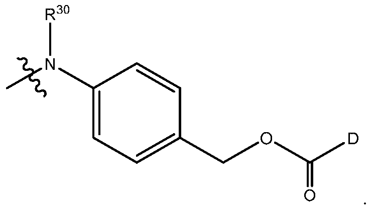
When n is 0, as described elsewhere herein, the bond between the hindered linker carbonyl carbon atom and the drug moiety may suitably be an ether or may be a tertiary amine formed from an acyclic or cyclic amine drug atom. When n is 1, the linkage of the spacer carbonyl carbon atom to the drug moiety, as described elsewhere herein, may suitably be a secondary amine formed from an aliphatic (acyclic) primary amine drug atom, may suitably be a tertiary amine formed from an aliphatic secondary amine drug atom, or may suitably be a tertiary amine formed from a cyclic amine drug atom.
In some aspects, activated hindered linking groups suitable for conjugation to a drug may be suitably prepared according to the following reaction scheme (1)

wherein X, R1 and R2 are as described elsewhere herein and X is bound to the linker by a disulfide bond.
In the first step, a stoichiometric excess of sulfuryl chloride is added to a stirred suspension or solution of a leaving group, X, in a dry solvent. The reaction product may optionally be isolated, such as for instance by solvent evaporation. The leaving group reaction product is combined with a linker precursor solution in a dry solvent and reacted with stirring to form a reaction product mixture comprising the activated linker intermediate. Suitable solvents and reaction conditions are derivable by those skilled in the art. An example of a solvent is dichloromethane (“DCM”), the leaving group concentration may be from about 0.1 to about 1 mol/L, the linker group concentration may be from about 0.1 to about 1 mol/L, the temperature for each reaction may be from about -10°C to about 30°C, and each reaction may be done under an inert atmosphere. In some aspects, each reaction is initiated at from about -10°C to about
5°C and then warmed to room temperature and held for a period of time sufficient to complete the reaction. Reaction completion may be determined by analytical methods known to those skilled in the art, such as LC/MS. The activated linker intermediate may be isolated from the reaction product mixture by methods known in the art. For instance, precipitate in the reaction product mixture may be removed by solid-liquid methods known to those skilled in the art, such as filtration or centrifugation. The activated linker intermediate may then be isolated from the filtrate by solvent evaporation. The isolated activated linker intermediate may optionally be purified. For instance, the isolated activated linker intermediate may be: (1) treated with basified water; (2) extracted into a solvent (e.g., DCM); (3) washed with water and/or brine; (4) dried (e.g., with MgSO4); (5) filtered and isolated as a solid by solvent removal (e.g., evaporation); and, optionally, (6) purified by a chromatographic method known the art, such as reverse phase high pressure liquid chromatography, ion exchange chromatography or flash chromatography.
In the second step, an equivalent excess of triphosgene (or diphosgene or phosgene) is added to a stirred solution of the activated linker intermediate in dry solvent (e.g., DCM) and reacted under an inert atmosphere until completion to form the activated linker. The activated linker intermediate concentration may be from about 0.05 to about 0.5 mol/L. The activated linker may be isolated as a solid by solvent removal or may be directly conjugated with a drug as described elsewhere herein.
CARRIERS
The carriers of the present disclosure are any cell-targeting biologic compound comprising at least one cysteine sulfhydryl moiety. Cell-targeting compounds include antibodies, peptides and polypeptides, hormones, growth factors and proteins.
Certain types of cells, such as cancer cells, express surface molecules (antigens) that are unique as compared to surrounding tissue. Cell targeting moieties that bind to these surface molecules enable the targeted delivery of a drug described elsewhere herein specifically to the target cells. For instance and without limitation, a cell targeting moiety may bind to and be internalized by a lung, breast, brain, prostate, spleen, pancreatic, cervical, ovarian, head and neck, esophageal, liver, skin, kidney, leukemia, bone, testicular, colon or bladder cell.
Tumor Associated Antigens
In some particular aspects of the disclosure, the target cells are cancer cells that express tumor-associated antigens (TAA). Tumor-associated antigens are known in the art, and can be
prepared for use in generating antibodies using methods and information which are well known in the art. In attempts to discover effective cellular targets for cancer diagnosis and therapy, researchers have sought to identify transmembrane or otherwise tumor-associated polypeptides that are specifically expressed on the surface of one or more particular type(s) of cancer cell as compared to on one or more normal non-cancerous cell(s). Often, such tumor-associated polypeptides are more abundantly expressed on the surface of the cancer cells as compared to on the surface of the non-cancerous cells. The identification of such tumor-associated cell surface antigen polypeptides has given rise to the ability to specifically target cancer cells for destruction via antibody-based therapies.
Examples of tumor-associated antigens TAA include, but are not limited to, TAA (1)- (53) listed herein. For convenience, information relating to these antigens, all of which are known in the art, is listed herein and includes names, alternative names, Genbank accession numbers and primary reference(s), following nucleic acid and protein sequence identification conventions of the National Center for Biotechnology Information (NCBI). Nucleic acid and protein sequences corresponding to TAA (1)-(53) are available in public databases such as GenBank. Tumor-associated antigens targeted by antibodies include all amino acid sequence variants and isoforms possessing at least about 70%, 80%, 85%, 90%, or 95% sequence identity relative to the sequences identified in the cited references, or which exhibit substantially the same biological properties or characteristics as a TAA having a sequence found in the cited references. For example, a TAA having a variant sequence generally is able to bind specifically to an antibody that binds specifically to the TAA with the corresponding sequence listed. The sequences and disclosure in the reference specifically recited herein are expressly incorporated by reference.
(1) BMPR1B (bone morphogenetic protein receptor-type IB, Genbank accession no. NM_001203) ten Dijke, P., et al. Science 264 (5155):101-104 (1994), Oncogene 14 (11):1377- 1382 (1997)); WO2004063362 (Claim 2); WO2003042661 (Claim 12); US2003134790-A1 (Page 38-39); WO2002102235 (Claim 13; Page 296); WO2003055443 (Page 91-92); WO200299122 (Example 2; Page 528-530); WO2003029421 (Claim 6); WO2003024392 (Claim 2; Fig 112); WO200298358 (Claim 1; Page 183); WO200254940 (Page 100-101); WO200259377(Page 349-350); WO200230268 (Claim 27; Page 376); WO200148204 (Example; Fig 4) NP_001194 bone morphogenetic protein receptor, type IB /pid=NP_001194.1 - Cross-references: MIM:603248; NP_001194.1; AY065994.
(2) E16 (LAT1, SLC7A5, Genbank accession no. NM_003486) Biochem. Biophys. Res. Commun.255 (2), 283-288 (1999), Nature 395 (6699):288-291 (1998), Gaugitsch, H.W., et al.
(1992) J. Biol. Chem. 267 (16):11267-11273); WO2004048938 (Example 2); WO2004032842 (Example IV); WO2003042661 (Claim 12); WO2003016475 (Claim 1); WO200278524 (Example 2); WO200299074 (Claim 19; Page 127-129); WO200286443 (Claim 27; Pages 222, 393); WO2003003906 (Claim 10; Page 293); WO200264798 (Claim 33; Page 93-95); WO200014228 (Claim 5; Page 133-136); US2003224454 (Fig 3); WO2003025138 (Claim 12; Page 150); NP_003477 solute carrier family 7 (cationic amino acid transporter, y+ system), member 5 /pid=NP_003477.3 - Homo sapiens Cross-references: MIM:600182; NP_003477.3; NM_015923; NM_003486_1.
(3) STEAP1 (six transmembrane epithelial antigen of prostate, Genbank accession no. NM_012449) Cancer Res. 61 (15), 5857-5860 (2001), Hubert, R.S., et al. (1999) Proc. Natl. Acad. Sci. U.S.A.96 (25):14523-14528); WO2004065577 (Claim 6); WO2004027049 (Fig 1L); EP1394274 (Example 11); WO2004016225 (Claim 2); WO2003042661 (Claim 12); US2003157089 (Example 5); US2003185830 (Example 5); US2003064397 (Fig 2); WO200289747 (Example 5; Page 618-619); WO2003022995 (Example 9; Fig 13A, Example 53; Page 173, Example 2; Fig 2A); NP_036581 six transmembrane epithelial antigen of the prostate Cross-references: MIM:604415; NP_036581.1; NM_012449_1.
(4) 0772P (CA125, MUC16, Genbank accession no. AF361486) J. Biol. Chem. 276 (29):27371-27375 (2001)); WO2004045553 (Claim 14); WO200292836 (Claim 6; Fig 12); WO200283866 (Claim 15; Page 116-121); US2003124140 (Example 16); US 798959. Cross- references: GI:34501467; AAK74120.3; AF361486_1.
(5) MPF (MPF, MSLN, SMR, megakaryocyte potentiating factor, mesothelin, Genbank accession no. NM_005823) Yamaguchi, N., et al. Biol. Chem. 269 (2), 805-808 (1994), Proc. Natl. Acad. Sci. U.S.A.96 (20):11531-11536 (1999), Proc. Natl. Acad. Sci. U.S.A.93 (1):136- 140 (1996), J. Biol. Chem. 270 (37):21984-21990 (1995)); WO2003101283 (Claim 14); (WO2002102235 (Claim 13; Page 287-288); WO2002101075 (Claim 4; Page 308-309); WO200271928 (Page 320-321); WO9410312 (Page 52-57); Cross-references: MIM:601051; NP_005814.2; NM_005823_1.
(6) Napi3b (NAPI-3B, NPTIIb, SLC34A2, solute carrier family 34 (sodium phosphate), member 2, type II sodium-dependent phosphate transporter 3b,Genbank accession no. NM_006424) J. Biol. Chem. 277 (22):19665-19672 (2002), Genomics 62 (2):281-284 (1999), Feild, J.A., et al. (1999) Biochem. Biophys. Res. Commun. 258 (3):578-582); WO2004022778 (Claim 2); EP1394274 (Example 11); WO2002102235 (Claim 13; Page 326); EP875569 (Claim 1; Page 17-19); WO200157188 (Claim 20; Page 329); WO2004032842 (Example IV);
WO200175177 (Claim 24; Page 139-140); Cross-references: MIM:604217; NP_006415.1; NM_006424_1.
(7) Sema 5b (FLJ10372, KIAA1445, Mm.42015, SEMA5B, SEMAG, Semaphorin 5b Hlog, sema domain, seven thrombospondin repeats (type 1 and type 1-like), transmembrane domain (TM) and short cytoplasmic domain, (semaphorin) 5B, Genbank accession no. AB040878) Nagase T., et al. (2000) DNA Res. 7 (2):143-150); WO2004000997 (Claim 1); WO2003003984 (Claim 1); WO200206339 (Claim 1; Page 50); WO200188133 (Claim 1; Page 41-43, 48-58); WO2003054152 (Claim 20); WO2003101400 (Claim 11); Accession: Q9P283; EMBL; AB040878; BAA95969.1. Genew; HGNC:10737.
(8) PSCA hlg (2700050C12Rik, C530008O16Rik, RIKEN cDNA 2700050C12, RIKEN cDNA 2700050C12 gene, Genbank accession no. AY358628); Ross et al. (2002) Cancer Res. 62:2546-2553; US2003129192 (Claim 2); US2004044180 (Claim 12); US2004044179 (Claim 11); US2003096961 (Claim 11); US2003232056 (Example 5); WO2003105758 (Claim 12); US2003206918 (Example 5); EP1347046 (Claim 1); WO2003025148 (Claim 20); Cross- references: GI:37182378; AAQ88991.1; AY358628_1.
(9) ETBR (Endothelin type B receptor, Genbank accession no. AY275463); Nakamuta M., et al. Biochem. Biophys. Res. Commun. 177, 34-39, 1991; Ogawa Y., et al. Biochem. Biophys. Res. Commun. 178, 248-255, 1991; Arai H., et al. Jpn. Circ. J. 56, 1303-1307, 1992; Arai H., et al. J. Biol. Chem. 268, 3463-3470, 1993; Sakamoto A., Yanagisawa M., et al. Biochem. Biophys. Res. Commun.178, 656-663, 1991; Elshourbagy N.A., et al. J. Biol. Chem. 268, 3873-3879, 1993; Haendler B., et al. J. Cardiovasc. Pharmacol. 20, s1-S4, 1992; Tsutsumi M., et al. Gene 228, 43-49, 1999; Strausberg R.L., et al. Proc. Natl. Acad. Sci. U.S.A. 99, 16899-16903, 2002; Bourgeois C., et al. J. Clin. Endocrinol. Metab. 82, 3116-3123, 1997; Okamoto Y., et al. Biol. Chem.272, 21589-21596, 1997; Verheij J.B., et al. Am. J. Med. Genet. 108, 223-225, 2002; Hofstra R.M.W., et al. Eur. J. Hum. Genet.5, 180-185, 1997; Puffenberger E.G., et al. Cell 79, 1257-1266, 1994; Attie T., et al., Hum. Mol. Genet. 4, 2407-2409, 1995; Auricchio A., et al. Hum. Mol. Genet. 5:351-354, 1996; Amiel J., et al. Hum. Mol. Genet. 5, 355-357, 1996; Hofstra R.M.W., et al. Nat. Genet.12, 445-447, 1996; Svensson P.J., et al. Hum. Genet. 103, 145-148, 1998; Fuchs S., et al. Mol. Med. 7, 115-124, 2001; Pingault V., et al. (2002) Hum. Genet. 111, 198-206; WO2004045516 (Claim 1); WO2004048938 (Example 2); WO2004040000 (Claim 151); WO2003087768 (Claim 1); WO2003016475 (Claim 1); WO2003016475 (Claim 1); WO200261087 (Fig 1); WO2003016494 (Fig 6); WO2003025138 (Claim 12; Page 144); WO200198351 (Claim 1; Page 124-125); EP522868 (Claim 8; Fig 2);
WO200177172 (Claim 1; Page 297-299); US2003109676; US6518404 (Fig 3); US5773223 (Claim 1a; Col 31-34); WO2004001004.
(10) MSG783 (RNF124, hypothetical protein FLJ20315, Genbank accession no. NM_017763); WO2003104275 (Claim 1); WO2004046342 (Example 2); WO2003042661 (Claim 12); WO2003083074 (Claim 14; Page 61); WO2003018621 (Claim 1); WO2003024392 (Claim 2; Fig 93); WO200166689 (Example 6); Cross-references: LocusID:54894; NP_060233.2; NM_017763_1.
(11) STEAP2 (HGNC_8639, IPCA-1, PCANAP1, STAMP1, STEAP2, STMP, prostate cancer associated gene 1, prostate cancer associated protein 1, six transmembrane epithelial antigen of prostate 2, six transmembrane prostate protein, Genbank accession no. AF455138) Lab. Invest. 82 (11):1573-1582 (2002)); WO2003087306; US2003064397 (Claim 1; Fig 1); WO200272596 (Claim 13; Page 54-55); WO200172962 (Claim 1; Fig 4B); WO2003104270 (Claim 11); WO2003104270 (Claim 16); US2004005598 (Claim 22); WO2003042661 (Claim 12); US2003060612 (Claim 12; Fig 10); WO200226822 (Claim 23; Fig 2); WO200216429 (Claim 12; Fig 10); Cross-references: GI:22655488; AAN04080.1; AF455138_1.
(12) TrpM4 (BR22450, FLJ20041, TRPM4, TRPM4B, transient receptor potential cation channel, subfamily M, member 4, Genbank accession no. NM_017636) Xu, X.Z., et al. Proc. Natl. Acad. Sci. U.S.A. 98 (19):10692-10697 (2001), Cell 109 (3):397-407 (2002), J. Biol. Chem. 278 (33):30813-30820 (2003)); US2003143557 (Claim 4); WO200040614 (Claim 14; Page 100-103); WO200210382 (Claim 1; Fig 9A); WO2003042661 (Claim 12); WO200230268 (Claim 27; Page 391); US2003219806 (Claim 4); WO200162794 (Claim 14; Fig 1A-D); Cross- references: MIM:606936; NP_060106.2; NM_017636_1.
(13) CRIPTO (CR, CR1, CRGF, CRIPTO, TDGF1, teratocarcinoma-derived growth factor, Genbank accession no. NP_003203 or NM_003212) Ciccodicola, A., et al. EMBO J. 8 (7):1987-1991 (1989), Am. J. Hum. Genet. 49 (3):555-565 (1991)); US2003224411 (Claim 1); WO2003083041 (Example 1); WO2003034984 (Claim 12); WO200288170 (Claim 2; Page 52- 53); WO2003024392 (Claim 2; Fig 58); WO200216413 (Claim 1; Page 94-95, 105); WO200222808 (Claim 2; Fig 1); US5854399 (Example 2; Col 17-18); US5792616 (Fig 2); Cross-references: MIM:187395; NP_003203.1; NM_003212_1.
(14) CD21 (CR2 (Complement receptor 2) or C3DR (C3d/Epstein Barr virus receptor) or Hs.73792 Genbank accession no. M26004) Fujisaku et al. (1989) J. Biol. Chem. 264 (4):2118- 2125); Weis J.J., et al. J. Exp. Med. 167, 1047-1066, 1988; Moore M., et al. Proc. Natl. Acad. Sci. U.S.A. 84, 9194-9198, 1987; Barel M., et al. Mol. Immunol. 35, 1025-1031, 1998; Weis J.J., et al. Proc. Natl. Acad. Sci. U.S.A. 83, 5639-5643, 1986; Sinha S.K., et al. (1993) J.
Immunol. 150, 5311-5320; WO2004045520 (Example 4); US2004005538 (Example 1); WO2003062401 (Claim 9); WO2004045520 (Example 4); WO9102536 (Fig 9.1-9.9); WO2004020595 (Claim 1); Accession: P20023; Q13866; Q14212; EMBL; M26004; AAA35786.1.
(15) CD79b (CD79B, CD79β, IGb (immunoglobulin-associated beta), B29, Genbank accession no. NM_000626 or 11038674) Proc. Natl. Acad. Sci. U.S.A. (2003) 100 (7):4126- 4131, Blood (2002) 100 (9):3068-3076, Muller et al. (1992) Eur. J. Immunol. 22 (6):1621- 1625); WO2004016225 (claim 2, Fig 140); WO2003087768, US2004101874 (claim 1, page 102); WO2003062401 (claim 9); WO200278524 (Example 2); US2002150573 (claim 5, page 15); US5644033; WO2003048202 (claim 1, pages 306 and 309); WO 99/558658, US6534482 (claim 13, Fig 17A/B); WO200055351 (claim 11, pages 1145-1146); Cross-references: MIM:147245; NP_000617.1; NM_000626_1.
(16) FcRH2 (IFGP4, IRTA4, SPAP1A (SH2 domain containing phosphatase anchor protein 1a), SPAP1B, SPAP1C, Genbank accession no. NM_030764, AY358130) Genome Res. 13 (10):2265-2270 (2003), Immunogenetics 54 (2):87-95 (2002), Blood 99 (8):2662-2669 (2002), Proc. Natl. Acad. Sci. U.S.A. 98 (17):9772-9777 (2001), Xu, M.J., et al. (2001) Biochem. Biophys. Res. Commun. 280 (3):768-775; WO2004016225 (Claim 2); WO2003077836; WO200138490 (Claim 5; Fig 18D-1-18D-2); WO2003097803 (Claim 12); WO2003089624 (Claim 25); Cross-references: MIM:606509; NP_110391.2; NM_030764_1.
(17) HER2 (ErbB2, Genbank accession no. M11730) Coussens L., et al. Science (1985) 230(4730):1132-1139); Yamamoto T., et al. Nature 319, 230-234, 1986; Semba K., et al. Proc. Natl. Acad. Sci. U.S.A. 82, 6497-6501, 1985; Swiercz J.M., et al. J. Cell Biol. 165, 869-880, 2004; Kuhns J.J., et al. J. Biol. Chem. 274, 36422-36427, 1999; Cho H.-S., et al. Nature 421, 756-760, 2003; Ehsani A., et al. (1993) Genomics 15, 426-429; WO2004048938 (Example 2); WO2004027049 (Fig 1I); WO2004009622; WO2003081210; WO2003089904 (Claim 9); WO2003016475 (Claim 1); US2003118592; WO2003008537 (Claim 1); WO2003055439 (Claim 29; Fig 1A-B); WO2003025228 (Claim 37; Fig 5C); WO200222636 (Example 13; Page 95-107); WO200212341 (Claim 68; Fig 7); WO200213847 (Page 71-74); WO200214503 (Page 114-117); WO200153463 (Claim 2; Page 41-46); WO200141787 (Page 15); WO200044899 (Claim 52; Fig 7); WO200020579 (Claim 3; Fig 2); US5869445 (Claim 3; Col 31-38); WO9630514 (Claim 2; Page 56-61); EP1439393 (Claim 7); WO2004043361 (Claim 7); WO2004022709; WO200100244 (Example 3; Fig 4); Accession: P04626; EMBL; M11767; AAA35808.1. EMBL; M11761; AAA35808.1.
(18) NCA (CEACAM6, Genbank accession no. M18728); Barnett T., et al. Genomics 3, 59-66, 1988; Tawaragi Y., et al. Biochem. Biophys. Res. Commun. 150, 89-96, 1988; Strausberg R.L., et al. Proc. Natl. Acad. Sci. U.S.A. 99:16899-16903, 2002; WO2004063709; EP1439393 (Claim 7); WO2004044178 (Example 4); WO2004031238; WO2003042661 (Claim 12); WO200278524 (Example 2); WO200286443 (Claim 27; Page 427); WO200260317 (Claim 2); Accession: P40199; Q14920; EMBL; M29541; AAA59915.1. EMBL; M18728.
(19) MDP (DPEP1, Genbank accession no. BC017023) Proc. Natl. Acad. Sci. U.S.A.99 (26):16899-16903 (2002)); WO2003016475 (Claim 1); WO200264798 (Claim 33; Page 85-87); JP05003790 (Fig 6-8); WO9946284 (Fig 9); Cross-references: MIM:179780; AAH17023.1; BC017023_1.
(20) IL20Rα (IL20Ra, ZCYTOR7, Genbank accession no. AF184971); Clark H.F., et al. Genome Res. 13, 2265-2270, 2003; Mungall A.J., et al. Nature 425, 805-811, 2003; Blumberg H., et al. Cell 104, 9-19, 2001; Dumoutier L., et al. J. Immunol.167, 3545-3549, 2001; Parrish- Novak J., et al. J. Biol. Chem. 277, 47517-47523, 2002; Pletnev S., et al. (2003) Biochemistry 42:12617-12624; Sheikh F., et al. (2004) J. Immunol. 172, 2006-2010; EP1394274 (Example 11); US2004005320 (Example 5); WO2003029262 (Page 74-75); WO2003002717 (Claim 2; Page 63); WO200222153 (Page 45-47); US2002042366 (Page 20-21); WO200146261 (Page 57- 59); WO200146232 (Page 63-65); WO9837193 (Claim 1; Page 55-59); Accession: Q9UHF4; Q6UWA9; Q96SH8; EMBL; AF184971; AAF01320.1.
(21) Brevican (BCAN, BEHAB, Genbank accession no. AF229053) Gary S.C., et al. Gene 256, 139-147, 2000; Clark H.F., et al. Genome Res.13, 2265-2270, 2003; Strausberg R.L., et al. Proc. Natl. Acad. Sci. U.S.A. 99, 16899-16903, 2002; US2003186372 (Claim 11); US2003186373 (Claim 11); US2003119131 (Claim 1; Fig 52); US2003119122 (Claim 1; Fig 52); US2003119126 (Claim 1); US2003119121 (Claim 1; Fig 52); US2003119129 (Claim 1); US2003119130 (Claim 1); US2003119128 (Claim 1; Fig 52); US2003119125 (Claim 1); WO2003016475 (Claim 1); WO200202634 (Claim 1).
(22) EphB2R (DRT, ERK, Hek5, EPHT3, Tyro5, Genbank accession no. NM_004442) Chan, J. and Watt, V.M., Oncogene 6 (6), 1057-1061 (1991) Oncogene 10 (5):897-905 (1995), Annu. Rev. Neurosci. 21:309-345 (1998), Int. Rev. Cytol. 196:177-244 (2000)); WO2003042661 (Claim 12); WO200053216 (Claim 1; Page 41); WO2004065576 (Claim 1); WO2004020583 (Claim 9); WO2003004529 (Page 128-132); WO200053216 (Claim 1; Page 42); Cross-references: MIM:600997; NP_004433.2; NM_004442_1.
(23) ASLG659 (B7h, Genbank accession no. AX092328) US20040101899 (Claim 2); WO2003104399 (Claim 11); WO2004000221 (Fig 3); US2003165504 (Claim 1);
US2003124140 (Example 2); US2003065143 (Fig 60); WO2002102235 (Claim 13; Page 299); US2003091580 (Example 2); WO200210187 (Claim 6; Fig 10); WO200194641 (Claim 12; Fig 7b); WO200202624 (Claim 13; Fig 1A-1B); US2002034749 (Claim 54; Page 45-46); WO200206317 (Example 2; Page 320-321, Claim 34; Page 321-322); WO200271928 (Page 468-469); WO200202587 (Example 1; Fig 1); WO200140269 (Example 3; Pages 190-192); WO200036107 (Example 2; Page 205-207); WO2004053079 (Claim 12); WO2003004989 (Claim 1); WO200271928 (Page 233-234, 452-453); WO 0116318.
(24) PSCA (Prostate stem cell antigen precursor, Genbank accession no. AJ297436) Reiter R.E., et al. Proc. Natl. Acad. Sci. U.S.A.95, 1735-1740, 1998; Gu Z., et al. Oncogene 19, 1288-1296, 2000; Biochem. Biophys. Res. Commun. (2000) 275(3):783-788; WO2004022709; EP1394274 (Example 11); US2004018553 (Claim 17); WO2003008537 (Claim 1); WO200281646 (Claim 1; Page 164); WO2003003906 (Claim 10; Page 288); WO200140309 (Example 1; Fig 17); US2001055751 (Example 1; Fig 1b); WO200032752 (Claim 18; Fig 1); WO9851805 (Claim 17; Page 97); WO9851824 (Claim 10; Page 94); WO9840403 (Claim 2; Fig 1B); Accession: O43653; EMBL; AF043498; AAC39607.1.
(25) GEDA (Genbank accession No. AY260763); AAP14954 lipoma HMGIC fusion- partner-like protein /pid=AAP14954.1 - Homo sapiens Species: Homo sapiens (human) WO2003054152 (Claim 20); WO2003000842 (Claim 1); WO2003023013 (Example 3, Claim 20); US2003194704 (Claim 45); Cross-references: GI:30102449; AAP14954.1; AY260763_1.
(26) BAFF-R (B cell -activating factor receptor, BLyS receptor 3, BR3, Genbank accession No. AF116456); BAFF receptor /pid=NP_443177.1 - Homo sapiens Thompson, J.S., et al. Science 293 (5537), 2108-2111 (2001); WO2004058309; WO2004011611; WO2003045422 (Example; Page 32-33); WO2003014294 (Claim 35; Fig 6B); WO2003035846 (Claim 70; Page 615-616); WO200294852 (Col 136-137); WO200238766 (Claim 3; Page 133); WO200224909 (Example 3; Fig 3); Cross-references: MIM:606269; NP_443177.1; NM_052945_1; AF132600.
(27) CD22 (B-cell receptor CD22-B isoform, BL-CAM, Lyb-8, Lyb8, SIGLEC-2, FLJ22814, Genbank accession No. AK026467); Wilson et al. (1991) J. Exp. Med.173:137-146; WO2003072036 (Claim 1; Fig 1); Cross-references: MIM:107266; NP_001762.1; NM_001771_1.
(28) CD79a (CD79A, CD79α, immunoglobulin-associated alpha, a B cell-specific protein that covalently interacts with Ig beta (CD79B) and forms a complex on the surface with Ig M molecules, transduces a signal involved in B-cell differentiation), pI: 4.84, MW: 25028 TM: 2 [P] Gene Chromosome: 19q13.2, Genbank accession No. NP_001774.10)
WO2003088808, US20030228319; WO2003062401 (claim 9); US2002150573 (claim 4, pages 13-14); WO9958658 (claim 13, Fig 16); WO9207574 (Fig 1); US5644033; Ha et al. (1992) J. Immunol. 148(5):1526-1531; Mueller et al. (1992) Eur. J. Biochem. 22:1621-1625; Hashimoto et al. (1994) Immunogenetics 40(4):287-295; Preud’homme et al. (1992) Clin. Exp. Immunol. 90(1):141-146; Yu et al. (1992) J. Immunol. 148(2) 633-637; Sakaguchi et al. (1988) EMBO J. 7(11):3457-3464.
(29) CXCR5 (Burkitt's lymphoma receptor 1, a G protein-coupled receptor that is activated by the CXCL13 chemokine, functions in lymphocyte migration and humoral defense, plays a role in HIV-2 infection and perhaps development of AIDS, lymphoma, myeloma, and leukemia); 372 aa, pI: 8.54 MW: 41959 TM: 7 [P] Gene Chromosome: 11q23.3, Genbank accession No. NP_001707.1) WO2004040000; WO2004015426; US2003105292 (Example 2); US6555339 (Example 2); WO200261087 (Fig 1); WO200157188 (Claim 20, page 269); WO200172830 (pages 12-13); WO200022129 (Example 1, pages 152-153, Example 2, pages 254-256); WO9928468 (claim 1, page 38); US5440021 (Example 2, col 49-52); WO9428931 (pages 56-58); WO9217497 (claim 7, Fig 5); Dobner et al. (1992) Eur. J. Immunol. 22:2795- 2799; Barella et al. (1995) Biochem. J.309:773-779.
(30) HLA-DOB (Beta subunit of MHC class II molecule (Ia antigen) that binds peptides and presents them to CD4+ T lymphocytes); 273 aa, pI: 6.56 MW: 30820 TM: 1 [P] Gene Chromosome: 6p21.3, Genbank accession No. NP_002111.1) Tonnelle et al. (1985) EMBO J. 4(11):2839-2847; Jonsson et al. (1989) Immunogenetics 29(6):411-413; Beck et al. (1992) J. Mol. Biol. 228:433-441; Strausberg et al. (2002) Proc. Natl. Acad. Sci USA 99:16899-16903; Servenius et al. (1987) J. Biol. Chem.262:8759-8766; Beck et al. (1996) J. Mol. Biol.255:1-13; Naruse et al. (2002) Tissue Antigens 59:512-519; WO9958658 (claim 13, Fig 15); US6153408 (Col 35-38); US5976551 (col 168-170); US6011146 (col 145-146); Kasahara et al. (1989) Immunogenetics 30(1):66-68; Larhammar et al. (1985) J. Biol. Chem.260(26):14111-14119.
(31) P2X5 (Purinergic receptor P2X ligand-gated ion channel 5, an ion channel gated by extracellular ATP, may be involved in synaptic transmission and neurogenesis, deficiency may contribute to the pathophysiology of idiopathic detrusor instability); 422 aa), pI: 7.63, MW: 47206 TM: 1 [P] Gene Chromosome: 17p13.3, Genbank accession No. NP_002552.2) Le et al. (1997) FEBS Lett.418(1-2):195-199; WO2004047749; WO2003072035 (claim 10); Touchman et al. (2000) Genome Res. 10:165-173; WO200222660 (claim 20); WO2003093444 (claim 1); WO2003087768 (claim 1); WO2003029277 (page 82).
(32) CD72 (B-cell differentiation antigen CD72, Lyb-2) PROTEIN SEQUENCE Full maeaity...tafrfpd (1..359; 359 aa), pI: 8.66, MW: 40225 TM: 1 [P] Gene Chromosome: 9p13.3,
Genbank accession No. NP_001773.1) WO2004042346 (claim 65); WO2003026493 (pages 51- 52, 57-58); WO200075655 (pages 105-106); Von Hoegen et al. (1990) J. Immunol. 144(12):4870-4877; Strausberg et al. (2002) Proc. Natl. Acad. Sci USA 99:16899-16903.
(33) LY64 (Lymphocyte antigen 64 (RP105), type I membrane protein of the leucine rich repeat (LRR) family, regulates B-cell activation and apoptosis, loss of function is associated with increased disease activity in patients with systemic lupus erythematosis); 661 aa, pI: 6.20, MW: 74147 TM: 1 [P] Gene Chromosome: 5q12, Genbank accession No. NP_005573.1) US2002193567; WO9707198 (claim 11, pages 39-42); Miura et al. (1996) Genomics 38(3):299- 304; Miura et al. (1998) Blood 92:2815-2822; WO2003083047; WO9744452 (claim 8, pages 57-61); WO200012130 (pages 24-26).
(34) FcRH1 (Fc receptor-like protein 1, a putative receptor for the immunoglobulin Fc domain that contains C2 type Ig-like and ITAM domains, may have a role in B-lymphocyte differentiation); 429 aa, pI: 5.28, MW: 46925 TM: 1 [P] Gene Chromosome: 1q21-1q22, Genbank accession No. NP_443170.1) WO2003077836; WO200138490 (claim 6, Fig 18E-1- 18-E-2); Davis et al. (2001) Proc. Natl. Acad. Sci USA 98(17):9772-9777; WO2003089624 (claim 8); EP1347046 (claim 1); WO2003089624 (claim 7).
(35) IRTA2 (Immunoglobulin superfamily receptor translocation associated 2, a putative immunoreceptor with possible roles in B cell development and lymphomagenesis; deregulation of the gene by translocation occurs in some B cell malignancies); 977 aa, pI: 6.88 MW: 106468 TM: 1 [P] Gene Chromosome: 1q21, Genbank accession No. Human:AF343662, AF343663, AF343664, AF343665, AF369794, AF397453, AK090423, AK090475, AL834187, AY358085; Mouse:AK089756, AY158090, AY506558; NP_112571.1. WO2003024392 (claim 2, Fig 97); Nakayama et al. (2000) Biochem. Biophys. Res. Commun. 277(1):124-127; WO2003077836; WO200138490 (claim 3, Fig 18B-1-18B-2).
(36) TENB2 (TMEFF2, tomoregulin, TPEF, HPP1, TR, putative transmembrane proteoglycan, related to the EGF/heregulin family of growth factors and follistatin); 374 aa, NCBI Accession: AAD55776, AAF91397, AAG49451, NCBI RefSeq: NP_057276; NCBI Gene: 23671; OMIM: 605734; SwissProt Q9UIK5; Genbank accession No. AF179274; AY358907, CAF85723, CQ782436 WO2004074320 (SEQ ID NO 810); JP2004113151 (SEQ ID NOS 2, 4, 8); WO2003042661 (SEQ ID NO 580); WO2003009814 (SEQ ID NO 411); EP1295944 (pages 69-70); WO200230268 (page 329); WO200190304 (SEQ ID NO 2706); US2004249130; US2004022727; WO2004063355; US2004197325; US2003232350; US2004005563; US2003124579; Horie et al. (2000) Genomics 67:146-152; Uchida et al. (1999)
Biochem. Biophys. Res. Commun. 266:593-602; Liang et al. (2000) Cancer Res. 60:4907-12; Glynne-Jones et al. (2001) Int J Cancer. Oct 15;94(2):178-84.
(37) PMEL17 (silver homolog; SILV; D12S53E; PMEL17; SI; SIL); ME20; gp100) BC001414; BT007202; M32295; M77348; NM_006928; McGlinchey, R.P. et al. (2009) Proc. Natl. Acad. Sci. U.S.A.106 (33), 13731-13736; Kummer, M.P. et al. (2009) J. Biol. Chem.284 (4), 2296-2306.
(38) TMEFF1 (transmembrane protein with EGF-like and two follistatin-like domains 1; Tomoregulin-1); H7365; C9orf2; C9ORF2; U19878; X83961; NM_080655; NM_003692; Harms, P.W. (2003) Genes Dev. 17 (21), 2624-2629; Gery, S. et al. (2003) Oncogene 22 (18):2723-2727.
(39) GDNF-Ra1 (GDNF family receptor alpha 1; GFRA1; GDNFR; GDNFRA; RETL1; TRNR1; RET1L; GDNFR-alpha1; GFR-ALPHA-1); U95847; BC014962; NM_145793 NM_005264; Kim, M.H. et al. (2009) Mol. Cell. Biol. 29 (8), 2264-2277; Treanor, J.J. et al. (1996) Nature 382 (6586):80-83.
(40) Ly6E (lymphocyte antigen 6 complex, locus E; Ly67,RIG-E,SCA-2,TSA-1); NP_002337.1; NM_002346.2; de Nooij-van Dalen, A.G. et al. (2003) Int. J. Cancer 103 (6), 768-774; Zammit, D.J. et al. (2002) Mol. Cell. Biol.22 (3):946-952.
(41) TMEM46 (shisa homolog 2 (Xenopus laevis); SHISA2); NP_001007539.1; NM_001007538.1; Furushima, K. et al. (2007) Dev. Biol. 306 (2), 480-492; Clark, H.F. et al. (2003) Genome Res.13 (10):2265-2270.
(42) Ly6G6D (lymphocyte antigen 6 complex, locus G6D; Ly6-D, MEGT1); NP_067079.2; NM_021246.2; Mallya, M. et al. (2002) Genomics 80 (1):113-123; Ribas, G. et al. (1999) J. Immunol.163 (1):278-287.
(43) LGR5 (leucine-rich repeat-containing G protein-coupled receptor 5; GPR49, GPR67); NP_003658.1; NM_003667.2; Salanti, G. et al. (2009) Am. J. Epidemiol.170 (5):537- 545; Yamamoto, Y. et al. (2003) Hepatology 37 (3):528-533.
(44) RET (ret proto-oncogene; MEN2A; HSCR1; MEN2B; MTC1; PTC; CDHF12; Hs.168114; RET51; RET-ELE1); NP_066124.1; NM_020975.4; Tsukamoto, H. et al. (2009) Cancer Sci.100 (10):1895-1901; Narita, N. et al. (2009) Oncogene 28 (34):3058-3068.
(45) LY6K (lymphocyte antigen 6 complex, locus K; LY6K; HSJ001348; FLJ35226); NP_059997.3; NM_017527.3; Ishikawa, N. et al. (2007) Cancer Res. 67 (24):11601-11611; de Nooij-van Dalen, A.G. et al. (2003) Int. J. Cancer 103 (6):768-774.
(46) GPR19 (G protein-coupled receptor 19; Mm.4787); NP_006134.1; NM_006143.2; Montpetit, A. and Sinnett, D. (1999) Hum. Genet. 105 (1-2):162-164; O'Dowd, B.F. et al. (1996) FEBS Lett.394 (3):325-329.
(47) GPR54 (KISS1 receptor; KISS1R; GPR54; HOT7T175; AXOR12); NP_115940.2; NM_032551.4; Navenot, J.M. et al. (2009) Mol. Pharmacol. 75 (6):1300-1306; Hata, K. et al. (2009) Anticancer Res.29 (2):617-623.
(48) ASPHD1 (aspartate beta-hydroxylase domain containing 1; LOC253982); NP_859069.2; NM_181718.3; Gerhard, D.S. et al. (2004) Genome Res.14 (10B):2121-2127.
(49) Tyrosinase (TYR; OCAIA; OCA1A; tyrosinase; SHEP3); NP_000363.1; NM_000372.4; Bishop, D.T. et al. (2009) Nat. Genet.41 (8):920-925; Nan, H. et al. (2009) Int. J. Cancer 125 (4):909-917.
(50) TMEM118 (ring finger protein, transmembrane 2; RNFT2; FLJ14627); NP_001103373.1; NM_001109903.1; Clark, H.F. et al. (2003) Genome Res.13 (10):2265-2270; Scherer, S.E. et al. (2006) Nature 440 (7082):346-351.
(51) GPR172A (G protein-coupled receptor 172A; GPCR41; FLJ11856; D15Ertd747e); NP_078807.1; NM_024531.3; Ericsson, T.A. et al. (2003) Proc. Natl. Acad. Sci. U.S.A. 100 (11):6759-6764; Takeda, S. et al. (2002) FEBS Lett.520 (1-3):97-101.
(52) CD33, a member of the sialic acid binding, immunoglobulin-like lectin family, is a 67-kDa glycosylated transmembrane protein. CD33is expressed on most myeloid and monocytic leukemia cells in addition to committed myelomonocytic and erythroid progenitor cells. It is not seen on the earliest pluripotent stem cells, mature granulocytes, lymphoid cells, or nonhematopoietic cells (Sabbath et al., (1985) J. Clin. Invest.75:756-56; Andrews et al., (1986) Blood 68:1030-5). CD33 contains two tyrosine residues on its cytoplasmic tail, each of which is followed by hydrophobic residues similar to the immunoreceptor tyrosine-based inhibitory motif (ITIM) seen in many inhibitory receptors.
(53) CLL-1 (CLEC12A, MICL, and DCAL2), encodes a member of the C-type lectin/C- type lectin-like domain (CTL/CTLD) superfamily. Members of this family share a common protein fold and have diverse functions, such as cell adhesion, cell-cell signaling, glycoprotein turnover, and roles in inflammation and immune response. The protein encoded by this gene is a negative regulator of granulocyte and monocyte function. Several alternatively spliced transcript variants of this gene have been described, but the full-length nature of some of these variants has not been determined. This gene is closely linked to other CTL/CTLD superfamily members in the natural killer gene complex region on chromosome 12p13 (Drickamer K (1999) Curr. Opin. Struct. Biol.9 (5):585–90; van Rhenen A, et al., (2007) Blood 110 (7):2659–66; Chen CH, et al.
(2006) Blood 107 (4):1459–67; Marshall AS, et al. (2006) Eur. J. Immunol. 36 (8):2159–69; Bakker AB, et al. (2005) Cancer Res. 64 (22):8443–50; Marshall AS, , et al. (2004) J. Biol. Chem. 279 (15):14792–802). CLL-1 has been shown to be a type II transmembrane receptor comprising a single C-type lectin-like domain (which is not predicted to bind either calcium or sugar), a stalk region, a transmembrane domain and a short cytoplasmic tail containing an ITIM motif.
Antibodies
In any of the antibody embodiments of the disclosure, an antibody is humanized. In one embodiment, an antibody comprises HVRs as in any of the embodiments of the disclosure, and further comprises a human acceptor framework, e.g. a human immunoglobulin framework or a human consensus framework. In certain embodiments, the human acceptor framework is the human VL kappa I consensus (VLKI) framework and/or the VH framework VH1. In certain embodiments, the human acceptor framework is the human VL kappa I consensus (VLKI) framework and/or the VH framework VH1 comprising any one of the following mutations.
In another aspect, the antibody comprises a VH as in any of the embodiments provided herein, and a VL as in any of the embodiments provided herein.
In a further aspect of the disclosure, an antibody according to any of the embodiments herein is a monoclonal antibody, including a human antibody. In one embodiment, an antibody is an antibody fragment, e.g., a Fv, Fab, Fab’, scFv, diabody, or F(ab’)2 fragment. In another embodiment, the antibody is a substantially full length antibody, e.g., an IgG1 antibody, IgG2a antibody or other antibody class or isotype as defined herein.
In a further aspect, an antibody according to any of the embodiments herein may incorporate any of the features, singly or in combination, as described herein.
1. Antibody Affinity
In certain embodiments, an antibody provided herein has a dissociation constant (Kd) of ≤ 1μM,≤ 100 nM,≤ 50 nM,≤ 10 nM,≤ 5 nM,≤ 1 nM,≤ 0.1 nM,≤ 0.01 nM, or≤ 0.001 nM, and optionally is≥ 10-13 M. (e.g.10-8 M or less, e.g. from 10-8 M to 10-13 M, e.g., from 10-9 M to 10-13 M).
In one embodiment, Kd is measured by a radiolabeled antigen binding assay (RIA) performed with the Fab version of an antibody of interest and its antigen as described by the following assay. Solution binding affinity of Fabs for antigen is measured by equilibrating Fab with a minimal concentration of (125I)-labeled antigen in the presence of a titration series of unlabeled antigen, then capturing bound antigen with an anti-Fab antibody-coated plate (see,
e.g., Chen et al., J. Mol. Biol. 293:865-881(1999)). To establish conditions for the assay, MICROTITER® multi-well plates (Thermo Scientific) are coated overnight with 5 μg/ml of a capturing anti-Fab antibody (Cappel Labs) in 50 mM sodium carbonate (pH 9.6), and subsequently blocked with 2% (w/v) bovine serum albumin in PBS for two to five hours at room temperature (approximately 23°C). In a non-adsorbent plate (Nunc #269620), 100 pM or 26 pM [125I]-antigen are mixed with serial dilutions of a Fab of interest (e.g., consistent with assessment of the anti-VEGF antibody, Fab-12, in Presta et al., Cancer Res.57:4593-4599 (1997)). The Fab of interest is then incubated overnight; however, the incubation may continue for a longer period (e.g., about 65 hours) to ensure that equilibrium is reached. Thereafter, the mixtures are transferred to the capture plate for incubation at room temperature (e.g., for one hour). The solution is then removed and the plate washed eight times with 0.1% polysorbate 20 (TWEEN- 20®) in PBS. When the plates have dried, 150 μl/well of scintillant (MICROSCINT-20 TM; Packard) is added, and the plates are counted on a TOPCOUNT TM gamma counter (Packard) for ten minutes. Concentrations of each Fab that give less than or equal to 20% of maximal binding are chosen for use in competitive binding assays.
According to another embodiment, Kd is measured using surface plasmon resonance assays using a BIACORE®-2000, BAICORE®-T200 or a BIACORE ®-3000 (BIAcore, Inc., Piscataway, NJ) at 25°C with immobilized antigen CM5 chips at ~10 response units (RU). Briefly, carboxymethylated dextran biosensor chips (CM5, BIACORE, Inc.) are activated with N-ethyl-N’- (3-dimethylaminopropyl)-carbodiimide hydrochloride (EDC) and N- hydroxysuccinimide (NHS) according to the supplier’s instructions. Antigen is diluted with 10 mM sodium acetate, pH 4.8, to 5 μg/ml (~0.2 μM) and/or HBS-P (0.01 M Hepes pH7.4, 0.15M NaCl, 0.005% Surfactant P20) before injection at a flow rate of 5 μl/minute and/or 30 μl/minute to achieve approximately 10 response units (RU) of coupled protein. Following the injection of antigen, 1 M ethanolamine is injected to block unreacted groups. For kinetics measurements, two-fold serial dilutions of Fab (0.78 nM to 500 nM) are injected in PBS with 0.05% polysorbate 20 (TWEEN-20TM) surfactant (PBST) at 25°C at a flow rate of approximately 25 μl/min. Association rates (k on ) and dissociation rates (k off ) are calculated using a simple one- to-one Langmuir binding model (BIACORE ® Evaluation Software version 3.2) by simultaneously fitting the association and dissociation sensorgrams. The equilibrium dissociation constant (Kd) is calculated as the ratio
 See, e.g., Chen et al., J. Mol. Biol. 293:865-881 (1999). If the on-rate exceeds 10 6 M -1 s -1 by the surface plasmon resonance assay describe herein, then the on-rate can be determined by using a fluorescent quenching technique
that measures the increase or decrease in fluorescence emission intensity (excitation = 295 nm; emission = 340 nm, 16 nm band-pass) at 25 o C of a 20 nM anti-antigen antibody (Fab form) in PBS, pH 7.2, in the presence of increasing concentrations of antigen as measured in a spectrometer, such as a stop-flow equipped spectrophometer (Aviv Instruments) or a 8000-series SLM-AMINCO TM spectrophotometer (ThermoSpectronic) with a stirred cuvette.
See, e.g., Chen et al., J. Mol. Biol. 293:865-881 (1999). If the on-rate exceeds 10 6 M -1 s -1 by the surface plasmon resonance assay describe herein, then the on-rate can be determined by using a fluorescent quenching technique
that measures the increase or decrease in fluorescence emission intensity (excitation = 295 nm; emission = 340 nm, 16 nm band-pass) at 25 o C of a 20 nM anti-antigen antibody (Fab form) in PBS, pH 7.2, in the presence of increasing concentrations of antigen as measured in a spectrometer, such as a stop-flow equipped spectrophometer (Aviv Instruments) or a 8000-series SLM-AMINCO TM spectrophotometer (ThermoSpectronic) with a stirred cuvette.
2. Antibody Fragments
In certain embodiments, an antibody provided herein is an antibody fragment. Antibody fragments include, but are not limited to, Fab, Fab’, Fab’-SH, F(ab’)2, Fv, and scFv fragments, and other fragments described herein. For a review of certain antibody fragments, see Hudson et al. Nat. Med. 9:129-134 (2003). For a review of scFv fragments, see, e.g., Pluckthün, in The Pharmacology of Monoclonal Antibodies, vol. 113, Rosenburg and Moore eds., (Springer- Verlag, New York), pp.269-315 (1994); see also WO 93/16185; and U.S. Patent Nos.5,571,894 and 5,587,458. For discussion of Fab and F(ab')2 fragments comprising salvage receptor binding epitope residues and having increased in vivo half-life, see U.S. Patent No.5,869,046.
Diabodies are antibody fragments with two antigen-binding sites that may be bivalent or bispecific. See, for example, EP 404,097; WO 1993/01161; Hudson et al., Nat. Med.9:129-134 (2003); and Hollinger et al., Proc. Natl. Acad. Sci. USA 90: 6444-6448 (1993). Triabodies and tetrabodies are also described in Hudson et al., Nat. Med.9:129-134 (2003).
Single-domain antibodies are antibody fragments comprising all or a portion of the heavy chain variable domain or all or a portion of the light chain variable domain of an antibody. In certain embodiments, a single-domain antibody is a human single-domain antibody (Domantis, Inc., Waltham, MA; see, e.g., U.S. Patent No.6,248,516 B1).
Antibody fragments can be made by various techniques, including but not limited to proteolytic digestion of an intact antibody as well as production by recombinant host cells (e.g. E. coli or phage), as described herein.
3. Chimeric and Humanized Antibodies
In certain embodiments, an antibody provided herein is a chimeric antibody. Certain chimeric antibodies are described, e.g., in U.S. Patent No.4,816,567; and Morrison et al., Proc. Natl. Acad. Sci. USA, 81:6851-6855 (1984)). In one example, a chimeric antibody comprises a non-human variable region (e.g., a variable region derived from a mouse, rat, hamster, rabbit, or non-human primate, such as a monkey) and a human constant region. In a further example, a chimeric antibody is a“class switched” antibody in which the class or subclass has been
changed from that of the parent antibody. Chimeric antibodies include antigen-binding fragments thereof.
In certain embodiments, a chimeric antibody is a humanized antibody. Typically, a non- human antibody is humanized to reduce immunogenicity to humans, while retaining the specificity and affinity of the parental non-human antibody. Generally, a humanized antibody comprises one or more variable domains in which HVRs, e.g., CDRs, (or portions thereof) are derived from a non-human antibody, and FRs (or portions thereof) are derived from human antibody sequences. A humanized antibody optionally will also comprise at least a portion of a human constant region. In some embodiments, some FR residues in a humanized antibody are substituted with corresponding residues from a non-human antibody (e.g., the antibody from which the HVR residues are derived), e.g., to restore or improve antibody specificity or affinity.
Humanized antibodies and methods of making them are reviewed, e.g., in Almagro and Fransson, Front. Biosci. 13:1619-1633 (2008), and are further described, e.g., in Riechmann et al., Nature 332:323-329 (1988); Queen et al., Proc. Nat’l Acad. Sci. USA 86:10029-10033 (1989); US Patent Nos. 5, 821,337, 7,527,791, 6,982,321, and 7,087,409; Kashmiri et al., Methods 36:25-34 (2005) (describing SDR (a-CDR) grafting); Padlan, Mol. Immunol. 28:489- 498 (1991) (describing“resurfacing”); Dall’Acqua et al., Methods 36:43-60 (2005) (describing “FR shuffling”); and Osbourn et al., Methods 36:61-68 (2005) and Klimka et al., Br. J. Cancer, 83:252-260 (2000) (describing the“guided selection” approach to FR shuffling).
Human framework regions that may be used for humanization include but are not limited to: framework regions selected using the "best-fit" method (see, e.g., Sims et al. J. Immunol. 151:2296 (1993)); framework regions derived from the consensus sequence of human antibodies of a particular subgroup of light or heavy chain variable regions (see, e.g., Carter et al. Proc. Natl. Acad. Sci. USA, 89:4285 (1992); and Presta et al. J. Immunol., 151:2623 (1993)); human mature (somatically mutated) framework regions or human germline framework regions (see, e.g., Almagro and Fransson, Front. Biosci. 13:1619-1633 (2008)); and framework regions derived from screening FR libraries (see, e.g., Baca et al., J. Biol. Chem. 272:10678-10684 (1997) and Rosok et al., J. Biol. Chem.271:22611-22618 (1996)).
4. Human Antibodies
In certain embodiments, an antibody provided herein is a human antibody. Human antibodies can be produced using various techniques known in the art. Human antibodies are described generally in van Dijk and van de Winkel, Curr. Opin. Pharmacol. 5: 368-74 (2001) and Lonberg, Curr. Opin. Immunol.20:450-459 (2008).
Human antibodies may be prepared by administering an immunogen to a transgenic animal that has been modified to produce intact human antibodies or intact antibodies with human variable regions in response to antigenic challenge. Such animals typically contain all or a portion of the human immunoglobulin loci, which replace the endogenous immunoglobulin loci, or which are present extrachromosomally or integrated randomly into the animal’s chromosomes. In such transgenic mice, the endogenous immunoglobulin loci have generally been inactivated. For review of methods for obtaining human antibodies from transgenic animals, see Lonberg, Nat. Biotech. 23:1117-1125 (2005). See also, e.g., U.S. Patent Nos. 6,075,181 and 6,150,584 describing XENOMOUSETM technology; U.S. Patent No. 5,770,429 describing HUMAB® technology; U.S. Patent No. 7,041,870 describing K-M MOUSE® technology, and U.S. Patent Application Publication No. US 2007/0061900, describing VELOCIMOUSE® technology). Human variable regions from intact antibodies generated by such animals may be further modified, e.g., by combining with a different human constant region.
Human antibodies can also be made by hybridoma-based methods. Human myeloma and mouse-human heteromyeloma cell lines for the production of human monoclonal antibodies have been described. (See, e.g., Kozbor J. Immunol., 133: 3001 (1984); Brodeur et al., Monoclonal Antibody Production Techniques and Applications, pp.51-63 (Marcel Dekker, Inc., New York, 1987); and Boerner et al., J. Immunol., 147: 86 (1991).) Human antibodies generated via human B-cell hybridoma technology are also described in Li et al., Proc. Natl. Acad. Sci. USA, 103:3557-3562 (2006). Additional methods include those described, for example, in U.S. Patent No. 7,189,826 (describing production of monoclonal human IgM antibodies from hybridoma cell lines) and Ni, Xiandai Mianyixue, 26(4):265-268 (2006) (describing human- human hybridomas). Human hybridoma technology (Trioma technology) is also described in Vollmers and Brandlein, Histology and Histopathology, 20(3):927-937 (2005) and Vollmers and Brandlein, Methods and Findings in Experimental and Clinical Pharmacology, 27(3):185-91 (2005).
Human antibodies may also be generated by isolating Fv clone variable domain sequences selected from human-derived phage display libraries. Such variable domain sequences may then be combined with a desired human constant domain. Techniques for selecting human antibodies from antibody libraries are described herein.
5. Library-Derived Antibodies
Antibodies of the disclosure may be isolated by screening combinatorial libraries for antibodies with the desired activity or activities. For example, a variety of methods are known in
the art for generating phage display libraries and screening such libraries for antibodies possessing the desired binding characteristics. Such methods are reviewed, e.g., in Hoogenboom et al. in Methods in Molecular Biology 178:1-37 (O’Brien et al., ed., Human Press, Totowa, NJ, 2001) and further described, e.g., in the McCafferty et al., Nature 348:552-554; Clackson et al., Nature 352: 624-628 (1991); Marks et al., J. Mol. Biol. 222: 581-597 (1992); Marks and Bradbury, in Methods in Molecular Biology 248:161-175 (Lo, ed., Human Press, Totowa, NJ, 2003); Sidhu et al., J. Mol. Biol.338(2): 299-310 (2004); Lee et al., J. Mol. Biol.340(5): 1073- 1093 (2004); Fellouse, Proc. Natl. Acad. Sci. USA 101(34): 12467-12472 (2004); and Lee et al., J. Immunol. Methods 284(1-2): 119-132(2004).
In certain phage display methods, repertoires of VH and VL genes are separately cloned by polymerase chain reaction (PCR) and recombined randomly in phage libraries, which can then be screened for antigen-binding phage as described in Winter et al., Ann. Rev. Immunol., 12: 433-455 (1994). Phage typically display antibody fragments, either as single-chain Fv (scFv) fragments or as Fab fragments. Libraries from immunized sources provide high-affinity antibodies to the immunogen without the requirement of constructing hybridomas. Alternatively, the naive repertoire can be cloned (e.g., from human) to provide a single source of antibodies to a wide range of non-self and also self antigens without any immunization as described by Griffiths et al., EMBO J, 12: 725-734 (1993). Finally, naive libraries can also be made synthetically by cloning unrearranged V-gene segments from stem cells, and using PCR primers containing random sequence to encode the highly variable CDR3 regions and to accomplish rearrangement in vitro, as described by Hoogenboom and Winter, J. Mol. Biol., 227: 381-388 (1992). Patent publications describing human antibody phage libraries include, for example: US Patent No. 5,750,373, and US Patent Publication Nos. 2005/0079574, 2005/0119455, 2005/0266000, 2007/0117126, 2007/0160598, 2007/0237764, 2007/0292936, and 2009/0002360.
Antibodies or antibody fragments isolated from human antibody libraries are considered human antibodies or human antibody fragments herein.
6. Multispecific Antibodies
In certain embodiments, an antibody provided herein is a multispecific antibody, e.g. a bispecific antibody. The term“multispecific antibody” is used in the broadest sense and specifically covers an antibody comprising an antigen-binding domain that has polyepitopic specificity (i.e., is capable of specifically binding to two, or more, different epitopes on one biological molecule or is capable of specifically binding to epitopes on two, or more, different
biological molecules). In some embodiments, multispecific antibodies are monoclonal antibodies that have binding specificities for at least two different sites. In some embodiments, an antigen-binding domain of a multispecific antibody (such as a bispecific antibody) comprises two VH/VL units, wherein a first VH/VL unit specifically binds to a first epitope and a second VH/VL unit specifically binds to a second epitope, wherein each VH/VL unit comprises a heavy chain variable domain (VH) and a light chain variable domain (VL). Such multispecific antibodies include, but are not limited to, full length antibodies, antibodies having two or more VL and VH domains, antibody fragments such as Fab, Fv, dsFv, scFv, diabodies, bispecific diabodies and triabodies, antibody fragments that have been linked covalently or non-covalently. A VH/VL unit that further comprises at least a portion of a heavy chain variable region and/or at least a portion of a light chain variable region may also be referred to as an“arm” or“hemimer” or“half antibody.” In some embodiments, a hemimer comprises a sufficient portion of a heavy chain variable region to allow intramolecular disulfide bonds to be formed with a second hemimer. In some embodiments, a hemimer comprises a knob mutation or a hole mutation, for example, to allow heterodimerization with a second hemimer or half antibody that comprises a complementary hole mutation or knob mutation. Knob mutations and hole mutations are discussed further herein.
In certain embodiments, a multispecific antibody provided herein may be a bispecific antibody. The term“bispecific antibody” is used in the broadest sense and covers a multispecific antibody comprising an antigen-binding domain that is capable of specifically binding to two different epitopes on one biological molecule or is capable of specifically binding to epitopes on two different biological molecules. A bispecific antibody may also be referred to herein as having“dual specificity” or as being“dual specific.” Bispecific antibodies can be prepared as full length antibodies or antibody fragments. The term“biparatopic antibody” as used herein, refers to a bispecific antibody where a first antigen-binding domain and a second antigen-binding domain bind to two different epitopes on the same antigen molecule or it may bind to epitopes on two different antigen molecules.
In some embodiments, the first antigen-binding domain and the second antigen-binding domain of the biparatopic antibody may bind the two epitopes within one and the same antigen molecule (intramolecular binding). For example, the first antigen-binding domain and the second antigen-binding domain of the biparatopic antibody may bind to two different epitopes on the same antibody molecule. In certain embodiments, the two different epitopes that a biparatopic antibody binds are epitopes that are not normally bound at the same time by one
monospecific antibody, such as e.g. a conventional antibody or one immunoglobulin single variable domain.
In some embodiments, the first antigen-binding domain and the second antigen-binding domain of the biparatopic antibody may bind epitopes located within two distinct antigen molecules.
Techniques for making multispecific antibodies include, but are not limited to, recombinant co-expression of two immunoglobulin heavy chain-light chain pairs having different specificities (see Milstein and Cuello, Nature 305: 537 (1983)), WO 93/08829, and Traunecker et al., EMBO J. 10: 3655 (1991)), and“knob-in-hole” engineering (see, e.g., U.S. Patent No.5,731,168), WO2009/089004, US2009/0182127, US2011/0287009, Marvin and Zhu, Acta Pharmacol. Sin. (2005) 26(6):649-658, and Kontermann (2005) Acta Pharmacol. Sin., 26:1-9). The term“knob-into-hole” or“KnH” technology as used herein refers to the technology directing the pairing of two polypeptides together in vitro or in vivo by introducing a protuberance (knob) into one polypeptide and a cavity (hole) into the other polypeptide at an interface in which they interact. For example, KnHs have been introduced in the Fc:Fc binding interfaces, CL:CH1 interfaces or VH/VL interfaces of antibodies (see, e.g., US 2011/0287009, US2007/0178552, WO 96/027011, WO 98/050431, and Zhu et al., 1997, Protein Science 6:781- 788). In some embodiments, KnHs drive the pairing of two different heavy chains together during the manufacture of multispecific antibodies. For example, multispecific antibodies having KnH in their Fc regions can further comprise single variable domains linked to each Fc region, or further comprise different heavy chain variable domains that pair with similar or different light chain variable domains. KnH technology can be also be used to pair two different receptor extracellular domains together or any other polypeptide sequences that comprises different target recognition sequences (e.g., including affibodies, peptibodies and other Fc fusions).
The term“knob mutation” as used herein refers to a mutation that introduces a protuberance (knob) into a polypeptide at an interface in which the polypeptide interacts with another polypeptide. In some embodiments, the other polypeptide has a hole mutation.
A“protuberance” refers to at least one amino acid side chain which projects from the interface of a first polypeptide and is therefore positionable in a compensatory cavity in the adjacent interface (i.e. the interface of a second polypeptide) so as to stabilize the heteromultimer, and thereby favor heteromultimer formation over homomultimer formation, for example. The protuberance may exist in the original interface or may be introduced synthetically (e.g. by altering nucleic acid encoding the interface). In some embodiments,
nucleic acid encoding the interface of the first polypeptide is altered to encode the protuberance. To achieve this, the nucleic acid encoding at least one“original” amino acid residue in the interface of the first polypeptide is replaced with nucleic acid encoding at least one“import” amino acid residue which has a larger side chain volume than the original amino acid residue. It will be appreciated that there can be more than one original and corresponding import residue. The side chain volumes of the various amino residues are shown, for example, in Table 1 of US2011/0287009. A mutation to introduce a“protuberance” may be referred to as a“knob mutation.”
In some embodiments, import residues for the formation of a protuberance are naturally occurring amino acid residues selected from arginine (R), phenylalanine (F), tyrosine (Y) and tryptophan (W). In some embodiments, an import residue is tryptophan or tyrosine. In some embodiment, the original residue for the formation of the protuberance has a small side chain volume, such as alanine, asparagine, aspartic acid, glycine, serine, threonine or valine.
A“cavity” refers to at least one amino acid side chain which is recessed from the interface of a second polypeptide and therefore accommodates a corresponding protuberance on the adjacent interface of a first polypeptide. The cavity may exist in the original interface or may be introduced synthetically (e.g. by altering nucleic acid encoding the interface). In some embodiments, nucleic acid encoding the interface of the second polypeptide is altered to encode the cavity. To achieve this, the nucleic acid encoding at least one“original” amino acid residue in the interface of the second polypeptide is replaced with DNA encoding at least one“import” amino acid residue which has a smaller side chain volume than the original amino acid residue. It will be appreciated that there can be more than one original and corresponding import residue. In some embodiments, import residues for the formation of a cavity are naturally occurring amino acid residues selected from alanine (A), serine (S), threonine (T) and valine (V). In some embodiments, an import residue is serine, alanine or threonine. In some embodiments, the original residue for the formation of the cavity has a large side chain volume, such as tyrosine, arginine, phenylalanine or tryptophan. A mutation to introduce a“cavity” may be referred to as a“hole mutation.”
The protuberance is“positionable” in the cavity which means that the spatial location of the protuberance and cavity on the interface of a first polypeptide and second polypeptide respectively and the sizes of the protuberance and cavity are such that the protuberance can be located in the cavity without significantly perturbing the normal association of the first and second polypeptides at the interface. Since protuberances such as Tyr, Phe and Trp do not typically extend perpendicularly from the axis of the interface and have preferred
conformations, the alignment of a protuberance with a corresponding cavity may, in some instances, rely on modeling the protuberance/cavity pair based upon a three-dimensional structure such as that obtained by X-ray crystallography or nuclear magnetic resonance (NMR). This can be achieved using widely accepted techniques in the art.
In some embodiments, a knob mutation in an IgG1 constant region is T366W. In some embodiments, a hole mutation in an IgG1 constant region comprises one or more mutations selected from T366S, L368A and Y407V. In some embodiments, a hole mutation in an IgG1 constant region comprises T366S, L368A and Y407V.
In some embodiments, a knob mutation in an IgG4 constant region is T366W. In some embodiments, a hole mutation in an IgG4 constant region comprises one or more mutations selected from T366S, L368A, and Y407V. In some embodiments, a hole mutation in an IgG4 constant region comprises T366S, L368A, and Y407V.
Multi-specific antibodies may also be made by engineering electrostatic steering effects for making antibody Fc-heterodimeric molecules (WO 2009/089004A1); cross-linking two or more antibodies or fragments (see, e.g., US 4676980, and Brennan et al., Science, 229: 81 (1985)); using leucine zippers to produce bi-specific antibodies (see, e.g., Kostelny et al., J. Immunol., 148(5):1547-1553 (1992)); using "diabody" technology for making bispecific antibody fragments (Hollinger et al., Proc. Natl. Acad. Sci. USA, 90:6444-6448 (1993)); and using single-chain Fv (sFv) dimers (Gruber et al., J. Immunol., 152:5368 (1994)); and preparing trispecific antibodies as described, e.g., in Tutt et al. J. Immunol.147: 60 (1991).
Engineered antibodies with three or more functional antigen binding sites, including “Octopus antibodies” or“dual-variable domain immunoglobulins” (DVDs) are also included herein (US 2006/0025576A1, and Wu et al. (2007) Nature Biotechnology).
In certain embodiments, one or more amino acid modifications may be introduced into the Fc region of an antibody provided herein, thereby generating an Fc region variant. The Fc region variant may comprise a human Fc region sequence (e.g., a human IgG1, IgG2, IgG3 or IgG4 Fc region) comprising an amino acid modification (e.g. a substitution) at one or more amino acid positions.
In certain embodiments, the disclosure contemplates an antibody variant that possesses some but not all effector functions, which make it a desirable candidate for applications in which the half life of the antibody in vivo is important yet certain effector functions (such as complement and ADCC) are unnecessary or deleterious. In vitro and/or in vivo cytotoxicity assays can be conducted to confirm the reduction/depletion of CDC and/or ADCC activities. For example, Fc receptor (FcR) binding assays can be conducted to ensure that the antibody lacks
FcγR binding (hence likely lacking ADCC activity), but retains FcRn binding ability. The primary cells for mediating ADCC, NK cells, express Fc(RIII only, whereas monocytes express Fc(RI, Fc(RII and Fc(RIII. FcR expression on hematopoietic cells is summarized in Table 3 on page 464 of Ravetch and Kinet, Annu. Rev. Immunol.9:457-492 (1991). Non-limiting examples of in vitro assays to assess ADCC activity of a molecule of interest is described in US 5500362 (see, e.g. Hellstrom, I. et al. Proc. Nat’l Acad. Sci. USA 83:7059-7063 (1986)) and Hellstrom, I et al., Proc. Nat’l Acad. Sci. USA 82:1499-1502 (1985); 5,821,337 (see Bruggemann, M. et al., J. Exp. Med. 166:1351-1361 (1987)). Alternatively, non-radioactive assays methods may be employed (see, for example, ACTI™ non-radioactive cytotoxicity assay for flow cytometry (CellTechnology, Inc. Mountain View, CA; and CytoTox 96® non-radioactive cytotoxicity assay (Promega, Madison, WI). Useful effector cells for such assays include peripheral blood mononuclear cells (PBMC) and Natural Killer (NK) cells. Alternatively, or additionally, ADCC activity of the molecule of interest may be assessed in vivo, e.g., in an animal model such as that disclosed in Clynes et al. Proc. Nat’l Acad. Sci. USA 95:652-656 (1998). C1q binding assays may also be carried out to confirm that the antibody is unable to bind C1q and hence lacks CDC activity. See, e.g., C1q and C3c binding ELISA in WO 2006/029879 and WO 2005/100402. To assess complement activation, a CDC assay may be performed (see, for example, Gazzano- Santoro et al., J. Immunol. Methods 202:163 (1996); Cragg, M.S. et al., Blood 101:1045-1052 (2003); and Cragg, M.S. and M.J. Glennie, Blood 103:2738-2743 (2004)). FcRn binding and in vivo clearance/half life determinations can also be performed using methods known in the art (Petkova, S.B. et al., Int’l. Immunol.18(12):1759-1769 (2006)).
Antibodies with reduced effector function include those with substitution of one or more of Fc region residues 238, 265, 269, 270, 297, 327 and 329 (U.S. Patent No. 6,737,056). Such Fc mutants include Fc mutants with substitutions at two or more of amino acid positions 265, 269, 270, 297 and 327, including the so-called“DANA” Fc mutant with substitution of residues 265 and 297 to alanine (US 7332581).
In certain embodiments Pro329 of a wild-type human Fc region is substituted with glycine or arginine or an amino acid residue large enough to destroy the proline sandwich within the Fc/Fcγ receptor interface, that is formed between the proline329 of the Fc and tryptophan residues Trp 87 and Trp 110 of FcgRIII (Sondermann et al.: Nature 406, 267-273 (20 July 2000)). In a further embodiment, at least one further amino acid substitution in the Fc variant is S228P, E233P, L234A, L235A, L235E, N297A, N297D, or P331S and still in another embodiment said at least one further amino acid substitution is L234A and L235A of the human
IgG1 Fc region or S228P and L235E of the human IgG4 Fc region (US 8969526 which is incorporated by reference in its entirety).
In certain embodiments, a polypeptide comprises the Fc variant of a wild-type human IgG Fc region wherein the polypeptide has Pro329 of the human IgG Fc region substituted with glycine and wherein the Fc variant comprises at least two further amino acid substitutions at L234A and L235A of the human IgG1 Fc region or S228P and L235E of the human IgG4 Fc region, and wherein the residues are numbered according to the EU index of Kabat (U.S. Patent No. 8,969,526 which is incorporated by reference in its entirety). In certain embodiments, the polypeptide comprising the P329G, L234A and L235A substitutions exhibit a reduced affinity to the human FcγRIIIA and FcγRIIA, for down-modulation of ADCC to at least 20% of the ADCC induced by the polypeptide comprising the wild type human IgG Fc region, and/or for down- modulation of ADCP (U.S. Patent No. 8,969,526 which is incorporated by reference in its entirety).
In a specific embodiment the polypeptide comprising an Fc variant of a wild type human Fc polypeptide comprises a triple mutation: an amino acid substitution at position Pro329, a L234A and a L235A mutation (P329 / LALA) (U.S. Patent No.8,969,526 which is incorporated by reference in its entirety). In specific embodiments, the polypeptide comprises the following amino acid substitutions: P329G, L234A, and L235A.
Certain antibody variants with improved or diminished binding to FcRs are described. (See, e.g., U.S. Patent No.6,737,056; WO 2004/056312, and Shields et al., J. Biol. Chem.9(2): 6591-6604 (2001)).
In certain embodiments, an antibody variant comprises an Fc region with one or more amino acid substitutions which improve ADCC, e.g., substitutions at positions 298, 333, and/or 334 of the Fc region (EU numbering of residues).
In some embodiments, alterations are made in the Fc region that result in altered (i.e., either improved or diminished) C1q binding and/or Complement Dependent Cytotoxicity (CDC), e.g., as described in US Patent No. 6,194,551, WO 99/51642, and Idusogie et al., J. Immunol.164: 4178-4184 (2000).
Antibodies with increased half-lives and improved binding to the neonatal Fc receptor (FcRn), which is responsible for the transfer of maternal IgGs to the fetus (Guyer et al., J. Immunol. 117:587 (1976) and Kim et al., J. Immunol. 24:249 (1994)), are described in US2005/0014934A1 (Hinton et al.). Those antibodies comprise an Fc region with one or more substitutions therein which improve binding of the Fc region to FcRn. Such Fc variants include those with substitutions at one or more of Fc region residues: 238, 256, 265, 272, 286, 303, 305,
307, 311, 312, 317, 340, 356, 360, 362, 376, 378, 380, 382, 413, 424 or 434, e.g., substitution of Fc region residue 434 (US Patent No.7,371,826).
See also Duncan & Winter, Nature 322:738-40 (1988); U.S. Patent No.5,648,260; U.S. Patent No.5,624,821; and WO 94/29351 concerning other examples of Fc region variants.
7. Cysteine engineered antibody variants
In certain embodiments, it may be desirable to create cysteine engineered antibodies, e.g.,“THIOMAB™ antibody,” in which one or more residues of an antibody are substituted with cysteine residues. In particular embodiments, the substituted residues occur at accessible sites of the antibody. By substituting those residues with cysteine, reactive thiol groups are thereby positioned at accessible sites of the antibody and may be used to conjugate the antibody to the drug moiety to create an immunoconjugate, as described further herein. In certain embodiments, any one or more of the following residues may be substituted with cysteine: V205 (Kabat numbering) of the light chain; K149 (Kabat numbering) of the light chain; A118 (EU numbering) of the heavy chain; and S400 (EU numbering) of the heavy chain Fc region. Cysteine engineered antibodies may be generated as described, e.g., in U.S. Patent No. 7,521,541.
In some aspects, a THIOMAB™ antibody comprises one of the heavy or light chain cysteine substitutions listed in Table A below.
Table A


Table B

Table C

Table D

In some other aspects, a THIOMABTM antibody comprises a pair of heavy or light chain cysteine substitutions listed in any of Table A to D. Non-limiting examples of such pairs include: (i) LC K149C and LC S121C; (ii) HC A118C and LC S121C, and (iii) LC K149C and LC V205C.
Cysteine engineered antibodies which may be useful in the antibody-drug conjugates of the disclosure in the treatment of cancer include, but are not limited to, antibodies against cell surface receptors and tumor-associated antigens (TAA). Tumor-associated antigens are known in the art, and can be prepared for use in generating antibodies using methods and information which are well known in the art. In attempts to discover effective cellular targets for cancer diagnosis and therapy, researchers have sought to identify transmembrane or otherwise tumor- associated polypeptides that are specifically expressed on the surface of one or more particular type(s) of cancer cell as compared to on one or more normal non-cancerous cell(s). Often, such tumor-associated polypeptides are more abundantly expressed on the surface of the cancer cells as compared to on the surface of the non-cancerous cells. The identification of such tumor- associated cell surface antigen polypeptides has given rise to the ability to specifically target cancer cells for destruction via antibody-based therapies.
Examples of tumor-associated antigens TAA include, but are not limited to, TAA (1)- (53) listed herein. For convenience, information relating to these antigens, all of which are known in the art, is listed herein and includes names, alternative names, Genbank accession numbers and primary reference(s), following nucleic acid and protein sequence identification conventions of the National Center for Biotechnology Information (NCBI). Nucleic acid and protein sequences corresponding to TAA (1)-(53) are available in public databases such as GenBank. Tumor-associated antigens targeted by antibodies include all amino acid sequence variants and isoforms possessing at least about 70%, 80%, 85%, 90%, or 95% sequence identity
relative to the sequences identified in the cited references, or which exhibit substantially the same biological properties or characteristics as a TAA having a sequence found in the cited references. For example, a TAA having a variant sequence generally is able to bind specifically to an antibody that binds specifically to the TAA with the corresponding sequence listed. The sequences and disclosure in the reference specifically recited herein are expressly incorporated by reference.
Peptides and Polypeptides
In some embodiments of the present disclosure, the carrier may be a cell-penetrating peptide (“CPP”). CPPs are small peptides, typically consisting of less than 30 amino acids, that exhibit tumor-penetration and internalization ability. CPP drug conjugates are capable of translocation and internalization and therefore provide delivery of the drug payload. CPPs are known in the art and examples include the Tat protein from the HIV-1 virus and the Drosophila melanogaster Antennapedia homeodomain. CPPs further include engineered cationic peptides such as See. e.g., US 2015/0239947; Regberg, Jakob, et al.,“Applications of Cell-Penetrating Peptides for Tumor Targeting and Future Cancer Therapies”, Pharmaceuticals 2012, 5, 991- 1007; Munyendo, LL, et al.,“Cell Penetrating Peptides in the Delivery of Biopharmaceuticals”, Biomolecules 2012, 2, 187-202; and US 2005/0260715 , each of which is incorporated by reference herein in its entirety. The linker may be conjugated to a CPP cysteine sulfhydryl moiety by way of a disulfide bond. In some such aspects, the CPP comprises at least one cysteine residue per equivalent. In some other such aspects, a cysteine residue may be inserted at one or both ends of a linear CPP.
In some embodiments of the present disclosure, the carrier may be a cell targeting peptide or polypeptide (“CTP”) having a least one cysteine thiol moiety per equivalent.
CTPs having specificity and affinity for targeted cell lines are known in the art. For instance, MimoDB is a publically available mimotope database of over 20,000 peptide sequences and over 1400 targets wherein CTP-target affinity values are specifically provided. Further, TumorHoPe is a database of over 700 peptide sequences that references the target tumor and affinity.
In some embodiments of the disclosure, CTPs may be identified using in vivo phase display technology (biopanning) methods known in the art wherein peptide libraries may be screened against one or more cancer cell lines to identify peptides that may be suitable for use as a carrier having sufficient specificity for the cancer cell. In general, in phage display, a random peptide is expressed on the phage surface by fusion of the corresponding coding sequence to a
gene encoding one of the phage surface proteins. The desired phages are selected on the basis of their binding to the target such as isolated organ fragments (ex vivo procedure) or cultured cells (in vitro procedure). Identification of targeting peptides can also be done by an in vivo procedure that is achieved by injecting phage libraries into mice and subsequently rescuing the bound phages from the targeted organs. Selected peptides are identified by sequencing the genome phage region encoding the displayed peptide. In vivo organ screening can be successfully applied to isolate peptide sequences that conferred selective phage homing to the brain and kidney (Pasqualini et al., Nature 380 (1996) 364-366, incorporated by reference herein in its entirety), to the vasculature of lung, skin and pancreas (Rajotte et al., J. Clin. Invest. 102 (1998) 430-437, incorporated by reference herein in its entirety) and to several tumor types (Pasqualini et al., Nature Biotechnology 15 (1997) 542-546, incorporated by reference herein in its entirety). See further: Smith GP, "Filamentous fusion phage: novel expression vectors that display cloned antigens on the virion surface", Science 228 (1985), 1315–7; Scott JK and Smith GP,“Searching for peptide ligands with an epitope library”, Science 249 (1990) 386-390; McGuire MJ, et al.,“Identification and Characterization of a Suite of Tumor Targeting Peptides for Non-Small Cell Lung Cancer, Scientific Reports 4:4480 (2014); Thundimadathil, Jyothi “Cancer Treatment Using Peptides: Current Therapies and Future Prospects” Journal of Amino Acids, Vol. 2012, Article ID 967347 (2012); and Zhi, JL and Chi, HC,“Peptides as targeting probes against tumor vasculature for diagnosis and drug delivery” Journal of Translational Medicine 2012, 10 (Suppl 1):S1). Each of Smith, Scott, McGuire, Thundimadathil and Zhi are incorporated herein in its entirety. The displaying phage may then be screened against one or more cell types of interest, and phages that interact with an antigen expressed by the cell may be amplified and isolated. The targeting peptide may then be sequenced and identified.
In some aspects of the disclosure, CTPs may be isolated from natural sources using standard techniques (see, for instance, U.S. published app. no. 2003/0232013, incorporated by reference herein in its entirety). In some other aspects, CTPs may be synthesized using standard techniques. For instance, CTPs may be synthesized using the solid-phase synthetic techniques (see, for instance, Merrifield, RB, “Solid Phase Peptide Synthesis. I. Synthesis of a Tetrapeptide”, J. Am. Chem. Soc.1963, 85(14), 2149-2154, incorporated by reference herein in its entirety). Other peptide synthesis techniques are well known to those of skill in the art (see, for instance, Bodanszky et al.:“Peptide Synthesis”, Wiley-Interscience, 1976 (2nd ed.); and “Principles of Peptide Synthesis”, Springer-Verlag 1985. Each is incorporated by reference herein in its entirety). Appropriate protective groups for use in such syntheses will be found in the above texts, as well as in Protective Groups in Organic Chemistry, 1973 (incorporated by
reference herein in its entirety). These synthetic methods involve the sequential addition of one or more amino acid residues or suitable protected amino acid residues to a growing peptide chain. Normally, either the amino or carboxyl group of the first amino acid residue is protected by a suitable, selectively removable protecting group. A different, selectively removable protecting group is utilized for amino acids containing a reactive side group, such as lysine and cysteine.
Optionally, to increase affinity of the targeting peptide to an antigen, the peptide may be tetramerized on a trilysine core (see McGuire MJ, et al.). As disclosed by McGuire, tetramerization enhances affinity by providing for multivalent bonding to a cell, and by maintaining the peptide valency and orientation displayed on the phage. In some aspects of the disclosure, targeting peptide monomers may be oligomerized on a tetracysteine core.
The linker may be conjugated to a CTP cysteine sulfhydryl moiety by way of a disulfide bond. In some such aspects, the CTP comprises at least one cysteine residue per equivalent. In some other such aspects, a cysteine residue may be inserted at one or both ends of a linear CTP. In yet other aspects, a core bearing one or more CTPs may comprise at least one cysteine residue, or at least one cysteine residue may be derivatized to such a core.
Other Carriers
In some embodiments of the present disclosure, the carrier may be a growth factor, a hormone, a cytokine or a protein having at least one cysteine thiol moiety per equivalent.
In some such aspects, the cell targeting moiety may be a growth factor (see, for instance, U.S. published app. no. 2014/0349944, incorporated by reference herein in its entirety). For example, transforming growth factors, epidermal growth factors, insulin-like growth factor, fibroblast growth factors (1-23), B lymphocyte stimulator (BLyS), heregulin, platelet-derived growth factors, vascular endothelial growth factor (VEGF), or hypoxia inducible factor may be used as a cell targeting moiety according to the disclosure. These growth factors enable the targeting of constructs to cells that express the cognate growth factor receptors. For example, VEGF can be used to target cells that express FLK-1 and/or Flt-1.
In further aspects of the disclosure, the cell targeting moiety may be a hormone (see, for instance, U.S. 2014/0349944). Some examples of hormones for use in the disclosure include, but are not limited to, human chorionic gonadotropin, gonadotropin releasing hormone, an androgen, an estrogen, thyroid-stimulating hormone, follicle-stimulating hormone, luteinizing hormone, prolactin, growth hormone, adrenocorticotropic hormone, antidiuretic hormone, oxytocin, thyrotropin-releasing hormone, growth hormone releasing hormone, corticotropin-
releasing hormone, somatostatin, dopamine, melatonin, thyroxine, calcitonin, parathyroid hormone, glucocorticoids, mineralocorticoids, adrenaline, noradrenaline, progesterone, insulin, glucagon, amylin, erythropoitin, calcitriol, calciferol, atrial-natriuretic peptide, gastrin, secretin, cholecystokinin, neuropeptide Y, ghrelin, PYY3-36, insulin-like growth factor-1, leptin, thrombopoietin, angiotensinogen, IL-1, IL-2, IL-3, IL-4, IL-5, IL-6, IL-7, IL-8, IL-9, IL-10, IL- 11, IL-12, IL-13, IL-14, IL-15, IL-16, IL-17, IL-18, IL-19, IL-20, IL-21, IL-22, IL-23, IL-24, IL-25, IL-26, IL-27, IL-28, IL-29, IL-30, IL-31, IL-32, IL-33, IL-34, IL-35, or IL-36. In such aspects, targeting constructs that comprise a hormone enable method of targeting cell populations that comprise extracellular receptors for the indicated hormone.
In yet further such aspects, cell targeting moieties may be cytokines, such as, granulocyte-colony stimulating factor, macrophage-colony stimulating factor, granulocyte- macrophage colony stimulating factor, leukemia inhibitory factor, erythropoietin, granulocyte macrophage colony stimulating factor, oncostatin M, leukemia inhibitory factor, IFN-γ, IFN-α, IFN-β, LT-β, CD40 ligand, Fas ligand, CD27 ligand, CD30 ligand, 4-1BBL, TGF-β, IL 1α, IL-1 β, IL-1 RA, MIF and IGIF (see, for instance, U.S.2014/0349944).
In still other aspects of the disclosure, the carrier may be a protein. Examples of such proteins include cell adhesion proteins such as selectins, integrins and cadherins, including but not limited to, albumin, fibronectin, collagen, alpha-1-antitrypsin, alpha-2-macroglobulin, ceruloplasmin, transferrin, laminin, thrombospondin, integrins, N-CAM, PECAM, CD44, . (See, e.g., U.S. published app. no. 2010/0168019. See also, Gumbiner, B M (1996). "Cell Adhesion: The Molecular Basis of Tissue Architecture and Morphogenesis", Cell 84 (3): 345– 357. Each reference is incorporated herein in its entirety). Other examples include lipid binding proteins that form complexes with fatty acids or other hydrophobic ligands, and include, but are not limited to, myelin basic protein, spectrin, lipoprotein lipase, apolipoprotein B and E, LDL (see, e.g., US 2010/0168019. See also, Bernlohr, DA, et al.,“Intracellular Lipid-Binding Proteins and their Genes”, Annual Review of Nutrition, Vol. 17:277-303 (1997), incorporated by reference herein).
The linker may be conjugated to a growth factor, a hormone, a cytokine or a protein cysteine sulfhydryl moiety by way of a disulfide bond. In some such aspects, the growth factor, hormone, cytokine or protein comprises at least one cysteine residue. In some other such aspects, a cysteine residue may be inserted into or at one or both ends of the growth factor, hormone, cytokine or protein.
DRUG
In any of the various aspects of the disclosure, the drug is generally any drug comprising a functional group suitable for forming a conjugate with a linker. Examples of such linking conjugates include an aliphatic (acyclic) amine, a cyclic amine, an aliphatic alcohol, and a cyclic alcohol. As used herein“drug moiety” refers to the drug constituent of the conjugates of the present disclosure. The drug moiety is covalently bound to the linker or, optionally, to a spacer. The aliphatic amine may suitably be a primary amine or a secondary amine. An example of a drug having a secondary amine conjugation moiety is monomethyl auristatin E (“MMAE”). The cyclic amine comprises a nitrogen heteroatom in an unsaturated ring (e.g., aryl), partially saturated ring, or completely saturated ring. An example of a drug having a cyclic nitrogen conjugation moiety in an unsaturated ring is a pyrrolobenzodiazepine. An example of a drug having a cyclic alcohol conjugation moiety on an unsaturated ring is a maytansinoid.
In some embodiments, the drug is a maytansinoid. Maytansinoids are derivatives of maytansine, and are mitototic inhibitors which act by inhibiting tubulin polymerization. Maytansine was first isolated from the east African shrub Maytenus serrata (U.S. Patent No. 3896111). Subsequently, it was discovered that certain microbes also produce maytansinoids, such as maytansinol and C-3 maytansinol esters (U.S. Patent No. 4,151,042). Synthetic maytansinoids are disclosed, for example, in U.S. Patent Nos. 4,137,230; 4,248,870; 4,256,746; 4,260,608; 4,265,814; 4,294,757; 4,307,016; 4,308,268; 4,308,269; 4,309,428; 4,313,946; 4,315,929; 4,317,821; 4,322,348; 4,331,598; 4,361,650; 4,364,866; 4,424,219; 4,450,254; 4,362,663; and 4,371,533. Each patent cited in this paragraph is incorporated by reference herein in its entirety.
Maytansinoid drug moieties are attractive drug moieties in antibody-drug conjugates because they are: (i) relatively accessible to prepare by fermentation or chemical modification or derivatization of fermentation products, (ii) amenable to derivatization with functional groups suitable for conjugation through non-disulfide linkers to antibodies, (iii) stable in plasma, and (iv) effective against a variety of tumor cell lines.
Certain maytansinoids suitable for use as maytansinoid drug moieties are known in the art and can be isolated from natural sources according to known methods or produced using genetic engineering techniques (see, e.g., Yu et al (2002) PNAS 99:7968-7973, incorporated by reference herein in its entirety). Maytansinoids may also be prepared synthetically according to known methods.
Exemplary maytansinoid drug moieties include, but are not limited to, those having a modified aromatic ring, such as: C-19-dechloro (US Pat. No.4256746) (prepared, for example, by lithium aluminum hydride reduction of ansamytocin P2); C-20-hydroxy (or C-20-demethyl) +/-C-19-dechloro (US Pat. Nos. 4361650 and 4307016) (prepared, for example, by demethylation using Streptomyces or Actinomyces or dechlorination using LAH); and C-20- demethoxy, C-20-acyloxy (-OCOR), +/-dechloro (U.S. Pat. No. 4,294,757) (prepared, for example, by acylation using acyl chlorides), and those having modifications at other positions of the aromatic ring. Each patent cited in this paragraph is incorporated by reference herein in its entirety.
Exemplary maytansinoid drug moieties also include those having modifications such as: C-9-SH (US Pat. No.4424219) (prepared, for example, by the reaction of maytansinol with H2S or P2S5); C-14-alkoxymethyl(demethoxy/CH2 OR)(US 4331598); C-14-hydroxymethyl or acyloxymethyl (CH2OH or CH2OAc) (US Pat. No. 4450254) (prepared, for example, from Nocardia); C-15-hydroxy/acyloxy (US 4364866) (prepared, for example, by the conversion of maytansinol by Streptomyces); C-15-methoxy (US Pat. Nos. 4313946 and 4315929) (for example, isolated from Trewia nudlflora); C-18-N-demethyl (US Pat. Nos. 4362663 and 4322348) (prepared, for example, by the demethylation of maytansinol by Streptomyces); and 4,5-deoxy (US 4371533) (prepared, for example, by the titanium trichloride/LAH reduction of maytansinol). Each patent cited in this paragraph is incorporated by reference herein in its entirety.
Many positions on maytansinoid compounds are useful as the linkage position. For example, an ester linkage may be formed by reaction with a hydroxyl group using conventional coupling techniques. In some embodiments, the reaction may occur at the C-3 position having a hydroxyl group, the C-14 position modified with hydroxymethyl, the C-15 position modified with a hydroxyl group, and the C-20 position having a hydroxyl group. In some embodiments, the linkage is formed at the C-3 position of maytansinol or a maytansinol analogue.
Maytansinoid drug moieties within the scope of the present disclosure include those having the structure:

All stereoisomers of the maytansinoid drug moiety are contemplated for the conjugates of the disclosure, i.e. any combination of R and S configurations at the chiral carbons (US 7276497; US 6913748; US 6441163; US 633410 (RE39151); US 5208020; Widdison et al (2006) J. Med. Chem. 49:4392-4408, each of which are incorporated by reference herein in its entirety). In some embodiments, the maytansinoid drug moiety has the following stereochemistry:


In some embodiments, antibody-maytansinoid conjugates may be prepared by chemically linking a carrier (e.g., an antibody) to a maytansinoid molecule without significantly diminishing the biological activity of either the antibody or the maytansinoid molecule. See, e.g., U.S. Patent No. 5,208,020 (the disclosure of which is hereby expressly incorporated by reference). In some embodiments, ADC with an average of 3-4 maytansinoid molecules conjugated per antibody molecule has shown efficacy in enhancing cytotoxicity of target cells without negatively affecting the function or solubility of the antibody. In some instances, even one molecule of toxin/antibody is expected to enhance cytotoxicity over the use of naked antibody.
In some embodiments, the drugs are dolastatins, auristatins, and analogs and derivatives thereof (US 5635483; US 5780588; US 5767237; US 6124431). Auristatins are derivatives of the marine mollusk compound dolastatin-10. While not intending to be bound by any particular theory, dolastatins and auristatins have been shown to interfere with microtubule dynamics, GTP hydrolysis, and nuclear and cellular division (Woyke et al (2001) Antimicrob. Agents and Chemother.45(12):3580-3584) and have anticancer (US 5663149) and antifungal activity (Pettit et al (1998) Antimicrob. Agents Chemother. 42:2961-2965). The dolastatin/auristatin drug moiety may be attached to the antibody through the N (amino) terminus or the C (carboxyl) terminus of the peptidic drug moiety (WO 02/088172; Doronina et al (2003) Nature Biotechnology 21(7):778-784; Francisco et al (2003) Blood 102(4):1458-1465). Each reference cited in this paragraph is incorporated by reference herein in its entirety.
Exemplary auristatin embodiments include the N-terminus linked monomethylauristatin drug moieties DE and DF, disclosed in US 7498298 and US 7659241, the disclosures of which are expressly incorporated by reference in its entirety:

In some aspects, R3, R4 and R7 are independently isopropyl or sec-butyl and R5 is–H or methyl. In an exemplary embodiment, R3 and R4 are each isopropyl, R5 is -H, and R7 is sec- butyl. In other aspects, R2 and R6 are each methyl, and R9 is -H. In other aspects, each occurrence of R8 is -OCH3. In other aspects, R3 and R4 are each isopropyl, R2 and R6 are each methyl, R5 is -H, R7 is sec-butyl, each occurrence of R8 is -OCH3, and R9 is -H. In other aspects, Z is -O- or -NH-. In other aspects, R10 is aryl. In other aspects, R10 is–phenyl. In other aspects, when Z is -O-, R11 is -H, methyl or t-butyl. In other aspects, when Z is -NH, R11 is -CH(R15)2, wherein R15 is -(CH2)n-N(R16)2, and R16 is -C1-C8 alkyl or -(CH2)n-COOH. In other aspects, when Z is -NH, R11 is -CH(R15)2, wherein R15 is -(CH2)n-SO3H.
An exemplary auristatin embodiment of formula DE is MMAE, wherein the wavy line indicates the covalent attachment to a linker (L) of an antibody-drug conjugate:

An exemplary auristatin embodiment of formula DF is MMAF, wherein the wavy line indicates the covalent attachment to a linker (L) of an antibody-drug conjugate:

Other exemplary embodiments include monomethylvaline compounds having phenylalanine carboxy modifications at the C-terminus of the pentapeptide auristatin drug moiety (WO 2007/008848) and monomethylvaline compounds having phenylalanine sidechain modifications at the C-terminus of the pentapeptide auristatin drug moiety (WO 2007/008603). Each publication cited in this paragraph is incorporated by reference herein in its entirety.
Typically, peptide-based drug moieties can be prepared by forming a peptide bond between two or more amino acids and/or peptide fragments. Such peptide bonds can be prepared, for example, according to a liquid phase synthesis method (see, e.g., E. Schröder and K. Lübke,“The Peptides”, volume 1, pp 76-136, 1965, Academic Press). Auristatin/dolastatin drug moieties may, in some embodiments, be prepared according to the methods of: US 7498298; US 5635483; US 5780588; Pettit et al (1989) J. Am. Chem. Soc.111:5463-5465; Pettit et al (1998) Anti-Cancer Drug Design 13:243-277; Pettit, G.R., et al. Synthesis, 1996, 719-725; Pettit et al (1996) J. Chem. Soc. Perkin Trans. 1 5:859-863; and Doronina (2003) Nat. Biotechnol. 21(7):778-784. Each reference cited in this paragraph is incorporated by reference herein in its entirety.
In some embodiments, auristatin/dolastatin drug moieties of formulas DE such as MMAE, and DF, such as MMAF, and drug-linker intermediates and derivatives thereof, such as MC-MMAF, MC-MMAE, MC-vc-PAB-MMAF, and MC-vc-PAB-MMAE, may be prepared using methods described in US 7498298; Doronina et al. (2006) Bioconjugate Chem. 17:114- 124; and Doronina et al. (2003) Nat. Biotech.21:778-784 and then conjugated to an antibody of interest. Each reference cited in this paragraph is incorporated by reference herein in its entirety.
In some embodiments, the drug is calicheamicin. The calicheamicin family of antibiotics, and analogues thereof, are capable of producing double-stranded DNA breaks at sub-picomolar concentrations (Hinman et al., (1993) Cancer Research 53:3336-3342; Lode et al., (1998) Cancer Research 58:2925-2928). Calicheamicin has intracellular sites of action but, in certain instances, does not readily cross the plasma membrane. Therefore, cellular uptake of these agents through antibody-mediated internalization may, in some embodiments, greatly enhances their cytotoxic effects. Nonlimiting exemplary methods of preparing antibody-drug conjugates with a calicheamicin drug moiety are described, for example, in US 5712374; US 5714586; US 5739116; and US 5767285, each of which is incorporated by reference herein in its entirety.
In some aspects, the drug moiety is a calicheamicin compound having the formula:

In some embodiments, the drug is a pyrrolobenzodiazepine (PBD). In some embodiments, PDB dimers recognize and bind to specific DNA sequences. The natural product anthramycin, a PBD, was first reported in 1965 (Leimgruber, et al., (1965) J. Am. Chem. Soc., 87:5793-5795; Leimgruber, et al., (1965) J. Am. Chem. Soc., 87:5791-5793). Since then, a number of PBDs, both naturally-occurring and analogues, have been reported (Thurston, et al., (1994) Chem. Rev.1994, 433-465 including dimers of the tricyclic PBD scaffold (US 6884799; US 7049311; US 7067511; US 7265105; US 7511032; US 7528126; US 7557099). Without intending to be bound by any particular theory, it is believed that the dimer structure imparts the appropriate three-dimensional shape for isohelicity with the minor groove of B-form DNA, leading to a snug fit at the binding site (Kohn, In Antibiotics III. Springer-Verlag, New York, pp. 3-11 (1975); Hurley and Needham-VanDevanter, (1986) Acc. Chem. Res., 19:230-237). Dimeric PBD compounds bearing C2 aryl substituents have been shown to be useful as
cytotoxic agents (Hartley et al (2010) Cancer Res. 70(17):6849-6858; Antonow (2010) J. Med. Chem.53(7):2927-2941; Howard et al (2009) Bioorganic and Med. Chem. Letters 19(22):6463- 6466). Each reference cited in this paragraph is incorporated by reference herein in its entirety.
In some embodiments, PBD compounds can be employed as prodrugs by protecting them at the N10 position with a nitrogen protecting group which is removable in vivo (see WO 00/12507 and WO 2005/023814, each of which are incorporated by reference herein in its entirety).
PBD dimers have been conjugated to antibodies and the resulting ADC shown to have anti-cancer properties (US 2010/0203007). Nonlimiting exemplary linkage sites on the PBD dimer include the five-membered pyrrolo ring, the tether between the PBD units, and the N10- C11 imine group (WO 2009/016516; US 2009/304710; US 2010/047257; US 2009/036431; US 2011/0256157; WO 2011/130598). Each reference cited in this paragraph is incorporated by reference herein in its entirety.
In some embodiments, a linker may be attached at one of various sites of the PBD dimer drug moiety, including the N10 imine of the B ring, the C-2 endo/exo position of the C ring, or the tether unit linking the A rings (see structures C(I) and C(II) below).
f Formula A:

In some aspects, R and R’ are each independently selected from optionally substituted C1-12 alkyl, C3-20 heterocycle, and C5-20 aryl groups, and optionally in relation to the group NRR’, R and R’ together with the nitrogen atom to which they are attached form an optionally substituted 4-, 5-, 6- or 7-membered heterocyclic ring. In some aspects, R9 and R19 are H. In some aspects, R6 and R16 are H. In some aspects, R7 are R17 are both OR7A, where R7A is optionally substituted C1-4 alkyl. In some embodiments, R7A is Me. In some such aspects R7A is Ch2Ph, where Ph is a phenyl group. In some aspects, X is O. In some aspects, R11 is H. In some aspects, there is a double bond between C2 and C3 in each monomer unit.
In some aspects, R2 and R12 are independently selected from H and R. In some aspects, R2 and R12 are independently R. In some aspects, R2 and R12 are independently optionally substituted C5-20 aryl or C5-7 aryl or C8-10 aryl. In some aspects, R2 and R12 are independently optionally substituted phenyl, thienyl, napthyl, pyridyl, quinolinyl, or isoquinolinyl. In some aspects, R2 and R12 are independently selected from =O, =CH2,
 In some aspects, R2 and R12 are each =CH2. In some aspects, R2 and R12 are each H. In some aspects, R2 and R12 are each =O. In some aspects, R2 and R12 are each =CF2. In some aspects, R2 and/or R12 are independently =C(RD)2. In some aspects, R2 and/or R12 are independently =CH-RD.
In some aspects, when R2 and/or R12 is =CH-RD, each group may independently have either configuration shown below:

(I) (II) . In some such aspects, a =CH-RD is in configuration (I). In some aspects, R″ is a C3 alkylene group or a C5 alkylene group.
In some aspects, an exemplary PBD dimer component of an ADC has the structure of Formula A-1:

In some aspects, an exemplary PBD dimer component of an ADC has the structure of Formula A-2:

In some aspects, an exemplary PBD dimer component of an ADC has the structure of Formula A-3:

In some aspects, an exemplary PBD dimer component of an ADC has the structure of Formula A-4:
 wherein Ar1 and Ar2 are each independently optionally substituted C5-20 aryl; wherein Ar1 and Ar2 may be the same or different; and wherein n is 0 or 1.
In some aspects, an exemplary PBD dimer component of an ADC has the structure of Formula A-5:

In some aspects, Ar1 and Ar2 are each independently selected from optionally substituted phenyl, furanyl, thiophenyl and pyridyl. In some aspects, Ar1 and Ar2 are each independently optionally substituted phenyl. In some aspects, Ar1 and Ar2 are each independently optionally substituted thien-2-yl or thien-3-yl. In some embodiments, Ar1 and Ar2 are each independently optionally substituted quinolinyl or isoquinolinyl. The quinolinyl or isoquinolinyl group may be bound to the PBD core through any available ring position. For example, the quinolinyl may be quinolin-2-yl, quinolin-3-yl, quinolin-4yl, quinolin-5-yl, quinolin-6-yl, quinolin-7-yl and quinolin-8-yl. In some aspects, the quinolinyl is selected from quinolin-3-yl and quinolin-6-yl. The isoquinolinyl may be isoquinolin-1-yl, isoquinolin-3-yl, isoquinolin-4yl, isoquinolin-5-yl, isoquinolin-6-yl, isoquinolin-7-yl and isoquinolin-8-yl. In some embodiments, the isoquinolinyl is selected from isoquinolin-3-yl and isoquinolin-6-yl.
In some aspects, an exemplary PBD dimer component of an ADC has the structure of Formula A-6:


Other nonlimiting exemplary PBD dimer drugs include tether-linked Formulas C(I) and C(II):

Formulae C(I) and C(II) are are shown in their N10-C11 imine form.
Exemplary PBD drug moieties also include the carbinolamine and protected carbinolamine forms as well, as shown in the table below wherein the indicated from in the table refers to one or both of the seven-membered rings in the structure C(II) PBD illustrated above:
 wherein: X is CH2 (n = 1 to 5), N, or O; Z and Z’ are independently selected from OR and NR2, where R is a primary, secondary or tertiary alkyl chain containing 1 to 5 carbon atoms; R1, R’1,
R2 and R’2 are each independently selected from H, C1-C8 alkyl, C2-C8 alkenyl, C2-C8 alkynyl, C5-20 aryl (including substituted aryls), C5-20 heteroaryl groups,–NH2, -NHMe, -OH, and -SH, where, in some embodiments, alkyl, alkenyl and alkynyl chains comprise up to 5 carbon atoms; R3 and R’3 are independently selected from H, OR, NHR, and NR2, where R is a primary, secondary or tertiary alkyl chain containing 1 to 5 carbon atoms; R4 and R’4 are independently selected from H, Me, and OMe; R5 is selected from C1-C8 alkyl, C2-C8 alkenyl, C2-C8 alkynyl, C5-20 aryl (including aryls substituted by halo, nitro, cyano, alkoxy, alkyl, heterocyclyl) and C5-20 heteroaryl groups, where, in some embodiments, alkyl, alkenyl and alkynyl chains comprise up to 5 carbon atoms; R11 is H, C1-C8 alkyl, or a protecting group (such as acetyl, trifluoroacetyl, t- butoxycarbonyl (BOC), benzyloxycarbonyl (CBZ), 9-fluorenylmethylenoxycarbonyl (Fmoc), or a moiety comprising a self-immolating unit such as valine-citrulline-PAB); R12 is H, C1-C8 alkyl, or a protecting group; and wherein a hydrogen of one of R1, R’1, R2, R’2, R5, or R12 or a hydrogen of the -OCH2CH2(X)nCH2CH2O- spacer between the A rings is replaced with a bond connected to the linker of the ADC.
wherein: X is CH2 (n = 1 to 5), N, or O; Z and Z’ are independently selected from OR and NR2, where R is a primary, secondary or tertiary alkyl chain containing 1 to 5 carbon atoms; R1, R’1,
R2 and R’2 are each independently selected from H, C1-C8 alkyl, C2-C8 alkenyl, C2-C8 alkynyl, C5-20 aryl (including substituted aryls), C5-20 heteroaryl groups,–NH2, -NHMe, -OH, and -SH, where, in some embodiments, alkyl, alkenyl and alkynyl chains comprise up to 5 carbon atoms; R3 and R’3 are independently selected from H, OR, NHR, and NR2, where R is a primary, secondary or tertiary alkyl chain containing 1 to 5 carbon atoms; R4 and R’4 are independently selected from H, Me, and OMe; R5 is selected from C1-C8 alkyl, C2-C8 alkenyl, C2-C8 alkynyl, C5-20 aryl (including aryls substituted by halo, nitro, cyano, alkoxy, alkyl, heterocyclyl) and C5-20 heteroaryl groups, where, in some embodiments, alkyl, alkenyl and alkynyl chains comprise up to 5 carbon atoms; R11 is H, C1-C8 alkyl, or a protecting group (such as acetyl, trifluoroacetyl, t- butoxycarbonyl (BOC), benzyloxycarbonyl (CBZ), 9-fluorenylmethylenoxycarbonyl (Fmoc), or a moiety comprising a self-immolating unit such as valine-citrulline-PAB); R12 is H, C1-C8 alkyl, or a protecting group; and wherein a hydrogen of one of R1, R’1, R2, R’2, R5, or R12 or a hydrogen of the -OCH2CH2(X)nCH2CH2O- spacer between the A rings is replaced with a bond connected to the linker of the ADC.
In some embodiments, a conjugate comprising a PBD dimer described herein may be made by conjugating a linker-drug intermediate including a leaving group (as described elsewhere herein) via a sulfur atom with a cysteine thiol of a carrier (as described elsewhere herein) to form a disulfide linkage. Further, in some embodiments, a conjugate comprising a PBD dimer described herein may be made by conjugating a linker-drug intermediate including a leaving group (as described elsewhere herein). In some particular aspects, the leaving group comprises a pyridyl ring monosubstituted with -NO2. In some such aspects, the -NO2 monosubstitution is para relative to the disulfide. In some aspects, the PBD dimer is connected through the N10 position. For example, an exemplary ADC comprising a PBD dimer may be made by conjugating a monomethylethyl pyridyl disulfide, N10-linked PBD linker intermediate (shown below) to a carrier (e.g., an antibody):


Leaving Group 4; and Leaving Group 5. PBD dimers and ADCs comprising PBD dimers may be prepared according to methods known in the art. See, e.g., WO 2009/016516; US 2009/304710; US 2010/047257; US 2009/036431; US 2011/0256157; WO 2011/130598; WO 2013/055987, each of which is incorporated by reference herein in its entirety.
In some embodiments, the drug is an anthracycline. Anthracyclines are antibiotic compounds that exhibit cytotoxic activity. While not intending to be bound by any particular theory, studies have indicated that anthracyclines may operate to kill cells by a number of different mechanisms, including: 1) intercalation of the drug molecules into the DNA of the cell thereby inhibiting DNA-dependent nucleic acid synthesis; 2) production by the drug of free radicals which then react with cellular macromolecules to cause damage to the cells, and/or 3) interactions of the drug molecules with the cell membrane (see, e.g., C. Peterson et al., “Transport And Storage Of Anthracycline In Experimental Systems And Human Leukemia” in Anthracycline Antibiotics In Cancer Therapy; N.R. Bachur,“Free Radical Damage” id. at pp.97-102). Because of their cytotoxic potential anthracyclines have been used in the treatment of numerous cancers such as leukemia, breast carcinoma, lung carcinoma, ovarian
adenocarcinoma and sarcomas (see e.g., P.H- Wiernik, in Anthracycline: Current Status And New Developments p 11).
Nonlimiting exemplary anthracyclines include doxorubicin, epirubicin, idarubicin, daunomycin, nemorubicin, and derivatives thereof. Immunoconjugates and prodrugs of daunorubicin and doxorubicin have been prepared and studied (Kratz et al (2006) Current Med. Chem. 13:477-523; Jeffrey et al (2006) Bioorganic & Med. Chem. Letters 16:358-362; Torgov et al (2005) Bioconj. Chem.16:717-721; Nagy et al (2000) Proc. Natl. Acad. Sci. USA 97:829- 834; Dubowchik et al (2002) Bioorg. & Med. Chem. Letters 12:1529-1532; King et al (2002) J. Med. Chem. 45:4336-4343; EP 0328147; US 6630579). The antibody-drug conjugate BR96- doxorubicin reacts specifically with the tumor-associated antigen Lewis-Y and has been evaluated in phase I and II studies (Saleh et al (2000) J. Clin. Oncology 18:2282-2292; Ajani et al (2000) Cancer Jour. 6:78-81; Tolcher et al (1999) J. Clin. Oncology 17:478-484). Each reference cited in this paragraph is incorporated by reference herein in its entirety.
PNU-159682 is a potent metabolite (or derivative) of nemorubicin (Quintieri, et al. (2005) Clinical Cancer Research 11(4):1608-1617). Nemorubicin is a semisynthetic analog of doxorubicin with a 2-methoxymorpholino group on the glycoside amino of doxorubicin and has been under clinical evaluation (Grandi et al (1990) Cancer Treat. Rev. 17:133; Ripamonti et al (1992) Brit. J. Cancer 65:703; ), including phase II/III trials for hepatocellular carcinoma (Sun et al (2003) Proceedings of the American Society for Clinical Oncology 22, Abs1448; Quintieri (2003) Proceedings of the American Association of Cancer Research, 44:1st Ed, Abs 4649; Pacciarini et al (2006) Jour. Clin. Oncology 24:14116). Each reference cited in this paragraph is incorporated by reference herein in its entirety.
Anthracyclines, including PNU-159682, may be conjugated to antibodies through several linkage sites and a variety of linkers (see US 2011/0076287, WO2009/099741, US 2010/0034837 and WO 2010/009124, each of which is incorporated by reference herein in its entirety), including the linkers described herein. For example, an ester linkage may be formed by reaction with a hydroxyl group using conventional coupling techniques.

In some aspects the drug is a 1-(chloromethyl)-2,3-dihydro-1H-benzo[e]indole (CBI) dimer (WO 2015/023355, incorporated by reference herein in its entirety). In some such embodiments, the dimer is a heterodimer wherein one half of the dimer is a CBI moiety and the other half of the dimer is a PBD moiety.
In some aspects, a CBI dimer comprises the formula:


wherein: the wavy line indicates the site of attachment to T; X1 and X2 are independently selected from O and NR43, where R43 is selected from H and C1−C6 alkyl optionally substituted with one or more F; R44 is H, CO2R, or a bond to a linker (L), where R is C1−C6 alkyl or benzyl; and R45 is H or C1−C6 alkyl. Conjugation to the linker is as described elsewhere herein in connection with PBD.
In some embodiments, the drug is an amatoxin. Amatoxins are cyclic peptides composed of 8 amino acids and can be isolated from Amanita phalloides mushrooms or prepared synthetically. Amatoxins specifically inhibit the DNA-dependent RNA polymerase II of mammalian cells, and thereby also the transcription and protein biosynthesis of the affected cells. Inhibition of transcription in a cell causes stop of growth and proliferation. See e.g., Moldenhauer et al. JNCI 104:1-13 (2012), WO2010115629, WO2012041504, WO2012119787, WO2014043403, WO2014135282, and WO2012119787, each of which incorporated herein in its entirety. In some embodiments, the one or more amatoxin molecules are one or more α- amanitin molecules.
Other drug moieties within the scope of the present disclosure include geldanamycin (Mandler et al (2000) J. Nat. Cancer Inst.92(19):1573-1581; Mandler et al (2000) Bioorganic & Med. Chem. Letters 10:1025-1028; Mandler et al (2002) Bioconjugate Chem. 13:786-791); and
enzymatically active toxins and fragments thereof, including, but not limited to, diphtheria A chain, nonbinding active fragments of diphtheria toxin, exotoxin A chain (from Pseudomonas aeruginosa), ricin A chain, abrin A chain, modeccin A chain, alpha-sarcin, Aleurites fordii proteins, dianthin proteins, Phytolaca americana proteins (PAPI, PAPII, and PAP-S), momordica charantia inhibitor, curcin, crotin, sapaonaria officinalis inhibitor, gelonin, mitogellin, restrictocin, phenomycin, enomycin and the tricothecenes. See, e.g., WO 93/21232. Each reference cited in this paragraph is incorporated by reference herein in its entirety.
Yet further drug moieties include cyclopropane dolines, analogs thereof, derivatives thereof and/or stereoisomers thereof (see, e.g., US 2012/0165292, incorporated herein by reference in its entirety).
Other drug moieties include cyclopropylbenzindoles (CBI), analogs thereof, derivatives thereof and/or stereoisomers thereof (see, e.g., US 5,585,499 and US 5,846,545, each of which is incorporated herein by reference in its entirety).
Other drug moieties include elinafides, analogs thereof, derivatives thereof and/or stereoisomers thereof (see, e.g., Bailly, C; Brana, M. F.; Waring, M. J. Eur. J. Biochem. 1996, 240, 195.; Brana, M. F.; Castellano, J. M.; Mora, M.; Vega, M. J. P; Romerdahl, C. R.; Qian, X. D.; Bousquet, P.; Emling, F.; Schlick, R; Keilhauer, G. Anticancer Drug Des. 1993, 8, 257.; Brana, M. F.; Castellano, I M.; Mora, M.; Vega, M. J. P; Perron, D.; Conlon, D.; Bousquet, P. F.; Romerdahl, C. A.; Robinson, S. P. Anticancer Drug Des.1996, 11, 297; Thompson, J.; Pratt, C. B.; Stewart, C. F.; Bowman, L.; Zamboni, W. C; Pappo, A. InVest. New Drugs 1998, 16, 45, each of which is incorporated herein by reference in its entirety).
Drug moieties also include compounds with nucleolytic activity (e.g., a ribonuclease or a DNA endonuclease). It is to be understood that where more than one nucleophilic group reacts with a drug-linker intermediate or linker reagent, then the resulting product is a mixture of ADC compounds with a distribution of one or more drug moieties attached to an antibody. The average number of drugs per antibody (DAR) may be calculated from the mixture by a dual ELISA antibody assay, which is specific for antibody and specific for the drug. Individual ADC molecules may be identified in the mixture by mass spectroscopy and separated by HPLC, e.g. hydrophobic interaction chromatography (see, e.g., McDonagh et al (2006) Prot. Engr. Design & Selection 19(7):299-307; Hamblett et al (2004) Clin. Cancer Res. 10:7063-7070; Hamblett, K.J., et al.“Effect of drug loading on the pharmacology, pharmacokinetics, and toxicity of an anti-CD30 antibody-drug conjugate,” Abstract No. 624, American Association for Cancer Research, 2004 Annual Meeting, March 27-31, 2004, Proceedings of the AACR, Volume 45, March 2004; Alley, S.C., et al.“Controlling the location of drug attachment in antibody-drug
conjugates,” Abstract No. 627, American Association for Cancer Research, 2004 Annual Meeting, March 27-31, 2004, Proceedings of the AACR, Volume 45, March 2004). In certain embodiments, a homogeneous ADC with a single loading value may be isolated from the conjugation mixture by electrophoresis or chromatography. Each reference cited in this paragraph is incorporated by reference herein in its entirety.
LINKER-DRUG CONJUGATION
Conjugation of the linker to a drug amine may suitably be done according to the methods of WO 2013/055987, WO 2015/023355, WO 2010/009124 and WO 2015/095227, each of which is incorporated by reference herein in its entirety. In some such aspects, the activated linker as described elsewhere herein is combined with a solution of a drug to form a linker-drug conjugate. Generally, any solvent capable of providing a solution comprising from about 0.05 to about 1 mole per liter drug is suitable. In some aspects, the solvent is DCM. In some other linker-drug conjugation aspects, a solution of the drug, a stoichiometric excess of triphosgene (or diphosgene or phosgene), a base (e.g., 4-dimethylaminopyridine) is formed in a solvent (e.g., dry DCM). The linker intermediate having an alcohol moiety (as described elsewhere herein) is combined with the drug solution to form a reaction mixture that is stirred until the reaction is complete to form a product mixture comprising the linker-drug conjugate. The reaction mixture may suitably comprise from about drug concentration is suitably from about 0.005 moles per liter to about 0.5 moles per liter of the drug, from about 2 to about 10 equivalents of linker intermediate per equivalent of drug, from about 0.02 to about 0.5 equivalents of base. After reaction completion, the linker-drug conjugate may be isolated, such as by solvent evaporation, and purified by methods known in the art such as one or more of extraction, reverse phase high pressure liquid chromatography, ion exchange chromatography or flash chromatography.
In aspects of the disclosure wherein the drug comprises an aliphatic alcohol, the drug- linker conjugates of the present disclosure are ethers of the following general structure:


wherein the Carrier, X, R1, R2 and R30 are as defined elsewhere herein, R20 is a drug alkyl moiety and R21 is optionally H or substituted alkyl.
PREPARATION OF DISULFIDE CONJUGATE COMPOUNDS
In some embodiments, disulfide conjugate compounds of the disclosure of structure (II)


In any of the various aspects of such embodiments, the source of the carrier may be provided in solution in an aqueous buffer. In some such aspects, the buffer may suitably be N- (2-Acetamido)-aminoethanesulfonic acid (“ACES”); acetate salt; N-(2-Acetamido)- iminodiacetic acid (“ADA”); 2-Aminoethanesulfonic acid, Taurine (“AES”); 2-Amino-2- methyl-1-propanol (“AMP”); 2-Amino-2-methyl-1,3-propanediol, Ammediol (“AMPD”); N- (1,1-Dimethyl-2-hydroxyethyl)-3-amino-2-hydroxypropanesulfonic acid (“AMPSO”); N,N-Bis- (2-hydroxyethyl)-2-aminoethanesulfonic acid (“BES”); N,N’-Bis(2-hydroxyethyl)-glycine (“Bicine”); [Bis-(2-hydroxyethyl)-imino]-tris-(hydroxymethylmethane) (“BIS-Tris”); 1,3- Bis[tris(hydroxymethyl)-methylamino]propane (“BIS-Tris-Propane”); Dimethylarsinic acid (“Cacodylate”); 3-(Cyclohexylamino)-2-hydroxy-1-propanesulfonic acid (“CAPSO”); Cyclohexylaminoethanesulfonic acid (“CHES”); citric acid salt; 3-[N-Bis(hydroxyethyl)amino]- 2-hydroxypropanesulfonic acid (“DIPSO”); N-(2-Hydroxyethyl)-piperazine-N’-ethanesulfonic acid (“HEPES”); N-(2-Hydroxyethyl)-piperazine-N’-3-propanesulfonic acid (“HEPPS, EPPS”); N-(2-Hydroxyethyl)-piperazine-N’-2-hydroxypropanesulfonic acid (“HEPPSO”); 2-(N- Morpholino)-ethanesulfonic acid (“MES”); 3-(N-Morpholino)-propanesulfonic acid (“MOPS”); 3-(N-Morpholino)-2-hydroxypropanesulfonic acid (“MOPSO”); Piperazine-N,N’-bis(2- ethanesulfonic acid) (“PIPES”); Piperazine-N,N’-bis(2-hydroxypropanesulfonic acid) (“POPSO”); salt of succinic acid; 3-{[Tris(hydroxymethyl)-methyl]-amino}-propanesulfonic acid (“TAPS”); 3-[N-Tris(hydroxymethyl)-methylamino]-2-hydroxypropanesulfonic acid (“TAPSO”); 2-Aminoethanesulfonic acid, AES (“Taurine”); Triethanolamine (“TEA”); 2- [Tris(hydroxymethyl)-methylamino]-ethanesulfonic acid (“TES”); N-[Tris(hydroxymethyl)- methyl]-glycine (“Tricine”); Tris(hydroxymethyl)-aminomethane (“TRIS”). In some such aspects, the buffer may be succinate or tris. The buffer concentration is suitably about 5 mM, about 10 mM, about 15 mM, about 20 mM, about 50 mM, about 100 mM, or about 150 nM, such as from about 5 mM to about 150 mM, from about 5 mM to about 30 mM, from about 5 mM to about 20 mM, from about 5 mM to about 10 mM, or from about 50 mM to about 100 nM. The concentration of the carrier in the buffer may be about 1 mg/mL, about 5 mg/mL, about 10 mg/mL or more. The pH of the carrier solution is suitably from about 4 to about 8, from about 4 to about 6, or about 5.
In any of the various aspects of such embodiments, the source of a linker-drug conjugate compound (“activated linker-drug conjugate”) may generally be formed by dissolving an activated hindered linker-drug conjugate in a solvent comprising at least one polar aprotic solvent selected from acetonitrile, tetrahydrofuran, ethyl acetate, acetone, N,N- dimethylformamide, dimethyl acetamide, dimethylsulfoxide, propylene glycol, ethylene glycol and dichloromethane. In some aspects, the solvent comprises, predominantly comprises, or consists essentially of N,N-dimethylformamide and/or dimethyl acetamide.
The source of the carrier and the source of the activated linker-drug conjugate may suitably be admixed to form a reaction mixture. The carrier concentration in the reaction mixture is suitably about 1 mg/mL, about 5 mg/mL, about 10 mg/mL, about 15 mg/mL, about 20 mg/mL, or about 25 mg/mL, and ranges thereof, such as from about 1 mg/mL to about 25 mg/mL, from about 1 mg/mL to about 20 mg/mL, from about 1 mg/mL to about 15 mg/mL, from about 1 mg/mL to about 10 mg/mL, from about 5 mg/mL to about 25 mg/mL, from about 5 mg/mL to about 20 mg/mL or from about 5 mg/mL to about 15 mg/mL. The equivalent ratio of activated linker-drug conjugate to the carrier is suitably about 2:1, about 3:1, about 5:1, about 10:1, about 15:1 or about 20:1, and ranges thereof, such as from about 2:1 to about 20:1, from about 3:1 to about 15:1, from about 3:1 to about 10:1, or from about 5:1 to about 10:1. In some aspects of the present disclosure, the reaction mixture solvent system predominantly comprises water. In some other aspects, the reaction mixture solvent system may generally comprise at least 75 v/v% of a buffer as described elsewhere herein and about 5 v/v%, about 10 v/v%, about 15 v/v%, about 20 v/v%, about 25 v/v% or about 30 v/v%, and ranges thereof, such as from about 5 v/v% to about 30 v/v%, from about 5 v/v% to about 20 v/v%, from about 5 v/v% to about 15 v/v%, or about 10 v/v% of a polar aprotic solvent as described elsewhere herein. In some other aspects, the reaction mixture solvent system may generally comprise at least 50 v/v% of a buffer as described elsewhere herein and between about 10 v/v% and about 50 v/v%, such as about 10 v/v%, about 20 v/v%, about 30 v/v% about 40 v/v% or above 50 v/v% propylene glycol or ethylene glycol. Alternatively stated, the reaction mixture solvent system may comprise, about 50 v/v%, about 60 v/v%, about 70 v/v%, 75 v/v%, about 80 v/v%, about 85 v/v%, about 90 v/v% or about 95 v/v% water to about 95 v/v%, and ranges thereof, such as from about 50 v/v% to about 95 v/v% water, from about 75 v/v% to about 95 v/v% water, from about 80 v/v% to about 95 v/v% water or from about 85 v/v% to about 95 v/v% water. The pH of the reaction mixture is suitably about 5.0, about 5.5, about 6.0, about 6.5, about 7.0, about 7.5, about 8.0, about 8.5 or about 9.0, and ranges thereof, such as from about 5.0 to about 9.0,
from about 5.0 to about 9.0, from about 6.0 to about 9.0, from about 6.5 to about 9.0, or from about 7.0 to about 9.0, or from about 7.5 to about 8.5.
The reaction mixture is incubated at about 10°C, about 15°C, about 20°C, about 25°C, about 30°C, about 35°C, about 40°C, about 45°C or about 50°C, and ranges thereof such as from about 10°C to about 50°C, from about 15°C to about 45°C, from about 15°C to about 40°C, from about 20°C to about 40°C, from about 20°C to about 35°C, or from about 20°C to about 30°C for about 0.5 hours, about 1 hour, about 4 hours, about 8 hours, about 12 hours, about 18 hours about 24 hours, about 36 hours or about 48 hours, and ranges thereof, such as from about 0.5 hours to about 48 hours, or from about 1 hour to about 24 hours to form a product mixture comprising the disulfide conjugate compound of structure (II).
The product mixture may comprise at least 60 A% carrier-linker-drug conjugate as determined by MS/LC, at least 65 area%, at least 70 area%, at least 75 area%, at least 80 area%, at least 85 area%, or at least 90 area%. The product mixture may further comprise at least one leaving group byproduct species. In some aspects, the area percentage of total leaving group byproduct species as compared to the area percentage of formed disulfide conjugate compound as measured by MS/LC is less than 10 area%, less than 5 area%, less than 4 area%, less than 3 area%, less than 2 area%, less than 1 area%, less than 0.5 area%. In the case of antibody carrier groups, and based on experimental evidence to date, the leaving group byproduct species may comprise:

wherein X is as defined elsewhere herein and wherein Q refers to an X moiety not having a sulfur linking atom. Exemplary X and corresponding Q are illustrated below:

.
The reaction mixture may further comprise unconjugated carrier compounds comprising (i) unconjugated carrier monomer species comprising at least one disulfide bond formed by the reaction of two cysteine sulfhydryl moieties, (ii) unconjugated carrier dimer species comprising at least one disulfide bond formed by the reaction of two cysteine sulfhydryl moieties, and (iii) a
combination of unconjugated carrier monomer species and unconjugated carrier dimer species. In some aspects, the total concentration of unconjugated carrier compounds compared to the area percentage of formed disulfide conjugate compounds as measured by MS/LC is less than 10 area%, less than 5 area%, less than 4 area%, less than 3 area%, less than 2 area%, less than 1 area%, less than 0.5 area%, less than 0.3 area%, less than 0.1 area%, or is not detectable.
DISULFIDE CONJUGATE METHODS OF TREATMENT
It is contemplated that the disulfide conjugate compounds of the present disclosure may be used to treat various diseases or disorders, e.g. characterized by the overexpression of a tumor antigen. Exemplary conditions or hyperproliferative disorders include benign or malignant solid tumors and hematological disorders such as leukemia and lymphoid malignancies. Others include neuronal, glial, astrocytal, hypothalamic, glandular, macrophagal, epithelial, stromal, blastocoelic, inflammatory, angiogenic and immunologic, including autoimmune, disorders.
In one aspect, a disulfide conjugate compound provided herein is used in a method of inhibiting proliferation of a cancer cell, the method comprising exposing the cell to the antibody-drug conjugate under conditions permissive for binding of the antibody or antibody- drug conjugates to a tumor-associated antigen on the surface of the cell, thereby inhibiting the proliferation of the cell. In certain embodiments, the method is an in vitro or an in vivo method. In further embodiments, the cell is a lymphocyte, lymphoblast, monocyte, or myelomonocyte cell.
Inhibition of cell proliferation in vitro may be assayed using the CellTiter-GloTM Luminescent Cell Viability Assay, which is commercially available from Promega (Madison, WI). That assay determines the number of viable cells in culture based on quantitation of ATP present, which is an indication of metabolically active cells. See Crouch et al. (1993) J. Immunol. Meth. 160:81-88, US Pat. No. 6602677. The assay may be conducted in 96- or 384- well format, making it amenable to automated high-throughput screening (HTS). See Cree et al. (1995) AntiCancer Drugs 6:398-404. The assay procedure involves adding a single reagent (CellTiter-Glo® Reagent) directly to cultured cells. This results in cell lysis and generation of a luminescent signal produced by a luciferase reaction. The luminescent signal is proportional to the amount of ATP present, which is directly proportional to the number of viable cells present in culture. Data can be recorded by luminometer or CCD camera imaging device. The luminescence output is expressed as relative light units (RLU).
In another aspect, a disulfide conjugate compound for use as a medicament is provided. In further aspects, a disulfide conjugate compound for use in a method of treatment is provided. In certain embodiments, a disulfide conjugate compound for use in treating cancer is provided. In certain embodiments, the disclosure provides a disulfide conjugate compound for use in a method of treating an individual comprising administering to the individual an effective amount of the disulfide conjugate compound.
Disulfide conjugate compounds of the disclosure can be used either alone or in combination with other agents in a therapy. For instance, an antibody or immunoconjugate of the disclosure may be co-administered with at least one additional therapeutic agent. In some embodiments, the additional therapeutic agent is an anthracycline. In some embodiments, the anthracycline is daunorubicin or idarubicin. In some embodiments, the additional therapeutic agent is cytarabine. In some embodiments, the additional therapeutic agent is cladribine. In some embodiments, the additional therapeutic agent is fludarabine or topotecan. In some embodiments, the additional therapeutic agent is 5-azacytidine or decitabine.
Such combination therapies noted herein encompass combined administration (where two or more therapeutic agents are included in the same or separate formulations), and separate administration, in which case, administration of the compositions of the disclosure can occur prior to, simultaneously, and/or following, administration of the additional therapeutic agent and/or adjuvant. Compositions of the disclosure can also be used in combination with radiation therapy.
Compositions of the disclosure (and any additional therapeutic agent) can be administered by any suitable means, including parenteral, intrapulmonary, and intranasal, and, if desired for local treatment, intralesional administration. Parenteral infusions include intramuscular, intravenous, intraarterial, intraperitoneal, or subcutaneous administration. Dosing can be by any suitable route, e.g. by injections, such as intravenous or subcutaneous injections, depending in part on whether the administration is brief or chronic. Various dosing schedules including but not limited to single or multiple administrations over various time-points, bolus administration, and pulse infusion are contemplated herein.
Compositions of the disclosure would be formulated, dosed, and administered in a fashion consistent with good medical practice. Factors for consideration in this context include the particular disorder being treated, the particular mammal being treated, the clinical condition of the individual patient, the cause of the disorder, the site of delivery of the agent, the method of administration, the scheduling of administration, and other factors known to medical practitioners. Compositions of the disclosure need not be, but are optionally formulated with one
or more agents currently used to prevent or treat the disorder in question. The effective amount of such other agents depends on the amount of the composition of the disclosure present in the formulation, the type of disorder or treatment, and other factors discussed herein. These are generally used in the same dosages and with administration routes as described herein, or about from 1 to 99% of the dosages described herein, or in any dosage and by any route that is empirically/clinically determined to be appropriate.
For the prevention or treatment of disease, the appropriate dosage of a composition of the disclosure (when used alone or in combination with one or more other additional therapeutic agents) will depend on the type of disease to be treated, the type of composition, the severity and course of the disease, whether the composition is administered for preventive or therapeutic purposes, previous therapy, the patient’s clinical history and response to the composition, and the discretion of the attending physician. The composition is suitably administered to the patient at one time or over a series of treatments. Depending on the type and severity of the disease, about 1 µg/kg to 15 mg/kg (e.g. 0.1mg/kg-10mg/kg) of a composition of the disclosure can be an initial candidate dosage for administration to the patient, whether, for example, by one or more separate administrations, or by continuous infusion. One typical daily dosage might range from about 1 µg/kg to 100 mg/kg or more, depending on the factors mentioned herein. For repeated administrations over several days or longer, depending on the condition, the treatment would generally be sustained until a desired suppression of disease symptoms occurs. One exemplary dosage of the composition would be in the range from about 0.05 mg/kg to about 10 mg/kg. Thus, one or more doses of about 0.5 mg/kg, 2.0 mg/kg, 4.0 mg/kg or 10 mg/kg (or any combination thereof) may be administered to the patient. Such doses may be administered intermittently, e.g. every week or every three weeks (e.g. such that the patient receives from about two to about twenty, or e.g. about six doses of the antibody). An initial higher loading dose, followed by one or more lower doses may be administered. However, other dosage regimens may be useful. The progress of this therapy is easily monitored by conventional techniques and assays.
Intracellular release of the drug from the disulfide conjugate compound in a target cell is believed to result from a combination of reductive cleavage of the disulfide bond by glutathione and linker immolation. Glutathione-mediated release provides for advantages as compared to certain linkers known in the prior art, such as acid-labile hydrazine linkers. More particularly, blood concentration of glutathione is known to be very low, such as in the micromolar range, whereas intracellular glutathione concentration is typically up to three orders of magnitude greater, such as in the millimolar range. It is further believed that glutathione concentration in
cancer cells is even greater due to increased activity of reductive enzymes. It is yet further believed that steric hindrance at the linker carbon atom bearing a sulfur atom of certain conjugates of the present disclosure provides for improved blood stream stability and improved intracellular release (immolation). Therefore, it is believed that the disulfide conjugate compounds of the present disclosure provide for improved stability in the bloodstream and for improved intracellular release rates.
ARTICLES OF MANUFACTURE
In another aspect of the disclosure, an article of manufacture containing materials useful for the treatment, prevention and/or diagnosis of the disorders described herein is provided. The article of manufacture comprises a container and a label or package insert on or associated with the container. Suitable containers include, for example, bottles, vials, syringes, IV solution bags, etc. The containers may be formed from a variety of materials such as glass or plastic. The container holds a composition which is by itself or combined with another composition effective for treating, preventing and/or diagnosing the disorder and may have a sterile access port (for example the container may be an intravenous solution bag or a vial having a stopper pierceable by a hypodermic injection needle). At least one active agent in the composition is a disulfide conjugate compound of the disclosure. The label or package insert indicates that the composition is used for treating the condition of choice. Moreover, the article of manufacture may comprise (a) a first container with a composition contained therein, wherein the composition comprises a disulfide conjugate compound of the disclosure; and (b) a second container with a composition contained therein, wherein the composition comprises a further cytotoxic or otherwise therapeutic agent. The article of manufacture in this embodiment of the invention may further comprise a package insert indicating that the compositions can be used to treat a particular condition. Alternatively, or additionally, the article of manufacture may further comprise a second (or third) container comprising a pharmaceutically-acceptable buffer, such as bacteriostatic water for injection (BWFI), phosphate-buffered saline, Ringer’s solution or dextrose solution. It may further include other materials desirable from a commercial and user standpoint, including other buffers, diluents, filters, needles, and syringes.
EXAMPLES
The following are examples of methods and compositions of the disclosure. It is understood that various other embodiments may be practiced, given the general description provided herein.
Example 1: General Method for ADC Preparation
Cysteine engineered antibodies, such as those listed in Tables A to D, were made reactive for conjugation with linker-drug intermediates of the present disclosure by treatment with a reducing agent such as DTT (Cleland’s reagent, dithiothreitol) or TCEP (tris(2- carboxyethyl)phosphine hydrochloride; see Getz et al (1999) Anal. Biochem. Vol 273:73-80 (incorporated herein by reference); Soltec Ventures, Beverly, MA) followed by re-formation of the inter-chain disulfide bonds (re-oxidation) with a mild oxidant such as dehydroascorbic acid. Full length, cysteine engineered monoclonal antibodies (THIOMAB™ antibodies) expressed in CHO cells (see Gomez et al. (2010) Biotechnology and Bioeng. 105(4):748-760 (incorporated herein by reference); and Gomez et al (2010) Biotechnol. Prog. 26:1438-1445 (incorporated herein by reference)) were reduced, for example, with about a 50 fold excess of DTT overnight in 50 mM Tris, pH 8.0 with 2 mM EDTA at room temperature, which removes cysteine and glutathione adducts as well as reduces interchain disulfide bonds in the antibody. Removal of the adducts was monitored by reverse-phase liquid chromatography/mass spectrometric Analysis (“LC/MS”) using a PLRP-S column. The reduced THIOMAB™ antibody was diluted and acidified by addition to at least four volumes of 10 mM sodium succinate, pH 5 buffer. Alternatively, the antibody was diluted and acidified by adding to at least four volumes of 10 mM succinate, pH 5 and titration with 10% acetic acid until pH was approximately five. The pH-lowered and diluted THIOMAB™ antibody was subsequently loaded onto a HiTrap S cation exchange column, washed with several column volumes of 10 mM sodium acetate, pH 5 and eluted with 50 mM Tris, pH 8.0, 150 mM sodium chloride.
Disulfide bonds were reestablished between cysteine residues present in the parent Mab by carrying out reoxidation. The eluted reduced THIOMAB™ antibody described above was treated with 15X dehydroascorbic acid (DHAA) for about 3 hours or, alternatively, with 200 nM to 2 mM aqueous copper sulfate (CuSO4) at room temperature overnight. Other oxidants, i.e. oxidizing agents, and oxidizing conditions, which are known in the art may be used. Ambient air oxidation may also be effective. This mild, partial reoxidation step formed intrachain disulfides efficiently with high fidelity. Reoxidation was monitored by reverse-phase LC/MS using a PLRP-S column. The reoxidized THIOMAB™ antibody was diluted with succinate buffer as described above to reach pH approximately 5 and purification on an S column was carried out as described above with the exception that elution was performed with a gradient of 10 mM succinate, pH 5, 300 mM sodium chloride (buffer B) in 10 mM succinate, pH 5 (buffer
A). EDTA was added to the eluted THIOMAB™ antibody to a final concentration of 2 mM and concentrated, if necessary, to reach a final concentration of more than 5 mg/mL.
The resulting THIOMAB™ antibody, suitable for conjugation, was stored at -20°C in aliquots. LC/MS Analysis was performed on a 6200 series TOF or QTOF Agilent LC/MS. Samples were chromatographed on a PRLP-S®, 1000 A, microbore column (50mm x 2.1mm, Polymer Laboratories, Shropshire, UK) heated to 80 °C. A linear gradient from 30-40% B (solvent A: 0.05% TFA in water, solvent B: 0.04% TFA in acetonitrile) was used and the eluent was directly ionized using the electrospray source. Data were collected and deconvoluted by the MassHunter software. Prior to LC/MS analysis, antibodies or drug conjugates (50 micrograms) were treated with PeptideN-Glucosidase F (“PNGase F”) (2 units/ml; PROzyme, San Leandro, CA) for 2 hours at 37 °C to remove N-linked carbohydrates. Alternatively, antibodies or drug conjugates were partially digested with LysC (0.25 ug per 50 ug antibody or conjugate) for 15 minutes at 37 C to give a Fab and Fc fragment for analysis by LC/MS. Peaks in the deconvoluted LC/MS spectra were assigned and quantitated. Drug-to-antibody ratios (DAR) were calculated by calculating the ratio of intensities of the peak or peaks corresponding to drug-conjugated antibody relative to all peaks observed.
The THIOMAB™ antibody for conjugation, in 10 mM succinate, pH 5, 150 mM NaCl, 2 mM EDTA, was adjusted to pH 7.5-8.5 with 1M Tris. An excess, from about 3 molar to 20 equivalents of a linker-drug intermediate of the present disclosure with a thiol-reactive pyridyl disulfide group was dissolved in DMF or DMA and added to the reduced, reoxidized, and pH- adjusted antibody. The reaction was incubated at room temperature or 37°C and monitored until completion (1 to about 24 hours), as determined by LC/MS analysis of the reaction mixture. When the reaction is complete, the conjugate may be purified by one or any combination of several methods to remove remaining unreacted linker-drug intermediate and aggregated protein (if present at significant levels). For example, the conjugate may be diluted with 10 mM histidine-acetate, pH 5.5 until final pH is approximately 5.5 and purified by S cation exchange chromatography using either HiTrap S columns connected to an Akta purification system (GE Healthcare) or S maxi spin columns (Pierce). Alternatively, the conjugate may be purified by gel filtration chromatography using an S200 column connected to an Akta purification system or Zeba spin columns. Alternatively, dialysis may be used. The THIOMAB™ antibody-drug conjugates were formulated into 20 mM His/acetate, pH 5, with 240 mM sucrose using either gel filtration or dialysis. The purified conjugate is concentrated by centrifugal ultrafiltration and filtered through a 0.2-µm filter under sterile conditions and frozen for storage. The antibody- drug conjugates were characterized by BCA assay to determine protein concentration, analytical
SEC (size-exclusion chromatography) for aggregation analysis and LC/MS after treatment with Lysine C endopeptidase (LysC) to calculate DAR.
Size exclusion chromatography was performed on conjugates using a Shodex KW802.5 column in 0.2M potassium phosphate pH 6.2 with 0.25 mM potassium chloride and 15% IPA at a flow rate of 0.75 ml/min. The aggregation state of the conjugate was determined by integration of eluted peak area absorbance at 280 nm.
LC/MS analysis may be performed on conjugates using an Agilent QTOF 6520 ESI instrument. As an example, the antibody-drug conjugate was treated with 1:500 w/w Endoproteinase Lys C (Promega) in Tris, pH 7.5, for 30 min at 37°C. The resulting cleavage fragments were loaded onto a 1000Å (Angstrom), 8 μm (micron) PLRP-S (highly cross-linked polystyrene) column heated to 80°C and eluted with a gradient of 30% B to 40% B in 5 minutes. Mobile phase A was H2O with 0.05% TFA and mobile phase B was acetonitrile with 0.04% TFA. The flow rate was 0.5ml/min. Protein elution was monitored by UV absorbance detection at 280nm prior to electrospray ionization and MS analysis. Chromatographic resolution of the unconjugated Fc fragment, residual unconjugated Fab and drugged Fab was usually achieved. The obtained m/z spectra were deconvoluted using Mass Hunter™ software (Agilent Technologies) to calculate the mass of the antibody fragments.
Example 2: Herceptin HC A118C antibody-probe conjugates
Various probe compounds comprising a linker and a thiol leaving group were conjugated with Herceptin HC A118C. In each conjugation, 5 mg/mL antibody in a solvent system was contacted with a probe-linker-leaving group compound at an equivalent ratio of probe compound to antibody of 10:1 wherein the probe-linker is conjugated to the antibody via a disulfide bond. The solvent system comprised 75 mM Tris, pH 8.5 and 10 v/v% DMF. The conjugation reaction was run at room temperature for 24 hours. The reaction product mixture was analyzed by LC/MS to determine a drug to antibody ratio (DAR), an area percent of leaving group byproduct as compared to antibody-probe conjugate, an area percent of unconjugated dimer as compared to antibody-probe conjugate, and an area percent of unconjugated monomer as compared to antibody-probe conjugate.
The probe was of the formula below where the wavy line indicates the point of attachment to the linker:
 Probe-linker-leaving group compounds evaluated included:


The results are reported in Table 1 below where“DAR” refers to drug-antibody ratio. Table 1

The probe compounds of Example 2 were evaluated for conjugation to Herceptin K149C via a disulfide bond under the reaction conditions of Example 2. The reaction product mixture
was analyzed by LC/MS to determine a drug to antibody ratio (DAR). The results are reported in Table 2 below.
Table 2

Various drug compounds comprising a linker and a 5-nitroPDS thiol leaving group were conjugated via a disulfide bond with Herceptin24D5 HC A118C antibody. In each conjugation, 5 mg/mL antibody in a solvent system was contacted with a drug-linker-leaving group compound at an equivalent ratio of drug-linker-leaving group compound to antibody of 3:1. The solvent system comprised 75 mM Tris, pH 8.5. The conjugation reaction was run at room temperature for 3 hours. The reaction product mixture was analyzed by LC/MS to determine a drug to antibody ratio (DAR) and an area percent of leaving group byproduct as compared to antibody-probe conjugate.
The drug was of the formula below where the wavy line indicates the point of attachment to the linker:


The results are reported in Table 3 below and the LC/MS results for cyclopropyl and cyclobutyl leaving groups are reproduced in Figures 1 and 2, respectively.
Table 3

A linker-drug compound comprising a methylsulfonate leaving group (MTS) (Linker 1) was conjugated via a disulfide bond with CD22 LC K149C antibody in a first evaluation. In the second evaluation, a linker-drug compound comprising a MTS leaving group (Linker 2) was conjugated with antibody in a second evaluation. In each conjugation, 5 mg/mL antibody in a solvent system was contacted with a drug-linker-leaving group compound at an equivalent ratio
of probe compound to antibody of 3:1. The solvent system comprised 75 mM Tris, pH 8.5. The conjugation reactions were run at room temperature for 3 hours. Linkers 1 and 2 are illustrated below:

The reaction product mixture was analyzed by LC/MS to determine a drug to antibody ratio (DAR). The LC/MS results are reported in Figures 3A and 3B. For Linker 1, the drug to antibody ratio was 2.0. For Linker 2, the drug to antibody ratio was 1.5.
Example 6: CD22 LC K149C antibody-probe conjugates formed at varying pH
The dimethyl/5-nitroPDS probe-linker-leaving group compound of Example 2 was evaluated for conjugation via a disulfide bond with CD22 LC K149C antibody in the following buffers: (1) 10 mM succinate, pH 5.0; (2) 75 mM Bis-Tris, pH 6.3; (3) 75 mM Tris, pH 7.5; and (4) 75 mM Tris, pH 8.5. In each evaluation, 5 mg/mL antibody was contacted with the probe- linker-leaving group at an equivalent ratio of probe compound to antibody of 10:1, and reacted over night at room temperature. The LC/MS results are depicted in Figures 4A to 4D. The LC/MS results show that lowering the pH does not improve dimethyl/nitro-PDS conjugation with the antibody.
Example 7: CD22 LC K149C antibody-probe conjugates formed at varying pH
A probe-linker-dinitro PDS leaving group compound of the following structure


. Example 8: CD22 LC K149C antibody-probe conjugates formed at varying pH
A probe-linker-2-mercapto-thiazole leaving group compound of the following structure

was evaluated for conjugation via a disulfide bond with CD22 LC K149C antibody in pH 5.0, 6.3, 7.5 and 8.5 buffers in accordance with the method of Example 6. The LC/MS results are reported in Table 4 and are depicted in Figures 6A to 6D.
Table 4


A probe-linker-2-mercapto-oxazole PDS leaving group compound of the following structure

was evaluated for conjugation via a disulfide bond with CD22 LC K149C antibody in pH 5.0, 6.3, 7.5 and 8.5 buffers in accordance with the method of Example 6. The LC/MS results are reported in Table 5 and are depicted in Figures 7A to 7D.
Table 5


wherein the SNAr species is the major LG byproduct peak. It is further believed that the SNAr species predominantly forms at a pH of 6.3 or less.
Example 10: CD22 LC K149C antibody-probe conjugates formed at pH 8.5.
Probe-linker-leaving group compounds of the following structures

Table 6

The leaving group-linker-drug compound below was evaluated for conjugation with CD22 LC K149C antibody in the following buffers: (1) 10 mM succinate, pH 5.0; (2) 75 mM Bis-Tris, pH 6.3; (3) 75 mM Tris, pH 7.5; and (4) 75 mM Tris, pH 8.5. In each evaluation, 5 mg/mL antibody was contacted the leaving group-linker-drug compound at an equivalent ratio of probe compound to antibody of 10:1, and reacted overnight at room temperature. The LC/MS results are depicted in Figures 9A to 9D and indicate that DAR for each conjugation reaction was 0.

A THIOMAB™ antibody as described herein was conjugated with various activated linker-probe conjugates of the present disclosure by methods disclosed herein. The reaction product mixtures were evaluated for DAR by methods described herein.
In a first series of evaluations, the activated linker-probe compound


was conjugated with the following antibodies: a THIOMABTM antibody with a LC K149C mutation (results are in Table 8A); a THIOMABTM antibody with a HC A140C mutation (results are in Table 8B); a THIOMABTM antibody with a LC S121C mutation (results are in Table 8B); a THIOMABTM antibody with a LC V205C mutation (results are in Table 8B); and a THIOMABTM antibody with a HC A118C mutation (results are in Table 8B). Each conjugation experiment was done at a 10X linker to drug ratio, in 75 mM Tris and 10% DMF for 15 to 17
hours at pH 8.5 and room temperature. In Tables 8A and 8B, the wavy line indicates the point of attachment of R to the disulfide moiety.
Table 8A


Each of the following activated drug compounds were conjugated with each of a THIOMABTM antibody with a LC K149C mutation, a THIOMABTM antibody with an HC A140C mutation, a THIOMABTM antibody with a LC S121C mutation, a THIOMABTM antibody with a LC V205C mutation, and a THIOMABTM antibody with an HC A118C mutation:


The Table 10 values were normalized to LC K149C, where the LC K149C results for each antibody normalized to 100. The normalized results are reported in Table 11 below.
Table 11

The Table 10 results show, for a given leaving group and Cys mutation site, conjugation of activated MMAE is greater than the conjugation of activated CBI-PBD Dimer is greater than the conjugation of activated PBD Dimer for the evaluated leaving group. These differences may be due to differential solubility among MMAE, PBD dimer and CBI-PBD dimer linker-drugs. The normalized data results in Table 11 indicate that the DAR trends are comparable across the Cys mutation sites for MMAE, CBI-PBD Dimer and PBD Dimer with the same leaving group, suggesting that Cys site selectivity is predominantly governed by the leaving group.
Example 14: Conjugation evaluation of activated drugs to THIOMAB™ anti-CD22 LC K149C and to THIOMAB™ anti-Her24D5 LC K149C
According to the method of Example 13, the CBI-PBD Dimer and the PBD Dimer of Example 13 and the following activated MMAE drug were conjugated with each of THIOMAB™ antibody anti-CD22 LC K149C and to THIOMAB™ anti-Her24D5 LC K149C:

. The DAR results are presented in Table 12 below.
Table 12

Example 15: THIOMAB™ antibody conjugates comprising a maytansinoid or a MMAE Maytansinoid and MMAE as depicted below were conjugated with Her24D5 LC K149C with various linker-leaving group compounds:

Table 13


Example 16: Synthesis of Activated Linker
A linker was activated with 5-nitropyridine-2-thiol as follows: Sulfuryl chloride (2.35 mL of a 1.0M solution in DCM, 2.35 mmol) was added drop-wise to a stirred suspension of 5- nitropyridine-2-thiol (334 mg, 2.14 mmol) in dry DCM (7.5 mL) at 0°C (ice/acetone) under an argon atmosphere. The reaction mixture turned from a yellow suspension to a yellow solution and was allowed to warm to room temperature then stirred for 2 hours after which time the solvent was removed by evaporation in vacuo to provide a yellow solid. The solid was re- dissolved in DCM (15 mL) and treated drop-wise with a solution of (R)-2-mercaptopropan-1-ol (213 mg, 2.31 mmol) in dry DCM (7.5 mL) at 0°C under an argon atmosphere. The reaction mixture was allowed to warm to room temperature and stirred for 20 hours at which point analysis by LC/MS revealed substantial product formation at retention time 1.41 minutes (ES+) m/z 247 ([M+ H]+., ~100% relative intensity). The precipitate was removed by filtration and the filtrate evaporated in vacuo to give an orange solid which was treated with H2O (20 mL) and basified with ammonium hydroxide solution. The mixture was extracted with DCM (3 x 25 mL) and the combined extracts washed with H2O (20 mL), brine (20 mL), dried (MgSO4), filtered and evaporated in vacuo to give the crude product. Purification by flash chromatography (gradient elution in 1% increments: 100% DCM to 98:2 v/v DCM/MeOH) gave (R)-2-((5- nitropyridin-2-yl)disulfanyl)propan-1-olas an oil (111 mg, 21% yield).
Triphosgene (48 mg, 0.16 mmol) was added to a stirred solution of (R)-2-((5- nitropyridin-2-yl)disulfanyl)propan-1-ol (111 mg, 0.45 mmol) and pyridine (34 µL, 33.5 mg, 0.42 mmol) in dry DCM (5 mL). The reaction mixture was allowed to stir under an argon atmosphere for 45 minutes after which time the solvent was removed by evaporation in vacuo to provide (R)-2-((5-nitropyridin-2-yl)disulfanyl)propyl carbonochloridate as a yellow film.
Example 17: Tumor growth inhibition in Her2 Model
The in vivo efficacy of two antibody-drug conjugates (ADC) of the invention was measured by a high expressing HER2 transgenic explant mouse model. Anti-Her2 antibody- drug conjugates were tested with the Fo5 model, a transgenic mouse model in which the human
HER2 gene is over-expressed in mammary epithelium under transcriptional regulation of the murine mammary tumor virus promoter (MMTV-HER2). The HER2 over-expression causes spontaneous development of a mammary tumor. The mammary tumor of one of these founder animals (founder #5 [Fo5]) was propagated in subsequent generations of FVB mice by serial transplantation of tumor fragments (~ 2 × 2 mm in size). All studies were conducted in accordance with the Guide for the Care and Use of Laboratory Animals.
Tumors were established and allowed to grow to 150-200 mm3 in volume (as measured using calipers) before a single treatment on day 0. Initial tumor size was about 200 mm3 volume. Subjects were treated once intravenously with 1 milligram per kilogram (“mpk”) ADC1, 3 mpk ADC1, 6 mpk ADC1 or 1 mpk ADC2, or a placebo buffer control (Vehicle) and monitored over 3 weeks to measure tumor volume. Tumor volume was measured using calipers according to the formula: V (mm3) = 0.5A X B2, where A and B are the long and short diameters, respectively. ADC1 and ADC2 are illustrated below and differ with respect to a methyl substituent on the linker carbon atom bearing the S atom forming a disulfide bond with the antibody. The antibody for both ADC1 and ADC2 was THIOMABTM with a LC K149C mutation.

Table 14: Tumor volume versus time and ADC

Scheme 1:

A solution of compound A2 (627 mg, 2.03 mmol) in DCM (10 mL) was added dropwise to a solution of compound A3 (prepared as described in WO2013055987, 1.50 g, 1.57 mmol) and pyridine (161.79 mg, 2.05 mmol) at 20°C. The reaction mixture was stirred at 20°C for 3 h. The solvent was removed and the residue was purified by flash column chromatography (0 to 30% EtOAc in petroleum ether) to give the product (1.6 g, 83%) as yellow foam. LCMS (Condition A, 5-95AB,1.5 min): RT = 1.22 min, m/z = 1225.4 [M+H]+.
Glacial HOAc (15 mL) was added to a solution of compound A4 (900 mg, 0.734 mmol) in a mixture of THF and H2O (10 mL/10 mL) at 20°C. The reaction mixture was stirred at 20°C for 24 h. The reaction mixture was diluted with EtOAc (50 mL) and was washed with water (2 x 20 mL), sat. aq. NaHCO3 (30 mL) and brine (30 mL). The organic layer was dried over Na2SO4, filtered, and was concentrated to give the crude product. This material was purified by flash column chromatography (1 to 5% CH3OH in DCM) to give the product (700 mg, 96%) as a yellow foam. LCMS (Condition A, 5-95AB, 1.5 min): RT = 0.83 min, m/z = 997.4 [M+23]+.
DMP (255 mg, 0.601 mmol) was added to a solution of compound A5 (200 mg, 0.200 mmol) in DCM (10 mL) at 0°C. After the reaction mixture was stirred at 20°C for 2 h, it was quenched with a saturated solution of NaHCO3/Na2SO3 (5 mL/5 mL), and extracted with DCM (3 x 10 mL). The combined organic layers were washed with NaHCO3/Na2SO3 (5 mL/5 mL) and brine (10 mL), then were dried over Na2SO4, filtered and were concentrated. The residue was purified by prep-TLC (DCM:MeOH = 15:1) to give the product (130 mg, 65%) as a yellow foam. LCMS (Condition A, 5-95AB, 1.5 min): RT = 0.77 min, m/z = 1015.4 [M+23]+.
Cold TFA (10 mL) was added to compound A6 (650 mg, 0.655 mmol) at 0 oC. The reaction mixture was stirred at 0°C for 30 min then was added dropwise to sat. aq. NaHCO3 (150 mL) at 0oC. The resulting mixture was extracted with DCM (4 x 40 mL) and the combined organic layers were washed with brine (50 mL), dried over Na2SO4, filtered, and were concentrated to give the crude product. This material was purified by prep-TLC (DCM:MeOH=15:1) to give compound 1 (300 mg, 52%) as a yellow foam. LCMS (Condition A, 5-95AB, 1.5 min): RT = 0.87 min, m/z = 875.5 [M+H]+; 1H NMR (400 MHz, CDCl3) δ 9.14 (s, 1H), 8.23 (d, J = 7.6 Hz, 1 H), 7.62 (d, J = 3.6 Hz, 1 H), 7.56 (d, J = 8.4 Hz, 1 H), 7.41 (s, 1 H), 7.18 (s, 1 H), 6.71 (s, 1 H), 6.68 (s, 1 H), 5.52 (d, J = 6.8 Hz, 1 H), 5.13–5.06 (m, 4 H), 4.51
(s, 1 H), 4.23–4.12 (m, 4 H), 4.08–3.90 (m, 5 H), 3.85–3.75 (s, 6 H), 3.75–3.70 (m, 2 H), 3.62– 3.52 (m, 1 H), 3.12–3.02 (m, 2 H), 2.88–2.78 (m, 2 H), 2.67–2.63 (m, 1 H), 1.84–1.56 (m, 6 H), 1.19 (d, J = 6.0 Hz, 3 H).
PBD dimer compound 2 comprising an activated disulfide linker hindered with a cyclopropyl moiety was synthesized according to the following overall reaction scheme 2:
Scheme 2:
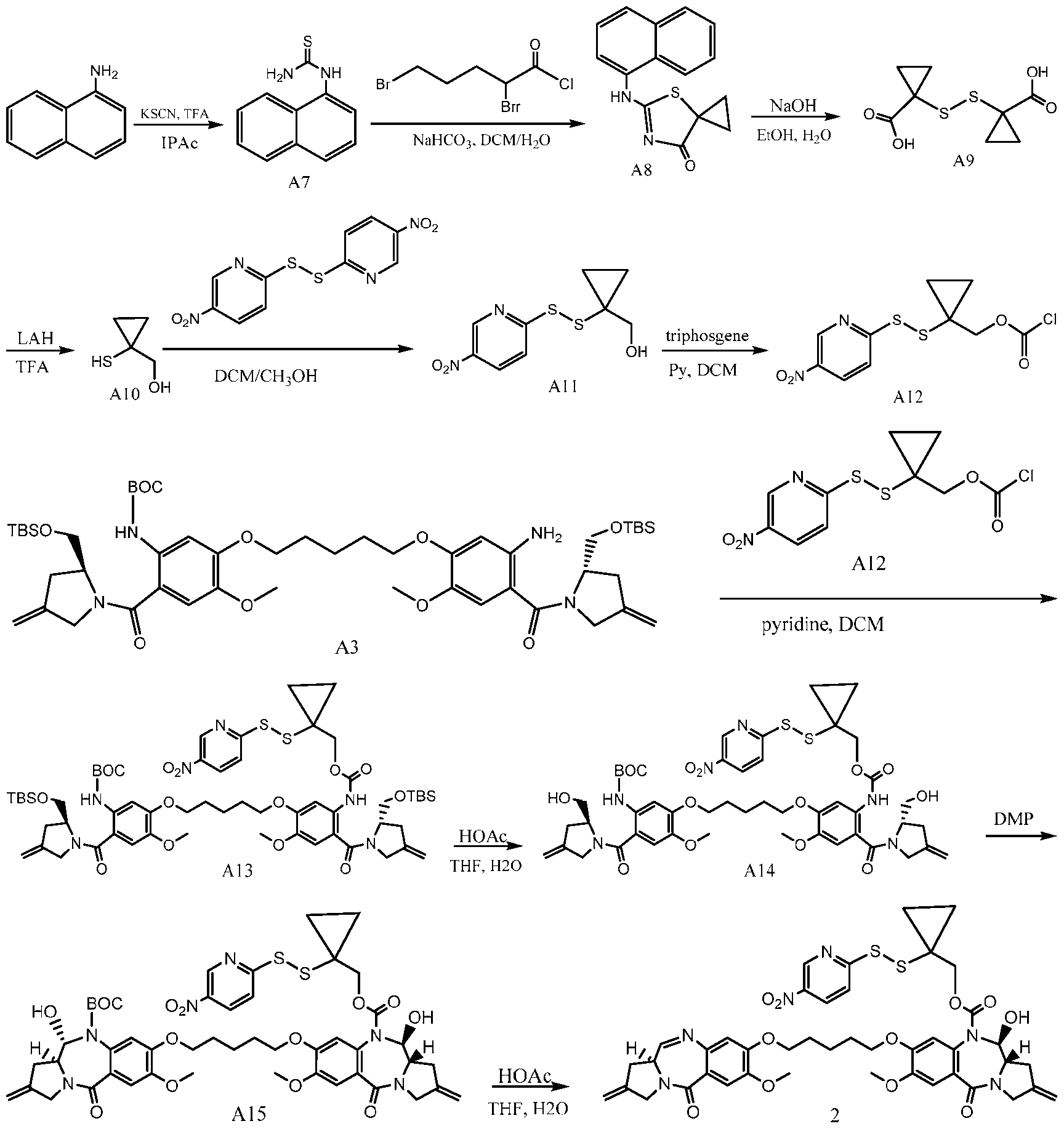
2,4-Dibromobutanoyl chloride (13.5 g, 51.07 mmol) was added to a mixture of compound A7 (10.33 g, 51.07 mmol) in saturated NaHCO3 solution (80 mL) and DCM (150 mL) slowly at 0oC. The mixture was stirred at 0oC for 30 min, then 15 % aq. NaOH solution (50 mL) and benzyltriethylammonium chloride (200 mg) were added sequentially. After stirring overnight at room temperature, H2O (100 mL) was added and the mixture was extracted with DCM (200 mL × 3). The combined organic layers were dried over Na2SO4, filtered, and were concentrated. The residue was purified by flash column chromatography (20% EtOAc in petroleum ether) to give compound A8 (9.0 g, 44%) as a yellow solid. 1H NMR (400 MHz, CDCl3) δ 7.99 (d, J = 8.0 Hz, 1H), 7.95 -7.92 (m, 1H), 7.61 -7.55 (m, 4H), 7.46 (d, J = 7.2 Hz, 1H), 1.90 -1.84 (m, 2H), 1.47 -1.41 (m, 2H).
The mixture of compound A8 (5.0 g, 18.63 mmol) in 15% aq. NaOH solution (50 mL) and EtOH (50 mL) was stirred under reflux for 6 h. After the EtOH was removed under vacuum, the aqueous phase was extracted with EtOAc (70 mL × 4) to remove unwanted byproducts. The aqueous phase was acidified to pH 3 by the slow addition of an aq. HCl solution, and then was extracted with EtOAc (70 mL × 4). The combined organic layers were dried over Na2SO4 and MnO2 (3.2 g, 36.8 mmol) was subsequently added.
The mixture was stirred at room temperature for 0.5 h then was filtered and concentrated. The residue was purified by flash column chromatography (5% CH3OH in DCM) to give compound A9 (1.3 g, 60%) as a yellow solid. 1H NMR (400 MHz, CDCl3) δ 1.92 (s, 4H), 1.62 (s, 4H).
A solution of compound A9 (400 mg, 1.71 mmol) in anhydrous THF (10 mL) was added to a suspension of LAH (389 mg, 10.2 mmol) in anhydrous THF (10 mL) at 25oC. The reaction mixture was stirred under reflux for 1 h then was cooled to 0oC and carefully quenched with 2 N aqueous HCl solution. The resulting mixture was diluted with DCM/MeOH (see below) and used directly in next step without further purification.
A mixture of crude compound A10 (356 mg, 3.42 mmol) and 1,2-bis(5-nitropyridin-2- yl)disulfane (2.12 g, 6.84 mmol) in DCM/MeOH (60 mL/60 mL) was stirred at room temperature overnight. MnO2 (3.0 g, 34.5 mmol) was then added and the mixture was stirred for another 0.5 h at room temperature. The mixture was filtered, and the filter cake was washed with DCM (100 mL). The combined filtrate and washings were concentrated under vacuum and
the residue was purified by flash column chromatography (0 to 1% DCM in MeOH) to give the desired product. This material was further purified by prep-TLC (DCM/MeOH = 20/1) to afford A11 (400 mg, 46% over 2 steps) as brown oil. LCMS (5-95AB, 1.5 min): RT = 0.80 min, m/z = 258.8 [M+H]+; 1H NMR (400 MHz, CDCl3) δ 9.35 (s, 1H), 8.36 (dd, J = 8.8, 2.4 Hz, 1H), 7.65 (d, J = 8.8 Hz, 1H), 4.41 (s, 1H), 3.47 (s, 2H), 1.15 -1.12 (m, 2H), 1.01 -0.98 (m, 2H).
A solution of compound A11 (85.0 mg, 0.33 mmol) and pyridine (25.0 mg, 0.32 mmol) in DCM (3 mL) was added dropwise to a solution of triphosgene (39.0 mg, 0.13 mmol) in DCM (3.0 mL) at 0oC. The reaction mixture was stirred at 0oC for 15 min then was used directly in the next step.
The solution of crude compound A12 prepared above (96.0 mg, 0.30 mmol) was added dropwise to a solution of compound A3 (220.0 mg, 0.23 mmol) and pyridine (24.0 mg, 0.30 mmol) in DCM (6.0 mL) at 26oC. The reaction mixture was stirred at 26oC for 2 h whereupon TLC analysis showed appearance of desired product (50% EtOAc in petroleum ether, Rf = 0.30) and consumption of the starting material (Rf = 0.25). The solvent was removed under reduced pressure and the residue was purified by prep-TLC (30% EtOAc in petroleum ether) to afford compound A13 (150 mg, 53%) as a yellow foam. LCMS (Condition A, 5-95AB, 1.5 min): RT = 1.25 min, m/z = 1237.4 [M+H]+.
Glacial HOAc (3.0 mL, 52 mmol) was added to a solution of compound A13 (150.0 mg, 0.12 mmol) in H2O (2.0 mL) and THF (2.0 mL) at 20oC. The reaction mixture was stirred at 20oC for 24 h whereupon TLC analysis showed appearance of the desired product (3.3% MeOH in DCM, Rf = 0.5) and consumption of the starting material (Rf = 0.65). The reaction mixture was diluted with EtOAc (10 mL) and was washed with water (2 x 4 mL), saturated aq. NaHCO3 (6 mL) and brine (6 mL). The organic layer was dried over Na2SO4, filtered, and was concentrated. The residue was purified by prep-TLC (5% MeOH in DCM, Rf = 0.5) to afford compound A14 (70 mg, 57%) as a yellow foam. LCMS (Condition A, 5-95AB, 1.5 min): RT = 0.87 min, m/z = 1009.4 [M+H]+.
Dess-Martin periodinane (88.0 mg, 0.210 mmol) was added to a solution of compound A14 (70.0 mg, 0.070 mmol) in DCM (4.0 mL) at 0oC. The reaction mixture was warmed to 20oC and was stirred for 2 h. A saturated solution of NaHCO3/Na2SO3 (3.0 mL/3.0 mL) was then added and the mixture was extracted with DCM (3 x 20 mL). The combined organic layers were washed with NaHCO3/Na2SO3 (3.0 mL/3.0 mL), brine (5 mL), dried over Na2SO4, filtered, and were concentrated. The residue was purified by prep-TLC (6.25% MeOH in DCM, Rf = 0.5) to give compound A15 (45 mg, 65%) as a yellow foam. LCMS (Condition B, 10-80AB, 7.0 min): RT = 2.70 min, m/z = 887.5 [M-100-18]+.
Cold TFA (95% in water, 2.0 mL) was added to compound A15 (45 mg, 0.045 mmol) at 0oC. After the reaction mixture was stirred at 0oC for 30 min, it was added dropwise to a solution of saturated aq. NaHCO3 (4.0 mL) at 0oC. The resulting mixture was extracted with DCM (4 x 8.0 mL), and the combined organic layers were washed with brine (20 mL), dried over Na2SO4, filtered, and were concentrated to give the crude product. This material was purified by prep-TLC (6.25% MeOH in DCM, Rf = 0.5) to afford compound 2 (22 mg, 55%) as a yellow solid. LCMS (Condition A, 5-95AB, 1.5 min): RT = 0.75 min, m/z = 887.5 [M+H]+; 1H NMR (400 MHz, CDCl3) δ 9.18 (s, 1H), 8.22-8.20 (m, 1H), 7.66 (d, J = 4.4 Hz, 1H), 7.51 (d, J = 8.8 Hz, 1H), 7.47 (s, 1H), 7.25 (s, 1H), 6.89 (s, 1H), 6.76 (s, 1H), 5.56 (d, J = 9.6 Hz, 1H), 7.19-5.13 (m, 4H), 4.48 (br, 1H), 4.32-4.23 (m, 4H), 4.16-4.02 (m, 5H), 3.93-3.86 (m, 6H), 3.81-3.79 (m, 1H), 3.70-3.68 (m, 2H), 3.14-3.08 (m, 1H), 2.95-2.91(m, 2H), 2.26 (d, J = 16.0 Hz, 1H), 1.93-1.89 (m, 4H), 1.63-1.59 (m, 2H), 1.06-1.02 (m, 2H), 0.94-0.84 (m, 2H).
PBD dimer compound 11 comprising an activated disulfide linker hindered with a cyclobutyl moiety was synthesized according to the following overall reaction scheme 3:
Scheme 3:


N-Bromosuccinimide (17.2 g, 96.6 mmol) and AIBN (1.28 g, 7.8 mmol) were added sequentially to a solution of ethyl cyclobutanecarboxylate (10.0 g, 78.0 mmol) in CCl4 (140 mL) at 25oC. The reaction mixture was heated to 80oC and was maintained at that temperature for 16 h whereupon TLC analysis showed that the desired product was formed (3% EtOAc in petroleum ether, Rf = 0.4). The mixture was cooled to room temperature then was diluted with DCM (100 mL) and washed with water (100 mL x 2) and brine (100 mL). The organic layer was dried over Na2SO4, filtered, and was concentrated. The residue was purified by flash column chromatography (1-3% EtOAc in petroleum ether) to afford compound A16 (6.4 g, 40%) as colorless oil, which was used without additional characterization.
KSAc (3.88 g, 34.0 mmol) was added to a mixture of compound A16 (6.4 g, 30.9 mmol) in acetone (100 mL) at 25oC. The reaction mixture was stirred at 80oC for 30 h whereupon TLC analysis showed that the desired product was formed (10% EtOAc in petroleum ether, Rf = 0.6). The mixture was cooled to room temperature then was poured into H2O (200 mL) and extracted with EtOAc (100 mL x 3). The combined organic layers were washed with brine (100 mL), dried over Na2SO4, filtered, and were concentrated. The residue was purified by flash column chromatography (0-5% EtOAc in petroleum ether) to afford compound A17 (2.5 g, 40%) as yellow oil. 1H NMR (400 MHz, CDCl3) δ 4.22-4.17 (m, 2H), 2.89-2.83 (m, 2H), 2.27 (s, 3H), 2.22-2.03 (m, 4H), 1.26 (t, J = 6.8 Hz, 3H).
A solution of compound A17 (2.5 g, 12.36 mmol) in THF (20 mL) was added to a mixture of LAH (1.88 g, 49.4 mmol) in THF (50 mL) at 0oC. After completing the addition, the mixture was heated to reflux at 80oC for 2 h whereupon TLC analysis showed formation of the desired product (10% EtOAc in petroleum ether, Rf = 0.2). The reaction mixture was cooled to 0oC and was carefully quenched by the slow addition of aqueous HCl (6 mL, 2.0 M). The mixture was diluted with DCM/MeOH (see below) and was used directly in the next step.
The reaction mixture prepared in the previous step was added to a suspension of 1,2- bis(5-nitropyridin-2-yl)disulfane (7.67 g, 24.7 mmol) in DCM (60 mL) and MeOH (60 mL) at 15oC. The reaction mixture was then stirred at 15oC for 16 h whereupon TLC analysis showed that the desired product was formed (100% DCM, Rf = 0.3). MnO2 (5.0 g) was added and the
mixture was stirred at room temperature for 30 min then filtered. The filtrate was concentrated to give a yellow solid which was triturated with 50 mL of MeOH and filtered a second time. The filtrate was once again concentrated and the residue was purified by flash column chromatography (0-1% MeOH in DCM) to afford compound A19 (1.46 g, 43% over two steps) as yellow solid. LCMS (Condition A, 5-95AB, 1.5 min): RT = 0.70 min, m/z = 272.9 [M+H]+.
A solution of compound A19 (200.0 mg, 0.73 mmol) and pyridine (58.0 mg, 0.73 mmol) in DCM (3.0 mL) was added dropwise to a solution of triphosgene (87.0 mg, 0.29 mmol) in DCM (2.0 mL) at 0oC. The mixture was stirred at 0oC for 1 h then was used directly in the next step.
The crude solution of compound A20 prepared above (245.0 mg, 0.50 mmol) in DCM (5.0 mL) was added dropwise to a stirred mixture of Compound A3 (500.0 mg, 0.52 mmol) and pyridine (0.06 mL, 0.74 mmol) in DCM (5.0 mL) at 25oC. After the addition was complete, the mixture was stirred at 25oC for 2 h and was subsequently concentrated. The residue was purified by flash column chromatography (0-33% EtOAc in petroleum ether) to afford compound A21 (510 mg, 76%) as a yellow solid. LCMS (Condition A, 5-95AB, 1.5 min): RT = 1.25 min, m/z = 1273.7 [M+Na]+.
Glacial HOAc (11.76 mL, 205 mmol) was added to a mixture of compound A21 (510.0 mg, 0.40 mmol) in THF (8.0 mL) and water (8.0 mL) at 25oC. The reaction mixture was stirred at 25oC for 16 h then was diluted with EtOAc (70 mL) and was washed sequentially with water (25 mL x 3), saturated aq. NaHCO3 (20 mL x 3), and brine (30 mL). The organic phase was dried over Na2SO4, filtered, and was concentrated. The residue was purified by flash column chromatography (0-5% MeOH in DCM) to afford compound A22 (350 mg, 84%) as a yellow solid. LCMS (Condition A, 5-95AB, 1.5 min): RT = 0.86 min, m/z = 1023.5 [M+H]+.
Dess-Martin periodinane (185 mg, 0.44 mmol) was added to a stirred mixture of compound A22 (154.0 mg, 0.15 mmol) in DCM (10 mL) at 0oC. After the reaction was stirred at 25oC for 20 h, it was poured into aqueous Na2S2O3 solution (10%, 30 mL) and the resulting mixture was extracted with EtOAc (20 mL x 3). The combined organic layers were washed with sat. aq. NaHCO3 (30 mL) and brine (30 mL), then were dried over Na2SO4, filtered, and concentrated. The residue was purified by prep-TLC (6.25% MeOH in DCM, Rf = 0.5) to give compound A23 (80 mg, 52%) as a white solid. LCMS (Condition A, 5-95AB, 1.5 min): RT =0.82 min, m/z = 1041.5 [M+Na]+.
Cold TFA (1.9 mL, 25 mmol) and water (0.10 mL) were added sequentially to compound A23 (100.0 mg, 0.10 mmol) at 0oC. The reaction mixture was stirred at 0oC for 1 h, then was added dropwise to a sat. aq. solution of NaHCO3 (150 mL) at 0oC. The resulting
mixture was extracted with DCM (40 mL x 4), and the combined organic layers were washed with brine (50 mL), dried over Na2SO4, filtered, and concentrated to give the crude product. This material was purified by prep-TLC (10% MeOH in DCM, Rf = 0.4) to give compound 11 (20 mg, 23%) as a yellow solid. LCMS (Condition A, 5-95AB, 1.5 min): RT = 0.77 min, m/z = 901.4 [M+H]+; 1H NMR (400 MHz, CDCl3) δ9.21 (d, J = 1.98 Hz, 1H) 8.36 (dd, J = 8.8, 2.4 Hz, 1 H) 7.70 (d, J = 4.4 Hz, 1H) 7.56 -7.51 (m, 2H), 7.49 (s, 1H), 6.80 (s, 1H), 6.74 (s, 1 H), 5.59 (d, J = 10.0 Hz, 1H), 5.20 (d, J = 11.2 Hz, 2H), 5.16 (br., 2H), 4.64 -4.48 (m, 1 H), 4.40 - 4.25 (m, 5H), 4.21–3.98 (m, 6H), 3.91 -3.73 (m, 2H), 3.65 (t, J = 8.80 Hz, 1H) 3.50 (s, 1H) 3.22 -3.08 (m, 1H) 2.99 -2.87 (m, 2H) 2.73 (m, 1H) 2.04 -1.86 (m, 8H) 1.38 -1.15 (m, 1H).
Example 19: Preparation of MMAE compounds comprising an activated hindered disulfide
Activated MMAE compound A was synthesized according to the following overall reaction scheme 1.
Scheme 1:

Activated MMAE compound B was synthesized according to the following overall reaction scheme 2.
Scheme 2:

To a stirred solution of Compound 3 (50.0 mg, 0.215 mmol), PNP (131 mg, 0.43 mmol) in DMF (3.0 mL) and DIPEA (56 mg, 0.43 mmol) were added at 15 °C. The mixture was stirred at 15 °C for 16 h under N2. The mixture was concentrated and purified by prep-TLC (DCM/MeOH = 30/1) to give compound 4 (60 mg, yield: 74%).
To a stirred solution of compound 4 (30 mg, 0.0756 mmol), were added MMAE (49 mg, 0.068 mmol), HOBt (2.0 mg, 0.015 mmol) in DMF (3.0 mL) and pyridine (60 mg, 0.756 mmol) at 22 °C. After the mixture was stirred at 20 °C for 16 h, it was purified by prep-HPLC (FA) to give compound B (60.0 mg, yield: 80%). LCMS: (5-95, AB, 1.5 min), 1.007 min, MS = 998.6[M+23]+; 1H NMR (400 MHz, DMSO-d6) δ 9.23 (s, 1 H), 8.53 (d, J = 7.6 Hz, 1 H), 7.44– 8.02 (d, J = 8.0 Hz, 1 H), 7.58 (s, 1 H), 7.35 - 7.18 (m, 7 H), 5.10 (s, 1 H), 4.70 - 4.45 (m, 2 H), 4.31 (s, 2 H), 4.18 (s, 1 H), 4.03 (s, 2 H), 3.80 (s, 1 H), 3.46 (s, 1 H), 3.40 - 3.18 (m, 12 H), 2.84 (d, J = 6.0 Hz, 3 H), 2.41 (s, 2 H), 2.18 (s, 2 H), 1.81 (s, 3 H), 1.74 - 1.50 (m, 2 H), 1.37 (s, 1 H), 1.04 - 0.78 (m, 28 H). The molecular weight determined by NMR was 975.48.
Activated MMAE compound C was synthesized according to the following overall reaction scheme 3.
Scheme 3:

To a stirred solution of MMAE (30 mg, 0.042 mmol) and compound 2 (34 mg, 0.084 mmol) in dry DMF (2.0 mL) was added HOBt (1.0 mg, 0.0042 mmol) and pyridine (33 mg, 0.42 mmol). The mixture was stirred at 27 °C for 12 h and purified by prep-HPLC (FA) to give compound C (23 mg, yield: 55%) as a white solid. 1H NMR (400 MHz, MeOH-d4) δ ppm 9.24 (br. s., 1H), 8.54 (dd, J = 8.9, 2.6 Hz, 1H), 8.07 (dd, J = 9.0, 2.0 Hz, 1H), 7.53 - 7.09 (m, 5 H), 4.73 - 4.56 (m, 5H), 4.36 - 4.15 (m, 5H), 3.92 - 3.51 (m, 1H), 3.49 - 3.37 (m, 2H), 3.35 (s, 5H), 3.25 - 3.09 (m, 2H), 3.02 - 2.86 (m, 3H), 2.56 - 2.39 (m, 2H), 2.32 - 1.51 (m, 2H), 1.38 (dd, J = 6.8, 2.8 Hz, 4H), 1.23 - 1.09 (m, 6H), 1.06 - 0.77 (m, 19H). The molecular weight determined by NMR was 989.50.
Activated MMAE compounds E, F and G were synthesized according to the following overall reaction scheme 4.
Scheme 4:


G
A mixture of compound 1 (500.0 mg, 1.92 mmol) and PNP carbonate (1169 mg, 3.84 mmol) in DMF (2.0 mL) was added DIEA (744.7 mg, 5.76 mmol). The reaction mixture was stirred at 14 °C for 8 h under N2. The reaction mixture was concentrated in vacuo and purified by column chromatography (100% DCM ) to give compound 2 (300 mg, 34% yield) as a yellow oil. LCMS (5-95AB/1.5min): RT = 0.850 min, [M+H]+ 425.8.
To a mixture of compound 2 (100.0 mg, 0.240 mmol) and pyridine (186 mg, 2.35 mmol) in DMF (10 mL) was added MMAE (107 mg, 0.210 mmol) and HOBt (3.18 mg, 0.020 mmol). The reaction mixture was stirred at 30 °C for 8 h. The mixture was concentrated in vacuo and purified prep-TLC (5% MeOH in DCM) to give compound D (110 mg, 46%) as a white solid. LCMS (5-95AB/1.5min): RT=0.899 min, [M+Na]+1026.3. 1H NMR (400 MHz,CDCl3) δ 9.18 (s, 1H), 8.30 (d, J = 8.4 Hz, 1H), 7.88 (d, J = 12.8 Hz, 1H), 7.32-7.19 (m, 4H), 6.42 (d, J = 9.2 Hz, 1H), 4.88 (s, 1H) , 4.72-4.60 (m, 1H), 4.25-4.20 (m, 1H), 4.14-3.94 (m, 5H), 3.78 (d, J = 5.6, 1H), 3.44 (m,1H), 3.35-3.31 (m, 4H), 3.24 (s, 3H), 3.04 (s, 1H), 2.84-3.00 (m, 2H), 2.44- 2.34 (m, 3H), 2.34-2.30 (m, 1H), 1.88-2.05 (m, 3), 1.84-1.75 (m, 3H), 1.30 (s, 7H), 1.20 (m, 3H) 1.00-0.75 (m, 24H). The molecular weight determined by NMR was 1003.51.
A mixture of compound D (20.0 mg, 0.020 mmol), compound 3 (14.96 mg, 0.1000mmol), compound 4 (29.71 mg, 0.100 mmol), and DMAP (7.3 mg, 0.060 mmol) in DMF (2.0 mL) was stirred at 30 °C for 8 h. The mixture was purified by prep-HPLC to give compound E (4.1 mg, 20.2%) as a white solid. LCMS (5-95AB/1.5min): RT = 0.758 min, [M/2+H]+500.1.
A solution of compound 5 (58.02 mg, 0.30 mmol) in DMF (5.0 mL) was added MnO2 (51.99 mg, 0.600 mmol). After the reaction mixture was stirred at 15 °C for 30 min, it was
filtered and the filtrate was added to compound D (30.0 mg, 0.030 mmol), compound 5 (29 mg, 0.15 mmol) and DMAP (10.96 mg, 0.090 mmol). The mixture was stirred at 30 °C for 8 h. The mixture was purified by prep-HPLC to give compound F (4.8 mg, 0.0047 mmol, 15.8% yield) as a white solid. LCMS (5-95AB/1.5min): RT = 0.767 min, [M/2+Na]+522.1.
To a solution of compound 6 (25 mg, 0.167 mmol) in DMF (5.0 mL) was added MnO2 (58.68 mg, 0.670 mmol). After the reaction mixture was stirred at 18 °C for 30 min, it was filtered, and the filtrates was added to compound D (30.0 mg, 0.0300 mmol), compound 6 (51 mg, 0.339 mmol) and DMAP (12.37 mg, 0.100 mmol). The reaction mixture was stirred at 40 °C for 8 h. The reaction mixture was purified by prep-HPLC to give compound G (9.8 mg, 27%) as a white solid. LCMS (5-95AB/1.5min): RT = 0.849 min, [M+H]+1043.5.
Activated MMAE compound H was synthesized according to the following overall reaction scheme 5.
Scheme 5:

A solution of compound 3 (51.0 mg, 0.339 mmol) in DMF (5.0 mL) was added MnO2 (58.68 mg, 0.670 mmol), and the reaction mixture was stirred at 18 °C for 30 min. The mixture was filtered, and to the filtrates was added to compound 2 (30.0 mg, 0.030 mmol), compound 3 (25.0 mg, 0.167 mmol) and DMAP (12.37 mg, 0.100 mmol). The reaction mixture was stirred at 40 °C for 8 h. The reaction mixture was purified by prep-HPLC (column: waters Xbridge Prep OBD C18150*30 5u, condition: 0.225%FA-ACN) to give compound H (9.8 mg, 0.0091 mmol, 27% yield) as a white solid. LCMS (5-95AB/1.5min): RT = 0.849 min, [M+H]+1043.5.
Activated MMAE compound I was synthesized according to the following overall reaction scheme 6.
Scheme 6:

To the mixture of compound 2 (45.0 mg, 0.300 mmol) in DMF (2.0 mL) was added MnO2 (129 mg, 1.49 mmol). The mixture was stirred at 25 °C for 15 min. The mixture was filtered and the filtrate was used directly in the next step.
To the mixture of compound 1 (30.0 mg, 0.030 mmol), compound 2a (44.0 mg, 0.150 mmol) and compound 2 (45.2 mg, 0.300 mmol) in DMF (3.0 mL) was added DMAP (18.3 mg, 0.150 mmol). The mixture was stirred at 40 °C for 2 h. The mixture was purified by prep-HPLC (acetonitrile 45-75% / 10mM NH4HCO3-ACN) to afford compound I (7.8 mg, 25.8%) as a white solid. LCMS (5-95AB/1.5min): RT = 0.735 min, [M+H]+999.6.
Activated MMAE compound J was synthesized according to the following overall reaction scheme 7.
Scheme 7:

Carbon disulfide (191 mg, 2.52mmol) was added to a mixture of KOH (141 mg, 2.52 mmol) in EtOH (4.0 mL) and water (1.0 mL). The reaction mixture was stirred at 20 °C for 15 min. Then compound 3 (250 mg, 2.29 mmol) was added. The reaction mixture was heated under microwave irradiation at 130 °C for 15 min and cooled to r.t. The reaction mixture was diluted with H2O (10 mL), adjusted to pH = 5 with HOAc, and cooled to 0 °C. The solid was filtered and dried under reduced pressure to give compound 2 (230 mg, 66.4%) as a white solid. 1H NMR (400 MHz, DMSO-d6) δ 12.87-12.84 (m, 2H), 8.36 (s, 1H), 8.23 (d, J = 5.2 Hz, 1H), 7.15 (d, J = 5.2 Hz, 1H).
To the mixture of compound 2 (45.0 mg, 0.300 mmol) in DMF (2.0 mL) was added MnO2 (129 mg, 1.49 mmol) and the mixture was stirred at 25 °C for 15 min. The mixture was filtrated and the filtrate was used directly to the next step.
To a mixture of compound 1 (30.0 mg, 0.030 mmol), compound 2a (44.0 mg, 0.150 mmol) and compound 2 (45.16 mg, 0.300 mmol) in DMF (3.0 mL) was added DMAP (18.25 mg, 0.150 mmol). The mixture was stirred at 40 °C for 15 h. The mixture was purified by prep- HPLC (acetonitrile 35-65% / 0.225%FA-ACN) to afford compound J (8.8 mg, 29.5%) as a white solid. LCMS (5-95AB/1.5min): RT = 0.717 min, [M+H]+ 999.5.
Activated MMAE compound K was synthesized according to the following overall reaction scheme 8.
Scheme 8:

To a solution of compound 3 (250.0 mg, 1.63 mmol) in DMF (1.0 mL) was added carbon disulfide (174 mg, 2.29 mmol). The mixture was stirred at 25 °C for 72 h. The reaction mixture was diluted with H2O (10.0 mL), adjusted to pH = 5.0 with HOAc, and filtered under reduced pressure to give compound 2 (200 mg, 62.8%) as a yellow solid, which was used in the next step directly. 1H NMR (400 MHz, DMSO-d6) δ 13.18-13.07 (m, 2H), 7.91 (d, J = 8.0 Hz, 1H), 7.49 (d, J = 8.0 Hz, 1H), 7.29 (t, J = 8.4 Hz, 1H).
To the mixture of compound 2 (60.0 mg, 0.310 mmol) in DMF (2.0 mL) was added MnO2 (134 mg, 1.54 mmol). The mixture was stirred at 25 °C for 15 min, then filtered. The filtrate was used directly in the next step.
To a mixture of compound 1 (30.0 mg, 0.030 mmol), compound 2a (58.0 mg, 0.150 mmol) and compound 2 (58.3 mg, 0.300 mmol) in DMF (3.0 mL) was added DMAP (18.25 mg, 0.150 mmol). The mixture was stirred at 40 °C for 2 h. The mixture was purified by prep-HPLC (acetonitrile 50-80% / 0.225%FA-ACN) to afford compound K (7.7 mg, 0.0072 mmol, 23.9% yield) as a yellow solid. LCMS (5-95AB/1.5min): RT = 0.863 min, [M+H]+ 1043.7.
Activated MMAE compound L was synthesized according to the following overall reaction scheme 9.
Scheme 9:

To the mixture of compound 2 (45.0 mg, 0.300 mmol) in DMF (3.0 mL) was added MnO2 (128.5 mg, 1.48 mmol). The mixture was stirred at 25 °C for 15 min and filtered. The filtrate was used directly used in the next step without further purification.
To a mixture of compound 1 (30.0 mg, 0.030 mmol), compound 2a (44.0 mg, 0.150 mmol) and compound 2 (45.5 mg, 0.300 mmol) in DMF (3.0 mL) was added DMAP (18.26 mg, 0.150 mmol). The mixture was stirred at 40 °C for 15 h. The mixture was purified by prep- HPLC (acetonitrile 30-60% / 0.1% NH4OH in water) to afford compound L (9.2 mg, 30.1%) as a white solid. LCMS (5-95AB/1.5min): RT = 0.873min, [M+H]+1001.0.
Activated MMAE compound M was synthesized according to the following overall reaction scheme 10.
Scheme 10:

To a solution of pyrazine-2,3-diamine (3) (250.0 mg, 2.27 mmol) and carbon disulfide (242.0 mg, 3.18 mmol) in DMF (2.0 mL). The reaction mixture was stirred at 25 °C for 16 h. The reaction mixture was concentrated under reduced pressure, and filtered to give compound 2 (200 mg,1.31 mmol, 57.9% yield) as a yellow solid, which was used directly in the next step.1H NMR (400 MHz, CDCl3) δ 13.45 (s, 2H), 8.08 (s, 2H).
A mixture of compound 1 (30.0 mg, 0.030 mmol) and compound 2a (101.9 mg, 0.340 mmol), DMAP (12.35 mg, 0.100 mmol) in DMF (1.0 mL) was added compound 2 (51.3 mg, 0.340 mmol). After the reaction mixture was stirred at 40 °C for 8 h, it was purified by prep- HPLC (Condition: water (0.225%FA)-CAN) to give the compound M (6.0 mg, 0.0059 mmol, 17.6% yield) as a white solid. LCMS (5-95AB/1.5min): RT = 0.788 min, [M+H]+1000.5.
Activated MMAE compound N was synthesized according to the following overall reaction scheme 11.
Scheme 11:


Activated MMAE compound O was synthesized according to the following overall reaction scheme 12.
Scheme 12:

To a solution of compound 1 (80.0 mg, 0.080 mmol) in DMF (4.0 mL) was added compound 2 (60.64 mg, 0.800 mmol), compound 2a (119.67 mg, 0.800 mmol) and DMAP (29.2 mg, 0.240 mmol). The mixture was stirred at 38 °C for 12 h. The mixture was purified by prep- HPLC (HCl) to give compound O (31 mg, 41.7%) as a white solid. LCMS (5-95AB/1.5min): RT = 0.706 min, [M+H]+ 924.5.
Activated MMAE compound P was synthesized according to the following overall reaction scheme 13.
Scheme 13:

Compound 3 (200.0 mg, 1.31 mmol) and KOH (110.6 mg, 1.97 mmol) in EtOH (4.0 mL) and water (1.0 mL) was added CS2 (150.1 mg, 1.97 mmol). The reaction mixture was stirred at 130 °C for 1.0 h under microwave irradiation. The mixture was poured into ice water, adjusted pH = 5 with HOAc, and extracted with DCM (10 mL x 3). The organic layer was concentrated to afford compound 2 (150 mg, 0.772 mmol, 58.8% yield) as a black oil.
A mixture of compound 2 (100.0 mg, 0.510 mmol) and MnO2 (259.5 mg, 2.99 mmol) in DMF (10.0 mL) was stirred at 26 °C for 20 min. The reaction mixture was filtered, and the filtrate was add to a solution of compound 1 (20 mg, 0.02 mmol) and compound 2 (50 mg, 0.26 mmol) and DMAP (0.24 mg, 0.002 mmol) in DMF (2.0 mL) at 26 °C. The mixture was stirred at 26 °C for 12 h. The mixture was concentrated in vacuo, and purified by prep-HPLC (acetonitrile 55-85%/0.225% FA in water) to give compound P (5.6 mg, 24.1%) as a white solid. LCMS (5-95AB/1.5min): RT = 0.810 min, [M+H]+ 1043.6.
Activated MMAE compound Q was synthesized according to the following overall reaction scheme 14.
Scheme 14:

To a solution of compound 3 (300.0 mg, 2.40 mmol) in DMF (12.0 mL) was added carbon disulfide (365 mg, 4.80 mmol). After the mixture was stirred at 20 °C for 24 h, it was concentrated and washed with water (15 mL x 2), and solid was collected to give compound 2 (120 mg, 29.9% yield) as a gray solid which was used directly in next step.
To a solution of compound 3 (300.0 mg, 2.40 mmol) in DMF (12.0 mL) was added carbon disulfide (365 mg, 4.80 mmol). After the mixture was stirred at 20 °C for 24 h, it was concentrated and washed with water (15 mL x 2), and solid was collected to give compound 2 (120 mg, 29.9% yield) as a gray solid which was used directly in next step.
The solution of compound 1 (30.0 mg, 0.030 mmol), compound 2 (24.9 mg, 0.150 mmol) and compound 2a (49.64 mg, 0.150 mmol) was stirred at 20 °C for 2.0 h. The reaction mixture was purified by prep-HPLC (acetonitrile 20-50/water (0.225FA)-ACN) to give compound Q (2.6 mg, 8.6%) as a white solid. LCMS (5-95AB/1.5min): RT = 0.722 min,

Activated MMAE compound R was synthesized according to the following overall reaction scheme 15.
Scheme 15:

4-Methoxybenzylamine (3) (6.00 g, 43.74 mmol) was dissolved in anhydrous THF (120 mL). Under ice cooling, triethylamine (7.32 mL, 52.81 mmol) and carbon disulfide (2.9 mL, 47.91 mmol) was added and the mixture was stirred for 1 h. To the reaction mixture was added iodomethane (3.33 mL, 53.47 mmol). The reaction mixture was warmed to 26 °C and stirred for 16 h. The mixture was concentrated, the residue was diluted with EtOAc (120 mL), washed successively with an aqueous solution of citric acid (60 mL × 2), sat. Na2CO3 (60 mL), and brine (60 mL). The organic layer was dried over Na2SO4, filtered, and concentrated in vacuo to compound 4 (9.00 g, 90.5%) as a colorless oil.
To a solution of compound 4 (10.00 g, 43.99 mmol) in EtOH (100 mL)/water (20 mL) was added sodium azide (5.05 g, 77.68 mmol). The reaction mixture was stirred at 80 °C for 16 h. The solution was concentrated in vacuo to remove most of EtOH. The remaining solution was diluted with EtOAc (60 mL), adjusted to pH = 3 with 1.0 M HCl solution, and extracted with EtOAc (60 mL × 3). The combined organic layers were dried over Na2SO4, filtered, and concentrated in vacuo. The residue was purified by column chromatography (0 - 10 % MeOH in DCM) to afford compound 5 (7.00 g, 71.6%) as a white solid.
A solution of compound 5 (444.0 mg, 2.0 mmol), anisole (0.70 mL, 6.44 mmol) and TFA (3.3 mL, 44.43 mmol) was heated at 100 °C under microwave irradiation for 2 h. After the
reaction mixture was cooled to r.t., the resulting precipitates were re-dissolved in EtOAc (4.0 mL). The solvent was removed under reduced pressure to afford a yellow solid, which was dissolved in EtOAc (10 mL) and water (5 mL). To the mixture was added NaOH (1.12 g, 28 mmol). After the mixture was stirred for 30 min, the aqueous layer was separated. And the pH was adjusted to 1~2 with concentrated HCl. The solution was extracted with EtOAc (20 mL x 2) dried over Na2SO4, concentrated in vacuo to afford 1H-tetrazole-5-thiol (180 mg, 88.2%) as a white solid.
To a solution of compound 2 (50.2 mg, 0.500 mmol) in anhydrous DMF (1.0 mL) was added MnO2 (216 mg, 2.49 mmol). The mixture was stirred at 24 °C for 20 min. Then the mixture was filtered, the filtrate was added to a solution of compound 1 (50.0 mg, 0.050 mmol), compound 2 (26 mg, 0.500 mmol), and DMAP (18.3 mg, 0.150 mmol) in anhydrous DMF (3.0 mL). The reaction solution was stirred at 40 °C for 16 h. The solution was purified by prep- HPLC (acetonitrile 50-80/0.225% FA in water) to afford compound R (14.1 mg, 27.7%) as a white solid. LCMS (5-95AB/1.5min): RT = 0.916 min, [M+H] + 950.7.
Activated MMAE compound S was synthesized according to the following overall reaction scheme 16.
Scheme 16:

A solution of compound 4 (80.0 mg, 0.360 mmol) in a 5:3:1 mixture of THF/EtOH/H2O (4.5 mL) was cooled to 0 °C in an ice-water bath, LiOH H2O (75 mg, 1.78mmol) was added, the reaction mixture was allowed to warm to 10 °C and it was stirred for 1 h. It was cooled to 0 °C, diluted with H2O (3.0 mL), and the pH was adjusted to 6 with HOAc. The solvent was concentrated to give compound 2 (68 mg, 0.347 mmol, 97.2% yield) as a yellow solid, which was used in the next step without further purification.
A mixture of compound 1 (30.0 mg, 0.030 mmol), compound 2a, (58.3 mg, 0.150 mmol), compound 2 (29.3 mg, 0.150 mmol) and DMAP (7.3 mg, 0.060 mmol) in DMF (2.0 mL) was stirred for 12 h at 38 °C. The mixture was purified by prep-HPLC (acetonitrile 35- 60%/0.225% FA in water) to give compound S (9.0 mg, 28.9%) as a white solid. LCMS (5- 95AB/1.5min): RT = 0.746 min, [M+H]+1044.4. HPLC (10-80AB/15min): RT = 8.55 min.
Activated MMAE compound T was synthesized according to the following overall reaction scheme 17.
Scheme 17:


A mixture of 4-chloronicotinic acid 3 (5.00 g, 31.74 mmol) in thionyl chloride (100.0 mL, 31.74 mmol) was stirred at 80 °C for 1.5 h. The mixture was concentrated in vacuo, the residue was diluted with toluene, and concentrated again to afford compound 4 (5.50 g, 98.5% yield) as a yellow solid which was used directly for next step.
To a solution of compound 4 (5.50 mg, 31.25 mmol) in anhydrous DCM (150 mL) was added triethylamine (9.49 g, 93.75 mmol) at 0 °C. A solution of N,N-dimethylamine (15.63 mL, 31.25 mmol) in anhydrous THF (15.6 mL) was added dropwise at 0 °C. The reaction mixture was stirred at 0 °C for 1 h. The mixture was concentrated in vacuo and the residue was purified by column chromatography (0 - 12 % MeOH in DCM) to afford compound 5 (4.10 g, 71.1%) as a brown oil.
Sodium sulfide nonahydrate (4.94 g, 20.58 mmol) and sulfur (659 mg, 20.58 mmol) in anhydrous EtOH (80 mL) was stirred at 80 °C for 15 min. The mixture was filtered while hot and the filtrate was added to compound 5 (3.80 g, 20.58 mmol). The mixture was stirred at 80 °C under N2 for another 16 h. The mixture was cooled to r.t., and MnO2 (8.70 g) was added, the mixture was stirred at r.t. for another 2 h. The mixture was filtered, the filtrate was concentrated in vacuo, the residue was purified by column chromatography (0 - 15 % MeOH in DCM) to afford compound 2a (1500 mg, 27.7%) as pale brown oil. LCMS (0-60AB/2min): RT = 0.923 min, [M+H] + 362.9.
To a solution of compound 2a (200.0 mg, 0.550 mmol) in THF (5 mL) / Water (5.0 mL) was added 3-[bis(2-carboxyethyl)phosphanyl]propanoic acid (6) (414 mg, 1.66 mmol). The reaction solution was stirred at 26 °C for 1 h. The solution was adjusted to pH 7 with aq. NaHCO3 solution, and extracted with DCM/MeOH (10:1, 30 mL × 3). The combined organic layers were dried over Na2SO4, filtered, and concentrated in vacuo to afford compound 2 (50 mg, 24.9%) as crude brown oil.
A solution of compound 1 (50.0 mg, 0.050 mmol), compound 2 (45.38 mg, 0.250 mmol), compound 2a (144 mg, 0.400 mmol), and DMAP (18.25mg, 0.1500mmol) in anhydrous DMF (4.0 mL) was stirred at 40 °C for 16 h. The solution was purified by prep-HPLC (acetonitrile 45-75/0.225% FA in water) to afford compound T (34.1 mg, 65.8%) as a white solid. LCMS (5-95AB/1.5min): RT = 0.878 min, [M+H]+ 1030.9.
Activated MMAE compound U was synthesized according to the following overall reaction scheme 18.
Scheme 18:

To a solution of compound 2 (28.9 mg, 0.250 mmol) in DMF (2.0 mL) was added MnO2 (10.8 mg, 0.120 mmol). After the reaction mixture was stirred at 15°C for 30 min, it was filtered, and to the filtrate were added compound 1 (25.0 mg, 0.020 mmol), DMAP (9.1 mg, 0.070 mmol) and compound 2 (28.9 mg, 0.250 mmol). The reaction mixture was stirred at 40 °C for 3 h. The reaction mixture was purified by prep-HPLC (Column: Waters Xbridge Prep OBD C18150*305u, Condition: water (0.225%FA)-CAN to give compound U (11.1 mg, 43.5%) as a white solid. LCMS (5-95AB/1.5min): RT = 0.831 min, [M+H]+964.6. 1H NMR (400 MHz, DMSO-d6 ) δ 8.05 (m, 1H), 7.88-7.60 (m, 1H), 7.29-7.22 (m, 4H), 7.15 (d, J = 4.0 Hz, 1H), 5.40-5.32 (m, 1H), 4.47-4.39 (m, 1H), 4.23-4.05 (m, 5H) , 3.98-3.93 (m, 2H), 3.60-3.51 (m, 2H), 3.21 (d, J = 8.0 Hz, 5H), 3.16 (d, J = 8.0 Hz, 3H), 3.10 (s, 2H), 3.03-2.95 (m, 2H), 2.85-2.79 (m, 3H), 2.65 (s, 1H), 2.38 (d, J = 12.0 Hz, 1H), 2.31-2.25 (m, 1H), 1.21-1.99 (m, 3H), 1.78-1.69 (m, 3H), 1.51-1.44 (m, 2H), 1.33-1.27 (m, 7H), 1.03-0.95 (m, 7H), 0.86-0.71 (m, 19H). The molecular weight determined by NMR was 963.53.
Activated MMAE compound V was synthesized according to the following overall reaction scheme 19.
Scheme 19:


Activated MMAE compound W was synthesized according to the following overall reaction scheme 20.
Scheme 20:

To a solution of compound 2 (34 mg, 0.300 mmol) in anhydrous DMF (4.0 mL) was added manganese dioxide (130 mg, 1.49 mmol). After the mixture was stirred at 24 °C for 0.5 h, it was filtered, the filtrated was added to a mixture of compound 1 (30.0 mg, 0.030 mmol), compound 2 (17 mg, 0.150 mmol) and DMAP (10.95 mg, 0.090 mmol). The reaction solution was stirred at 40 °C for 16 h. The solution was purified by prep-HPLC (Waters Xbridge Prep OBD C18 100*19mm*5um, acetonitrile 45-75/0.225% FA in water) to afford compound W (8.0 mg, 0.0079 mmol, 26.4% yield) as a white solid. LCMS (5-95AB/1.5min): RT = 0.770 min, [M+H]+ 963.6.
Activated MMAE compound X was synthesized according to the following overall reaction scheme 21.
Scheme 21:

To a solution of compound 1 (300.0 mg, 1.74 mmol) in NMP (10.0 mL) was added DIEA (675 mg, 5.23 mmol), followed by compound 2 (409 mg, 2.61 mmol). After the reaction solution was stirred at 20 °C for 16 h, it was diluted with EtOAc (60 mL), washed with brine (30 mL × 5), dried over Na2SO4, filtered, and concentrated. The residue was purified by prep-TLC (5 % MeOH in DCM, Rf = 0.6) to afford compound 3 (500 mg, 1.71 mmol, 98.2% yield) as a white solid.
To a solution of compound 1 (300.0 mg, 1.74 mmol) in NMP (10.0 mL) was added DIEA (675 mg, 5.23 mmol), followed by compound 2 (409 mg, 2.61 mmol). After the reaction solution was stirred at 20 °C for 16 h, it was diluted with EtOAc (60 mL), washed with brine (30 mL × 5), dried over Na2SO4, filtered, and concentrated. The residue was purified by prep-TLC
(5 % MeOH in DCM, Rf = 0.6) to afford compound 3 (500 mg, 1.71 mmol, 98.2% yield) as a white solid.
To a mixture of compound 4 (500 mg, 1.63 mmol) in MeOH (6.0 mL) / water (3.0 mL) was added NaOH (196 mg, 4.90 mmol). The reaction mixture was stirred at 40 °C for 2 h. The solution was concentrated in vacuo to remove most of MeOH, and the water slurry was diluted with H2O (20 mL), washed with DCM (20 mL × 3). The water phase was adjusted to pH = 2 with HCl solution, and extracted with DCM/MeOH (10:1, 30 mL × 3). The combined organic layers were dried over Na2SO4, filtered, and concentrated in vacuo to afford compound 5 (300 mg, 0.830 mmol, 50.9% yield) as a brown solid. LCMS (0-60AB/7min): RT = 2.66 min, [M+H]+ 278.9.1H NMR (400 MHz, DMSO-d6) δ 7.86 (s, 1H), 7.18 (d, J = 8.4 Hz, 2H), 6.84 (d, J = 8.4 Hz, 2H), 4.22 (s, 2H), 3.72 (s, 3H), 3.41 (s, 3H).
A solution of compound 5 (300 mg, 1.08 mmol) in TFA (6.0 mL) / anisole (1.0 mL) was stirred at 40 °C for 24 h. The solution was concentrated in vacuo to remove the solvent, the residue was washed with DCM (20 mL) to afford compound 6 (100 mg, 0.632 mmol, 58.7% yield) as a white solid. 1H NMR (400 MHz, DMSO-d6) δ 12.77 (s, 1H), 7.80 (s, 1H), 3.45 (s, 3H).
To a solution of compound 6 (48 mg, 0.300 mmol) in anhydrous DMF (5.0 mL) was added manganese dioxide (130 mg, 1.50 mmol). After the mixture was stirred at 24 °C for 1 h, it was filtered, the filtrated was added to a mixture of compound 7 (30.0 mg, 0.030 mmol), compound 6 (24 mg, 0.150 mmol) and DMAP (18.3 mg, 0.150 mmol). The reaction solution was stirred at 40 °C for 2 days. The solution was purified by prep-HPLC (Waters Xbridge Prep OBD C18 150*30mm*5um, acetonitrile 42-88/0.225% FA in water) to afford Compound X (3.80 mg, 0.0038 mmol, 12.7% yield) as a white solid. LCMS (5-95AB/1.5min): RT = 0.888 min, [M+Na]+ 1028.8.
Activated MMAE compound Y was synthesized according to the following overall reaction scheme 22.
Scheme 22:


To a solution of Compound 2 (47.0 mg, 0.300 mmol) in anhydrous DMF (4.0 mL) was added MnO2 (129.85 mg, 1.49 mmol). After the mixture was stirred at 24 °C for 0.5 h, it was filtered, and the filtrated was added to a mixture of Compound 3 (30.0 mg, 0.030 mmol), compound 2 (23 mg, 0.150 mmol), and DMAP (10.95 mg, 0.090 mmol). After the reaction solution was stirred at 40 °C for 2 days, it was purified by prep-HPLC to afford compound Y (4.8 mg, 0.0047 mmol, 15.8% yield) as a white solid. LCMS (5-95AB/1.5min): RT = 0.886 min,

Activated MMAE compound Z was synthesized according to the following overall reaction scheme 23.
Scheme 23:


To a solution of MMAE (100.0 mg, 0.140 mmol) in anhydrous DMF (5.0 mL) was added compound 2 (68.76 mg, 0.170 mmol), 1-HOBt (3.76 mg, 0.030 mmol), and pyridine (110.17 mg, 1.39 mmol). The solution was stirred at 15 °C for 2 days. The solution was concentrated in vacuo, and the residue was purified by prep-TLC (5% MeOH in DCM, Rf = 0.5) to afford compound 3 (100 mg, 0.098 mmol, 70.3% yield) as a white solid. LCMS (5- 95AB/1.5min): RT = 0.996 min, [M+H] + 990.6.
To a solution of compound 4 (47 mg, 0.410 mmol) in anhydrous DMF (5.0 mL) was added MnO2 (175.59 mg, 2.02 mmol). The mixture was stirred at 24 °C for 1 h. Then the mixture was filtered, the filtrated was added to a mixture of compound 3 (40.0 mg, 0.040 mmol), compound 4A (22mg, 0.200 mmol) and DMAP (14.8 mg, 0.120 mmol). The reaction solution was stirred at 40 °C for 16 h. The solution was purified by prep-HPLC (Diamonsil 150*20mm*5um, acetonitrile 35-65/0.225% FA in water). It was further purified by prep-TLC (10% MeOH in DCM, Rf = 0.5) to afford GNT_B343_837-1 (28.2 mg, 0.0282 mmol, 69.9% yield) as a white solid. LCMS (5-95AB/1.5min): RT = 0.866 min, [M+H]+ 949.5.
Activated MMAE compound AA was synthesized according to the following overall reaction scheme 24.
Scheme 24:


A mixture of MMAE (30.0 mg, 0.04 mmol) and compound 2 (60.0 mg, 0.15 mmol) in DMF (3.0 mL) was added pyridine (33.05 mg, 0.42 mmol) and HOBt (0.56 mg, 0.01mmol). The reaction mixture was stirred at 30 °C for 8 h. The mixture was concentrated in vacuo and purified by prep-TLC (5% MeOH in DCM) to give Compound 3 (35 mg, 0.035 mmol, 83.7 % yield) as a white solid. LCMS (5-95AB/1.5min): RT = 0.994 min, [M+H]+ 990.5.
To a solution Compound 4 (200.0 mg, 1.74 mmol) in DMF (10.0 mL) was added MnO2 (754.9 mg, 8.68 mmol). The mixture was stirred at 15 °C for 2 h. The mixture was filtered and concentrated to give Compound 4A (150 mg, 0.66 mmol, 75.7 % yield) as a red solid.
To a solution of Compound 3 (40.0 mg, 0.04 mmol) in DMF (4.0 mL) was added Compound 4 (46.52 mg, 0.40 mmol), Compound 4A (92.22 mg, 0.40 mmol) and DMAP (14.8 mg, 0.12 mmol). The mixture was stirred at 38 °C for 12 h. The mixture was purified by prep- HPLC (acetonitrile 30–60% / 0.1 % NH4HCO3 in water) to give compound AA (20 mg, 0.02 mmol, 50.1 % yield) as a white solid. LCMS (5-95AB/1.5min): RT = 0.875 min, [M+H]+949.5. HPLC (10-80AB/15min): RT = 4.79 min.
Activated MMAE compound BB was synthesized according to the following overall reaction scheme 25.
Scheme 25:

To the mixture of potassium ethanethioate (7.28 g, 63.75 mmol) in acetone (80.0 mL) was added a solution of compound 2 (11.0 g, 53.12 mmol) in water (50.0 mL). The reaction mixture was stirred at 80 °C for 3 h. The mixture was concentrated, diluted with MTBE (150 mL) and washed with water (150 mL x 3). The combined organic phase was concentrated and purified by column chromatography (0-5% EtOAc in PE) to afford compound 3 (4.60 g, 22.74 mmol, 42.8% yield) as a yellow oil.
To the mixture of LAH (4.32 g, 113.0 mmol) in THF (70 mL) at 0 °C was added a solution of compound 3 (4.60 g, 22.74 mmol) in THF (20 mL). After addition, the mixture was heated to reflux at 80 °C for 3 h under N2. The mixture was quenched with an aq. HCl solution (2.0 M, 20.0 mL) at 0 °C. It was filtered, and the filtrate was concentrated and dissolved in EtOAc (150 mL). The organic phase was washed with water (150 mL x 3) and concentrated to give the crude compound 4 (2.5 g, 18.91 mmol, 83.1% yield) as a yellow oil, which was used in
the next reaction directly without further purification. 1H NMR (400 MHz, CDCl3) δ 3.51 (s, 2H), 2.25 (s, 1H), 1.91-1.67 (m, 8H).
Iodine (2.879 g, 11.34 mmol) was added slowly to a solution of compound 4 (500 mg, 3.78 mmol) and compound 5 (4.355 g, 37.82 mmol) in MeOH (30.0 mL). The mixture was stirred at 10 °C for 2 h. The mixture was concentrated and dissolved in DCM (80 mL), washed withe sat.Na2S2O3 solution (80 mL x 3). The organic layer was concentrated and purified by prep-TLC (20% EtOAc in PE, Rf = 0.4) to give compound 6 (350 mg, 1.355 mmol, 35.8% yield) as a colorless oil. LCMS (0-60AB_2min): RT (220/254nm) = 1.043 min, [M+H]+245.8.
A mixture of bis(4-nitrophenyl)carbonate (372 mg, 1.22 mmol) and DIEA (0.320 mL, 1.83 mmol) in DMF (15.0 mL) was added compound 6 (150.0 mg, 0.610 mmol). The reaction mixture was stirred at 20 °C for 12 h under N2. The reaction mixture was concentrated in vacuo and purified by column chromatography (50%-100% EtOAc in PE, Rf = 0.5) to give compound 7 (200 mg, 0.468 mmol, 76.5% yield) as a white solid. LCMS (5-95AB/1.5min): RT = 0.723min, [M+H]+ 411.0.
A mixture of HOBt (1.65 mg, 0.010 mmol) and MMAE (78.6 mg, 0.110 mmol) in DMF (5.0 mL) was added pyridine (0.10 mL, 1.22 mmol) and compound 7 (50.0 mg, 0.120 mmol). After the reaction mixture was stirred at 20 °C for 18 h, it was concentrated in vacuo and purified prep-HPLC (acetonitrile 50-75/0.225% FA in water) to give compound BB (50 mg, 0.050 mmol, 41.1% yield) as a white solid. LCMS (5-95AB/1.5min): RT = 0.795 min, [M+H]+ 989.4. HPLC (10-80AB/8min): RT = 5.11 min.
Activated MMAE compound CC was synthesized according to the following overall reaction scheme 26.
Scheme 26:


A solution of compound 5 (1.10 g, 10.36 mmol) in DCM (20 mL) and MeOH (20 mL) was added compound 6 (2.28 g, 10.36 mmol). The reaction mixture was stirred at 20 °C for 16 h. MnO2 (10.0 g) was added to the mixture and stirred at r.t. for 2h. The reaction mixture was filtered, the filtrate was concentrated in vacuo, and purified by chromatography on silica (0-50% EtOAC in PE) to give compound 7 (320 mg, 1.486 mmol, 14.3% yield) as a yellow solid. LCMS (5-95AB/ 7 min): RT = 1.546 min, [M+H]+215.7.
A solution of compound 7 (100.0 mg, 0.460 mmol) in DMF (10.0 mL) and was added PNP carbonate (282 mg, 0.930 mmol) and DIEA (180 mg, 1.39 mmol). The reaction mixture was stirred at 18 °C for 16 h. The mixture was concentrated in vacuo and purified by column chromatography (0-50% EtOAC in PE, Rf = 0.5) to give compound 8 (85 mg, 0.217 mmol, 46.7% yield) as a yellow solid. LCMS (5-95_1.5min): RT (220/254nm) = 0.780 min,

A solution of compound 7 (100.0 mg, 0.460 mmol) in DMF (10.0 mL) and was added PNP carbonate (282 mg, 0.930 mmol) and DIEA (180 mg, 1.39 mmol). The reaction mixture was stirred at 18 °C for 16 h. The mixture was concentrated in vacuo and purified by column chromatography (0-50% EtOAC in PE, Rf = 0.5) to give compound 8 (85 mg, 0.217 mmol, 46.7% yield) as a yellow solid. LCMS (5-95_1.5min): RT (220/254nm) = 0.780 min,

To a mixture of compound 9 (24.79 mg, 0.060 mmol) in DMF (5.0 mL) was added MMAE (15.0 mg, 0.020 mmol), pyridine (16.53 mg, 0.210 mmol) and HOBt (1.41 mg, 0.010 mmol). The mixture was stirred at 35 °C for 12 h under N2. The mixture was filtered and purified by prep-HPLC (Column : Phenomenex Synergi C18 250*21.2mm*4um, Condition: water (0.05%HCl)-ACN) to give compound CC (10.3 mg, 0.0101 mmol, 48.6% yield) as a white solid. LCMS (5-95_1.5min): RT (220/254nm) = 0.708 min, [M+H]+ 974.0.
Activated MMAE compound DD was synthesized according to the following overall reaction scheme 27.
Scheme 27:

To a solution of 2,4-dichloro-5-nitropyrimidine (3) (7.80 g, 40.2 mmol) and DIEA (10.39 g, 80.4 mmol) in THF (200 mL) was added 4-methoxybenzylamine (4.96 g, 36.2 mmol) dropwise at 0 °C. After the mixture was stirred at 20 °C for 12 h, it was concentrated and purified on column chromatography (0-33.3% EtOAc in PE, Rf = 0.6) to give compound 4 (6.80 g, 23.1 mmol, 57.4% yield) as a yellow oil. LCMS (5-95AB_1.5min): RT (220/254nm) = 0.788 min, [M+H]+ 294.9.
To a solution of triethylamine (4.58 mL, 32.6 mmol), XantPhos (251.33 mg, 0.430 mmol), MeOH (4.4 mL,108 mmol) and compound 4 (3.20 g, 10.86 mmol) in toluene (30 mL) was added palladium(II) acetate (97.5 mg, 0.430 mmol) . The mixture was stirred under CO (15 psi) at 70 °C for 16 h. The mixture was filtrated and the filtrate was concentrated and purified by column chromatography on silica ( 20% EtOAc in PE, Rf = 0.4) to give compound 5 (1.71 g, 5.89 mmol, 54.3% yield) as a brown solid.
A mixture of ceric ammonium nitrate (9.09 g, 16.59 mmol) in acetonitrile (60 mL) and water (15 mL) was added compound 5 (1.70 g, 5.86 mmol). The mixture was stirred at 20 °C for 12 h. The reaction was quenched with a.q. NaHCO3 (80 mL) and extracted with EtOAc (70 mL x 3), washed with brine. The organic layer was concentrated and purified by
column chromatography (40% EtOAc in PE, Rf = 0.4) to compound 6 (610 mg, 2.87 mmol, 44.5% yield) as a brown solid.
A mixture of compound 6 (560 mg, 3.29 mmol) in MeOH (30 mL) was added 10% Pd on carbon (56.4 mg). After the mixture was stirred at 20 °C under H2 (15 psi) for 1 h, it was filtered and the filtrate was concentrated to give compound 7 (310 mg, 2.21 mmol, 67.2% yield) as a brown solid.
To a mixture of KOH (192 mg, 3.43 mmol) in EtOH (8.0 mL) and water (2.0 mL) was added compound 7 (160.0 mg, 1.14 mmol) and carbon disulfide (261 mg, 3.43 mmol). The mixture was stirred at 130 °C under microwave for 1 h. The mixture was concentrated to remove EtOH, and adjusted pH = 5 with HOAc. The mixture was filtrated, the solid was washed with water (2.0 mL x 2) to give 2 as a brown solid (140 mg, 0.768 mmol, 67.3% yield). 1H NMR (400MHz, DMSO-d6) δ 13.30 (br. s., 1H), 12.81 (br. s., 1H), 8.18 (s, 1H), 3.34 (s, 3H).
To a solution of compound 2 (60.0 mg, 0.330 mmol) in DMF (5.0 mL) was added MnO2 (143.2 mg, 1.65 mmol). After the mixture was stirred at 15 °C for 1 h, it was filtrated, and the filtrate was concentrated to give compound 2A (45.0 mg, 0.124 mmol, 37.7% yield) as a brown solid, which was used in next step without further purification. LCMS (5-95_1.5min): RT (220/254nm) = 0.653 min, [M+H]+ 363.0.
To a solution of DMAP (12.16 mg, 0.100 mmol) in DMF (5.0 mL) was added compound 1 (20.0 mg, 0.020 mmol), 2 (18.14 mg, 0.100 mmol) and 2A (36.08 mg, 0.1000 mmol). After the mixture was stirred at 40 °C for 12 h, it was filtered and the filtrate was purified by prep- HPLC (Acetonitrile 45-75% / 0.225% FA in water) to give compound DD (5.7 mg, 0.0055 mmol, 27.8% yield) as an off white solid. LCMS (5-95AB/1.5min): RT = 0.905 min, [M+1]+ 1030.6.
Example 20: Preparation of maytansinoid compounds comprising an activated hindered disulfide
Activated maytansinoid compound A was synthesized according to the following overall reaction scheme 1.
Scheme 1:

Activated maytansinoid compound B was synthesized according to the following overall reaction scheme 2.
Scheme 2:

Activated maytansinoid compound C was synthesized according to the following overall reaction scheme 3.
Scheme 3:

Activated maytansinoid compound D was synthesized according to the following overall reaction scheme 4.
Scheme 4:

Activated maytansinoid compound E was synthesized according to the following overall reaction scheme 5.
Scheme 5:

Example 21: Linker Immolation and Cell Killing Activity of Disulfide-Linked PBD ADCs
The in vitro linker immolation and release parameters associated with PBD dimers containing β-mercaptoethyl-carbamate disulfide linkers with methyl-, cyclopropyl-, and cyclobutyl-substitutions were investigated. The cell killing activity of related ADSs were also investigated.
In a first phase of the experiment, disulfide cleavage and immolation by incubation with cysteine and glutathione - with the thiol concentrations mimic those in plasma and cancer cells - was investigated using unconjugated small molecule model systems with para-nitro-pyridyl disulfide-containing linker drug analogs 1, 2 and 11 below. In particular, cleavage and immolation was evaluated in vitro by incubation at 15 μM with cysteine (30 μM or 200 μM) or glutathione (GSH, 4 mM) in 100 mM Tris buffer pH 7.0 containing 5% methanol at 37°C. Aliquots were taken at specified time points and the samples were analyzed by LC/MS on Sciex TripleTOF 5600 on a Hypersil Gold C18 column (100x2.1, 1.9 μM, Thermo Scientific). The column was eluted by a gradient of buffer A (0.1% formic acid in 10 mM ammonium acetate) to buffer B (0.1% formic acid in 10 mM ammonium acetate in 90% acetonitrile), 5% B 0-0.5 min, 5- 25% B 0.5-8 min, 25- 75% B 8-13 min, and 75-95% B 13-13.5 min, 95% B 13.5-14.5 min, 95-5% B 14.5-15 min at 0.4 mL/min. Under these conditions, para-nitropyridyl thiol was eluted at 5.3 min. All products (compounds 1, 2, 11, 3a, 3b, 4a, 4b, 12a, 12 b, 6, 7, 13 and 8) were separated and characterized by LC/MS/MS in a positive ESI ion mode. All analytes had the protonated molecular MH+ as the major species with little source fragmentation. Full scan accurate mass peak areas were used to estimate relative abundance of each component and the disappearance of 1, 2, and 11 estimated relative full scan peak areas was consistent with that estimated based on the relative abundance compared to time 0 by MS or UV. The results are reported in Table 15 below and the analytical results are presented in Table 16 below where “Cmpd” refers to compound,“RT” refers to retention time in minutes, and“Calc. Mass” refers to calculated mass. The relative abundance of disulfide cleavage and immolation products over time were evaluated and are disclosed in Figure 12 A1 to D3 where the products were monitored by LC/MS from incubations of linker drugs 1, 2, and 11 at pH 7.0 and 37°C in the presence of: 0.2 mM cysteine (Figures A1, A2 and A3); 0.03 mM cysteine (Figures B1, B2 and B3); 4 mM glutathione (Figures C1, C2 and C3); and 0.03 mM glutathione (Figures D1, D2 and D3). In Figures 12 A1 to D3, compound reference numbers 1, 2, 3a, 3b, 4a, 4b, 6, 7, 8, 11, 12a, 12b and 13 correspond to the structures below.
The structures of drug analogs 1, 2 and 11 are depicted below:

where R’ and R (PBD dimer) are:


Disulfide reduction/immolation of compounds 3a and 3b is believed to generate compound 6 and PBD dimer 8 below; disulfide reduction/immolation of compounds 4a and 4b is believed to generate compound 7 and PBD dimer 8 below; and disulfide reduction/immolation of compounds 12a and 12b is believed to generate compound 13 and PBD dimer 8 below.

8
Table 15: Characterization of disulfide cleavage products of linker drugs 1, 2, and 11 in the resence of csteine and lutathione.


Table 16: LC/MS characterization of disulfide cleavage products of linker drugs 1, 2, and 11 in the presence of cysteine and glutathione (in a positive ESI mode)


The data show that the para-nitropyridyl disulfide moieties in these molecules underwent rapid disulfide cleavage with a half-life of minutes (Figures 12 A1 to D3, Table 15). Analysis by LC/MS/UV showed that reduction of compounds1 and 2 gave different products: PBD-dimer compound 8 was obtained as a major product from methyl-containing compound 1 while the thiol compound 7 was formed from cyclopropyl-containing compound 2 (Tables 15 and 16 and Figures 12A1, 12A2, 12C1 and 12C2) when an excess level of a reducing thiol was present (0.2 mM cysteine or 4 mM GSH). In contrast, when a relatively low concentration of cysteine (0.03 mM) was used (Figures 12B1 and 12B2), thiol-cysteine adduct compounds 3a and 4a were the major and stable products in the 24-h incubations due to consumption of the reducing thiol. Similarly, thiol-GSH adduct compounds 3b and 4b were observed as the major products when compounds 1 and 2 were exposed to a low concentration of GSH (0.03 mM) for the same time period (Figures 12D1 and 12D2). These cleavage reactions also formed the para-nitropyridyl thiol compound 5 as a depicted in the proposed mechanism below). Reversible disulfide exchange was observed between para-nitropyridyl thiol compound 5 and cyclopropyl-containing
thiol compound 7 with a comparable pKa value (4.7-4.8) at 0.03 mM GSH concentration (Figure 12D2). The observation of intermediate compounds 3a and 6 (albeit at low levels) was consistent with a proposed mechanism - without being bound to any particular mechanism - for formation of PBD-dimer compound 8 from compound 1 that involves (1) cysteine attack on the 2-mercaptoethyl-derived disulfide sulfur atom to form compound 3a, (2) 2nd cysteine attack on the less hindered sulfur atom of the resulting thiol-cysteine disulfide to afford a thiolate intermediate, and (3) the thiolate immolation to liberate PBD-dimer compound 8 or capture of a proton to form the corresponding thiol compound 6 (See the proposed mechanism below). The depicted regiochemistry of initial cysteine attack on compound 1 is believed to be influenced by both disulfide electronics and the acidic nitropyridyl thiolate pKa (i.e., this fragment is a good leaving group) (See Singh, R.; Whitesides, G. M. Suppl. S Chem. sulfur-containing Funct. groups 1993, 633. See also Freter, Rolf; Pohl, Eric R.; Wilson, Janet M.; Hupe, D. J. J Organ Chem 1979, 44 (11), 1771). The thiol-cysteine disulfide compound 3a is identical to the proposed intermediate produced by intracellular catabolism of the corresponding ADC following internalization and lysosomal proteolysis in cells (See Erickson, H. K.; Widdison, W. C.; Mayo, M. F.; Whiteman, K.; Audette, C.; Wilhelm, S. D.; Singh, R. Bioconjug Chem 2013, 21 (1), 84).
One mechanism, without being bound to any particular mechanism, for PBD dimer- linker immolation and release is depicted below:


As shown in Figures 12A1 and 12C1 and Table 15, the methyl-containing thiol intermediate compound 6 could be detected by LC/MS at low levels in the initial phase of the incubations but immolated quickly to afford the PBD-dimer compound 8. However, the corresponding cyclopropyl-containing thiol compound 7 produced from disulfide compound 2 did not appreciably immolate and appeared to be stable. Furthermore, the thiirane compound 9 was detected via LC/MS in the cleavage experiment utilizing compound 1 while the corresponding thiirane compound 10 was not identified from studies employing compound 2 (See above proposed mechanism). Subsequently, the cyclobutyl analog compound 11 was prepared. Both compound 11 and its thiol-cysteine adduct compound 12a underwent rapid disulfide cleavage followed by efficient immolation of its thiolate and thiol 13 to form thiirane compound 14 and PBD-dimer compound 8 (Figures 12A3, 12B3 and 12C3). The cyclobutyl- substituted disulfide in compound 11 is more stable than the methyl analog compound 1 since greater amounts of starting material compound 11 were observed relative to compound 1 in the incubation with a lower concentration of glutathione (0.03 mM) (Figures 12D1 and 12D3). Immolation of cyclobutyl-containing thiol compound 13 may also be slightly more efficient than the methyl-containing thiol compound 6, as less compound 13 was detected than compound 6 under all incubation conditions (Table 15). Non-immolation of the cyclopropyl linker compared to efficient immolation of the cyclobutyl or methyl-containing linkers is believed to be due to multiple factors. For example, the bond angles associated with the cyclopropyl ring are believed to result in increased ring strain in the fused di-cyclopropyl thiirane compound 10 relative to thiolate compounds 9 and 14 and thereby impede the desired immolation reaction (See Jung, M. E.; Piizzi, G. Chem. Rev. 2005, 105 (5), 1735). The cyclopropyl ring may also exert an electronic effect (p-orbital character) on the thiolate intermediate which reduces its nucleophilicity and therefore its ability to cyclize. Consistent with this possibility, a significantly lower pKa value (more acidic) was measured for the cyclopropyl-containing thiol compound 15 relative to that measured for the cyclobutyl-containing thiol compound 16 (4.8
versus 9.6, respectively). Collectively, these results demonstrate that minor structural modifications to the disulfide linker can lead to differences in the corresponding immolation and release efficiencies.
The effects of immolation on cell-killing activities of related ADCs were evaluated. Anti-CD22 conjugates (LC-K149C-anti-CD22-PBD) 19-1 (methyl-), 20-1 (cyclopropyl-), and 21-1 (cyclobutyl-) and the corresponding control conjugates (LC-K149C-anti-NaPi2b-PBD) 19- 2, 20-2, and 21-2 were prepared from compounds 1, 2, and 11. The CD22 antigen was chosen because of its high expression on cancers of B-cell origin and relatively low prevalence on non- B cell-related normal cells and tissues (See Polson, a G.; Williams, M.; Gray, a M.; Fuji, R. N.; Poon, K. a; McBride, J.; Raab, H.; Januario, T.; Go, M.; Lau, J.; Yu, S.-F.; Du, C.; Fuh, F.; Tan, C.; Wu, Y.; Liang, W.-C.; Prabhu, S.; Stephan, J.-P.; Hongo, J.; Dere, R. C.; Deng, R.; Cullen, M.; de Tute, R.; Bennett, F.; Rawstron, a; Jack, a; Ebens, a. Leukemia 2010, 24 (9), 1566). The following LC-K149C-anti-CD22-PBD and LC-K149C-anti-NaPi2b-PBD ADCs were prepared where PBD-N10 corresponds to PBD compound designated as“R” above.

During and after the THIOMAB™ antibody preparations (reduction/reoxidation) and subsequent conjugations, the reactions were monitored for both aggregation and drug-to- antibody ratio (DAR). For aggregation measurements, analytical SEC was used (Shodex KW- 802.5 column; 0.2 M potassium phosphate, pH 6.2, 0.25 M potassium chloride buffer). To measure DAR, we analyzed conjugates either intact or partially digested by the protease Lys-C (separating Fab and Fc domains) by reverse-phase LCMS (PLRP-S column, 1000 angstrom, 50 mm x 2.1 mm, Polymer Laboratories) using an Agilent 1100 series TOF mass spectrometer (Agilent Technologies). Data were collected by Agilent Chemstation acquisition software and deconvolution was performed using Agilent MassHunter software (Novatia, LLC, New Jersey). DAR values were calculated by comparing peaks corresponding to drug-modified antibody with those corresponding to antibody without drug. Protein concentrations were determined using a BCA assay (Pierce). All conjugates had aggregation levels <5%, amounts of remaining unconjugated linker drug <5%, concentrations >2 mg/mL and endotoxin levels <0.5 EU/mg. DAR values for the various conjugates are provided in Table 17 below.
Table 17

BJAB, and WSU-DLCL2, and Jurkat2 cell lines were seeded at 4-7,000 cells/well and grown on Corning® 384 well Flat Clear Bottom White Polystyrene TC-Treated Microplates in cysteine-free RPMI or DMEM_Ham’s F-12 media containing 10% FBS, 2 mM L-Glutamine and 0.015 g/L methionine supplemented with 50 μM fresh cystine to confluence. ADC
conjugates (19, 20, and 21) at 10 concentrations (1 to 3 dilutions) were incubated for 4 days before the cell viability was determined using Promega CellTiter-Glo luminescent reagent, which measures ATP level (an indirect measure of cell number). The luminescent intensity was measured on PerkinElmer Envision reader. The relative cell viability was calculated by normalizing to non-drug treatment control and was graphed using KleidaGraph software package. IC50 value was determined as the concentrations to obtain 50% of the maximum cell killing (N=4). The IC50 results are presented in Table 18 and the cell viability results are presented in Figures 11A to 11F.
Table 18: Cell-killing activities of methyl-, cyclopropyl, and cyclobutyl-containing conjugates 19-1, 20-1, and 21-1 in CD22-expressing BJAB and WSU-DLCL2 cell cultures

The methyl- and cyclobutyl-containing conjugates 19-1 and 21-1 showed potent, target- dependent cell-killing activities in two CD22-expressing cell lines (WSU-DLCL2 and BJAB, Table 18). However, the cyclopropyl-containing conjugate 20-1 was significantly (>50-fold) weaker than 19-1 and 21-1 in these experiments and also exhibited almost no potency differences from the corresponding non-target control conjugate (20-2). These results were consistent with the in vitro data depicted in Figure 11 and Table 15 which illustrate the inability of the cyclopropyl-containing thiol compound 7 to both efficiently alkylate DNA Oligos and to release PBD-dimer compound 8 via immolation. The ADC cell data was also consistent with the efficient release of a potent DNA alkylating agent (di-imine compound 8) from in vitro model systems related to conjugates 19-1 and 21-1 (See Table 15 and the proposed immolation and release mechanism above).
Example 22: In Vivo Tumor Killing Activity of Disulfide-Linked PBD ADCs
The efficacy of the anti-CD22 antibody-drug conjugate ADCs of the structures 1-1, 2-1 and 3-1 (corresponding to structures 19-1, 20-1 and 21-1 depicted above in Example 21) was
investigated in a mouse xenograft model of human diffuse large B-cell lymphoma WSU-DLCL2 (German Collection of Microorganisms and Cell Cultures; Braunschweig, Germany) versus non-target control anti-NaPi antibody-drug conjugate ADCs of the structures 1-2, 2-2, and 3-2 (corresponding to structures 19-2, 20-2 and 21-2 depicted above in Example 22). PBD cyclopropyl thiol compound 7 of the following structure was also evaluated:

To establish subcutaneous xenograft model, the tumor cells (20 million cells in 0.2 mL Hank’s Balanced Salt Solution; Hyclone) were inoculated subcutaneously into the flanks of female CB17 SCID mice (Charles Rivers Laboratories). When tumor size reached the desired volume, animals were divided into groups of 8 mice each and received a single intravenous injection of anti-Napi (non-target control), LC-K149C-anti-CD22-methyl-PBD compound 1-1, cyclopropyl-PBD compound 2-1, or cyclobutyl-PBD compound 3-1 at 0.5 or 1 mg/kg through the tail vein (Day 0 of the study). Tumors and body weights of mice were measured 1-2 times a week throughout the study. Mice were promptly euthanized when body weight loss was >20% of their starting weight. All animals were euthanized before tumors reached 3000 mm3 or showed signs of impending ulceration. Tumor volume was measured in two dimensions (length and width) using calipers and the tumor volume was calculated using the formula: Tumor size (mm3) = (longer measurement x shorter measurement2) x 0.5.
Selected tissues including tumor, liver, and plasma were collected at 24 and 96 h post- dose of single IV 5m/kg of compound 2-1 (cyclopropyl-) and compound 3-1 (cyclobutyl-) and kept frozen at -80C until being analyzed for exposure assessment on total antibody and payloads. Selected plasma samples were collected at 1, 4, and 7 days post-dose of single IV 1 m/kg of compound 1-1 (methyl-), compound 2-1 (cyclopropyl-) and compound 3-1 (cyclobutyl- ) and kept frozen at -80C until analysis for DAR values.
Total mAb and catabolites were characterized in tumors and other tissues after xenograft mice were dosed with cyclopropyl-containing compound 2-1 or cyclobutyl-containing compound 3-1 with a single IV 5 mg/kg. The plasma, liver and tumor were collected at 24 and 96 h, and homogenized in control mouse plasma and analyzed by LC/MS/MS for catabolites following extraction with an organic solvent. Another portion of the tissues were homogenized in PBS buffer containing protease inhibitor cocktail (Sigma-Aldrich) and analyzed by affinity- capture LC/MS for total antibody following Protein A capture and trypsin digestion.
To quantitate the concentrations of catabolites in the mouse plasma and tissues, 50 μL plasma or approximately 50 mg tissues (liver or tumor) were mixed with ice-cooled 150 μL blank mouse plasma. The samples were thoroughly homogenized 2 cycles of 30 seconds each by pre-cooled beads (10 beads, -80°C) and then extracted by ice-cooled 400 μL acetonitrile containing 10 nM internal standard (589.2/261.0). After 6,500xg centrifuge for 15 min, 10 μL supernatant was injected to an AB Sciex Triple Quad 6500 mass spectrometer (MS) coupled with a Shimadzu liquid chromatography (LC). The LC conditions use Phenomenex Kinetex C18, 1.7 μm, 100 Å, 100 × 2.1 mm with mobile phase A (0.1% formic acid) and B (100% acetonitrile) and a gradient of 0–0.5 min 5% B, 0.5–3.5 min 5–90% B, 3.5-4.0 min 90% B, 4.0– 4.5 min 90-5% B, 4.5–5.0 min 5% B at a flow rate of 0.5 mL/min (column temperature of 35°C). The retention times of PBD-dimer 8, catabolite thiol 5 and IS were 2.6, 3.1 and 2.9 min, respectively. The multiple reaction monitor (MRM) transitions in MS were PBD 4 (585.1/504.2) and catabolite thiol compound 5 (732.2/504.2). The compound-dependent MS parameters were 61, 10, 29-41, 14 for DP, EP, CE, and CXP, respectively. The MS instrument- dependent parameters were CAD (-3), CUR (10), GS1 (90), GS2 (50), IS (5500), and TEM (500). The standard curve samples for quantitation were 0.24, 0.49, 0.98, 1.96, and 3.91 and 0.24, 0.49, 0.98, 1.96, and 3.91, and 7.81 nM for PBD-dimer compound 8 and thiol compound 7, respectively. The lower limits of quantification (LLOQs) of compounds 7 and 8 were both 0.24 nM. The matrix effects of the tissue samples were minimized by homogenizing the tissues in blank mouse plasma.
The samples were analyzed by LC/MS on Sciex TripleTOF 5600 on a Hypersil Gold C18 column (100x2.1, 1.9 μM, Thermo Scientific). The column was eluted by a gradient of 0.1% formic acid in 10 mM ammonium formate to acetonitrile containing 0.1% formic acid in 10 mM ammonium formate. PBD-dimer compound 8 was identified by LC/MS at M/Z found: 585.2708 and calculated: 585.2711, C33H36N4O6 and major fragments at M/Z: 504.2144, 492.2144, 411.1570, 327.1724, 259.1096, and 246.1139. Cyclopropyl-containing thiol compound 7 was identified by LC/MS at M/Z found: 733.2901 and calculated: 733.2902, C38H44N4O9S and major fragments at M/Z: 715.2814, 585.2716, 536.2040, 504.2140, and 492.2140.
An affinity capture approach using protein A magnetic beads was used to enrich the cyclobutyl- or cyclopropyl-containing compounds 2-1 and 3-1 from the mouse tissue homogenate in PBS buffer, pH 7.4. The bound ADCs were subjected to“on-bead” proteolysis with trypsin, following standard protein denaturation, reduction, and alkylation processing steps. Briefly, quantification of the total antibody concentration was achieved by using LC-MS/MS measurement of its surrogate peptide(s) produced by proteolytic digestion. A surrogate peptide, TTPPVLDSDGSFFLYSK, generated from the human Fc region unique to allow the differentiation of cyclobutyl- or cyclopropyl-containing compounds 2-1 and 3-1 from the endogenous matrix components, was quantified by the MRM transition of 938.0/836.7. In addition, several other peptides characteristic to the human Fc region were monitored for the conformational and troubleshooting purposes as described previously (Xu et al.,“A multiplexed hybrid LC-MS/MS pharmacokinetic assay to measure two co-administered monoclonal antibodies in a clinical study” Bioanalysis 6:1781–94 (2014)).
The DAR was determined as described previously (Xu et al., Characterization of intact antibody-drug conjugates from plasma/serum in vivo by affinity capture capillary liquid chromatography-mass spectrometry. Anal Biochem 412:56–66 (2011)). Briefly, an appropriate volume of plasma of mice after IV administration of ADCs was incubated at room temperature with the biotinylated CD22 target antigen, which was coupled to the streptavidin paramagnetic beads (Invitrogen). The bead captured ADC analytes were washed and deglycosylated at 37°C overnight. The ADC was eluted by 30% acetonitrile in water with 1% formic acid and injected onto a Triple TOF 5600 mass spectrometer (AB Sciex) coupled with a reversed phase HPLC column. A gradient condition with a flow rate at 15 μL/min was applied. Mobile phase A was water with 0.1 % formic acid and mobile phase B was acetonitrile with 0.1 % formic acid. Positive time-of-flight (TOF) MS scan was acquired and processed. Peak deconvolution was performed to obtain the distribution profile of DAR0, DAR1 and DAR2 species (depicted
below), and the corresponding peak areas were measured. Subsequently, the relative ratio of each DAR and the average DAR value at each time point could be calculated.

For DNA binding analysis, compounds 7 and 8 were incubated at 1 μM with 1 mg/mL calf DNA for 1 hour in 0.5 mL of 10 mM Bis-Tris, pH 7.1 at 37°C. Ethyl acetate (1 mL) was used to extract the reaction mixture twice by vortex (20 seconds) and centrifugation (4 min at 5,000 x g). The combined organic extracts were concentrated under vacuum and reconstituted in 0.15 mL of 1:1 water/methanol before injection on LC/MS. The samples were analyzed by LC/MS/UV on Sciex TripleTOF 5600 on a Hypersil Gold C18 column (100x2.1, 1.9 μM, Thermo Scientific) in a positive ESI ion mode. The column was eluted by a gradient of buffer A (0.1% formic acid) to buffer B (0.1% formic acid in acetonitrile), 5% B 0-0.5 min, 5- 25% B 0.5-8 min, 25- 75% B 8-13 min, and 75-95% B 13-13.5 min, 95% B 13.5-14.5 min, 95-5% B 14.5-15 min at 0.4 mL/min.
The efficacy of conjugates 1-1 (methyl-), 2-1 (cyclopropyl-), and 3-1 (cyclobutyl-) was studied in vivo in a CD22-expressing WSU-DLCL2 xenograft mouse model. When dosed once IV at both 0.5 and 1 mg/kg, conjugates containing disulfide linkers bearing methyl (1-1) and cyclobutyl (2-1) substituents exhibited strong efficacy with partial responses observed beginning in week 1 and lasting for 35 days (Figure 13). These entities were well-tolerated in mice as the corresponding body weight losses were within 5% in all treatment groups (Figure 14). The related non-target NaPi (Lin et al., 2015) control conjugates (3-2, and 2-2) displayed minimal efficacy in the same mouse model. A 1.0 mg/kg IV administration of the cyclopropyl-containing anti-CD22 conjugate (2-1) afforded almost no anti-tumor activity and all animals in this group together with the NaPi control group were euthanized due to tumor growth within two weeks of ADC dosing (Figures 13 and 14).
A study was done to evaluate whether the systemic pharmacokinetics could be a possibility for the weak efficacy exhibited by the cyclopropyl-containing conjugate 2-1 in the experiment described above. To explore this possibility, total mAb and catabolite concentrations were measured in plasma, liver, and tumors of WSU-DLCL2 xenograft mice after animals were administered a single IV 5 mg/kg dose of both cyclopropyl-containing 2-1 and cyclobutyl-containing 3-1. The tissue sampling was performed at relatively early time points (24 and 96 h) to ensure sufficient tumor quantities were available for analysis. As shown in Table 19, the measured tumor and plasma exposures of total antibody for both entities were similar at both 24 and 96 h after dosing with much higher total mAb concentrations in plasma (216-370 nM) than in tumors (16-59 nM). In a separate
experiment employing 1 mg/kg IV doses, the drug-antibody ratios of the two conjugates determined from plasma samples were similar and relatively stable over a 7-day period (Figure 15). Consequently, the conjugated antibody should also have similar concentrations between the cyclopropyl- and cyclobutyl-containing conjugates in mice. These results indicated that there was no significant PK and/or in vivo stability difference that could be responsible for the drastically different efficacy outcomes observed for cyclopropyl-containing 2-1 and cyclobutyl-containing 3-1 ADCs.
Table 19: Total antibody (mAb) and catabolite concentrations in tissues of WSU-DLCL2 xenograft mice following a single 5 mg/kg IV dose of PBD-containing ADCs 2-1 (cyclopropyl-) and 3-1 (cyclobutyl) (n = 2)

Examination of the associated catabolites present in tumors helped to explain the disparate efficacy results. In experiments employing cyclobutyl-containing 3-1, the PBD-dimer 8 was detected in marked quantities in tumors (1.0-2.0 nM) at both 24 and 96 h after dosing (Table 19). As expected for an ADC-mediated delivery, the tumor concentrations of PBD-dimer 8 were significantly higher than those detected in plasma and liver at both time points. In addition, the concentration of PBD-dimer 8 was maintained or slightly increased in tumors from 24 to 96 h but decreased to a very low level in plasma. In contrast, PBD dimer 8 was not detected at appreciable levels in tumors or plasma in experiments which utilized the cyclopropyl-containing 2-1 conjugate. Instead, measurable levels of the thiol-containing catabolite 7 were observed in tumor (4.3-7.5 nM) as well as plasma and liver (0.5-1.4 nM at 24 h) that were much higher than the corresponding concentrations of PBD-dimer 8 from the cyclobutyl-containing ADC 3-1.
The DNA binding potential of the catabolites PBD-dimer 8 and cyclopropyl thiol 7 was evaluated in vitro. As shown in Figure 16D, after 1-hour incubation of PBD-dimer 8 in a DNA solution approximately 94% of PBD-dimer 8 became unextractable (<10% recovery). In contrast the cyclopropyl thiol 7 was quantitatively recovered from similar incubations (Figure 16B). These results suggest that cyclopropyl thiol 7 does not bind to DNA while PBD-dimer 8 quickly binds to DNA, which supports their distinct efficacy profiles. Therefore, the lower PBD-dimer 8 concentrations in the tumor and liver relative to 7 (approximately 2.5-4-fold) are consistent with loss of PBD-dimer 8 due to efficient DNA alkylation in tissues (Figure 15, cyclopropyl thiol 7 does not appreciably bind DNA). Based on these tissue analysis results, an IV dose of 0.5 mg/kg of 3-1 (cyclobutyl-) is predicted to generate PBD-dimer 8 at a concentration range of 0.10-0.20 nM in tumors (representing <5% of total mAb measured in tumor) assuming linear PK, uptake, and catabolism in mice. Such PBD-dimer 8 concentrations are consistent with the strong tumor killing observed in the corresponding efficacy experiments performed with cyclobutyl-containing 3-1 given the high potency of PBD-dimer 8 (IC50 = ~50 pM) in target-expressing cell lines (Hartley, JA“The development of pyrrolobenzodiazepines as antitumour agents” Expert Opin Investig Drugs 20:733– 744 (2011)) and the likely underestimation of actual intra-tumor quantities due to DNA alkylation. The similar concentrations of total antibody for cyclopropyl-containing 2-1 and cyclobutyl- containing 3-1 in plasma as well as in tumor would predict a similar efficacy between the two ADCs in vivo. However, under one theory and without being bound to any particular theory, drug catabolites in tumors and associated DNA binding properties helps to explain the efficacy differences exhibited by these two conjugates. Thus, the measurement of ADC-delivered payload in tumors, which is responsible for DNA binding/alkylation and ultimate efficacy, is a factor in understanding the efficacy of a given ADC. These results demonstrate that tumor catabolite analysis may be used to predict in vivo efficacy outcomes of ADCs.
Example 23: In Vitro Stability and Cell Killing Activity of Maytansinoid and MMAE ADCs As disclosed in Table 20 below, activated maytansinoid and MMAE drugs depicted in Example 15 with substitutions indicated below were conjugated with human THIOMABTM antibodies according to methods described elsewhere herein.
Table 20: Maytansinoid and MMAE ADCs


The 24-hour stability, as measured by DAR loss due to disulfide exchange, of May 1 to May 3 and MMAE 1 to MMAE 3 in cynomolgus monkey whole blood, human whole blood, mouse whole blood and rat whole, blood was determined. The results are presented in Table 21 below.
Table 21: 24-hour whole blood stability

As disclosed in Table 22 below, activated maytansinoid and MMAE drugs depicted in Example 15 with substitutions indicated below were conjugated with human THIOMABTM antibodies according to methods described elsewhere herein.
Table 22: Maytansinoid and MMAE ADCs


M MAE 6 A DC was p repared fro m the follo wing MMA E (where X was H, Y was CH3 and Z w as para nitr o pyridine depicted in Table 22)

and the antibody: A nti-Her27 C2 LC K149C.
T he in vitro IC50 of A DC compo unds May 4 to May 10 and MM AE 1 to M MAE 4 of Table 22 against B JAB, WSU -DLCL2 an d Jurkat ce lls was eva luated. Th e cells wer e plated in 96-well plates (100 ,000 cells p er mL). T he media w as cysteine free RPMI + 10% FB S + 20mM glutami ne + 50μM cysteine. Media w as then rem oved and was replac ed with fr esh media containi ng differen t ADC co ncentration s. Cell v iability wa s measure d 4 days after drug adminis tration usin g Cell Titer -Glo. The results are presented in Table 23 b elow.
T able 23: I C50 of may tansinoid and MMAE ADCs ag ainst BJAB , WSU-D LCL2 and Jurkat c ells


The in vitro IC50 of ADC compounds MMAE 1, MMAE 2, MMAE 5 and MMAE 6 against SK-BR-3 and KPL-4 cells was evaluated. The cells were plated in 96-well plates (SK- BR-3 at 5000 cell per well; KPL-4 at 1500 cells per mL) in cysteine free RPMI + 10% FBS + 20mM glutamine + 50μM cysteine media and allowed to adhere overnight. Media was then removed and was replaced with fresh media containing different ADC concentrations. Cell viability was measured 5 days after drug administration using Cell Titer-Glo. The results are presented in Table 24 below.
Table 24: IC50 of MMAE ADCs against SK-BR-3 and KPL-4 cells

MMAE and maytansinoid ADCs were evaluated in vivo in mouse xenograph BJAB- luciniferase tumor models to study efficacy and catabolism.
The efficacy of anti-CD22 antibody-MMAE conjugates anti-CD22 antibody- maytansinoid conjugates and was investigated in a mouse xenograft model of BJAB-luc (human Burkitt's lymphoma). The BJAB cell line was obtained from DSMZ (German Collection of
Microorganisms and Cell Cultures; Braunschweig, Germany), and a sub-line BJAB-luc was generated at Genentech to stably express the luciferase gene.
Female SCID mice (Charles River Laboratories; Hollister, Calif.) were each inoculated subcutaneously in the flank area with about 20 million cells of BJAB-luc. When the xenograft tumors reached an average tumor volume of about 200 mm3 (referred to as Day 0), the mice were randomized into groups and animal received a single intravenous injection of the ADC or vehicle. Tumors and body weights of mice were measured at various times. Mice were promptly euthanized when body weight loss was >20% of their starting weight, before tumors reached about 1500 mm3 or showed signs of impending ulceration.
Plasma drug to antibody ratios were measured according to the method disclosed in Zhang, D., et al., Chemical Structure and Concentration of Intratumor Catabolites Determine Efficacy of Antibody Drug Conjugates, Drug Meta Dispos.44:1517-1523, September 2016. In general, an appropriate volume of mouse plasma after intravenous administration of ADCs was incubated at room temperature with the biotinylated CD22 target antigen, which was coupled to the streptavidin paramagnetic beads (Invitrogen/Thermo Fisher Scientific). The bead-captured ADC analytes were washed and deglycosylated at 37°C overnight. The resulting samples in 30% acetonitrile in water containing 1% formic acid were injected onto a Triple TOF 5600 mass spectrometer (AB Sciex) coupled with HPLC using a reversed-phase HPLC column. The compounds were eluted by a gradient of mobile phase A (water with 0.1% formic acid) and mobile phase B (acetonitrile with 0.1% formic acid) at a flow rate of 5 ml/min. Positive time- of-flight (TOF) MS scan was acquired and processed. Peak deconvolution was performed to obtain the distribution profiles of DAR0, DAR1, and DAR2 species, and the corresponding peak areas were measured. Subsequently, the relative ratio of each DAR and the average DAR value at each time point were calculated.
In a first evaluation, as disclosed in Table 25 below, activated maytansinoid drugs depicted in Example 15 with substitutions indicated below were conjugated with human THIOMABTM antibodies according to methods described elsewhere herein. The mice were randomized into groups. The mice described above received a single IV dose of the vehicle, May 1 to 4, or the control at day 0. The May 1 does was 3.1 mg/kg (100 μg/m2) and the dose for the remaining ADCs was 2.8 mg/kg (100 μg/m2). Tumor volume and body weights of the mice were measured at 0, 4, 7, 11, 14, 18 and 21 days, or was terminated when tumor volume reached about 1000 mm3. Plasma from mice dosed with an ADC was collected at days 1, 4 and 7 and evaluated for DAR.
Table 25: Maytansinoid ADCs

The tumor volume results (in mm3) and normalized DAR results are reported in Table 26 below.
Table 26: Tumor volume and DAR results for the ADC compounds of table 25


In a second evaluation, as disclosed in Table 27 below, activated maytansinoid drugs depicted in Example 15 with substitutions indicated below were conjugated with human THIOMABTM antibodies according to methods described elsewhere herein. The mice were randomized into groups. The mice described above received a single IV dose of the vehicle and May 5 to 12 at day 0. The dose for each ADC was 50 μg/m2. Tumor volume and body weights of the mice were measured at 0, 3, 7, 10, 14, 17 and 21 days, or was terminated when tumor volume exceeded about 1000 mm3. Plasma from mice dosed with an ADC was collected at days 1, 3 and 7 and evaluated for DAR.
Table 27: Maytansinoid ADCs


The tumor volume results (in mm3) and normalized DAR results are reported in Table 28 below.
Table 28: Tumor volume and DAR results for the ADC compounds of table 27


In a third evaluation, activated MMAE drugs depicted below and in corresponding Table 29 below were conjugated with human THIOMABTM antibodies according to methods described elsewhere herein. The mice were randomized into groups. The mice described above received a single IV dose of the vehicle, MMAE 1 to 4, or control 1 or 2 at day 0. For tumor efficacy studies, the dose for MMAE 1 was 1 mg/kg and the dose for MMAE 2 to 4 and controls 1 and 2 was 20 mg/kg. For DAR studies, the dose for MMAE 1 was 1 mg/kg, and the dose for MMAE 2 to 4 was 20 mg/kg. Tumor volume and body weights of the mice were measured at 0, 4, 7, 11, 14, 18, 21, 25 and 32 days, or was terminated when tumor volume exceeded about 1000 mm3. Plasma from mice dosed with an ADC was collected at days 1, 4 and 7 and evaluated for DAR.

Table 29: MMAE ADCs


Table 30: Tumor volume and DAR results for the ADC compounds of table 29

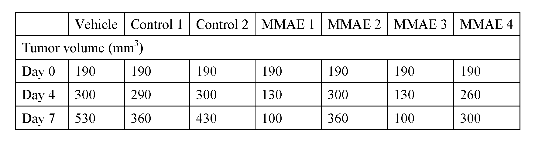

Example 25: Conjugation of activated disulfide-MMAE molecules to albumin and penetratin In a first evaluation, MMAE was conjugated to human serum albumin (HSA). Human serum albumin bears a single free endogenous cysteine residue, Cys-34, to which Cys-reactive molecules can ultimately be conjugated (e.g., Bioconj Chem, 2015, 26(5), 941-949). This Cys residue is blocked as a mixed disulfide with Cys or glutathione, preventing complete conjugation of other molecules to this residue. In a manner analogous to removing such blocking groups from THIOMAB™ antibodies, the blocking group on Cys-34 of HSA was removed through a reduction-reoxidation procedure and the protein was stored in a buffer of 10 mM succinate, pH 5, 150 mM NaCl, 2 mM EDTA. Disulfide-MMAE molecules with varying leaving groups were conjugated to HSA by mixing the protein at 2.2 mg/mL with 1M Tris, pH 8.5 (final buffer concentration of 75 mM Tris, pH 8.5) and with the disulfide MMAE compound at 10 molar equivalents relative to HSA for ~16 hours at room temperature. Conjugate reactions were analyzed by LC/MS. The extent of the expected MMAE-HSA conjugate formed during the reaction is represented by the“Conjugate Ratio”, measured by dividing the abundance of the MS peak for the MMAE-HSA conjugate by the sum total abundance of all peaks observed. The ideal value for the Conjugate Ratio is 1.0. The results are reported in table 31 below wherein the wavy line for the leaving group represents the point of attachment to the disulfide bond, and “HSA CR” refers to human serum albumin conjugate ratio.
In a second evaluation, MMAE was conjugated to penetratin (AntP) peptide. The AntP peptide, with sequence H2N-RQIKIWFQNRRMKWKKC-CONH2, bearing a free Cys thiol at the C-terminus, was synthesized via standard Fmoc solid-phase peptide synthesis methods. This peptide was conjugated at a concentration of 25 uM to disulfide MMAE molecules with varying leaving groups (5 equivalents) in a buffered solution composed of 75 mM Tris, pH 8.5 and 10% DMF for ~16 hours at room temperature. Extent of the reaction was measured by dividing the integrated area for the UV peak of the MMAE-AntP conjugate by the sum of all peaks (“%Product”). The ideal value for %Product is 100%. The results are reported in table 31 below wherein the wavy line for the leaving group represent the point of attachment to the disulfide bond, and“AntP %P” refers to penetratin (AntP) peptide %product.
Table 31: HSA Conjugation Ratio and AntP %Product


When introducing elements of the present disclosure or the preferred embodiments(s) thereof, the articles "a", "an", "the" and "said" are intended to mean that there are one or more of the elements. The terms "comprising", "including" and "having" are intended to be inclusive and mean that there may be additional elements other than the listed elements.
This written description uses examples to disclose the invention, including the best mode, and also to enable any person skilled in the art to practice the invention, including making and using any devices or systems and performing any incorporated methods. The patentable scope of the invention is defined by the claims, and may include other examples that occur to those skilled in the art. Such other examples are intended to be within the scope of the claims if they have structural elements that do not differ from the literal language of the claims, or if they include equivalent structural elements with insubstantial differences from the literal languages of the claims.
Claims
)
189
WHAT IS CLAIMED IS:
1. A linker-drug compound of structure (I):

wherein:
X is a leaving group;
the leaving group, X, and the sulfur atom, S, are bound via a disulfide bond;
R1 and R2 are independently selected from H and C1-3 alkyl, wherein only one of R1 and R2 can be H, or R1 and R2 together with the carbon atom to which they are bound form a three- to six-membered ring or a four- to six-membered ring comprising an oxygen heteroatom;
Sp is a spacer wherein n is 0 or 1 ; and
D is a drug moiety.
2. The linker-drug compound of claim 1 wherein X is selected from the following:

Leaving Group 4; and Leaving Group 5. wherein:
X1, X2, X3, X4 and X5 are independently C, N, S or O, at least one of X1 to X5 is N, the dashed lines represent optional double bonds, and A denotes a six-membered ring;
Y1, Y2 Y3 and Y4 are independently C, N, S or O, at least one of Y1 to Y4 is N, the dashed lines represent optional double bonds, and B denotes a five-membered ring;
Z1, Z2, Z3, Z4, Zs and Z6 are independently C, N, S or O, at least one of Z1 and Z2 is N, the dashed lines represent optional double bonds, E denotes a six-membered ring, and D denotes a fused five-membered ring;
each R3 is independently selected from - C1-4 alkyl, -NO2, -NH2, -C(O)OH, - S(O)(O)R5, -C(O)N(R5)(R6), -OR5, -CI, -F, -CN and -Br;
R5 and R6 are independently selected from H, unsubstituted or substituted C1-6 hydrocarbyl, unsubstituted or substituted C5-6 carbocycle, and unsubstituted or substituted C5-6 heterocycle;
R5 is C1-4 alkyl;
q is 0, 1, 2 or 3;
each carbon atom in the ring structure of leaving group 1, leaving group 2, leaving group 3 and/or leaving group 4 is unsubstituted or substituted with R5;
each nitrogen atom in the ring structure of leaving group 1, leaving group 2, leaving group 3 and/or leaving group 4 is unsubstituted or substituted with R5 to form a tertiary amine or a quaternary amine; and
the wavy lines indicate the point of attachment of the leaving group to the sulfur atom, S, thereby forming a disulfide bond.
3. The linker-drug compound of claim 2 wherein:
X1, X2, X3, X4 and X5 are independently C or N, no more than two of X1 to X5 are N, and ring A is unsaturated;
Y1, Y2 Y3 and Y4 are independently C or N, and ring B is unsaturated;
Z1 is N, Z2 is selected from N, S and O, Z3 to Z6 are independently selected from C and N, no more than two of Z3 to Z6 are N, and ring E is unsaturated; and
each R3 is independently selected from -CH3, -NO2, -NH2, -C(O)OH, - S(O)(O)CH3, -OCH3 and -C(O)N(CH3)2.
4. The linker-drug compound of any one of claims 1 to 3 wherein X is selected from:


5. The linker-drug compound of any one of claims 1 to 4 wherein R1 and R2 are independently selected from H, -CH3 and -CH2CH3, wherein only one of R1 and R2 can be H, or R1 and R2 together with the carbon atom to which they are attached form a ring selected from cyclopropyl, cyclobutyl, cyclopentyl, tetrahydrofuran, cyclohexyl and tetrahydropyran.
6. The linker-drug compound of any one of claims 1 to 4 wherein R1 and R2 are each -CH3 or wherein R1 and R2 together with the carbon atom to which they are bound form a ring selected from cyclopentyl, cyclohexyl, tetrahydrofuran and tetrahydropyran, and wherein X is selected from leaving group 3 and leaving group 4.
7. The linker-drug compound of claim 6 wherein X is selected from:


The linker-drug compound of claim 7 wherein R1 and R2 are each -CH3 and X is

9. The linker-drug compound of any one of claims 1 to 5 wherein (i) R1 is -C¾ and R2 is H, (ii) R1 is -CH2CH3 and R2 is H, or (iii) R1 and R2 together with the carbon atom to which they are bound form a cyclobutyl ring, and wherein X is leaving group 1.
10. The linker-drug compound of claim 9 wherein X is:

11 The linker-drug compound of any one of claims 1 to 10 wherein n is 0 and the bond between the hindered linker carbonyl carbon atom and the drug moiety is an ether or is a tertiary amine formed from an acyclic or cyclic amine drug moiety atom.
12 The linker-drug compound of any one of claims 1 to 10 wherein n is 1, Sp-D is of the structure:

wherein
the wavy line indicates the point of attachment of the spacer to the hindered linker carbonyl carbon atom,
R30 is selected from is selected from H, unsubstituted or substituted C1-6 hydrocarbyl, unsubstituted or substituted C5-6 carbocycle, and unsubstituted or substituted C5-6 heterocycle, and
the linkage of the Sp carbonyl carbon atom to the drug moiety is a secondary amine formed from an aliphatic primary amine drug moiety atom, is a tertiary amine formed from an aliphatic secondary amine drug moiety atom, or is a tertiary amine formed from an acyclic or cyclic amine drug moiety atom.
13. The linker-drug compound of claim 12 wherein R30 is H.
14. The linker-drug compound of any one of claims 1 to 13 wherein the drug moiety is selected from: maytansine, an analog thereof, a derivative thereof and/or a stereoisomer thereof; auristatin, an analog thereof, a derivative thereof and/or a stereoisomer thereof; dolastatin, an analog thereof, a derivative thereof and/or a stereoisomer thereof; calicheamicin, an analog thereof, a derivative thereof and/or a stereoisomer thereof; pyrrolobenzodiazepine (PBD) monomer or dimer, an analog thereof, a derivative thereof and/or a stereoisomer thereof; a l-(chloromethyl)-2,3-dihydro-lH-benzo[e]indole (CBI) monomer or dimer, an analog thereof, a derivative thereof and/or a stereoisomer thereof; CBI-PBD dimer, an analog thereof, a derivative thereof and/or a stereoisomer thereof; amatoxin, an analog thereof, a derivative thereof and/or a stereoisomer thereof; geldanamycin, an analog thereof, a derivative thereof, an enzymatically active toxin thereof, an enzymatically active fragment thereof and/or a stereoisomer thereof; cyclopropane doline, an analog thereof, a derivative thereof and/or a stereoisomer thereof; cyclopropylbenzindole, an analog thereof, a derivative thereof, and/or a
stereoisomer thereof; and elinafide, an analog thereof, a derivative thereof and/or a stereoisomer thereof.
15. The linker-drug compound of any one of claims 1 to 14 wherein X has a pKa of from about -4 to about 12 or from about 2 to about 12.
A disulfide con ugate compound of structure (II)

wherein
the carrier comprises a cysteine sulfur atom SA;
p is 1, 2, 3, 4, 5, 6, 7 or 8;
R1 and R2 are independently selected from H and C1-3 alkyl, wherein only one of R1 and R2 can be H, or R1 and R2 together with the carbon atom to which they are bound form a three- to six-membered ring or a four- to six-membered ring comprising an oxygen heteroatom;
Sp is a spacer wherein n is 0 or 1 ; and
D is a drug moiety.
17. The disulfide conjugate compound of claim 16 wherein R1 and R2 are independently selected from H, -CH3 and -CH2CH3, wherein only one of R1 and R2 can be H, or R1 and R2 together with the carbon atom to which they are bound form a ring selected from cyclopropyl, cyclobutyl, cyclopentyl, cyclohexyl, tetrahydrofuran and tetrahydropyran.
18. The disulfide conjugate compound of claim 17 wherein R1 and R2 are each -CH3 or wherein R1 and R2 together with the carbon atom to which they are bound form a ring selected from cyclopentyl, cyclohexyl, tetrahydrofuran and tetrahydropyran.
19. The disulfide conjugate compound of any one of claims 16 to 18 wherein n is 0 and the bond between the carbonyl carbon atom and the drug moiety is an ether or is a tertiary amine formed from an acyclic or cyclic amine drug moiety atom.
20. The disulfide conjugate compound of any one of claims 16 to 19 wherein n is 1 and Sp- D is of the structure:

wherein
R is selected from is selected from H, unsubstituted or substituted Q1-6 hydrocarbyl, unsubstituted or substituted C5-6 carbocycle, and unsubstituted or substituted C5-6 heterocycle,
the wavy line indicates the point of attachment of the spacer to the carbonyl carbon atom, and
the linkage of the Sp carbonyl carbon atom to the drug moiety is a secondary amine formed from an aliphatic primary amine drug moiety atom, is a tertiary amine formed from an aliphatic secondary amine drug moiety atom, or is a tertiary amine formed from an acyclic or cyclic amine drug atom.
21. The disulfide conjugate compound of claim 20 wherein R30 is H.
22. The disulfide conjugate compound of any one of claims 16 to 21 wherein the carrier is an antibody which binds to one or more tumor-associated antigens or cell-surface receptors.
23. The disulfide conjugate compound of claim 22 wherein the antibody comprises at least one cysteine sulfhydryl moiety engineered for conjugation, wherein the antibody is selected from:
(1) BMPR1B (bone morphogenetic protein receptor-type IB); (2) El 6 (LAT1, SLC7A5); (3) STEAP1 (six transmembrane epithelial antigen of prostate); (4) MUC16 (0772P, CA125); (5) MPF (MPF, MSLN, SMR, megakaryocyte potentiating factor, mesothelin); (6) Napi2b (NAPI-3B, NPTIIb, SLC34A2, solute carrier family 34 (sodium phosphate), member 2, type II sodium-dependent phosphate transporter 3b); (7) Sema 5b (FLJ10372, KIAA1445, Mm.42015, SEMA5B, SEMAG, Semaphorin 5b Hlog, sema domain, seven thrombospondin repeats (type 1 and type 1-like), transmembrane domain
(TM) and short cytoplasmic domain, (semaphorin) 5B); (8) PSCA hlg (2700050C12Rik, C530008O16Rik, RIKEN cDNA 2700050C12, RIKEN cDNA 2700050C12 gene); (9) ETBR (Endothelin type B receptor); (10) MSG783 (RNF124, hypothetical protein FLJ20315); (11) STEAP2 (HGNC_8639, IPCA-1, PCANAP1, STAMP1, STEAP2, STMP, prostate cancer associated gene 1, prostate cancer associated protein 1, six transmembrane epithelial antigen of prostate 2, six transmembrane prostate protein); (12) TrpM4 (BR22450, FLJ20041, TRPM4, TRPM4B, transient receptor potential cation channel, subfamily M, member 4); (13) CRIPTO (CR, CR1, CRGF, CRIPTO, TDGF1, teratocarcinoma-derived growth factor); (14) CD21 (CR2 (Complement receptor 2) or C3DR (C3d/Epstein Barr virus receptor) or Hs 73792); (15) CD79b (CD79B, CD79β, IGb (immunoglobulin-associated beta), B29); (16) FcRH2 (IFGP4, IRTA4, SPAP1A (SH2 domain containing phosphatase anchor protein la), SPAPIB, SPAP1C); (17) HER2; (18) NCA; (19) MDP; (20) IL20Rα ; (21) Brevican; (22) EphB2R; (23) ASLG659; (24) PSCA; (25) GEDA; (26)-BAFF-R (B cell -activating factor receptor, BLyS receptor 3, BR3); (27) CD22 (B-cell receptor CD22-B isoform); (28) CD79a (CD79A, CD79oc, immunoglobulin-associated alpha); (29' CXCR5 (Burkitt's lymphoma receptor 1); (30) HLA-DOB (Beta subunit of MHC class II molecule (la antigen)); (31) P2X5 (Purinergic receptor P2X ligand-gated ion channel 5); (32) CD72 (B-cell differentiation antigen CD72, Lyb-2); (33) LY64 (Lymphocyte antigen 64 (RP105), type I membrane protein of the leucine rich repeat (LRR) family); (34) FcRHl (Fc receptorlike protein 1); (35) FcRH5 (IRTA2, Immunoglobulin superfamily receptor translocation associated 2); (36) TENB2 (putative transmembrane proteoglycan); (37) PMEL17 (silver homolog; SILV; D12S53E; PMEL17; SI; SIL); (38) TMEFF1 (transmembrane protein with EGF-like and two follistatin-like domains 1; Tomoregulin-1); (39) GDNF-Ral (GDNF family receptor alpha 1; GFRAl; GDNFR; GDNFRA; RETL1; TRNR1; RET1L; GDNFR-alphal; GFR-ALPHA-1); (40) Ly6E (lymphocyte antigen 6 complex, locus E; Ly67,RIG-E,SCA-2,TSA-l); (41) TMEM46 (shisa homolog 2 (Xenopus laevis); SHISA2); (42) Ly6G6D (lymphocyte antigen 6 complex, locus G6D; Ly6-D, MEGT1); (43) LGR5 (leucine-rich repeat-containing G protein-coupled receptor 5; GPR49, GPR67); (44) RET (ret proto-oncogene; MEN2A; HSCR1; MEN2B; MTC1; PTC; CDHF12; Hs.168114; RET51; RET-ELEl); (45) LY6K (lymphocyte antigen 6 complex, locus K; LY6K; HSJ001348; FLJ35226); (46) GPR19 (G protein-coupled receptor 19; Mm.4787); (47) GPR54 (KISS1 receptor; KISS1R; GPR54; HOT7T175;
AXOR12); (48) AS PHD 1 (aspartate beta-hydroxylase domain containing 1; LOC253982); (49) Tyrosinase (TYR; OCAIA; OCA1A; tyrosinase; SHEP3); (50) TMEM118 (ring finger protein, transmembrane 2; RNFT2; FLJ14627); (51) GPR172A (G protein-coupled receptor 172A; GPCR41; FLJ11856; D15Ertd747e); (52) CD33; and (53) CLL-1.
24. The disulfide conjugate compound of claim 22 or 23 wherein the antibody is a cysteine- engineered antibody.
25. The disulfide conjugate compound of claim 24 wherein the cysteine-engineered antibody comprises LC K149C, HC A118C, LC S121C or LC V205C as the site of linker conjugation.
26. The disulfide conjugate compound of any one of claims 22 to 25 wherein the antibody is selected from anti-HER2, anti-CD22, anti-CD33, anti-Napi2b, and anti-CLL-1.
27. The disulfide conjugate compound of any one of claims 16 to 26 wherein p is 1, 2, 3, or
4.
28. The disulfide conjugate compound of any one of claims 16 to 27 comprising a mixture of the antibody-drug conjugate compounds, wherein the average drug loading per antibody in the mixture of antibody-drug conjugate compounds is about 2 to about 5.
29. The disulfide conjugate compound of any one of claims 16 to 21 wherein the carrier is a protein.
30. The disulfide conjugate compound of claim 29 wherein the protein is albumin.
31. The disulfide conjugate compound of any one of claims 16 to 21 wherein the carrier is a peptide.
32. The disulfide conjugate compound of claim 31 wherein the peptide is selected from the cell-penetrating peptides antennapedia homeodomain, Tat HIV protein, and engineered cationic peptides.
33. The disulfide conjugate compound of any one of claims 16 to 32 wherein the drug moiety is selected from: maytansine, an analog thereof, a derivative thereof and/or a stereoisomer thereof; auristatin, an analog thereof, a derivative thereof and/or a stereoisomer thereof; dolastatin, an analog thereof, a derivative thereof and/or a stereoisomer thereof; calicheamicin, an analog thereof, a derivative thereof and/or a stereoisomer thereof; pyrrolobenzodiazepine (PBD) monomer or dimer, an analog
thereof, a derivative thereof and/or a stereoisomer thereof; l-(chloromethyl)-2,3- dihydro-lH-benzo[e]indole (CBI) monomer or dimer, an analog thereof, a derivative thereof and/or a stereoisomer thereof; CBI-PBD dimer, an analog thereof, a derivative thereof and/or a stereoisomer thereof; amatoxin, an analog thereof, a derivative thereof and/or a stereoisomer thereof; geldanamycin, an analog thereof, a derivative thereof, an enzymatically active toxin thereof, an enzymatically active fragment thereof and/or a stereoisomer thereof; cyclopropane doline, an analog thereof, a derivative thereof and/or a stereoisomer thereof; cylopropylbenzindole, an analog thereof, a derivative thereof, and/or a stereoisomer thereof; and elinafide, an analog thereof, a derivative thereof and/or a stereoisomer thereof.
34. A method of reparing a disulfide conjugate compound of structure (II):

the method comprising:
(A) forming a reaction mixture comprising
(i) a solvent system comprising water,
(ii) a carrier comprising at least one cysteine having a sulfliydryl moiety having a sulfur atom SA capable of bonding to a linker sulfur atom to form the disulfide bond, and
(iii) a stoichiometric excess of a linker-drug conjugate compound of structure (I):

X is a leaving group;
the leaving group and the linker are bound via disulfide bond;
R1 and R2 are independently selected from H and C1-3 alkyl, wherein only one of R1 and R2 can H, or R1 and R2 together with the carbon atom to which they are bound form a three- to six-membered ring or a four- to six-membered ring comprising an oxygen heteroatom;
Sp is a spacer wherein n is 0 or 1 ; and
D is a drug moiety; and
(B) reacting the reaction mixture to form a product mixture comprising the disulfide conjugate compound of structure (II) wherein p is 1, 2, 3, 4, 5, 6, 7 or 8, and wherein the average value of p for a plurality of formed disulfide conjugate compounds in the product mixture is from about 1 to about 5, from about 1.5 to about 3, from about 1.5 to about 2.5, from about 1.7 to about 2.3, from about 1.8 to about 2.2, or about 2.
35. The method of claim 34 wherein the product mixture further comprises at least one leaving group byproduct species wherein the area percentage of total leaving group byproduct species as compared to the area percentage of formed disulfide conjugate compound as measured by LC/MS is less than 5 area%, less than 4 area%, less than 3 area%, less than 2 area%, less than 1 area%, less than 0.5 area%.
36. The method of claim 34 or claim 35 wherein the product mixture further comprises unconjugated carrier compounds comprising (i) unconjugated carrier monomer species comprising at least one disulfide bond formed by the reaction of two cysteine sulfhydryl moieties, (ii) unconjugated carrier dimer species comprising at least one disulfide bond formed by the reaction of two cysteine sulfhydryl moieties, and (iii) a combination of unconjugated carrier monomer species and unconjugated carrier dimer species, wherein the total concentration of unconjugated carrier compounds compared to the area percentage of formed disulfide conjugate compounds as measured by LC/MS is less than
5 area%, less than 4 area%, less than 3 area%, less than 2 area%, less than 1 area%, less than 0.5 area%, less than 0.3 area%, less than 0.1 area%, or is not detectable.
37. The method of any one of claims 34 to 36 wherein the solvent system comprises a biological buffer and has a pH of about 5.0, about 5.5, about 6.0, about 6.5, about 7.0, about 7.5, about 8.0, about 8.5 or about 9.0, and ranges thereof, such as from about 5.0 to about 9.0, from about 5.0 to about 9.0, from about 6.0 to about 9.0, from about 6.5 to about 9.0, or from about 7.0 to about 9.0, or from about 7.5 to about 8.5.
38. The method of any one of claims 34 to 37 wherein the solvent system further comprises: at least one polar aprotic solvent selected from acetonitrile, tetrahydrofuran, ethyl acetate, acetone, NN-dimethylformamide, dimethyl acetamide, dimethylsulfoxide, propylene glycol, ethylene glycol and dichloromethane,
wherein the polar aprotic solvent concentration in the solvent system is about 5 v/v%, about 10 v/v%, about 15 v/v%, about 20 v/v%, about 25 v/v%, about 30 v/v%, about 40 v/v% or about 50 v/v%, and ranges thereof, such as from about 5 v/v% to about 50 v/v%, from about 25 v/v% to about 50 v/v%, from about 5 v/v% to about 30 v/v%, from about 5 v/v% to about 20 v/v%, from about 5 v/v% to about 15 v/v%, or about 10 v/v%.
39. The method of any one of claims 34 to 38 wherein the solvent system comprises:
at least 50 v/v% or at least 75 v/v% of a biological buffer selected from 50 mM to 100 mM TRIS, BIS-TRIS or HEPES having a pH of from about 6.0 to about 9.0, from about 6.0 to about 8.5, from about 7.0 to about 8.5 or from about 7.5 to about 8.5; and from about 5 v/v% to about 25 v/v% NN-dimethylformamide or dimethyl acetamide or between about 10 v/v% to about 50 v/v% propylene glycol or ethylene glycol.
40. The method of any one of claims 34 to 39 wherein the antibody concentration in the reaction mixture is about 1 mg/mL, about 5 mg/mL, about 10 mg/mL, about 15 mg/mL, about 20 mg/mL, or about 25 mg/mL, and ranges thereof, such as from about 1 mg/mL to about 25 mg/mL, from about 1 mg/mL to about 20 mg/mL, from about 1 mg/mL to about 15 mg/mL, from about 1 mg/mL to about 10 mg/mL, from about 5 mg/mL to about 25 mg/mL, from about 5 mg/mL to about 20 mg/mL or from about 5 mg/mL to about 15 mg/mL.
41. The method of any one of claims 34 to 40 wherein the equivalent ratio of linker-drug conjugate compound to the carrier is about 2:1, about 3: 1, about 5:1, about 10:1, about 15:1 or about 20:1, and ranges thereof, such as from about 2: 1 to about 20:1, from about 3: 1 to about 15:1, from about 3:1 to about 10: 1, or from about 5:1 to about 10: 1.
42. The method of any one of claims 34 to 41 wherein:
the reaction temperature is about 10°C, about 15°C, about 20°C, about 25°C, about 30°C, about 35°C, about 40°C, about 45°C or about 50°C, and ranges thereof such as from about 10°C to about 50°C, from about 15°C to about 45°C, from about 15°C to
about 40°C, from about 20°C to about 40°C, from about 20°C to about 35°C, or from about 20°C to about 30°C; and
the reaction time is about 05 hours, about 1 hour, about 4 hours, about 8 hours, about 12 hours, about 18 hours, about 24 hours, about 36 hours or about 48 hours, and ranges thereof, such as from about 0.5 hours to about 48 hours, or from about 1 hour to about 24 hours.
43. The method of any one of claims 34 to 42 wherein X is selected from the following:

wherein:
X1, X2, X3, X4 and X5 are independently C, N, S or O, at least one of X1 to Xs is N, the dashed lines represent optional double bonds, and A denotes a six-membered ring;
Y1, Y2 Y3 and Y4 are independently C, N, S or O, at least one of Y1 to Y4 is N, the dashed lines represent optional double bonds, and B denotes a five-membered ring;
Z1, Z2, Z3, Z4, Z5 and Z6 are independently C, N, S or O, at least one of Z1 and Z2 is N, the dashed lines represent optional double bonds, E denotes a six-membered ring, and D denotes a fused five-membered ring;
each R3 is independently selected from -C1-4 alkyl, -N02, -NH2, -C(O)OH, - S(O)(O)R5, -C(O)N(R5)(R6), -OR5, -CI, -F, -CN and -Br;
R5 and R6 axe independently selected from H, unsubstituted or substituted C1-6 hydrocarbyl, unsubstituted or substituted C5-6 carbocycle, and unsubstituted or substituted C5-6 heterocycle;
R5 is C1-4 alkyl;
q is 0, 1, 2 or 3;
each carbon atom in the ring structure of leaving group 1, leaving group 2, leaving group 3 and/or leaving group 4 is unsubstituted or substituted with R5;
each nitrogen atom in the ring structure of leaving group 1, leaving group 2, leaving group 3 and/or leaving group 4 is unsubstituted or substituted with R5 to form a tertiary amine or a quaternary amine; and
the wavy lines indicate the point of attachment of the leaving group to the sulfur atom, S, thereby forming a disulfide bond.
44 The method of claim 43 wherein:
X1, X2, X3, X4 and X5 are independently C or N, no more than two of X1 to X5 are N, and ring A is unsaturated;
Y1, Y2 Y3 and Y4 are independently C or N, and ring B is unsaturated;
Z1 is N, Z2 is selected from N, S and O, Z3 to Z6 are independently selected from C and N, no more than two of Z3 to Z6 are N, and ring E is unsaturated; and
each R3 is independently selected from -CH3, -N02, -NH2, -C(O)OH, - S(O)(O)CH3, -OCH3, and -C(O)N(CH3)2.
45 The method of any one of claims 42 to 44 wherein X is selected from:

02N , 02N

N02

COOH

N02
 HOOC
HOOC

46. The method of any one of claims 34 to 45 wherein R1 and R2 are independently selected from H, -CH3 and -CH2CH3, wherein only one of R1 and R2 is H, or R1 and R2 together with the carbon atom to which they are bound form a ring selected from cyclopropyl, cyclobutyl, cyclopentyl, cyclohexyl, tetrahydrofuran and tetrahydropyran.
47. The method of any one of claims 34 to 46 wherein R1 and R2 are each -C¾ or wherein R1 and R2 together with the carbon atom to which they are bound form a ring selected from cyclopentyl, cyclohexyl, tetrahydrofuran and tetrahydropyran, and wherein X is selected from leaving group 3 and leaving group 4.

49. The linker-drug compound of claim 48 wherein R1 and R2 are each -CH3 and X is O.
50. The method of any one of claims 34 to 46 wherein: (i) R1 is -CH3 and R2 is H, (ii) R1 is - CH2CH3 and R2 is H, or (iii) R1 and R2 together with the carbon atom to which they are bound form a cyclobutyl ring, and wherein X is leaving group 1.
51. The method of claim 50 wherein X is:
02N,

52. The method of any one of claims 34 to 51 wherein n is 0 and the bond between the hindered linker carbonyl carbon atom and the drug moiety is an ether or is a tertiary amine formed from an acyclic or cyclic amine drug moiety atom.
53. The linker-drug compound of any one of claims 34 to 52 wherein n is 1, Sp-D is of the structure:

wherein
R30 is selected from is selected from H, unsubstituted or substituted C1-6 hydrocarbyl, unsubstituted or substituted C5-6 carbocycle, and unsubstituted or substituted C5-6 heterocycle,
the wavy line indicates the point of attachment of the spacer to the hindered linker carbonyl carbon atom, and
the linkage of the Sp carbonyl carbon atom to the drug moiety is a secondary amine formed from an aliphatic primary amine drug moiety atom, is a tertiary amine formed from an aliphatic secondary amine drug moiety atom, or is a tertiary amine formed from an acyclic or cyclic amine drug moiety atom.
54. The method of claim 53 wherein R30 is H.
55. The method of any one of claims 34 to 54 wherein the drug moiety is selected from: maytansine, an analog thereof, a derivative thereof and/or a stereoisomer thereof; auristatin, an analog thereof, a derivative thereof and/or a stereoisomer thereof; dolastatin, an analog thereof, a derivative thereof and/or a stereoisomer thereof; calicheamicin, an analog thereof, a derivative thereof and/or a stereoisomer thereof; pyrrolobenzodiazepine (PBD) monomer or dimer, an analog thereof, a derivative thereof and/or a stereoisomer thereof; l-(chloromethyl)-2,3-dihydro-lH-benzo[e]indole (CBI) monomer or dimer, an analog thereof, a derivative thereof and/or a stereoisomer thereof; CBI-PBD dimer, an analog thereof, a derivative thereof and/or a stereoisomer thereof; amatoxin, an analog thereof, a derivative thereof and/or a stereoisomer thereof; geldanamycin, an analog thereof, a derivative thereof, an enzymatically active toxin thereof, an enzymatically active fragment thereof and/or a stereoisomer thereof; cyclopropane doline, an analog thereof, a derivative thereof and/or a stereoisomer thereof; cylopropylbenzindole, an analog thereof, a derivative thereof, and/or a stereoisomer thereof; and elinafide, an analog thereof, a derivative thereof and/or a stereoisomer thereof.
56. The method of any one of claims 34 to 55 wherein X has a pKa of from about -4 to about 12 or from about 2 to about 12.
57. A pharmaceutical composition comprising the disulfide conjugate compound according to any one of claims 16 to 33 and a pharmaceutically acceptable diluent, carrier or excipient.
58. The pharmaceutical composition of claim 57, further comprising a therapeutically effective amount of a chemotherapeutic agent.
59. A method of treating cancer comprising administering to a patient the pharmaceutical composition of claim 57 or claim 58.
60. The method of claim 59 wherein the patient is administered a chemotherapeutic agent, in combination with the antibody-drug conjugate.
61. Use of an antibody-drug conjugate compound according to any one of claims 16 to 33 in the manufacture of a medicament for the treatment of cancer in a mammal.
62. An antibody-drug conjugate compound according to any one of claims 16 to 33 for use in a method for treating cancer.
63. An article of manufacture comprising a pharmaceutical composition of claim 57 or claim 58, a container, and a package insert or label indicating that the pharmaceutical composition can be used to treat cancer.
Priority Applications (3)
| Application Number | Priority Date | Filing Date | Title |
|---|---|---|---|
| JP2018519319A JP6621919B2 (en) | 2015-10-16 | 2016-10-14 | Constrained disulfide drug conjugate |
| EP16788243.0A EP3362100B1 (en) | 2015-10-16 | 2016-10-14 | Hindered disulfide drug conjugates |
| CN201680070988.6A CN108472384B (en) | 2015-10-16 | 2016-10-14 | Hindered disulfide drug conjugates |
Applications Claiming Priority (2)
| Application Number | Priority Date | Filing Date | Title |
|---|---|---|---|
| CN2015092084 | 2015-10-16 | ||
| CNPCT/CN2015/092084 | 2015-10-16 |
Publications (1)
| Publication Number | Publication Date |
|---|---|
| WO2017064675A1 true WO2017064675A1 (en) | 2017-04-20 |
Family
ID=57209664
Family Applications (1)
| Application Number | Title | Priority Date | Filing Date |
|---|---|---|---|
| PCT/IB2016/056186 WO2017064675A1 (en) | 2015-10-16 | 2016-10-14 | Hindered disulfide drug conjugates |
Country Status (6)
| Country | Link |
|---|---|
| US (1) | US10729738B2 (en) |
| EP (1) | EP3362100B1 (en) |
| JP (1) | JP6621919B2 (en) |
| CN (1) | CN108472384B (en) |
| MA (1) | MA43354A (en) |
| WO (1) | WO2017064675A1 (en) |
Cited By (10)
| Publication number | Priority date | Publication date | Assignee | Title |
|---|---|---|---|---|
| WO2018086593A1 (en) * | 2016-11-11 | 2018-05-17 | 礼沃(上海)医药科技有限公司 | Nitrogen-containing heterocyclic compound, preparation method, intermediate, pharmaceutical composition and use |
| US10532108B2 (en) | 2016-08-11 | 2020-01-14 | Genentech, Inc. | Pyrrolobenzodiazepine prodrugs and antibody conjugates thereof |
| WO2020123275A1 (en) | 2018-12-10 | 2020-06-18 | Genentech, Inc. | Photocrosslinking peptides for site specific conjugation to fc-containing proteins |
| US20210299137A1 (en) * | 2018-01-05 | 2021-09-30 | Cybrexa 1, Inc. | Compounds, compositions, and methods for treatment of diseases involving acidic or hypoxic diseased tissues |
| EP3720437A4 (en) * | 2017-12-06 | 2021-11-03 | Yale University | Prodrugs activated by reduction in the cytosol |
| US20220088204A1 (en) * | 2019-01-25 | 2022-03-24 | Yale University | Anticancer drugs and methods of making and using same |
| WO2022132058A1 (en) * | 2020-12-14 | 2022-06-23 | Centrum Experimentálnej Medicíny Sav | Cemtirestat disulfide, prodrug of aldo-keto reductase inhibitor, preparation, pharmaceutical composition and use thereof |
| US11547762B2 (en) | 2016-10-05 | 2023-01-10 | Genentech, Inc. | Methods for preparing antibody drug conjugates |
| US11555019B2 (en) | 2019-07-10 | 2023-01-17 | Cybrexa 3, Inc. | Peptide conjugates of microtubule-targeting agents as therapeutics |
| US11634508B2 (en) | 2019-07-10 | 2023-04-25 | Cybrexa 2, Inc. | Peptide conjugates of cytotoxins as therapeutics |
Families Citing this family (5)
| Publication number | Priority date | Publication date | Assignee | Title |
|---|---|---|---|---|
| MX2018015683A (en) | 2016-06-17 | 2019-05-27 | Magenta Therapeutics Inc | Compositions and methods for the depletion of cells. |
| CA3049501A1 (en) | 2017-01-20 | 2018-07-26 | Magenta Therapeutics, Inc. | Compositions and methods for the depletion of cd137+ cells |
| US11566044B2 (en) | 2017-07-26 | 2023-01-31 | The University Of Queensland | Disulfide bond containing compounds and uses thereof |
| JP7210770B2 (en) * | 2019-03-29 | 2023-01-23 | メドイミューン・リミテッド | Compounds and their complexes |
| CN112661986B (en) * | 2020-12-07 | 2023-08-22 | 南京师范大学 | Amphoteric ion-based hydrogen sulfide driven nanomotor and preparation method and application thereof |
Citations (356)
| Publication number | Priority date | Publication date | Assignee | Title |
|---|---|---|---|---|
| US3896111A (en) | 1973-02-20 | 1975-07-22 | Research Corp | Ansa macrolides |
| US4137230A (en) | 1977-11-14 | 1979-01-30 | Takeda Chemical Industries, Ltd. | Method for the production of maytansinoids |
| US4151042A (en) | 1977-03-31 | 1979-04-24 | Takeda Chemical Industries, Ltd. | Method for producing maytansinol and its derivatives |
| US4248870A (en) | 1978-10-27 | 1981-02-03 | Takeda Chemical Industries, Ltd. | Maytansinoids and use |
| US4256746A (en) | 1978-11-14 | 1981-03-17 | Takeda Chemical Industries | Dechloromaytansinoids, their pharmaceutical compositions and method of use |
| US4260608A (en) | 1978-11-14 | 1981-04-07 | Takeda Chemical Industries, Ltd. | Maytansinoids, pharmaceutical compositions thereof and methods of use thereof |
| US4265814A (en) | 1978-03-24 | 1981-05-05 | Takeda Chemical Industries | Matansinol 3-n-hexadecanoate |
| US4294757A (en) | 1979-01-31 | 1981-10-13 | Takeda Chemical Industries, Ltd | 20-O-Acylmaytansinoids |
| US4307016A (en) | 1978-03-24 | 1981-12-22 | Takeda Chemical Industries, Ltd. | Demethyl maytansinoids |
| US4308269A (en) | 1979-06-11 | 1981-12-29 | Takeda Chemical Industries, Ltd. | Maytansinoids, pharmaceutical compositions thereof and method of use thereof |
| US4308268A (en) | 1979-06-11 | 1981-12-29 | Takeda Chemical Industries, Ltd. | Maytansinoids, pharmaceutical compositions thereof and method of use thereof |
| US4309428A (en) | 1979-07-30 | 1982-01-05 | Takeda Chemical Industries, Ltd. | Maytansinoids |
| US4313946A (en) | 1981-01-27 | 1982-02-02 | The United States Of America As Represented By The Secretary Of Agriculture | Chemotherapeutically active maytansinoids from Trewia nudiflora |
| US4315929A (en) | 1981-01-27 | 1982-02-16 | The United States Of America As Represented By The Secretary Of Agriculture | Method of controlling the European corn borer with trewiasine |
| US4317821A (en) | 1979-06-08 | 1982-03-02 | Takeda Chemical Industries, Ltd. | Maytansinoids, their use and pharmaceutical compositions thereof |
| US4322348A (en) | 1979-06-05 | 1982-03-30 | Takeda Chemical Industries, Ltd. | Maytansinoids |
| US4331598A (en) | 1979-09-19 | 1982-05-25 | Takeda Chemical Industries, Ltd. | Maytansinoids |
| US4362663A (en) | 1979-09-21 | 1982-12-07 | Takeda Chemical Industries, Ltd. | Maytansinoid compound |
| US4364866A (en) | 1979-09-21 | 1982-12-21 | Takeda Chemical Industries, Ltd. | Maytansinoids |
| US4371533A (en) | 1980-10-08 | 1983-02-01 | Takeda Chemical Industries, Ltd. | 4,5-Deoxymaytansinoids, their use and pharmaceutical compositions thereof |
| US4424219A (en) | 1981-05-20 | 1984-01-03 | Takeda Chemical Industries, Ltd. | 9-Thiomaytansinoids and their pharmaceutical compositions and use |
| US4450254A (en) | 1980-11-03 | 1984-05-22 | Standard Oil Company | Impact improvement of high nitrile resins |
| US4676980A (en) | 1985-09-23 | 1987-06-30 | The United States Of America As Represented By The Secretary Of The Department Of Health And Human Services | Target specific cross-linked heteroantibodies |
| US4816567A (en) | 1983-04-08 | 1989-03-28 | Genentech, Inc. | Recombinant immunoglobin preparations |
| EP0328147A2 (en) | 1988-02-11 | 1989-08-16 | Bristol-Myers Squibb Company | Anthracycline immunoconjugates having a novel linker and methods for their production |
| EP0404097A2 (en) | 1989-06-22 | 1990-12-27 | BEHRINGWERKE Aktiengesellschaft | Bispecific and oligospecific, mono- and oligovalent receptors, production and applications thereof |
| WO1991002536A1 (en) | 1989-08-23 | 1991-03-07 | Scripps Clinic And Research Foundation | Compositions and methods for detection and treatment of epstein-barr virus infection and immune disorders |
| WO1992007574A1 (en) | 1990-10-25 | 1992-05-14 | Tanox Biosystems, Inc. | Glycoproteins associated with membrane-bound immunoglobulins as antibody targets on b cells |
| WO1992017497A1 (en) | 1991-03-29 | 1992-10-15 | Genentech, Inc. | Human pf4a receptors and their use |
| EP0522868A1 (en) | 1991-07-12 | 1993-01-13 | SHIONOGI SEIYAKU KABUSHIKI KAISHA trading under the name of SHIONOGI & CO. LTD. | A human endothelin receptor |
| JPH053790B2 (en) | 1983-11-02 | 1993-01-18 | Canon Kk | |
| WO1993001161A1 (en) | 1991-07-11 | 1993-01-21 | Pfizer Limited | Process for preparing sertraline intermediates |
| US5208020A (en) | 1989-10-25 | 1993-05-04 | Immunogen Inc. | Cytotoxic agents comprising maytansinoids and their therapeutic use |
| WO1993008829A1 (en) | 1991-11-04 | 1993-05-13 | The Regents Of The University Of California | Compositions that mediate killing of hiv-infected cells |
| WO1993016185A2 (en) | 1992-02-06 | 1993-08-19 | Creative Biomolecules, Inc. | Biosynthetic binding protein for cancer marker |
| WO1993021232A1 (en) | 1992-04-10 | 1993-10-28 | Research Development Foundation | IMMUNOTOXINS DIRECTED AGAINST c-erbB-2 (HER-2/neu) RELATED SURFACE ANTIGENS |
| WO1994010312A1 (en) | 1992-10-23 | 1994-05-11 | Chugai Seiyaku Kabushiki Kaisha | Gene coding for megakaryocyte potentiator |
| WO1994029351A2 (en) | 1993-06-16 | 1994-12-22 | Celltech Limited | Antibodies |
| WO1994028931A1 (en) | 1993-06-11 | 1994-12-22 | Genentech, Inc. | Methods for treating inflammatory disorders |
| US5440021A (en) | 1991-03-29 | 1995-08-08 | Chuntharapai; Anan | Antibodies to human IL-8 type B receptor |
| US5500362A (en) | 1987-01-08 | 1996-03-19 | Xoma Corporation | Chimeric antibody with specificity to human B cell surface antigen |
| WO1996027011A1 (en) | 1995-03-01 | 1996-09-06 | Genentech, Inc. | A method for making heteromultimeric polypeptides |
| EP0425235B1 (en) | 1989-10-25 | 1996-09-25 | Immunogen Inc | Cytotoxic agents comprising maytansinoids and their therapeutic use |
| WO1996030514A1 (en) | 1995-03-31 | 1996-10-03 | University Of Washington | Intracellular domain of the her-2/neu protein for prevention or treatment of malignancies |
| US5571894A (en) | 1991-02-05 | 1996-11-05 | Ciba-Geigy Corporation | Recombinant antibodies specific for a growth factor receptor |
| US5585499A (en) | 1992-03-25 | 1996-12-17 | Immunogen Inc. | Cyclopropylbenzindole-containing cytotoxic drugs |
| US5587458A (en) | 1991-10-07 | 1996-12-24 | Aronex Pharmaceuticals, Inc. | Anti-erbB-2 antibodies, combinations thereof, and therapeutic and diagnostic uses thereof |
| WO1997007198A2 (en) | 1995-08-11 | 1997-02-27 | Genetics Institute, Inc. | Dna sequences and secreted proteins encoded thereby |
| US5624821A (en) | 1987-03-18 | 1997-04-29 | Scotgen Biopharmaceuticals Incorporated | Antibodies with altered effector functions |
| US5635483A (en) | 1992-12-03 | 1997-06-03 | Arizona Board Of Regents Acting On Behalf Of Arizona State University | Tumor inhibiting tetrapeptide bearing modified phenethyl amides |
| US5644033A (en) | 1992-12-22 | 1997-07-01 | Health Research, Inc. | Monoclonal antibodies that define a unique antigen of human B cell antigen receptor complex and methods of using same for diagnosis and treatment |
| US5663149A (en) | 1994-12-13 | 1997-09-02 | Arizona Board Of Regents Acting On Behalf Of Arizona State University | Human cancer inhibitory pentapeptide heterocyclic and halophenyl amides |
| WO1997044452A1 (en) | 1996-05-17 | 1997-11-27 | Schering Corporation | Human b-cell antigens, related reagents |
| US5712374A (en) | 1995-06-07 | 1998-01-27 | American Cyanamid Company | Method for the preparation of substantiallly monomeric calicheamicin derivative/carrier conjugates |
| US5714586A (en) | 1995-06-07 | 1998-02-03 | American Cyanamid Company | Methods for the preparation of monomeric calicheamicin derivative/carrier conjugates |
| US5739116A (en) | 1994-06-03 | 1998-04-14 | American Cyanamid Company | Enediyne derivatives useful for the synthesis of conjugates of methyltrithio antitumor agents |
| US5750373A (en) | 1990-12-03 | 1998-05-12 | Genentech, Inc. | Enrichment method for variant proteins having altered binding properties, M13 phagemids, and growth hormone variants |
| US5767237A (en) | 1993-10-01 | 1998-06-16 | Teikoku Hormone Mfg. Co., Ltd. | Peptide derivatives |
| US5770429A (en) | 1990-08-29 | 1998-06-23 | Genpharm International, Inc. | Transgenic non-human animals capable of producing heterologous antibodies |
| US5773223A (en) | 1993-09-02 | 1998-06-30 | Chiron Corporation | Endothelin B1, (ETB1) receptor polypeptide and its encoding nucleic acid methods, and uses thereof |
| US5780588A (en) | 1993-01-26 | 1998-07-14 | Arizona Board Of Regents | Elucidation and synthesis of selected pentapeptides |
| US5792616A (en) | 1990-05-29 | 1998-08-11 | The United States Of America | Antibodies to human cripto protein |
| WO1998037193A1 (en) | 1997-02-20 | 1998-08-27 | Zymogenetics, Inc. | Zcytor7 cytokine receptor |
| US5807715A (en) | 1984-08-27 | 1998-09-15 | The Board Of Trustees Of The Leland Stanford Junior University | Methods and transformed mammalian lymphocyte cells for producing functional antigen-binding protein including chimeric immunoglobulin |
| WO1998040403A1 (en) | 1997-03-10 | 1998-09-17 | The Regents Of The University Of California | Psca: prostate stem cell antigen |
| US5821337A (en) | 1991-06-14 | 1998-10-13 | Genentech, Inc. | Immunoglobulin variants |
| EP0875569A1 (en) | 1997-04-28 | 1998-11-04 | Smithkline Beecham Corporation | A human sodium dependent phosphate transporter (IPT-1) |
| WO1998050431A2 (en) | 1997-05-02 | 1998-11-12 | Genentech, Inc. | A method for making multispecific antibodies having heteromultimeric and common components |
| WO1998051805A1 (en) | 1997-05-15 | 1998-11-19 | Abbott Laboratories | Reagents and methods useful for detecting diseases of the prostate |
| WO1998051824A1 (en) | 1997-05-15 | 1998-11-19 | Abbott Laboratories | Reagents and methods useful for detecting disease of the urinary tract |
| US5854399A (en) | 1991-08-23 | 1998-12-29 | The United States Of America As Represented By The Department Of Health And Human Services | Antibodies specific for human cripto-related polypeptide CR-3 |
| US5869445A (en) | 1993-03-17 | 1999-02-09 | University Of Washington | Methods for eliciting or enhancing reactivity to HER-2/neu protein |
| US5869046A (en) | 1995-04-14 | 1999-02-09 | Genentech, Inc. | Altered polypeptides with increased half-life |
| WO1999028468A1 (en) | 1997-12-02 | 1999-06-10 | The Regents Of The University Of California | Modulating b lymphocyte chemokine/receptor interactions |
| WO1999046284A2 (en) | 1998-03-13 | 1999-09-16 | The Burnham Institute | Molecules that home to various selected organs or tissues |
| WO1999051642A1 (en) | 1998-04-02 | 1999-10-14 | Genentech, Inc. | Antibody variants and fragments thereof |
| US5976551A (en) | 1991-11-15 | 1999-11-02 | Institut Pasteur And Institut Nationale De La Sante Et De La Recherche Medicale | Altered major histocompatibility complex (MHC) determinant and method of using the determinant |
| WO1999058658A2 (en) | 1998-05-13 | 1999-11-18 | Epimmune, Inc. | Expression vectors for stimulating an immune response and methods of using the same |
| WO2000012507A2 (en) | 1998-08-27 | 2000-03-09 | Spirogen Limited | Pyrrolobenzodiazepines |
| WO2000012130A1 (en) | 1998-08-27 | 2000-03-09 | Smithkline Beecham Corporation | Rp105 agonists and antagonists |
| WO2000014228A1 (en) | 1998-09-03 | 2000-03-16 | Japan Science And Technology Corporation | Neutral amino acid transporter and gene thereof |
| WO2000020579A1 (en) | 1998-10-02 | 2000-04-13 | Mcmaster University | Spliced form of erbb-2/neu oncogene |
| WO2000022129A1 (en) | 1998-10-13 | 2000-04-20 | Arena Pharmaceuticals, Inc. | Non-endogenous, constitutively activated human g protein-coupled receptors |
| WO2000032752A1 (en) | 1998-12-02 | 2000-06-08 | The Regents Of The University Of California | Psca: prostate stem cell antigen and uses thereof |
| US6075181A (en) | 1990-01-12 | 2000-06-13 | Abgenix, Inc. | Human antibodies derived from immunized xenomice |
| WO2000036107A2 (en) | 1998-12-17 | 2000-06-22 | Corixa Corporation | Compositions and methods for therapy and diagnosis of ovarian cancer |
| WO2000040614A2 (en) | 1998-12-30 | 2000-07-13 | Beth Israel Deaconess Medical Center, Inc. | Characterization of the soc/crac calcium channel protein family |
| WO2000044899A1 (en) | 1999-01-29 | 2000-08-03 | Corixa Corporation | Her-2/neu fusion proteins |
| WO2000053216A2 (en) | 1999-03-05 | 2000-09-14 | Smithkline Beecham Biologicals S.A. | Use of casb616 polypeptides and polynucleotides for cancer treatment |
| WO2000055351A1 (en) | 1999-03-12 | 2000-09-21 | Human Genome Sciences, Inc. | Human colon cancer associated gene sequences and polypeptides |
| US6150584A (en) | 1990-01-12 | 2000-11-21 | Abgenix, Inc. | Human antibodies derived from immunized xenomice |
| US6153408A (en) | 1991-11-15 | 2000-11-28 | Institut Pasteur And Institut National De La Sante Et De La Recherche Medicale | Altered major histocompatibility complex (MHC) determinant and methods of using the determinant |
| WO2000075655A1 (en) | 1999-06-03 | 2000-12-14 | Takeda Chemical Industries, Ltd. | Screening method with the use of cd100 |
| WO2001000244A2 (en) | 1999-06-25 | 2001-01-04 | Genentech, Inc. | METHODS OF TREATMENT USING ANTI-ErbB ANTIBODY-MAYTANSINOID CONJUGATES |
| US6194551B1 (en) | 1998-04-02 | 2001-02-27 | Genentech, Inc. | Polypeptide variants |
| WO2001016318A2 (en) | 1999-09-01 | 2001-03-08 | Genentech, Inc. | Secreted and transmembrane polypeptides and nucleic acids encoding the same |
| WO2001038490A2 (en) | 1999-11-29 | 2001-05-31 | The Trustees Of Columbia University In The City Of New York | ISOLATION OF FIVE NOVEL GENES CODING FOR NEW Fc RECEPTORS-TYPE MELANOMA INVOLVED IN THE PATHOGENESIS OF LYMPHOMA/MELANOMA |
| WO2001040269A2 (en) | 1999-11-30 | 2001-06-07 | Corixa Corporation | Compositions and methods for therapy and diagnosis of breast cancer |
| WO2001040309A2 (en) | 1999-10-29 | 2001-06-07 | Genentech, Inc. | Anti-prostate stem cell antigen (psca) antibody compositions and methods of use |
| WO2001041787A1 (en) | 1999-12-10 | 2001-06-14 | Epimmune Inc. | INDUCING CELLULAR IMMUNE RESPONSES TO HER2/neu USING PEPTIDE AND NUCLEIC ACID COMPOSITIONS |
| US6248516B1 (en) | 1988-11-11 | 2001-06-19 | Medical Research Council | Single domain ligands, receptors comprising said ligands methods for their production, and use of said ligands and receptors |
| WO2001046261A1 (en) | 1999-12-23 | 2001-06-28 | Zymogenetics, Inc. | Method for treating inflammation |
| WO2001046232A2 (en) | 1999-12-23 | 2001-06-28 | Zymogenetics, Inc. | Soluble interleukin-20 receptor |
| WO2001048204A1 (en) | 1999-12-23 | 2001-07-05 | Agresearch Limited | Mutated bmp1b receptor as regulator of ovulation rate |
| WO2001053463A2 (en) | 2000-01-21 | 2001-07-26 | Corixa Corporation | COMPOUNDS AND METHODS FOR PREVENTION AND TREATMENT OF HER-2/neu ASSOCIATED MALIGNANCIES |
| WO2001057188A2 (en) | 2000-02-03 | 2001-08-09 | Hyseq, Inc. | Novel nucleic acids and polypeptides |
| WO2001062794A2 (en) | 2000-02-22 | 2001-08-30 | Millennium Pharmaceuticals, Inc. | 18607, a human calcium channel |
| WO2001066689A2 (en) | 2000-03-07 | 2001-09-13 | Hyseq, Inc. | Novel nucleic acids and polypeptides |
| WO2001072830A2 (en) | 2000-03-31 | 2001-10-04 | Ipf Pharmaceuticals Gmbh | Diagnostic and medicament for analysing the cell surface proteome of tumour and inflammatory cells and for treating tumorous and inflammatory diseases, preferably using a specific chemokine receptor analysis and the chemokine receptor-ligand interaction |
| WO2001072962A2 (en) | 2000-03-24 | 2001-10-04 | Fahri Saatcioglu | Novel prostate-specific or testis-specific nucleic acid molecules, polypeptides, and diagnostic and therapeutic methods |
| WO2001075177A2 (en) | 2000-04-03 | 2001-10-11 | The Government Of The United States Of America, As Represented By The Secretary, Department Of Health And Human Services | Tumor markers in ovarian cancer |
| WO2001077172A2 (en) | 2000-04-07 | 2001-10-18 | Arena Pharmaceuticals, Inc. | Non-endogenous, constitutively activated known g protein-coupled receptors |
| WO2001088133A2 (en) | 2000-05-18 | 2001-11-22 | Lexicon Genetics Incorporated | Human semaphorin homologs and polynucleotides encoding the same |
| WO2001090304A2 (en) | 2000-05-19 | 2001-11-29 | Human Genome Sciences, Inc. | Nucleic acids, proteins, and antibodies |
| WO2001094641A2 (en) | 2000-06-09 | 2001-12-13 | Idec Pharmaceuticals Corporation | Gene targets and ligands that bind thereto for treatment and diagnosis of ovarian carcinomas |
| WO2001098351A2 (en) | 2000-06-16 | 2001-12-27 | Incyte Genomics, Inc. | G-protein coupled receptors |
| US20010055751A1 (en) | 1997-03-10 | 2001-12-27 | Reiter Robert E | PSCA: Prostate stem cell antigen and uses thereof |
| WO2002002624A2 (en) | 2000-06-30 | 2002-01-10 | Amgen, Inc. | B7-like molecules and uses thereof |
| WO2002002587A1 (en) | 2000-06-30 | 2002-01-10 | Human Genome Sciences, Inc. | B7-like polynucleotides, polypeptides, and antibodies |
| WO2002002634A2 (en) | 2000-06-30 | 2002-01-10 | Incyte Genomics, Inc. | Human extracellular matrix and cell adhesion polypeptides |
| WO2002006317A2 (en) | 2000-07-17 | 2002-01-24 | Corixa Corporation | Compositions and methods for the therapy and diagnosis of ovarian cancer |
| WO2002006339A2 (en) | 2000-07-03 | 2002-01-24 | Curagen Corporation | Proteins and nucleic acids encoding same |
| WO2002010187A1 (en) | 2000-07-27 | 2002-02-07 | Mayo Foundation For Medical Education And Research | B7-h3 and b7-h4, novel immunoregulatory molecules |
| WO2002010382A2 (en) | 2000-07-28 | 2002-02-07 | Ulrich Wissenbach | Trp8, trp9 and trp10, markers for cancer |
| WO2002012341A2 (en) | 2000-08-03 | 2002-02-14 | Corixa Corporation | Her-2/neu fusion proteins |
| WO2002013847A2 (en) | 2000-08-14 | 2002-02-21 | Corixa Corporation | Methods for diagnosis and therapy of hematological and virus-associated malignancies |
| WO2002014503A2 (en) | 2000-08-14 | 2002-02-21 | Corixa Corporation | Compositions and methods for the therapy and diagnosis of her-2/neu-associated malignancies |
| WO2002016429A2 (en) | 2000-08-24 | 2002-02-28 | Genentech, Inc. | Compositions and methods for the diagnosis and treatment of tumor |
| WO2002016413A2 (en) | 2000-08-24 | 2002-02-28 | Glaxosmithkline Biologicals S.A. | Cripto tumour polypeptide |
| WO2002022153A2 (en) | 2000-09-15 | 2002-03-21 | Zymogenetics, Inc. | Use of a polypeptide comprising the extracellular domains of il-20rb for the treatment of inflammation |
| US20020034749A1 (en) | 1997-11-18 | 2002-03-21 | Billing-Medel Patricia A. | Reagents and methods useful for detecting diseases of the breast |
| WO2002022660A2 (en) | 2000-09-11 | 2002-03-21 | Hyseq, Inc. | Novel nucleic acids and polypeptides |
| WO2002022808A2 (en) | 2000-09-18 | 2002-03-21 | Biogen, Inc. | Cripto mutant and uses thereof |
| WO2002022636A1 (en) | 2000-09-15 | 2002-03-21 | Isis Pharmaceuticals, Inc. | Antisense modulation of her-2 expression |
| WO2002024909A2 (en) | 2000-09-18 | 2002-03-28 | Biogen, Inc. | Receptor nucleic acids and polypeptides |
| US20020042366A1 (en) | 1999-12-23 | 2002-04-11 | Penny Thompson | Method for treating inflammation |
| WO2002030268A2 (en) | 2000-10-13 | 2002-04-18 | Eos Biotechnology, Inc. | Methods of diagnosis of prostate cancer, compositions and methods of screening for modulators of prostate cancer |
| WO2002038766A2 (en) | 2000-11-07 | 2002-05-16 | Zymogenetics, Inc. | Human tumor necrosis factor receptor |
| WO2002054940A2 (en) | 2001-01-12 | 2002-07-18 | University Of Medicine & Dentistry Of New Jersey | Bone morphogenetic protein-2 in the treatment and diagnosis of cancer |
| WO2002059377A2 (en) | 2001-01-24 | 2002-08-01 | Protein Design Labs | Methods of diagnosis of breast cancer, compositions and methods of screening for modulators of breast cancer |
| WO2002060317A2 (en) | 2001-01-30 | 2002-08-08 | Corixa Corporation | Compositions and methods for the therapy and diagnosis of pancreatic cancer |
| WO2002061087A2 (en) | 2000-12-19 | 2002-08-08 | Lifespan Biosciences, Inc. | Antigenic peptides, such as for g protein-coupled receptors (gpcrs), antibodies thereto, and systems for identifying such antigenic peptides |
| WO2002064798A1 (en) | 2001-02-12 | 2002-08-22 | Bionomics Limited | Dna sequences differentially expressed in tumour cell lines |
| US6441163B1 (en) | 2001-05-31 | 2002-08-27 | Immunogen, Inc. | Methods for preparation of cytotoxic conjugates of maytansinoids and cell binding agents |
| WO2002072596A1 (en) | 2001-03-09 | 2002-09-19 | Incyte Genomics, Inc. | Steap-related protein |
| WO2002071928A2 (en) | 2001-03-14 | 2002-09-19 | Millennium Pharmaceuticals, Inc. | Nucleic acid molecules and proteins for the identification, assessment, prevention, and therapy of ovarian cancer |
| WO2002078524A2 (en) | 2001-03-28 | 2002-10-10 | Zycos Inc. | Translational profiling |
| WO2002081646A2 (en) | 2001-04-06 | 2002-10-17 | Mannkind Corporation | Epitope sequences |
| US20020150573A1 (en) | 2000-11-10 | 2002-10-17 | The Rockefeller University | Anti-Igalpha-Igbeta antibody for lymphoma therapy |
| WO2002083866A2 (en) | 2001-04-17 | 2002-10-24 | The Board Of Trustees Of The University Of Arkansas | Repeat sequences of the ca125 gene and their use for diagnostic and therapeutic interventions |
| WO2002086443A2 (en) | 2001-04-18 | 2002-10-31 | Protein Design Labs, Inc | Methods of diagnosis of lung cancer, compositions and methods of screening for modulators of lung cancer |
| WO2002088170A2 (en) | 2001-04-26 | 2002-11-07 | Biogen, Inc. | Cripto blocking antibodies and uses thereof |
| WO2002088172A2 (en) | 2001-04-30 | 2002-11-07 | Seattle Genetics, Inc. | Pentapeptide compounds and uses related thereto |
| WO2002089747A2 (en) | 2001-05-09 | 2002-11-14 | Corixa Corporation | Compositions and methods for the therapy and diagnosis of prostate cancer |
| WO2002092836A2 (en) | 2001-05-11 | 2002-11-21 | Sloan-Kettering Institute For Cancer Research | Nucleic acid sequence encoding ovarian antigen, ca125, and uses thereof |
| WO2002094852A2 (en) | 2001-05-24 | 2002-11-28 | Zymogenetics, Inc. | Taci-immunoglobulin fusion proteins |
| WO2002098358A2 (en) | 2001-06-04 | 2002-12-12 | Eos Biotechnology, Inc. | Methods of diagnosis and treatment of androgen-dependent prostate cancer, prostate cancer undergoing androgen-withdrawal, and androgen-independent prostate cancer |
| WO2002099122A1 (en) | 2001-06-05 | 2002-12-12 | Exelixis, Inc. | Modifiers of the p53 pathway and methods of use |
| WO2002099074A2 (en) | 2001-06-05 | 2002-12-12 | Exelixis, Inc. | Slc7s as modifiers of the p53 pathway and methods of use |
| US20020193567A1 (en) | 1995-08-11 | 2002-12-19 | Genetics Institute, Inc. | Secreted proteins and polynucleotides encoding them |
| WO2002101075A2 (en) | 2001-06-13 | 2002-12-19 | Millennium Pharmaceuticals, Inc. | Novel genes, compositions, kits, and methods for identification, assessment, prevention, and therapy of cervical cancer |
| WO2002102235A2 (en) | 2001-06-18 | 2002-12-27 | Eos Biotechnology Inc. | Methods of diagnosis of ovarian cancer, compositions and methods of screening for modulators of ovarian cancer |
| WO2003000842A2 (en) | 2001-06-04 | 2003-01-03 | Curagen Corporation | Novel proteins and nucleic acids encoding same |
| WO2003002717A2 (en) | 2001-06-28 | 2003-01-09 | Schering Corporation | Biological activity of ak155 |
| WO2003004529A2 (en) | 2001-07-02 | 2003-01-16 | Licentia Ltd. | Ephrin-tie receptor materials and methods |
| WO2003003984A2 (en) | 2001-07-05 | 2003-01-16 | Curagen Corporation | Novel proteins and nucleic acids encoding same |
| WO2003004989A2 (en) | 2001-06-21 | 2003-01-16 | Millennium Pharmaceuticals, Inc. | Compositions, kits, and methods for identification, assessment, prevention, and therapy of breast cancer |
| WO2003003906A2 (en) | 2001-07-03 | 2003-01-16 | Eos Biotechnology, Inc. | Diagnostic and screening methods for bladder cancer |
| WO2003009814A2 (en) | 2001-07-25 | 2003-02-06 | Millennium Pharmaceuticals, Inc. | Novel genes, compositions, kits, and methods for identification, assessment, prevention, and therapy of prostate cancer |
| US6518404B1 (en) | 1994-10-17 | 2003-02-11 | Human Genome Sciences, Inc. | Human endothelin-bombesin receptor antibodies |
| WO2003014294A2 (en) | 2001-08-03 | 2003-02-20 | Genentech, Inc. | Tacis and br3 polypeptides and uses thereof |
| WO2003016494A2 (en) | 2001-08-16 | 2003-02-27 | Vitivity, Inc. | Diagnosis and treatment of vascular disease |
| WO2003016475A2 (en) | 2001-08-14 | 2003-02-27 | The General Hospital Corporation | Nucleic acid and amino acid sequences involved in pain |
| WO2003018621A2 (en) | 2001-08-23 | 2003-03-06 | Oxford Biomedica (Uk) Limited | Genes |
| WO2003023013A2 (en) | 2001-09-13 | 2003-03-20 | Nuvelo, Inc. | Novel nucleic acids and polypeptides |
| WO2003022995A2 (en) | 2001-09-06 | 2003-03-20 | Agensys, Inc. | Nucleic acid and corresponding protein entitled steap-1 useful in treatment and detection of cancer |
| EP1295944A2 (en) | 1996-03-19 | 2003-03-26 | Otsuka Pharmaceutical Co., Ltd. | GDP dissociation stimulating protein, brain-specific nucleosome assembly protein, skeletal muscle specific ubiquitin-conjugating enzyme, cell proliferation protein, phosphatidylinositolkinase, nel related proteins |
| WO2003025228A1 (en) | 2001-09-18 | 2003-03-27 | Proteologics, Inc. | Methods and compositions for treating hcap associated diseases |
| US20030060612A1 (en) | 1997-10-28 | 2003-03-27 | Genentech, Inc. | Compositions and methods for the diagnosis and treatment of tumor |
| WO2003025148A2 (en) | 2001-09-19 | 2003-03-27 | Nuvelo, Inc. | Novel nucleic acids and polypeptides |
| WO2003024392A2 (en) | 2001-09-18 | 2003-03-27 | Genentech, Inc. | Compositions and methods for the diagnosis and treatment of tumor |
| WO2003025138A2 (en) | 2001-09-17 | 2003-03-27 | Protein Design Labs, Inc. | Methods of diagnosis of cancer compositions and methods of screening for modulators of cancer |
| US20030065143A1 (en) | 1998-12-30 | 2003-04-03 | Genentech, Inc. | Secreted and transmembrane polypeptides and nucleic acids encoding the same |
| WO2003026493A2 (en) | 2001-09-28 | 2003-04-03 | Bing Yang | Diagnosis and treatment of diseases caused by mutations in cd72 |
| US20030064397A1 (en) | 1998-05-22 | 2003-04-03 | Incyte Genomics, Inc. | Transmembrane protein differentially expressed in prostate and lung tumors |
| WO2003029421A2 (en) | 2001-10-03 | 2003-04-10 | Origene Technologies, Inc. | Regulated breast cancer genes |
| WO2003029277A2 (en) | 2001-10-03 | 2003-04-10 | Rigel Pharmaceuticals, Inc. | Modulators of lymphocyte activation and migration |
| WO2003029262A2 (en) | 2001-08-29 | 2003-04-10 | Vanderbilt University | The human mob-5 (il-24) receptors and uses thereof |
| WO2003035846A2 (en) | 2001-10-24 | 2003-05-01 | National Jewish Medical And Research Center | Structure of tall-1 and its cognate receptor |
| WO2003034984A2 (en) | 2001-10-19 | 2003-05-01 | Genentech, Inc. | Compositions and methods for the diagnosis and treatment of inflammatory bowel disorders |
| US20030091580A1 (en) | 2001-06-18 | 2003-05-15 | Mitcham Jennifer L. | Compositions and methods for the therapy and diagnosis of ovarian cancer |
| US20030096961A1 (en) | 2001-06-01 | 2003-05-22 | Genentech, Inc. | Secreted and transmembrane polypeptides and nucleic acids encoding the same |
| WO2003042661A2 (en) | 2001-11-13 | 2003-05-22 | Protein Design Labs, Inc. | Methods of diagnosis of cancer, compositions and methods of screening for modulators of cancer |
| WO2003045422A1 (en) | 2001-11-29 | 2003-06-05 | Genset S.A. | Agonists and antagonists of prolixin for the treatment of metabolic disorders |
| WO2003048202A2 (en) | 2001-12-03 | 2003-06-12 | Asahi Kasei Pharma Corporation | Nf-kappab activating genes |
| US20030119128A1 (en) | 1999-07-20 | 2003-06-26 | Genentech, Inc. | Secreted and transmembrane polypeptides and nucleic acids encoding the same |
| US20030119126A1 (en) | 2001-01-16 | 2003-06-26 | Genentech, Inc. | Secreted and transmembrane polypeptides and nucleic acids encoding the same |
| US20030119131A1 (en) | 2000-01-20 | 2003-06-26 | Genentech, Inc. | Secreted and transmembrane polypeptides and nucleic acids encoding the same |
| US20030119122A1 (en) | 1999-05-11 | 2003-06-26 | Genentech, Inc. | Secreted and transmembrane polypeptides and nucleic acids encoding the same |
| US20030118592A1 (en) | 2001-01-17 | 2003-06-26 | Genecraft, Inc. | Binding domain-immunoglobulin fusion proteins |
| US20030119125A1 (en) | 2001-01-16 | 2003-06-26 | Genentech, Inc. | Secreted and transmembrane polypeptides and nucleic acids encoding the same |
| US20030119121A1 (en) | 2000-09-15 | 2003-06-26 | Genentech, Inc. | Secreted and transmembrane polypeptides and nucleic acids encoding the same |
| US20030119129A1 (en) | 1999-08-10 | 2003-06-26 | Genentech, Inc. | Secreted and transmembrane polypeptides and nucleic acids encoding the same |
| US20030119130A1 (en) | 1999-08-17 | 2003-06-26 | Genentech, Inc. | Secreted and transmembrane polypeptides and nucleic acids encoding the same |
| WO2003054152A2 (en) | 2001-12-10 | 2003-07-03 | Nuvelo, Inc. | Novel nucleic acids and polypeptides |
| US20030124140A1 (en) | 1998-12-17 | 2003-07-03 | Corixa Corporation | Compositions and methods for the therapy and diagnosis of ovarian cancer |
| US20030124579A1 (en) | 2001-09-05 | 2003-07-03 | Eos Biotechnology, Inc. | Methods of diagnosis of ovarian cancer, compositions and methods of screening for modulators of ovarian cancer |
| US20030129192A1 (en) | 1999-09-10 | 2003-07-10 | Corixa Corporation | Compositions and methods for the therapy and diagnosis of ovarian cancer |
| WO2003055439A2 (en) | 2001-07-18 | 2003-07-10 | The Regents Of The University Of California | Her2/neu target antigen and use of same to stimulate an immune response |
| WO2003055443A2 (en) | 2001-10-31 | 2003-07-10 | Alcon, Inc. | Bone morphogenic proteins (bmp), bmp receptors and bmp binding proteins and their use in the diagnosis and treatment of glaucoma |
| US20030134790A1 (en) | 2002-01-11 | 2003-07-17 | University Of Medicine And Dentistry Of New Jersey | Bone Morphogenetic Protein-2 And Bone Morphogenetic Protein-4 In The Treatment And Diagnosis Of Cancer |
| WO2003062401A2 (en) | 2002-01-22 | 2003-07-31 | Corixa Corporation | Compositions and methods for the detection, diagnosis and therapy of hematological malignancies |
| US20030143557A1 (en) | 2002-01-25 | 2003-07-31 | Reinhold Penner | Methods of screening for TRPM4b modulators |
| US6602677B1 (en) | 1997-09-19 | 2003-08-05 | Promega Corporation | Thermostable luciferases and methods of production |
| US20030157089A1 (en) | 1997-02-25 | 2003-08-21 | Corixa Corporation | Compositions and methods for the therapy and diagnosis of prostate cancer |
| WO2003072036A2 (en) | 2002-02-21 | 2003-09-04 | Duke University | Treatment methods using anti-cd22 antibodies |
| WO2003072035A2 (en) | 2002-02-22 | 2003-09-04 | Genentech, Inc. | Compositions and methods for the treatment of immune related diseases |
| US20030165504A1 (en) | 1999-09-24 | 2003-09-04 | Retter Marc W. | Compositions and methods for the therapy and diagnosis of ovarian cancer |
| EP1347046A1 (en) | 2002-03-22 | 2003-09-24 | Research Association for Biotechnology | Full-length cDNA sequences |
| WO2003077836A2 (en) | 2001-11-06 | 2003-09-25 | Corixa Corporation | Compositions and methods for the detection, diagnosis and therapy of hematological malignancies |
| US20030186372A1 (en) | 2000-02-11 | 2003-10-02 | Genentech, Inc. | Secreted and transmembrane polypeptides and nucleic acids encoding the same |
| WO2003081210A2 (en) | 2002-03-21 | 2003-10-02 | Sunesis Pharmaceuticals, Inc. | Identification of kinase inhibitors |
| US20030185830A1 (en) | 1997-02-25 | 2003-10-02 | Corixa Corporation | Compositions and methods for the therapy and diagnosis of prostate cancer |
| US6630579B2 (en) | 1999-12-29 | 2003-10-07 | Immunogen Inc. | Cytotoxic agents comprising modified doxorubicins and daunorubicins and their therapeutic use |
| WO2003083047A2 (en) | 2002-03-01 | 2003-10-09 | Exelixis, Inc. | MP53s AS MODIFIERS OF THE p53 PATHWAY AND METHODS OF USE |
| WO2003083074A2 (en) | 2002-03-28 | 2003-10-09 | Idec Pharmaceuticals Corporation | Novel gene targets and ligands that bind thereto for treatment and diagnosis of colon carcinomas |
| WO2003083041A2 (en) | 2002-03-22 | 2003-10-09 | Biogen, Inc. | Cripto-specific antibodies |
| US20030194704A1 (en) | 2002-04-03 | 2003-10-16 | Penn Sharron Gaynor | Human genome-derived single exon nucleic acid probes useful for gene expression analysis two |
| WO2003087768A2 (en) | 2002-04-12 | 2003-10-23 | Mitokor | Targets for therapeutic intervention identified in the mitochondrial proteome |
| WO2003087306A2 (en) | 2002-04-05 | 2003-10-23 | Agensys, Inc. | Nucleic acid and corresponding protein entitled 98p4b6 useful in treatment and detection of cancer |
| WO2003089624A2 (en) | 2002-03-25 | 2003-10-30 | Uab Research Foundation | Fc receptor homolog, reagents, and uses thereof |
| WO2003088808A2 (en) | 2002-04-16 | 2003-10-30 | Genentech, Inc. | Compositions and methods for the diagnosis and treatment of tumor |
| WO2003089904A2 (en) | 2002-04-17 | 2003-10-30 | Baylor College Of Medicine | Aib1 as a prognostic marker and predictor of resistance to encocrine therapy |
| US20030206918A1 (en) | 1999-09-10 | 2003-11-06 | Corixa Corporation | Compositions and methods for the therapy and diagnosis of ovarian cancer |
| WO2003093444A2 (en) | 2002-05-03 | 2003-11-13 | Incyte Corporation | Transporters and ion channels |
| WO2003097803A2 (en) | 2002-05-15 | 2003-11-27 | Avalon Pharmaceuticals | Cancer-linked gene as target for chemotherapy |
| US20030219806A1 (en) | 2000-02-22 | 2003-11-27 | Millennium Pharmaceuticals, Inc. | Novel 18607, 15603, 69318, 12303, 48000, 52920, 5433, 38554, 57301, 58324, 55063, 52991, 59914, 59921 and 33751 molecules and uses therefor |
| US20030224411A1 (en) | 2003-03-13 | 2003-12-04 | Stanton Lawrence W. | Genes that are up- or down-regulated during differentiation of human embryonic stem cells |
| US20030224454A1 (en) | 2002-05-30 | 2003-12-04 | Ryseck Rolf Peter | Human solute carrier family 7, member 11 (hSLC7A11) |
| WO2003101283A2 (en) | 2002-06-04 | 2003-12-11 | Incyte Corporation | Diagnostics markers for lung cancer |
| WO2003101400A2 (en) | 2002-06-04 | 2003-12-11 | Avalon Pharmaceuticals, Inc. | Cancer-linked gene as target for chemotherapy |
| WO2003104270A2 (en) | 2002-06-06 | 2003-12-18 | Ingenium Pharmaceuticals Ag | Dudulin 2 genes, expression products, non-human animal model: uses in human hematological disease |
| US20030232013A1 (en) | 2002-02-22 | 2003-12-18 | Gary Sieckman | Therapeutic and diagnostic targeting of cancers cells with tumor homing peptides |
| US20030232350A1 (en) | 2001-11-13 | 2003-12-18 | Eos Biotechnology, Inc. | Methods of diagnosis of cancer, compositions and methods of screening for modulators of cancer |
| WO2003104399A2 (en) | 2002-06-07 | 2003-12-18 | Avalon Pharmaceuticals, Inc | Cancer-linked gene as target for chemotherapy |
| WO2003104275A2 (en) | 2002-06-06 | 2003-12-18 | Oncotherapy Science, Inc. | Genes and polypeptides relating to human colon cancers |
| US20030232056A1 (en) | 1999-09-10 | 2003-12-18 | Corixa Corporation | Compositions and methods for the therapy and diagnosis of ovarian cancer |
| WO2003105758A2 (en) | 2002-06-12 | 2003-12-24 | Avalon Pharmaceuticals, Inc. | Cancer-linked gene as target for chemotherapy |
| WO2004000997A2 (en) | 2002-03-19 | 2003-12-31 | Curagen Corporation | Therapeutic polypeptides, nucleic acids encoding same, and methods of use |
| WO2004000221A2 (en) | 2002-06-20 | 2003-12-31 | The Regents Of The University Of California | Compositions and methods for modulating lymphocyte activity |
| WO2004001004A2 (en) | 2002-06-21 | 2003-12-31 | Johns Hopkins University School Of Medicine | Membrane associated tumor endothelium markers |
| US20040005563A1 (en) | 2001-06-18 | 2004-01-08 | Eos Biotechnology, Inc. | Methods of diagnosis of ovarian cancer, compositions and methods of screening for modulators of ovarian cancer |
| US20040005538A1 (en) | 2001-04-11 | 2004-01-08 | Xiaojiang Chen | Three-dimensional structure of complement receptor type 2 and uses thereof |
| WO2004009622A2 (en) | 2002-07-19 | 2004-01-29 | Cellzome Ag | Protein complexes of cellular networks underlying the development of cancer and other diseases |
| WO2004011611A2 (en) | 2002-07-25 | 2004-02-05 | Genentech, Inc. | Taci antibodies and uses thereof |
| US20040022727A1 (en) | 2002-06-18 | 2004-02-05 | Martin Stanton | Aptamer-toxin molecules and methods for using same |
| WO2004015426A1 (en) | 2002-08-06 | 2004-02-19 | Bayer Healthcare Ag | Diagnostics and therapeutics for diseases associated with human cxc chemokine receptor 5(cxcr5) |
| WO2004016225A2 (en) | 2002-08-19 | 2004-02-26 | Genentech, Inc. | Compositions and methods for the diagnosis and treatment of tumor |
| EP1394274A2 (en) | 2002-08-06 | 2004-03-03 | Genox Research, Inc. | Methods of testing for bronchial asthma or chronic obstructive pulmonary disease |
| US20040044179A1 (en) | 2000-07-25 | 2004-03-04 | Genentech, Inc. | Secreted and transmembrane polypeptides and nucleic acids encoding the same |
| WO2004020583A2 (en) | 2002-08-27 | 2004-03-11 | Bristol-Myers Squibb Company | Polynucleotide predictor set for identifying protein tyrosine kinase modulators |
| WO2004020595A2 (en) | 2002-08-29 | 2004-03-11 | Five Prime Therapeutics, Inc. | Novel human polypeptides encoded by polynucleotides |
| WO2004022709A2 (en) | 2002-09-06 | 2004-03-18 | Mannkind Corporation | Epitope sequences |
| WO2004022778A1 (en) | 2002-09-05 | 2004-03-18 | Garvan Institute Of Medical Research | Methods of diagnosis and prognosis of ovarian cancer |
| WO2004027049A2 (en) | 2002-09-20 | 2004-04-01 | Astral, Inc. | Methods and compositions to generate and control the effector profile of t cells by simultaneous loading and activation of selected subsets of antigen presenting cells |
| WO2004031238A2 (en) | 2002-10-03 | 2004-04-15 | Mcgill Univeristy | Antibodies and cyclic peptides which bind cea (carcinoembryonic antigen) and their use as cancer therapeutics |
| JP2004113151A (en) | 2002-09-27 | 2004-04-15 | Sankyo Co Ltd | Oncogene and its application |
| WO2004032842A2 (en) | 2002-10-04 | 2004-04-22 | Van Andel Research Institute | Molecular sub-classification of kidney tumors and the discovery of new diagnostic markers |
| WO2004040000A2 (en) | 2002-09-09 | 2004-05-13 | Nura, Inc | G protein coupled receptors and uses thereof |
| US6737056B1 (en) | 1999-01-15 | 2004-05-18 | Genentech, Inc. | Polypeptide variants with altered effector function |
| WO2004042346A2 (en) | 2002-04-24 | 2004-05-21 | Expression Diagnostics, Inc. | Methods and compositions for diagnosing and monitoring transplant rejection |
| WO2004044178A2 (en) | 2002-11-13 | 2004-05-27 | Genentech, Inc. | Methods and compositions for diagnosing dysplasia |
| WO2004043361A2 (en) | 2002-11-08 | 2004-05-27 | Genentech, Inc. | Compositions and methods for the treatment of natural killer cell related diseases |
| WO2004045516A2 (en) | 2002-11-15 | 2004-06-03 | Genentech, Inc. | Compositions and methods for the diagnosis and treatment of tumor |
| WO2004045553A2 (en) | 2002-11-15 | 2004-06-03 | The Board Of Trustees Of The University Of Arkansas | Ca125 gene and its use for diagnostic and therapeutic interventions |
| WO2004046342A2 (en) | 2002-11-20 | 2004-06-03 | Biogen Idec Inc. | Novel gene targets and ligands that bind thereto for treatment and diagnosis of carcinomas |
| WO2004045520A2 (en) | 2002-11-15 | 2004-06-03 | Musc Foundation For Research Development | Complement receptor 2 targeted complement modulators |
| WO2004048938A2 (en) | 2002-11-26 | 2004-06-10 | Protein Design Labs, Inc. | Methods of detecting soft tissue sarcoma, compositions and methods of screening for soft tissue sarcoma modulators |
| WO2004047749A2 (en) | 2002-11-21 | 2004-06-10 | University Of Utah Research Foundation | Purinergic modulation of smell |
| WO2004053079A2 (en) | 2002-12-06 | 2004-06-24 | Diadexus, Inc. | Compositions, splice variants and methods relating to ovarian specific genes and proteins |
| WO2004056312A2 (en) | 2002-12-16 | 2004-07-08 | Genentech, Inc. | Immunoglobulin variants and uses thereof |
| WO2004058309A1 (en) | 2002-12-23 | 2004-07-15 | Human Genome Sciences, Inc. | Neutrokine-alpha conjugate, neutrokine-alpha complex, and uses thereof |
| EP1439393A2 (en) | 2002-12-13 | 2004-07-21 | Bayer Healthcare LLC | Detection methods using TIMP 1 for colon cancer diagnosis |
| WO2004063362A2 (en) | 2003-01-10 | 2004-07-29 | Cyclacel Limited | Cell cycle progression proteins |
| WO2004063355A2 (en) | 2003-01-10 | 2004-07-29 | Protein Design Labs, Inc. | Novel methods of diagnosis of metastatic cancer, compositions and methods of screening for modulators of matastatic cancer |
| WO2004063709A2 (en) | 2003-01-08 | 2004-07-29 | Bristol-Myers Squibb Company | Biomarkers and methods for determining sensitivity to epidermal growth factor receptor modulators |
| WO2004065576A2 (en) | 2003-01-15 | 2004-08-05 | Millennium Pharmaceuticals, Inc. | Methods and compositions for the treatment of urological disorder using differential expressed polypeptides |
| WO2004065577A2 (en) | 2003-01-14 | 2004-08-05 | Bristol-Myers Squibb Company | Polynucleotides and polypeptides associated with the nf-kb pathway |
| WO2004074320A2 (en) | 2003-02-14 | 2004-09-02 | Sagres Discovery, Inc. | Therapeutic targets in cancer |
| US20040197325A1 (en) | 2002-12-20 | 2004-10-07 | Debbie Law | Antibodies against GPR64 and uses thereof |
| US20040229310A1 (en) | 2003-01-23 | 2004-11-18 | Genentech, Inc. | Methods for producing humanized antibodies and improving yield of antibodies or antigen binding fragments in cell culture |
| US20040249130A1 (en) | 2002-06-18 | 2004-12-09 | Martin Stanton | Aptamer-toxin molecules and methods for using same |
| US20050014934A1 (en) | 2002-10-15 | 2005-01-20 | Hinton Paul R. | Alteration of FcRn binding affinities or serum half-lives of antibodies by mutagenesis |
| US20050048572A1 (en) | 2002-10-31 | 2005-03-03 | Genentech, Inc. | Methods and compositions for increasing antibody production |
| WO2005023814A1 (en) | 2003-09-11 | 2005-03-17 | Spirogen Limited | Synthesis of protected pyrrolobenzodiazepines |
| US20050079574A1 (en) | 2003-01-16 | 2005-04-14 | Genentech, Inc. | Synthetic antibody phage libraries |
| US6884799B2 (en) | 2003-03-31 | 2005-04-26 | Council Of Scientific And Industrial Research | Non-cross-linking pyrrolo[2,1-c][1,4]benzodiazepines and process thereof |
| US20050119455A1 (en) | 2002-06-03 | 2005-06-02 | Genentech, Inc. | Synthetic antibody phage libraries |
| US6913748B2 (en) | 2002-08-16 | 2005-07-05 | Immunogen, Inc. | Cross-linkers with high reactivity and solubility and their use in the preparation of conjugates for targeted delivery of small molecule drugs |
| WO2005100402A1 (en) | 2004-04-13 | 2005-10-27 | F.Hoffmann-La Roche Ag | Anti-p-selectin antibodies |
| US20050260715A1 (en) | 1998-11-20 | 2005-11-24 | Migenix Inc. | Efficient methods for producing antimicrobial cationic peptides in host cells |
| US20050266000A1 (en) | 2004-04-09 | 2005-12-01 | Genentech, Inc. | Variable domain library and uses |
| US20050276812A1 (en) | 2004-06-01 | 2005-12-15 | Genentech, Inc. | Antibody-drug conjugates and methods |
| US6982321B2 (en) | 1986-03-27 | 2006-01-03 | Medical Research Council | Altered antibodies |
| US20060025576A1 (en) | 2000-04-11 | 2006-02-02 | Genentech, Inc. | Multivalent antibodies and uses therefor |
| WO2006029879A2 (en) | 2004-09-17 | 2006-03-23 | F.Hoffmann-La Roche Ag | Anti-ox40l antibodies |
| US7041870B2 (en) | 2000-11-30 | 2006-05-09 | Medarex, Inc. | Transgenic transchromosomal rodents for making human antibodies |
| US7049311B1 (en) | 1998-08-27 | 2006-05-23 | Spirogen Limited | Pyrrolbenzodiazepines |
| US7087409B2 (en) | 1997-12-05 | 2006-08-08 | The Scripps Research Institute | Humanization of murine antibody |
| WO2007008603A1 (en) | 2005-07-07 | 2007-01-18 | Seattle Genetics, Inc. | Monomethylvaline compounds having phenylalanine side-chain modifications at the c-terminus |
| WO2007008848A2 (en) | 2005-07-07 | 2007-01-18 | Seattle Genetics, Inc. | Monomethylvaline compounds having phenylalanine carboxy modifications at the c-terminus |
| US7189826B2 (en) | 1997-11-24 | 2007-03-13 | Institute For Human Genetics And Biochemistry | Monoclonal human natural antibodies |
| US20070061900A1 (en) | 2000-10-31 | 2007-03-15 | Murphy Andrew J | Methods of modifying eukaryotic cells |
| US20070117126A1 (en) | 1999-12-15 | 2007-05-24 | Genentech, Inc. | Shotgun scanning |
| US20070160598A1 (en) | 2005-11-07 | 2007-07-12 | Dennis Mark S | Binding polypeptides with diversified and consensus vh/vl hypervariable sequences |
| US7276497B2 (en) | 2003-05-20 | 2007-10-02 | Immunogen Inc. | Cytotoxic agents comprising new maytansinoids |
| US20070237764A1 (en) | 2005-12-02 | 2007-10-11 | Genentech, Inc. | Binding polypeptides with restricted diversity sequences |
| US20070292936A1 (en) | 2006-05-09 | 2007-12-20 | Genentech, Inc. | Binding polypeptides with optimized scaffolds |
| US7371826B2 (en) | 1999-01-15 | 2008-05-13 | Genentech, Inc. | Polypeptide variants with altered effector function |
| US20090002360A1 (en) | 2007-05-25 | 2009-01-01 | Innolux Display Corp. | Liquid crystal display device and method for driving same |
| WO2009016516A2 (en) | 2007-07-19 | 2009-02-05 | Sanofi-Aventis | Cytotoxic agents comprising new tomaymycin derivatives and their therapeutic use |
| US20090036431A1 (en) | 2006-01-25 | 2009-02-05 | Sanofi-Aventis | Cytotoxic Agents Comprising New Tomaymycin Derivatives |
| US7498298B2 (en) | 2003-11-06 | 2009-03-03 | Seattle Genetics, Inc. | Monomethylvaline compounds capable of conjugation to ligands |
| US7511032B2 (en) | 2003-10-22 | 2009-03-31 | The United States Of America As Represented By The Secretary, Department Of Health And Human Services | Pyrrolobenzodiazepine derivatives, compositions comprising the same and methods related thereto |
| US7521541B2 (en) | 2004-09-23 | 2009-04-21 | Genetech Inc. | Cysteine engineered antibodies and conjugates |
| US7528126B2 (en) | 2004-03-09 | 2009-05-05 | Spirogen Limited | Pyrrolobenzodiazepines |
| US7527791B2 (en) | 2004-03-31 | 2009-05-05 | Genentech, Inc. | Humanized anti-TGF-beta antibodies |
| US7557099B2 (en) | 2004-03-01 | 2009-07-07 | Spirogen Limited | Pyrrolobenzodiazepines as key intermediates in the synthesis of dimeric cytotoxic pyrrolobenzodiazepines |
| US20090182127A1 (en) | 2006-06-22 | 2009-07-16 | Novo Nordisk A/S | Production of Bispecific Antibodies |
| WO2009089004A1 (en) | 2008-01-07 | 2009-07-16 | Amgen Inc. | Method for making antibody fc-heterodimeric molecules using electrostatic steering effects |
| WO2009099741A1 (en) | 2008-02-01 | 2009-08-13 | Genentech, Inc. | Nemorubicin metabolite and analog reagents, antibody-drug conjugates and methods |
| US20090304710A1 (en) | 2006-10-19 | 2009-12-10 | Sanofi-Aventis | Novel anti-cd38 antibodies for the treatment of cancer |
| WO2010009124A2 (en) | 2008-07-15 | 2010-01-21 | Genentech, Inc. | Anthracycline derivative conjugates, process for their preparation and their use as antitumor compounds |
| US7659241B2 (en) | 2002-07-31 | 2010-02-09 | Seattle Genetics, Inc. | Drug conjugates and their use for treating cancer, an autoimmune disease or an infectious disease |
| US20100047257A1 (en) | 2006-07-18 | 2010-02-25 | Sanofi-Aventis | Antagonist antibody for the treatment of cancer |
| US20100168019A1 (en) | 2000-09-05 | 2010-07-01 | Stela Gengrinovitch | Peptide conjugated anti-cancer prodrugs |
| US20100203007A1 (en) | 2009-02-05 | 2010-08-12 | Immunogen Inc. | Novel benzodiazepine derivatives |
| WO2010115629A2 (en) | 2009-04-08 | 2010-10-14 | Deutsches Krebsforschungszentrum | Amatoxin-armed therapeutic cell surface binding components designed for tumour therapy |
| US7989595B2 (en) | 2005-06-20 | 2011-08-02 | Genentech, Inc. | Compositions and methods for the diagnosis and treatment of tumor |
| WO2011130598A1 (en) | 2010-04-15 | 2011-10-20 | Spirogen Limited | Pyrrolobenzodiazepines and conjugates thereof |
| US20110287009A1 (en) | 2010-04-23 | 2011-11-24 | Genentech, Inc. | Production of Heteromultimeric Proteins |
| WO2012041504A1 (en) | 2010-09-30 | 2012-04-05 | Heidelberg Pharma Gmbh | Amatoxin-conjugates with improved linkers |
| US20120121615A1 (en) | 2010-11-17 | 2012-05-17 | Flygare John A | Alaninyl maytansinol antibody conjugates |
| US20120165292A2 (en) | 2002-05-17 | 2012-06-28 | Celgene Corporation | METHODS FOR TREATMENT OF MULTIPLE MYELOMA USING CYCLOPROPANE CARBOXYLIC ACID {2-(Is)-(3-eTHOXY-4METHOXY-PHENYL)-2-METHANESULFONYL-ETHYL}-3-OXO-2.3-DIHYDRO-1H-ISOINDOL-4-YL}-AMIDE |
| WO2012119787A1 (en) | 2011-03-10 | 2012-09-13 | Heidelberg Pharma Gmbh | Amatoxin-conjugates with improved linkages |
| WO2013055987A1 (en) | 2011-10-14 | 2013-04-18 | Spirogen Sàrl | Pyrrolobenzodiazepines and conjugates thereof |
| WO2014011518A1 (en) * | 2012-07-09 | 2014-01-16 | Genentech, Inc. | Immunoconjugates comprising anti-cd22 antibodies |
| WO2014043403A1 (en) | 2012-09-12 | 2014-03-20 | Agensys, Inc. | Amatoxin derivatives and cell-permeable conjugates thereof as inhibitors of rna polymerase |
| WO2014068443A1 (en) * | 2012-11-05 | 2014-05-08 | Pfizer Inc. | Spliceostatin analogs |
| WO2014135282A1 (en) | 2013-03-04 | 2014-09-12 | Heidelberg Pharma Gmbh | Amatoxin derivatives |
| US20140349944A1 (en) | 2013-05-23 | 2014-11-27 | Musc Foundation For Research Development | Chaperone-based integrin inhibitors for the treatment of cancer and inflammatory diseases |
| WO2015023355A1 (en) | 2013-08-12 | 2015-02-19 | Genentech, Inc. | 1-(chloromethyl)-2,3-dihydro-1h-benzo[e]indole dimer antibody-drug conjugate compounds, and methods of use and treatment |
| US8969526B2 (en) | 2011-03-29 | 2015-03-03 | Roche Glycart Ag | Antibody Fc variants |
| WO2015095227A2 (en) | 2013-12-16 | 2015-06-25 | Genentech, Inc. | Peptidomimetic compounds and antibody-drug conjugates thereof |
| US20150239947A1 (en) | 2010-06-14 | 2015-08-27 | Hoffmann-La Roche Inc. | Cell-penetrating peptides and uses thereof |
| WO2016044560A1 (en) * | 2014-09-17 | 2016-03-24 | Genentech, Inc. | Pyrrolobenzodiazepines and antibody disulfide conjugates thereof |
Family Cites Families (10)
| Publication number | Priority date | Publication date | Assignee | Title |
|---|---|---|---|---|
| GB2315067B (en) | 1996-07-11 | 2000-02-16 | Pharmacia Spa | Morpholinyl anthracycline derivatives |
| EP1243276A1 (en) | 2001-03-23 | 2002-09-25 | Franciscus Marinus Hendrikus De Groot | Elongated and multiple spacers containing activatible prodrugs |
| US6716821B2 (en) | 2001-12-21 | 2004-04-06 | Immunogen Inc. | Cytotoxic agents bearing a reactive polyethylene glycol moiety, cytotoxic conjugates comprising polyethylene glycol linking groups, and methods of making and using the same |
| US20070060534A1 (en) | 2005-06-30 | 2007-03-15 | Threshold Pharmaceuticals, Inc. | Anthracycline analogs |
| US8470984B2 (en) | 2010-12-02 | 2013-06-25 | Nerviano Medical Sciences S.R.L. | Process for the preparation of morpholinyl anthracycline derivatives |
| AU2014244245C1 (en) * | 2013-03-13 | 2018-04-19 | Genentech, Inc. | Pyrrolobenzodiazepines and conjugates thereof |
| MX2016007578A (en) | 2013-12-16 | 2016-10-03 | Genentech Inc | 1-(chloromethyl)-2,3-dihydro-1h-benzo[e]indole dimer antibody-drug conjugate compounds, and methods of use and treatment. |
| CN106459055B (en) | 2014-01-27 | 2019-11-01 | 辉瑞大药厂 | Difunctional cytotoxic drug agent |
| EP3191134B1 (en) | 2014-09-12 | 2019-11-20 | Genentech, Inc. | Anthracycline disulfide intermediates, antibody-drug conjugates and methods |
| MA43345A (en) * | 2015-10-02 | 2018-08-08 | Hoffmann La Roche | PYRROLOBENZODIAZEPINE ANTIBODY-DRUG CONJUGATES AND METHODS OF USE |
-
0
- MA MA43354A patent/MA43354A/en unknown
-
2016
- 2016-10-14 CN CN201680070988.6A patent/CN108472384B/en active Active
- 2016-10-14 EP EP16788243.0A patent/EP3362100B1/en active Active
- 2016-10-14 US US15/293,743 patent/US10729738B2/en active Active
- 2016-10-14 WO PCT/IB2016/056186 patent/WO2017064675A1/en active Application Filing
- 2016-10-14 JP JP2018519319A patent/JP6621919B2/en active Active
Patent Citations (385)
| Publication number | Priority date | Publication date | Assignee | Title |
|---|---|---|---|---|
| US3896111A (en) | 1973-02-20 | 1975-07-22 | Research Corp | Ansa macrolides |
| US4151042A (en) | 1977-03-31 | 1979-04-24 | Takeda Chemical Industries, Ltd. | Method for producing maytansinol and its derivatives |
| US4137230A (en) | 1977-11-14 | 1979-01-30 | Takeda Chemical Industries, Ltd. | Method for the production of maytansinoids |
| US4307016A (en) | 1978-03-24 | 1981-12-22 | Takeda Chemical Industries, Ltd. | Demethyl maytansinoids |
| US4265814A (en) | 1978-03-24 | 1981-05-05 | Takeda Chemical Industries | Matansinol 3-n-hexadecanoate |
| US4361650A (en) | 1978-03-24 | 1982-11-30 | Takeda Chemical Industries, Ltd. | Fermentation process of preparing demethyl maytansinoids |
| US4248870A (en) | 1978-10-27 | 1981-02-03 | Takeda Chemical Industries, Ltd. | Maytansinoids and use |
| US4256746A (en) | 1978-11-14 | 1981-03-17 | Takeda Chemical Industries | Dechloromaytansinoids, their pharmaceutical compositions and method of use |
| US4260608A (en) | 1978-11-14 | 1981-04-07 | Takeda Chemical Industries, Ltd. | Maytansinoids, pharmaceutical compositions thereof and methods of use thereof |
| US4294757A (en) | 1979-01-31 | 1981-10-13 | Takeda Chemical Industries, Ltd | 20-O-Acylmaytansinoids |
| US4322348A (en) | 1979-06-05 | 1982-03-30 | Takeda Chemical Industries, Ltd. | Maytansinoids |
| US4317821A (en) | 1979-06-08 | 1982-03-02 | Takeda Chemical Industries, Ltd. | Maytansinoids, their use and pharmaceutical compositions thereof |
| US4308269A (en) | 1979-06-11 | 1981-12-29 | Takeda Chemical Industries, Ltd. | Maytansinoids, pharmaceutical compositions thereof and method of use thereof |
| US4308268A (en) | 1979-06-11 | 1981-12-29 | Takeda Chemical Industries, Ltd. | Maytansinoids, pharmaceutical compositions thereof and method of use thereof |
| US4309428A (en) | 1979-07-30 | 1982-01-05 | Takeda Chemical Industries, Ltd. | Maytansinoids |
| US4331598A (en) | 1979-09-19 | 1982-05-25 | Takeda Chemical Industries, Ltd. | Maytansinoids |
| US4362663A (en) | 1979-09-21 | 1982-12-07 | Takeda Chemical Industries, Ltd. | Maytansinoid compound |
| US4364866A (en) | 1979-09-21 | 1982-12-21 | Takeda Chemical Industries, Ltd. | Maytansinoids |
| US4371533A (en) | 1980-10-08 | 1983-02-01 | Takeda Chemical Industries, Ltd. | 4,5-Deoxymaytansinoids, their use and pharmaceutical compositions thereof |
| US4450254A (en) | 1980-11-03 | 1984-05-22 | Standard Oil Company | Impact improvement of high nitrile resins |
| US4313946A (en) | 1981-01-27 | 1982-02-02 | The United States Of America As Represented By The Secretary Of Agriculture | Chemotherapeutically active maytansinoids from Trewia nudiflora |
| US4315929A (en) | 1981-01-27 | 1982-02-16 | The United States Of America As Represented By The Secretary Of Agriculture | Method of controlling the European corn borer with trewiasine |
| US4424219A (en) | 1981-05-20 | 1984-01-03 | Takeda Chemical Industries, Ltd. | 9-Thiomaytansinoids and their pharmaceutical compositions and use |
| US4816567A (en) | 1983-04-08 | 1989-03-28 | Genentech, Inc. | Recombinant immunoglobin preparations |
| JPH053790B2 (en) | 1983-11-02 | 1993-01-18 | Canon Kk | |
| US5807715A (en) | 1984-08-27 | 1998-09-15 | The Board Of Trustees Of The Leland Stanford Junior University | Methods and transformed mammalian lymphocyte cells for producing functional antigen-binding protein including chimeric immunoglobulin |
| US4676980A (en) | 1985-09-23 | 1987-06-30 | The United States Of America As Represented By The Secretary Of The Department Of Health And Human Services | Target specific cross-linked heteroantibodies |
| US6982321B2 (en) | 1986-03-27 | 2006-01-03 | Medical Research Council | Altered antibodies |
| US5500362A (en) | 1987-01-08 | 1996-03-19 | Xoma Corporation | Chimeric antibody with specificity to human B cell surface antigen |
| US5624821A (en) | 1987-03-18 | 1997-04-29 | Scotgen Biopharmaceuticals Incorporated | Antibodies with altered effector functions |
| US5648260A (en) | 1987-03-18 | 1997-07-15 | Scotgen Biopharmaceuticals Incorporated | DNA encoding antibodies with altered effector functions |
| EP0328147A2 (en) | 1988-02-11 | 1989-08-16 | Bristol-Myers Squibb Company | Anthracycline immunoconjugates having a novel linker and methods for their production |
| US6248516B1 (en) | 1988-11-11 | 2001-06-19 | Medical Research Council | Single domain ligands, receptors comprising said ligands methods for their production, and use of said ligands and receptors |
| EP0404097A2 (en) | 1989-06-22 | 1990-12-27 | BEHRINGWERKE Aktiengesellschaft | Bispecific and oligospecific, mono- and oligovalent receptors, production and applications thereof |
| WO1991002536A1 (en) | 1989-08-23 | 1991-03-07 | Scripps Clinic And Research Foundation | Compositions and methods for detection and treatment of epstein-barr virus infection and immune disorders |
| US5208020A (en) | 1989-10-25 | 1993-05-04 | Immunogen Inc. | Cytotoxic agents comprising maytansinoids and their therapeutic use |
| EP0425235B1 (en) | 1989-10-25 | 1996-09-25 | Immunogen Inc | Cytotoxic agents comprising maytansinoids and their therapeutic use |
| US5416064A (en) | 1989-10-25 | 1995-05-16 | Immunogen, Inc. | Cytotoxic agents comprising maytansinoids and their therapeutic use |
| US6075181A (en) | 1990-01-12 | 2000-06-13 | Abgenix, Inc. | Human antibodies derived from immunized xenomice |
| US6150584A (en) | 1990-01-12 | 2000-11-21 | Abgenix, Inc. | Human antibodies derived from immunized xenomice |
| US5792616A (en) | 1990-05-29 | 1998-08-11 | The United States Of America | Antibodies to human cripto protein |
| US5770429A (en) | 1990-08-29 | 1998-06-23 | Genpharm International, Inc. | Transgenic non-human animals capable of producing heterologous antibodies |
| WO1992007574A1 (en) | 1990-10-25 | 1992-05-14 | Tanox Biosystems, Inc. | Glycoproteins associated with membrane-bound immunoglobulins as antibody targets on b cells |
| US5750373A (en) | 1990-12-03 | 1998-05-12 | Genentech, Inc. | Enrichment method for variant proteins having altered binding properties, M13 phagemids, and growth hormone variants |
| US5571894A (en) | 1991-02-05 | 1996-11-05 | Ciba-Geigy Corporation | Recombinant antibodies specific for a growth factor receptor |
| US5440021A (en) | 1991-03-29 | 1995-08-08 | Chuntharapai; Anan | Antibodies to human IL-8 type B receptor |
| WO1992017497A1 (en) | 1991-03-29 | 1992-10-15 | Genentech, Inc. | Human pf4a receptors and their use |
| US5821337A (en) | 1991-06-14 | 1998-10-13 | Genentech, Inc. | Immunoglobulin variants |
| WO1993001161A1 (en) | 1991-07-11 | 1993-01-21 | Pfizer Limited | Process for preparing sertraline intermediates |
| EP0522868A1 (en) | 1991-07-12 | 1993-01-13 | SHIONOGI SEIYAKU KABUSHIKI KAISHA trading under the name of SHIONOGI & CO. LTD. | A human endothelin receptor |
| US5854399A (en) | 1991-08-23 | 1998-12-29 | The United States Of America As Represented By The Department Of Health And Human Services | Antibodies specific for human cripto-related polypeptide CR-3 |
| US5587458A (en) | 1991-10-07 | 1996-12-24 | Aronex Pharmaceuticals, Inc. | Anti-erbB-2 antibodies, combinations thereof, and therapeutic and diagnostic uses thereof |
| WO1993008829A1 (en) | 1991-11-04 | 1993-05-13 | The Regents Of The University Of California | Compositions that mediate killing of hiv-infected cells |
| US5976551A (en) | 1991-11-15 | 1999-11-02 | Institut Pasteur And Institut Nationale De La Sante Et De La Recherche Medicale | Altered major histocompatibility complex (MHC) determinant and method of using the determinant |
| US6011146A (en) | 1991-11-15 | 2000-01-04 | Institut Pasteur | Altered major histocompatibility complex (MHC) determinant and methods of using the determinant |
| US6153408A (en) | 1991-11-15 | 2000-11-28 | Institut Pasteur And Institut National De La Sante Et De La Recherche Medicale | Altered major histocompatibility complex (MHC) determinant and methods of using the determinant |
| WO1993016185A2 (en) | 1992-02-06 | 1993-08-19 | Creative Biomolecules, Inc. | Biosynthetic binding protein for cancer marker |
| US5585499A (en) | 1992-03-25 | 1996-12-17 | Immunogen Inc. | Cyclopropylbenzindole-containing cytotoxic drugs |
| US5846545A (en) | 1992-03-25 | 1998-12-08 | Immunogen, Inc. | Targeted delivery of cyclopropylbenzindole-containing cytotoxic drugs |
| WO1993021232A1 (en) | 1992-04-10 | 1993-10-28 | Research Development Foundation | IMMUNOTOXINS DIRECTED AGAINST c-erbB-2 (HER-2/neu) RELATED SURFACE ANTIGENS |
| WO1994010312A1 (en) | 1992-10-23 | 1994-05-11 | Chugai Seiyaku Kabushiki Kaisha | Gene coding for megakaryocyte potentiator |
| US5635483A (en) | 1992-12-03 | 1997-06-03 | Arizona Board Of Regents Acting On Behalf Of Arizona State University | Tumor inhibiting tetrapeptide bearing modified phenethyl amides |
| US5644033A (en) | 1992-12-22 | 1997-07-01 | Health Research, Inc. | Monoclonal antibodies that define a unique antigen of human B cell antigen receptor complex and methods of using same for diagnosis and treatment |
| US5780588A (en) | 1993-01-26 | 1998-07-14 | Arizona Board Of Regents | Elucidation and synthesis of selected pentapeptides |
| US5869445A (en) | 1993-03-17 | 1999-02-09 | University Of Washington | Methods for eliciting or enhancing reactivity to HER-2/neu protein |
| WO1994028931A1 (en) | 1993-06-11 | 1994-12-22 | Genentech, Inc. | Methods for treating inflammatory disorders |
| WO1994029351A2 (en) | 1993-06-16 | 1994-12-22 | Celltech Limited | Antibodies |
| US5773223A (en) | 1993-09-02 | 1998-06-30 | Chiron Corporation | Endothelin B1, (ETB1) receptor polypeptide and its encoding nucleic acid methods, and uses thereof |
| US5767237A (en) | 1993-10-01 | 1998-06-16 | Teikoku Hormone Mfg. Co., Ltd. | Peptide derivatives |
| US6124431A (en) | 1993-10-01 | 2000-09-26 | Teikoku Hormone Mfg. Co., Ltd. | Peptide derivatives |
| US5767285A (en) | 1994-06-03 | 1998-06-16 | American Cyanamid Company | Linkers useful for the synthesis of conjugates of methyltrithio antitumor agents |
| US5739116A (en) | 1994-06-03 | 1998-04-14 | American Cyanamid Company | Enediyne derivatives useful for the synthesis of conjugates of methyltrithio antitumor agents |
| US6518404B1 (en) | 1994-10-17 | 2003-02-11 | Human Genome Sciences, Inc. | Human endothelin-bombesin receptor antibodies |
| US20030109676A1 (en) | 1994-10-17 | 2003-06-12 | Human Genome Sciences, Inc. | Human endothelin-bombesin receptor |
| US5663149A (en) | 1994-12-13 | 1997-09-02 | Arizona Board Of Regents Acting On Behalf Of Arizona State University | Human cancer inhibitory pentapeptide heterocyclic and halophenyl amides |
| WO1996027011A1 (en) | 1995-03-01 | 1996-09-06 | Genentech, Inc. | A method for making heteromultimeric polypeptides |
| US5731168A (en) | 1995-03-01 | 1998-03-24 | Genentech, Inc. | Method for making heteromultimeric polypeptides |
| WO1996030514A1 (en) | 1995-03-31 | 1996-10-03 | University Of Washington | Intracellular domain of the her-2/neu protein for prevention or treatment of malignancies |
| US5869046A (en) | 1995-04-14 | 1999-02-09 | Genentech, Inc. | Altered polypeptides with increased half-life |
| US5712374A (en) | 1995-06-07 | 1998-01-27 | American Cyanamid Company | Method for the preparation of substantiallly monomeric calicheamicin derivative/carrier conjugates |
| US5714586A (en) | 1995-06-07 | 1998-02-03 | American Cyanamid Company | Methods for the preparation of monomeric calicheamicin derivative/carrier conjugates |
| US20020193567A1 (en) | 1995-08-11 | 2002-12-19 | Genetics Institute, Inc. | Secreted proteins and polynucleotides encoding them |
| WO1997007198A2 (en) | 1995-08-11 | 1997-02-27 | Genetics Institute, Inc. | Dna sequences and secreted proteins encoded thereby |
| EP1295944A2 (en) | 1996-03-19 | 2003-03-26 | Otsuka Pharmaceutical Co., Ltd. | GDP dissociation stimulating protein, brain-specific nucleosome assembly protein, skeletal muscle specific ubiquitin-conjugating enzyme, cell proliferation protein, phosphatidylinositolkinase, nel related proteins |
| WO1997044452A1 (en) | 1996-05-17 | 1997-11-27 | Schering Corporation | Human b-cell antigens, related reagents |
| WO1998037193A1 (en) | 1997-02-20 | 1998-08-27 | Zymogenetics, Inc. | Zcytor7 cytokine receptor |
| US20030157089A1 (en) | 1997-02-25 | 2003-08-21 | Corixa Corporation | Compositions and methods for the therapy and diagnosis of prostate cancer |
| US20030185830A1 (en) | 1997-02-25 | 2003-10-02 | Corixa Corporation | Compositions and methods for the therapy and diagnosis of prostate cancer |
| US20010055751A1 (en) | 1997-03-10 | 2001-12-27 | Reiter Robert E | PSCA: Prostate stem cell antigen and uses thereof |
| WO1998040403A1 (en) | 1997-03-10 | 1998-09-17 | The Regents Of The University Of California | Psca: prostate stem cell antigen |
| US20030105292A1 (en) | 1997-04-14 | 2003-06-05 | Liaw Chen W. | Non-endogenous, constitutively activated human G protein-coupled receptors |
| US6555339B1 (en) | 1997-04-14 | 2003-04-29 | Arena Pharmaceuticals, Inc. | Non-endogenous, constitutively activated human protein-coupled receptors |
| EP0875569A1 (en) | 1997-04-28 | 1998-11-04 | Smithkline Beecham Corporation | A human sodium dependent phosphate transporter (IPT-1) |
| WO1998050431A2 (en) | 1997-05-02 | 1998-11-12 | Genentech, Inc. | A method for making multispecific antibodies having heteromultimeric and common components |
| US20070178552A1 (en) | 1997-05-02 | 2007-08-02 | Genentech, Inc. | Method for Making Multispecific Antibodies Having Heteromultimeric and Common Components |
| US20040018553A1 (en) | 1997-05-15 | 2004-01-29 | Patricia A. Billing-Medel | Reagents and methods useful for detecting diseases of the prostate |
| WO1998051824A1 (en) | 1997-05-15 | 1998-11-19 | Abbott Laboratories | Reagents and methods useful for detecting disease of the urinary tract |
| WO1998051805A1 (en) | 1997-05-15 | 1998-11-19 | Abbott Laboratories | Reagents and methods useful for detecting diseases of the prostate |
| US6602677B1 (en) | 1997-09-19 | 2003-08-05 | Promega Corporation | Thermostable luciferases and methods of production |
| US20030060612A1 (en) | 1997-10-28 | 2003-03-27 | Genentech, Inc. | Compositions and methods for the diagnosis and treatment of tumor |
| US20020034749A1 (en) | 1997-11-18 | 2002-03-21 | Billing-Medel Patricia A. | Reagents and methods useful for detecting diseases of the breast |
| US7189826B2 (en) | 1997-11-24 | 2007-03-13 | Institute For Human Genetics And Biochemistry | Monoclonal human natural antibodies |
| WO1999028468A1 (en) | 1997-12-02 | 1999-06-10 | The Regents Of The University Of California | Modulating b lymphocyte chemokine/receptor interactions |
| US7087409B2 (en) | 1997-12-05 | 2006-08-08 | The Scripps Research Institute | Humanization of murine antibody |
| WO1999046284A2 (en) | 1998-03-13 | 1999-09-16 | The Burnham Institute | Molecules that home to various selected organs or tissues |
| WO1999051642A1 (en) | 1998-04-02 | 1999-10-14 | Genentech, Inc. | Antibody variants and fragments thereof |
| US6194551B1 (en) | 1998-04-02 | 2001-02-27 | Genentech, Inc. | Polypeptide variants |
| WO1999058658A2 (en) | 1998-05-13 | 1999-11-18 | Epimmune, Inc. | Expression vectors for stimulating an immune response and methods of using the same |
| US6534482B1 (en) | 1998-05-13 | 2003-03-18 | Epimmune, Inc. | Expression vectors for stimulating an immune response and methods of using the same |
| US20030064397A1 (en) | 1998-05-22 | 2003-04-03 | Incyte Genomics, Inc. | Transmembrane protein differentially expressed in prostate and lung tumors |
| US7067511B2 (en) | 1998-08-27 | 2006-06-27 | Spirogen Limited | Pyrrolobenzodiazepines |
| US7049311B1 (en) | 1998-08-27 | 2006-05-23 | Spirogen Limited | Pyrrolbenzodiazepines |
| WO2000012507A2 (en) | 1998-08-27 | 2000-03-09 | Spirogen Limited | Pyrrolobenzodiazepines |
| WO2000012130A1 (en) | 1998-08-27 | 2000-03-09 | Smithkline Beecham Corporation | Rp105 agonists and antagonists |
| US7265105B2 (en) | 1998-08-27 | 2007-09-04 | Spirogen Limited | Pyrrolobenzodiazepines |
| WO2000014228A1 (en) | 1998-09-03 | 2000-03-16 | Japan Science And Technology Corporation | Neutral amino acid transporter and gene thereof |
| WO2000020579A1 (en) | 1998-10-02 | 2000-04-13 | Mcmaster University | Spliced form of erbb-2/neu oncogene |
| WO2000022129A1 (en) | 1998-10-13 | 2000-04-20 | Arena Pharmaceuticals, Inc. | Non-endogenous, constitutively activated human g protein-coupled receptors |
| US20050260715A1 (en) | 1998-11-20 | 2005-11-24 | Migenix Inc. | Efficient methods for producing antimicrobial cationic peptides in host cells |
| WO2000032752A1 (en) | 1998-12-02 | 2000-06-08 | The Regents Of The University Of California | Psca: prostate stem cell antigen and uses thereof |
| US20030124140A1 (en) | 1998-12-17 | 2003-07-03 | Corixa Corporation | Compositions and methods for the therapy and diagnosis of ovarian cancer |
| WO2000036107A2 (en) | 1998-12-17 | 2000-06-22 | Corixa Corporation | Compositions and methods for therapy and diagnosis of ovarian cancer |
| US20030065143A1 (en) | 1998-12-30 | 2003-04-03 | Genentech, Inc. | Secreted and transmembrane polypeptides and nucleic acids encoding the same |
| WO2000040614A2 (en) | 1998-12-30 | 2000-07-13 | Beth Israel Deaconess Medical Center, Inc. | Characterization of the soc/crac calcium channel protein family |
| US7332581B2 (en) | 1999-01-15 | 2008-02-19 | Genentech, Inc. | Polypeptide variants with altered effector function |
| US7371826B2 (en) | 1999-01-15 | 2008-05-13 | Genentech, Inc. | Polypeptide variants with altered effector function |
| US6737056B1 (en) | 1999-01-15 | 2004-05-18 | Genentech, Inc. | Polypeptide variants with altered effector function |
| WO2000044899A1 (en) | 1999-01-29 | 2000-08-03 | Corixa Corporation | Her-2/neu fusion proteins |
| WO2000053216A2 (en) | 1999-03-05 | 2000-09-14 | Smithkline Beecham Biologicals S.A. | Use of casb616 polypeptides and polynucleotides for cancer treatment |
| WO2000055351A1 (en) | 1999-03-12 | 2000-09-21 | Human Genome Sciences, Inc. | Human colon cancer associated gene sequences and polypeptides |
| US20030119122A1 (en) | 1999-05-11 | 2003-06-26 | Genentech, Inc. | Secreted and transmembrane polypeptides and nucleic acids encoding the same |
| WO2000075655A1 (en) | 1999-06-03 | 2000-12-14 | Takeda Chemical Industries, Ltd. | Screening method with the use of cd100 |
| WO2001000244A2 (en) | 1999-06-25 | 2001-01-04 | Genentech, Inc. | METHODS OF TREATMENT USING ANTI-ErbB ANTIBODY-MAYTANSINOID CONJUGATES |
| US20030119128A1 (en) | 1999-07-20 | 2003-06-26 | Genentech, Inc. | Secreted and transmembrane polypeptides and nucleic acids encoding the same |
| US20030119129A1 (en) | 1999-08-10 | 2003-06-26 | Genentech, Inc. | Secreted and transmembrane polypeptides and nucleic acids encoding the same |
| US20030119130A1 (en) | 1999-08-17 | 2003-06-26 | Genentech, Inc. | Secreted and transmembrane polypeptides and nucleic acids encoding the same |
| WO2001016318A2 (en) | 1999-09-01 | 2001-03-08 | Genentech, Inc. | Secreted and transmembrane polypeptides and nucleic acids encoding the same |
| US20030206918A1 (en) | 1999-09-10 | 2003-11-06 | Corixa Corporation | Compositions and methods for the therapy and diagnosis of ovarian cancer |
| US20030129192A1 (en) | 1999-09-10 | 2003-07-10 | Corixa Corporation | Compositions and methods for the therapy and diagnosis of ovarian cancer |
| US20030232056A1 (en) | 1999-09-10 | 2003-12-18 | Corixa Corporation | Compositions and methods for the therapy and diagnosis of ovarian cancer |
| US20030165504A1 (en) | 1999-09-24 | 2003-09-04 | Retter Marc W. | Compositions and methods for the therapy and diagnosis of ovarian cancer |
| WO2001040309A2 (en) | 1999-10-29 | 2001-06-07 | Genentech, Inc. | Anti-prostate stem cell antigen (psca) antibody compositions and methods of use |
| WO2001038490A2 (en) | 1999-11-29 | 2001-05-31 | The Trustees Of Columbia University In The City Of New York | ISOLATION OF FIVE NOVEL GENES CODING FOR NEW Fc RECEPTORS-TYPE MELANOMA INVOLVED IN THE PATHOGENESIS OF LYMPHOMA/MELANOMA |
| WO2001040269A2 (en) | 1999-11-30 | 2001-06-07 | Corixa Corporation | Compositions and methods for therapy and diagnosis of breast cancer |
| US20040101899A1 (en) | 1999-11-30 | 2004-05-27 | Corixa Corporation | Compositions and methods for the therapy and diagnosis of breast cancer |
| WO2001041787A1 (en) | 1999-12-10 | 2001-06-14 | Epimmune Inc. | INDUCING CELLULAR IMMUNE RESPONSES TO HER2/neu USING PEPTIDE AND NUCLEIC ACID COMPOSITIONS |
| US20070117126A1 (en) | 1999-12-15 | 2007-05-24 | Genentech, Inc. | Shotgun scanning |
| WO2001046261A1 (en) | 1999-12-23 | 2001-06-28 | Zymogenetics, Inc. | Method for treating inflammation |
| US20040005320A1 (en) | 1999-12-23 | 2004-01-08 | Penny Thompson | Method for treating inflammation |
| US20020042366A1 (en) | 1999-12-23 | 2002-04-11 | Penny Thompson | Method for treating inflammation |
| WO2001048204A1 (en) | 1999-12-23 | 2001-07-05 | Agresearch Limited | Mutated bmp1b receptor as regulator of ovulation rate |
| WO2001046232A2 (en) | 1999-12-23 | 2001-06-28 | Zymogenetics, Inc. | Soluble interleukin-20 receptor |
| US6630579B2 (en) | 1999-12-29 | 2003-10-07 | Immunogen Inc. | Cytotoxic agents comprising modified doxorubicins and daunorubicins and their therapeutic use |
| US20030119131A1 (en) | 2000-01-20 | 2003-06-26 | Genentech, Inc. | Secreted and transmembrane polypeptides and nucleic acids encoding the same |
| WO2001053463A2 (en) | 2000-01-21 | 2001-07-26 | Corixa Corporation | COMPOUNDS AND METHODS FOR PREVENTION AND TREATMENT OF HER-2/neu ASSOCIATED MALIGNANCIES |
| WO2001057188A2 (en) | 2000-02-03 | 2001-08-09 | Hyseq, Inc. | Novel nucleic acids and polypeptides |
| US20030186372A1 (en) | 2000-02-11 | 2003-10-02 | Genentech, Inc. | Secreted and transmembrane polypeptides and nucleic acids encoding the same |
| US20030219806A1 (en) | 2000-02-22 | 2003-11-27 | Millennium Pharmaceuticals, Inc. | Novel 18607, 15603, 69318, 12303, 48000, 52920, 5433, 38554, 57301, 58324, 55063, 52991, 59914, 59921 and 33751 molecules and uses therefor |
| WO2001062794A2 (en) | 2000-02-22 | 2001-08-30 | Millennium Pharmaceuticals, Inc. | 18607, a human calcium channel |
| WO2001066689A2 (en) | 2000-03-07 | 2001-09-13 | Hyseq, Inc. | Novel nucleic acids and polypeptides |
| WO2001072962A2 (en) | 2000-03-24 | 2001-10-04 | Fahri Saatcioglu | Novel prostate-specific or testis-specific nucleic acid molecules, polypeptides, and diagnostic and therapeutic methods |
| WO2001072830A2 (en) | 2000-03-31 | 2001-10-04 | Ipf Pharmaceuticals Gmbh | Diagnostic and medicament for analysing the cell surface proteome of tumour and inflammatory cells and for treating tumorous and inflammatory diseases, preferably using a specific chemokine receptor analysis and the chemokine receptor-ligand interaction |
| WO2001075177A2 (en) | 2000-04-03 | 2001-10-11 | The Government Of The United States Of America, As Represented By The Secretary, Department Of Health And Human Services | Tumor markers in ovarian cancer |
| WO2001077172A2 (en) | 2000-04-07 | 2001-10-18 | Arena Pharmaceuticals, Inc. | Non-endogenous, constitutively activated known g protein-coupled receptors |
| US20060025576A1 (en) | 2000-04-11 | 2006-02-02 | Genentech, Inc. | Multivalent antibodies and uses therefor |
| WO2001088133A2 (en) | 2000-05-18 | 2001-11-22 | Lexicon Genetics Incorporated | Human semaphorin homologs and polynucleotides encoding the same |
| WO2001090304A2 (en) | 2000-05-19 | 2001-11-29 | Human Genome Sciences, Inc. | Nucleic acids, proteins, and antibodies |
| WO2001094641A2 (en) | 2000-06-09 | 2001-12-13 | Idec Pharmaceuticals Corporation | Gene targets and ligands that bind thereto for treatment and diagnosis of ovarian carcinomas |
| WO2001098351A2 (en) | 2000-06-16 | 2001-12-27 | Incyte Genomics, Inc. | G-protein coupled receptors |
| WO2002002624A2 (en) | 2000-06-30 | 2002-01-10 | Amgen, Inc. | B7-like molecules and uses thereof |
| WO2002002587A1 (en) | 2000-06-30 | 2002-01-10 | Human Genome Sciences, Inc. | B7-like polynucleotides, polypeptides, and antibodies |
| WO2002002634A2 (en) | 2000-06-30 | 2002-01-10 | Incyte Genomics, Inc. | Human extracellular matrix and cell adhesion polypeptides |
| WO2002006339A2 (en) | 2000-07-03 | 2002-01-24 | Curagen Corporation | Proteins and nucleic acids encoding same |
| WO2002006317A2 (en) | 2000-07-17 | 2002-01-24 | Corixa Corporation | Compositions and methods for the therapy and diagnosis of ovarian cancer |
| US20040044179A1 (en) | 2000-07-25 | 2004-03-04 | Genentech, Inc. | Secreted and transmembrane polypeptides and nucleic acids encoding the same |
| WO2002010187A1 (en) | 2000-07-27 | 2002-02-07 | Mayo Foundation For Medical Education And Research | B7-h3 and b7-h4, novel immunoregulatory molecules |
| WO2002010382A2 (en) | 2000-07-28 | 2002-02-07 | Ulrich Wissenbach | Trp8, trp9 and trp10, markers for cancer |
| WO2002012341A2 (en) | 2000-08-03 | 2002-02-14 | Corixa Corporation | Her-2/neu fusion proteins |
| WO2002014503A2 (en) | 2000-08-14 | 2002-02-21 | Corixa Corporation | Compositions and methods for the therapy and diagnosis of her-2/neu-associated malignancies |
| WO2002013847A2 (en) | 2000-08-14 | 2002-02-21 | Corixa Corporation | Methods for diagnosis and therapy of hematological and virus-associated malignancies |
| WO2002016413A2 (en) | 2000-08-24 | 2002-02-28 | Glaxosmithkline Biologicals S.A. | Cripto tumour polypeptide |
| WO2002016429A2 (en) | 2000-08-24 | 2002-02-28 | Genentech, Inc. | Compositions and methods for the diagnosis and treatment of tumor |
| US20100168019A1 (en) | 2000-09-05 | 2010-07-01 | Stela Gengrinovitch | Peptide conjugated anti-cancer prodrugs |
| WO2002022660A2 (en) | 2000-09-11 | 2002-03-21 | Hyseq, Inc. | Novel nucleic acids and polypeptides |
| US20030186373A1 (en) | 2000-09-15 | 2003-10-02 | Genentech, Inc. | Secreted and transmembrane polypeptides and nucleic acids encoding the same |
| US20030119121A1 (en) | 2000-09-15 | 2003-06-26 | Genentech, Inc. | Secreted and transmembrane polypeptides and nucleic acids encoding the same |
| WO2002022153A2 (en) | 2000-09-15 | 2002-03-21 | Zymogenetics, Inc. | Use of a polypeptide comprising the extracellular domains of il-20rb for the treatment of inflammation |
| WO2002022636A1 (en) | 2000-09-15 | 2002-03-21 | Isis Pharmaceuticals, Inc. | Antisense modulation of her-2 expression |
| WO2002024909A2 (en) | 2000-09-18 | 2002-03-28 | Biogen, Inc. | Receptor nucleic acids and polypeptides |
| WO2002022808A2 (en) | 2000-09-18 | 2002-03-21 | Biogen, Inc. | Cripto mutant and uses thereof |
| US20040005598A1 (en) | 2000-09-26 | 2004-01-08 | Genentech, Inc. | PUMPCn compositions and uses thereof |
| WO2002026822A2 (en) | 2000-09-26 | 2002-04-04 | Genentech, Inc. | Pumpcn compositions and uses thereof |
| WO2002030268A2 (en) | 2000-10-13 | 2002-04-18 | Eos Biotechnology, Inc. | Methods of diagnosis of prostate cancer, compositions and methods of screening for modulators of prostate cancer |
| US20070061900A1 (en) | 2000-10-31 | 2007-03-15 | Murphy Andrew J | Methods of modifying eukaryotic cells |
| WO2002038766A2 (en) | 2000-11-07 | 2002-05-16 | Zymogenetics, Inc. | Human tumor necrosis factor receptor |
| US20020150573A1 (en) | 2000-11-10 | 2002-10-17 | The Rockefeller University | Anti-Igalpha-Igbeta antibody for lymphoma therapy |
| US7041870B2 (en) | 2000-11-30 | 2006-05-09 | Medarex, Inc. | Transgenic transchromosomal rodents for making human antibodies |
| WO2002061087A2 (en) | 2000-12-19 | 2002-08-08 | Lifespan Biosciences, Inc. | Antigenic peptides, such as for g protein-coupled receptors (gpcrs), antibodies thereto, and systems for identifying such antigenic peptides |
| WO2002054940A2 (en) | 2001-01-12 | 2002-07-18 | University Of Medicine & Dentistry Of New Jersey | Bone morphogenetic protein-2 in the treatment and diagnosis of cancer |
| US20030119125A1 (en) | 2001-01-16 | 2003-06-26 | Genentech, Inc. | Secreted and transmembrane polypeptides and nucleic acids encoding the same |
| US20030119126A1 (en) | 2001-01-16 | 2003-06-26 | Genentech, Inc. | Secreted and transmembrane polypeptides and nucleic acids encoding the same |
| US20030118592A1 (en) | 2001-01-17 | 2003-06-26 | Genecraft, Inc. | Binding domain-immunoglobulin fusion proteins |
| WO2002059377A2 (en) | 2001-01-24 | 2002-08-01 | Protein Design Labs | Methods of diagnosis of breast cancer, compositions and methods of screening for modulators of breast cancer |
| WO2002060317A2 (en) | 2001-01-30 | 2002-08-08 | Corixa Corporation | Compositions and methods for the therapy and diagnosis of pancreatic cancer |
| WO2002064798A1 (en) | 2001-02-12 | 2002-08-22 | Bionomics Limited | Dna sequences differentially expressed in tumour cell lines |
| WO2002072596A1 (en) | 2001-03-09 | 2002-09-19 | Incyte Genomics, Inc. | Steap-related protein |
| WO2002071928A2 (en) | 2001-03-14 | 2002-09-19 | Millennium Pharmaceuticals, Inc. | Nucleic acid molecules and proteins for the identification, assessment, prevention, and therapy of ovarian cancer |
| WO2002078524A2 (en) | 2001-03-28 | 2002-10-10 | Zycos Inc. | Translational profiling |
| WO2002081646A2 (en) | 2001-04-06 | 2002-10-17 | Mannkind Corporation | Epitope sequences |
| WO2003008537A2 (en) | 2001-04-06 | 2003-01-30 | Mannkind Corporation | Epitope sequences |
| US20040005538A1 (en) | 2001-04-11 | 2004-01-08 | Xiaojiang Chen | Three-dimensional structure of complement receptor type 2 and uses thereof |
| WO2002083866A2 (en) | 2001-04-17 | 2002-10-24 | The Board Of Trustees Of The University Of Arkansas | Repeat sequences of the ca125 gene and their use for diagnostic and therapeutic interventions |
| WO2002086443A2 (en) | 2001-04-18 | 2002-10-31 | Protein Design Labs, Inc | Methods of diagnosis of lung cancer, compositions and methods of screening for modulators of lung cancer |
| WO2002088170A2 (en) | 2001-04-26 | 2002-11-07 | Biogen, Inc. | Cripto blocking antibodies and uses thereof |
| WO2002088172A2 (en) | 2001-04-30 | 2002-11-07 | Seattle Genetics, Inc. | Pentapeptide compounds and uses related thereto |
| WO2002089747A2 (en) | 2001-05-09 | 2002-11-14 | Corixa Corporation | Compositions and methods for the therapy and diagnosis of prostate cancer |
| WO2002092836A2 (en) | 2001-05-11 | 2002-11-21 | Sloan-Kettering Institute For Cancer Research | Nucleic acid sequence encoding ovarian antigen, ca125, and uses thereof |
| WO2002094852A2 (en) | 2001-05-24 | 2002-11-28 | Zymogenetics, Inc. | Taci-immunoglobulin fusion proteins |
| US6441163B1 (en) | 2001-05-31 | 2002-08-27 | Immunogen, Inc. | Methods for preparation of cytotoxic conjugates of maytansinoids and cell binding agents |
| US20040044180A1 (en) | 2001-06-01 | 2004-03-04 | Genentech, Inc. | Secreted and transmembrane polypeptides and nucleic acids encoding the same |
| US20030096961A1 (en) | 2001-06-01 | 2003-05-22 | Genentech, Inc. | Secreted and transmembrane polypeptides and nucleic acids encoding the same |
| WO2002098358A2 (en) | 2001-06-04 | 2002-12-12 | Eos Biotechnology, Inc. | Methods of diagnosis and treatment of androgen-dependent prostate cancer, prostate cancer undergoing androgen-withdrawal, and androgen-independent prostate cancer |
| WO2003000842A2 (en) | 2001-06-04 | 2003-01-03 | Curagen Corporation | Novel proteins and nucleic acids encoding same |
| WO2002099074A2 (en) | 2001-06-05 | 2002-12-12 | Exelixis, Inc. | Slc7s as modifiers of the p53 pathway and methods of use |
| WO2002099122A1 (en) | 2001-06-05 | 2002-12-12 | Exelixis, Inc. | Modifiers of the p53 pathway and methods of use |
| WO2002101075A2 (en) | 2001-06-13 | 2002-12-19 | Millennium Pharmaceuticals, Inc. | Novel genes, compositions, kits, and methods for identification, assessment, prevention, and therapy of cervical cancer |
| US20040005563A1 (en) | 2001-06-18 | 2004-01-08 | Eos Biotechnology, Inc. | Methods of diagnosis of ovarian cancer, compositions and methods of screening for modulators of ovarian cancer |
| WO2002102235A2 (en) | 2001-06-18 | 2002-12-27 | Eos Biotechnology Inc. | Methods of diagnosis of ovarian cancer, compositions and methods of screening for modulators of ovarian cancer |
| US20030091580A1 (en) | 2001-06-18 | 2003-05-15 | Mitcham Jennifer L. | Compositions and methods for the therapy and diagnosis of ovarian cancer |
| WO2003004989A2 (en) | 2001-06-21 | 2003-01-16 | Millennium Pharmaceuticals, Inc. | Compositions, kits, and methods for identification, assessment, prevention, and therapy of breast cancer |
| WO2003002717A2 (en) | 2001-06-28 | 2003-01-09 | Schering Corporation | Biological activity of ak155 |
| WO2003004529A2 (en) | 2001-07-02 | 2003-01-16 | Licentia Ltd. | Ephrin-tie receptor materials and methods |
| WO2003003906A2 (en) | 2001-07-03 | 2003-01-16 | Eos Biotechnology, Inc. | Diagnostic and screening methods for bladder cancer |
| WO2003003984A2 (en) | 2001-07-05 | 2003-01-16 | Curagen Corporation | Novel proteins and nucleic acids encoding same |
| WO2003055439A2 (en) | 2001-07-18 | 2003-07-10 | The Regents Of The University Of California | Her2/neu target antigen and use of same to stimulate an immune response |
| WO2003009814A2 (en) | 2001-07-25 | 2003-02-06 | Millennium Pharmaceuticals, Inc. | Novel genes, compositions, kits, and methods for identification, assessment, prevention, and therapy of prostate cancer |
| WO2003014294A2 (en) | 2001-08-03 | 2003-02-20 | Genentech, Inc. | Tacis and br3 polypeptides and uses thereof |
| WO2003016475A2 (en) | 2001-08-14 | 2003-02-27 | The General Hospital Corporation | Nucleic acid and amino acid sequences involved in pain |
| WO2003016494A2 (en) | 2001-08-16 | 2003-02-27 | Vitivity, Inc. | Diagnosis and treatment of vascular disease |
| WO2003018621A2 (en) | 2001-08-23 | 2003-03-06 | Oxford Biomedica (Uk) Limited | Genes |
| WO2003029262A2 (en) | 2001-08-29 | 2003-04-10 | Vanderbilt University | The human mob-5 (il-24) receptors and uses thereof |
| US20030124579A1 (en) | 2001-09-05 | 2003-07-03 | Eos Biotechnology, Inc. | Methods of diagnosis of ovarian cancer, compositions and methods of screening for modulators of ovarian cancer |
| WO2003022995A2 (en) | 2001-09-06 | 2003-03-20 | Agensys, Inc. | Nucleic acid and corresponding protein entitled steap-1 useful in treatment and detection of cancer |
| WO2003023013A2 (en) | 2001-09-13 | 2003-03-20 | Nuvelo, Inc. | Novel nucleic acids and polypeptides |
| WO2003025138A2 (en) | 2001-09-17 | 2003-03-27 | Protein Design Labs, Inc. | Methods of diagnosis of cancer compositions and methods of screening for modulators of cancer |
| WO2003024392A2 (en) | 2001-09-18 | 2003-03-27 | Genentech, Inc. | Compositions and methods for the diagnosis and treatment of tumor |
| WO2003025228A1 (en) | 2001-09-18 | 2003-03-27 | Proteologics, Inc. | Methods and compositions for treating hcap associated diseases |
| WO2003025148A2 (en) | 2001-09-19 | 2003-03-27 | Nuvelo, Inc. | Novel nucleic acids and polypeptides |
| WO2003026493A2 (en) | 2001-09-28 | 2003-04-03 | Bing Yang | Diagnosis and treatment of diseases caused by mutations in cd72 |
| WO2003029277A2 (en) | 2001-10-03 | 2003-04-10 | Rigel Pharmaceuticals, Inc. | Modulators of lymphocyte activation and migration |
| WO2003029421A2 (en) | 2001-10-03 | 2003-04-10 | Origene Technologies, Inc. | Regulated breast cancer genes |
| WO2003034984A2 (en) | 2001-10-19 | 2003-05-01 | Genentech, Inc. | Compositions and methods for the diagnosis and treatment of inflammatory bowel disorders |
| WO2003035846A2 (en) | 2001-10-24 | 2003-05-01 | National Jewish Medical And Research Center | Structure of tall-1 and its cognate receptor |
| WO2003055443A2 (en) | 2001-10-31 | 2003-07-10 | Alcon, Inc. | Bone morphogenic proteins (bmp), bmp receptors and bmp binding proteins and their use in the diagnosis and treatment of glaucoma |
| WO2003077836A2 (en) | 2001-11-06 | 2003-09-25 | Corixa Corporation | Compositions and methods for the detection, diagnosis and therapy of hematological malignancies |
| US20030232350A1 (en) | 2001-11-13 | 2003-12-18 | Eos Biotechnology, Inc. | Methods of diagnosis of cancer, compositions and methods of screening for modulators of cancer |
| WO2003042661A2 (en) | 2001-11-13 | 2003-05-22 | Protein Design Labs, Inc. | Methods of diagnosis of cancer, compositions and methods of screening for modulators of cancer |
| WO2003045422A1 (en) | 2001-11-29 | 2003-06-05 | Genset S.A. | Agonists and antagonists of prolixin for the treatment of metabolic disorders |
| WO2003048202A2 (en) | 2001-12-03 | 2003-06-12 | Asahi Kasei Pharma Corporation | Nf-kappab activating genes |
| WO2003054152A2 (en) | 2001-12-10 | 2003-07-03 | Nuvelo, Inc. | Novel nucleic acids and polypeptides |
| US20030134790A1 (en) | 2002-01-11 | 2003-07-17 | University Of Medicine And Dentistry Of New Jersey | Bone Morphogenetic Protein-2 And Bone Morphogenetic Protein-4 In The Treatment And Diagnosis Of Cancer |
| WO2003062401A2 (en) | 2002-01-22 | 2003-07-31 | Corixa Corporation | Compositions and methods for the detection, diagnosis and therapy of hematological malignancies |
| US20030143557A1 (en) | 2002-01-25 | 2003-07-31 | Reinhold Penner | Methods of screening for TRPM4b modulators |
| WO2003072036A2 (en) | 2002-02-21 | 2003-09-04 | Duke University | Treatment methods using anti-cd22 antibodies |
| US20030232013A1 (en) | 2002-02-22 | 2003-12-18 | Gary Sieckman | Therapeutic and diagnostic targeting of cancers cells with tumor homing peptides |
| WO2003072035A2 (en) | 2002-02-22 | 2003-09-04 | Genentech, Inc. | Compositions and methods for the treatment of immune related diseases |
| WO2003083047A2 (en) | 2002-03-01 | 2003-10-09 | Exelixis, Inc. | MP53s AS MODIFIERS OF THE p53 PATHWAY AND METHODS OF USE |
| WO2004000997A2 (en) | 2002-03-19 | 2003-12-31 | Curagen Corporation | Therapeutic polypeptides, nucleic acids encoding same, and methods of use |
| WO2003081210A2 (en) | 2002-03-21 | 2003-10-02 | Sunesis Pharmaceuticals, Inc. | Identification of kinase inhibitors |
| EP1347046A1 (en) | 2002-03-22 | 2003-09-24 | Research Association for Biotechnology | Full-length cDNA sequences |
| WO2003083041A2 (en) | 2002-03-22 | 2003-10-09 | Biogen, Inc. | Cripto-specific antibodies |
| WO2003089624A2 (en) | 2002-03-25 | 2003-10-30 | Uab Research Foundation | Fc receptor homolog, reagents, and uses thereof |
| WO2003083074A2 (en) | 2002-03-28 | 2003-10-09 | Idec Pharmaceuticals Corporation | Novel gene targets and ligands that bind thereto for treatment and diagnosis of colon carcinomas |
| US20030194704A1 (en) | 2002-04-03 | 2003-10-16 | Penn Sharron Gaynor | Human genome-derived single exon nucleic acid probes useful for gene expression analysis two |
| WO2003087306A2 (en) | 2002-04-05 | 2003-10-23 | Agensys, Inc. | Nucleic acid and corresponding protein entitled 98p4b6 useful in treatment and detection of cancer |
| WO2003087768A2 (en) | 2002-04-12 | 2003-10-23 | Mitokor | Targets for therapeutic intervention identified in the mitochondrial proteome |
| US20040101874A1 (en) | 2002-04-12 | 2004-05-27 | Mitokor Inc. | Targets for therapeutic intervention identified in the mitochondrial proteome |
| WO2003088808A2 (en) | 2002-04-16 | 2003-10-30 | Genentech, Inc. | Compositions and methods for the diagnosis and treatment of tumor |
| US20030228319A1 (en) | 2002-04-16 | 2003-12-11 | Genentech, Inc. | Compositions and methods for the diagnosis and treatment of tumor |
| WO2003089904A2 (en) | 2002-04-17 | 2003-10-30 | Baylor College Of Medicine | Aib1 as a prognostic marker and predictor of resistance to encocrine therapy |
| WO2004042346A2 (en) | 2002-04-24 | 2004-05-21 | Expression Diagnostics, Inc. | Methods and compositions for diagnosing and monitoring transplant rejection |
| WO2003093444A2 (en) | 2002-05-03 | 2003-11-13 | Incyte Corporation | Transporters and ion channels |
| WO2003097803A2 (en) | 2002-05-15 | 2003-11-27 | Avalon Pharmaceuticals | Cancer-linked gene as target for chemotherapy |
| US20120165292A2 (en) | 2002-05-17 | 2012-06-28 | Celgene Corporation | METHODS FOR TREATMENT OF MULTIPLE MYELOMA USING CYCLOPROPANE CARBOXYLIC ACID {2-(Is)-(3-eTHOXY-4METHOXY-PHENYL)-2-METHANESULFONYL-ETHYL}-3-OXO-2.3-DIHYDRO-1H-ISOINDOL-4-YL}-AMIDE |
| US20030224454A1 (en) | 2002-05-30 | 2003-12-04 | Ryseck Rolf Peter | Human solute carrier family 7, member 11 (hSLC7A11) |
| US20050119455A1 (en) | 2002-06-03 | 2005-06-02 | Genentech, Inc. | Synthetic antibody phage libraries |
| WO2003101400A2 (en) | 2002-06-04 | 2003-12-11 | Avalon Pharmaceuticals, Inc. | Cancer-linked gene as target for chemotherapy |
| WO2003101283A2 (en) | 2002-06-04 | 2003-12-11 | Incyte Corporation | Diagnostics markers for lung cancer |
| WO2003104270A2 (en) | 2002-06-06 | 2003-12-18 | Ingenium Pharmaceuticals Ag | Dudulin 2 genes, expression products, non-human animal model: uses in human hematological disease |
| WO2003104275A2 (en) | 2002-06-06 | 2003-12-18 | Oncotherapy Science, Inc. | Genes and polypeptides relating to human colon cancers |
| WO2003104399A2 (en) | 2002-06-07 | 2003-12-18 | Avalon Pharmaceuticals, Inc | Cancer-linked gene as target for chemotherapy |
| WO2003105758A2 (en) | 2002-06-12 | 2003-12-24 | Avalon Pharmaceuticals, Inc. | Cancer-linked gene as target for chemotherapy |
| US20040022727A1 (en) | 2002-06-18 | 2004-02-05 | Martin Stanton | Aptamer-toxin molecules and methods for using same |
| US20040249130A1 (en) | 2002-06-18 | 2004-12-09 | Martin Stanton | Aptamer-toxin molecules and methods for using same |
| WO2004000221A2 (en) | 2002-06-20 | 2003-12-31 | The Regents Of The University Of California | Compositions and methods for modulating lymphocyte activity |
| WO2004001004A2 (en) | 2002-06-21 | 2003-12-31 | Johns Hopkins University School Of Medicine | Membrane associated tumor endothelium markers |
| WO2004009622A2 (en) | 2002-07-19 | 2004-01-29 | Cellzome Ag | Protein complexes of cellular networks underlying the development of cancer and other diseases |
| WO2004011611A2 (en) | 2002-07-25 | 2004-02-05 | Genentech, Inc. | Taci antibodies and uses thereof |
| US7659241B2 (en) | 2002-07-31 | 2010-02-09 | Seattle Genetics, Inc. | Drug conjugates and their use for treating cancer, an autoimmune disease or an infectious disease |
| EP1394274A2 (en) | 2002-08-06 | 2004-03-03 | Genox Research, Inc. | Methods of testing for bronchial asthma or chronic obstructive pulmonary disease |
| WO2004015426A1 (en) | 2002-08-06 | 2004-02-19 | Bayer Healthcare Ag | Diagnostics and therapeutics for diseases associated with human cxc chemokine receptor 5(cxcr5) |
| US6913748B2 (en) | 2002-08-16 | 2005-07-05 | Immunogen, Inc. | Cross-linkers with high reactivity and solubility and their use in the preparation of conjugates for targeted delivery of small molecule drugs |
| WO2004016225A2 (en) | 2002-08-19 | 2004-02-26 | Genentech, Inc. | Compositions and methods for the diagnosis and treatment of tumor |
| WO2004020583A2 (en) | 2002-08-27 | 2004-03-11 | Bristol-Myers Squibb Company | Polynucleotide predictor set for identifying protein tyrosine kinase modulators |
| WO2004020595A2 (en) | 2002-08-29 | 2004-03-11 | Five Prime Therapeutics, Inc. | Novel human polypeptides encoded by polynucleotides |
| WO2004022778A1 (en) | 2002-09-05 | 2004-03-18 | Garvan Institute Of Medical Research | Methods of diagnosis and prognosis of ovarian cancer |
| WO2004022709A2 (en) | 2002-09-06 | 2004-03-18 | Mannkind Corporation | Epitope sequences |
| WO2004040000A2 (en) | 2002-09-09 | 2004-05-13 | Nura, Inc | G protein coupled receptors and uses thereof |
| WO2004027049A2 (en) | 2002-09-20 | 2004-04-01 | Astral, Inc. | Methods and compositions to generate and control the effector profile of t cells by simultaneous loading and activation of selected subsets of antigen presenting cells |
| JP2004113151A (en) | 2002-09-27 | 2004-04-15 | Sankyo Co Ltd | Oncogene and its application |
| WO2004031238A2 (en) | 2002-10-03 | 2004-04-15 | Mcgill Univeristy | Antibodies and cyclic peptides which bind cea (carcinoembryonic antigen) and their use as cancer therapeutics |
| WO2004032842A2 (en) | 2002-10-04 | 2004-04-22 | Van Andel Research Institute | Molecular sub-classification of kidney tumors and the discovery of new diagnostic markers |
| US20050014934A1 (en) | 2002-10-15 | 2005-01-20 | Hinton Paul R. | Alteration of FcRn binding affinities or serum half-lives of antibodies by mutagenesis |
| US20050048572A1 (en) | 2002-10-31 | 2005-03-03 | Genentech, Inc. | Methods and compositions for increasing antibody production |
| WO2004043361A2 (en) | 2002-11-08 | 2004-05-27 | Genentech, Inc. | Compositions and methods for the treatment of natural killer cell related diseases |
| WO2004044178A2 (en) | 2002-11-13 | 2004-05-27 | Genentech, Inc. | Methods and compositions for diagnosing dysplasia |
| WO2004045516A2 (en) | 2002-11-15 | 2004-06-03 | Genentech, Inc. | Compositions and methods for the diagnosis and treatment of tumor |
| WO2004045520A2 (en) | 2002-11-15 | 2004-06-03 | Musc Foundation For Research Development | Complement receptor 2 targeted complement modulators |
| WO2004045553A2 (en) | 2002-11-15 | 2004-06-03 | The Board Of Trustees Of The University Of Arkansas | Ca125 gene and its use for diagnostic and therapeutic interventions |
| WO2004046342A2 (en) | 2002-11-20 | 2004-06-03 | Biogen Idec Inc. | Novel gene targets and ligands that bind thereto for treatment and diagnosis of carcinomas |
| WO2004047749A2 (en) | 2002-11-21 | 2004-06-10 | University Of Utah Research Foundation | Purinergic modulation of smell |
| WO2004048938A2 (en) | 2002-11-26 | 2004-06-10 | Protein Design Labs, Inc. | Methods of detecting soft tissue sarcoma, compositions and methods of screening for soft tissue sarcoma modulators |
| WO2004053079A2 (en) | 2002-12-06 | 2004-06-24 | Diadexus, Inc. | Compositions, splice variants and methods relating to ovarian specific genes and proteins |
| EP1439393A2 (en) | 2002-12-13 | 2004-07-21 | Bayer Healthcare LLC | Detection methods using TIMP 1 for colon cancer diagnosis |
| WO2004056312A2 (en) | 2002-12-16 | 2004-07-08 | Genentech, Inc. | Immunoglobulin variants and uses thereof |
| US20040197325A1 (en) | 2002-12-20 | 2004-10-07 | Debbie Law | Antibodies against GPR64 and uses thereof |
| WO2004058309A1 (en) | 2002-12-23 | 2004-07-15 | Human Genome Sciences, Inc. | Neutrokine-alpha conjugate, neutrokine-alpha complex, and uses thereof |
| WO2004063709A2 (en) | 2003-01-08 | 2004-07-29 | Bristol-Myers Squibb Company | Biomarkers and methods for determining sensitivity to epidermal growth factor receptor modulators |
| WO2004063355A2 (en) | 2003-01-10 | 2004-07-29 | Protein Design Labs, Inc. | Novel methods of diagnosis of metastatic cancer, compositions and methods of screening for modulators of matastatic cancer |
| WO2004063362A2 (en) | 2003-01-10 | 2004-07-29 | Cyclacel Limited | Cell cycle progression proteins |
| WO2004065577A2 (en) | 2003-01-14 | 2004-08-05 | Bristol-Myers Squibb Company | Polynucleotides and polypeptides associated with the nf-kb pathway |
| WO2004065576A2 (en) | 2003-01-15 | 2004-08-05 | Millennium Pharmaceuticals, Inc. | Methods and compositions for the treatment of urological disorder using differential expressed polypeptides |
| US20050079574A1 (en) | 2003-01-16 | 2005-04-14 | Genentech, Inc. | Synthetic antibody phage libraries |
| US20040229310A1 (en) | 2003-01-23 | 2004-11-18 | Genentech, Inc. | Methods for producing humanized antibodies and improving yield of antibodies or antigen binding fragments in cell culture |
| WO2004074320A2 (en) | 2003-02-14 | 2004-09-02 | Sagres Discovery, Inc. | Therapeutic targets in cancer |
| US20030224411A1 (en) | 2003-03-13 | 2003-12-04 | Stanton Lawrence W. | Genes that are up- or down-regulated during differentiation of human embryonic stem cells |
| US6884799B2 (en) | 2003-03-31 | 2005-04-26 | Council Of Scientific And Industrial Research | Non-cross-linking pyrrolo[2,1-c][1,4]benzodiazepines and process thereof |
| US7276497B2 (en) | 2003-05-20 | 2007-10-02 | Immunogen Inc. | Cytotoxic agents comprising new maytansinoids |
| WO2005023814A1 (en) | 2003-09-11 | 2005-03-17 | Spirogen Limited | Synthesis of protected pyrrolobenzodiazepines |
| US7511032B2 (en) | 2003-10-22 | 2009-03-31 | The United States Of America As Represented By The Secretary, Department Of Health And Human Services | Pyrrolobenzodiazepine derivatives, compositions comprising the same and methods related thereto |
| US7498298B2 (en) | 2003-11-06 | 2009-03-03 | Seattle Genetics, Inc. | Monomethylvaline compounds capable of conjugation to ligands |
| US7557099B2 (en) | 2004-03-01 | 2009-07-07 | Spirogen Limited | Pyrrolobenzodiazepines as key intermediates in the synthesis of dimeric cytotoxic pyrrolobenzodiazepines |
| US7528126B2 (en) | 2004-03-09 | 2009-05-05 | Spirogen Limited | Pyrrolobenzodiazepines |
| US7527791B2 (en) | 2004-03-31 | 2009-05-05 | Genentech, Inc. | Humanized anti-TGF-beta antibodies |
| US20050266000A1 (en) | 2004-04-09 | 2005-12-01 | Genentech, Inc. | Variable domain library and uses |
| WO2005100402A1 (en) | 2004-04-13 | 2005-10-27 | F.Hoffmann-La Roche Ag | Anti-p-selectin antibodies |
| US20050276812A1 (en) | 2004-06-01 | 2005-12-15 | Genentech, Inc. | Antibody-drug conjugates and methods |
| WO2006029879A2 (en) | 2004-09-17 | 2006-03-23 | F.Hoffmann-La Roche Ag | Anti-ox40l antibodies |
| US7521541B2 (en) | 2004-09-23 | 2009-04-21 | Genetech Inc. | Cysteine engineered antibodies and conjugates |
| US7989595B2 (en) | 2005-06-20 | 2011-08-02 | Genentech, Inc. | Compositions and methods for the diagnosis and treatment of tumor |
| WO2007008848A2 (en) | 2005-07-07 | 2007-01-18 | Seattle Genetics, Inc. | Monomethylvaline compounds having phenylalanine carboxy modifications at the c-terminus |
| WO2007008603A1 (en) | 2005-07-07 | 2007-01-18 | Seattle Genetics, Inc. | Monomethylvaline compounds having phenylalanine side-chain modifications at the c-terminus |
| US20070160598A1 (en) | 2005-11-07 | 2007-07-12 | Dennis Mark S | Binding polypeptides with diversified and consensus vh/vl hypervariable sequences |
| US20070237764A1 (en) | 2005-12-02 | 2007-10-11 | Genentech, Inc. | Binding polypeptides with restricted diversity sequences |
| US20090036431A1 (en) | 2006-01-25 | 2009-02-05 | Sanofi-Aventis | Cytotoxic Agents Comprising New Tomaymycin Derivatives |
| US20070292936A1 (en) | 2006-05-09 | 2007-12-20 | Genentech, Inc. | Binding polypeptides with optimized scaffolds |
| US20090182127A1 (en) | 2006-06-22 | 2009-07-16 | Novo Nordisk A/S | Production of Bispecific Antibodies |
| US20100047257A1 (en) | 2006-07-18 | 2010-02-25 | Sanofi-Aventis | Antagonist antibody for the treatment of cancer |
| US20090304710A1 (en) | 2006-10-19 | 2009-12-10 | Sanofi-Aventis | Novel anti-cd38 antibodies for the treatment of cancer |
| US20090002360A1 (en) | 2007-05-25 | 2009-01-01 | Innolux Display Corp. | Liquid crystal display device and method for driving same |
| WO2009016516A2 (en) | 2007-07-19 | 2009-02-05 | Sanofi-Aventis | Cytotoxic agents comprising new tomaymycin derivatives and their therapeutic use |
| WO2009089004A1 (en) | 2008-01-07 | 2009-07-16 | Amgen Inc. | Method for making antibody fc-heterodimeric molecules using electrostatic steering effects |
| WO2009099741A1 (en) | 2008-02-01 | 2009-08-13 | Genentech, Inc. | Nemorubicin metabolite and analog reagents, antibody-drug conjugates and methods |
| US20110076287A1 (en) | 2008-02-01 | 2011-03-31 | Robert L Cohen | Nemorubicin metabolite and analog reagents, antibody-drug conjugates and methods |
| US20100034837A1 (en) | 2008-07-15 | 2010-02-11 | Italo Beria | Anthracycline derivative conjugates, process for their preparation and their use as antitumor compounds |
| WO2010009124A2 (en) | 2008-07-15 | 2010-01-21 | Genentech, Inc. | Anthracycline derivative conjugates, process for their preparation and their use as antitumor compounds |
| US20100203007A1 (en) | 2009-02-05 | 2010-08-12 | Immunogen Inc. | Novel benzodiazepine derivatives |
| WO2010115629A2 (en) | 2009-04-08 | 2010-10-14 | Deutsches Krebsforschungszentrum | Amatoxin-armed therapeutic cell surface binding components designed for tumour therapy |
| US20110256157A1 (en) | 2010-04-15 | 2011-10-20 | Spirogen Limited | Pyrrolobenzodiazepines and conjugates thereof |
| WO2011130598A1 (en) | 2010-04-15 | 2011-10-20 | Spirogen Limited | Pyrrolobenzodiazepines and conjugates thereof |
| US20110287009A1 (en) | 2010-04-23 | 2011-11-24 | Genentech, Inc. | Production of Heteromultimeric Proteins |
| US20150239947A1 (en) | 2010-06-14 | 2015-08-27 | Hoffmann-La Roche Inc. | Cell-penetrating peptides and uses thereof |
| WO2012041504A1 (en) | 2010-09-30 | 2012-04-05 | Heidelberg Pharma Gmbh | Amatoxin-conjugates with improved linkers |
| US20120121615A1 (en) | 2010-11-17 | 2012-05-17 | Flygare John A | Alaninyl maytansinol antibody conjugates |
| WO2012119787A1 (en) | 2011-03-10 | 2012-09-13 | Heidelberg Pharma Gmbh | Amatoxin-conjugates with improved linkages |
| US8969526B2 (en) | 2011-03-29 | 2015-03-03 | Roche Glycart Ag | Antibody Fc variants |
| WO2013055987A1 (en) | 2011-10-14 | 2013-04-18 | Spirogen Sàrl | Pyrrolobenzodiazepines and conjugates thereof |
| WO2014011518A1 (en) * | 2012-07-09 | 2014-01-16 | Genentech, Inc. | Immunoconjugates comprising anti-cd22 antibodies |
| WO2014043403A1 (en) | 2012-09-12 | 2014-03-20 | Agensys, Inc. | Amatoxin derivatives and cell-permeable conjugates thereof as inhibitors of rna polymerase |
| WO2014068443A1 (en) * | 2012-11-05 | 2014-05-08 | Pfizer Inc. | Spliceostatin analogs |
| WO2014135282A1 (en) | 2013-03-04 | 2014-09-12 | Heidelberg Pharma Gmbh | Amatoxin derivatives |
| US20140349944A1 (en) | 2013-05-23 | 2014-11-27 | Musc Foundation For Research Development | Chaperone-based integrin inhibitors for the treatment of cancer and inflammatory diseases |
| WO2015023355A1 (en) | 2013-08-12 | 2015-02-19 | Genentech, Inc. | 1-(chloromethyl)-2,3-dihydro-1h-benzo[e]indole dimer antibody-drug conjugate compounds, and methods of use and treatment |
| WO2015095227A2 (en) | 2013-12-16 | 2015-06-25 | Genentech, Inc. | Peptidomimetic compounds and antibody-drug conjugates thereof |
| WO2016044560A1 (en) * | 2014-09-17 | 2016-03-24 | Genentech, Inc. | Pyrrolobenzodiazepines and antibody disulfide conjugates thereof |
Non-Patent Citations (330)
| Title |
|---|
| "Principles of Peptide Synthesis", 1985, SPRINGER-VERLAG |
| AJANI ET AL., CANCER JOUR., vol. 6, 2000, pages 78 - 81 |
| ALLEY, S.C. ET AL.: "Controlling the location of drug attachment in antibody-drug conjugates", PROCEEDINGS OF THE AACR, vol. 45, 27 March 2004 (2004-03-27) |
| ALMAGRO; FRANSSON, FRONT. BIOSCI, vol. 13, 2008, pages 1619 - 1633 |
| ALMAGRO; FRANSSON, FRONT. BIOSCI., vol. 13, 2008, pages 1619 - 1633 |
| AM. J. HUM. GENET., vol. 49, no. 3, 1991, pages 555 - 565 |
| AMIEL J. ET AL., HUM. MOL. GENET., vol. 5, 1996, pages 355 - 357 |
| ANDREWS ET AL., BLOOD, vol. 68, 1986, pages 1030 - 5 |
| ANNU. REV. NEUROSCI., vol. 21, 1998, pages 309 - 345 |
| ANTONOW, J. MED. CHEM., vol. 53, no. 7, 2010, pages 2927 - 2941 |
| ARAI H. ET AL., J. BIOL. CHEM., vol. 268, 1993, pages 3463 - 3470 |
| ARAI H. ET AL., JPN. CIRC. J., vol. 56, 1992, pages 1303 - 1307 |
| ATTIE T. ET AL., HUM. MOL. GENET., vol. 4, 1995, pages 2407 - 2409 |
| ATWELL ET AL., J. MED. CHEM., vol. 42, 1999, pages 3400 |
| AURICCHIO A ET AL., HUM. MOL. GENET., vol. 5, 1996, pages 351 - 354 |
| BACA ET AL., J. BIOL. CHEM., vol. 272, 1997, pages 10678 - 10684 |
| BAILLY, C; BRANA, M. F.; WARING, M. J., EUR. J. BIOCHEM., vol. 240, 1996, pages 195 |
| BAKKER AB ET AL., CANCER RES., vol. 64, no. 22, 2005, pages 8443 - 50 |
| BAREL M. ET AL., MOL. IMMUNOL., vol. 35, 1998, pages 1025 - 1031 |
| BARELLA ET AL., BIOCHEM. J., vol. 309, 1995, pages 773 - 779 |
| BARNETT T. ET AL., GENOMICS, vol. 3, 1988, pages 59 - 66 |
| BECK ET AL., J. MOL. BIOL., vol. 228, 1992, pages 433 - 441 |
| BECK ET AL., J. MOL. BIOL., vol. 255, 1996, pages 1 - 13 |
| BERNLOHR, DA ET AL.: "Intracellular Lipid-Binding Proteins and their Genes", ANNUAL REVIEW OF NUTRITION, vol. 17, 1997, pages 277 - 303 |
| BIOCHEM. BIOPHYS. RES. COMMUN., vol. 255, no. 2, 1999, pages 283 - 288 |
| BIOCHEM. BIOPHYS. RES. COMMUN., vol. 275, no. 3, 2000, pages 783 - 788 |
| BIOCONJ CHEM, vol. 26, no. 5, 2015, pages 941 - 949 |
| BISHOP, D.T ET AL., NAT. GENET., vol. 41, no. 8, 2009, pages 920 - 925 |
| BLOOD, vol. 100, no. 9, 2002, pages 3068 - 3076 |
| BLOOD, vol. 99, no. 8, 2002, pages 2662 - 2669 |
| BLUMBERG H. ET AL., CELL, vol. 104, 2001, pages 9 - 19 |
| BODANSZKY ET AL.: "Peptide Synthesis, 2nd ed.", 1976, WILEY-INTERSCIENCE |
| BOERNER ET AL., J. IMMUNOL., vol. 147, 1991, pages 86 |
| BOURGEOIS C. ET AL., J. CLIN. ENDOCRINOL. METAB, vol. 82, 1997, pages 3116 - 3123 |
| BRANA, M. F.; CASTELLANO, I M.; MORA, M.; VEGA, M. J. P; PERRON, D; CONLON, D.; BOUSQUET, P. F.; ROMERDAHL, C. A.; ROBINSON, S. P., ANTICANCER DRUG DES., vol. 11, 1996, pages 297 |
| BRANA, M. F.; CASTELLANO, J. M.; MORA, M.; VEGA, M. J. P; ROMERDAHL, C. R.; QIAN, X. D.; BOUSQUET, P.; EMLING, F.; SCHLICK, R; KEI, ANTICANCER DRUG DES., vol. 8, 1993, pages 257 |
| BRENNAN ET AL., SCIENCE, vol. 229, 1985, pages 81 |
| BRODEUR ET AL.: "Monoclonal Antibody Production Techniques and Applications", 1987, MARCEL DEKKER, INC, pages: 51 - 63 |
| BRUGGEMANN, M. ET AL., J. EXP. MED., vol. 166, 1987, pages 1351 - 1361 |
| C. PETERSON ET AL.: "Transport And Storage Of Anthracycline In Experimental Systems And Human Leukemia", ANTHRACYCLINE ANTIBIOTICS IN CANCER THERAPY |
| CANCER RES., vol. 61, no. 15, 2001, pages 5857 - 5860 |
| CARTER ET AL., PROC. NATL. ACAD. SCI. USA, vol. 89, 1992, pages 4285 |
| CELL, vol. 109, no. 3, 2002, pages 397 - 407 |
| CHAN, J.; WATT, V.M., ONCOGENE, vol. 6, no. 6, 1991, pages 1057 - 1061 |
| CHARI ET AL., CANCER RES., vol. 52, 1992, pages 127 - 131 |
| CHARI ET AL., CANCER RESEARCH, vol. 52, 1992, pages 127 - 131 |
| CHEN CH ET AL., BLOOD, vol. 107, no. 4, 2006, pages 1459 - 67 |
| CHEN ET AL., J. MOL BIOL, vol. 293, 1999, pages 865 - 881 |
| CHEN ET AL., J. MOL. BIOL., vol. 293, 1999, pages 865 - 881 |
| CHEN, Y. ET AL., J. MOL BIOL, vol. 293, 1999, pages 865 - 881 |
| CHO H.-S. ET AL., NATURE, vol. 421, 2003, pages 756 - 760 |
| CHOTHIA; LESK, J. MOL. BIOL., vol. 196, 1987, pages 901 - 917 |
| CICCODICOLA, A. ET AL., EMBO J., vol. 8, no. 7, 1989, pages 1987 - 1991 |
| CLACKSON ET AL., NATURE, vol. 352, 1991, pages 624 - 628 |
| CLARK H.F. ET AL., GENOME RES., vol. 13, 2003, pages 2265 - 2270 |
| CLARK, H.F ET AL., GENOME RES., vol. 13, no. 10, 2003, pages 2265 - 2270 |
| CLARK, H.F. ET AL., GENOME RES., vol. 13, no. 10, 2003, pages 2265 - 2270 |
| CLARKSON ET AL., NATURE, vol. 352, 1991, pages 624 - 628 |
| CLYNES ET AL., PROC. NAT'L ACAD. SCI. USA, vol. 95, 1998, pages 652 - 656 |
| COUSSENS L. ET AL., SCIENCE, vol. 230, no. 4730, 1985, pages 1132 - 1139 |
| CRAGG, M.S. ET AL., BLOOD, vol. 101, 2003, pages 1045 - 1052 |
| CRAGG, M.S.; M.J. GLENNIE, BLOOD, vol. 103, 2004, pages 2738 - 2743 |
| CREE ET AL., ANTICANCER DRUGS, vol. 6, 1995, pages 398 - 404 |
| CROUCH ET AL., J. IMMUNOL. METH., vol. 160, 1993, pages 81 - 88 |
| DALL'ACQUA ET AL., METHODS, vol. 36, 2005, pages 43 - 60 |
| DAVIS ET AL., PROC. NATL. ACAD. SCI USA, vol. 98, no. 17, 2001, pages 9772 - 9777 |
| DE NOOIJ-VAN DALEN, A.G. ET AL., INT. J. CANCER, vol. 103, no. 6, 2003, pages 768 - 774 |
| DIJKE, P. ET AL., SCIENCE, vol. 264, no. 5155, 1994, pages 101 - 104 |
| DOBNER ET AL., EUR. J. IMMUNOL., vol. 22, 1992, pages 2795 - 2799 |
| DONGLU ZHANG ET AL: "Linker Immolation Determines Cell Killing Activity of Disulfide-Linked Pyrrolobenzodiazepine Antibody-Drug Conjugates", ACS MEDICINAL CHEMISTRY LETTERS, vol. 7, no. 11, 26 August 2016 (2016-08-26), United States, pages 988 - 993, XP055327679, ISSN: 1948-5875, DOI: 10.1021/acsmedchemlett.6b00233 * |
| DORONINA ET AL., BIOCONJUGATE CHEM., vol. 17, 2006, pages 114 - 124 |
| DORONINA ET AL., NAT. BIOTECH., vol. 21, 2003, pages 778 - 784 |
| DORONINA ET AL., NATURE BIOTECHNOLOGY, vol. 21, no. 7, 2003, pages 778 - 784 |
| DORONINA, NAT. BIOTECHNOL., vol. 21, no. 7, 2003, pages 778 - 784 |
| DRICKAMER K, CURR. OPIN. STRUCT. BIOL., vol. 9, no. 5, 1999, pages 585 - 90 |
| DUBOWCHIK ET AL., BIOORG. & MED. CHEM. LETTERS, vol. 12, 2002, pages 1529 - 1532 |
| DUMOUTIER L. ET AL., J. IMMUNOL., vol. 167, 2001, pages 3545 - 3549 |
| DUNCAN; WINTER, NATURE, vol. 322, 1988, pages 738 - 40 |
| E. SCHRODER; K. LÜBKE: "The Peptides", vol. 1, 1965, ACADEMIC PRESS, pages: 76 - 136 |
| EHSANI A. ET AL., GENOMICS, vol. 15, 1993, pages 426 - 429 |
| ELSHOURBAGY N.A. ET AL., J. BIOL. CHEM., vol. 268, 1993, pages 3873 - 3879 |
| ERICKSON, H. K.; WIDDISON, W. C.; MAYO, M. F.; WHITEMAN, K.; AUDETTE, C.; WILHELM, S. D.; SINGH, R, BIOCONJUG CHEM, vol. 21, no. 1, 2013, pages 84 |
| ERICSSON, T.A. ET AL., PROC. NATL. ACAD. SCI. U.S.A., vol. 100, no. 11, 2003, pages 6759 - 6764 |
| FEILD, J.A. ET AL., BIOCHEM. BIOPHYS. RES. COMMUN., vol. 258, no. 3, 1999, pages 578 - 582 |
| FELLOUSE, PROC. NATL. ACAD. SCI. USA, vol. 101, no. 34, 2004, pages 12467 - 12472 |
| FLATMAN ET AL., J. CHROMATOGR. B, vol. 848, 2007, pages 79 - 87 |
| FRANCISCO ET AL., BLOOD, vol. 102, no. 4, 2003, pages 1458 - 1465 |
| FRETER, ROLF; POHL, ERIC R.; WILSON, JANET M.; HUPE, D. J., J ORGAN CHEM, vol. 44, no. 11, 1979, pages 1771 |
| FUCHS S. ET AL., MOL. MED, vol. 7, 2001, pages 115 - 124 |
| FUJISAKU ET AL., J. BIOL. CHEM., vol. 264, no. 4, 1989, pages 2118 - 2125 |
| FURUSHIMA, K. ET AL., DEV. BIOL., vol. 306, no. 2, 2007, pages 480 - 492 |
| GARY S.C. ET AL., GENE, vol. 256, 2000, pages 139 - 147 |
| GAUGITSCH, H.W. ET AL., J. BIOL. CHEM., vol. 267, no. 16, 1992, pages 11267 - 11273 |
| GENOME RES., vol. 13, no. 10, 2003, pages 2265 - 2270 |
| GENOMICS, vol. 62, no. 2, 1999, pages 281 - 284 |
| GERHARD, D.S ET AL., GENOME RES., vol. 14, no. 10B, 2004, pages 2121 - 2127 |
| GERY, S. ET AL., ONCOGENE, vol. 22, no. 18, 2003, pages 2723 - 2727 |
| GETZ ET AL., ANAL. BIOCHEM., vol. 273, 1999, pages 73 - 80 |
| GLYNNE-JONES ET AL., INT J CANCER., vol. 94, no. 2, 15 October 2001 (2001-10-15), pages 178 - 84 |
| GOMEZ ET AL., BIOTECHNOL. PROG., vol. 26, 2010, pages 1438 - 1445 |
| GOMEZ ET AL., BIOTECHNOLOGY AND BIOENG, vol. 105, no. 4, 2010, pages 748 - 760 |
| GRANDI ET AL., CANCER TREAT. REV., vol. 17, 1990, pages 133 |
| GRIFFITHS ET AL., EMBO J, vol. 12, 1993, pages 725 - 734 |
| GRUBER ET AL., J. IMMUNOL., vol. 152, 1994, pages 5368 |
| GU Z. ET AL., ONCOGENE, vol. 19, 2000, pages 1288 - 1296 |
| GUMBINER, B M: "Cell Adhesion: The Molecular Basis of Tissue Architecture and Morphogenesis", CELL, vol. 84, no. 3, 1996, pages 345 - 357 |
| GUYER ET AL., J. IMMUNOL., vol. 117, 1976, pages 587 |
| HA ET AL., J. IMMUNOL., vol. 148, no. 5, 1992, pages 1526 - 1531 |
| HAENDLER B. ET AL., J. CARDIOVASC. PHARMACOL., vol. 20, 1992, pages S1 - S4 |
| HAMBLETT ET AL., CLIN. CANCER RES., vol. 10, 2004, pages 7063 - 7070 |
| HAMBLETT, K.J. ET AL.: "Effect of drug loading on the pharmacology, pharmacokinetics, and toxicity of an anti-CD30 antibody-drug conjugate", PROCEEDINGS OF THE AACR, vol. 45, 27 March 2004 (2004-03-27) |
| HARMS, P.W., GENES DEV., vol. 17, no. 21, 2003, pages 2624 - 2629 |
| HARTLEY ET AL., CANCER RES., vol. 70, no. 17, 2010, pages 6849 - 6858 |
| HARTLEY, JA: "The development of pyrrolobenzodiazepines as antitumour agents", EXPERT OPIN INVESTIG DRUGS, vol. 20, 2011, pages 733 - 744, XP008139716 |
| HASHIMOTO ET AL., IMMUNOGENETICS, vol. 40, no. 4, 1994, pages 287 - 295 |
| HATA, K. ET AL., ANTICANCER RES., vol. 29, no. 2, 2009, pages 617 - 623 |
| HAY ET AL., J. MED. CHEM., vol. 46, 2003, pages 2456 |
| HELLSTROM, I ET AL., PROC. NAT'L ACAD. SCI. USA, vol. 82, 1985, pages 1499 - 1502 |
| HELLSTROM, I. ET AL., PROC. NAT'L ACAD. SCI. USA, vol. 83, 1986, pages 7059 - 7063 |
| HINMAN ET AL., CANCER RESEARCH, vol. 53, 1993, pages 3336 - 3342 |
| HOFSTRA R.M.W. ET AL., EUR. J. HUM. GENET., vol. 5, 1997, pages 180 - 185 |
| HOFSTRA R.M.W. ET AL., NAT. GENET., vol. 12, 1996, pages 445 - 447 |
| HOLLINGER ET AL., PROC. NATL. ACAD. SCI. USA, vol. 90, 1993, pages 6444 - 6448 |
| HOOGENBOOM ET AL.: "Methods in Molecular Biology", vol. 178, 2001, HUMAN PRESS, pages: 1 - 37 |
| HOOGENBOOM; WINTER, J. MOL. BIOL., vol. 227, 1992, pages 381 - 388 |
| HORIE ET AL., GENOMICS, vol. 67, 2000, pages 146 - 152 |
| HOWARD ET AL., BIOORGANIC AND MED. CHEM. LETTERS, vol. 19, no. 22, 2009, pages 6463 - 6466 |
| HUBERT, R.S. ET AL., PROC. NATL. ACAD. SCI. U.S.A., vol. 96, no. 25, 1999, pages 14523 - 14528 |
| HUDSON ET AL., NAT. MED., vol. 9, 2003, pages 129 - 134 |
| HURLEY; NEEDHAM-VANDEVANTER, ACC. CHEM. RES., vol. 19, 1986, pages 230 - 237 |
| IDUSOGIE ET AL., J. IMMUNOL., vol. 164, 2000, pages 4178 - 4184 |
| IMMUNOGENETICS, vol. 54, no. 2, 2002, pages 87 - 95 |
| INT. REV. CYTOL., vol. 196, 2000, pages 177 - 244 |
| ISHIKAWA, N. ET AL., CANCER RES., vol. 67, no. 24, 2007, pages 11601 - 11611 |
| J. BIOL. CHEM., vol. 270, no. 37, 1995, pages 21984 - 21990 |
| J. BIOL. CHEM., vol. 276, no. 29, 2001, pages 27371 - 27375 |
| J. BIOL. CHEM., vol. 277, no. 22, 2002, pages 19665 - 19672 |
| J. BIOL. CHEM., vol. 278, no. 33, 2003, pages 30813 - 30820 |
| J. IMMUNOL., vol. 151, 1993, pages 2296 |
| JANEWAY, C.; TRAVERS, P.; WALPORT, M.: "Immuno Biology, 5th ed.", 2001, GARLAND PUBLISHING |
| JANEWAY, C.; TRAVERS, P.; WALPORT, M.: "Immunobiology, 5th ed.", 2001, GARLAND PUBLISHING |
| JEFFREY ET AL., BIOORGANIC & MED. CHEM. LETTERS, vol. 16, 2006, pages 358 - 362 |
| JEFFREY ET AL., J. MED. CHEM., vol. 48, 2005, pages 1344 |
| JONSSON ET AL., IMMUNOGENETICS, vol. 29, no. 6, 1989, pages 411 - 413 |
| JUNG, M. E.; PIIZZI, G., CHEM. REV., vol. 105, no. 5, 2005, pages 1735 |
| KABAT ET AL.: "Sequences of Proteins of Immunological Interest", vol. 1-3, 1991, NIH PUBLICATION 91-3242 |
| KABAT ET AL.: "Sequences of Proteins of Immunological Interest, 5th ed.", 1991, NATIONAL INSTITUTES OF HEALTH |
| KASAHARA ET AL., IMMUNOGENETICS, vol. 30, no. 1, 1989, pages 66 - 68 |
| KASHMIRI ET AL., METHODS, vol. 36, 2005, pages 25 - 34 |
| KIM ET AL., J. IMMUNOL., vol. 24, 1994, pages 249 |
| KIM, M.H ET AL., MOL. CELL. BIOL., vol. 29, no. 8, 2009, pages 2264 - 2277 |
| KINDT ET AL.: "Kuby Immunology, 6th ed.", 2007, W.H. FREEMAN AND CO., pages: 91 |
| KING ET AL., J. MED. CHEM., vol. 45, 2002, pages 4336 - 4343 |
| KLIMKA ET AL., BR. J. CANCER, vol. 83, 2000, pages 252 - 260 |
| KLUSSMAN ET AL., BIOCONJUGATE CHEMISTRY, vol. 15, no. 4, 2004, pages 765 - 773 |
| KOHN: "Antibiotics III", 1975, SPRINGER-VERLAG, pages: 3 - 11 |
| KONTERMANN, ACTA PHARMACOL. SIN., vol. 26, 2005, pages 1 - 9 |
| KOSTELNY ET AL., J. IMMUNOL., vol. 148, no. 5, 1992, pages 1547 - 1553 |
| KOZBOR, J. IMMUNOL., vol. 133, 1984, pages 3001 |
| KRATZ ET AL., CURRENT MED. CHEM., vol. 13, 2006, pages 477 - 523 |
| KUHNS J.J. ET AL., J. BIOL. CHEM., vol. 274, 1999, pages 36422 - 36427 |
| KUMMER, M.P. ET AL., J. BIOL. CHEM., vol. 284, no. 4, 2009, pages 2296 - 2306 |
| LAB. INVEST., vol. 82, no. 11, 2002, pages 1573 - 1582 |
| LARHAMMAR ET AL., J. BIOL. CHEM., vol. 260, no. 26, 1985, pages 14111 - 14119 |
| LE ET AL., FEBS LETT., vol. 418, no. 1-2, 1997, pages 195 - 199 |
| LEE ET AL., J. IMMUNOL. METHODS, vol. 284, no. 1-2, 2004, pages 119 - 132 |
| LEE ET AL., J. MOL. BIOL., vol. 340, no. 5, 2004, pages 1073 - 1093 |
| LEIMGRUBER ET AL., J. AM. CHEM. SOC., vol. 87, 1965, pages 5791 - 5793 |
| LEIMGRUBER ET AL., J. AM. CHEM. SOC., vol. 87, 1965, pages 5793 - 5795 |
| LI ET AL., PROC. NATL. ACAD. SCI. USA, vol. 103, 2006, pages 3557 - 3562 |
| LIANG ET AL., CANCER RES., vol. 60, 2000, pages 4907 - 12 |
| LIU ET AL., PROC. NATL. ACAD. SCI USA, vol. 93, 1996, pages 8618 - 8623 |
| LIU ET AL., PROC. NATL. ACAD. SCI. USA, vol. 93, 1996, pages 8618 - 8623 |
| LODE ET AL., CANCER RESEARCH, vol. 58, 1998, pages 2925 - 2928 |
| LONBERG, CURR. OPIN. IMMUNOL., vol. 20, 2008, pages 450 - 459 |
| LONBERG, NAT. BIOTECH., vol. 23, 2005, pages 1117 - 1125 |
| LUND ET AL., EUR. J. BIOCHEM., vol. 267, 2000, pages 7246 - 7256 |
| MALLYA, M ET AL., GENOMICS, vol. 80, no. 1, 2002, pages 113 - 123 |
| MANDLER ET AL., BIOCONJUGATE CHEM., vol. 13, 2002, pages 786 - 791 |
| MANDLER ET AL., BIOORGANIC & MED. CHEM. LETTERS, vol. 10, 2000, pages 1025 - 1028 |
| MANDLER ET AL., J. NAT. CANCER INST, vol. 92, no. 19, 2000, pages 1573 - 1581 |
| MARKS ET AL., J. MOL. BIOL., vol. 222, 1991, pages 581 - 597 |
| MARKS ET AL., J. MOL. BIOL., vol. 222, 1992, pages 581 - 597 |
| MARKS; BRADBURY: "Methods in Molecular Biology", vol. 248, 2003, HUMAN PRESS, pages: 161 - 175 |
| MARSHALL AS ET AL., EUR. J. IMMUNOL., vol. 36, no. 8, 2006, pages 2159 - 69 |
| MARSHALL AS ET AL., J. BIOL. CHEM., vol. 279, no. 15, 2004, pages 14792 - 802 |
| MARVIN; ZHU, ACTA PHARMACOL. SIN, vol. 26, no. 6, 2005, pages 649 - 658 |
| MCCAFFERTY ET AL., NATURE, vol. 348, pages 552 - 554 |
| MCDONAGH ET AL., PROT. ENGR. DESIGN & SELECTION, vol. 19, no. 7, 2006, pages 299 - 307 |
| MCGLINCHEY, RP ET AL., PROC. NATL. ACAD. SCI. U.S.A., vol. 106, no. 33, 2009, pages 13731 - 13736 |
| MCGUIRE MJ ET AL.: "Identification and Characterization of a Suite of Tumor Targeting Peptides for Non-Small Cell Lung Cancer", SCIENTIFIC REPORTS, vol. 4, 2014, pages 4480 |
| MERRIFIELD, RB: "Solid Phase Peptide Synthesis. I. Synthesis of a Tetrapeptide", J. AM. CHEM. SOC., vol. 85, no. 14, 1963, pages 2149 - 2154, XP002257754, DOI: doi:10.1021/ja00897a025 |
| MILLER ET AL., JOUR. OF IMMUNOLOGY, vol. 170, 2003, pages 4854 - 4861 |
| MILSTEIN; CUELLO, NATURE, vol. 305, 1983, pages 537 |
| MIURA ET AL., BLOOD, vol. 92, 1998, pages 2815 - 2822 |
| MIURA ET AL., GENOMICS, vol. 38, no. 3, 1996, pages 299 - 304 |
| MOLDENHAUER ET AL., JNCI, vol. 104, 2012, pages 1 - 13 |
| MONTPETIT, A.; SINNETT, D., HUM. GENET., vol. 105, no. 1-2, 1999, pages 162 - 164 |
| MOORE M. ET AL., PROC. NATL. ACAD. SCI. U.S.A., vol. 84, 1987, pages 9194 - 9198 |
| MORRISON ET AL., PROC. NATL. ACAD. SCI. USA, vol. 81, 1984, pages 6851 - 6855 |
| MUELLER ET AL., EUR. J. BIOCHEM., vol. 22, 1992, pages 1621 - 1625 |
| MULLER ET AL., EUR. J. IMMUNOL., vol. 22, no. 6, 1992, pages 1621 - 1625 |
| MUNGALL A.J. ET AL., NATURE, vol. 425, 2003, pages 805 - 811 |
| MUNYENDO, LL ET AL.: "Cell Penetrating Peptides in the Delivery of Biopharmaceuticals", BIOMOLECULES, vol. 2, 2012, pages 187 - 202, XP055248851, DOI: doi:10.3390/biom2020187 |
| N.R. BACHUR, FREE RADICAL DAMAGE, pages 97 - 102 |
| NAGASE T. ET AL., DNA RES., vol. 7, no. 2, 2000, pages 143 - 150 |
| NAGY ET AL., PROC. NATL. ACAD. SCI. USA, vol. 97, 2000, pages 829 - 834 |
| NAKAMUTA M. ET AL., BIOCHEM. BIOPHYS. RES. COMMUN., vol. 177, 1991, pages 34 - 39 |
| NAKAYAMA ET AL., BIOCHEM. BIOPHYS. RES. COMMUN, vol. 277, no. 1, 2000, pages 124 - 127 |
| NAN, H. ET AL., INT. J. CANCER, vol. 125, no. 4, 2009, pages 909 - 917 |
| NARITA, N. ET AL., ONCOGENE, vol. 28, no. 34, 2009, pages 3058 - 3068 |
| NARUSE ET AL., TISSUE ANTIGENS, vol. 59, 2002, pages 512 - 519 |
| NATURE, vol. 395, no. 6699, 1998, pages 288 - 291 |
| NAVENOT, J.M ET AL., MOL. PHARMACOL., vol. 75, no. 6, 2009, pages 1300 - 1306 |
| NI, XIANDAI MIANYIXUE, vol. 26, no. 4, 2006, pages 265 - 268 |
| O. R. THIEL ET AL., J. ORG. CHEM., vol. 73, 2008, pages 3508 |
| O'DOWD, B.F. ET AL., FEBS LETT., vol. 394, no. 3, 1996, pages 325 - 329 |
| OGAWA Y. ET AL., BIOCHEM. BIOPHYS. RES. COMMUN, vol. 178, 1991, pages 248 - 255 |
| OKAMOTO Y. ET AL., BIOL. CHEM., vol. 272, 1997, pages 21589 - 21596 |
| OLAFSEN ET AL., PROTEIN ENG. DESIGN & SEL., vol. 17, no. 4, 2004, pages 315 - 323 |
| ONCOGENE, vol. 10, no. 5, 1995, pages 897 - 905 |
| ONCOGENE, vol. 14, no. 11, 1997, pages 1377 - 1382 |
| OSBOURN ET AL., METHODS, vol. 36, 2005, pages 61 - 68 |
| P.H- WIERNIK, ANTHRACYCLINE: CURRENT STATUS AND NEW DEVELOPMENTS, pages 11 |
| PACCIARINI ET AL., JOUR. CLIN. ONCOLOGY, vol. 24, 2006, pages 14116 |
| PADLAN, MOL. IMMUNOL., vol. 28, 1991, pages 489 - 498 |
| PARRISH-NOVAK J. ET AL., J. BIOL. CHEM., vol. 277, 2002, pages 47517 - 47523 |
| PASQUALINI ET AL., NATURE BIOTECHNOLOGY, vol. 15, 1997, pages 542 - 546 |
| PASQUALINI ET AL., NATURE, vol. 380, 1996, pages 364 - 366 |
| PETKOVA, S.B ET AL., INT'L. IMMUNOL., vol. 18, no. 12, 2006, pages 1759 - 1769 |
| PETTIT ET AL., ANTI-CANCER DRUG DESIGN, vol. 13, 1998, pages 243 - 277 |
| PETTIT ET AL., ANTIMICROB. AGENTS CHEMOTHER., vol. 42, 1998, pages 2961 - 2965 |
| PETTIT ET AL., J. AM. CHEM. SOC., vol. 111, 1989, pages 5463 - 5465 |
| PETTIT ET AL., J. CHEM. SOC. PERKIN TRANS., vol. 1, no. 5, 1996, pages 859 - 863 |
| PETTIT, G.R. ET AL., SYNTHESIS, 1996, pages 719 - 725 |
| PINGAULT V. ET AL., HUM. GENET., vol. 111, 2002, pages 198 - 206 |
| PLETNEV S. ET AL., BIOCHEMISTRY, vol. 42, 2003, pages 12617 - 12624 |
| PLUCKTHIIN: "The Pharmacology of Monoclonal Antibodies", vol. 113, 1994, SPRINGER-VERLAG, pages: 269 - 315 |
| POISON, A G.; WILLIAMS, M.; GRAY, A M.; FUJI, R. N.; POON, K. A; MCBRIDE, J.; RAAB, H.; JANUARIO, T.; GO, M.; LAU, J., LEUKEMIA, vol. 24, no. 9, 2010, pages 1566 |
| PORTOLANO ET AL., J. IMMUNOL., vol. 150, 1993, pages 880 - 887 |
| PRESTA ET AL., CANCER RES., vol. 57, 1997, pages 4593 - 4599 |
| PRESTA ET AL., J. IMMUNOL., vol. 151, 1993, pages 2623 |
| PREUD'HOMME ET AL., CLIN. EXP. IMMUNOL., vol. 90, no. 1, 1992, pages 141 - 146 |
| PROC. NATL. ACAD. SCI. U.S.A., vol. 100, no. 7, 2003, pages 4126 - 4131 |
| PROC. NATL. ACAD. SCI. U.S.A., vol. 93, no. 1, 1996, pages 136 - 140 |
| PROC. NATL. ACAD. SCI. U.S.A., vol. 96, no. 20, 1999, pages 11531 - 11536 |
| PROC. NATL. ACAD. SCI. U.S.A., vol. 98, no. 17, 2001, pages 9772 - 9777 |
| PROC. NATL. ACAD. SCI. U.S.A., vol. 99, no. 26, 2002, pages 16899 - 16903 |
| PROTECTIVE GROUPS IN ORGANIC CHEMISTRY, 1973 |
| PUFFENBERGER E.G. ET AL., CELL, vol. 79, 1994, pages 1257 - 1266 |
| QUEEN ET AL., PROC. NAT'L ACAD. SCI. USA, vol. 86, 1989, pages 10029 - 10033 |
| QUINTIERI ET AL., CLINICAL CANCER RESEARCH, vol. 11, no. 4, 2005, pages 1608 - 1617 |
| QUINTIERI: "Proceedings of the American Association of Cancer Research", vol. 44, 2003, article "Abs 4649" |
| RAJOTTE ET AL., J. CLIN. IN, vol. 102, 1998, pages 430 - 437 |
| RAVETCH; KINET, ANMI. REV. IMMUNOL., vol. 9, 1991, pages 457 - 492 |
| REGBERG, JAKOB ET AL.: "Applications of Cell-Penetrating Peptides for Tumor Targeting and Future Cancer Therapies", PHARMACEUTICALS, vol. 5, 2012, pages 991 - 1007 |
| REITER R.E. ET AL., PROC. NATL. ACAD. SCI. U.S.A., vol. 95, 1998, pages 1735 - 1740 |
| RIBAS, G. ET AL., J. IMMUNOL., vol. 163, no. 1, 1999, pages 278 - 287 |
| RIECHMANN ET AL., NATURE, vol. 332, 1988, pages 323 - 329 |
| RIPAMONTI ET AL., BRIT. J. CANCER, vol. 65, 1992, pages 703 |
| ROSOK ET AL., J. BIOL. CHEM., vol. 271, 1996, pages 22611 - 22618 |
| ROSS ET AL., CANCER RES., vol. 62, 2002, pages 2546 - 2553 |
| ROUX ET AL., J. IMMUNOL., vol. 161, 1998, pages 4083 - 4090 |
| SABBATH ET AL., J. CLIN. INVEST., vol. 75, 1985, pages 756 - 56 |
| SAKAGUCHI ET AL., EMBO J., vol. 7, no. 11, 1988, pages 3457 - 3464 |
| SAKAMOTO A.; YANAGISAWA M. ET AL., BIOCHEM. BIOPHYS. RES. COMMUN., vol. 178, 1991, pages 656 - 663 |
| SALANTI, G ET AL., AM. J. EPIDEMIOL., vol. 170, no. 5, 2009, pages 537 - 545 |
| SALEH ET AL., J. CLIN. ONCOLOGY, vol. 18, 2000, pages 2282 - 2292 |
| SANTORO ET AL., J. IMMUNOL. METHODS, vol. 202, 1996, pages 163 |
| SCHERER, S.E. ET AL., NATURE, vol. 440, no. 7082, 2006, pages 346 - 351 |
| SCOTT JK; SMITH GP: "Searching for peptide ligands with an epitope library", SCIENCE, vol. 249, 1990, pages 386 - 390, XP000571645, DOI: doi:10.1126/science.1696028 |
| SEMBA K ET AL., PROC. NATL. ACAD. SCI. U.S.A., vol. 82, 1985, pages 6497 - 6501 |
| SERVENIUS ET AL., J. BIOL. CHEM., vol. 262, 1987, pages 8759 - 8766 |
| SHEIKH F. ET AL., J. IMMUNOL., vol. 172, 2004, pages 2006 - 2010 |
| SHIELDS ET AL., J. BIOL. CHEM., vol. 9, no. 2, 2001, pages 6591 - 6604 |
| SIDHU ET AL., J. MOL. BIOL., vol. 338, no. 2, 2004, pages 299 - 310 |
| SINGH, R.; WHITESIDES, G. M., CHEM. SULFUR-CONTAINING FUNCT. GROUPS, 1993, pages 633 |
| SINGLETON ET AL.: "Dictionary of Microbiology and Molecular Biology, 2nd ed.", 1994, J. WILEY & SONS |
| SINHA S.K. ET AL., J. IMMUNOL., vol. 150, 1993, pages 5311 - 5320 |
| SMITH GP: "Filamentous fusion phage: novel expression vectors that display cloned antigens on the virion surface", SCIENCE, vol. 228, 1985, pages 1315 - 7, XP002464311, DOI: doi:10.1126/science.4001944 |
| SONDERMANN ET AL., NATURE, vol. 406, 20 July 2000 (2000-07-20), pages 267 - 273 |
| STRAUSBERG ET AL., PROC. NATL. ACAD. SCI USA, vol. 99, 2002, pages 16899 - 16903 |
| STRAUSBERG R.L. ET AL., PROC. NATL. ACAD. SCI. U.S.A., vol. 99, 2002, pages 16899 - 16903 |
| SUN ET AL., PROCEEDINGS OF THE AMERICAN SOCIETY FOR CLINICAL ONCOLOGY, vol. 22, 2003 |
| SVENSSON P.J. ET AL., HUM. GENET., vol. 103, 1998, pages 145 - 148 |
| SWIERCZ J.M. ET AL., J. CELL BIOL., vol. 165, 2004, pages 869 - 880 |
| TAKEDA, S. ET AL., FEBS LETT, vol. 520, no. 1-3, 2002, pages 97 - 101 |
| TAWARAGI Y. ET AL., BIOCHEM. BIOPHYS. RES. COMMUN., vol. 150, 1988, pages 89 - 96 |
| TERCEL ET AL., ANGEW. CHEM., INT. ED., vol. 50, 2011, pages 2606 - 2609 |
| TERCEL ET AL., J. MED. CHEM, vol. 46, 2003, pages 2132 - 2151 |
| THOMPSON, J.; PRATT, C. B.; STEWART, C. F.; BOWMAN, L.; ZAMBONI, W. C; PAPPO, A, INVEST. NEW DRUGS, vol. 16, 1998, pages 45 |
| THOMPSON, J.S. ET AL., SCIENCE, vol. 293, no. 5537, 2001, pages 2108 - 2111 |
| THUNDIMADATHIL, JYOTHI: "Cancer Treatment Using Peptides: Current Therapies and Future Prospects", JOURNAL OF AMINO ACIDS, vol. 2012, 2012 |
| THURSTON ET AL., CHEM. REV., 1994, pages 433 - 465 |
| TOLCHER ET AL., J. CLIN. ONCOLOGY, vol. 17, 1999, pages 478 - 484 |
| TONNELLE ET AL., EMBO J., vol. 4, no. 11, 1985, pages 2839 - 2847 |
| TORGOV ET AL., BIOCONJ. CHEM., vol. 16, 2005, pages 717 - 721 |
| TOUCHMAN ET AL., GENOME RES., vol. 10, 2000, pages 165 - 173 |
| TRAUNECKER ET AL., EMBO J., vol. 10, 1991, pages 3655 |
| TREANOR, J.J. ET AL., NATURE, vol. 382, no. 6586, 1996, pages 80 - 83 |
| TSUKAMOTO, H. ET AL., CANCER SCI., vol. 100, no. 10, 2009, pages 1895 - 1901 |
| TSUTSUMI M. ET AL., GENE, vol. 228, 1999, pages 43 - 49 |
| TUTT ET AL., J. IMMUNOL., vol. 147, 1991, pages 60 |
| UCHIDA ET AL., BIOCHEM. BIOPHYS. RES. COMMUN, vol. 266, 1999, pages 593 - 602 |
| VAN DIJK; VAN DE WINKEL; 2001, CURR. OPIN. PHARMACOL., vol. 5, pages 368 - 74 |
| VAN RHENEN A ET AL., BLOOD, vol. 110, no. 7, 2007, pages 2659 - 66 |
| VERHEIJ J.B. ET AL., AM. J. MED. GENET., vol. 108, 2002, pages 223 - 225 |
| VOLLMERS; BRANDLEIN, HISTOLOGY AND HISTOPATHOLOGY, vol. 20, no. 3, 2005, pages 927 - 937 |
| VOLLMERS; BRANDLEIN, METHODS AND FINDINGS IN EXPERIMENTAL AND CLINICAL PHARMACOLOGY, vol. 27, no. 3, 2005, pages 185 - 91 |
| VON HOEGEN ET AL., J. IMMUNOL., vol. 144, no. 12, 1990, pages 4870 - 4877 |
| WEIS J.J. ET AL., J. EXP. MED., vol. 167, 1988, pages 1047 - 1066 |
| WEIS J.J. ET AL., PROC. NATL. ACAD. SCI. U.S.A., vol. 83, 1986, pages 5639 - 5643 |
| WIDDISON ET AL., J. MED. CHEM., vol. 49, 2006, pages 4392 - 4408 |
| WILSON ET AL., J. EXP. MED., vol. 173, 1991, pages 137 - 146 |
| WINTER ET AL., ANN. REV. IMMUNOL., vol. 12, 1994, pages 433 - 455 |
| WOYKE ET AL., ANTIMICROB. AGENTS AND CHEMOTHER., vol. 45, no. 12, 2001, pages 3580 - 3584 |
| WU ET AL., NATURE BIOTECHNOLOGY, 2007 |
| XU ET AL.: "A multiplexed hybrid LC-MS/MS pharmacokinetic assay to measure two co-administered monoclonal antibodies in a clinical study", BIOANALYSIS, vol. 6, 2014, pages 1781 - 94, XP009188591 |
| XU ET AL.: "Characterization of intact antibody-drug conjugates from plasma/serum in vivo by affinity capture capillary liquid chromatography-mass spectrometry", ANAL BIOCHEM, vol. 412, 2011, pages 56 - 66 |
| XU, M.J. ET AL., BIOCHEM. BIOPHYS. RES. COMMUN., vol. 280, no. 3, 2001, pages 768 - 775 |
| XU, X.Z. ET AL., PROC. NATL. ACAD. SCI. U.S.A., vol. 98, no. 19, 2001, pages 10692 - 10697 |
| YAMAGUCHI, N. ET AL., BIOL. CHEM., vol. 269, no. 2, 1994, pages 805 - 808 |
| YAMAMOTO T. ET AL., NATURE, vol. 319, 1986, pages 230 - 234 |
| YAMAMOTO, Y ET AL., HEPATOLOGY, vol. 37, no. 3, 2003, pages 528 - 533 |
| YU ET AL., J. IMMUNOL., vol. 148, no. 2, 1992, pages 633 - 637 |
| YU ET AL., PNAS, vol. 99, 2002, pages 7968 - 7973 |
| ZAMMIT, D.J ET AL., MOL. CELL. BIOL, vol. 22, no. 3, 2002, pages 946 - 952 |
| ZHANG, D. ET AL.: "Chemical Structure and Concentration of Intratumor Catabolites Determine Efficacy of Antibody Drug Conjugates", DRUG META DISPOS., vol. 44, September 2016 (2016-09-01), pages 1517 - 1523, XP055333023, DOI: doi:10.1124/dmd.116.070631 |
| ZHI, JL; CHI, HC: "Peptides as targeting probes against tumor vasculature for diagnosis and drug delivery", JOURNAL OF TRANSLATIONAL MEDICINE, vol. 10, no. 1, 2012, pages 1 |
| ZHU ET AL., PROTEIN SCIENCE, vol. 6, 1997, pages 781 - 788 |
Cited By (12)
| Publication number | Priority date | Publication date | Assignee | Title |
|---|---|---|---|---|
| US10532108B2 (en) | 2016-08-11 | 2020-01-14 | Genentech, Inc. | Pyrrolobenzodiazepine prodrugs and antibody conjugates thereof |
| US11576979B2 (en) | 2016-08-11 | 2023-02-14 | Genentech, Inc. | Pyrrolobenzodiazepine prodrugs and antibody conjugates thereof |
| US11547762B2 (en) | 2016-10-05 | 2023-01-10 | Genentech, Inc. | Methods for preparing antibody drug conjugates |
| WO2018086593A1 (en) * | 2016-11-11 | 2018-05-17 | 礼沃(上海)医药科技有限公司 | Nitrogen-containing heterocyclic compound, preparation method, intermediate, pharmaceutical composition and use |
| US11084818B2 (en) | 2016-11-11 | 2021-08-10 | Hepo Pharmaceutical Co., Ltd. | Nitrogen-containing heterocyclic compound, preparation method, intermediate, pharmaceutical composition and use |
| EP3720437A4 (en) * | 2017-12-06 | 2021-11-03 | Yale University | Prodrugs activated by reduction in the cytosol |
| US20210299137A1 (en) * | 2018-01-05 | 2021-09-30 | Cybrexa 1, Inc. | Compounds, compositions, and methods for treatment of diseases involving acidic or hypoxic diseased tissues |
| WO2020123275A1 (en) | 2018-12-10 | 2020-06-18 | Genentech, Inc. | Photocrosslinking peptides for site specific conjugation to fc-containing proteins |
| US20220088204A1 (en) * | 2019-01-25 | 2022-03-24 | Yale University | Anticancer drugs and methods of making and using same |
| US11555019B2 (en) | 2019-07-10 | 2023-01-17 | Cybrexa 3, Inc. | Peptide conjugates of microtubule-targeting agents as therapeutics |
| US11634508B2 (en) | 2019-07-10 | 2023-04-25 | Cybrexa 2, Inc. | Peptide conjugates of cytotoxins as therapeutics |
| WO2022132058A1 (en) * | 2020-12-14 | 2022-06-23 | Centrum Experimentálnej Medicíny Sav | Cemtirestat disulfide, prodrug of aldo-keto reductase inhibitor, preparation, pharmaceutical composition and use thereof |
Also Published As
| Publication number | Publication date |
|---|---|
| US20170112891A1 (en) | 2017-04-27 |
| MA43354A (en) | 2018-08-22 |
| JP6621919B2 (en) | 2019-12-18 |
| CN108472384B (en) | 2021-09-24 |
| EP3362100B1 (en) | 2022-06-22 |
| US10729738B2 (en) | 2020-08-04 |
| EP3362100A1 (en) | 2018-08-22 |
| JP2018537416A (en) | 2018-12-20 |
| CN108472384A (en) | 2018-08-31 |
Similar Documents
| Publication | Publication Date | Title |
|---|---|---|
| US10729738B2 (en) | Hindered disulfide drug conjugates | |
| KR102337414B1 (en) | Peptidomimetic compounds and antibody-drug conjugates thereof | |
| US11576979B2 (en) | Pyrrolobenzodiazepine prodrugs and antibody conjugates thereof | |
| JP6671292B2 (en) | Peptidomimetic compounds and antibody-drug conjugates thereof | |
| US11648315B2 (en) | Silvestrol antibody-drug conjugates and methods of use | |
| US10730900B2 (en) | Calicheamicin-antibody-drug conjugates and methods of use | |
| JP6622293B2 (en) | Anthracycline disulfide intermediates, antibody-drug conjugates, and methods |
Legal Events
| Date | Code | Title | Description |
|---|---|---|---|
| 121 | Ep: the epo has been informed by wipo that ep was designated in this application |
Ref document number: 16788243 Country of ref document: EP Kind code of ref document: A1 |
|
| WWE | Wipo information: entry into national phase |
Ref document number: 2018519319 Country of ref document: JP |
|
| NENP | Non-entry into the national phase |
Ref country code: DE |
|
| WWE | Wipo information: entry into national phase |
Ref document number: 2016788243 Country of ref document: EP |