WO2017151979A1 - Eribulin-based antibody-drug conjugates and methods of use - Google Patents
Eribulin-based antibody-drug conjugates and methods of use Download PDFInfo
- Publication number
- WO2017151979A1 WO2017151979A1 PCT/US2017/020529 US2017020529W WO2017151979A1 WO 2017151979 A1 WO2017151979 A1 WO 2017151979A1 US 2017020529 W US2017020529 W US 2017020529W WO 2017151979 A1 WO2017151979 A1 WO 2017151979A1
- Authority
- WO
- WIPO (PCT)
- Prior art keywords
- antibody
- seq
- drug conjugate
- moiety
- cleavable
- Prior art date
Links
- 239000000611 antibody drug conjugate Substances 0.000 title claims abstract description 485
- 229940049595 antibody-drug conjugate Drugs 0.000 title claims abstract description 485
- 229960003649 eribulin Drugs 0.000 title claims abstract description 174
- 238000000034 method Methods 0.000 title claims abstract description 124
- UFNVPOGXISZXJD-XJPMSQCNSA-N eribulin Chemical compound C([C@H]1CC[C@@H]2O[C@@H]3[C@H]4O[C@H]5C[C@](O[C@H]4[C@H]2O1)(O[C@@H]53)CC[C@@H]1O[C@H](C(C1)=C)CC1)C(=O)C[C@@H]2[C@@H](OC)[C@@H](C[C@H](O)CN)O[C@H]2C[C@@H]2C(=C)[C@H](C)C[C@H]1O2 UFNVPOGXISZXJD-XJPMSQCNSA-N 0.000 title claims abstract 29
- 239000003814 drug Substances 0.000 claims abstract description 214
- 239000000427 antigen Substances 0.000 claims abstract description 209
- 108091007433 antigens Proteins 0.000 claims abstract description 209
- 102000036639 antigens Human genes 0.000 claims abstract description 209
- 206010028980 Neoplasm Diseases 0.000 claims abstract description 188
- 201000011510 cancer Diseases 0.000 claims abstract description 111
- 102000010451 Folate receptor alpha Human genes 0.000 claims abstract description 91
- 108050001931 Folate receptor alpha Proteins 0.000 claims abstract description 91
- 239000000203 mixture Substances 0.000 claims abstract description 80
- 238000011282 treatment Methods 0.000 claims abstract description 64
- 210000004027 cell Anatomy 0.000 claims description 239
- 239000002202 Polyethylene glycol Substances 0.000 claims description 212
- 229920001223 polyethylene glycol Polymers 0.000 claims description 212
- 230000027455 binding Effects 0.000 claims description 183
- 125000003275 alpha amino acid group Chemical group 0.000 claims description 168
- 239000012634 fragment Substances 0.000 claims description 152
- 108010047041 Complementarity Determining Regions Proteins 0.000 claims description 114
- 125000006850 spacer group Chemical group 0.000 claims description 82
- 108090000765 processed proteins & peptides Proteins 0.000 claims description 71
- 210000004881 tumor cell Anatomy 0.000 claims description 41
- 238000003776 cleavage reaction Methods 0.000 claims description 40
- 230000007017 scission Effects 0.000 claims description 40
- 102000003735 Mesothelin Human genes 0.000 claims description 39
- 108090000015 Mesothelin Proteins 0.000 claims description 39
- 230000021615 conjugation Effects 0.000 claims description 37
- NFGXHKASABOEEW-UHFFFAOYSA-N 1-methylethyl 11-methoxy-3,7,11-trimethyl-2,4-dodecadienoate Chemical compound COC(C)(C)CCCC(C)CC=CC(C)=CC(=O)OC(C)C NFGXHKASABOEEW-UHFFFAOYSA-N 0.000 claims description 35
- 206010033128 Ovarian cancer Diseases 0.000 claims description 33
- 206010061535 Ovarian neoplasm Diseases 0.000 claims description 31
- 206010014733 Endometrial cancer Diseases 0.000 claims description 25
- 206010014759 Endometrial neoplasm Diseases 0.000 claims description 25
- -1 p- aminobenzyloxycarbonyl Chemical group 0.000 claims description 24
- 239000000562 conjugate Substances 0.000 claims description 23
- 206010017758 gastric cancer Diseases 0.000 claims description 21
- 208000005718 Stomach Neoplasms Diseases 0.000 claims description 20
- 208000002154 non-small cell lung carcinoma Diseases 0.000 claims description 20
- 201000011549 stomach cancer Diseases 0.000 claims description 20
- 125000000151 cysteine group Chemical group N[C@@H](CS)C(=O)* 0.000 claims description 19
- 229940124530 sulfonamide Drugs 0.000 claims description 19
- 101001012157 Homo sapiens Receptor tyrosine-protein kinase erbB-2 Proteins 0.000 claims description 18
- 102100030086 Receptor tyrosine-protein kinase erbB-2 Human genes 0.000 claims description 18
- 229960002173 citrulline Drugs 0.000 claims description 17
- 125000002228 disulfide group Chemical group 0.000 claims description 16
- 208000029729 tumor suppressor gene on chromosome 11 Diseases 0.000 claims description 16
- 208000003721 Triple Negative Breast Neoplasms Diseases 0.000 claims description 14
- 208000022679 triple-negative breast carcinoma Diseases 0.000 claims description 14
- 206010006187 Breast cancer Diseases 0.000 claims description 13
- 208000026310 Breast neoplasm Diseases 0.000 claims description 13
- 206010009944 Colon cancer Diseases 0.000 claims description 13
- 102000004190 Enzymes Human genes 0.000 claims description 13
- 108090000790 Enzymes Proteins 0.000 claims description 13
- 210000004072 lung Anatomy 0.000 claims description 13
- 208000001333 Colorectal Neoplasms Diseases 0.000 claims description 12
- 125000000217 alkyl group Chemical group 0.000 claims description 12
- 201000008968 osteosarcoma Diseases 0.000 claims description 12
- 239000008194 pharmaceutical composition Substances 0.000 claims description 12
- 125000000565 sulfonamide group Chemical group 0.000 claims description 12
- 150000001412 amines Chemical class 0.000 claims description 11
- 206010005003 Bladder cancer Diseases 0.000 claims description 10
- 208000007097 Urinary Bladder Neoplasms Diseases 0.000 claims description 10
- 208000002458 carcinoid tumor Diseases 0.000 claims description 10
- 201000003914 endometrial carcinoma Diseases 0.000 claims description 10
- 239000004052 folic acid antagonist Substances 0.000 claims description 10
- 125000005439 maleimidyl group Chemical group C1(C=CC(N1*)=O)=O 0.000 claims description 10
- 102000005962 receptors Human genes 0.000 claims description 10
- 108020003175 receptors Proteins 0.000 claims description 10
- 201000005112 urinary bladder cancer Diseases 0.000 claims description 10
- 230000003432 anti-folate effect Effects 0.000 claims description 9
- 229940127074 antifolate Drugs 0.000 claims description 9
- 238000004128 high performance liquid chromatography Methods 0.000 claims description 9
- 238000004895 liquid chromatography mass spectrometry Methods 0.000 claims description 9
- 125000003588 lysine group Chemical group [H]N([H])C([H])([H])C([H])([H])C([H])([H])C([H])([H])C([H])(N([H])[H])C(*)=O 0.000 claims description 9
- 229960001230 asparagine Drugs 0.000 claims description 8
- 239000012472 biological sample Substances 0.000 claims description 8
- 230000002209 hydrophobic effect Effects 0.000 claims description 8
- 230000002401 inhibitory effect Effects 0.000 claims description 8
- 238000004519 manufacturing process Methods 0.000 claims description 8
- KZNICNPSHKQLFF-UHFFFAOYSA-N succinimide Chemical group O=C1CCC(=O)N1 KZNICNPSHKQLFF-UHFFFAOYSA-N 0.000 claims description 8
- 206010044412 transitional cell carcinoma Diseases 0.000 claims description 8
- 108090000712 Cathepsin B Proteins 0.000 claims description 6
- 102000004225 Cathepsin B Human genes 0.000 claims description 6
- 102000005600 Cathepsins Human genes 0.000 claims description 6
- 108010084457 Cathepsins Proteins 0.000 claims description 6
- 102000001301 EGF receptor Human genes 0.000 claims description 6
- 108060006698 EGF receptor Proteins 0.000 claims description 6
- 238000001574 biopsy Methods 0.000 claims description 6
- 239000003937 drug carrier Substances 0.000 claims description 6
- 229940116978 human epidermal growth factor Drugs 0.000 claims description 5
- 230000003993 interaction Effects 0.000 claims description 5
- 102000006815 folate receptor Human genes 0.000 claims description 4
- 108020005243 folate receptor Proteins 0.000 claims description 4
- 230000012010 growth Effects 0.000 claims description 4
- 101000851181 Homo sapiens Epidermal growth factor receptor Proteins 0.000 claims description 3
- UCKMPCXJQFINFW-UHFFFAOYSA-N Sulphide Chemical compound [S-2] UCKMPCXJQFINFW-UHFFFAOYSA-N 0.000 claims 1
- 229940079593 drug Drugs 0.000 abstract description 203
- 230000000694 effects Effects 0.000 abstract description 20
- 239000003053 toxin Substances 0.000 abstract description 16
- 231100000765 toxin Toxicity 0.000 abstract description 10
- 108700012359 toxins Proteins 0.000 abstract description 10
- 125000005647 linker group Chemical group 0.000 description 364
- UFNVPOGXISZXJD-JBQZKEIOSA-N eribulin Chemical compound C([C@H]1CC[C@@H]2O[C@@H]3[C@H]4O[C@@H]5C[C@](O[C@H]4[C@H]2O1)(O[C@@H]53)CC[C@@H]1O[C@H](C(C1)=C)CC1)C(=O)C[C@@H]2[C@@H](OC)[C@@H](C[C@H](O)CN)O[C@H]2C[C@@H]2C(=C)[C@H](C)C[C@H]1O2 UFNVPOGXISZXJD-JBQZKEIOSA-N 0.000 description 87
- 230000002147 killing effect Effects 0.000 description 64
- 101100314454 Caenorhabditis elegans tra-1 gene Proteins 0.000 description 50
- 230000002829 reductive effect Effects 0.000 description 41
- 238000011068 loading method Methods 0.000 description 39
- 230000000981 bystander Effects 0.000 description 35
- 108090000623 proteins and genes Proteins 0.000 description 34
- DNIAPMSPPWPWGF-UHFFFAOYSA-N Propylene glycol Chemical compound CC(O)CO DNIAPMSPPWPWGF-UHFFFAOYSA-N 0.000 description 33
- PZBFGYYEXUXCOF-UHFFFAOYSA-N TCEP Chemical compound OC(=O)CCP(CCC(O)=O)CCC(O)=O PZBFGYYEXUXCOF-UHFFFAOYSA-N 0.000 description 33
- 238000004458 analytical method Methods 0.000 description 33
- 210000001519 tissue Anatomy 0.000 description 33
- 229940024606 amino acid Drugs 0.000 description 32
- 102000004169 proteins and genes Human genes 0.000 description 32
- 239000003795 chemical substances by application Substances 0.000 description 31
- 150000001875 compounds Chemical class 0.000 description 31
- 230000001225 therapeutic effect Effects 0.000 description 31
- 235000018102 proteins Nutrition 0.000 description 30
- 231100000135 cytotoxicity Toxicity 0.000 description 29
- 230000003013 cytotoxicity Effects 0.000 description 28
- 230000036515 potency Effects 0.000 description 26
- 235000001014 amino acid Nutrition 0.000 description 25
- 238000003556 assay Methods 0.000 description 25
- 238000004220 aggregation Methods 0.000 description 23
- 230000002776 aggregation Effects 0.000 description 23
- 231100000599 cytotoxic agent Toxicity 0.000 description 22
- 230000009467 reduction Effects 0.000 description 22
- 150000001413 amino acids Chemical class 0.000 description 21
- 230000001965 increasing effect Effects 0.000 description 21
- 229940127089 cytotoxic agent Drugs 0.000 description 19
- 239000002619 cytotoxin Substances 0.000 description 19
- 238000005304 joining Methods 0.000 description 19
- LOKCTEFSRHRXRJ-UHFFFAOYSA-I dipotassium trisodium dihydrogen phosphate hydrogen phosphate dichloride Chemical compound P(=O)(O)(O)[O-].[K+].P(=O)(O)([O-])[O-].[Na+].[Na+].[Cl-].[K+].[Cl-].[Na+] LOKCTEFSRHRXRJ-UHFFFAOYSA-I 0.000 description 17
- 230000003834 intracellular effect Effects 0.000 description 17
- 239000002953 phosphate buffered saline Substances 0.000 description 17
- 150000003573 thiols Chemical class 0.000 description 17
- 239000002246 antineoplastic agent Substances 0.000 description 16
- 150000007523 nucleic acids Chemical class 0.000 description 16
- 102100022524 Alpha-1-antichymotrypsin Human genes 0.000 description 15
- 101000678026 Homo sapiens Alpha-1-antichymotrypsin Proteins 0.000 description 15
- ZMXDDKWLCZADIW-UHFFFAOYSA-N N,N-Dimethylformamide Chemical compound CN(C)C=O ZMXDDKWLCZADIW-UHFFFAOYSA-N 0.000 description 15
- 238000006243 chemical reaction Methods 0.000 description 15
- 235000018417 cysteine Nutrition 0.000 description 15
- 230000014509 gene expression Effects 0.000 description 15
- 238000001990 intravenous administration Methods 0.000 description 15
- 229960000575 trastuzumab Drugs 0.000 description 15
- 239000004472 Lysine Substances 0.000 description 14
- 241000699670 Mus sp. Species 0.000 description 14
- 102000035195 Peptidases Human genes 0.000 description 14
- 108091005804 Peptidases Proteins 0.000 description 14
- 230000001976 improved effect Effects 0.000 description 14
- 235000018977 lysine Nutrition 0.000 description 14
- 108010039224 Amidophosphoribosyltransferase Proteins 0.000 description 13
- XUJNEKJLAYXESH-REOHCLBHSA-N L-Cysteine Chemical compound SC[C@H](N)C(O)=O XUJNEKJLAYXESH-REOHCLBHSA-N 0.000 description 13
- 238000000338 in vitro Methods 0.000 description 13
- 241000894007 species Species 0.000 description 13
- 101710112752 Cytotoxin Proteins 0.000 description 12
- IAZDPXIOMUYVGZ-UHFFFAOYSA-N Dimethylsulphoxide Chemical compound CS(C)=O IAZDPXIOMUYVGZ-UHFFFAOYSA-N 0.000 description 12
- 239000012591 Dulbecco’s Phosphate Buffered Saline Substances 0.000 description 12
- KDXKERNSBIXSRK-UHFFFAOYSA-N Lysine Natural products NCCCCC(N)C(O)=O KDXKERNSBIXSRK-UHFFFAOYSA-N 0.000 description 12
- 229940088598 enzyme Drugs 0.000 description 12
- 238000001727 in vivo Methods 0.000 description 12
- 102000004196 processed proteins & peptides Human genes 0.000 description 12
- 210000002966 serum Anatomy 0.000 description 12
- 230000004614 tumor growth Effects 0.000 description 12
- 108060003951 Immunoglobulin Proteins 0.000 description 11
- 208000037265 diseases, disorders, signs and symptoms Diseases 0.000 description 11
- 102000018358 immunoglobulin Human genes 0.000 description 11
- 238000002360 preparation method Methods 0.000 description 11
- 238000002415 sodium dodecyl sulfate polyacrylamide gel electrophoresis Methods 0.000 description 11
- 239000002904 solvent Substances 0.000 description 11
- 239000000126 substance Substances 0.000 description 11
- 238000006467 substitution reaction Methods 0.000 description 11
- 239000002253 acid Substances 0.000 description 10
- 239000013078 crystal Substances 0.000 description 10
- XUJNEKJLAYXESH-UHFFFAOYSA-N cysteine Natural products SCC(N)C(O)=O XUJNEKJLAYXESH-UHFFFAOYSA-N 0.000 description 10
- 231100000433 cytotoxic Toxicity 0.000 description 10
- 230000001472 cytotoxic effect Effects 0.000 description 10
- 238000011534 incubation Methods 0.000 description 10
- 230000036961 partial effect Effects 0.000 description 10
- 229920001184 polypeptide Polymers 0.000 description 10
- 150000003456 sulfonamides Chemical class 0.000 description 10
- 229930188224 Cryptophycin Natural products 0.000 description 9
- 108020004414 DNA Proteins 0.000 description 9
- BWGNESOTFCXPMA-UHFFFAOYSA-N Dihydrogen disulfide Chemical compound SS BWGNESOTFCXPMA-UHFFFAOYSA-N 0.000 description 9
- 101001128634 Homo sapiens NADH dehydrogenase [ubiquinone] 1 beta subcomplex subunit 2, mitochondrial Proteins 0.000 description 9
- 102100032194 NADH dehydrogenase [ubiquinone] 1 beta subcomplex subunit 2, mitochondrial Human genes 0.000 description 9
- 238000011579 SCID mouse model Methods 0.000 description 9
- 230000022534 cell killing Effects 0.000 description 9
- 125000003729 nucleotide group Chemical group 0.000 description 9
- 239000003981 vehicle Substances 0.000 description 9
- XLYOFNOQVPJJNP-UHFFFAOYSA-N water Substances O XLYOFNOQVPJJNP-UHFFFAOYSA-N 0.000 description 9
- YBJHBAHKTGYVGT-ZKWXMUAHSA-N (+)-Biotin Chemical compound N1C(=O)N[C@@H]2[C@H](CCCCC(=O)O)SC[C@@H]21 YBJHBAHKTGYVGT-ZKWXMUAHSA-N 0.000 description 8
- DJQYYYCQOZMCRC-UHFFFAOYSA-N 2-aminopropane-1,3-dithiol Chemical group SCC(N)CS DJQYYYCQOZMCRC-UHFFFAOYSA-N 0.000 description 8
- PKFBJSDMCRJYDC-GEZSXCAASA-N N-acetyl-s-geranylgeranyl-l-cysteine Chemical compound CC(C)=CCC\C(C)=C\CC\C(C)=C\CC\C(C)=C\CSC[C@@H](C(O)=O)NC(C)=O PKFBJSDMCRJYDC-GEZSXCAASA-N 0.000 description 8
- 239000004365 Protease Substances 0.000 description 8
- 102000004243 Tubulin Human genes 0.000 description 8
- 108090000704 Tubulin Proteins 0.000 description 8
- 229950001537 amatuximab Drugs 0.000 description 8
- 125000003277 amino group Chemical group 0.000 description 8
- 229960002685 biotin Drugs 0.000 description 8
- 239000011616 biotin Substances 0.000 description 8
- 230000001413 cellular effect Effects 0.000 description 8
- 238000004587 chromatography analysis Methods 0.000 description 8
- 108010006226 cryptophycin Proteins 0.000 description 8
- PSNOPSMXOBPNNV-UHFFFAOYSA-N cryptophycin-327 Natural products C1=C(Cl)C(OC)=CC=C1CC1C(=O)NCC(C)C(=O)OC(CC(C)C)C(=O)OC(C(C)C2C(O2)C=2C=CC=CC=2)CC=CC(=O)N1 PSNOPSMXOBPNNV-UHFFFAOYSA-N 0.000 description 8
- 230000003247 decreasing effect Effects 0.000 description 8
- 230000006334 disulfide bridging Effects 0.000 description 8
- 230000002349 favourable effect Effects 0.000 description 8
- BDAGIHXWWSANSR-UHFFFAOYSA-N methanoic acid Natural products OC=O BDAGIHXWWSANSR-UHFFFAOYSA-N 0.000 description 8
- 239000011541 reaction mixture Substances 0.000 description 8
- 239000000523 sample Substances 0.000 description 8
- FVFVNNKYKYZTJU-UHFFFAOYSA-N 6-chloro-1,3,5-triazine-2,4-diamine Chemical compound NC1=NC(N)=NC(Cl)=N1 FVFVNNKYKYZTJU-UHFFFAOYSA-N 0.000 description 7
- 102000014914 Carrier Proteins Human genes 0.000 description 7
- 108010021625 Immunoglobulin Fragments Proteins 0.000 description 7
- 102000008394 Immunoglobulin Fragments Human genes 0.000 description 7
- 206010058467 Lung neoplasm malignant Diseases 0.000 description 7
- RHGKLRLOHDJJDR-UHFFFAOYSA-N Ndelta-carbamoyl-DL-ornithine Natural products OC(=O)C(N)CCCNC(N)=O RHGKLRLOHDJJDR-UHFFFAOYSA-N 0.000 description 7
- 230000006907 apoptotic process Effects 0.000 description 7
- 108091008324 binding proteins Proteins 0.000 description 7
- 230000004071 biological effect Effects 0.000 description 7
- 235000013477 citrulline Nutrition 0.000 description 7
- 230000000875 corresponding effect Effects 0.000 description 7
- 230000001085 cytostatic effect Effects 0.000 description 7
- 238000002784 cytotoxicity assay Methods 0.000 description 7
- 230000034994 death Effects 0.000 description 7
- 231100000517 death Toxicity 0.000 description 7
- 238000011033 desalting Methods 0.000 description 7
- 239000000975 dye Substances 0.000 description 7
- 230000000670 limiting effect Effects 0.000 description 7
- 239000007788 liquid Substances 0.000 description 7
- 201000005202 lung cancer Diseases 0.000 description 7
- 208000020816 lung neoplasm Diseases 0.000 description 7
- 102000039446 nucleic acids Human genes 0.000 description 7
- 108020004707 nucleic acids Proteins 0.000 description 7
- 239000002773 nucleotide Substances 0.000 description 7
- 235000019419 proteases Nutrition 0.000 description 7
- 125000003396 thiol group Chemical group [H]S* 0.000 description 7
- LFQSCWFLJHTTHZ-UHFFFAOYSA-N Ethanol Chemical compound CCO LFQSCWFLJHTTHZ-UHFFFAOYSA-N 0.000 description 6
- 241000124008 Mammalia Species 0.000 description 6
- 102000029749 Microtubule Human genes 0.000 description 6
- 108091022875 Microtubule Proteins 0.000 description 6
- FAPWRFPIFSIZLT-UHFFFAOYSA-M Sodium chloride Chemical compound [Na+].[Cl-] FAPWRFPIFSIZLT-UHFFFAOYSA-M 0.000 description 6
- 125000000539 amino acid group Chemical group 0.000 description 6
- 230000010261 cell growth Effects 0.000 description 6
- 239000003638 chemical reducing agent Substances 0.000 description 6
- PSNOPSMXOBPNNV-VVCTWANISA-N cryptophycin 1 Chemical compound C1=C(Cl)C(OC)=CC=C1C[C@@H]1C(=O)NC[C@@H](C)C(=O)O[C@@H](CC(C)C)C(=O)O[C@H]([C@H](C)[C@@H]2[C@H](O2)C=2C=CC=CC=2)C/C=C/C(=O)N1 PSNOPSMXOBPNNV-VVCTWANISA-N 0.000 description 6
- 239000002254 cytotoxic agent Substances 0.000 description 6
- 231100000263 cytotoxicity test Toxicity 0.000 description 6
- 208000035475 disorder Diseases 0.000 description 6
- VHJLVAABSRFDPM-QWWZWVQMSA-N dithiothreitol Chemical compound SC[C@@H](O)[C@H](O)CS VHJLVAABSRFDPM-QWWZWVQMSA-N 0.000 description 6
- 238000001962 electrophoresis Methods 0.000 description 6
- 239000000499 gel Substances 0.000 description 6
- 230000003902 lesion Effects 0.000 description 6
- 230000002132 lysosomal effect Effects 0.000 description 6
- WKPWGQKGSOKKOO-RSFHAFMBSA-N maytansine Chemical compound CO[C@@H]([C@@]1(O)C[C@](OC(=O)N1)([C@H]([C@@H]1O[C@@]1(C)[C@@H](OC(=O)[C@H](C)N(C)C(C)=O)CC(=O)N1C)C)[H])\C=C\C=C(C)\CC2=CC(OC)=C(Cl)C1=C2 WKPWGQKGSOKKOO-RSFHAFMBSA-N 0.000 description 6
- 210000004688 microtubule Anatomy 0.000 description 6
- 230000035772 mutation Effects 0.000 description 6
- 239000000047 product Substances 0.000 description 6
- 238000012216 screening Methods 0.000 description 6
- 238000012360 testing method Methods 0.000 description 6
- 229940124597 therapeutic agent Drugs 0.000 description 6
- DCXYFEDJOCDNAF-UHFFFAOYSA-N Asparagine Natural products OC(=O)C(N)CC(N)=O DCXYFEDJOCDNAF-UHFFFAOYSA-N 0.000 description 5
- KCXVZYZYPLLWCC-UHFFFAOYSA-N EDTA Chemical compound OC(=O)CN(CC(O)=O)CCN(CC(O)=O)CC(O)=O KCXVZYZYPLLWCC-UHFFFAOYSA-N 0.000 description 5
- RHGKLRLOHDJJDR-BYPYZUCNSA-N L-citrulline Chemical compound NC(=O)NCCC[C@H]([NH3+])C([O-])=O RHGKLRLOHDJJDR-BYPYZUCNSA-N 0.000 description 5
- 229930126263 Maytansine Natural products 0.000 description 5
- 102000025171 antigen binding proteins Human genes 0.000 description 5
- 108091000831 antigen binding proteins Proteins 0.000 description 5
- 238000013459 approach Methods 0.000 description 5
- 235000009582 asparagine Nutrition 0.000 description 5
- 230000008901 benefit Effects 0.000 description 5
- 230000015556 catabolic process Effects 0.000 description 5
- 210000000170 cell membrane Anatomy 0.000 description 5
- 239000003153 chemical reaction reagent Substances 0.000 description 5
- 230000009137 competitive binding Effects 0.000 description 5
- 239000000824 cytostatic agent Substances 0.000 description 5
- 238000006731 degradation reaction Methods 0.000 description 5
- 238000012217 deletion Methods 0.000 description 5
- 230000037430 deletion Effects 0.000 description 5
- 201000010099 disease Diseases 0.000 description 5
- 238000009472 formulation Methods 0.000 description 5
- 230000006870 function Effects 0.000 description 5
- 230000005764 inhibitory process Effects 0.000 description 5
- 239000000463 material Substances 0.000 description 5
- 230000007246 mechanism Effects 0.000 description 5
- 230000001394 metastastic effect Effects 0.000 description 5
- 206010061289 metastatic neoplasm Diseases 0.000 description 5
- 238000002703 mutagenesis Methods 0.000 description 5
- 231100000350 mutagenesis Toxicity 0.000 description 5
- 238000005457 optimization Methods 0.000 description 5
- 230000003389 potentiating effect Effects 0.000 description 5
- 235000019833 protease Nutrition 0.000 description 5
- 238000000746 purification Methods 0.000 description 5
- 239000000243 solution Substances 0.000 description 5
- 238000004704 ultra performance liquid chromatography Methods 0.000 description 5
- 239000004474 valine Substances 0.000 description 5
- AXKGIPZJYUNAIW-UHFFFAOYSA-N (4-aminophenyl)methanol Chemical compound NC1=CC=C(CO)C=C1 AXKGIPZJYUNAIW-UHFFFAOYSA-N 0.000 description 4
- OSWFIVFLDKOXQC-UHFFFAOYSA-N 4-(3-methoxyphenyl)aniline Chemical compound COC1=CC=CC(C=2C=CC(N)=CC=2)=C1 OSWFIVFLDKOXQC-UHFFFAOYSA-N 0.000 description 4
- 208000031648 Body Weight Changes Diseases 0.000 description 4
- 208000000461 Esophageal Neoplasms Diseases 0.000 description 4
- DCXYFEDJOCDNAF-REOHCLBHSA-N L-asparagine Chemical compound OC(=O)[C@@H](N)CC(N)=O DCXYFEDJOCDNAF-REOHCLBHSA-N 0.000 description 4
- KZSNJWFQEVHDMF-BYPYZUCNSA-N L-valine Chemical compound CC(C)[C@H](N)C(O)=O KZSNJWFQEVHDMF-BYPYZUCNSA-N 0.000 description 4
- 206010027476 Metastases Diseases 0.000 description 4
- 241001465754 Metazoa Species 0.000 description 4
- JGFZNNIVVJXRND-UHFFFAOYSA-N N,N-Diisopropylethylamine (DIPEA) Chemical compound CCN(C(C)C)C(C)C JGFZNNIVVJXRND-UHFFFAOYSA-N 0.000 description 4
- NQTADLQHYWFPDB-UHFFFAOYSA-N N-Hydroxysuccinimide Chemical compound ON1C(=O)CCC1=O NQTADLQHYWFPDB-UHFFFAOYSA-N 0.000 description 4
- 206010030155 Oesophageal carcinoma Diseases 0.000 description 4
- 206010061902 Pancreatic neoplasm Diseases 0.000 description 4
- 108010029485 Protein Isoforms Proteins 0.000 description 4
- 102000001708 Protein Isoforms Human genes 0.000 description 4
- IQFYYKKMVGJFEH-XLPZGREQSA-N Thymidine Chemical compound O=C1NC(=O)C(C)=CN1[C@@H]1O[C@H](CO)[C@@H](O)C1 IQFYYKKMVGJFEH-XLPZGREQSA-N 0.000 description 4
- KZSNJWFQEVHDMF-UHFFFAOYSA-N Valine Natural products CC(C)C(N)C(O)=O KZSNJWFQEVHDMF-UHFFFAOYSA-N 0.000 description 4
- 238000007792 addition Methods 0.000 description 4
- 239000011230 binding agent Substances 0.000 description 4
- 230000015572 biosynthetic process Effects 0.000 description 4
- 235000020958 biotin Nutrition 0.000 description 4
- 230000004579 body weight change Effects 0.000 description 4
- 239000000872 buffer Substances 0.000 description 4
- 230000005754 cellular signaling Effects 0.000 description 4
- 150000002019 disulfides Chemical class 0.000 description 4
- 238000005516 engineering process Methods 0.000 description 4
- 238000011067 equilibration Methods 0.000 description 4
- 201000004101 esophageal cancer Diseases 0.000 description 4
- 238000002474 experimental method Methods 0.000 description 4
- OVBPIULPVIDEAO-LBPRGKRZSA-N folic acid Chemical compound C=1N=C2NC(N)=NC(=O)C2=NC=1CNC1=CC=C(C(=O)N[C@@H](CCC(O)=O)C(O)=O)C=C1 OVBPIULPVIDEAO-LBPRGKRZSA-N 0.000 description 4
- 235000019253 formic acid Nutrition 0.000 description 4
- 239000001963 growth medium Substances 0.000 description 4
- 229910052739 hydrogen Inorganic materials 0.000 description 4
- 230000007062 hydrolysis Effects 0.000 description 4
- 238000006460 hydrolysis reaction Methods 0.000 description 4
- 229940127121 immunoconjugate Drugs 0.000 description 4
- 230000002055 immunohistochemical effect Effects 0.000 description 4
- 239000003112 inhibitor Substances 0.000 description 4
- 208000015486 malignant pancreatic neoplasm Diseases 0.000 description 4
- 238000004949 mass spectrometry Methods 0.000 description 4
- 230000001404 mediated effect Effects 0.000 description 4
- 238000012544 monitoring process Methods 0.000 description 4
- CJWXCNXHAIFFMH-AVZHFPDBSA-N n-[(2s,3r,4s,5s,6r)-2-[(2r,3r,4s,5r)-2-acetamido-4,5,6-trihydroxy-1-oxohexan-3-yl]oxy-3,5-dihydroxy-6-methyloxan-4-yl]acetamide Chemical compound C[C@H]1O[C@@H](O[C@@H]([C@@H](O)[C@H](O)CO)[C@@H](NC(C)=O)C=O)[C@H](O)[C@@H](NC(C)=O)[C@@H]1O CJWXCNXHAIFFMH-AVZHFPDBSA-N 0.000 description 4
- ZVEZMVFBMOOHAT-UHFFFAOYSA-N nonane-1-thiol Chemical compound CCCCCCCCCS ZVEZMVFBMOOHAT-UHFFFAOYSA-N 0.000 description 4
- 201000002528 pancreatic cancer Diseases 0.000 description 4
- 208000008443 pancreatic carcinoma Diseases 0.000 description 4
- 239000011148 porous material Substances 0.000 description 4
- 230000000069 prophylactic effect Effects 0.000 description 4
- 239000011780 sodium chloride Substances 0.000 description 4
- 238000003860 storage Methods 0.000 description 4
- 230000008685 targeting Effects 0.000 description 4
- 238000002560 therapeutic procedure Methods 0.000 description 4
- 230000001988 toxicity Effects 0.000 description 4
- 231100000419 toxicity Toxicity 0.000 description 4
- JTTIOYHBNXDJOD-UHFFFAOYSA-N 2,4,6-triaminopyrimidine Chemical compound NC1=CC(N)=NC(N)=N1 JTTIOYHBNXDJOD-UHFFFAOYSA-N 0.000 description 3
- WEVYAHXRMPXWCK-UHFFFAOYSA-N Acetonitrile Chemical compound CC#N WEVYAHXRMPXWCK-UHFFFAOYSA-N 0.000 description 3
- WVDDGKGOMKODPV-UHFFFAOYSA-N Benzyl alcohol Chemical compound OCC1=CC=CC=C1 WVDDGKGOMKODPV-UHFFFAOYSA-N 0.000 description 3
- 102100024940 Cathepsin K Human genes 0.000 description 3
- 206010008342 Cervix carcinoma Diseases 0.000 description 3
- 108010016626 Dipeptides Proteins 0.000 description 3
- 238000012286 ELISA Assay Methods 0.000 description 3
- WHUUTDBJXJRKMK-UHFFFAOYSA-N Glutamic acid Natural products OC(=O)C(N)CCC(O)=O WHUUTDBJXJRKMK-UHFFFAOYSA-N 0.000 description 3
- PEDCQBHIVMGVHV-UHFFFAOYSA-N Glycerine Chemical compound OCC(O)CO PEDCQBHIVMGVHV-UHFFFAOYSA-N 0.000 description 3
- DHMQDGOQFOQNFH-UHFFFAOYSA-N Glycine Chemical compound NCC(O)=O DHMQDGOQFOQNFH-UHFFFAOYSA-N 0.000 description 3
- 101000724418 Homo sapiens Neutral amino acid transporter B(0) Proteins 0.000 description 3
- WHUUTDBJXJRKMK-VKHMYHEASA-N L-glutamic acid Chemical compound OC(=O)[C@@H](N)CCC(O)=O WHUUTDBJXJRKMK-VKHMYHEASA-N 0.000 description 3
- 206010025323 Lymphomas Diseases 0.000 description 3
- 206010027406 Mesothelioma Diseases 0.000 description 3
- AFVFQIVMOAPDHO-UHFFFAOYSA-N Methanesulfonic acid Chemical class CS(O)(=O)=O AFVFQIVMOAPDHO-UHFFFAOYSA-N 0.000 description 3
- 241000699666 Mus <mouse, genus> Species 0.000 description 3
- 102100028267 Neutral amino acid transporter B(0) Human genes 0.000 description 3
- 108091028043 Nucleic acid sequence Proteins 0.000 description 3
- 108010043958 Peptoids Proteins 0.000 description 3
- 208000006105 Uterine Cervical Neoplasms Diseases 0.000 description 3
- 230000004075 alteration Effects 0.000 description 3
- 238000010171 animal model Methods 0.000 description 3
- 230000001093 anti-cancer Effects 0.000 description 3
- 230000001640 apoptogenic effect Effects 0.000 description 3
- AFYNADDZULBEJA-UHFFFAOYSA-N bicinchoninic acid Chemical compound C1=CC=CC2=NC(C=3C=C(C4=CC=CC=C4N=3)C(=O)O)=CC(C(O)=O)=C21 AFYNADDZULBEJA-UHFFFAOYSA-N 0.000 description 3
- 210000004369 blood Anatomy 0.000 description 3
- 239000008280 blood Substances 0.000 description 3
- 230000037396 body weight Effects 0.000 description 3
- 125000003178 carboxy group Chemical group [H]OC(*)=O 0.000 description 3
- 201000010881 cervical cancer Diseases 0.000 description 3
- 230000001268 conjugating effect Effects 0.000 description 3
- 238000007796 conventional method Methods 0.000 description 3
- 238000010790 dilution Methods 0.000 description 3
- 239000012895 dilution Substances 0.000 description 3
- 238000010494 dissociation reaction Methods 0.000 description 3
- 230000005593 dissociations Effects 0.000 description 3
- 229960005542 ethidium bromide Drugs 0.000 description 3
- ZMMJGEGLRURXTF-UHFFFAOYSA-N ethidium bromide Chemical compound [Br-].C12=CC(N)=CC=C2C2=CC=C(N)C=C2[N+](CC)=C1C1=CC=CC=C1 ZMMJGEGLRURXTF-UHFFFAOYSA-N 0.000 description 3
- 238000011156 evaluation Methods 0.000 description 3
- 235000019152 folic acid Nutrition 0.000 description 3
- RWSXRVCMGQZWBV-WDSKDSINSA-N glutathione Chemical compound OC(=O)[C@@H](N)CCC(=O)N[C@@H](CS)C(=O)NCC(O)=O RWSXRVCMGQZWBV-WDSKDSINSA-N 0.000 description 3
- 230000001900 immune effect Effects 0.000 description 3
- 238000010348 incorporation Methods 0.000 description 3
- 238000003780 insertion Methods 0.000 description 3
- 230000037431 insertion Effects 0.000 description 3
- 208000014018 liver neoplasm Diseases 0.000 description 3
- 201000005249 lung adenocarcinoma Diseases 0.000 description 3
- 210000003712 lysosome Anatomy 0.000 description 3
- 230000001868 lysosomic effect Effects 0.000 description 3
- 239000002609 medium Substances 0.000 description 3
- 201000001441 melanoma Diseases 0.000 description 3
- 210000004379 membrane Anatomy 0.000 description 3
- 239000012528 membrane Substances 0.000 description 3
- 230000009401 metastasis Effects 0.000 description 3
- 230000000394 mitotic effect Effects 0.000 description 3
- 230000004048 modification Effects 0.000 description 3
- 238000012986 modification Methods 0.000 description 3
- 230000017074 necrotic cell death Effects 0.000 description 3
- 238000007254 oxidation reaction Methods 0.000 description 3
- 239000000816 peptidomimetic Chemical group 0.000 description 3
- 239000003755 preservative agent Substances 0.000 description 3
- 238000010188 recombinant method Methods 0.000 description 3
- 239000001488 sodium phosphate Substances 0.000 description 3
- 229910000162 sodium phosphate Inorganic materials 0.000 description 3
- 238000012289 standard assay Methods 0.000 description 3
- 230000004083 survival effect Effects 0.000 description 3
- 208000024891 symptom Diseases 0.000 description 3
- 150000003568 thioethers Chemical class 0.000 description 3
- 238000004448 titration Methods 0.000 description 3
- 230000032258 transport Effects 0.000 description 3
- RYFMWSXOAZQYPI-UHFFFAOYSA-K trisodium phosphate Chemical compound [Na+].[Na+].[Na+].[O-]P([O-])([O-])=O RYFMWSXOAZQYPI-UHFFFAOYSA-K 0.000 description 3
- JWDFQMWEFLOOED-UHFFFAOYSA-N (2,5-dioxopyrrolidin-1-yl) 3-(pyridin-2-yldisulfanyl)propanoate Chemical compound O=C1CCC(=O)N1OC(=O)CCSSC1=CC=CC=N1 JWDFQMWEFLOOED-UHFFFAOYSA-N 0.000 description 2
- JSHOVKSMJRQOGY-UHFFFAOYSA-N (2,5-dioxopyrrolidin-1-yl) 4-(pyridin-2-yldisulfanyl)butanoate Chemical compound O=C1CCC(=O)N1OC(=O)CCCSSC1=CC=CC=N1 JSHOVKSMJRQOGY-UHFFFAOYSA-N 0.000 description 2
- GKSPIZSKQWTXQG-UHFFFAOYSA-N (2,5-dioxopyrrolidin-1-yl) 4-[1-(pyridin-2-yldisulfanyl)ethyl]benzoate Chemical compound C=1C=C(C(=O)ON2C(CCC2=O)=O)C=CC=1C(C)SSC1=CC=CC=N1 GKSPIZSKQWTXQG-UHFFFAOYSA-N 0.000 description 2
- MFRNYXJJRJQHNW-DEMKXPNLSA-N (2s)-2-[[(2r,3r)-3-methoxy-3-[(2s)-1-[(3r,4s,5s)-3-methoxy-5-methyl-4-[methyl-[(2s)-3-methyl-2-[[(2s)-3-methyl-2-(methylamino)butanoyl]amino]butanoyl]amino]heptanoyl]pyrrolidin-2-yl]-2-methylpropanoyl]amino]-3-phenylpropanoic acid Chemical compound CN[C@@H](C(C)C)C(=O)N[C@@H](C(C)C)C(=O)N(C)[C@@H]([C@@H](C)CC)[C@H](OC)CC(=O)N1CCC[C@H]1[C@H](OC)[C@@H](C)C(=O)N[C@H](C(O)=O)CC1=CC=CC=C1 MFRNYXJJRJQHNW-DEMKXPNLSA-N 0.000 description 2
- IOOMXAQUNPWDLL-UHFFFAOYSA-N 2-[6-(diethylamino)-3-(diethyliminiumyl)-3h-xanthen-9-yl]-5-sulfobenzene-1-sulfonate Chemical compound C=12C=CC(=[N+](CC)CC)C=C2OC2=CC(N(CC)CC)=CC=C2C=1C1=CC=C(S(O)(=O)=O)C=C1S([O-])(=O)=O IOOMXAQUNPWDLL-UHFFFAOYSA-N 0.000 description 2
- GOJUJUVQIVIZAV-UHFFFAOYSA-N 2-amino-4,6-dichloropyrimidine-5-carbaldehyde Chemical group NC1=NC(Cl)=C(C=O)C(Cl)=N1 GOJUJUVQIVIZAV-UHFFFAOYSA-N 0.000 description 2
- VPFUWHKTPYPNGT-UHFFFAOYSA-N 3-(3,4-dihydroxyphenyl)-1-(5-hydroxy-2,2-dimethylchromen-6-yl)propan-1-one Chemical compound OC1=C2C=CC(C)(C)OC2=CC=C1C(=O)CCC1=CC=C(O)C(O)=C1 VPFUWHKTPYPNGT-UHFFFAOYSA-N 0.000 description 2
- KDCGOANMDULRCW-UHFFFAOYSA-N 7H-purine Chemical compound N1=CNC2=NC=NC2=C1 KDCGOANMDULRCW-UHFFFAOYSA-N 0.000 description 2
- 108010066676 Abrin Proteins 0.000 description 2
- 102000007469 Actins Human genes 0.000 description 2
- 108010085238 Actins Proteins 0.000 description 2
- 208000010507 Adenocarcinoma of Lung Diseases 0.000 description 2
- 206010052747 Adenocarcinoma pancreas Diseases 0.000 description 2
- IJGRMHOSHXDMSA-UHFFFAOYSA-N Atomic nitrogen Chemical compound N#N IJGRMHOSHXDMSA-UHFFFAOYSA-N 0.000 description 2
- QIHHTRNUHMRKQK-MNFMTBGBSA-N CC[C@H](C)[C@@H]([C@@H](CC(=O)N1CCC[C@H]1[C@H](OC)[C@@H](C)C(=O)N[C@H](CN=[N+]=[N-])Cc1ccc(N)cc1)OC)N(C)C(=O)[C@@H](NC(=O)[C@H](C(C)C)N(C)C)C(C)C Chemical compound CC[C@H](C)[C@@H]([C@@H](CC(=O)N1CCC[C@H]1[C@H](OC)[C@@H](C)C(=O)N[C@H](CN=[N+]=[N-])Cc1ccc(N)cc1)OC)N(C)C(=O)[C@@H](NC(=O)[C@H](C(C)C)N(C)C)C(C)C QIHHTRNUHMRKQK-MNFMTBGBSA-N 0.000 description 2
- 201000009030 Carcinoma Diseases 0.000 description 2
- 102000003902 Cathepsin C Human genes 0.000 description 2
- 108090000267 Cathepsin C Proteins 0.000 description 2
- 102100035654 Cathepsin S Human genes 0.000 description 2
- 108090000613 Cathepsin S Proteins 0.000 description 2
- AOJJSUZBOXZQNB-TZSSRYMLSA-N Doxorubicin Chemical compound O([C@H]1C[C@@](O)(CC=2C(O)=C3C(=O)C=4C=CC=C(C=4C(=O)C3=C(O)C=21)OC)C(=O)CO)[C@H]1C[C@H](N)[C@H](O)[C@H](C)O1 AOJJSUZBOXZQNB-TZSSRYMLSA-N 0.000 description 2
- 101150029707 ERBB2 gene Proteins 0.000 description 2
- 108090000371 Esterases Proteins 0.000 description 2
- LYCAIKOWRPUZTN-UHFFFAOYSA-N Ethylene glycol Chemical compound OCCO LYCAIKOWRPUZTN-UHFFFAOYSA-N 0.000 description 2
- 108010024636 Glutathione Proteins 0.000 description 2
- 239000004471 Glycine Substances 0.000 description 2
- 241000282412 Homo Species 0.000 description 2
- 101001130286 Homo sapiens Rab GTPase-binding effector protein 2 Proteins 0.000 description 2
- 108010054477 Immunoglobulin Fab Fragments Proteins 0.000 description 2
- 102000001706 Immunoglobulin Fab Fragments Human genes 0.000 description 2
- 108010067060 Immunoglobulin Variable Region Proteins 0.000 description 2
- 102000017727 Immunoglobulin Variable Region Human genes 0.000 description 2
- KFZMGEQAYNKOFK-UHFFFAOYSA-N Isopropanol Chemical compound CC(C)O KFZMGEQAYNKOFK-UHFFFAOYSA-N 0.000 description 2
- ONIBWKKTOPOVIA-BYPYZUCNSA-N L-Proline Chemical compound OC(=O)[C@@H]1CCCN1 ONIBWKKTOPOVIA-BYPYZUCNSA-N 0.000 description 2
- QNAYBMKLOCPYGJ-REOHCLBHSA-N L-alanine Chemical compound C[C@H](N)C(O)=O QNAYBMKLOCPYGJ-REOHCLBHSA-N 0.000 description 2
- CKLJMWTZIZZHCS-REOHCLBHSA-N L-aspartic acid Chemical compound OC(=O)[C@@H](N)CC(O)=O CKLJMWTZIZZHCS-REOHCLBHSA-N 0.000 description 2
- PEEHTFAAVSWFBL-UHFFFAOYSA-N Maleimide Chemical compound O=C1NC(=O)C=C1 PEEHTFAAVSWFBL-UHFFFAOYSA-N 0.000 description 2
- 206010048723 Multiple-drug resistance Diseases 0.000 description 2
- FXHOOIRPVKKKFG-UHFFFAOYSA-N N,N-Dimethylacetamide Chemical compound CN(C)C(C)=O FXHOOIRPVKKKFG-UHFFFAOYSA-N 0.000 description 2
- 108091034117 Oligonucleotide Proteins 0.000 description 2
- 102000015636 Oligopeptides Human genes 0.000 description 2
- 108010038807 Oligopeptides Proteins 0.000 description 2
- 102000000447 Peptide-N4-(N-acetyl-beta-glucosaminyl) Asparagine Amidase Human genes 0.000 description 2
- 108010055817 Peptide-N4-(N-acetyl-beta-glucosaminyl) Asparagine Amidase Proteins 0.000 description 2
- ONIBWKKTOPOVIA-UHFFFAOYSA-N Proline Natural products OC(=O)C1CCCN1 ONIBWKKTOPOVIA-UHFFFAOYSA-N 0.000 description 2
- 206010060862 Prostate cancer Diseases 0.000 description 2
- 208000000236 Prostatic Neoplasms Diseases 0.000 description 2
- 102000007056 Recombinant Fusion Proteins Human genes 0.000 description 2
- 108010008281 Recombinant Fusion Proteins Proteins 0.000 description 2
- 108010039491 Ricin Proteins 0.000 description 2
- 239000002262 Schiff base Substances 0.000 description 2
- 150000004753 Schiff bases Chemical class 0.000 description 2
- PXIPVTKHYLBLMZ-UHFFFAOYSA-N Sodium azide Chemical compound [Na+].[N-]=[N+]=[N-] PXIPVTKHYLBLMZ-UHFFFAOYSA-N 0.000 description 2
- 208000024770 Thyroid neoplasm Diseases 0.000 description 2
- 229940122803 Vinca alkaloid Drugs 0.000 description 2
- IEDXPSOJFSVCKU-HOKPPMCLSA-N [4-[[(2S)-5-(carbamoylamino)-2-[[(2S)-2-[6-(2,5-dioxopyrrolidin-1-yl)hexanoylamino]-3-methylbutanoyl]amino]pentanoyl]amino]phenyl]methyl N-[(2S)-1-[[(2S)-1-[[(3R,4S,5S)-1-[(2S)-2-[(1R,2R)-3-[[(1S,2R)-1-hydroxy-1-phenylpropan-2-yl]amino]-1-methoxy-2-methyl-3-oxopropyl]pyrrolidin-1-yl]-3-methoxy-5-methyl-1-oxoheptan-4-yl]-methylamino]-3-methyl-1-oxobutan-2-yl]amino]-3-methyl-1-oxobutan-2-yl]-N-methylcarbamate Chemical compound CC[C@H](C)[C@@H]([C@@H](CC(=O)N1CCC[C@H]1[C@H](OC)[C@@H](C)C(=O)N[C@H](C)[C@@H](O)c1ccccc1)OC)N(C)C(=O)[C@@H](NC(=O)[C@H](C(C)C)N(C)C(=O)OCc1ccc(NC(=O)[C@H](CCCNC(N)=O)NC(=O)[C@@H](NC(=O)CCCCCN2C(=O)CCC2=O)C(C)C)cc1)C(C)C IEDXPSOJFSVCKU-HOKPPMCLSA-N 0.000 description 2
- DPKHZNPWBDQZCN-UHFFFAOYSA-N acridine orange free base Chemical compound C1=CC(N(C)C)=CC2=NC3=CC(N(C)C)=CC=C3C=C21 DPKHZNPWBDQZCN-UHFFFAOYSA-N 0.000 description 2
- 230000004913 activation Effects 0.000 description 2
- 235000004279 alanine Nutrition 0.000 description 2
- 239000002168 alkylating agent Substances 0.000 description 2
- 229940100198 alkylating agent Drugs 0.000 description 2
- 230000006229 amino acid addition Effects 0.000 description 2
- 239000003708 ampul Substances 0.000 description 2
- 229940125644 antibody drug Drugs 0.000 description 2
- 239000003080 antimitotic agent Substances 0.000 description 2
- 239000003972 antineoplastic antibiotic Substances 0.000 description 2
- 230000009286 beneficial effect Effects 0.000 description 2
- DZBUGLKDJFMEHC-UHFFFAOYSA-N benzoquinolinylidene Natural products C1=CC=CC2=CC3=CC=CC=C3N=C21 DZBUGLKDJFMEHC-UHFFFAOYSA-N 0.000 description 2
- 230000008827 biological function Effects 0.000 description 2
- 238000013378 biophysical characterization Methods 0.000 description 2
- 210000001124 body fluid Anatomy 0.000 description 2
- 239000010839 body fluid Substances 0.000 description 2
- 210000004271 bone marrow stromal cell Anatomy 0.000 description 2
- 210000004899 c-terminal region Anatomy 0.000 description 2
- 230000005880 cancer cell killing Effects 0.000 description 2
- 239000003560 cancer drug Substances 0.000 description 2
- 125000002843 carboxylic acid group Chemical group 0.000 description 2
- 230000030833 cell death Effects 0.000 description 2
- 230000032823 cell division Effects 0.000 description 2
- 230000003833 cell viability Effects 0.000 description 2
- 238000005119 centrifugation Methods 0.000 description 2
- 238000009833 condensation Methods 0.000 description 2
- 230000005494 condensation Effects 0.000 description 2
- 238000009295 crossflow filtration Methods 0.000 description 2
- 238000012258 culturing Methods 0.000 description 2
- 230000003413 degradative effect Effects 0.000 description 2
- 238000004807 desolvation Methods 0.000 description 2
- 238000001514 detection method Methods 0.000 description 2
- 239000003085 diluting agent Substances 0.000 description 2
- 238000009826 distribution Methods 0.000 description 2
- 238000009509 drug development Methods 0.000 description 2
- 238000007876 drug discovery Methods 0.000 description 2
- 238000000132 electrospray ionisation Methods 0.000 description 2
- 230000002255 enzymatic effect Effects 0.000 description 2
- 229960000439 eribulin mesylate Drugs 0.000 description 2
- 102000015694 estrogen receptors Human genes 0.000 description 2
- 108010038795 estrogen receptors Proteins 0.000 description 2
- 229940014144 folate Drugs 0.000 description 2
- 239000011724 folic acid Substances 0.000 description 2
- 238000013467 fragmentation Methods 0.000 description 2
- 238000006062 fragmentation reaction Methods 0.000 description 2
- 125000000524 functional group Chemical group 0.000 description 2
- 208000005017 glioblastoma Diseases 0.000 description 2
- 235000013922 glutamic acid Nutrition 0.000 description 2
- 239000004220 glutamic acid Substances 0.000 description 2
- 201000010536 head and neck cancer Diseases 0.000 description 2
- 208000014829 head and neck neoplasm Diseases 0.000 description 2
- 206010073071 hepatocellular carcinoma Diseases 0.000 description 2
- 102000051894 human RABEP2 Human genes 0.000 description 2
- 210000004408 hybridoma Anatomy 0.000 description 2
- 150000007857 hydrazones Chemical class 0.000 description 2
- 238000004191 hydrophobic interaction chromatography Methods 0.000 description 2
- 125000002887 hydroxy group Chemical group [H]O* 0.000 description 2
- 230000028993 immune response Effects 0.000 description 2
- 230000005847 immunogenicity Effects 0.000 description 2
- 239000002596 immunotoxin Substances 0.000 description 2
- 229940051026 immunotoxin Drugs 0.000 description 2
- 230000002637 immunotoxin Effects 0.000 description 2
- 231100000608 immunotoxin Toxicity 0.000 description 2
- 239000007924 injection Substances 0.000 description 2
- 238000002347 injection Methods 0.000 description 2
- 230000002452 interceptive effect Effects 0.000 description 2
- 238000007918 intramuscular administration Methods 0.000 description 2
- 238000007912 intraperitoneal administration Methods 0.000 description 2
- 238000002955 isolation Methods 0.000 description 2
- 150000002540 isothiocyanates Chemical class 0.000 description 2
- 239000007951 isotonicity adjuster Substances 0.000 description 2
- 208000032839 leukemia Diseases 0.000 description 2
- YFVGRULMIQXYNE-UHFFFAOYSA-M lithium;dodecyl sulfate Chemical compound [Li+].CCCCCCCCCCCCOS([O-])(=O)=O YFVGRULMIQXYNE-UHFFFAOYSA-M 0.000 description 2
- 201000007270 liver cancer Diseases 0.000 description 2
- 239000008176 lyophilized powder Substances 0.000 description 2
- 150000002669 lysines Chemical class 0.000 description 2
- 229920002521 macromolecule Polymers 0.000 description 2
- 238000013507 mapping Methods 0.000 description 2
- GTCAXTIRRLKXRU-UHFFFAOYSA-N methyl carbamate Chemical compound COC(N)=O GTCAXTIRRLKXRU-UHFFFAOYSA-N 0.000 description 2
- 230000008880 microtubule cytoskeleton organization Effects 0.000 description 2
- 108010010621 modeccin Proteins 0.000 description 2
- 239000003607 modifier Substances 0.000 description 2
- 108010093470 monomethyl auristatin E Proteins 0.000 description 2
- 108010059074 monomethylauristatin F Proteins 0.000 description 2
- 230000004660 morphological change Effects 0.000 description 2
- 210000003205 muscle Anatomy 0.000 description 2
- MBHINSULENHCMF-UHFFFAOYSA-N n,n-dimethylpropanamide Chemical compound CCC(=O)N(C)C MBHINSULENHCMF-UHFFFAOYSA-N 0.000 description 2
- FEMOMIGRRWSMCU-UHFFFAOYSA-N ninhydrin Chemical compound C1=CC=C2C(=O)C(O)(O)C(=O)C2=C1 FEMOMIGRRWSMCU-UHFFFAOYSA-N 0.000 description 2
- 229910052757 nitrogen Inorganic materials 0.000 description 2
- 231100001221 nontumorigenic Toxicity 0.000 description 2
- 238000010899 nucleation Methods 0.000 description 2
- 230000000269 nucleophilic effect Effects 0.000 description 2
- 230000000771 oncological effect Effects 0.000 description 2
- 230000002611 ovarian Effects 0.000 description 2
- 230000003647 oxidation Effects 0.000 description 2
- 201000002094 pancreatic adenocarcinoma Diseases 0.000 description 2
- 238000007911 parenteral administration Methods 0.000 description 2
- 239000008188 pellet Substances 0.000 description 2
- 230000035699 permeability Effects 0.000 description 2
- 239000000546 pharmaceutical excipient Substances 0.000 description 2
- 239000000843 powder Substances 0.000 description 2
- 230000002335 preservative effect Effects 0.000 description 2
- 125000002924 primary amino group Chemical group [H]N([H])* 0.000 description 2
- 238000012545 processing Methods 0.000 description 2
- 102000003998 progesterone receptors Human genes 0.000 description 2
- 108090000468 progesterone receptors Proteins 0.000 description 2
- 230000000750 progressive effect Effects 0.000 description 2
- 230000035755 proliferation Effects 0.000 description 2
- 230000002035 prolonged effect Effects 0.000 description 2
- 230000017854 proteolysis Effects 0.000 description 2
- 230000002685 pulmonary effect Effects 0.000 description 2
- 238000011946 reduction process Methods 0.000 description 2
- 230000001105 regulatory effect Effects 0.000 description 2
- 230000000717 retained effect Effects 0.000 description 2
- 239000012146 running buffer Substances 0.000 description 2
- 230000009870 specific binding Effects 0.000 description 2
- 206010041823 squamous cell carcinoma Diseases 0.000 description 2
- 210000000130 stem cell Anatomy 0.000 description 2
- 238000007920 subcutaneous administration Methods 0.000 description 2
- 229960002317 succinimide Drugs 0.000 description 2
- 238000010189 synthetic method Methods 0.000 description 2
- 201000002510 thyroid cancer Diseases 0.000 description 2
- 239000003643 water by type Substances 0.000 description 2
- 239000000080 wetting agent Substances 0.000 description 2
- UFIVODCEJLHUTQ-UHFFFAOYSA-N (2,5-dioxopyrrolidin-1-yl) 2-(1-phenylethyldisulfanyl)-2h-pyridine-1-carboxylate Chemical compound C=1C=CC=CC=1C(C)SSC1C=CC=CN1C(=O)ON1C(=O)CCC1=O UFIVODCEJLHUTQ-UHFFFAOYSA-N 0.000 description 1
- VQZYZXLBKBUOHE-UHFFFAOYSA-N (2,5-dioxopyrrolidin-1-yl) 3-(pyridin-2-yldisulfanyl)butanoate Chemical compound C=1C=CC=NC=1SSC(C)CC(=O)ON1C(=O)CCC1=O VQZYZXLBKBUOHE-UHFFFAOYSA-N 0.000 description 1
- LIAOYFMFNCCCGC-STYFAFABSA-N (2S)-2-amino-3-phenylpropanoic acid (2S)-2,6-diaminohexanoic acid Chemical compound NCCCC[C@H](N)C(O)=O.N[C@@H](Cc1ccccc1)C(O)=O.N[C@@H](Cc1ccccc1)C(O)=O LIAOYFMFNCCCGC-STYFAFABSA-N 0.000 description 1
- KQRHTCDQWJLLME-XUXIUFHCSA-N (2s)-2-[[(2s)-2-[[(2s)-2-[[(2s)-2-aminopropanoyl]amino]-4-methylpentanoyl]amino]propanoyl]amino]-4-methylpentanoic acid Chemical compound CC(C)C[C@@H](C(O)=O)NC(=O)[C@H](C)NC(=O)[C@H](CC(C)C)NC(=O)[C@H](C)N KQRHTCDQWJLLME-XUXIUFHCSA-N 0.000 description 1
- ALBODLTZUXKBGZ-JUUVMNCLSA-N (2s)-2-amino-3-phenylpropanoic acid;(2s)-2,6-diaminohexanoic acid Chemical compound NCCCC[C@H](N)C(O)=O.OC(=O)[C@@H](N)CC1=CC=CC=C1 ALBODLTZUXKBGZ-JUUVMNCLSA-N 0.000 description 1
- 125000003088 (fluoren-9-ylmethoxy)carbonyl group Chemical group 0.000 description 1
- WEZDRVHTDXTVLT-GJZGRUSLSA-N 2-[[(2s)-2-[[(2s)-2-[(2-aminoacetyl)amino]-3-phenylpropanoyl]amino]-4-methylpentanoyl]amino]acetic acid Chemical compound OC(=O)CNC(=O)[C@H](CC(C)C)NC(=O)[C@@H](NC(=O)CN)CC1=CC=CC=C1 WEZDRVHTDXTVLT-GJZGRUSLSA-N 0.000 description 1
- FBUTXZSKZCQABC-UHFFFAOYSA-N 2-amino-1-methyl-7h-purine-6-thione Chemical compound S=C1N(C)C(N)=NC2=C1NC=N2 FBUTXZSKZCQABC-UHFFFAOYSA-N 0.000 description 1
- MFZCCULJNROUEF-PZMMFNHRSA-N 2-aminoacetic acid;(2s)-2-amino-3-phenylpropanoic acid;(2s)-2,6-diaminohexanoic acid Chemical compound NCC(O)=O.NCCCC[C@H](N)C(O)=O.OC(=O)[C@@H](N)CC1=CC=CC=C1 MFZCCULJNROUEF-PZMMFNHRSA-N 0.000 description 1
- BMUXBWLKTHLRQC-UHFFFAOYSA-N 2-azanylethanoic acid Chemical compound NCC(O)=O.NCC(O)=O.NCC(O)=O BMUXBWLKTHLRQC-UHFFFAOYSA-N 0.000 description 1
- JUIKUQOUMZUFQT-UHFFFAOYSA-N 2-bromoacetamide Chemical compound NC(=O)CBr JUIKUQOUMZUFQT-UHFFFAOYSA-N 0.000 description 1
- KGIGUEBEKRSTEW-UHFFFAOYSA-N 2-vinylpyridine Chemical compound C=CC1=CC=CC=N1 KGIGUEBEKRSTEW-UHFFFAOYSA-N 0.000 description 1
- FWMNVWWHGCHHJJ-SKKKGAJSSA-N 4-amino-1-[(2r)-6-amino-2-[[(2r)-2-[[(2r)-2-[[(2r)-2-amino-3-phenylpropanoyl]amino]-3-phenylpropanoyl]amino]-4-methylpentanoyl]amino]hexanoyl]piperidine-4-carboxylic acid Chemical compound C([C@H](C(=O)N[C@H](CC(C)C)C(=O)N[C@H](CCCCN)C(=O)N1CCC(N)(CC1)C(O)=O)NC(=O)[C@H](N)CC=1C=CC=CC=1)C1=CC=CC=C1 FWMNVWWHGCHHJJ-SKKKGAJSSA-N 0.000 description 1
- STQGQHZAVUOBTE-UHFFFAOYSA-N 7-Cyan-hept-2t-en-4,6-diinsaeure Natural products C1=2C(O)=C3C(=O)C=4C(OC)=CC=CC=4C(=O)C3=C(O)C=2CC(O)(C(C)=O)CC1OC1CC(N)C(O)C(C)O1 STQGQHZAVUOBTE-UHFFFAOYSA-N 0.000 description 1
- IPWKGIFRRBGCJO-IMJSIDKUSA-N Ala-Ser Chemical compound C[C@H]([NH3+])C(=O)N[C@@H](CO)C([O-])=O IPWKGIFRRBGCJO-IMJSIDKUSA-N 0.000 description 1
- JQFZHHSQMKZLRU-IUCAKERBSA-N Arg-Lys Chemical compound NCCCC[C@@H](C(O)=O)NC(=O)[C@@H](N)CCCN=C(N)N JQFZHHSQMKZLRU-IUCAKERBSA-N 0.000 description 1
- CKAJHWFHHFSCDT-WHFBIAKZSA-N Asp-Glu Chemical compound OC(=O)C[C@H](N)C(=O)N[C@H](C(O)=O)CCC(O)=O CKAJHWFHHFSCDT-WHFBIAKZSA-N 0.000 description 1
- 101000669426 Aspergillus restrictus Ribonuclease mitogillin Proteins 0.000 description 1
- NOWKCMXCCJGMRR-UHFFFAOYSA-N Aziridine Chemical class C1CN1 NOWKCMXCCJGMRR-UHFFFAOYSA-N 0.000 description 1
- DWRXFEITVBNRMK-UHFFFAOYSA-N Beta-D-1-Arabinofuranosylthymine Natural products O=C1NC(=O)C(C)=CN1C1C(O)C(O)C(CO)O1 DWRXFEITVBNRMK-UHFFFAOYSA-N 0.000 description 1
- 241000283690 Bos taurus Species 0.000 description 1
- 208000003174 Brain Neoplasms Diseases 0.000 description 1
- KXDHJXZQYSOELW-UHFFFAOYSA-M Carbamate Chemical compound NC([O-])=O KXDHJXZQYSOELW-UHFFFAOYSA-M 0.000 description 1
- BVKZGUZCCUSVTD-UHFFFAOYSA-L Carbonate Chemical compound [O-]C([O-])=O BVKZGUZCCUSVTD-UHFFFAOYSA-L 0.000 description 1
- 108010078791 Carrier Proteins Proteins 0.000 description 1
- 108010076667 Caspases Proteins 0.000 description 1
- 102000011727 Caspases Human genes 0.000 description 1
- 102000003908 Cathepsin D Human genes 0.000 description 1
- 108090000258 Cathepsin D Proteins 0.000 description 1
- 108090000610 Cathepsin F Proteins 0.000 description 1
- 102000004176 Cathepsin F Human genes 0.000 description 1
- 108090000619 Cathepsin H Proteins 0.000 description 1
- 108090000625 Cathepsin K Proteins 0.000 description 1
- 108090000624 Cathepsin L Proteins 0.000 description 1
- 102000004172 Cathepsin L Human genes 0.000 description 1
- 102100026540 Cathepsin L2 Human genes 0.000 description 1
- 101710177066 Cathepsin O Proteins 0.000 description 1
- 108010061112 Cathepsin W Proteins 0.000 description 1
- 102000011933 Cathepsin W Human genes 0.000 description 1
- 108091026890 Coding region Proteins 0.000 description 1
- 108020004705 Codon Proteins 0.000 description 1
- 108700032819 Croton tiglium crotin II Proteins 0.000 description 1
- YXQDRIRSAHTJKM-IMJSIDKUSA-N Cys-Ser Chemical compound SC[C@H](N)C(=O)N[C@@H](CO)C(O)=O YXQDRIRSAHTJKM-IMJSIDKUSA-N 0.000 description 1
- 102000010831 Cytoskeletal Proteins Human genes 0.000 description 1
- 108010037414 Cytoskeletal Proteins Proteins 0.000 description 1
- FBPFZTCFMRRESA-FSIIMWSLSA-N D-Glucitol Natural products OC[C@H](O)[C@H](O)[C@@H](O)[C@H](O)CO FBPFZTCFMRRESA-FSIIMWSLSA-N 0.000 description 1
- FBPFZTCFMRRESA-KVTDHHQDSA-N D-Mannitol Chemical compound OC[C@@H](O)[C@@H](O)[C@H](O)[C@H](O)CO FBPFZTCFMRRESA-KVTDHHQDSA-N 0.000 description 1
- FBPFZTCFMRRESA-JGWLITMVSA-N D-glucitol Chemical compound OC[C@H](O)[C@@H](O)[C@H](O)[C@H](O)CO FBPFZTCFMRRESA-JGWLITMVSA-N 0.000 description 1
- 239000012626 DNA minor groove binder Substances 0.000 description 1
- 108010002156 Depsipeptides Proteins 0.000 description 1
- 108010053187 Diphtheria Toxin Proteins 0.000 description 1
- 102000016607 Diphtheria Toxin Human genes 0.000 description 1
- 238000002965 ELISA Methods 0.000 description 1
- 241000196324 Embryophyta Species 0.000 description 1
- 108010042407 Endonucleases Proteins 0.000 description 1
- 102000004533 Endonucleases Human genes 0.000 description 1
- 230000004668 G2/M phase Effects 0.000 description 1
- 206010017993 Gastrointestinal neoplasms Diseases 0.000 description 1
- 108700004714 Gelonium multiflorum GEL Proteins 0.000 description 1
- FYYSIASRLDJUNP-WHFBIAKZSA-N Glu-Asp Chemical compound OC(=O)CC[C@H](N)C(=O)N[C@@H](CC(O)=O)C(O)=O FYYSIASRLDJUNP-WHFBIAKZSA-N 0.000 description 1
- WQZGKKKJIJFFOK-GASJEMHNSA-N Glucose Natural products OC[C@H]1OC(O)[C@H](O)[C@@H](O)[C@@H]1O WQZGKKKJIJFFOK-GASJEMHNSA-N 0.000 description 1
- 108010009504 Gly-Phe-Leu-Gly Proteins 0.000 description 1
- 229940123011 Growth factor receptor antagonist Drugs 0.000 description 1
- 241000353756 Halichondria okadai Species 0.000 description 1
- ZBLLGPUWGCOJNG-UHFFFAOYSA-N Halichondrin B Natural products CC1CC2(CC(C)C3OC4(CC5OC6C(CC5O4)OC7CC8OC9CCC%10OC(CC(C(C9)C8=C)C%11%12CC%13OC%14C(OC%15CCC(CC(=O)OC7C6C)OC%15C%14O%11)C%13O%12)CC%10=C)CC3O2)OC%16OC(CC1%16)C(O)CC(O)CO ZBLLGPUWGCOJNG-UHFFFAOYSA-N 0.000 description 1
- 108091005886 Hemoglobin subunit gamma Proteins 0.000 description 1
- 102100038617 Hemoglobin subunit gamma-2 Human genes 0.000 description 1
- WSDOHRLQDGAOGU-BQBZGAKWSA-N His-Asn Chemical compound NC(=O)C[C@@H](C(O)=O)NC(=O)[C@@H](N)CC1=CN=CN1 WSDOHRLQDGAOGU-BQBZGAKWSA-N 0.000 description 1
- 208000017604 Hodgkin disease Diseases 0.000 description 1
- 208000010747 Hodgkins lymphoma Diseases 0.000 description 1
- 101000761509 Homo sapiens Cathepsin K Proteins 0.000 description 1
- 101000983577 Homo sapiens Cathepsin L2 Proteins 0.000 description 1
- 101000910979 Homo sapiens Cathepsin Z Proteins 0.000 description 1
- PMMYEEVYMWASQN-DMTCNVIQSA-N Hydroxyproline Chemical compound O[C@H]1CN[C@H](C(O)=O)C1 PMMYEEVYMWASQN-DMTCNVIQSA-N 0.000 description 1
- 102000009786 Immunoglobulin Constant Regions Human genes 0.000 description 1
- 108010009817 Immunoglobulin Constant Regions Proteins 0.000 description 1
- 208000007766 Kaposi sarcoma Diseases 0.000 description 1
- 208000008839 Kidney Neoplasms Diseases 0.000 description 1
- AHLPHDHHMVZTML-BYPYZUCNSA-N L-Ornithine Chemical compound NCCC[C@H](N)C(O)=O AHLPHDHHMVZTML-BYPYZUCNSA-N 0.000 description 1
- JTTHKOPSMAVJFE-VIFPVBQESA-N L-homophenylalanine Chemical compound OC(=O)[C@@H](N)CCC1=CC=CC=C1 JTTHKOPSMAVJFE-VIFPVBQESA-N 0.000 description 1
- LRQKBLKVPFOOQJ-YFKPBYRVSA-N L-norleucine Chemical compound CCCC[C@H]([NH3+])C([O-])=O LRQKBLKVPFOOQJ-YFKPBYRVSA-N 0.000 description 1
- COLNVLDHVKWLRT-QMMMGPOBSA-N L-phenylalanine Chemical compound OC(=O)[C@@H](N)CC1=CC=CC=C1 COLNVLDHVKWLRT-QMMMGPOBSA-N 0.000 description 1
- 239000000232 Lipid Bilayer Substances 0.000 description 1
- 229910052765 Lutetium Inorganic materials 0.000 description 1
- NPBGTPKLVJEOBE-IUCAKERBSA-N Lys-Arg Chemical compound NCCCC[C@H](N)C(=O)N[C@H](C(O)=O)CCCNC(N)=N NPBGTPKLVJEOBE-IUCAKERBSA-N 0.000 description 1
- 239000007993 MOPS buffer Substances 0.000 description 1
- 229930195725 Mannitol Natural products 0.000 description 1
- 102000018697 Membrane Proteins Human genes 0.000 description 1
- 108010052285 Membrane Proteins Proteins 0.000 description 1
- 229930192392 Mitomycin Natural products 0.000 description 1
- 241001529936 Murinae Species 0.000 description 1
- OVBPIULPVIDEAO-UHFFFAOYSA-N N-Pteroyl-L-glutaminsaeure Natural products C=1N=C2NC(N)=NC(=O)C2=NC=1CNC1=CC=C(C(=O)NC(CCC(O)=O)C(O)=O)C=C1 OVBPIULPVIDEAO-UHFFFAOYSA-N 0.000 description 1
- KZNQNBZMBZJQJO-UHFFFAOYSA-N N-glycyl-L-proline Natural products NCC(=O)N1CCCC1C(O)=O KZNQNBZMBZJQJO-UHFFFAOYSA-N 0.000 description 1
- 208000015914 Non-Hodgkin lymphomas Diseases 0.000 description 1
- AHLPHDHHMVZTML-UHFFFAOYSA-N Orn-delta-NH2 Natural products NCCCC(N)C(O)=O AHLPHDHHMVZTML-UHFFFAOYSA-N 0.000 description 1
- UTJLXEIPEHZYQJ-UHFFFAOYSA-N Ornithine Natural products OC(=O)C(C)CCCN UTJLXEIPEHZYQJ-UHFFFAOYSA-N 0.000 description 1
- 208000007571 Ovarian Epithelial Carcinoma Diseases 0.000 description 1
- 229930012538 Paclitaxel Natural products 0.000 description 1
- RBRNEFJTEHPDSL-ACRUOGEOSA-N Phe-Phe-Lys Chemical compound C([C@@H](C(=O)N[C@@H](CCCCN)C(O)=O)NC(=O)[C@@H](N)CC=1C=CC=CC=1)C1=CC=CC=C1 RBRNEFJTEHPDSL-ACRUOGEOSA-N 0.000 description 1
- 206010035226 Plasma cell myeloma Diseases 0.000 description 1
- 102100025974 Pro-cathepsin H Human genes 0.000 description 1
- 102000006010 Protein Disulfide-Isomerase Human genes 0.000 description 1
- 108700033844 Pseudomonas aeruginosa toxA Proteins 0.000 description 1
- 239000012979 RPMI medium Substances 0.000 description 1
- 102000004278 Receptor Protein-Tyrosine Kinases Human genes 0.000 description 1
- 108090000873 Receptor Protein-Tyrosine Kinases Proteins 0.000 description 1
- 108020004511 Recombinant DNA Proteins 0.000 description 1
- 206010038389 Renal cancer Diseases 0.000 description 1
- 241000283984 Rodentia Species 0.000 description 1
- 206010061934 Salivary gland cancer Diseases 0.000 description 1
- 206010039491 Sarcoma Diseases 0.000 description 1
- 208000000453 Skin Neoplasms Diseases 0.000 description 1
- 206010041067 Small cell lung cancer Diseases 0.000 description 1
- 108010090804 Streptavidin Proteins 0.000 description 1
- 229940123237 Taxane Drugs 0.000 description 1
- 201000000331 Testicular germ cell cancer Diseases 0.000 description 1
- GLNADSQYFUSGOU-GPTZEZBUSA-J Trypan blue Chemical compound [Na+].[Na+].[Na+].[Na+].C1=C(S([O-])(=O)=O)C=C2C=C(S([O-])(=O)=O)C(/N=N/C3=CC=C(C=C3C)C=3C=C(C(=CC=3)\N=N\C=3C(=CC4=CC(=CC(N)=C4C=3O)S([O-])(=O)=O)S([O-])(=O)=O)C)=C(O)C2=C1N GLNADSQYFUSGOU-GPTZEZBUSA-J 0.000 description 1
- 208000002495 Uterine Neoplasms Diseases 0.000 description 1
- JXLYSJRDGCGARV-WWYNWVTFSA-N Vinblastine Natural products O=C(O[C@H]1[C@](O)(C(=O)OC)[C@@H]2N(C)c3c(cc(c(OC)c3)[C@]3(C(=O)OC)c4[nH]c5c(c4CCN4C[C@](O)(CC)C[C@H](C3)C4)cccc5)[C@@]32[C@H]2[C@@]1(CC)C=CCN2CC3)C JXLYSJRDGCGARV-WWYNWVTFSA-N 0.000 description 1
- 206010047741 Vulval cancer Diseases 0.000 description 1
- JLCPHMBAVCMARE-UHFFFAOYSA-N [3-[[3-[[3-[[3-[[3-[[3-[[3-[[3-[[3-[[3-[[3-[[5-(2-amino-6-oxo-1H-purin-9-yl)-3-[[3-[[3-[[3-[[3-[[3-[[5-(2-amino-6-oxo-1H-purin-9-yl)-3-[[5-(2-amino-6-oxo-1H-purin-9-yl)-3-hydroxyoxolan-2-yl]methoxy-hydroxyphosphoryl]oxyoxolan-2-yl]methoxy-hydroxyphosphoryl]oxy-5-(5-methyl-2,4-dioxopyrimidin-1-yl)oxolan-2-yl]methoxy-hydroxyphosphoryl]oxy-5-(6-aminopurin-9-yl)oxolan-2-yl]methoxy-hydroxyphosphoryl]oxy-5-(6-aminopurin-9-yl)oxolan-2-yl]methoxy-hydroxyphosphoryl]oxy-5-(6-aminopurin-9-yl)oxolan-2-yl]methoxy-hydroxyphosphoryl]oxy-5-(6-aminopurin-9-yl)oxolan-2-yl]methoxy-hydroxyphosphoryl]oxyoxolan-2-yl]methoxy-hydroxyphosphoryl]oxy-5-(5-methyl-2,4-dioxopyrimidin-1-yl)oxolan-2-yl]methoxy-hydroxyphosphoryl]oxy-5-(4-amino-2-oxopyrimidin-1-yl)oxolan-2-yl]methoxy-hydroxyphosphoryl]oxy-5-(5-methyl-2,4-dioxopyrimidin-1-yl)oxolan-2-yl]methoxy-hydroxyphosphoryl]oxy-5-(5-methyl-2,4-dioxopyrimidin-1-yl)oxolan-2-yl]methoxy-hydroxyphosphoryl]oxy-5-(6-aminopurin-9-yl)oxolan-2-yl]methoxy-hydroxyphosphoryl]oxy-5-(6-aminopurin-9-yl)oxolan-2-yl]methoxy-hydroxyphosphoryl]oxy-5-(4-amino-2-oxopyrimidin-1-yl)oxolan-2-yl]methoxy-hydroxyphosphoryl]oxy-5-(4-amino-2-oxopyrimidin-1-yl)oxolan-2-yl]methoxy-hydroxyphosphoryl]oxy-5-(4-amino-2-oxopyrimidin-1-yl)oxolan-2-yl]methoxy-hydroxyphosphoryl]oxy-5-(6-aminopurin-9-yl)oxolan-2-yl]methoxy-hydroxyphosphoryl]oxy-5-(4-amino-2-oxopyrimidin-1-yl)oxolan-2-yl]methyl [5-(6-aminopurin-9-yl)-2-(hydroxymethyl)oxolan-3-yl] hydrogen phosphate Polymers Cc1cn(C2CC(OP(O)(=O)OCC3OC(CC3OP(O)(=O)OCC3OC(CC3O)n3cnc4c3nc(N)[nH]c4=O)n3cnc4c3nc(N)[nH]c4=O)C(COP(O)(=O)OC3CC(OC3COP(O)(=O)OC3CC(OC3COP(O)(=O)OC3CC(OC3COP(O)(=O)OC3CC(OC3COP(O)(=O)OC3CC(OC3COP(O)(=O)OC3CC(OC3COP(O)(=O)OC3CC(OC3COP(O)(=O)OC3CC(OC3COP(O)(=O)OC3CC(OC3COP(O)(=O)OC3CC(OC3COP(O)(=O)OC3CC(OC3COP(O)(=O)OC3CC(OC3COP(O)(=O)OC3CC(OC3COP(O)(=O)OC3CC(OC3COP(O)(=O)OC3CC(OC3COP(O)(=O)OC3CC(OC3COP(O)(=O)OC3CC(OC3CO)n3cnc4c(N)ncnc34)n3ccc(N)nc3=O)n3cnc4c(N)ncnc34)n3ccc(N)nc3=O)n3ccc(N)nc3=O)n3ccc(N)nc3=O)n3cnc4c(N)ncnc34)n3cnc4c(N)ncnc34)n3cc(C)c(=O)[nH]c3=O)n3cc(C)c(=O)[nH]c3=O)n3ccc(N)nc3=O)n3cc(C)c(=O)[nH]c3=O)n3cnc4c3nc(N)[nH]c4=O)n3cnc4c(N)ncnc34)n3cnc4c(N)ncnc34)n3cnc4c(N)ncnc34)n3cnc4c(N)ncnc34)O2)c(=O)[nH]c1=O JLCPHMBAVCMARE-UHFFFAOYSA-N 0.000 description 1
- 239000003070 absorption delaying agent Substances 0.000 description 1
- 238000010521 absorption reaction Methods 0.000 description 1
- DHKHKXVYLBGOIT-UHFFFAOYSA-N acetaldehyde Diethyl Acetal Natural products CCOC(C)OCC DHKHKXVYLBGOIT-UHFFFAOYSA-N 0.000 description 1
- 150000001241 acetals Chemical class 0.000 description 1
- 230000002378 acidificating effect Effects 0.000 description 1
- 150000007513 acids Chemical class 0.000 description 1
- 229930183665 actinomycin Natural products 0.000 description 1
- 239000004480 active ingredient Substances 0.000 description 1
- 230000006978 adaptation Effects 0.000 description 1
- 208000009956 adenocarcinoma Diseases 0.000 description 1
- 210000001789 adipocyte Anatomy 0.000 description 1
- 239000002671 adjuvant Substances 0.000 description 1
- 238000013019 agitation Methods 0.000 description 1
- 125000003295 alanine group Chemical group N[C@@H](C)C(=O)* 0.000 description 1
- 108010054982 alanyl-leucyl-alanyl-leucine Proteins 0.000 description 1
- 150000001299 aldehydes Chemical class 0.000 description 1
- HAXFWIACAGNFHA-UHFFFAOYSA-N aldrithiol Chemical compound C=1C=CC=NC=1SSC1=CC=CC=N1 HAXFWIACAGNFHA-UHFFFAOYSA-N 0.000 description 1
- 108010001818 alpha-sarcin Proteins 0.000 description 1
- 150000001408 amides Chemical class 0.000 description 1
- 150000003862 amino acid derivatives Chemical class 0.000 description 1
- BFNBIHQBYMNNAN-UHFFFAOYSA-N ammonium sulfate Chemical compound N.N.OS(O)(=O)=O BFNBIHQBYMNNAN-UHFFFAOYSA-N 0.000 description 1
- 229910052921 ammonium sulfate Inorganic materials 0.000 description 1
- 235000011130 ammonium sulphate Nutrition 0.000 description 1
- 230000003321 amplification Effects 0.000 description 1
- 230000033115 angiogenesis Effects 0.000 description 1
- 229940045799 anthracyclines and related substance Drugs 0.000 description 1
- 239000003242 anti bacterial agent Substances 0.000 description 1
- 230000000844 anti-bacterial effect Effects 0.000 description 1
- 230000000340 anti-metabolite Effects 0.000 description 1
- 230000000259 anti-tumor effect Effects 0.000 description 1
- 230000009830 antibody antigen interaction Effects 0.000 description 1
- 238000011394 anticancer treatment Methods 0.000 description 1
- 239000003429 antifungal agent Substances 0.000 description 1
- 229940121375 antifungal agent Drugs 0.000 description 1
- 230000000890 antigenic effect Effects 0.000 description 1
- 229940100197 antimetabolite Drugs 0.000 description 1
- 239000002256 antimetabolite Substances 0.000 description 1
- 238000003782 apoptosis assay Methods 0.000 description 1
- 125000000637 arginyl group Chemical group N[C@@H](CCCNC(N)=N)C(=O)* 0.000 description 1
- 108010062796 arginyllysine Proteins 0.000 description 1
- 235000003704 aspartic acid Nutrition 0.000 description 1
- 108010038633 aspartylglutamate Proteins 0.000 description 1
- 238000002820 assay format Methods 0.000 description 1
- 108010044540 auristatin Proteins 0.000 description 1
- 230000001580 bacterial effect Effects 0.000 description 1
- 230000003385 bacteriostatic effect Effects 0.000 description 1
- 235000019445 benzyl alcohol Nutrition 0.000 description 1
- 208000036815 beta tubulin Diseases 0.000 description 1
- WQZGKKKJIJFFOK-VFUOTHLCSA-N beta-D-glucose Chemical compound OC[C@H]1O[C@@H](O)[C@H](O)[C@@H](O)[C@@H]1O WQZGKKKJIJFFOK-VFUOTHLCSA-N 0.000 description 1
- IQFYYKKMVGJFEH-UHFFFAOYSA-N beta-L-thymidine Natural products O=C1NC(=O)C(C)=CN1C1OC(CO)C(O)C1 IQFYYKKMVGJFEH-UHFFFAOYSA-N 0.000 description 1
- OQFSQFPPLPISGP-UHFFFAOYSA-N beta-carboxyaspartic acid Natural products OC(=O)C(N)C(C(O)=O)C(O)=O OQFSQFPPLPISGP-UHFFFAOYSA-N 0.000 description 1
- 230000001588 bifunctional effect Effects 0.000 description 1
- 238000002306 biochemical method Methods 0.000 description 1
- 239000012620 biological material Substances 0.000 description 1
- 230000031018 biological processes and functions Effects 0.000 description 1
- 238000007413 biotinylation Methods 0.000 description 1
- 230000006287 biotinylation Effects 0.000 description 1
- OWMVSZAMULFTJU-UHFFFAOYSA-N bis-tris Chemical compound OCCN(CCO)C(CO)(CO)CO OWMVSZAMULFTJU-UHFFFAOYSA-N 0.000 description 1
- 201000000053 blastoma Diseases 0.000 description 1
- 238000010504 bond cleavage reaction Methods 0.000 description 1
- 210000000988 bone and bone Anatomy 0.000 description 1
- 210000000481 breast Anatomy 0.000 description 1
- 208000014581 breast ductal adenocarcinoma Diseases 0.000 description 1
- 201000010983 breast ductal carcinoma Diseases 0.000 description 1
- 239000006172 buffering agent Substances 0.000 description 1
- 239000006227 byproduct Substances 0.000 description 1
- 238000004364 calculation method Methods 0.000 description 1
- 229930195731 calicheamicin Natural products 0.000 description 1
- 208000035269 cancer or benign tumor Diseases 0.000 description 1
- 230000005773 cancer-related death Effects 0.000 description 1
- 150000001718 carbodiimides Chemical class 0.000 description 1
- 150000001720 carbohydrates Chemical class 0.000 description 1
- 235000014633 carbohydrates Nutrition 0.000 description 1
- 229910052799 carbon Inorganic materials 0.000 description 1
- 239000000969 carrier Substances 0.000 description 1
- 238000012219 cassette mutagenesis Methods 0.000 description 1
- 239000006143 cell culture medium Substances 0.000 description 1
- 230000025084 cell cycle arrest Effects 0.000 description 1
- 230000006369 cell cycle progression Effects 0.000 description 1
- 230000003915 cell function Effects 0.000 description 1
- 230000004663 cell proliferation Effects 0.000 description 1
- 239000006285 cell suspension Substances 0.000 description 1
- 230000004700 cellular uptake Effects 0.000 description 1
- 239000000919 ceramic Substances 0.000 description 1
- 230000008859 change Effects 0.000 description 1
- 238000007385 chemical modification Methods 0.000 description 1
- 238000002512 chemotherapy Methods 0.000 description 1
- 230000010428 chromatin condensation Effects 0.000 description 1
- 210000000349 chromosome Anatomy 0.000 description 1
- DQLATGHUWYMOKM-UHFFFAOYSA-L cisplatin Chemical compound N[Pt](N)(Cl)Cl DQLATGHUWYMOKM-UHFFFAOYSA-L 0.000 description 1
- 229960004316 cisplatin Drugs 0.000 description 1
- 238000011260 co-administration Methods 0.000 description 1
- 238000003501 co-culture Methods 0.000 description 1
- 238000000576 coating method Methods 0.000 description 1
- 208000029742 colonic neoplasm Diseases 0.000 description 1
- 230000002860 competitive effect Effects 0.000 description 1
- 230000000295 complement effect Effects 0.000 description 1
- 230000001010 compromised effect Effects 0.000 description 1
- 239000012141 concentrate Substances 0.000 description 1
- 230000006552 constitutive activation Effects 0.000 description 1
- 230000008878 coupling Effects 0.000 description 1
- 239000007822 coupling agent Substances 0.000 description 1
- 238000010168 coupling process Methods 0.000 description 1
- 238000005859 coupling reaction Methods 0.000 description 1
- 125000000113 cyclohexyl group Chemical group [H]C1([H])C([H])([H])C([H])([H])C([H])(*)C([H])([H])C1([H])[H] 0.000 description 1
- 210000000805 cytoplasm Anatomy 0.000 description 1
- 210000000172 cytosol Anatomy 0.000 description 1
- 230000001086 cytosolic effect Effects 0.000 description 1
- 230000006378 damage Effects 0.000 description 1
- 230000000254 damaging effect Effects 0.000 description 1
- 229960000975 daunorubicin Drugs 0.000 description 1
- STQGQHZAVUOBTE-VGBVRHCVSA-N daunorubicin Chemical compound O([C@H]1C[C@@](O)(CC=2C(O)=C3C(=O)C=4C=CC=C(C=4C(=O)C3=C(O)C=21)OC)C(C)=O)[C@H]1C[C@H](N)[C@H](O)[C@H](C)O1 STQGQHZAVUOBTE-VGBVRHCVSA-N 0.000 description 1
- 230000002939 deleterious effect Effects 0.000 description 1
- 238000002716 delivery method Methods 0.000 description 1
- 230000001419 dependent effect Effects 0.000 description 1
- 230000000368 destabilizing effect Effects 0.000 description 1
- 229910052805 deuterium Inorganic materials 0.000 description 1
- 238000011161 development Methods 0.000 description 1
- 239000008121 dextrose Substances 0.000 description 1
- 238000003745 diagnosis Methods 0.000 description 1
- 238000000502 dialysis Methods 0.000 description 1
- HPNMFZURTQLUMO-UHFFFAOYSA-N diethylamine Chemical compound CCNCC HPNMFZURTQLUMO-UHFFFAOYSA-N 0.000 description 1
- 230000004069 differentiation Effects 0.000 description 1
- 238000009792 diffusion process Methods 0.000 description 1
- 239000012470 diluted sample Substances 0.000 description 1
- 239000000539 dimer Substances 0.000 description 1
- 125000000118 dimethyl group Chemical group [H]C([H])([H])* 0.000 description 1
- 239000006185 dispersion Substances 0.000 description 1
- 239000002612 dispersion medium Substances 0.000 description 1
- PMMYEEVYMWASQN-UHFFFAOYSA-N dl-hydroxyproline Natural products OC1C[NH2+]C(C([O-])=O)C1 PMMYEEVYMWASQN-UHFFFAOYSA-N 0.000 description 1
- 239000003534 dna topoisomerase inhibitor Substances 0.000 description 1
- 229930188854 dolastatin Natural products 0.000 description 1
- 239000002552 dosage form Substances 0.000 description 1
- 229960004679 doxorubicin Drugs 0.000 description 1
- 229960005501 duocarmycin Drugs 0.000 description 1
- 229930184221 duocarmycin Natural products 0.000 description 1
- 238000000835 electrochemical detection Methods 0.000 description 1
- 238000002330 electrospray ionisation mass spectrometry Methods 0.000 description 1
- 238000003379 elimination reaction Methods 0.000 description 1
- 201000008184 embryoma Diseases 0.000 description 1
- 239000003995 emulsifying agent Substances 0.000 description 1
- 230000002357 endometrial effect Effects 0.000 description 1
- 210000001163 endosome Anatomy 0.000 description 1
- 239000002158 endotoxin Substances 0.000 description 1
- 230000008029 eradication Effects 0.000 description 1
- 210000003238 esophagus Anatomy 0.000 description 1
- VJJPUSNTGOMMGY-MRVIYFEKSA-N etoposide Chemical compound COC1=C(O)C(OC)=CC([C@@H]2C3=CC=4OCOC=4C=C3[C@@H](O[C@H]3[C@@H]([C@@H](O)[C@@H]4O[C@H](C)OC[C@H]4O3)O)[C@@H]3[C@@H]2C(OC3)=O)=C1 VJJPUSNTGOMMGY-MRVIYFEKSA-N 0.000 description 1
- 229960005420 etoposide Drugs 0.000 description 1
- 210000000416 exudates and transudate Anatomy 0.000 description 1
- 229950009929 farletuzumab Drugs 0.000 description 1
- 238000001914 filtration Methods 0.000 description 1
- 238000000684 flow cytometry Methods 0.000 description 1
- 239000012530 fluid Substances 0.000 description 1
- 239000007850 fluorescent dye Substances 0.000 description 1
- 229910052731 fluorine Inorganic materials 0.000 description 1
- 229960000304 folic acid Drugs 0.000 description 1
- 239000012537 formulation buffer Substances 0.000 description 1
- 238000004108 freeze drying Methods 0.000 description 1
- 239000012737 fresh medium Substances 0.000 description 1
- 230000002538 fungal effect Effects 0.000 description 1
- 108020001507 fusion proteins Proteins 0.000 description 1
- 102000037865 fusion proteins Human genes 0.000 description 1
- 108010063718 gamma-glutamylaspartic acid Proteins 0.000 description 1
- 239000007789 gas Substances 0.000 description 1
- 208000010749 gastric carcinoma Diseases 0.000 description 1
- 239000003862 glucocorticoid Substances 0.000 description 1
- 125000000291 glutamic acid group Chemical group N[C@@H](CCC(O)=O)C(=O)* 0.000 description 1
- 229960003180 glutathione Drugs 0.000 description 1
- KZNQNBZMBZJQJO-YFKPBYRVSA-N glyclproline Chemical compound NCC(=O)N1CCC[C@H]1C(O)=O KZNQNBZMBZJQJO-YFKPBYRVSA-N 0.000 description 1
- 230000013595 glycosylation Effects 0.000 description 1
- 238000006206 glycosylation reaction Methods 0.000 description 1
- 108010077515 glycylproline Proteins 0.000 description 1
- 239000003102 growth factor Substances 0.000 description 1
- FXNFULJVOQMBCW-VZBLNRDYSA-N halichondrin b Chemical compound O([C@@H]1[C@@H](C)[C@@H]2O[C@@H]3C[C@@]4(O[C@H]5[C@@H](C)C[C@@]6(C[C@@H]([C@@H]7O[C@@H](C[C@@H]7O6)[C@@H](O)C[C@@H](O)CO)C)O[C@H]5C4)O[C@@H]3C[C@@H]2O[C@H]1C[C@@H]1C(=C)[C@H](C)C[C@@H](O1)CC[C@H]1C(=C)C[C@@H](O1)CC1)C(=O)C[C@H](O2)CC[C@H]3[C@H]2[C@H](O2)[C@@H]4O[C@@H]5C[C@@]21O[C@@H]5[C@@H]4O3 FXNFULJVOQMBCW-VZBLNRDYSA-N 0.000 description 1
- 230000036541 health Effects 0.000 description 1
- 230000002440 hepatic effect Effects 0.000 description 1
- 125000000487 histidyl group Chemical group [H]N([H])C(C(=O)O*)C([H])([H])C1=C([H])N([H])C([H])=N1 0.000 description 1
- 229940098197 human immunoglobulin g Drugs 0.000 description 1
- 239000001257 hydrogen Substances 0.000 description 1
- 125000001165 hydrophobic group Chemical group 0.000 description 1
- WGCNASOHLSPBMP-UHFFFAOYSA-N hydroxyacetaldehyde Natural products OCC=O WGCNASOHLSPBMP-UHFFFAOYSA-N 0.000 description 1
- 229910052588 hydroxylapatite Inorganic materials 0.000 description 1
- 229960002591 hydroxyproline Drugs 0.000 description 1
- 150000002463 imidates Chemical class 0.000 description 1
- 125000001841 imino group Chemical group [H]N=* 0.000 description 1
- 239000012642 immune effector Substances 0.000 description 1
- 210000000987 immune system Anatomy 0.000 description 1
- 230000036039 immunity Effects 0.000 description 1
- 230000002163 immunogen Effects 0.000 description 1
- 230000002998 immunogenetic effect Effects 0.000 description 1
- 229940072221 immunoglobulins Drugs 0.000 description 1
- 238000011532 immunohistochemical staining Methods 0.000 description 1
- 229940121354 immunomodulator Drugs 0.000 description 1
- 238000009169 immunotherapy Methods 0.000 description 1
- 230000006872 improvement Effects 0.000 description 1
- 238000000099 in vitro assay Methods 0.000 description 1
- 238000005462 in vivo assay Methods 0.000 description 1
- 238000011065 in-situ storage Methods 0.000 description 1
- 230000006882 induction of apoptosis Effects 0.000 description 1
- 230000001939 inductive effect Effects 0.000 description 1
- 230000002757 inflammatory effect Effects 0.000 description 1
- 238000001802 infusion Methods 0.000 description 1
- 238000011850 initial investigation Methods 0.000 description 1
- 229940102223 injectable solution Drugs 0.000 description 1
- 238000007689 inspection Methods 0.000 description 1
- 239000000138 intercalating agent Substances 0.000 description 1
- 210000004347 intestinal mucosa Anatomy 0.000 description 1
- 230000002601 intratumoral effect Effects 0.000 description 1
- 208000030776 invasive breast carcinoma Diseases 0.000 description 1
- 206010073095 invasive ductal breast carcinoma Diseases 0.000 description 1
- 238000011835 investigation Methods 0.000 description 1
- PGLTVOMIXTUURA-UHFFFAOYSA-N iodoacetamide Chemical compound NC(=O)CI PGLTVOMIXTUURA-UHFFFAOYSA-N 0.000 description 1
- 238000004255 ion exchange chromatography Methods 0.000 description 1
- 150000002500 ions Chemical class 0.000 description 1
- 239000012948 isocyanate Substances 0.000 description 1
- 150000002513 isocyanates Chemical class 0.000 description 1
- 125000000741 isoleucyl group Chemical group [H]N([H])C(C(C([H])([H])[H])C([H])([H])C([H])([H])[H])C(=O)O* 0.000 description 1
- 229950003188 isovaleryl diethylamide Drugs 0.000 description 1
- 201000010982 kidney cancer Diseases 0.000 description 1
- 125000001909 leucine group Chemical group [H]N(*)C(C(*)=O)C([H])([H])C(C([H])([H])[H])C([H])([H])[H] 0.000 description 1
- 150000002632 lipids Chemical class 0.000 description 1
- 239000002502 liposome Substances 0.000 description 1
- 239000008297 liquid dosage form Substances 0.000 description 1
- 239000006193 liquid solution Substances 0.000 description 1
- 210000004185 liver Anatomy 0.000 description 1
- OHSVLFRHMCKCQY-UHFFFAOYSA-N lutetium atom Chemical compound [Lu] OHSVLFRHMCKCQY-UHFFFAOYSA-N 0.000 description 1
- 150000002678 macrocyclic compounds Chemical class 0.000 description 1
- 239000006249 magnetic particle Substances 0.000 description 1
- 238000012423 maintenance Methods 0.000 description 1
- 230000003211 malignant effect Effects 0.000 description 1
- 210000004962 mammalian cell Anatomy 0.000 description 1
- 239000000594 mannitol Substances 0.000 description 1
- 235000010355 mannitol Nutrition 0.000 description 1
- 231100000682 maximum tolerated dose Toxicity 0.000 description 1
- 230000010534 mechanism of action Effects 0.000 description 1
- 230000004060 metabolic process Effects 0.000 description 1
- 125000002496 methyl group Chemical group [H]C([H])([H])* 0.000 description 1
- 230000005012 migration Effects 0.000 description 1
- 238000013508 migration Methods 0.000 description 1
- 230000011278 mitosis Effects 0.000 description 1
- 238000012434 mixed-mode chromatography Methods 0.000 description 1
- 238000002156 mixing Methods 0.000 description 1
- 210000002200 mouth mucosa Anatomy 0.000 description 1
- 238000002887 multiple sequence alignment Methods 0.000 description 1
- 201000000050 myeloid neoplasm Diseases 0.000 description 1
- 239000006199 nebulizer Substances 0.000 description 1
- 238000007857 nested PCR Methods 0.000 description 1
- 230000007935 neutral effect Effects 0.000 description 1
- PGSADBUBUOPOJS-UHFFFAOYSA-N neutral red Chemical compound Cl.C1=C(C)C(N)=CC2=NC3=CC(N(C)C)=CC=C3N=C21 PGSADBUBUOPOJS-UHFFFAOYSA-N 0.000 description 1
- 125000004433 nitrogen atom Chemical group N* 0.000 description 1
- 231100001083 no cytotoxicity Toxicity 0.000 description 1
- 210000000633 nuclear envelope Anatomy 0.000 description 1
- 238000003199 nucleic acid amplification method Methods 0.000 description 1
- 210000004940 nucleus Anatomy 0.000 description 1
- 238000011580 nude mouse model Methods 0.000 description 1
- 231100000590 oncogenic Toxicity 0.000 description 1
- 230000002246 oncogenic effect Effects 0.000 description 1
- 238000012803 optimization experiment Methods 0.000 description 1
- 229960003104 ornithine Drugs 0.000 description 1
- 150000002905 orthoesters Chemical class 0.000 description 1
- 201000004228 ovarian endometrial cancer Diseases 0.000 description 1
- 230000002018 overexpression Effects 0.000 description 1
- 230000001590 oxidative effect Effects 0.000 description 1
- 229910052760 oxygen Inorganic materials 0.000 description 1
- 239000003002 pH adjusting agent Substances 0.000 description 1
- 238000010979 pH adjustment Methods 0.000 description 1
- 230000037361 pathway Effects 0.000 description 1
- 230000035515 penetration Effects 0.000 description 1
- XYJRXVWERLGGKC-UHFFFAOYSA-D pentacalcium;hydroxide;triphosphate Chemical compound [OH-].[Ca+2].[Ca+2].[Ca+2].[Ca+2].[Ca+2].[O-]P([O-])([O-])=O.[O-]P([O-])([O-])=O.[O-]P([O-])([O-])=O XYJRXVWERLGGKC-UHFFFAOYSA-D 0.000 description 1
- 210000003668 pericyte Anatomy 0.000 description 1
- KHIWWQKSHDUIBK-UHFFFAOYSA-N periodic acid Chemical compound OI(=O)(=O)=O KHIWWQKSHDUIBK-UHFFFAOYSA-N 0.000 description 1
- 201000002628 peritoneum cancer Diseases 0.000 description 1
- 229940124531 pharmaceutical excipient Drugs 0.000 description 1
- 230000003285 pharmacodynamic effect Effects 0.000 description 1
- 238000009521 phase II clinical trial Methods 0.000 description 1
- COLNVLDHVKWLRT-UHFFFAOYSA-N phenylalanine Natural products OC(=O)C(N)CC1=CC=CC=C1 COLNVLDHVKWLRT-UHFFFAOYSA-N 0.000 description 1
- 125000000405 phenylalanyl group Chemical group 0.000 description 1
- 230000004962 physiological condition Effects 0.000 description 1
- 239000006187 pill Substances 0.000 description 1
- 238000006116 polymerization reaction Methods 0.000 description 1
- 108091033319 polynucleotide Proteins 0.000 description 1
- 102000040430 polynucleotide Human genes 0.000 description 1
- 239000002157 polynucleotide Substances 0.000 description 1
- 229910052700 potassium Inorganic materials 0.000 description 1
- 238000001556 precipitation Methods 0.000 description 1
- 239000002243 precursor Substances 0.000 description 1
- 229940071643 prefilled syringe Drugs 0.000 description 1
- 230000002028 premature Effects 0.000 description 1
- 230000002265 prevention Effects 0.000 description 1
- 230000005522 programmed cell death Effects 0.000 description 1
- XJMOSONTPMZWPB-UHFFFAOYSA-M propidium iodide Chemical compound [I-].[I-].C12=CC(N)=CC=C2C2=CC=C(N)C=C2[N+](CCC[N+](C)(CC)CC)=C1C1=CC=CC=C1 XJMOSONTPMZWPB-UHFFFAOYSA-M 0.000 description 1
- 125000006239 protecting group Chemical group 0.000 description 1
- 238000000159 protein binding assay Methods 0.000 description 1
- 108020003519 protein disulfide isomerase Proteins 0.000 description 1
- 239000000649 purine antagonist Substances 0.000 description 1
- 239000003790 pyrimidine antagonist Substances 0.000 description 1
- 238000011002 quantification Methods 0.000 description 1
- 230000002285 radioactive effect Effects 0.000 description 1
- 238000001959 radiotherapy Methods 0.000 description 1
- 238000002708 random mutagenesis Methods 0.000 description 1
- 230000035484 reaction time Effects 0.000 description 1
- 238000006722 reduction reaction Methods 0.000 description 1
- 208000016691 refractory malignant neoplasm Diseases 0.000 description 1
- 230000010076 replication Effects 0.000 description 1
- 230000002441 reversible effect Effects 0.000 description 1
- 201000003804 salivary gland carcinoma Diseases 0.000 description 1
- 150000003839 salts Chemical class 0.000 description 1
- 239000012723 sample buffer Substances 0.000 description 1
- 238000005070 sampling Methods 0.000 description 1
- 150000007659 semicarbazones Chemical class 0.000 description 1
- 239000008299 semisolid dosage form Substances 0.000 description 1
- 230000035945 sensitivity Effects 0.000 description 1
- 238000000926 separation method Methods 0.000 description 1
- 238000012163 sequencing technique Methods 0.000 description 1
- 125000003607 serino group Chemical group [H]N([H])[C@]([H])(C(=O)[*])C(O[H])([H])[H] 0.000 description 1
- 230000019491 signal transduction Effects 0.000 description 1
- 238000002741 site-directed mutagenesis Methods 0.000 description 1
- 238000001542 size-exclusion chromatography Methods 0.000 description 1
- 201000000849 skin cancer Diseases 0.000 description 1
- 208000000587 small cell lung carcinoma Diseases 0.000 description 1
- 150000003384 small molecules Chemical class 0.000 description 1
- 239000007909 solid dosage form Substances 0.000 description 1
- 239000000600 sorbitol Substances 0.000 description 1
- 238000001228 spectrum Methods 0.000 description 1
- 230000007046 spindle assembly involved in mitosis Effects 0.000 description 1
- 230000019130 spindle checkpoint Effects 0.000 description 1
- 230000002269 spontaneous effect Effects 0.000 description 1
- 208000017572 squamous cell neoplasm Diseases 0.000 description 1
- 238000010186 staining Methods 0.000 description 1
- 239000012192 staining solution Substances 0.000 description 1
- 238000010561 standard procedure Methods 0.000 description 1
- 239000008223 sterile water Substances 0.000 description 1
- 210000002784 stomach Anatomy 0.000 description 1
- 201000000498 stomach carcinoma Diseases 0.000 description 1
- 239000000758 substrate Substances 0.000 description 1
- JJAHTWIKCUJRDK-UHFFFAOYSA-N succinimidyl 4-(N-maleimidomethyl)cyclohexane-1-carboxylate Chemical compound C1CC(CN2C(C=CC2=O)=O)CCC1C(=O)ON1C(=O)CCC1=O JJAHTWIKCUJRDK-UHFFFAOYSA-N 0.000 description 1
- 235000000346 sugar Nutrition 0.000 description 1
- 150000005846 sugar alcohols Polymers 0.000 description 1
- 150000008163 sugars Chemical class 0.000 description 1
- 125000000472 sulfonyl group Chemical group *S(*)(=O)=O 0.000 description 1
- 229910052717 sulfur Inorganic materials 0.000 description 1
- 239000000829 suppository Substances 0.000 description 1
- 239000000725 suspension Substances 0.000 description 1
- 230000008961 swelling Effects 0.000 description 1
- 238000003786 synthesis reaction Methods 0.000 description 1
- 230000002194 synthesizing effect Effects 0.000 description 1
- 230000009897 systematic effect Effects 0.000 description 1
- 230000009885 systemic effect Effects 0.000 description 1
- 231100000057 systemic toxicity Toxicity 0.000 description 1
- 239000003826 tablet Substances 0.000 description 1
- 239000008399 tap water Substances 0.000 description 1
- 235000020679 tap water Nutrition 0.000 description 1
- 150000004579 taxol derivatives Chemical class 0.000 description 1
- NRUKOCRGYNPUPR-QBPJDGROSA-N teniposide Chemical compound COC1=C(O)C(OC)=CC([C@@H]2C3=CC=4OCOC=4C=C3[C@@H](O[C@H]3[C@@H]([C@@H](O)[C@@H]4O[C@@H](OC[C@H]4O3)C=3SC=CC=3)O)[C@@H]3[C@@H]2C(OC3)=O)=C1 NRUKOCRGYNPUPR-QBPJDGROSA-N 0.000 description 1
- 229960001278 teniposide Drugs 0.000 description 1
- 231100001274 therapeutic index Toxicity 0.000 description 1
- SRVJKTDHMYAMHA-WUXMJOGZSA-N thioacetazone Chemical compound CC(=O)NC1=CC=C(\C=N\NC(N)=S)C=C1 SRVJKTDHMYAMHA-WUXMJOGZSA-N 0.000 description 1
- 125000000341 threoninyl group Chemical group [H]OC([H])(C([H])([H])[H])C([H])(N([H])[H])C(*)=O 0.000 description 1
- 229940104230 thymidine Drugs 0.000 description 1
- 229940044693 topoisomerase inhibitor Drugs 0.000 description 1
- 231100000331 toxic Toxicity 0.000 description 1
- 230000002588 toxic effect Effects 0.000 description 1
- 230000002110 toxicologic effect Effects 0.000 description 1
- 231100000027 toxicology Toxicity 0.000 description 1
- FGMPLJWBKKVCDB-UHFFFAOYSA-N trans-L-hydroxy-proline Natural products ON1CCCC1C(O)=O FGMPLJWBKKVCDB-UHFFFAOYSA-N 0.000 description 1
- 239000001003 triarylmethane dye Substances 0.000 description 1
- 230000001960 triggered effect Effects 0.000 description 1
- 239000003744 tubulin modulator Substances 0.000 description 1
- 230000005740 tumor formation Effects 0.000 description 1
- 206010046766 uterine cancer Diseases 0.000 description 1
- 125000002987 valine group Chemical group [H]N([H])C([H])(C(*)=O)C([H])(C([H])([H])[H])C([H])([H])[H] 0.000 description 1
- 229910052720 vanadium Inorganic materials 0.000 description 1
- 230000035899 viability Effects 0.000 description 1
- 229960003048 vinblastine Drugs 0.000 description 1
- JXLYSJRDGCGARV-XQKSVPLYSA-N vincaleukoblastine Chemical compound C([C@@H](C[C@]1(C(=O)OC)C=2C(=CC3=C([C@]45[C@H]([C@@]([C@H](OC(C)=O)[C@]6(CC)C=CCN([C@H]56)CC4)(O)C(=O)OC)N3C)C=2)OC)C[C@@](C2)(O)CC)N2CCC2=C1NC1=CC=CC=C21 JXLYSJRDGCGARV-XQKSVPLYSA-N 0.000 description 1
- OGWKCGZFUXNPDA-XQKSVPLYSA-N vincristine Chemical compound C([N@]1C[C@@H](C[C@]2(C(=O)OC)C=3C(=CC4=C([C@]56[C@H]([C@@]([C@H](OC(C)=O)[C@]7(CC)C=CCN([C@H]67)CC5)(O)C(=O)OC)N4C=O)C=3)OC)C[C@@](C1)(O)CC)CC1=C2NC2=CC=CC=C12 OGWKCGZFUXNPDA-XQKSVPLYSA-N 0.000 description 1
- 229960004528 vincristine Drugs 0.000 description 1
- OGWKCGZFUXNPDA-UHFFFAOYSA-N vincristine Natural products C1C(CC)(O)CC(CC2(C(=O)OC)C=3C(=CC4=C(C56C(C(C(OC(C)=O)C7(CC)C=CCN(C67)CC5)(O)C(=O)OC)N4C=O)C=3)OC)CN1CCC1=C2NC2=CC=CC=C12 OGWKCGZFUXNPDA-UHFFFAOYSA-N 0.000 description 1
- 238000012800 visualization Methods 0.000 description 1
- 201000005102 vulva cancer Diseases 0.000 description 1
- 238000009736 wetting Methods 0.000 description 1
- 238000002424 x-ray crystallography Methods 0.000 description 1
Classifications
-
- A—HUMAN NECESSITIES
- A61—MEDICAL OR VETERINARY SCIENCE; HYGIENE
- A61K—PREPARATIONS FOR MEDICAL, DENTAL OR TOILETRY PURPOSES
- A61K47/00—Medicinal preparations characterised by the non-active ingredients used, e.g. carriers or inert additives; Targeting or modifying agents chemically bound to the active ingredient
- A61K47/50—Medicinal preparations characterised by the non-active ingredients used, e.g. carriers or inert additives; Targeting or modifying agents chemically bound to the active ingredient the non-active ingredient being chemically bound to the active ingredient, e.g. polymer-drug conjugates
- A61K47/51—Medicinal preparations characterised by the non-active ingredients used, e.g. carriers or inert additives; Targeting or modifying agents chemically bound to the active ingredient the non-active ingredient being chemically bound to the active ingredient, e.g. polymer-drug conjugates the non-active ingredient being a modifying agent
- A61K47/68—Medicinal preparations characterised by the non-active ingredients used, e.g. carriers or inert additives; Targeting or modifying agents chemically bound to the active ingredient the non-active ingredient being chemically bound to the active ingredient, e.g. polymer-drug conjugates the non-active ingredient being a modifying agent the modifying agent being an antibody, an immunoglobulin or a fragment thereof, e.g. an Fc-fragment
- A61K47/6801—Drug-antibody or immunoglobulin conjugates defined by the pharmacologically or therapeutically active agent
- A61K47/6803—Drugs conjugated to an antibody or immunoglobulin, e.g. cisplatin-antibody conjugates
-
- A—HUMAN NECESSITIES
- A61—MEDICAL OR VETERINARY SCIENCE; HYGIENE
- A61K—PREPARATIONS FOR MEDICAL, DENTAL OR TOILETRY PURPOSES
- A61K31/00—Medicinal preparations containing organic active ingredients
- A61K31/33—Heterocyclic compounds
- A61K31/335—Heterocyclic compounds having oxygen as the only ring hetero atom, e.g. fungichromin
- A61K31/357—Heterocyclic compounds having oxygen as the only ring hetero atom, e.g. fungichromin having two or more oxygen atoms in the same ring, e.g. crown ethers, guanadrel
-
- A—HUMAN NECESSITIES
- A61—MEDICAL OR VETERINARY SCIENCE; HYGIENE
- A61K—PREPARATIONS FOR MEDICAL, DENTAL OR TOILETRY PURPOSES
- A61K47/00—Medicinal preparations characterised by the non-active ingredients used, e.g. carriers or inert additives; Targeting or modifying agents chemically bound to the active ingredient
- A61K47/50—Medicinal preparations characterised by the non-active ingredients used, e.g. carriers or inert additives; Targeting or modifying agents chemically bound to the active ingredient the non-active ingredient being chemically bound to the active ingredient, e.g. polymer-drug conjugates
- A61K47/51—Medicinal preparations characterised by the non-active ingredients used, e.g. carriers or inert additives; Targeting or modifying agents chemically bound to the active ingredient the non-active ingredient being chemically bound to the active ingredient, e.g. polymer-drug conjugates the non-active ingredient being a modifying agent
- A61K47/62—Medicinal preparations characterised by the non-active ingredients used, e.g. carriers or inert additives; Targeting or modifying agents chemically bound to the active ingredient the non-active ingredient being chemically bound to the active ingredient, e.g. polymer-drug conjugates the non-active ingredient being a modifying agent the modifying agent being a protein, peptide or polyamino acid
- A61K47/65—Peptidic linkers, binders or spacers, e.g. peptidic enzyme-labile linkers
-
- A—HUMAN NECESSITIES
- A61—MEDICAL OR VETERINARY SCIENCE; HYGIENE
- A61K—PREPARATIONS FOR MEDICAL, DENTAL OR TOILETRY PURPOSES
- A61K47/00—Medicinal preparations characterised by the non-active ingredients used, e.g. carriers or inert additives; Targeting or modifying agents chemically bound to the active ingredient
- A61K47/50—Medicinal preparations characterised by the non-active ingredients used, e.g. carriers or inert additives; Targeting or modifying agents chemically bound to the active ingredient the non-active ingredient being chemically bound to the active ingredient, e.g. polymer-drug conjugates
- A61K47/51—Medicinal preparations characterised by the non-active ingredients used, e.g. carriers or inert additives; Targeting or modifying agents chemically bound to the active ingredient the non-active ingredient being chemically bound to the active ingredient, e.g. polymer-drug conjugates the non-active ingredient being a modifying agent
- A61K47/68—Medicinal preparations characterised by the non-active ingredients used, e.g. carriers or inert additives; Targeting or modifying agents chemically bound to the active ingredient the non-active ingredient being chemically bound to the active ingredient, e.g. polymer-drug conjugates the non-active ingredient being a modifying agent the modifying agent being an antibody, an immunoglobulin or a fragment thereof, e.g. an Fc-fragment
- A61K47/6835—Medicinal preparations characterised by the non-active ingredients used, e.g. carriers or inert additives; Targeting or modifying agents chemically bound to the active ingredient the non-active ingredient being chemically bound to the active ingredient, e.g. polymer-drug conjugates the non-active ingredient being a modifying agent the modifying agent being an antibody, an immunoglobulin or a fragment thereof, e.g. an Fc-fragment the modifying agent being an antibody or an immunoglobulin bearing at least one antigen-binding site
- A61K47/6849—Medicinal preparations characterised by the non-active ingredients used, e.g. carriers or inert additives; Targeting or modifying agents chemically bound to the active ingredient the non-active ingredient being chemically bound to the active ingredient, e.g. polymer-drug conjugates the non-active ingredient being a modifying agent the modifying agent being an antibody, an immunoglobulin or a fragment thereof, e.g. an Fc-fragment the modifying agent being an antibody or an immunoglobulin bearing at least one antigen-binding site the antibody targeting a receptor, a cell surface antigen or a cell surface determinant
-
- A—HUMAN NECESSITIES
- A61—MEDICAL OR VETERINARY SCIENCE; HYGIENE
- A61K—PREPARATIONS FOR MEDICAL, DENTAL OR TOILETRY PURPOSES
- A61K47/00—Medicinal preparations characterised by the non-active ingredients used, e.g. carriers or inert additives; Targeting or modifying agents chemically bound to the active ingredient
- A61K47/50—Medicinal preparations characterised by the non-active ingredients used, e.g. carriers or inert additives; Targeting or modifying agents chemically bound to the active ingredient the non-active ingredient being chemically bound to the active ingredient, e.g. polymer-drug conjugates
- A61K47/51—Medicinal preparations characterised by the non-active ingredients used, e.g. carriers or inert additives; Targeting or modifying agents chemically bound to the active ingredient the non-active ingredient being chemically bound to the active ingredient, e.g. polymer-drug conjugates the non-active ingredient being a modifying agent
- A61K47/68—Medicinal preparations characterised by the non-active ingredients used, e.g. carriers or inert additives; Targeting or modifying agents chemically bound to the active ingredient the non-active ingredient being chemically bound to the active ingredient, e.g. polymer-drug conjugates the non-active ingredient being a modifying agent the modifying agent being an antibody, an immunoglobulin or a fragment thereof, e.g. an Fc-fragment
- A61K47/6889—Conjugates wherein the antibody being the modifying agent and wherein the linker, binder or spacer confers particular properties to the conjugates, e.g. peptidic enzyme-labile linkers or acid-labile linkers, providing for an acid-labile immuno conjugate wherein the drug may be released from its antibody conjugated part in an acidic, e.g. tumoural or environment
-
- A—HUMAN NECESSITIES
- A61—MEDICAL OR VETERINARY SCIENCE; HYGIENE
- A61P—SPECIFIC THERAPEUTIC ACTIVITY OF CHEMICAL COMPOUNDS OR MEDICINAL PREPARATIONS
- A61P35/00—Antineoplastic agents
-
- C—CHEMISTRY; METALLURGY
- C07—ORGANIC CHEMISTRY
- C07K—PEPTIDES
- C07K16/00—Immunoglobulins [IGs], e.g. monoclonal or polyclonal antibodies
- C07K16/18—Immunoglobulins [IGs], e.g. monoclonal or polyclonal antibodies against material from animals or humans
- C07K16/28—Immunoglobulins [IGs], e.g. monoclonal or polyclonal antibodies against material from animals or humans against receptors, cell surface antigens or cell surface determinants
- C07K16/30—Immunoglobulins [IGs], e.g. monoclonal or polyclonal antibodies against material from animals or humans against receptors, cell surface antigens or cell surface determinants from tumour cells
-
- C—CHEMISTRY; METALLURGY
- C07—ORGANIC CHEMISTRY
- C07K—PEPTIDES
- C07K16/00—Immunoglobulins [IGs], e.g. monoclonal or polyclonal antibodies
- C07K16/18—Immunoglobulins [IGs], e.g. monoclonal or polyclonal antibodies against material from animals or humans
- C07K16/32—Immunoglobulins [IGs], e.g. monoclonal or polyclonal antibodies against material from animals or humans against translation products of oncogenes
-
- C—CHEMISTRY; METALLURGY
- C07—ORGANIC CHEMISTRY
- C07K—PEPTIDES
- C07K2317/00—Immunoglobulins specific features
- C07K2317/10—Immunoglobulins specific features characterized by their source of isolation or production
- C07K2317/14—Specific host cells or culture conditions, e.g. components, pH or temperature
-
- C—CHEMISTRY; METALLURGY
- C07—ORGANIC CHEMISTRY
- C07K—PEPTIDES
- C07K2317/00—Immunoglobulins specific features
- C07K2317/40—Immunoglobulins specific features characterized by post-translational modification
-
- C—CHEMISTRY; METALLURGY
- C07—ORGANIC CHEMISTRY
- C07K—PEPTIDES
- C07K2317/00—Immunoglobulins specific features
- C07K2317/50—Immunoglobulins specific features characterized by immunoglobulin fragments
- C07K2317/56—Immunoglobulins specific features characterized by immunoglobulin fragments variable (Fv) region, i.e. VH and/or VL
- C07K2317/565—Complementarity determining region [CDR]
-
- C—CHEMISTRY; METALLURGY
- C07—ORGANIC CHEMISTRY
- C07K—PEPTIDES
- C07K2317/00—Immunoglobulins specific features
- C07K2317/70—Immunoglobulins specific features characterized by effect upon binding to a cell or to an antigen
- C07K2317/77—Internalization into the cell
-
- C—CHEMISTRY; METALLURGY
- C07—ORGANIC CHEMISTRY
- C07K—PEPTIDES
- C07K2317/00—Immunoglobulins specific features
- C07K2317/90—Immunoglobulins specific features characterized by (pharmaco)kinetic aspects or by stability of the immunoglobulin
- C07K2317/92—Affinity (KD), association rate (Ka), dissociation rate (Kd) or EC50 value
Definitions
- the present disclosure relates to antibody drug conjugates (ADCs) that bind human oncology antigen targets such as folate receptor alpha and/or provide anti-tubulin drug activity.
- ADCs antibody drug conjugates
- the disclosure further relates to methods and compositions useful in the treatment and diagnosis of cancers that express folate receptor alpha and/or are amenable to treatment by disrupting tubulin.
- Cancer is among the leading causes of morbidity and mortality worldwide, with approximately 14 million new cases and 8.2 million cancer-related deaths in 2012.
- the most common causes of cancer death are cancers of: lung (1.59 million deaths); liver (745,000 deaths); stomach (723,000 deaths); colorectal (694,000 deaths); breast (521,000 deaths); and esophagus (400,000 deaths).
- the number of new cancer cases is expected to rise by about 70% over the next two decades, to approximately 22 million new cancer cases per year (World Cancer Report 2014).
- Microtubules are dynamic filamentous cytoskeletal proteins that are involved in a variety of cellular functions, including intracellular migration and transport, cell signaling, and the maintenance of cell shape. Microtubules also play a critical role in mitotic cell division by forming the mitotic spindle required to segregate chromosomes into two daughter cells. The biological functions of microtubules in all cells are regulated in large part by their polymerization dynamics, which occurs by the reversible, non-covalent addition of a and ⁇ tubulin dimers at both ends of microtubules.
- tubulin inhibitors and other microtubule-targeted agents have become a promising class of drugs for the treatment of cancer (Dumontet and Jordan
- FRA Folate receptor alpha
- GPI glycophosphatidylinositol
- the proto-oncogene human epidermal growth factor receptor 2 encodes a transmembrane tyrosine kinase receptor that belongs to the human epidermal growth factor receptor (EGFR) family (King et al. (1985) Science 229:974-6).
- FIER2 Overexpression of FIER2 enables constitutive activation of growth factor signaling pathways, such as the PI3K-AKT-mTOR pathway, and thereby serves as an oncogenic driver in several types of cancers, including approximately 20% of invasive breast carcinomas (Slamon et al. (1989) Science 244:707-12; Gajria and Chandarlapaty (2011) Expert Rev. Anticancer Ther. 11 :263-75). Given that FIER2 amplification mediates the transformed phenotype, FIER2 is another promising target for cancer treatment.
- the present disclosure provides, in part, novel compounds with biological activity against tumor cells.
- the compounds may inhibit tumor growth in mammals, and may be useful for treating human cancer patients.
- ADC antibody-drug conjugate
- Ab is an internalizing antibody or an internalizing antigen-binding fragment thereof which targets a tumor cell
- D is eribulin
- L is a cleavable linker that covalently attaches Ab to D
- p is an integer from 1 to 20.
- the linker is stable outside a cell, such that the ADC remains intact when present in extracellular conditions but is capable of being cleaved on internalization in a cell, e.g., a cancer cell.
- the eribulin drug moiety is cleaved from the antibody moiety when the ADC enters a cell that expresses an antigen specific for the antibody moiety of the ADC, and cleavage releases an unmodified form of eribulin.
- the linker comprises a cleavable moiety that is positioned such that no part of the linker or the antibody moiety remains bound to the eribulin drug moiety upon cleavage.
- the cleavable moiety in the linker is a cleavable peptide moiety.
- an ADC that comprises a cleavable peptide moiety demonstrates lower aggregation levels, improved antibody:drug ratio, increased on- target killing of cancer cells, decreased off-target killing of non-cancer cells, and/or higher drug loading (p) relative to an ADC that comprises an alternate cleavable moiety.
- adding a cleavable moiety increases cytotoxicity and/or potency relative to a non-cleavable linker.
- the increased potency and/or cytotoxicity is in a cancer expressing moderate levels of the antigen targeted by the antibody moiety of the ADC (e.g., moderate FRA expression).
- the cleavable peptide moiety is cleavable by an enzyme
- the linker is an enzyme- cleavable linker.
- the enzyme is cathepsin
- the linker is a cathepsin-cleavable linker.
- the enzyme-cleavable linker e.g., the cathepsin-cleavable linker
- the cleavable peptide moiety in the linker comprises an amino acid unit.
- the amino acid unit comprises valine-citrulline (Val-Cit).
- an ADC that comprises Val-Cit demonstrates increased stability, decreased off-target cell killing, increased on-target cell killing, lower aggregation levels, and/or higher drug loading relative to an ADC that comprises an alternate amino acid unit or alternate cleavable moiety.
- the linker comprises at least one spacer unit joining the antibody moiety to the cleavable moiety.
- the spacer unit in the linker may comprise at least one polyethylene glycol (PEG) moiety.
- the PEG moiety may, for example, comprise -(PEG) m -, wherein m is an integer from 1 to 10.
- the spacer unit in the linker comprises (PEG) 2 .
- an ADC that comprises a shorter spacer unit demonstrates lower aggregation levels and/or higher drug loading relative to an ADC that comprises a longer spacer unit (e.g., (PEG)s) despite the shorter linker length.
- the spacer unit in the linker attaches to the antibody moiety of the ADC via a maleimide moiety (Mai).
- a maleimide moiety (Mai)
- an ADC that comprises a linker attached to the antibody moiety via a Mai demonstrates higher drug loading relative to an ADC that comprises a linker attached to the antibody moiety via an alternate moiety.
- the Mai in the linker is reactive with a cysteine residue on the antibody moiety.
- the Mai in the linker is joined to the antibody moiety via a cysteine residue.
- the Mal- spacer unit comprises a PEG moiety.
- the linker comprises Mal- (PEG) m , e.g., Mal-(PEG) 2 In some embodiments, the linker comprises Mal-(PEG) 2 . In some embodiments, the Mal-spacer unit attaches the antibody moiety to the cleavable moiety in the linker. In some embodiments, the cleavable moiety in the linker is a cleavable peptide moiety, e.g., an amino acid unit. In some embodiments, the linker comprises Mal-(PEG) 2 -Val-Cit.
- the cleavable moiety in the linker is directly joined to the eribulin drug moiety of the ADC, and the cleavable moiety is either directly connected to the antibody moiety or connected through a spacer unit.
- a spacer unit also attaches the cleavable moiety in the linker to the eribulin drug moiety.
- the spacer unit that attaches the cleavable moiety in the linker to the eribulin drug moiety is self-immolative.
- the self-immolative spacer is capable of releasing unmodified eribulin in a target cell.
- the self-immolative spacer unit comprises a p- aminobenzyl alcohol.
- the self-immolative spacer unit comprises p-aminobenzyloxycarbonyl (pAB).
- the pAB in the linker attaches the cleavable moiety to the eribulin drug moiety.
- the cleavable moiety is a cleavable peptide moiety, e.g., an amino acid unit.
- the linker comprises Val-Cit-pAB.
- the linker comprises Val-Cit-pAB and a PEG spacer unit joining the linker to the antibody moiety through a Mai.
- /? is an integer from 1 to 6, from 2 to 5, or preferably, from 3 to 4. In the some embodiments, /? is 4. In some embodiments, a pool of ADCs are provided, and the average p in the pool is about 4 (e.g., 3.5-4.5, such as about 3.8). In some embodiments, the linker comprises Mal-(PEG)2-Val-Cit-pAB. In some embodiments, the linker comprises Mal-(PEG)2-Val-Cit-pAB and p is 4.
- a pool of ADCs are provided, wherein each ADC comprises a Mal- (PEG) 2 -Val-Cit-pAB linker, and the average p in the pool is about 4 (e.g., 3.5-4.5, such as about 3.8).
- the internalizing antibody or internalizing antigen- binding fragment (Ab or Ab moiety) of the ADC is an anti-folate receptor alpha (FRA) antibody or internalizing antibody fragment, and can bind FRA-expressing tumor cells (i.e., the ADC targets FRA-expressing cells).
- the ADC comprising an anti-FRA Ab moiety and a cleavable peptide moiety demonstrates lower aggregation levels, improved antibody:drug ratio, increased on-target killing of cancer cells, decreased off-target killing of non-cancer cells, higher drug loading (p), increased cytotoxicity, and/or potency relative to a non-cleavable linker or an alternate cleavage mechanism.
- the increased potency and/or cytotoxicity is in a cancer expressing moderate levels of the antigen targeted by the antibody moiety of the ADC (e.g., moderate FRA expression).
- the cleavable peptide moiety is cleavable by an enzyme
- the linker is an enzyme-cleavable linker.
- the enzyme is cathepsin
- the linker is a cathepsin-cleavable linker.
- the enzyme-cleavable linker e.g., the cathepsin- cleavable linker
- the linker is a Mal-(PEG) m - Val-Cit-p AB .
- the internalizing antibody or internalizing antigen- binding fragment binds to folate receptor alpha (FRA) and targets FRA-expressing tumor cells.
- the internalizing antibody or internalizing antigen- binding fragment comprises three heavy chain complementarity determining regions (CDRs) and three light chain CDRs, wherein the heavy chain CDRs comprise heavy chain CDRl consisting of SEQ ID NO: 2, heavy chain CDR2 consisting of SEQ ID NO:3, and heavy chain CDR3 consisting of SEQ ID NO:4; and the three light chain CDRs comprise light chain CDRl consisting of SEQ ID NO:7, light chain CDR2 consisting of SEQ ID NO:8, and light chain CDR3 consisting of SEQ ID NO:9, as defined by the Kabat numbering system; or wherein the heavy chain CDRs comprise heavy chain CDRl consisting of SEQ ID NO: 13, heavy chain CDR2 consisting of SEQ ID NO: 14, and heavy chain CDR3 consisting of SEQ ID NO: 15; and
- the internalizing antibody or internalizing antigen-binding fragment comprises human framework sequences. In some embodiments, the internalizing antibody or internalizing antigen- binding fragment comprises a heavy chain variable domain of SEQ ID NO:23 and a light chain variable domain of SEQ ID NO:24. In some embodiments, the internalizing antibody or internalizing antigen-binding fragment comprises a human IgGl heavy chain constant domain and an Ig kappa light chain constant domain. In some embodiments, the internalizing antibody or internalizing antigen-binding competes for binding and/or binds the same epitope as an antibody comprising a heavy chain variable domain of SEQ ID NO:23 and a light chain variable domain of SEQ ID NO:24.
- the internalizing antibody or internalizing antigen-binding fragment binds to an epitope comprising alanine-histadine-lysine-aspartic acid (AHKD) (SEQ ID NO:365) (O' Shannessy et al., (2011) Oncotarget 2: 1227-43). In some embodiments, the internalizing antibody or internalizing antigen-binding fragment binds to an epitope comprising NTSQEAHKDVSYL (SEQ ID NO:366).
- the internalizing antibody or internalizing antigen- binding fragment is an internalizing anti-FRA antibody or internalizing antigen-binding fragment.
- the internalizing antibody or internalizing antigen- binding fragment comprises three heavy chain CDRs and three light chain CDRs, wherein the heavy chain CDRs comprise heavy chain CDR1 consisting of SEQ ID NO:2, heavy chain CDR2 consisting of SEQ ID NO:3, and heavy chain CDR3 consisting of SEQ ID NO:4; and the three light chain CDRs comprise light chain CDR1 consisting of SEQ ID NO:7, light chain CDR2 consisting of SEQ ID NO:8, and light chain CDR3 consisting of SEQ ID NO:9, as defined by the Kabat numbering system; or wherein the heavy chain CDRs comprise heavy chain CDR1 consisting of SEQ ID NO: 13, heavy chain CDR2 consisting of SEQ ID NO: 14, and heavy chain CDR3 consisting of SEQ ID NO: 15; and the light chain CDRs comprise light chain CDR
- the internalizing antibody or internalizing antigen-binding fragment comprises a heavy chain variable domain of SEQ ID NO:23 and a light chain variable domain of SEQ ID NO:24. In some embodiments, the internalizing antibody or internalizing antigen-binding fragment comprises a human IgGl heavy chain constant domain and an Ig kappa light chain constant domain. In some embodiments, the internalizing antibody or internalizing antigen-binding competes for binding and/or binds the same epitope as an antibody comprising a heavy chain variable domain of SEQ ID NO:23 and a light chain variable domain of SEQ ID NO:24. In some embodiments, the internalizing antibody or internalizing antigen-binding fragment binds to an epitope comprising SEQ ID NO:365. In some embodiments, the internalizing antibody or internalizing antigen-binding fragment binds to an epitope comprising SEQ ID NO:366.
- the internalizing antibody or internalizing antigen- binding fragment binds to human epidermal growth factor receptor 2 (her2) and targets her2-expressing tumor cells.
- the internalizing antibody or internalizing antigen-binding fragment comprises three heavy chain complementarity determining regions (CDRs) and three light chain CDRs, wherein the heavy chain CDRs comprise heavy chain CDR1 consisting of SEQ ID NO:71 heavy chain CDR2 consisting of SEQ ID NO: 72, and heavy chain CDR3 consisting of SEQ ID NO: 73; and the three light chain CDRs comprise light chain CDR1 consisting of SEQ ID NO:74, light chain CDR2 consisting of SEQ ID NO:75, and light chain CDR3 consisting of SEQ ID NO: 76, as defined by the Kabat numbering system; or wherein the heavy chain CDRs comprise heavy chain CDR1 consisting of SEQ ID NO: 191, heavy chain CDR2 consisting of SEQ ID NO: 192, and heavy chain CDR3 consisting
- the antibody or internalizing antigen-binding fragment comprises human framework sequences. In some embodiments, the internalizing antibody or internalizing antigen- binding fragment comprises a heavy chain variable domain of SEQ ID NO:27 and a light chain variable domain of SEQ ID NO:28. In some embodiments, the internalizing antibody or internalizing antigen-binding fragment comprises a human IgGl heavy chain constant domain and an Ig kappa light chain constant domain. In some embodiments, the internalizing antibody or internalizing antigen-binding competes for binding and/or binds the same epitope as an antibody comprising a heavy chain variable domain of SEQ ID NO:27 and a light chain variable domain of SEQ ID NO:28.
- the internalizing antibody or internalizing antigen- binding fragment is an internalizing anti-her2 antibody or internalizing antigen-binding fragment.
- a pool of such ADCs are provided and p is about 4 (e.g., about 3.8).
- the internalizing antibody or internalizing antigen-binding fragment comprises a heavy chain variable domain of SEQ ID NO:27 and a light chain variable domain of SEQ ID NO:28.
- the internalizing antibody or internalizing antigen-binding fragment comprises a human IgGl heavy chain constant domain and an Ig kappa light chain constant domain.
- the internalizing antibody or internalizing antigen-binding competes for binding and/or binds the same epitope as an antibody comprising a heavy chain variable domain of SEQ ID NO:27 and a light chain variable domain of SEQ ID NO:28.
- the internalizing antibody or internalizing antigen- binding fragment binds to mesothelin (MSLN) and targets MSLN-expressing tumor cells.
- the internalizing antibody or internalizing antigen-binding fragment comprises three heavy chain complementarity determining regions (CDRs) and three light chain CDRs, wherein the heavy chain CDRs comprise heavy chain CDR1 consisting of SEQ ID NO:65 heavy chain CDR2 consisting of SEQ ID NO:66, and heavy chain CDR3 consisting of SEQ ID NO:67; and the three light chain CDRs comprise light chain CDR1 consisting of SEQ ID NO:68, light chain CDR2 consisting of SEQ ID NO:69, and light chain CDR3 consisting of SEQ ID NO:70, as defined by the Kabat numbering system; or wherein the heavy chain CDRs comprise heavy chain CDR1 consisting of SEQ ID NO: 185, heavy chain CDR2 consisting of SEQ ID
- the internalizing antibody or internalizing antigen-binding fragment comprises a heavy chain variable domain of SEQ ID NO:25 and a light chain variable domain of SEQ ID NO:26.
- the internalizing antibody or internalizing antigen-binding fragment comprises a human IgGl heavy chain constant domain and an Ig kappa light chain constant domain.
- the internalizing antibody or internalizing antigen-binding competes for binding and/or binds the same epitope as an antibody comprising a heavy chain variable domain of SEQ ID NO:25 and a light chain variable domain of SEQ ID NO:26.
- the internalizing antibody or internalizing antigen- binding fragment is an internalizing anti-MSLN antibody or internalizing antigen- binding fragment.
- a pool of such ADCs are provided and p is about 4 (e.g., about 3.8).
- the internalizing antibody or internalizing antigen-binding fragment comprises a heavy chain variable domain of SEQ ID NO:25 and a light chain variable domain of SEQ ID NO:26.
- the internalizing antibody or internalizing antigen-binding fragment comprises a human IgGl heavy chain constant domain and an Ig kappa light chain constant domain.
- the internalizing antibody or internalizing antigen-binding competes for binding and/or binds the same epitope as an antibody comprising a heavy chain variable domain of SEQ ID NO:25 and a light chain variable domain of SEQ ID NO:26.
- compositions comprising multiple copies of any of the described ADCs, wherein the average drug loading (average p) of the ADCs in the composition is between about 3 and 4, or about 3.5 to about 4.5, or about 4. In some embodiments, the average p is between about 3.2 and 3.8. In some embodiments, the average p is between about 3.6 and 4.4.
- compositions comprising -L-D, wherein D is eribulin; and L is a cleavable linker that covalently attaches to D.
- the cleavable linker covalently attaches to the C-35 amine on eribulin.
- the cleavable linker comprises Val-Cit. In some embodiments, the cleavable linker comprises a PEG spacer unit. In some embodiments, the cleavable linker comprises Mal-(PEG) 2 -Val-Cit-pAB.
- compositions comprising an ADC and a pharmaceutically acceptable diluent, carrier, and/or excipient.
- Another aspect of the present disclosure includes therapeutic and diagnostic uses for the described ADC compounds and compositions, e.g., in treating cancer.
- Another aspect includes methods of treating a cancer that expresses an antigen targeted by the antibody moiety of the ADC, such as FRA.
- methods are provided of killing or inhibiting the proliferation of tumor cells or cancer cells by administering a therapeutically effective amount and/or regimen of any one of the described ADCs.
- Another aspect includes methods for detecting tumor cells or cancer cells that express FRA using the disclosed ADCs, and methods of screening for cancer patients that will be responsive to treatment with the described ADCs.
- the cancer is a gastric cancer, a serous ovarian cancer, a clear cell ovarian cancer, a non-small cell lung cancer, a colorectal cancer, a triple negative breast cancer, an endometrial cancer, a serous endometrial carcinoma, a lung carcinoid, or an osteosarcoma.
- Methods of producing the described ADCs are also disclosed.
- Figure 1 shows one of the methodologies used to prepare MORAb-003 ADCs, as disclosed in certain embodiments.
- unpaired cysteines are generated through partial reduction with limited molar equivalents of the non-thiol reducing agent TCEP.
- This approach preferentially reduces the interchain disulfide bonds that link the light chain and heavy chain (one pair per H-L pairing) and the two heavy chains in the hinge region (two pairs per H-H pairing in the case of human IgGl), while leaving the intrachain disulfide bonds intact.
- Figure 2 shows a method of synthesizing maleimide-(PEG) 2 -Val-Cit-pAB- eribulin (mal-(PEG) 2 -VCP-eribulin), as disclosed in certain embodiments.
- Figure 3 shows an SDS-PAGE analysis of reduction conditions for MORAb- 003. Lanes are indicated to the right of the figure. Lane M corresponds to protein standard; lane 1 corresponds to untreated MORAb-003; lane 2 corresponds to 5.3 mg/mL reduced in 70.6 ⁇ TCEP; lane 3 corresponds to MORAb-003 5.3 mg/mL reduced in 141.2 ⁇ TCEP; lane 4 corresponds to MORAb-003 1.5 mg/mL reduced in 20 ⁇ TCEP; and lane 5 corresponds to MORAb-003 1.5 mg/mL reduced in 40 ⁇ TCEP. Identities of each band are indicated on the lower right gel. "H” indicates heavy chain. "L” indicates light chain.
- Figure 4 shows an SDS-PAGE analysis of reduction conditions for MORAb- 003.
- Lane 1 corresponds to protein standard;
- lane 2 corresponds to untreated MORAb- 003;
- lane 3 corresponds to MORAb-003 treated at a ratio of MORAb-003 : TCEP of 1 : 1;
- lane 4 corresponds to MORAb-003 treated at a ratio of MORAb-003 : TCEP of 1 :2;
- lane 1 corresponds to protein standard
- lane 2 corresponds to untreated MORAb- 003
- lane 3 corresponds to MORAb-003 treated at a ratio of MORAb-003 : TCEP of 1 : 1
- lane 4 corresponds to MORAb-003 treated at a ratio of MORAb-003 : TCEP of 1 :2;
- lane 1 corresponds to protein standard
- lane 2 corresponds to untreated MORAb- 003
- Figure 5 shows a non-reducing SDS-PAGE analysis of select MORAb-003 ADCs, including M-MMAE (lane 2), M-DM1 (lane 3), M-0026 (lane 4), M-0260 (lane 5), M-0267 (lane 6), M-0272 (lane 7), M-0285 (lane 8), M-0292 (lane 9), M-027-0381 (lane 10), and M-0284 (lane 11).
- Figure 6 A shows the results of a bystander cytotoxicity assay of MORAb-003 - maleimido-PEG2-Val-Cit-pAB-eribulin (M3-VCP-eribulin, or "MORAb-202").
- Figure 6B shows the results of a bystander cytotoxicity assay of MORAb-003 -maleimido- (CH 2 ) 5 -Val-Cit-pAB-ER-001150828 (M3-ER-61318).
- Figure 6C shows the results of a bystander cytotoxicity assay of MORAb-003-PEG-pAB-duostatin 3 (M3-027-0285).
- the information shown in the respective figure legends provides cell line:agent tested (cell line/cell lines cultured, seeding density of l st /2 nd cell line).
- Figures 7A and 7B show drug-to-antibody ratio (DAR) distribution for ADCs MORAb-003-VCP-eribulin ( Figure 7A) and MORAb-003-0285 ( Figure 7B) relative to unconjugated MORAb-003, as disclosed in certain embodiments. Numbers over each peak indicate the DAR of the individual species.
- Figure 8 shows the results of a cytotoxicity analysis - competition of MORAb- 003 -VCP-eribulin with unconjugated MORAb-003 (2 ⁇ ) in IGROV1 or SJSA-1 cells.
- Figure 9 shows body weight kinetics for each group of CD-I mice (group average and SEM) treated with a single intravenous dose of vehicle (PBS), or MORAb- 202 at 10, 20, 40, or 80 mg/kg.
- Figure 10 shows body weight kinetics for each group of CD-I mice (group average and SEM) treated intravenously with PBS, or with eribulin at 0.4, 0.8, 1.6, or 3.2 mg/kg, according to a q4dx3 dosing regimen (doses administered once every four days for 3 doses total).
- Figure 11 shows tumor growth kinetics for each group of CB17-SCID mice implanted with hNSCLC NCI-H2110 cells (group average and SEM) and treated with a single intravenous dose of PBS, MORAb-003 -VCP-eribulin (MORAb-202) at 1, 2.5, or 5 mg/kg, or MORAb-003-0285 at 5 mg/kg.
- Figure 12 shows tumor volumes of individual CB17-SCID mice implanted with hNSCLC NCI-H2110 cells, as well as group average and SEM, on day 17. Groups were treated with a single intravenous dose of PBS, MORAb-003 -VCP-eribulin
- MORAb-202 at 1, 2.5, or 5 mg/kg
- MORAb-003-0285 at 5 mg/kg.
- Figure 13 shows body weight kinetics for each group of NCI-H2110-implanted CB 17-SCID mice (group average and SEM) treated with a single intravenous dose of PBS, MORAb-003 -VCP-eribulin (MORAb-202) at 1, 2.5, or 5 mg/kg, or MORAb-003- 0285 at 5 mg/kg.
- Figure 14 shows tumor growth kinetics for each group of NCI-H2110- implanted CB17-SCID mice (group average and SEM) treated intravenously with vehicle (PBS), or with eribulin at 0.5, 0.2, 0.8, or 1.6 mg/kg, according to a q4dx3 dosing regimen.
- Figure 15 shows tumor volumes of individual NCI-H2110-implanted CB 17- SCID mice, as well as group average and SEM, on day 24. Groups were treated intravenously with vehicle (PBS), or with eribulin at 0.5, 0.2, 0.8, or 1.6 mg/kg, according to a q4dx3 dosing regimen.
- PBS vehicle
- eribulin 0.5, 0.2, 0.8, or 1.6 mg/kg
- Figure 16 shows body weight change kinetics for each group of NCI-H2110- implanted CB17-SCID mice (group average and SEM) treated intravenously with vehicle (PBS), or with eribulin at 0.5, 0.2, 0.8, or 1.6 mg/kg, according to a q4dx3 dosing regimen.
- Figure 17 shows the potency of MORAb-003-VCP-eribulin (MORAb-202) on IGROV1, OVCAR3, NCI-H2110, A431-A3, and SJSA-1 cells, as measured by Crystal Violet cytotoxicity assay.
- Figure 18 shows tumor growth kinetics for each group of NCI-H2110- implanted CB17-SCID mice (group average and SEM) treated with a single intravenous dose of PBS, or MORAb-003-VCP-eribulin (MORAb-202) at 1, 2.5, or 5 mg/kg.
- Figures 19A and 19B show tumor growth kinetics (Figure 19 A) and body weight change kinetics (Figure 19B) for each group of NSCLC PDx (LXFA-737) tumor-bearing mice (group average and SEM) treated with a single intravenous dose of vehicle (PBS), MORAb-003 at 5 mg/kg, or MORAb-003-VCP-eribulin (MORAb-202) at 5 mg/kg.
- PBS tumor growth kinetics
- MORAb-003 MORAb-003 at 5 mg/kg
- MORAb-202 MORAb-003-VCP-eribulin
- Figures 20A and 20B show individual tumor volume ratios (Figure 20A) and body weight change kinetics (Figure 20B) for each group of endometrial cancer PDx (Endo- 12961) tumor-bearing mice (group average and SEM) treated with a single intravenous dose of PBS, eribulin at 0.1 or 3.2 mg/kg, or MORAb-003 -VCP-eribulin (MORAb-202) at 5 mg/kg.
- Figures 20C and 20D show tumor growth kinetics (Figure 20C) and body weight change kinetics (Figure 20D) for each group of endometrial cancer PDx (Endo-10590) tumor-bearing mice (group average and SEM) treated with a single intravenous dose of PBS, eribulin at 0.1 or 3.2 mg/kg, or MORAb-003 -VCP- eribulin (MORAb-202) at 5 mg/kg.
- Figure 21 A shows immunohistochemical (IHC) staining of tumor tissue in
- T BC PDx (OD-BRE-0631) tumor-bearing mice with an anti-human IgG antibody.
- FIG. 1 shows IHC staining of tumor tissue in T BC PDx (OD-BRE-0631) tumor-bearing mice with an a-smooth muscle actin (SMA)-FITC antibody.
- T BC PDx OD-BRE-0631
- SMA smooth muscle actin
- Figure 21C shows tumor growth kinetics for each group of TNBC PDx (OD-BRE-0631) tumor-bearing mice (group average and SEM) treated with a single intravenous dose of vehicle (PBS), or MORAb-003-VCP-eribulin (MORAb-202) at 5 mg/kg.
- PBS vehicle
- MORAb-003-VCP-eribulin MORAb-202
- Figure 22 shows the differentiation of human bone marrow-mesenchymal stem cells (BM-MSCs) in culture with MKN-74 cells following treatment with vehicle (PBS or ethanol), eribulin, MORAb-003, or MORAb-003-VCP-eribulin (MORAb-202), as measured by flow cytometry analysis.
- Stro-l7'CDI057 CD347CD3 ⁇ , and NG2 + are markers of MSCs, adipocytes, and pericytes, respectively.
- compositions and methods are not limited to the specific compositions and methods described and/or shown herein, and that the terminology used herein is for the purpose of describing particular embodiments by way of example only and is not intended to be limiting of the claimed compositions and methods.
- compositions and methods of using said compositions refer to compositions and methods of using said compositions.
- a feature or embodiment associated with a composition such a feature or embodiment is equally applicable to the methods of using said composition.
- a feature or embodiment associated with a method of using a composition such a feature or embodiment is equally applicable to the composition.
- compositions and methods that are, for brevity, described in the context of a single embodiment, may also be provided separately or in any subcombination.
- antibody is used in the broadest sense to refer to an
- the heavy chain of an antibody is composed of a heavy chain variable domain (V H ) and a heavy chain constant region (C H ).
- the light chain is composed of a light chain variable domain (V L ) and a light chain constant domain (C L ).
- the mature heavy chain and light chain variable domains each comprise three complementarity determining regions (CDRl, CDR2 and CDR3) within four framework regions (FRl, FR2, FR3 and FR4) arranged from N- terminus to C-terminus: FRl, CDRl, FR2, CDR2, FR3, CDR3 and FR4.
- An "antibody” can be naturally occurring or man-made, such as monoclonal antibodies produced by conventional hybridoma technology.
- the term “antibody” includes full-length monoclonal antibodies and full-length polyclonal antibodies, as well as antibody fragments such as Fab, Fab', F(ab')2, Fv, and single chain antibodies.
- An antibody can be any one of the five major classes of immunoglobulins: IgA, IgD, IgE, IgG, and IgM, or subclasses thereof (e.g., isotypes IgGl, IgG2, IgG3, IgG4).
- the term further encompasses human antibodies, chimeric antibodies, humanized antibodies and any modified immunoglobulin molecule containing an antigen recognition site, so long as it demonstrates the desired biological activity.
- the term "monoclonal antibody,” as used herein, refers to an antibody obtained from a population of substantially homogeneous antibodies, i.e., the individual antibodies comprising the population are identical except for possible naturally occurring mutations that may be present in minor amounts. Monoclonal antibodies are highly specific, being directed against a single antigenic epitope. In contrast, conventional (polyclonal) antibody preparations typically include a multitude of antibodies directed against (or specific for) different epitopes. The modifier
- monoclonal indicates the character of the antibody as being obtained from a substantially homogeneous population of antibodies, and is not to be construed as requiring production of the antibody by any particular method.
- the monoclonal antibodies to be used in accordance with the present disclosure may be made by the hybridoma method first described by Kohler et al. (1975) Nature 256:495, or may be made by recombinant DNA methods (see, e.g., U.S. Pat. No. 4,816,567).
- Monoclonal antibodies may also be isolated from phage antibody libraries using the techniques described in Clackson et al. (1991) Nature 352:624-8, and Marks et al. (1991) J. Mol. Biol. 222:581-97, for example.
- the monoclonal antibodies described herein specifically include "chimeric" antibodies, in which a portion of the heavy and/or light chain is identical with or homologous to corresponding sequences in antibodies derived from a particular species or belonging to a particular antibody class or subclass, while the remainder of the chain(s) is identical with or homologous to corresponding sequences in antibodies derived from another species or belonging to another antibody class or subclass, as well as fragments of such antibodies, so long as they specifically bind the target antigen and/or exhibit the desired biological activity.
- human antibody refers an antibody produced by a human or an antibody having an amino acid sequence of an antibody produced by a human.
- chimeric antibody refers to antibodies wherein the amino acid sequence of the immunoglobulin molecule is derived from two or more species.
- the variable regions of both heavy and light chains corresponds to the variable regions of antibodies derived from one species with the desired specificity, affinity, and activity while the constant regions are homologous to antibodies derived from another species (e.g., human) to minimize an immune response in the latter species.
- humanized antibody refers to forms of antibodies that contain sequences from non-human (e.g., murine) antibodies as well as human antibodies. Such antibodies are chimeric antibodies which contain minimal sequence derived from non-human immunoglobulin.
- the humanized antibody will comprise substantially all of at least one, and typically two, variable domains, in which all or substantially all of the hypervariable loops correspond to those of a non-human immunoglobulin and all or substantially all of the framework (FR) regions are those of a human immunoglobulin sequence.
- the humanized antibody optionally also will comprise at least a portion of an immunoglobulin constant region (Fc), typically that of a human immunoglobulin.
- the humanized antibody can be further modified by the substitution of residues, either in the Fv framework region and/or within the replaced non-human residues to refine and optimize antibody specificity, affinity, and/or activity.
- antigen-binding fragment or "antigen-binding portion" of an antibody, as used herein, refers to one or more fragments of an antibody that retain the ability to specifically bind to an antigen (e.g., FRA). Antigen-binding fragments preferably also retain the ability to internalize into an antigen-expressing cell. In some embodiments, antigen-binding fragments also retain immune effector activity. It has been shown that fragments of a full-length antibody can perform the antigen-binding function of a full-length antibody. Examples of binding fragments encompassed within the term "antigen-binding fragment" or "antigen-binding portion" of an antibody include
- a Fab fragment a monovalent fragment consisting of the V L , V H , C L , and C HI domains;
- a F(ab') 2 fragment a bivalent fragment comprising two Fab fragments linked by a disulfide bridge at the hinge region;
- a Fd fragment consisting of the V H and C HI domains;
- a Fv fragment consisting of the V L and V H domains of a single arm of an antibody;
- a dAb fragment which comprises a single variable domain, e.g., a V H domain (see, e.g., Ward et al. (1989) Nature 341 :544-6; and Winter et al., WO
- CDR complementarity determining region
- V L and V H are coded for by separate genes, they can be joined, using recombinant methods, by a synthetic linker that enables them to be made as a single protein chain in which the V L and V H regions pair to form monovalent molecules (known as single chain Fv (scFv)).
- scFv single chain Fv
- Such single chain antibodies are also intended to be encompassed within the term "antigen-binding fragment” or "antigen-binding portion” of an antibody, and are known in the art as an exemplary type of binding fragment that can internalize into cells upon binding. See, e.g., Zhu et al. (2010) 9:2131-41; He et al. (2010) J. Nucl.
- scFv molecules may be incorporated into a fusion protein.
- Other forms of single chain antibodies, such as diabodies are also encompassed.
- Diabodies are bivalent, bispecific antibodies in which V H and V L domains are expressed on a single polypeptide chain, but using a linker that is too short to allow for pairing between the two domains on the same chain, thereby forcing the domains to pair with complementary domains of another chain and creating two antigen binding sites (see e.g., Holliger et al. (1993) Proc. Natl. Acad. Sci. USA 90:6444-8; and Poljak et al.
- Antigen- binding fragments are obtained using conventional techniques known to those of skill in the art, and the binding fragments are screened for utility (e.g., binding affinity, internalization) in the same manner as are intact antibodies.
- Antigen-binding fragments may be prepared by cleavage of the intact protein, e.g., by protease or chemical cleavage.
- Internalizing refers to an antibody or antigen-binding fragment that is capable of being taken through the cell's lipid bilayer membrane to an internal compartment (i.e., "internalized") upon binding to the cell, preferably into a degradative compartment in the cell.
- an internalizing anti-FRA antibody is one that is capable of being taken into the cell after binding to FRA on the cell membrane.
- FRA farnesoid receptor alpha
- FRA farnesoid receptor alpha
- the term encompasses full-length FRA (e.g., NCBI Reference Sequence: NP 000793; SEQ ID NO: 19), as well as any form of human FRA that results from cellular processing.
- the term also encompasses naturally occurring variants of FRA, including but not limited to splice variants, allelic variants, and isoforms.
- FRA can be isolated from a human, or may be produced recombinantly or by synthetic methods.
- anti-FRA antibody or “antibody that specifically binds FRA” refers to any form of antibody or fragment thereof that specifically binds FRA, and
- the anti-FRA antibody used in the ADCs disclosed herein is an internalizing antibody or internalizing antibody fragment.
- MORAb-003 is an exemplary internalizing anti-human FRA antibody.
- telomere binding refers to the selective binding of the antibody to the target antigen epitope.
- Antibodies can be tested for specificity of binding by comparing binding to appropriate antigen to binding to irrelevant antigen or antigen mixture under a given set of conditions. If the antibody binds to the appropriate antigen with at least 2, 5, 7, and preferably 10 times more affinity than to irrelevant antigen or antigen mixture, then it is considered to be specific.
- a specific antibody is one that only binds the FRA antigen, but does not bind (or exhibits minimal binding) to other antigens.
- human epidermal growth factor receptor 2 refers to any native form of human her2.
- the term encompasses full- length her2 (e.g., NCBI Reference Sequence: P_004439.2; SEQ ID NO: 21), as well as any form of human her2 that results from cellular processing.
- the term also encompasses naturally occurring variants of her2, including but not limited to splice variants, allelic variants, and isoforms.
- Her2 can be isolated from human, or may be produced recombinantly or by synthetic methods.
- anti-her2 antibody or “antibody that specifically binds her2” refers to any form of antibody or fragment thereof that specifically binds her2, and
- U.S. Pat. No. 5,821,337 (incorporated herein by reference) provides exemplary her2 -binding sequences, including exemplary anti-her2 antibody sequences.
- the anti-her2 antibody used in the ADCs disclosed herein is an internalizing antibody or internalizing antibody fragment.
- Trastuzumab is an exemplary internalizing anti-human her2 antibody.
- epitope refers to the portion of an antigen capable of being recognized and specifically bound by an antibody.
- the antigen is a polypeptide
- epitopes can be formed from contiguous amino acids or noncontiguous amino acids juxtaposed by tertiary folding of the polypeptide.
- the epitope bound by an antibody may be identified using any epitope mapping technique known in the art, including X- ray crystallography for epitope identification by direct visualization of the antigen- antibody complex, as well as monitoring the binding of the antibody to fragments or mutated variations of the antigen, or monitoring solvent accessibility of different parts of the antibody and the antigen.
- Exemplary strategies used to map antibody epitopes include, but are not limited to, array -based oligo-peptide scanning, limited proteolysis, site-directed mutagenesis, high-throughput mutagenesis mapping, hydrogen-deuterium exchange, and mass spectrometry (see, e.g., Gershoni et al. (2007) 21 : 145-56; and Hager-Braun and Tomer (2005) Expert Rev. Proteomics 2:745-56).
- competitive binding is identified when a test antibody or binding protein reduces binding of a reference antibody or binding protein to a target antigen such as FRA or her2 (e.g., a binding protein comprising CDRs and/or variable domains selected from those identified in Tables 2, 4, and 6), by at least about 50% in the cross-blocking assay (e.g., 50%, 60%, 70%, 80%, 90%, 95%, 99%), 99.5%), or more, or any percentage in between), and/or vice versa.
- competitive binding can be due to shared or similar (e.g., partially overlapping) epitopes, or due to steric hindrance where antibodies or binding proteins bind at nearby epitopes.
- competitive binding can be used to sort groups of binding proteins that share similar epitopes, e.g., those that compete for binding can be "binned" as a group of binding proteins that have overlapping or nearby epitopes, while those that do not compete are placed in a separate group of binding proteins that do not have overlapping or nearby epitopes.
- k on or "k a” refers to the on rate constant for association of an antibody to the antigen to form the antibody/antigen complex.
- the rate can be determined using standard assays, such as a Biacore or ELISA assay.
- k off or "k d” refers to the off rate constant for dissociation of an antibody from the antibody/antigen complex.
- the rate can be determined using standard assays, such as a Biacore or ELISA assay.
- K D refers to the equilibrium dissociation constant of a particular antibody-antigen interaction. K D is calculated by k a /k d . The rate can be determined using standard assays, such as a Biacore or ELISA assay.
- p or "antibody: drug ratio” or “drug-to-antibody ratio” or “DAR” refers to the number of drug moieties per antibody moiety, i.e., drug loading, or the number of -L-D moieties per antibody or antigen-binding fragment (Ab) in ADCs of Formula I.
- > refers to the average number of -L-D moieties per antibody or antigen-binding fragment, also referred to as average drug loading.
- a “linker” or “linker moiety” is any chemical moiety that is capable of covalently joining a compound, usually a drug moiety such as a chemotherapeutic agent, to another moiety such as an antibody moiety.
- Linkers can be susceptible to or substantially resistant to acid-induced cleavage, peptidase-induced cleavage, light-based cleavage, esterase-induced cleavage, and/or disulfide bond cleavage, at conditions under which the compound or the antibody remains active.
- agent is used herein to refer to a chemical compound, a mixture of chemical compounds, a biological macromolecule, or an extract made from biological materials.
- therapeutic agent refers to an agent that is capable of modulating a biological process and/or has biological activity.
- chemotherapeutic agent or "anti-cancer agent” is used herein to refer to all chemical compounds that are effective in treating cancer regardless of mechanism of action. Inhibition of metastasis or angiogenesis is frequently a property of a chemotherapeutic agent.
- Non-limiting examples of chemotherapeutic agents include alkylating agents, for example, nitrogen mustards, ethyleneimine compounds, and alkyl sulphonates; antimetabolites, for example, folic acid, purine or pyrimidine antagonists; anti-mitotic agents, for example, anti-tubulin agents such as eribulin or eribulin mesylate (HalavenTM) or derivatives thereof, vinca alkaloids, and auristatins; cytotoxic antibiotics; compounds that damage or interfere with DNA expression or replication, for example, DNA minor groove binders; and growth factor receptor antagonists.
- chemotherapeutic agents include antibodies, biological molecules, and small molecules.
- a chemotherapeutic agent may be a cytotoxic or cytostatic agent.
- cytostatic agent refers to an agent that inhibits or suppresses cell growth and/or multiplication of cells.
- cytotoxic agent refers to a substance that causes cell death primarily by interfering with a cell's expression activity and/or functioning.
- cytotoxic agents include, but are not limited to, anti-mitotic agents, such as eribulin, auristatins (e.g., monomethyl auristatin E (MMAE), monomethyl auristatin F (MMAF)), maytansinoids (e.g., maytansine), dolastatins, duostatins, cryptophycins, vinca alkaloids
- anthracyclines e.g., daunorubicin, doxorubicin, dihydroxyanthracindione
- cytotoxic antibiotics e.g., mitomycins, actinomycins, duocarmycins (e.g., CC-1065), auromycins, duomycins, calicheamicins, endomycins, phenomycins
- alkylating agents e.g., cisplatin
- intercalating agents e.g., ethidium bromide
- topoisomerase inhibitors e.g., etoposide, tenoposide
- radioisotopes such as At 211 , 1 131 , 1 125 , Y 90 , Re 186 , Re 188 , Sm 153 , Bi 212 or 213 , P 32 , and radioactive isotopes of lutetium (e.g., Lu 177 ); and toxins of bacterial, fungal, plant or animal origin (e.g., ricin (e.g., ricin A-chain), diphtheria toxin, Pseudomonas exotoxin A (e.g., PE40), endotoxin, mitogellin, combrestatin, restrictocin, gelonin, alpha-sarcin, abrin (e.g., abrin A-chain), modeccin (e.g., modeccin
- eribulin refers to a synthetic analog of halichondrin B, a macrocyclic compound that was originally isolated from the marine sponge Halichondria okadais.
- eribulin drug moiety refers to the component of an ADC that has the structure of eribulin, and is attached to the linker of the ADC via its C-35 amine. Eribulin is a microtubule dynamics inhibitor, which is thought to bind tubulin and induce cell cycle arrest at the G2/M phase by inhibiting mitotic spindle assembly.
- eribulin mesylate refers to the mesylate salt of eribulin, which is marketed under the trade name HalavenTM.
- homolog refers to a molecule which exhibits homology to another molecule, by for example, having sequences of chemical residues that are the same or similar at corresponding positions.
- inhibitor means to reduce by a measurable amount, and can include but does not require complete prevention or inhibition.
- target-negative or “target antigen-negative” refers to the absence of target antigen expression by a cell or tissue.
- target-positive or “target antigen-positive” refers to the presence of target antigen expression.
- a cell or a cell line that does not express a target antigen may be described as target-negative, whereas a cell or cell line that expresses a target antigen may be described as target- positive.
- bystander killing or “bystander effect” refers to the killing of target-negative cells in the presence of target-positive cells, wherein killing of target- negative cells is not observed in the absence of target-positive cells.
- Cell-to-cell contact, or at least proximity between target-positive and target-negative cells enables bystander killing. This type of killing is distinguishable from “off-target killing,” which refers to the indiscriminate killing of target-negative cells. "Off-target killing" may be observed in the absence of target-positive cells.
- cancer refers to the physiological condition in mammals in which a population of cells is characterized by unregulated cell growth.
- examples of cancers include, but are not limited to, carcinoma, lymphoma, blastoma, sarcoma, and leukemia.
- cancers include squamous cell cancer, small cell lung cancer, nonsmall cell lung cancer, adenocarcinoma of the lung, squamous carcinoma of the lung, cancer of the peritoneum, hepatocellular cancer, gastrointestinal cancer, pancreatic cancer, glioblastoma, cervical cancer, ovarian cancer, liver cancer, bladder cancer, hepatoma, breast cancer (e.g., triple negative breast cancer), osteosarcoma, melanoma, colon cancer, colorectal cancer, endometrial (e.g., serous) or uterine cancer, salivary gland carcinoma, kidney cancer, liver cancer, prostate cancer, vulval cancer, thyroid cancer, hepatic carcinoma, and various types of head and neck cancers.
- Triple negative breast cancer refers to breast cancer that is negative for expression of the genes for estrogen receptor (ER), progesterone receptor (PR), or Her2/neu.
- tumor and “neoplasm” refer to any mass of tissue that results from excessive cell growth or proliferation, either benign or malignant, including
- cancer cell and “tumor cell” refer to individual cells or the total population of cells derived from a tumor, including both non-tumorigenic cells and cancer stem cells.
- tumor cell will be modified by the term “non-tumorigenic” when referring solely to those tumor cells lacking the capacity to renew and differentiate to distinguish those tumor cells from cancer stem cells.
- subject and “patient” are used interchangeably herein to refer to any animal, such as any mammal, including but not limited to, humans, non-human primates, rodents, and the like.
- the mammal is a mouse.
- the mammal is a human.
- a “pharmaceutical composition” refers to a preparation which is in such form as to permit administration and subsequently provide the intended biological activity of the active ingredient(s) and/or to achieve a therapeutic effect, and which contains no additional components which are unacceptably toxic to a subject to which the formulation would be administered.
- the pharmaceutical composition may be sterile.
- a “pharmaceutical excipient” comprises a material such as an adjuvant, a carrier, pH-adjusting and buffering agents, tonicity adjusting agents, wetting agents, preservative, and the like.
- “Pharmaceutically acceptable” means approved or approvable by a regulatory agency of the Federal or a state government, or listed in the U.S. Pharmacopeia or other generally recognized pharmacopeia, for use in animals, and more particularly in humans.
- an "effective amount" of an ADC as disclosed herein is an amount sufficient to perform a specifically stated purpose, for example to produce a therapeutic effect after administration, such as a reduction in tumor growth rate or tumor volume, a reduction in a symptom of cancer, or some other indicia of treatment efficacy.
- An effective amount can be determined in a routine manner in relation to the stated purpose.
- the term "therapeutically effective amount” refers to an amount of an ADC effective to treat a disease or disorder in a subject.
- a therapeutically effective amount of ADC can reduce the number of cancer cells, reduce tumor size, inhibit (e.g., slow or stop) tumor metastasis, inhibit (e.g., slow or stop) tumor growth, and/or relieve one or more symptoms.
- a “prophylactically effective amount” refers to an amount effective, at dosages and for periods of time necessary, to achieve the desired
- prophylactic result typically, since a prophylactic dose is used in subjects prior to or at an earlier stage of disease, the prophylactically effective amount will be less than the therapeutically effective amount.
- to treat or "therapeutic” and grammatically related terms, refer to any improvement of any consequence of disease, such as prolonged survival, less morbidity, and/or a lessening of side effects which are the byproducts of an alternative therapeutic modality. As is readily appreciated in the art, full eradication of disease is a preferred but albeit not a requirement for a treatment act. "Treatment" or
- treat refers to the administration of a described ADC to a subject, e.g., a patient.
- the treatment can be to cure, heal, alleviate, relieve, alter, remedy, ameliorate, palliate, improve or affect the disorder, the symptoms of the disorder or the predisposition toward the disorder, e.g., a cancer.
- a labeled ADC is used.
- Suitable "labels” include radionuclides, enzymes, substrates, cofactors, inhibitors, fluorescent moieties, chemiluminescent moieties, magnetic particles, and the like.
- protein is meant at least two covalently attached amino acids.
- the term encompasses polypeptides, oligopeptides, and peptides.
- the two or more covalently attached amino acids are attached by a peptide bond.
- the protein may be made up of naturally occurring amino acids and peptide bonds, for example when the protein is made recombinantly using expression systems and host cells.
- the protein may include synthetic amino acids (e.g., homophenylalanine, citrulline, ornithine, and norleucine), or peptidomimetic structures, i.e., "peptide or protein analogs,” such as peptoids.
- Peptoids are an exemplary class of peptidomimetics whose side chains are appended to the nitrogen atom of the peptide backbone, rather than to the a-carbons (as they are in amino acids), and have different hydrogen bonding and conformational characteristics in comparison to peptides (see, e.g., Simon et al. (1992) Proc. Natl. Acad. Sci. USA 89:9367). As such, peptoids can be resistant to proteolysis or other physiological or storage conditions, and effective at permeating cell membranes. Such synthetic amino acids may be incorporated in particular when the antibody is synthesized in vitro by conventional methods well known in the art.
- amino acid also includes imino acid residues, such as proline and hydroxyproline.
- the amino acid “R group” or “side chain” may be in either the (L)- or the (S)- configuration. In a specific embodiment, the amino acids are in the (L)- or (S)- configuration.
- a "recombinant protein” is a protein made using recombinant techniques using any techniques and methods known in the art, i.e., through the expression of a recombinant nucleic acid. Methods and techniques for the production of recombinant proteins are well known in the art.
- An "isolated" protein is unaccompanied by at least some of the material with which it is normally associated in its natural state, for example constituting at least about 5%, or at least about 50% by weight of the total protein in a given sample. It is understood that the isolated protein may constitute from 5 to 99.9% by weight of the total protein content depending on the circumstances. For example, the protein may be made at a significantly higher concentration through the use of an inducible promoter or high expression promoter, such that the protein is made at increased concentration levels.
- the definition includes the production of an antibody in a wide variety of organisms and/or host cells that are known in the art.
- sequence identity and/or similarity may be determined using standard techniques known in the art, including, but not limited to, the local sequence identity algorithm of Smith and Waterman (1981) Adv. Appl. Math.
- percent identity is calculated by
- PILEUP creates a multiple sequence alignment from a group of related sequences using progressive, pairwise alignments. It can also plot a tree showing the clustering relationships used to create the alignment. PILEUP uses a simplification of the progressive alignment method of Feng
- Useful PILEUP parameters including a default gap weight of 3.00, a default gap length weight of 0.10, and weighted end gaps.
- Another example of a useful algorithm is the BLAST algorithm, described in:
- a particularly useful BLAST program is the WU-BLAST-2 program which was obtained from Altschul et al. (1996) Methods in Enzymology 266:460-80.
- WU-BLAST-2 uses several search parameters, most of which are set to the default values.
- the HSP S and HSP S2 parameters are dynamic values and are established by the program itself depending upon the composition of the particular sequence and composition of the particular database against which the sequence of interest is being searched; however, the values may be adjusted to increase sensitivity.
- Gapped BLAST uses BLOSUM-62 substitution scores; threshold T parameter set to 9; the two-hit method to trigger ungapped extensions, charges gap lengths of k a cost of 10+k; Xu set to 16, and Xg set to 40 for database search stage and to 67 for the output stage of the algorithms. Gapped alignments are triggered by a score corresponding to about 22 bits.
- amino acid homology, similarity, or identity between proteins disclosed herein and variants thereof, including variants of FRA, variants of her2, variants of tubulin sequences, and variants of antibody variable domains (including individual variant CDRs), are at least 80% to the sequences depicted herein, and more typically with preferably increasing homologies or identities of at least 85%, 90%, 91%, 92%, 93%, 94%, 95%, 96%, 97%, 98%, 99%, and almost 100% or 100%.
- percent (%) nucleic acid sequence identity with respect to the nucleic acid sequence of the antibodies and other proteins identified herein is defined as the percentage of nucleotide residues in a candidate sequence that are identical with the nucleotide residues in the coding sequence of the antigen binding protein.
- a specific method utilizes the BLASTN module of WU-BLAST-2 set to the default parameters, with overlap span and overlap fraction set to 1 and 0.125, respectively.
- the site or region for introducing an amino acid sequence variation is predetermined, the mutation per se need not be predetermined.
- random mutagenesis may be conducted at the target codon or region and the expressed antigen binding protein CDR variants screened for the optimal combination of desired activity.
- Techniques for making substitution mutations at predetermined sites in DNA having a known sequence are well known, for example, MI3 primer mutagenesis and PCR mutagenesis.
- the compounds of the present disclosure include those with anti-cancer activity.
- the compounds include an antibody moiety (including an antigen-binding fragment thereof) conjugated (i.e., covalently attached by a linker) to a drug moiety, wherein the drug moiety when not conjugated to an antibody moiety has a cytotoxic or cytostatic effect.
- the drug moiety exhibits reduced or no cytotoxicity when bound in a conjugate but resumes cytotoxicity after cleavage from the linker and antibody moiety.
- the drug moiety exhibits reduced or no bystander killing when bound in a conjugate (e.g., using a non-cleavable linker) but exhibits increased bystander killing after cleavage from a conjugate (e.g., a conjugate having a cleavable Val-Cit cleavable moiety).
- an ADC for use as a human therapeutic agent may require more than the identification of an antibody capable of binding to a desired target or targets and attaching to a drug used on its own to treat cancer.
- Linking the antibody to the drug may have significant and unpredictable effects on the activity of one or both of the antibody and the drug, effects which will vary depending on the type of linker and/or drug chosen.
- the components of the ADC are selected to (i) retain one or more therapeutic properties exhibited by the antibody and drug moieties in isolation, (ii) maintain the specific binding properties of the antibody moiety; (iii) optimize drug loading and drug-to-antibody ratios; (iv) allow delivery, e.g., intracellular delivery, of the drug moiety via stable attachment to the antibody moiety; (v) retain ADC stability as an intact conjugate until transport or delivery to a target site; (vi) minimize aggregation of the ADC prior to or after administration; (vii) allow for the therapeutic effect, e.g., cytotoxic effect, of the drug moiety after cleavage in the cellular environment; (viii) exhibit in vivo anti-cancer treatment efficacy comparable to or superior to that of the antibody and drug moieties in isolation; (ix) minimize off-target killing by the drug moiety; and/or (x) exhibit desirable pharmacokinetic and pharmacodynamics properties, formulatability, and toxicologic/i
- the ADCs disclosed herein exhibit unexpectedly favorable properties in some or each of the categories listed above.
- ADC constructs comprising a Mai attachment to an antibody, a PEG spacer unit (preferably a short PEG spacer unit), and/or peptide cleavable linker (e.g., a Val-Cit linker) exhibit surprisingly favorable drug loading, aggregation, and/or stability profiles, and/or preserve antibody binding function, drug activity, and/or improved bystander killing, while reducing off-target killing, as compared to ADCs using other cleavable or non-cleavable linker structures.
- an ADC comprising a Mal-(PEG) 2 -Val-Cit-pAB linker joining eribulin to an antibody (e.g., an anti-FRA antibody such as MORAb-003) exhibits particularly favorable properties across the listed categories, as compared to other cleavable or non-cleavable linkers joining eribulin to an antibody moiety.
- an ADC comprising a Mal-(PEG) 2 -Val-Cit-pAB linker joining eribulin to an antibody (e.g., an anti-FRA antibody such as MORAb-003) exhibits particularly favorable bystander killing properties as compared to an uncleavable ADC.
- an ADC comprising a Mal-(PEG) 2 -Val-Cit-pAB linker joining eribulin to an antibody (e.g., an anti-FRA antibody such as MORAb-003) exhibits particularly favorable bystander killing properties as compared to an ADC using alternate cleavable linker structures.
- an ADC comprising a Mal-(PEG) 2 -Val-Cit-pAB linker joining eribulin to MORAb-003 exhibits a higher and more desirable
- an ADC comprising a Mal-(PEG) 2 -Val-Cit-pAB linker joining eribulin to MORAb-003 exhibits a higher and more desirable drug:antibody ratio, and/or lower aggregation levels, relative to an ADC, e.g., comprising a longer spacer unit (e.g., (PEG) 8 ).
- a longer spacer unit e.g., (PEG) 8
- an ADC comprising a Mal-(PEG) 2 -Val-Cit- pAB linker joining eribulin to MORAb-003 demonstrates a higher and more desirable drug:antibody ratio, lower aggregation levels, increased on-target killing, and/or decreased off-target killing relative to an ADC, e.g., comprising an alternate cleavable moiety (i.e., a non-peptide cleavable moiety, such as a cleavable disulfide or sulfonamide).
- an alternate cleavable moiety i.e., a non-peptide cleavable moiety, such as a cleavable disulfide or sulfonamide.
- an ADC comprising a Mal-(PEG) 2 -Val-Cit-pAB linker joining eribulin to MORAb-003 demonstrates increased stability, increased on- target killing, decreased off-target killing, lower aggregation levels, and/or a higher and more desirable drug:antibody ratio relative to an ADC, e.g., comprising an alternate amino acid unit (e.g., Ala-Ala-Asn) or alternate cleavable moiety (e.g., a cleavable disulfide or sulfonamide).
- an alternate amino acid unit e.g., Ala-Ala-Asn
- alternate cleavable moiety e.g., a cleavable disulfide or sulfonamide
- ADCs comprising a Mal-(PEG)2-Val-Cit-pAB linker joining eribulin to MORAb- 003 may be observed with ADCs comprising the Mal-(PEG)2-Val-Cit-pAB-eribulin linker-toxin conjugated to an anti-her2 antibody such as trastuzumab, or an anti- mesothelin antibody.
- the ADC compounds of the present disclosure may selectively deliver an effective dose of a cytotoxic or cytostatic agent to cancer cells or to tumor tissue. It has been discovered that the disclosed ADCs have potent cytotoxic and/or cytostatic activity against cells expressing the respective target antigen (e.g., FRA or her2). In some embodiments, the cytotoxic and/or cytostatic activity of the ADC is dependent on the target antigen expression level in a cell. In some embodiments, the disclosed ADCs are particularly effective at killing cancer cells expressing a high level of target antigen, as compared to cancer cells expressing the same antigen at a low level.
- a target antigen e.g., FRA or her2
- the disclosed ADCs are particularly effective at killing cancer cells expressing the target antigen at a moderate level, as compared to cancer cells expressing the same antigen at a low level.
- Exemplary high FRA-expressing cancers include but are not limited to ovarian cancer (e.g., serous ovarian cancer, clear cell ovarian cancer), lung carcinoid, triple negative breast cancer, endometrial cancer, and nonsmall cell lung cancer (e.g., adenocarcinoma).
- Exemplary moderate FRA-expressing cancers include but are not limited to gastric cancer and colorectal cancer.
- Exemplary low FRA- expressing cancers include but are not limited to melanoma and lymphoma.
- Exemplary high her2-expressing cancers include but are not limited to breast cancer, gastric cancer, esophageal cancer, ovarian cancer, and endometrial cancer.
- Exemplary moderate her2- expressing cancers include but are not limited to lung cancer and bladder cancer.
- cleavage of an ADC releases eribulin from the antibody moiety and linker. In some embodiments, cleavage and release of the eribulin improves cytotoxicity of the ADC. In some embodiments, an ADC comprising a cleavable linker is particularly effective at killing cancer cells, including bystander killing, as compared to comparable treatment with an ADC comprising a non-cleavable linker.
- an ADC comprising a cleavable linker demonstrates increased on-target cell killing and/or decreased off-target cell killing relative to an ADC comprising a non-cleavable linker (e.g., a non-cleavable (PEG) 2 or (PEG) 4 linker), particularly wherein the cells and/or cancer treated with the ADC do not express high levels of the target antigen.
- a cleavable linker e.g., a Val-Cit linker
- the disclosed ADCs also demonstrate bystander killing activity, but low off-target cytotoxicity.
- the bystander killing activity of an ADC may be particularly beneficial where its penetration into a solid tumor is limited and/or target antigen expression among tumor cells is heterogeneous.
- an ADC comprising a cleavable linker is particularly effective at bystander killing and/or demonstrates improved bystander killing activity, as compared to comparable treatment with an ADC comprising a non- cleavable linker.
- ADC compounds comprising an antibody or antigen- binding fragment thereof (Ab) which targets a tumor cell, a drug moiety (D), and a linker moiety (L) that covalently attaches Ab to D.
- the antibody or antigen-binding fragment is able to bind to a tumor-associated antigen (e.g., FRA or her2) with high specificity and high affinity.
- the antibody or antigen-binding fragment is internalized into a target cell upon binding, e.g., into a degradative compartment in the cell.
- Preferred ADCs are thus those that internalize upon binding to a target cell, undergo degradation, and release the drug moiety to kill cancer cells.
- the drug moiety may be released from the antibody and/or the linker moiety of the ADC by enzymatic action, hydrolysis, oxidation, or any other mechanism.
- An exemplary ADC has Formula I:
- the antibody moiety (Ab) of Formula I includes within its scope any antibody or antigen-binding fragment that specifically binds to a target antigen on a cancer cell.
- the antibody or antigen-binding fragment may bind to a target antigen with a dissociation constant (K D ) of ⁇ 1 mM, ⁇ 100 nM or ⁇ 10 nM, or any amount in between, as measured by, e.g., BIAcore® analysis.
- the K D is 1 pM to 500 pM.
- the K D is between 500 pM to 1 ⁇ , 1 ⁇ to 100 nM, or lOO mM to 10 nM.
- the antibody moiety is a four-chain antibody (also referred to as an immunoglobulin), comprising two heavy chains and two light chains.
- the antibody moiety is a two-chain half body (one light chain and one heavy chain), or an antigen-binding fragment of an immunoglobulin.
- the antibody moiety is an internalizing antibody or internalizing antigen-binding fragment thereof.
- the internalizing antibody binds to a target cancer antigen expressed on the surface of a cell and enters the cell upon binding.
- the drug moiety of the ADC is released from the antibody moiety of the ADC after the ADC enters and is present in a cell expressing the target cancer antigen (i.e., after the ADC has been internalized).
- VYNNNNLAWFQQKPGQPPKLLIYLASTL ASGVPSRFSGSGSGTQFTLTISGVQCDD AATYYCLGGCDDDADTFAFGGGTEVWK
- 201C15-xi LC CDR1 164 CAGGCCAGTGAGAGCATTTATCGCGTATTGGCC
- 201C15-ZU LC CDR1 170 CAGGCCTCCGAGTCCATCTACCGGGTGCTGGCC
- 201C15-xi LC CDR3 306 CAAGGCGGTTATTATGCTGATAGTTATGGTATT
- 201C15-ZU HC CDR1 307 GGAATCGACCTGTCCTCCTACGCT
- 201C15-ZU HC CDR2 308 ATCAACATCGGCGGCAGAGTG
- 201C15-ZU LC CDR3 312 CAGGGCGGCTACTACGCCGACTCCTACGGAATC
- trastuzumab Heavy chain 327 EVQLVESGGGLVQPGGSLRLSCAASGFNIKDT
- trastuzumab Light chain 328 DIQMTQSPSSLSASVGDRVTITCRASQDVNTA
- LAWYQQKPGKAPKLLIYDTSTLASGVPSRFSG SGSGTEFTLTI SSLQCDDAATYYCQGGYYADS YGI AFGGGTKVE I KRTVAAPSVF I FPPSDEQL KSGTASWCLLNNFYPREAKVQWKVDNALQSG NSQESVTEQDSKDSTYSLSSTLTLSKADYEKH KVYACE VTHQGL S S P VT KSFNRGEC
- MORAb-003 Light chain 346 GACATCCAGCTGACCCAGAGCCCAAGCAGCCTG
- MORAb-009 Heavy chain 347 CAGGTACAACTGCAGCAGTCTGGGCCTGAGCTG
- an ADC disclosed herein may comprise any set of heavy and light chain variable domains listed in the tables above (e.g., MORAb-003 heavy and light chain variable domains, or trastuzumab heavy and light chain variable domains), or the set of six CDR sequences from the heavy and light chain set.
- the ADC further comprises human heavy and light chain constant
- the ADC may comprise a human IgG
- the antibody moiety of the described ADCs comprises a human immunoglobulin G subtype 1 (IgGl) heavy chain constant domain with a human Ig kappa light chain constant domain.
- IgGl immunoglobulin G subtype 1
- the target cancer antigen for an ADC is folate
- FAA receptor alpha
- the anti-FRA antibody or antigen-binding fragment thereof comprises three heavy chain CDRs and three light chain CDRs as follows:
- heavy chain CDRl consisting of SEQ ID NO:2
- heavy chain CDR2 consisting of SEQ ID NO:3
- heavy chain CDR3 consisting of SEQ ID NO:4;
- LCDR1 light chain CDRl
- LCDR2 light chain CDR2
- LCDR3 light chain CDR3
- the anti-FRA antibody or antigen-binding fragment thereof comprises three heavy chain CDRs and three light chain CDRs as follows:
- heavy chain CDRl consisting of SEQ ID NO: 13
- heavy chain CDR2 consisting of SEQ ID NO: 14
- heavy chain CDR3 consisting of SEQ ID NO: 15
- light chain CDRl consisting of SEQ ID NO: 16
- light chain CDR2 consisting of SEQ ID NO: 17
- light chain CDR3 consisting of SEQ ID NO: 18, as defined by the EVIGT numbering system (International ImMunoGeneTics Information System (IMGT®)).
- EVIGT numbering system International ImMunoGeneTics Information System (IMGT®)
- the anti-FRA antibody or antigen-binding fragment thereof comprises a heavy chain variable region comprising the amino acid sequence of SEQ ID NO:23, and a light chain variable region comprising the amino acid sequence of SEQ ID NO:24.
- the anti-FRA antibody or antigen-binding fragment thereof comprises the heavy chain variable region amino acid sequence of SEQ ID NO:23 and the light chain variable region amino acid sequence of SEQ ID NO:24, or sequences that are at least 95% identical to the above-mentioned sequences.
- the anti-FRA antibody or antigen-binding fragment thereof has a heavy chain variable region amino acid sequence that is at least 96%, at least 97%, at least 98%), or at least 99% identical to SEQ ID NO:23 and a light chain variable region amino acid sequence that is at least 96%, at least 97%, at least 98%, or at least 99% identical to SEQ ID NO:24.
- the anti-FRA antibody comprises a human IgGl heavy chain constant domain with a human Ig kappa light chain constant domain.
- the anti-FRA antibody comprises the heavy chain amino acid sequence of SEQ ID NO: 1 or a sequence that is at least 95% identical to
- the antibody comprises the heavy chain amino acid sequence of SEQ ID NO: 1 and the light chain amino acid sequence of SEQ ID NO: 6, or sequences that are at least 95% identical to the above-mentioned sequences.
- the anti-FRA antibody has a heavy chain amino acid sequence that is at least 96%, at least 97%, at least 98%, or at least 99% identical to SEQ ID NO: 1 and/or a light chain amino acid sequence that is at least 96%, at least 97%, at least 98%, or at least 99% identical to SEQ ID NO:6.
- the anti-FRA antibody comprises a heavy chain encoded by the nucleotide sequence of SEQ ID NO: 11 (with the nucleotides encoding the leader sequence), or SEQ ID NO:345 (without the nucleotides encoding the leader sequence); and a light chain encoded by the nucleotide sequence of SEQ ID NO: 12 (with the nucleotides encoding the leader sequence), or SEQ ID NO:346 (without the nucleotides encoding the leader sequence).
- the heavy chain amino acid sequence lacks the C-terminal lysine.
- the anti-FRA antibody has the amino acid sequence of the antibody produced by a cell line deposited under terms in accordance with the Budapest Treaty with the American Type Culture
- the anti-FRA antibody is MORAb-003 (USAN name: farletuzumab) (Ebel et al. (2007) Cancer Immunity 7:6), or an antigen- binding fragment thereof.
- the target cancer antigen for an ADC is human epidermal growth factor receptor 2 ("her2").
- the anti-her2 antibody or antigen-binding fragment thereof comprises three heavy chain CDRs and three light chain CDRs as follows:
- heavy chain CDR1 consisting of SEQ ID NO:71, heavy chain CDR2
- HCDR2 consisting of SEQ ID NO:72
- heavy chain CDR3 HCDR3
- LCDR3 consisting of SEQ ID NO:73
- light chain CDR1 LCDR1
- LCDR2 LCDR2
- LCDR3 LCDR3
- the anti-her2 antibody or antigen-binding fragment thereof comprises three heavy chain CDRs and three light chain CDRs as follows:
- heavy chain CDR1 consisting of SEQ ID NO: 191
- heavy chain CDR2 consisting of SEQ ID NO: 192
- heavy chain CDR3 consisting of SEQ ID NO: 193
- light chain CDR1 consisting of SEQ ID NO: 194
- light chain CDR2 consisting of SEQ ID NO: 195
- light chain CDR3 consisting of SEQ ID NO: 196, as defined by the IMGT numbering system.
- the anti-her2 antibody or antigen-binding fragment thereof comprises a heavy chain variable region comprising the amino acid sequence of
- the anti-her2 antibody or antigen-binding fragment thereof comprises the heavy chain variable region amino acid sequence of
- the anti-her2 antibody or antigen-binding fragment thereof has a heavy chain variable region amino acid sequence that is at least 96%, at least 97%, at least 98%, or at least 99% identical to SEQ ID NO:27 and/or a light chain variable region amino acid sequence that is at least 96%, at least 97%, at least 98%, or at least 99% identical to SEQ ID NO:28.
- the anti-her2 antibody comprises a human IgGl heavy chain constant domain and a human Ig kappa light chain constant domain.
- the anti-her2 antibody comprises the heavy chain amino acid sequence of SEQ ID NO:327 or a sequence that is at least 95% identical to SEQ ID NO:327, and the light chain amino acid sequence of SEQ ID NO:328 or a sequence that is at least 95% identical to SEQ ID NO:328.
- the antibody comprises the heavy chain amino acid sequence of SEQ ID NO:327 and the light chain amino acid sequence of SEQ ID NO:328, or sequences that are at least 95% identical to the above-mentioned sequences.
- the anti-her2 antibody has a heavy chain amino acid sequence that is at least 96%, at least 97%, at least 98%), or at least 99% identical to SEQ ID NO:327 and a light chain amino acid sequence that is at least 96%, at least 97%, at least 98%, or at least 99% identical to SEQ ID NO:328.
- the anti-her2 antibody is trastuzumab, or an antigen-binding fragment thereof.
- the anti-FRA antibody or antigen-binding fragment thereof comprises the three heavy chain CDRs and three light chain CDRs of MORAb- 003 or wherein the CDRs include no more than one, two, three, four, five, or six amino acid additions, deletions or substitutions of HCDRl (SEQ ID NO:2 according to Kabat, or SEQ ID NO: 13 according to IMGT), HCDR2 (SEQ ID NO: 3 according to Kabat, or SEQ ID NO: 14 according to IMGT), HCDR3 (SEQ ID NO:4 according to Kabat, or SEQ ID NO: 15 according to IMGT); LCDR1 (SEQ ID NO: 7 according to Kabat, or SEQ ID NO: 16 according to IMGT), LCDR2 (SEQ ID NO:8 according to Kabat, or SEQ ID NO: 17 according to IMGT), and LCDR3 (SEQ ID NO:9 according to Kabat, or SEQ ID NO: 18 according to IMGT).
- HCDRl SEQ ID NO:
- the anti-her2 antibody or antigen-binding fragment thereof comprises the three heavy chain CDRs and three light chain CDRs of trastuzumab or wherein the CDRs include no more than one, two, three, four, five, or six amino acid additions, deletions or substitutions of HCDRl (SEQ ID NO:71 according to Kabat, or SEQ ID NO: 191 according to IMGT), HCDR2 (SEQ ID NO: 72 according to Kabat, or SEQ ID NO: 192 according to IMGT), HCDR3 (SEQ ID NO:73 according to Kabat, or SEQ ID NO: 193 according to EVIGT); LCDR1 (SEQ ID NO:74 according to Kabat, or SEQ ID NO: 194 according to EVIGT), LCDR2 (SEQ ID NO:75 according to Kabat, or SEQ ID NO: 195 according to IMGT), and LCDR3 (SEQ ID NO: 76 according to Kabat, or SEQ ID NO: 196 according to
- amino acid substitutions are of single residues. Insertions usually will be on the order of from about 1 to about 20 amino acid residues, although considerably larger insertions may be tolerated as long as biological function is retained (e.g., binding to FRA or her2). Deletions usually range from about 1 to about 20 amino acid residues, although in some cases deletions may be much larger.
- substitutions, deletions, insertions, or any combination thereof may be used to arrive at a final derivative or variant. Generally these changes are done on a few amino acids to minimize the alteration of the molecule, particularly the immunogenicity and specificity of the antigen binding protein. However, larger changes may be tolerated in certain circumstances. Conservative substitutions are generally made in accordance with the following chart depicted as Table 10.
- substitutions that are less conservative than those shown in Table 10.
- substitutions may be made which more significantly affect: the structure of the polypeptide backbone in the area of the alteration, for example the alpha-helical or beta- sheet structure; the charge or hydrophobicity of the molecule at the target site; or the bulk of the side chain.
- substitutions which in general are expected to produce the greatest changes in the polypeptide's properties are those in which (a) a hydrophilic residue, e.g., seryl or threonyl, is substituted for (or by) a hydrophobic residue, e.g., leucyl, isoleucyl, phenylalanyl, valyl or alanyl; (b) a cysteine or proline is substituted for (or by) any other residue; (c) a residue having an electropositive side chain, e.g., lysyl, arginyl, or histidyl, is substituted for (or by) an electronegative residue, e.g., glutamyl or aspartyl; or (d) a residue having a bulky side chain, e.g., phenylalanine, is substituted for (or by) one not having a side chain, e.g., glycine.
- a hydrophilic residue
- variant antibody sequences typically exhibit the same qualitative biological activity and will elicit the same immune response, although variants may also be selected to modify the characteristics of the antigen binding proteins as needed.
- variants may be designed such that the biological activity of the antigen binding protein is altered. For example, glycosylation sites may be altered or removed, as discussed herein.
- Various antibodies may be used with the ADCs used herein to target cancer cells.
- the linker-toxins in the ADCs disclosed herein are surprisingly effective with different tumor antigen-targeting antibodies. Suitable antigens expressed on tumor cells but not healthy cells, or expressed on tumor cells at a higher level than on healthy cells, are known in the art, as are antibodies directed against them. These antibodies may be used with the linkers and toxin (e.g., eribulin) disclosed herein.
- the antibody moiety targets FRA.
- the FRA- targeting antibody moiety is MORAb-003.
- FRA-targeting antibody moieties such as MORAb-003 provided particularly improved drug:antibody ratio, tumor targeting, bystander killing, treatment efficacy, and reduced off-target killing. Improved treatment efficacy can be measured in vitro or in vivo, and may include reduced tumor growth rate and/or reduced tumor volume.
- antibodies to other antigen targets are used and provide at least some of the favorable functional properties of an ADC comprising an FRA-targeting antibody moiety such as MORAb-003 (e.g., improved drug:antibody ratio, improved treatment efficacy, reduced off-target killing, etc.).
- MORAb-003 e.g., improved drug:antibody ratio, improved treatment efficacy, reduced off-target killing, etc.
- some or all of these favorable functional properties are observed when the disclosed linkers and toxin (eribulin) are conjugated to a her2 -targeting antibody moiety such as trastuzumab.
- the antibody moiety targets her2.
- the her2 -targeting antibody moiety is trastuzumab.
- some or all of these favorable functional properties are observed when the disclosed linkers and toxin (eribulin) are conjugated to a MSLN-targeting antibody moiety such as MORAb-009.
- the antibody moiety targets MSLN.
- the MSLN-targeting antibody moiety is MORAb-009. Linkers
- the linker in an ADC is stable extracellularly in a sufficient manner to be therapeutically effective.
- the linker is stable outside a cell, such that the ADC remains intact when present in extracellular conditions (e.g., prior to transport or delivery into a cell).
- the term "intact,” used in the context of an ADC, means that the antibody moiety remains attached to the drug moiety.
- stable in the context of a linker or ADC comprising a linker, means that no more than 20%, no more than about 15%, no more than about 10%, no more than about 5%, no more than about 3%, or no more than about 1% of the linkers (or any percentage in between) in a sample of ADC are cleaved (or in the case of an overall ADC are otherwise not intact) when the ADC is present in extracellular conditions.
- Whether a linker is stable extracellularly can be determined, for example, by including an ADC in plasma for a predetermined time period (e.g., 2, 4, 6, 8, 16, or 24 hours) and then quantifying the amount of free drug moiety present in the plasma.
- a predetermined time period e.g. 2, 4, 6, 8, 16, or 24 hours
- the linker is stable outside of a target cell and releases the drug moiety from the ADC once inside of the cell, such that the drug moiety can bind to its target (e.g., to microtubules).
- an effective linker will: (i) maintain the specific binding properties of the antibody moiety; (ii) allow delivery, e.g., intracellular delivery, of the drug moiety via stable attachment to the antibody moiety; (iii) remain stable and intact until the ADC has been transported or delivered to its target site; and (iv) allow for the therapeutic effect, e.g., cytotoxic effect, of the drug moiety after cleavage.
- Linkers may impact the physico-chemical properties of an ADC. As many cytotoxic agents are hydrophobic in nature, linking them to the antibody with an additional hydrophobic moiety may lead to aggregation. ADC aggregates are insoluble and often limit achievable drug loading onto the antibody, which can negatively affect the potency of the ADC. Protein aggregates of biologies, in general, have also been linked to increased immunogenicity. As shown below, linkers disclosed herein result in ADCs with low aggregation levels and desirable levels of drug loading.
- a linker may be "cleavable” or “non-cleavable” (Ducry and Stump,
- Cleavable linkers are designed to release the drug when subjected to certain environment factors, e.g., when internalized into the target cell, whereas non-cleavable linkers generally rely on the degradation of the antibody moiety itself.
- the linker is a non-cleavable linker.
- the drug moiety of the ADC is released by degradation of the antibody moiety.
- Non-cleavable linkers tend to remain covalently associated with at least one amino acid of the antibody and the drug upon internalization by and degradation within the target cell.
- Non-cleavable linkers commonly include a thioether linkage, which is prepared by the conjugation of a thiol group on the drug or the antibody with a maleimide or haloacetamide group on the antibody or drug, respectively (Goldmacher et. al., In Cancer Drug Discovery and Development: Antibody-Drug Conjugates and
- An exemplary non-cleavable linker comprises thioether, cyclohexyl, N-succinimidyl 4-(N-maleimidomethyl) cyclohexane-1 carboxylate (SMCC), N-hydroxysuccinimide (NHS), or one or more polyethylene glycol (PEG) moieties, e.g., 1, 2, 3, 4, 5, or 6 PEG moieties.
- the non-cleavable linker comprises (PEG) 2 .
- the non-cleavable linker comprises (PEG) 4 .
- the linker is a cleavable linker.
- a cleavable linker refers to any linker that comprises a cleavable moiety. As used herein, the term
- cleavable moiety refers to any chemical bond that can be cleaved. Suitable cleavable chemical bonds are well known in the art and include, but are not limited to, acid labile bonds, protease/peptidase labile bonds, photolabile bonds, disulfide bonds, and esterase labile bonds. Linkers comprising a cleavable moiety can allow for the release of the drug moiety from the ADC via cleavage at a particular site in the linker. In various embodiments, cleavage of the antibody from the linked toxin activates or increases the activity of the toxin.
- an ADC comprising a cleavable linker demonstrates increased on-target cell killing and/or decreased off-target cell killing, as compared to an ADC comprising a non-cleavable linker (e.g., a non-cleavable (PEG) 2 or (PEG) 4 linker).
- a non-cleavable linker e.g., a non-cleavable (PEG) 2 or (PEG) 4 linker.
- an ADC comprising a cleavable linker exhibits improved treatment efficacy relative to an ADC comprising a non-cleavable linker when the cells and/or the cancer treated with the ADC does not express high levels of the target antigen (e.g., FRA or her2).
- cleavage of the antibody from the linked toxin is required to achieve improved treatment efficacy of an ADC, as measured in vitro and/or in vivo.
- the linker is cleavable under intracellular conditions, such that cleavage of the linker sufficiently releases the drug moiety from the antibody moiety in the intracellular environment to activate the drug and/or render the drug therapeutically effective.
- the drug moiety is not cleaved from the antibody moiety until the ADC enters a cell that expresses an antigen specific for the antibody moiety of the ADC, and the drug moiety is cleaved from the antibody moiety upon entering the cell.
- the linker comprises a cleavable moiety that is positioned such that no part of the linker or the antibody moiety remains bound to the drug moiety upon cleavage.
- Exemplary cleavable linkers include acid labile linkers, protease/peptidase-sensitive linkers, photolabile linkers, dimethyl-, disulfide-, or sulfonamide-containing linkers.
- the linker is a pH-sensitive linker, and is sensitive to hydrolysis at certain pH values.
- the pH-sensitive linker is cleavable under acidic conditions. This cleavage strategy generally takes advantage of the lower pH in the endosomal (pH ⁇ 5-6) and lysosomal (pH ⁇ 4.8) intracellular compartments, as compared to the cytosol (pH ⁇ 7.4), to trigger hydrolysis of an acid labile group in the linker, such as a hydrazone (Jain et al. (2015) Pharm Res 32:3526-40).
- the linker is an acid labile and/or hydrolyzable linker.
- an acid labile linker that is hydrolyzable in the lysosome, and contains an acid labile group (e.g., a hydrazone, a semicarbazone, a thiosemicarbazone, a cis-aconitic amide, an orthoester, an acetal, a ketal, or the like) can be used.
- an acid labile group e.g., a hydrazone, a semicarbazone, a thiosemicarbazone, a cis-aconitic amide, an orthoester, an acetal, a ketal, or the like. See, e.g., U.S. Pat. Nos.
- the hydrolyzable linker is a thioether linker (such as, e.g., a thioether attached to the therapeutic agent via an acylhydrazone bond). See, e.g., U.S. Pat. No. 5,622,929.
- the linker is cleavable under reducing conditions. In some embodiments, the linker is cleavable in the presence of a reducing agent, such as glutathione or dithiothreitol. In some embodiments, the linker is a cleavable disulfide linker or a cleavable sulfonamide linker.
- a reducing agent such as glutathione or dithiothreitol.
- the linker is a cleavable disulfide linker or a cleavable sulfonamide linker.
- the linker is a cleavable disulfide linker.
- disulfide linkers are known in the art, including, for example, those that can be formed using SATA (N-succinimidyl-5-acetylthioacetate), SPDP (N-succinimidyl-3-(2- pyridyldithio)propionate), SPDB (N-succinimidyl-3-(2-pyridyldithio)butyrate) and
- Disulfide linkers are typically used to exploit the abundance of intracellular thiols, which can facilitate the cleavage of their disulfide bonds.
- the intracellular concentrations of the most abundance intracellular thiol, reduced glutathione, are generally in the range of 1-10 nM, which is about 1,000-fold higher than that of the most abundant low-molecular thiol in the blood (i.e., cysteine) at about 5 ⁇
- cleavable disulfide linker refers to any linker that comprises a cleavable disulfide moiety.
- cleavable disulfide moiety refers to a disulfide bond that can be cleaved and/or reduced, e.g., by a thiol or enzyme. In some embodiments, the cleavable disulfide moiety is disulfidyl- dimethyl.
- the linker is a cleavable sulfonamide linker.
- a cleavable sulfonamide linker refers to any linker that comprises a cleavable sulfonamide moiety.
- the term "cleavable sulfonamide moiety" refers to a sulfonamide group, i.e., sulfonyl group connected to an amine group, wherein the sulfur-nitrogen bond can be cleaved.
- the linker may be a dendritic type linker for covalent attachment of more than one drug moiety to an antibody moiety through a branching, multifunctional linker moiety. See, e.g., Sun et al. (2002) Bioorg. Med. Chem. Lett.
- Dendritic linkers can increase the molar ratio of drug to antibody, i.e., drug loading, which is related to the potency of the ADC.
- drug loading i.e., drug loading
- a multitude of drug moieties may be attached through a dendritic linker.
- the linker moiety or linker-drug moiety may be attached to the antibody via reduced disulfide bridging chemistry or limited lysine utilization technology. See, e.g., Intl. Publ. Nos. WO2013173391 and WO2013173393.
- the linker is cleavable by a cleaving agent, e.g., an enzyme, that is present in the intracellular environment (e.g., within a lysosome or endosome or caveolea).
- the linker can be, e.g., a peptide linker that is cleaved by an intracellular peptidase or protease enzyme, including, but not limited to, a lysosomal or endosomal protease.
- the linker is a cleavable peptide linker.
- a cleavable peptide linker refers to any linker that comprises a cleavable peptide moiety.
- the term "cleavable peptide moiety" refers to any chemical bond linking amino acids (natural or synthetic amino acid derivatives) that can be cleaved by an agent that is present in the intracellular environment.
- a linker may comprise an alanine-alanine-asparagine (Ala-Ala-Asn) sequence or a valine-citrulline
- Val-Cit sequence that is cleavable by a peptidase such as cathepsin, e.g., cathepsin B.
- the linker is an enzyme-cleavable linker and a cleavable peptide moiety in the linker is cleavable by the enzyme.
- the cleavable peptide moiety is cleavable by a lysosomal enzyme, e.g., cathepsin.
- the linker is a cathepsin-cleavable linker.
- the cleavable peptide moiety in the linker is cleavable by a lysosomal cysteine cathepsin, such as cathepsin B, C, F, H, K, L, O, S, V, X, or W.
- the cleavable peptide moiety is cleavable by cathepsin B.
- An exemplary dipeptide that may be cleaved by cathepsin B is valine-citrulline (Val-Cit) (Dubowchik et al. (2002) Bioconjugate Chem. 13 :855-69).
- an ADC that comprises a cleavable peptide moiety demonstrates lower aggregation levels and/or higher drug loading (p) relative to an ADC that comprises an alternate cleavable moiety (e.g., a cleavable disulfide moiety or a cleavable sulfonamide moiety).
- an alternate cleavable moiety e.g., a cleavable disulfide moiety or a cleavable sulfonamide moiety.
- the linker or the cleavable peptide moiety in the linker comprises an amino acid unit.
- the amino acid unit allows for cleavage of the linker by a protease, thereby facilitating release of the drug moiety from the ADC upon exposure to one or more intracellular proteases, such as one or more lysosomal enzymes (Doronina et al. (2003) Nat. Biotechnol. 21 :778-84; Dubowchik and Walker (1999) Pharm. Therapeutics 83 :67-123).
- Exemplary amino acid units include, but are not limited to, dipeptides, tripeptides, tetrapeptides, and pentapeptides.
- Exemplary dipeptides include, but are not limited to, valine-citrulline (Val-Cit), alanine- asparagine (Ala-Asn), alanine-phenylalanine (Ala-Phe), phenylalanine-lysine (Phe-Lys), alanine-lysine (Ala-Lys), alanine-valine (Ala-Val), valine-alanine (Val-Ala), valine- lysine (Val-Lys), lysine-lysine (Lys-Lys), phenylalanine-citrulline (Phe-Cit), leucine- citrulline (Leu-Cit), isoleucine-citrulline (Ile-Cit), tryptophan-citrulline (Trp-Cit), and phenylalanine-alanine (Phe-Ala).
- Exemplary tripeptides include, but are not limited to, alanine-alanine-asparagine (Ala-Ala-Asn), glycine-valine-citrulline (Gly-Val-Cit), glycine-glycine-glycine (Gly-Gly-Gly), phenylalanine-phenylalanine-lysine (Phe-Phe- Lys), and glycine-phenylalanine-lysine (Gly-Phe-Lys).
- Al-Ala-Asn alanine-alanine-asparagine
- Ga-Val-Cit glycine-valine-citrulline
- Gly-Val-Cit glycine-glycine-glycine
- Phe-Phe- Lys phenylalanine-phenylalanine-lysine
- Gly-Phe-Lys glycine-phenylalanine-lysine
- amino acid units include, but are not limited to, Gly-Phe-Leu-Gly, Ala-Leu-Ala-Leu, Phe-N 9 -tosyl- Arg, and Phe-N 9 -Nitro-Arg, as described in, e.g., U.S. Pat. No. 6,214,345.
- the amino acid unit in the linker comprises Val-Cit.
- the amino acid unit in the linker comprises Ala-Ala-Asn.
- an ADC that comprises Val-Cit demonstrates decreased off-target cell killing, increased on-target cell killing, lower aggregation levels, and/or higher drug loading (p) relative to an ADC that comprises an alternate amino acid unit or an alternate cleavable moiety.
- An amino acid unit may comprise amino acid residues that occur naturally and/or minor amino acids and/or non-naturally occurring amino acid analogs, such as citrulline.
- Amino acid units can be designed and optimized for enzymatic cleavage by a particular enzyme, for example, a tumor-associated protease, a lysosomal protease such as cathepsin B, C, D, or S, or a plasmin protease.
- a tumor-associated protease for example, a tumor-associated protease, a lysosomal protease such as cathepsin B, C, D, or S, or a plasmin protease.
- the linker in any of the ADCs disclosed herein may comprise at least one spacer unit joining the antibody moiety to the drug moiety.
- the spacer unit joins a cleavage site (e.g., a cleavable peptide moiety) in the linker to the antibody moiety.
- the linker, and/or spacer unit in the linker is substantially hydrophilic.
- a hydrophilic linker may be used to reduce the extent to which the drug may be pumped out of resistant cancer cells through multiple drug resistance (MDR) or functionally similar transporters.
- the linker includes one or more polyethylene glycol (PEG) moieties, e.g., 1, 2, 3, 4, 5, or 6 PEG moieties.
- PEG polyethylene glycol
- the linker is a shorter PEG linker, and provides improved stability and reduced aggregation over longer PEG linkers.
- the spacer unit in the linker comprises one or more PEG moieties.
- the spacer unit comprises -(PEG) m -, and m is an integer from 1 to 10.
- m ranges from 1 to 10; from 2 to 8; from 2 to 6; from 2 to 5; from 2 to 4; or from 2 to 3.
- m is 8.
- m is 4.
- m is 3.
- m is 2.
- the spacer unit comprises (PEG) 2 , (PEG) 4 , (PEG) 8 , (PEG) 9 , (PEG) 3 - triazole-(PEG) 3 , (PEG) 4 -triazole-(PEG) 3 , or dibenzylcyclooctene-triazole-(PEG) 3 .
- the spacer unit comprises (PEG) 2 .
- an ADC that comprises a shorter spacer unit e.g., (PEG) 2
- the spacer unit in the linker comprises an alkyl moiety.
- the spacer unit comprises -(CH 2 ) endeavour-, and n is an integer from 1 to 10 (i.e., n may be 1, 2, 3, 4, 5, 6, 7, 8, 9, or 10). In some embodiments, n is 5.
- an ADC that comprises a shorter spacer unit e.g., (CH 2 ) 5
- a spacer unit may be used, for example, to link the antibody moiety to the drug moiety, either directly or indirectly.
- the spacer unit links the antibody moiety to the drug moiety directly.
- the antibody moiety and the drug moiety are attached via a spacer unit comprising one or more PEG moieties (e.g., (PEG) 2 or (PEG) 4 ).
- the spacer unit links the antibody moiety to the drug moiety indirectly.
- the spacer unit links the antibody moiety to the drug moiety indirectly through a cleavable moiety (e.g., a cleavable peptide, a cleavable disulfide, or a cleavable sulfonamide) and/or an attachment moiety to join the spacer unit to the antibody moiety, e.g., a maleimide moiety.
- a cleavable moiety e.g., a cleavable peptide, a cleavable disulfide, or a cleavable sulfonamide
- the spacer unit attaches to the antibody moiety (i.e., the antibody or antigen-binding fragment) via a maleimide moiety (Mai).
- an ADC that comprises a linker attached to the antibody moiety via a maleimide moiety demonstrates higher drug loading (p) relative to an ADC that comprises a linker attached to the antibody moiety via an alternate attachment moiety such as a succinimide moiety.
- maleimide moiety means a compound that contains a maleimide group and that is reactive with a sulfhydryl group, e.g., a sulfhydryl group of a cysteine residue on the antibody moiety.
- sulfhydryl groups include, but are not limited to, iodoacetamide, bromoacetamide, vinyl pyridine, disulfide, pyridyl disulfide, isocyanate, and isothiocyanate.
- the Mal-spacer unit is reactive with a cysteine residue on the antibody or antigen-binding fragment.
- the Mal-spacer unit is joined to the antibody or antigen-binding fragment via the cysteine residue.
- the Mal-spacer unit comprises a PEG moiety.
- the Mal-spacer unit comprises an alkyl moiety.
- the linker comprises the Mal-spacer unit and a cleavable peptide moiety.
- the cleavable peptide moiety comprises an amino acid unit.
- the amino acid unit comprises
- the amino acid unit comprises Ala-Ala-Asn.
- the linker comprises the Mal-spacer unit and Val-Cit.
- the linker comprises Mal-(PEG) 2 and Val-Cit.
- the linker comprises Mal-(PEG) m and Val-Cit, where m is 2 to 8 or 2 to 5, or 2, 3, 4, or 5.
- the linker comprises Mal-(PEG) 8 and Val-Cit.
- the linker comprises Mal-(CH 2 )5 and Val-Cit.
- the linker comprises the Mal-spacer unit and Ala-Ala-Asn.
- the linker comprises Mal-(PEG) 2 and Ala-Ala-Asn.
- the linker comprises the Mal-spacer unit and a cleavable disulfide moiety.
- the cleavable disulfide moiety is disulfidyl-dimethyl.
- the linker comprises the Mal-spacer unit and disulfidyl-dimethyl.
- the linker comprises Mal-(PEG) 4 - triazole-(PEG)3 and disulfidyl-dimethyl.
- the linker comprises the Mal-spacer unit and a cleavable sulfonamide moiety.
- the linker comprises Mal- (PEG) 4 -triazole-(PEG)3 and sulfonamide.
- the spacer unit attaches to the antibody or antigen- binding fragment via a succinimide moiety (OSu).
- a spacer unit that attaches to the antibody or antigen-binding fragment via an OSu is referred to herein as an "OSu-spacer unit.”
- succinimide moiety means a compound that contains a succinimide compound that is reactive with an amine group, e.g., an amine group of a lysine residue on the antibody moiety.
- An exemplary succinimide moiety is N- hydroxysuccinimide (NHS).
- the OSu-spacer unit is reactive with a lysine residue on the antibody or antigen-binding fragment.
- the OSu-spacer unit is joined to the antibody or antigen-binding fragment via the lysine residue.
- the OSu-spacer unit comprises a PEG moiety.
- the OSu-spacer unit comprises an alkyl moiety.
- the linker comprises the OSu-spacer unit and a cleavable peptide moiety.
- the cleavable peptide moiety comprises an amino acid unit.
- the amino acid unit comprises Val-Cit.
- the amino acid unit comprises Ala-Ala-Asn.
- the linker comprises the OSu-spacer unit and Val-Cit.
- the linker comprises OSu-(PEG) 2 and Val-Cit.
- the linker comprises OSu-(PEG)g and Val-Cit.
- the linker comprises OSu-(CH 2 )5 and Val-Cit.
- the linker comprises OSu-(PEG) 3 - triazole-(PEG) 3 and Val-Cit. In some embodiments, the linker comprises the OSu- spacer unit and Ala-Ala-Asn. In some embodiments, the linker comprises OSu-(PEG) 2 and Ala-Ala-Asn.
- the linker comprises the OSu-spacer unit and a cleavable disulfide moiety. In some embodiments, the cleavable disulfide moiety is disulfidyl-dimethyl. In some embodiments, the linker comprises the OSu-spacer unit and disulfidyl-dimethyl. In some embodiments, the linker comprises OSu-(PEG) 3 - triazole-(PEG)3 and disulfidyl-dimethyl. In other embodiments, the linker comprises OSu-dibenzylcyclooctene-triazole-(PEG) 3 and disulfidyl-dimethyl.
- the linker comprises the OSu-spacer unit and a cleavable sulfonamide moiety.
- the linker comprises OSu- (PEG) 3 -triazole-(PEG) 3 and sulfonamide.
- the linker comprises OSu-dibenzylcyclooctene-triazole-(PEG) 3 and sulfonamide.
- the Mal-spacer unit or the OSu-spacer unit attaches the antibody moiety (i.e., the antibody or antigen-binding fragment) to the cleavable moiety in the linker.
- the Mal-spacer unit or the OSu-spacer unit attaches the antibody or antigen-binding fragment to a cleavable peptide moiety.
- the cleavable peptide moiety comprises an amino acid unit.
- the linker comprises Mal-spacer unit-amino acid unit or OSu- spacer unit-amino acid unit.
- the Mal-spacer unit or the OSu- spacer unit comprises a PEG moiety.
- the Mal-spacer-unit or the OSu-spacer unit comprises an alkyl moiety.
- the amino acid unit comprises Val-Cit. In other embodiments, the amino acid unit comprises Ala-Ala-Asn.
- the linker comprises the structure: Mal-spacer unit- Val-Cit. In some embodiments, the linker comprises the structure: Mal-(PEG) 2 - Val-Cit. In some embodiments, the linker comprises the structure: Mal-(PEG) 2 -Val-Cit-pAB. In some embodiments, the linker comprises Mal-(PEG) 8 -Val-Cit. In certain embodiments, the linker comprises Mal-(CH 2 )5-Val-Cit. In some embodiments, the linker comprises the Mal-spacer unit-Ala-Ala-Asn. In some embodiments, the linker comprises Mal- (PEG) 2 -Ala-Ala-Asn.
- the linker comprises OSu-spacer unit- Val-Cit. In some embodiments, the linker comprises OSu-(PEG) 2 - Val-Cit. In other embodiments, the linker comprises OSu-(PEG)9- Val-Cit. In other embodiments, the linker comprises
- the linker comprises OSu-(CH 2 ) 5 -Val-Cit.
- the linker comprises OSu-(PEG) 3 -triazole- (PEG)3-Val-Cit.
- the linker comprises the OSu-spacer unit-Ala- Ala-Asn.
- the linker comprises OSu-(PEG)2-Ala-Ala-Asn.
- the Mal-spacer unit or the OSu-spacer unit attaches the antibody or antigen-binding fragment to a cleavable disulfide moiety.
- the linker comprises Mal-spacer unit-disulfide or OSu-spacer unit- disulfide.
- the disulfide is disulfidyl-dimethyl.
- the linker comprises Mal-spacer unit-disulfidyl-dimethyl.
- the linker comprises Mal-(PEG) 4 -triazole-(PEG)3.disulfidyl-dimethyl.
- the linker comprises OSu-spacer unit-disulfidyl-dimethyl. In some embodiments, the linker comprises OSu-(PEG)3-triazole-(PEG)3-disulfidyl-dimethyl. In other embodiments, the linker comprises OSu-dibenzylcyclooctene-triazole-(PEG)3- disulfidyl-dimethyl.
- the Mal-spacer unit or the OSu-spacer unit attaches the antibody or antigen-binding fragment to a cleavable sulfonamide moiety.
- the linker comprises Mal-spacer unit-sulfonamide or OSu-spacer unit- sulfonamide.
- the linker comprises Mal-(PEG) 4 -triazole-(PEG) 3 - sulfonamide.
- the linker comprises OSu-(PEG)3-triazole-(PEG)3- sulfonamide.
- the linker comprises OSu-dibenzylcyclooctene- tri azol e-(PEG)3 - sulf onami de .
- the cleavable moiety in the linker is joined directly to the drug moiety. In other embodiments, another spacer unit is used to attach the cleavable moiety in the linker to the drug moiety.
- the drug moiety is eribulin. In various embodiments, the eribulin is attached to the cleavable moiety in the linker by a spacer unit. In some embodiments, the eribulin is attached to the cleavable moiety in the linker by a self-immolative spacer unit.
- the eribulin is attached to the cleavable moiety in the linker by a self- immolative spacer unit, the cleavable moiety comprises Val-Cit, and a further spacer unit comprising PEG joins the cleavable moiety to the antibody moiety.
- the eribulin is joined to an anti-FRA antibody via a Mal-spacer unit in the linker joined to a Val-Cit cleavable moiety and a pAB self-immolative spacer unit.
- the eribulin is joined to an anti-her2 antibody via a Mal- spacer unit in the linker joined to a Val-Cit cleavable moiety and a pAB self-immolative spacer unit.
- a spacer unit may be "self-immolative" or "non-self-immolative.”
- a "non- self-immolative" spacer unit is one in which part or all of the spacer unit remains bound to the drug moiety upon cleavage of the linker. Examples of non-self-immolative spacer units include, but are not limited to, a glycine spacer unit and a glycine-glycine spacer unit.
- Non-self-immolative spacer units may eventually degrade over time but do not readily release a linked native drug entirely under cellular conditions.
- a "self- immolative" spacer unit allows for release of the native drug moiety under intracellular conditions.
- a “native drug” is one where no part of the spacer unit or other chemical modification remains after cleavage/degradation of the spacer unit.
- the spacer unit attaching the cleavable moiety in the linker to the drug moiety is self-immolative, and undergoes self-immolation concurrently with or shortly before/after cleavage of the cleavable moiety under intracellular conditions.
- the self-immolative spacer unit in the linker comprises a p-aminobenzyl unit.
- a p-aminobenzyl alcohol pABOH
- pABOH p-aminobenzyl alcohol
- the self-immolative spacer unit is or comprises p- aminobenzyloxycarbonyl (pAB).
- pAB p- aminobenzyloxycarbonyl
- pAB p-aminobenzyloxycarbonyl
- the self-immolative spacer unit attaches the cleavable moiety in the linker to the C-35 amine on eribulin.
- the self-immolative spacer unit is pAB.
- the pAB attaches the cleavable moiety in the linker to the C-35 amine on eribulin.
- the pAB undergoes self-immolation upon cleavage of the cleavable moiety, and eribulin is released from the ADC in its native, active form.
- an anti-FRA antibody e.g., MORAb-003
- an anti-her2 antibody e.g., trastuzumab
- an anti-her2 antibody is joined to the C-35 amine of eribulin by a linker comprising Mal- (PEG) 2 -Val-Cit-pAB.
- the pAB undergoes self-immolation upon cleavage of a cleavable peptide moiety in the linker.
- the cleavable peptide moiety comprises an amino acid unit.
- the linker comprises amino acid unit-pAB.
- the amino acid unit is Val-Cit.
- the linker comprises Val-Cit-pAB (VCP).
- the amino acid unit is Ala-Ala-Asn.
- the linker comprises Ala-Ala- Asn-pAB.
- the pAB undergoes self-immolation upon cleavage of a cleavable disulfide moiety in the linker.
- the linker comprises disulfide-pAB.
- the linker comprises disulfidyl-dimethyl-pAB.
- the pAB undergoes self-immolation upon cleavage of a cleavable sulfonamide moiety in the linker.
- the linker comprises sulfonamide-pAB.
- the antibody moiety of the ADC is conjugated to the drug moiety via a linker, wherein the linker comprises a Mal-spacer unit, a cleavable amino acid unit, and a pAB.
- the linker comprises a Mal-spacer unit, a cleavable amino acid unit, and a pAB.
- the spacer unit comprises a PEG moiety.
- the spacer unit comprises an alkyl moiety.
- the linker comprises Mal-(PEG) 2 -amino acid unit-pAB. In some embodiments, the linker comprises Mal-(PEG) 2 -Val-Cit-pAB. In other embodiments, the linker comprises Mal-(PEG) 2 -Ala-Ala-Asn-pAB. In some embodiments, the linker comprises, Mal-(PEG) 8 -amino acid unit-pAB. In some embodiments, the linker comprises Mal-(PEG) 8 -Val-Cit-pAB. In some embodiments, the linker comprises Mal- (CH 2 )5-amino acid unit-pAB. In some embodiments, the linker comprises Mal-(CH 2 )5- Val-Cit-pAB.
- the antibody moiety of the ADC is conjugated to the drug moiety via a linker, wherein the linker comprises Mal-spacer unit-disulfide-pAB.
- the spacer unit comprises a PEG moiety.
- the linker comprises Mal-(PEG) 4 -triazole-(PEG)3-disulfide-pAB.
- the linker comprises Mal-(PEG) 4 -triazole-(PEG)3-disulfidyl-dimethyl- pAB.
- the antibody moiety of the ADC is conjugated to the drug moiety via a linker, wherein the linker comprises Mal-spacer unit-sulfonamide- pAB.
- the spacer unit comprises a PEG moiety.
- the linker comprises Mal-(PEG) 4 -triazole-(PEG)3-sulfonamide-pAB.
- the antibody moiety of the ADC is conjugated to the drug moiety via a linker, wherein the linker comprises OSu-spacer unit-amino acid unit-pAB.
- the spacer unit comprises a PEG moiety.
- the spacer unit comprises an alkyl moiety.
- the linker comprises OSu-(PEG)2-amino acid unit-pAB.
- the linker comprises OSu- (PEG) 2 -Val-Cit-pAB.
- the linker comprises OSu-(PEG) 2 -Ala- Ala-Asn-pAB.
- the linker comprises, OSu-(PEG)9-amino acid unit-pAB. In some embodiments, the linker comprises OSu-(PEG)9-Val-Cit-pAB. In some embodiments, the linker comprises OSu-(CH 2 )5-amino acid unit-pAB. In some embodiments, the linker comprises OSu-(CH 2 )5-Val-Cit-pAB. In some embodiments, the linker comprises OSu-(PEG)3-triazole-(PEG) 3 -amino acid unit-pAB. In some embodiments, the linker comprises OSu-(PEG)3-triazole-(PEG)3-Val-Cit-pAB.
- the antibody moiety of the ADC is conjugated to the drug moiety via a linker, wherein the linker comprises OSu-spacer unit-disulfide-pAB.
- the spacer unit comprises a PEG moiety.
- the linker comprises OSu-(PEG)3-triazole-(PEG)3-disulfide-pAB.
- the linker comprises OSu-(PEG)3-triazole-(PEG)3-disulfidyl-dimethyl- pAB. In some embodiments, the linker comprises OSu-dibenzylcyclooctene-triazole- (PEG) 3 -disulfide-pAB. In some embodiments, the linker comprises OSu- dibenzylcyclooctene-triazole-(PEG)3-disulfidyl-dimethyl-pAB.
- the antibody moiety of the ADC is conjugated to the drug moiety via a linker, wherein the linker comprises OSu-spacer unit-sulfonamide- pAB.
- the spacer unit comprises a PEG moiety.
- the linker comprises OSu-(PEG)3-triazole-(PEG)3-sulfonamide-pAB.
- the linker comprises OSu-dibenzylcyclooctene-triazole-(PEG)3- sulfonamide-pAB.
- the linker is designed to facilitate bystander killing (the killing of neighboring cells) through cleavage after cellular internalization and diffusion of the linker-drug moiety and/or the drug moiety alone to neighboring cells. In some embodiments, the linker promotes cellular internalization. In some embodiments, the linker promotes cellular internalization.
- the linker is designed to minimize cleavage in the extracellular environment and thereby reduce toxicity to off-target tissue (e.g., non-cancerous tissue), while preserving ADC binding to target tissue and bystander killing of cancerous tissue that does not express an antigen targeted by the antibody moiety of an ADC, but surrounds target cancer tissue expressing that antigen.
- a linker comprising a maleimide moiety (Mai), a polyethylene glycol (PEG) moiety, valine- citrulline (Val-Cit or "vc”), and a pAB provides these functional features.
- a linker comprising Mal-(PEG)2-Val-Cit-pAB is particularly effective in providing these functional features, e.g., when joining an anti-FRA antibody moiety such as MORAb-003 and a drug moiety such as eribulin. In some embodiments, at least some of these functional features may also be observed without an anti-FRA antibody moiety, and/or without MORAb-003. For instance, in some embodiments, a linker comprising Mal-(PEG)2-Val-Cit-pAB is effective in providing some or all of these functional features , e.g., when joining an anti-her2 antibody moiety such as
- trastuzumab and a drug moiety such as eribulin.
- the antibody moiety is conjugated to the drug moiety via a linker comprising a maleimide moiety (Mai), a polyethylene glycol (PEG) moiety, valine citrulline (Val-Cit or "vc"), and a pAB.
- the maleimide moiety covalently attaches the linker-drug moiety to the antibody moiety, and the pAB acts as a self-immolative spacer unit.
- linker may be referred to as the "m-vc-pAB” linker, the "Mal-VCP” linker, the "Mal-(PEG) 2 -VCP” linker, or the "Mal-(PEG) 2 -Val-
- Cit-pAB Cit-pAB linker.
- the drug moiety is eribulin.
- ADCs comprising Mal-(PEG) 2 -Val-Cit-pAB- eribulin demonstrate a particular combination of desirable properties, particularly when paired with an anti-FRA antibody such as MORAb-003 or an antigen-binding fragment thereof.
- These properties include, but are not limited to, effective levels of drug loading (p ⁇ 4), low aggregation levels, stability under storage conditions or when in circulation in the body (e.g., serum stability), retained affinity for target-expressing cells comparable to unconjugated antibody, potent cytotoxicity against target-expressing cells, low levels of off-target cell killing, high levels of bystander killing, and/or effective in vivo anti-cancer activity, all as compared to ADCs using other linker-toxin and/or antibody moieties.
- the particular combination of a Mal-(PEG) 2 -Val- Cit-pAB linker joining eribulin to an antibody moiety such as an anti-FRA antibody (e.g., MORAb-003) may provide good or superior properties across the spectrum of desirable functional properties for a therapeutic ADC.
- the good or superior functional properties provided by the particular combination of a Mal- (PEG)2-Val-Cit-pAB linker joining eribulin to an antibody moiety may be observed with this linker-toxin conjugated to, e.g., an anti-her 2 antibody such as trastuzumab.
- the ADC comprises Mal-(PEG) 2 -Val-Cit-pAB- eribulin and an antibody moiety comprising an internalizing antibody or an antigen- binding fragment thereof that retains the ability to target and internalize in a tumor cell.
- the ADC comprises Mal-(PEG) 2 -Val-Cit-pAB-eribulin and an internalizing antibody or internalizing antigen-binding fragment thereof that targets an FRA-expressing tumor cell.
- the internalizing antibody or internalizing antigen-binding fragment thereof that targets an FRA-expressing tumor cell comprises three heavy chain complementarity determining regions (HCDRs) comprising amino acid sequences of SEQ ID NO:2 (HCDR1), SEQ ID NO:3 (HCDR2), and SEQ ID NO:4 (HCDR3); and three light chain complementarity determining regions (LCDRs) comprising amino acid sequences of SEQ ID NO:7 (LCDR1), SEQ ID NO:8 (LCDR2), and SEQ ID NO:9 (LCDR3), as defined by the Kabat numbering system; or three heavy chain complementarity determining regions (HCDRs) comprising amino acid sequences of SEQ ID NO: 13 (HCDR1), SEQ ID NO: 14
- HCDR2 SEQ ID NO: 14
- LCDR3 SEQ ID NO: 15
- LCDRs three light chain complementarity determining regions
- the internalizing antibody or internalizing antigen-binding fragment thereof that targets an FRA-expressing tumor cell comprises a heavy chain variable region comprising an amino acid sequence of SEQ ID NO:23, and a light chain variable region comprising an amino acid sequence of SEQ ID NO:24.
- the internalizing antibody or internalizing antigen-binding fragment thereof that targets an FRA-expressing tumor cell comprises a human IgGl heavy chain constant domain and an Ig kappa light chain constant domain.
- the ADC has Formula I:
- Ab is an internalizing anti-folate receptor alpha (FRA) antibody or internalizing antigen-binding fragment thereof comprising three heavy chain
- HCDRs complementarity determining regions comprising amino acid sequences of SEQ ID NO:2 (HCDR1), SEQ ID NO:3 (HCDR2), and SEQ ID NO:4 (HCDR3)
- LCDRs three light chain complementarity determining regions comprising amino acid sequences of SEQ ID NO: 7 (LCDR1), SEQ ID NO: 8 (LCDR2), and SEQ ID NO: 9 (LCDR3), as defined by the Kabat numbering system; or three heavy chain
- HCDRs complementarity determining regions
- LCDRs three light chain complementarity determining regions
- LCDRs comprising amino acid sequences of SEQ ID NO: 16 (LCDR1), SEQ ID NO: 17 (LCDR2), and SEQ ID NO: 18 (LCDR3), as defined by the IMGT numbering system
- L is a cleavable linker comprising Mal-(PEG)2-Val-Cit-pAB;
- the internalizing antibody or internalizing antigen- binding fragment thereof comprises a heavy chain variable region comprising an amino acid sequence of SEQ ID NO:23, and a light chain variable region comprising an amino acid sequence of SEQ ID NO:24.
- the internalizing antibody is MORAb-003.
- /? is from 1 to 8, or 1 to 6.
- p is from 2 to 8, or 2 to 5.
- /? is from 3 to 4. In some
- p is 4.
- the ADC comprises Mal-(PEG) 2 -Val-Cit-pAB-eribulin and an internalizing antibody or internalizing antigen-binding fragment thereof that targets a her2-expressing tumor cell.
- the internalizing antibody or internalizing antigen-binding fragment thereof that targets a her2-expressing tumor cell comprises three heavy chain complementarity determining regions (HCDRs) comprising amino acid sequences of SEQ ID NO:71 (HCDRl), SEQ ID NO:72
- HCDR2 SEQ ID NO:74
- LCDR3 SEQ ID NO:75
- LCDR3 SEQ ID NO:76
- HCDR3 heavy chain complementarity determining regions
- the internalizing antibody or internalizing antigen-binding fragment thereof that targets a her2-expressing tumor cell comprises a heavy chain variable region comprising an amino acid sequence of SEQ ID NO:27, and a light chain variable region comprising an amino acid sequence of SEQ ID NO:28.
- the internalizing antibody or internalizing antigen-binding fragment thereof that targets a her2-expressing tumor cell comprises a human IgGl heavy chain constant domain and an Ig kappa light chain constant domain.
- the ADC has Formula I:
- Ab is an internalizing anti-human epidermal growth factor receptor 2 (her2) antibody or internalizing antigen-binding fragment thereof comprising three heavy chain complementarity determining regions (HCDRs) comprising amino acid sequences of SEQ ID NO:71 (HCDRl), SEQ ID NO: 72 (HCDR2), and SEQ ID NO: 73 (HCDR3); and three light chain complementarity determining regions (LCDRs) comprising amino acid sequences of SEQ ID NO:74 (LCDR1), SEQ ID NO:75
- LCDR2 LCDR2
- SEQ ID NO:76 LCDR3
- HCDR3 three heavy chain complementarity determining regions (HCDRs) comprising amino acid sequences of SEQ ID NO: 191 (HCDRl), SEQ ID NO: 192 (HCDR2), and SEQ ID NO: 193 (HCDR3)
- LCDRs three light chain complementarity determining regions (LCDRs) comprising amino acid sequences of SEQ ID NO: 194 (LCDR1), SEQ ID NO: 195 (LCDR2), and SEQ ID NO: 196 (LCDR3), as defined by the EVIGT numbering system
- LCDR1 HCDRl
- SEQ ID NO: 192 HCDR2
- HCDR3 SEQ ID NO: 193
- LCDR3 three light chain complementarity determining regions
- L is a cleavable linker comprising Mal-(PEG)2-Val-Cit-pAB;
- the internalizing antibody or internalizing antigen- binding fragment thereof comprises a heavy chain variable region comprising an amino acid sequence of SEQ ID NO:27, and a light chain variable region comprising an amino acid sequence of SEQ ID NO:28.
- the internalizing antibody is trastuzumab.
- /? is from 1 to 8, or 1 to 6.
- p is from 2 to 8, or 2 to 5.
- /? is from 3 to 4.
- p is 4.
- the ADC comprises Mal-(PEG) 2 -Val-Cit-pAB-eribulin and an internalizing antibody or internalizing antigen-binding fragment thereof that targets a mesothelin (MSLN)-expressing tumor cell.
- MSLN mesothelin
- MSLN-expressing tumor cell comprises three heavy chain complementarity determining regions (HCDRs) comprising amino acid sequences of SEQ ID NO: 65 (HCDRl), SEQ
- LCDRs complementarity determining regions
- SEQ ID NO:68 (LCDR1), SEQ ID NO:69 (LCDR2), and SEQ ID NO:70 (LCDR3), as defined by the Kabat numbering system; or three heavy chain complementarity determining regions (HCDRs) comprising amino acid sequences of SEQ ID NO: 185
- HCDRl SEQ ID NO: 186
- HCDR3 SEQ ID NO: 187
- LCDRs three light chain complementarity determining regions comprising amino acid sequences of SEQ ID NO: 188 (LCDR1), SEQ ID NO: 189 (LCDR2), and SEQ ID NO: 190
- the internalizing antibody or internalizing antigen-binding fragment thereof that targets a MSLN-expressing tumor cell comprises a heavy chain variable region comprising an amino acid sequence of SEQ ID NO:25, and a light chain variable region comprising an amino acid sequence of SEQ ID NO:26.
- the internalizing antibody or internalizing antigen-binding fragment thereof that targets a MSLN- expressing tumor cell comprises a human IgGl heavy chain constant domain and an Ig kappa light chain constant domain.
- the ADC has Formula I:
- Ab is an internalizing anti-mesothelin antibody or internalizing antigen- binding fragment thereof comprising three heavy chain complementarity determining regions (HCDRs) comprising amino acid sequences of SEQ ID NO:65 (HCDRl), SEQ ID NO:66 (HCDR2), and SEQ ID NO:67 (HCDR3); and three light chain
- LCDRs complementarity determining regions comprising amino acid sequences of SEQ ID NO:68 (LCDR1), SEQ ID NO:69 (LCDR2), and SEQ ID NO:70 (LCDR3), as defined by the Kabat numbering system; or three heavy chain complementarity determining regions (HCDRs) comprising amino acid sequences of SEQ ID NO: 185 (HCDRl), SEQ ID NO: 186 (HCDR2), and SEQ ID NO: 187 (HCDR3); and three light chain complementarity determining regions (LCDRs) comprising amino acid sequences of SEQ ID NO: 188 (LCDR1), SEQ ID NO: 189 (LCDR2), and SEQ ID NO: 190 (LCDR3), as defined by the IMGT numbering system;
- L is a cleavable linker comprising Mal-(PEG)2-Val-Cit-pAB;
- the internalizing antibody or internalizing antigen- binding fragment thereof comprises a heavy chain variable region comprising an amino acid sequence of SEQ ID NO:25, and a light chain variable region comprising an amino acid sequence of SEQ ID NO:26.
- the internalizing antibody is MORAb-003, MORAb-009, or trastuzumab.
- /? is from 1 to 8, or 1 to 6.
- /? is from 2 to 8, or 2 to 5.
- ? is from 3 to 4.
- p is 4. Drug Moieties
- the drug moiety (D) of the ADCs described herein can be any drug moiety (D) of the ADCs described herein.
- chemotherapeutic agent useful classes of chemotherapeutic agents include, for example, anti-tubulin agents.
- the drug moiety is an anti-tubulin agent.
- anti-tubulin agents include cryptophycin and eribulin.
- the preferred drug moiety for use in the described ADCs is eribulin.
- the drug moiety is eribulin.
- the linker of the ADC is attached via the C-35 amine on eribulin.
- an intermediate which is the precursor of the linker, is reacted with the drug moiety under appropriate conditions.
- reactive groups are used on the drug and/or the intermediate or linker.
- the product of the reaction between the drug and the intermediate, or the derivatized drug is subsequently reacted with the antibody or antigen-binding fragment under appropriate conditions.
- the linker or intermediate may first be reacted with the antibody or a derivatized antibody, and then reacted with the drug or derivatized drug.
- a number of different reactions are available for covalent attachment of drugs and/or linkers to the antibody moiety. This is often accomplished by reaction of one or more amino acid residues of the antibody molecule, including the amine groups of lysine, the free carboxylic acid groups of glutamic acid and aspartic acid, the sulfhydryl groups of cysteine, and the various moieties of the aromatic amino acids.
- non-specific covalent attachment may be undertaken using a carbodiimide reaction to link a carboxy (or amino) group on a compound to an amino (or carboxy) group on an antibody moiety.
- bifunctional agents such as dialdehydes or imidoesters may also be used to link the amino group on a compound to an amino group on an antibody moiety.
- the Schiff base reaction also involves the periodate oxidation of a drug that contains glycol or hydroxy groups, thus forming an aldehyde which is then reacted with the binding agent. Attachment occurs via formation of a Schiff base with amino groups of the binding agent.
- Isothiocyanates may also be used as coupling agents for covalently attaching drugs to binding agents. Other techniques are known to the skilled artisan and within the scope of the present disclosure.
- Drug loading is represented by p, and is also referred to herein as the drug-to- antibody ratio (DAR). Drug loading may range from 1 to 20 drug moieties per antibody moiety. In some embodiments, /? is an integer from 1 to 20. In some embodiments, p is an integer from 1 to 10, 1 to 9, 1 to 8, 1 to 7, 1 to 6, 1 to 5, 1 to 4, 1 to 3, or 1 to 2. In some embodiments, p is an integer from 2 to 10, 2 to 9, 2 to 8, 2 to 7, 2 to 6, 2 to 5, 2 to 4, or 2 to 3. In some embodiments, p is an integer from 3 to 4. In other embodiments, p is 1, 2, 3, 4, 5, or 6, preferably 3 or 4.
- Drug loading may be limited by the number of attachment sites on the antibody moiety.
- the linker moiety (L) of the ADC attaches to the antibody moiety through a chemically active group on one or more amino acid residues on the antibody moiety.
- the linker may be attached to the antibody moiety via a free amino, imino, hydroxyl, thiol, or carboxyl group (e.g., to the
- the site to which the linker is attached can be a natural residue in the amino acid sequence of the antibody moiety, or it can be introduced into the antibody moiety, e.g., by DNA recombinant technology
- the number of drug moieties that can be conjugated to an antibody moiety is limited by the number of free cysteine residues.
- an antibody may have only one or a few cysteine thiol groups, or may have only one or a few sufficiently reactive thiol groups through which a linker may be attached.
- antibodies do not contain many free and reactive cysteine thiol groups that may be linked to a drug moiety. Indeed, most cysteine thiol residues in antibodies exist as disulfide bridges.
- an optimal drug:antibody ratio should increase potency of the ADC (by increasing the number of attached drug moieties per antibody) without destabilizing the antibody moiety.
- an optimal ratio may be about 3-4.
- a linker attached to an antibody moiety through a Mai moiety provides a ratio of about 3-4.
- a linker attached to an antibody moiety through an alternate moiety may provide a less optimal ratio (e.g., a lower ratio, such as about 0-3).
- a linker comprising a short spacer unit e.g., a short PEG spacer unit such as (PEG) 2 or (PEG) 4 , or a short alkyl spacer unit such as (CH 2 ) 5 ) provides a ratio of about 3-4.
- a linker that comprises a longer spacer unit may provide a less optimal ratio (e.g., a lower ratio, such as about 0-3).
- a linker comprising a peptide cleavable moiety provides a ratio of about 3-4.
- a linker that comprises an alternate cleavable moiety e.g., a cleavable disulfide or a cleavable sulfonamide
- may provide a less optimal ratio e.g., a lower ratio, such as about 0-3).
- an ADC comprising Mal-(PEG) 2 -Val- Cit-pAB-eribulin joined to an antibody such as an anti-FRA antibody (e.g., MORAb- 003) has a ratio of about 3-4. In some embodiments, a ratio of about 3-4 is observed with an ADC comprising Mal-(PEG) 2 -Val-Cit-pAB-eribulin joined to a different antibody, such as an anti-her2 antibody (e.g., trastuzumab). In some embodiments, the optimal ratio observed with ADCs comprising the Mal-(PEG) 2 -Val-Cit-pAB-eribulin linker-toxin is antibody-independent.
- an antibody moiety is exposed to reducing conditions prior to conjugation in order to generate one or more free cysteine residues.
- An antibody in some embodiments, may be reduced with a reducing agent such as dithiothreitol (DTT) or tris(2-carboxyethyl)phosphine (TCEP), under partial or total reducing conditions, to generate reactive cysteine thiol groups.
- DTT dithiothreitol
- TCEP tris(2-carboxyethyl)phosphine
- Unpaired cysteines may be generated through partial reduction with limited molar equivalents of TCEP, which preferentially reduces the interchain disulfide bonds which link the light chain and heavy chain (one pair per H-L pairing) and the two heavy chains in the hinge region (two pairs per H-H pairing in the case of human IgGl) while leaving the intrachain disulfide bonds intact (Stefano et al. (2013) Methods Mol. Biol. 1045: 145-71).
- disulfide bonds within the antibodies are reduced electrochemically, e.g., by employing a working electrode that applies an alternating reducing and oxidizing voltage.
- This approach can allow for on-line coupling of disulfide bond reduction to an analytical device (e.g., an electrochemical detection device, an MR spectrometer, or a mass spectrometer) or a chemical separation device (e.g., a liquid chromatograph (e.g., an HPLC) or an electrophoresis device (see, e.g., U.S. Publ. No. 20140069822)).
- an antibody is subjected to denaturing conditions to reveal reactive nucleophilic groups on amino acid residues, such as lysine or cysteine.
- the drug loading of an ADC may be controlled in different ways, e.g., by: (i) limiting the molar excess of drug-linker intermediate or linker reagent relative to antibody; (ii) limiting the conjugation reaction time or temperature; (iii) partial or limiting reductive conditions for cysteine thiol modification; and/or (iv) engineering by recombinant techniques the amino acid sequence of the antibody such that the number and position of cysteine residues is modified for control of the number and/or position of linker-drug attachments.
- cysteine engineered antibodies can be prepared wherein one or more amino acids of a parent antibody are replaced with a cysteine amino acid. Any form of antibody may be so engineered, i.e. mutated.
- a parent Fab antibody fragment may be engineered to form a cysteine engineered Fab referred to as a "ThioFab.”
- a parent monoclonal antibody may be engineered to form a "ThioMab.”
- a single site mutation yields a single engineered cysteine residue in a ThioFab, whereas a single site mutation yields two engineered cysteine residues in a ThioMab, due to the dimeric nature of the IgG antibody.
- DNA encoding an amino acid sequence variant of the parent polypeptide can be prepared by a variety of methods known in the art (see, e.g., the methods described in WO2006/034488).
- ADCs of Formula I include, but are not limited to, antibodies that have 1, 2, 3, or 4 engineered cysteine amino acids (Lyon et al. (2012) Methods Enzymol. 502: 123-38). In some embodiments, one or more free cysteine residues are already present in an antibody moiety, without the use of engineering, in which case the existing free cysteine residues may be used to conjugate the antibody moiety to a drug moiety.
- higher drug loading may cause aggregation, insolubility, toxicity, or loss of cellular permeability of certain antibody- drug conjugates. Higher drug loading may also negatively affect the pharmacokinetics (e.g., clearance) of certain ADCs.
- lower drug loading e.g., ? ⁇ 3
- the drug loading for an ADC of the present disclosure ranges from 1 to about 8; from about 2 to about 6; from about 2 to about 5; from about 3 to about 5; or from about 3 to about 4.
- the resulting product can be a mixture of ADC compounds with a distribution of one or more drug moieties attached to each copy of the antibody moiety in the mixture.
- the drug loading in a mixture of ADCs resulting from a conjugation reaction ranges from 1 to 20 drug moieties attached per antibody moiety.
- the average number of drug moieties per antibody moiety may be calculated by any conventional method known in the art, e.g., by mass spectrometry (e.g., reverse-phase LC-MS), and/or high-performance liquid
- the average number of drug moieties per antibody moiety is determined by hydrophobic interaction
- the average number of drug moieties per antibody moiety is determined by reverse-phase liquid chromatography-mass spectrometry (LC-MS). In some embodiments, the average number of drug moieties per antibody moiety is from about 3 to about 4; from about 3.1 to about 3.9; from about 3.2 to about 3.8; from about 3.2 to about 3.7; from about 3.2 to about 3.6; from about 3.3 to about 3.8; or from about 3.3 to about 3.7. In some embodiments, the average number of drug moieties per antibody moiety is from about 3.2 to about 3.8. In some embodiments, the average number of drug moieties per antibody moiety is about 3.8.
- the average number of drug moieties per antibody moiety is from 3 to 4; from 3.1 to 3.9; from 3.2 to 3.8; from 3.2 to 3.7; from 3.2 to 3.6; from 3.3 to 3.8; or from 3.3 to 3.7. In some embodiments, the average number of drug moieties per antibody moiety is from 3.2 to 3.8. In some embodiments, the average number of drug moieties per antibody moiety is 3.8.
- the average number of drug moieties per antibody moiety is from about 3.5 to about 4.5; from about 3.6 to about 4.4; from about 3.7 to about 4.3; from about 3.7 to about 4.2; or from about 3.8 to about 4.2. In some embodiments, the average number of drug moieties per antibody moiety is from about
- the average number of drug moieties per antibody moiety is about 4.0. In some embodiments, the average number of drug moieties per antibody moiety is from 3.5 to 4.5; from 3.6 to 4.4; from 3.7 to 4.3; from
- the average number of drug moieties per antibody moiety is from 3.6 to 4.4. In some embodiments, the average number of drug moieties per antibody moiety is 4.0.
- the term "about” as used with respect to the average number of drug moieties per antibody moiety means +/- 10%.
- ADC compounds may be identified in the mixture by mass spectroscopy and separated by UPLC or HPLC, e.g. hydrophobic interaction chromatography (HIC-HPLC).
- HPLC hydrophobic interaction chromatography
- a homogeneous or nearly homogenous ADC with a single loading value may be isolated from the conjugation mixture, e.g., by electrophoresis or chromatography.
- a drug loading and/or an average drug loading of about 4 provides beneficial properties.
- a drug loading and/or an average drug loading of less than about 4 may result in an unacceptably high level of unconjugated antibody species, which can compete with the ADC for binding to a target antigen and/or provide for reduced treatment efficacy.
- a drug loading and/or average drug loading of more than about 4 may result in an unacceptably high level of product heterogeneity and/or ADC aggregation.
- a drug loading and/or average drug loading of more than about 4 may also affect stability of the ADC, due to loss of one or more chemical bonds required to stabilize the antibody moiety.
- an ADC has Formula I:
- Ab is an internalizing anti-folate receptor alpha antibody or antigen- binding fragment thereof comprising a heavy chain variable region comprising an amino acid sequence of SEQ ID NO:23, and a light chain variable region comprising an amino acid sequence of SEQ ID NO:24;
- L is a cleavable linker comprising Mal-(PEG)2-Val-Cit-pAB;
- an ADC has Formula I:
- Ab is an internalizing anti-human epidermal growth factor receptor 2 antibody or antigen-binding fragment thereof comprising a heavy chain variable region comprising an amino acid sequence of SEQ ID NO:27, and a light chain variable region comprising an amino acid sequence of SEQ ID NO:28;
- L is a cleavable linker comprising Mal-(PEG)2-Val-Cit-pAB;
- /? is 4.
- the present disclosure includes methods of producing the described ADCs.
- the ADCs comprise an antibody or antigen-binding fragment as the antibody moiety, a drug moiety, and a linker that joins the drug moiety and the antibody moiety.
- the ADCs can be prepared using a linker having reactive functionalities for covalently attaching to the drug moiety and to the antibody moiety.
- a cysteine thiol of an antibody moiety can form a bond with a reactive functional group of a linker or a drug-linker intermediate (e.g., a maleimide moiety) to make an ADC.
- the generation of the ADCs can be accomplished by any technique known to the skilled artisan.
- an ADC is produced by contacting an antibody moiety with a linker and a drug moiety in a sequential manner, such that the antibody moiety is covalently linked to the linker first, and then the pre-formed antibody-linker
- an ADC is produced by contacting an antibody moiety with a linker drug compound pre-formed by reacting a linker with a drug moiety.
- the pre-formed linker- drug compound may or may not be subjected to a purification step prior to contacting the antibody moiety.
- the antibody moiety contacts the linker and the drug moiety in one reaction mixture, allowing simultaneous formation of the covalent bonds between the antibody moiety and the linker, and between the linker and the drug moiety.
- This method of producing ADCs may include a reaction, wherein the antibody moiety contacts the antibody moiety prior to the addition of the linker to the reaction mixture, and vice versa.
- an ADC is produced by reacting an antibody moiety with a linker joined to a drug moiety, such as Mal-(PEG)2- Val-Cit-pAB-eribulin, under conditions that allow conjugation.
- the ADCs prepared according to the methods described above may be subjected to a purification step.
- the purification step may involve any biochemical methods known in the art for purifying proteins, or any combination of methods thereof. These include, but are not limited to, tangential flow filtration (TFF), affinity
- chromatography ion exchange chromatography, any charge or isoelectric point-based chromatography, mixed mode chromatography, e.g., CHT (ceramic hydroxyapatite), hydrophobic interaction chromatography, size exclusion chromatography, dialysis, filtration, selective precipitation, or any combination thereof.
- CHT ceramic hydroxyapatite
- hydrophobic interaction chromatography size exclusion chromatography
- dialysis filtration, selective precipitation, or any combination thereof.
- ADCs may be administered alone or in combination with a second therapeutic agent, and may be administered in any pharmaceutically acceptable formulation, dosage, and dosing regimen.
- ADC treatment efficacy may be evaluated for toxicity as well as indicators of efficacy and adjusted accordingly. Efficacy measures include, but are not limited to, a cytostatic and/or cytotoxic effect observed in vitro or in vivo, reduced tumor volume, tumor growth inhibition, and/or prolonged survival.
- cytotoxic or cytostatic activity of an ADC can be measured by: exposing mammalian cells expressing a target protein of the ADC in a cell culture medium; culturing the cells for a period from about 6 hours to about 5 days; and measuring cell viability.
- Cell-based in vitro assays may also be used to measure viability (proliferation), cytotoxicity, and induction of apoptosis (caspase activation) of the ADC.
- a thymidine incorporation assay may be used.
- cancer cells expressing a target antigen at a density of 5,000 cells/well of a 96-well plated can be cultured for a 72-hour period and exposed to 0.5 ⁇ of 3 H-thymidine during the final 8 hours of the 72-hour period.
- the incorporation of 3 H-thymidine into cells of the culture is measured in the presence and absence of the ADC.
- necrosis or apoptosis may be measured.
- necrosis is typically accompanied by increased permeability of the plasma membrane; swelling of the cell, and rupture of the plasma membrane.
- Apoptosis is typically characterized by membrane blebbing, condensation of cytoplasm, and the activation of endogenous endonucleases. Determination of any of these effects on cancer cells indicates that an ADC is useful in the treatment of cancers.
- Cell viability may be measured, e.g., by determining in a cell the uptake of a dye such as neutral red, trypan blue, Crystal Violet, or ALAMARTM blue (see, e.g., Page et al. (1993) Intl. J. Oncology 3 :473-6).
- a dye such as neutral red, trypan blue, Crystal Violet, or ALAMARTM blue
- the cells are incubated in media containing the dye, the cells are washed, and the remaining dye, reflecting cellular uptake of the dye, is measured spectrophotometrically.
- in vitro potency of prepared ADCs is assessed using a Crystal Violet assay.
- Crystal Violet is a triarylmethane dye that accumulates in the nucleus of viable cells.
- cells are exposed to the ADCs or control agents for a defined period of time, after which, cells are stained with crystal violet, washed copiously with water, then solubilized with 1% SDS and read spectrophotometrically.
- the protein-binding dye sulforhodamine B (SRB) can also be used to measure cytoxicity (Skehan et al. (1990) J. Natl. Cancer Inst. 82: 1107-12).
- Apoptosis can be quantitated, for example, by measuring DNA
- Apoptosis may also be determined by measuring morphological changes in a cell. For example, as with necrosis, loss of plasma membrane integrity can be determined by measuring uptake of certain dyes (e.g., a fluorescent dye such as, for example, acridine orange or ethidium bromide).
- a fluorescent dye such as, for example, acridine orange or ethidium bromide.
- Cells also can be labeled with a DNA dye (e.g., acridine orange, ethidium bromide, or propidium iodide) and the cells observed for chromatin condensation and margination along the inner nuclear membrane.
- a DNA dye e.g., acridine orange, ethidium bromide, or propidium iodide
- Other morphological changes that can be measured to determine apoptosis include, e.g., cytoplasmic condensation, increased membrane blebbing, and cellular shrinkage.
- the disclosed ADCs may also be evaluated for bystander killing activity.
- Bystander killing activity may be determined, e.g., by an assay employing two cell lines, one positive for target antigen and one negative for target antigen.
- the cell lines are preferably labeled to differentiate them.
- IGROV1 cells (FRA+) labeled with NuclightTM Green (NLG) and HL-60 (FRA-) labeled with NuclightTM Red (NLR) may be co-cultured, treated with an anti-FRA ADC followed by monitoring of cytotoxicity. Killing of the target antigen negative cells when mixed with target antigen positive cells is indicative of bystander killing, whereas killing of the target antigen negative cells in the absence of the target antigen positive cells is indicative of off-target killing.
- the present disclosure features a method of killing, inhibiting or modulating the growth of, or interfering with the metabolism of, a cancer cell or tissue by disrupting tubulin.
- the method may be used with any subject where disruption of tubulin provides a therapeutic benefit.
- Subjects that may benefit from disrupting tubulin include, but are not limited to, those having or at risk of having a gastric cancer, ovarian cancer (e.g., serous ovarian cancer), lung cancer (e.g., non-small cell lung cancer), breast cancer (e.g., triple negative breast cancer), endometrial cancer (e.g., serous endometrial carcinoma), osteosarcoma, Kaposi's sarcoma, testicular germ cell cancer, leukemia, lymphoma (e.g., Hodgkin's disease, non-Hodgkin's lymphoma), myeloma, head and neck cancer, esophageal cancer, pancreatic cancer, prostate cancer, brain cancer (e.g., glioblastoma), thyroid cancer, colorectal cancer, and/or skin cancer (e.g., melanoma), or any metastases thereof (Dumontet and Jordan (2010) Nat.
- ovarian cancer e.g., serous
- the disclosed ADCs may be administered in any cell or tissue that expresses FRA, such as an FRA-expressing cancer cell or tissue.
- An exemplary embodiment includes a method of inhibiting FRA- mediated cell signaling or a method of killing a cell.
- the method may be used with any cell or tissue that expresses FRA, such as a cancerous cell or a metastatic lesion.
- FRA-expressing cancers include gastric cancer, serous ovarian cancer, clear cell ovarian cancer, non-small cell lung cancer, colorectal cancer, triple negative breast cancer, endometrial cancer, serous endometrial carcinoma, lung carcinoid, and osteosarcoma.
- Non-limiting examples of FRA-expressing cells include IGROV1 and OVCAR3 human ovarian carcinoma cells, NCI-H2110 human non-small cell lung carcinoma cells, and cells comprising a recombinant nucleic acid encoding FRA or a portion thereof.
- the disclosed ADCs may be administered in any cell or tissue that expresses her2, such as a her2-expressing cancer cell or tissue.
- An exemplary embodiment includes a method of inhibiting her2-mediated cell signaling or a method of killing a cell.
- the method may be used with any cell or tissue that expresses her2, such as a cancerous cell or a metastatic lesion.
- her2-expressing cancers include breast cancer, gastric cancer, bladder cancer, urothelial cell carcinoma, esophageal cancer, lung cancer, cervical cancer, endometrial cancer, and ovarian cancer (English et al. (2013) Mol. Diagn. Ther. 17:85-99).
- Non- limiting examples of her2-expressing cells include NCI-N87-luc human gastric carcinoma cells, ZR75 and BT-474 human breast ductal carcinoma cells, and cells comprising a recombinant nucleic acid encoding her2 or a portion thereof.
- the disclosed ADCs may be administered in any cell or tissue that expresses mesothelin (MSLN), such as a MSLN-expressing cancer cell or tissue.
- MSLN mesothelin
- An exemplary embodiment includes a method of inhibiting MSLN-mediated cell signaling or a method of killing a cell.
- the method may be used with any cell or tissue that expresses MSLN, such as a cancerous cell or a metastatic lesion.
- MSLN-expressing cancers include mesothelioma, pancreatic cancer (e.g., pancreatic adenocarcinoma), ovarian cancer, and lung cancer (e.g., lung adenocarcinoma) (Wang et al.
- Non-limiting examples of MSLN-expressing cells include OVCAR3 human ovarian carcinoma cells, HEC-251 human endometroid cells, H226 human lung squamous cell mesothelioma cells, and cells comprising a recombinant nucleic acid encoding MSLN or a portion thereof.
- Exemplary methods include the steps of contacting the cell with an ADC, as described herein, in an effective amount, i.e., amount sufficient to kill the cell.
- the method can be used on cells in culture, e.g. in vitro, in vivo, ex vivo, or in situ.
- cells that express FRA, her2, and/or MSLN e.g., cells collected by biopsy of a tumor or metastatic lesion; cells from an established cancer cell line; or recombinant cells
- the contacting step can be effected by adding the ADC to the culture medium.
- the method will result in killing of cells expressing FRA, her2, and/or MSLN, including in particular tumor cells expressing FRA, her2, and/or MSLN.
- the ADC can be administered to a subject by any suitable administration route (e.g., intravenous, subcutaneous, or direct contact with a tumor tissue) to have an effect in vivo.
- the in vivo effect of a disclosed ADC therapeutic composition can be evaluated in a suitable animal model.
- xenogenic cancer models can be used, wherein cancer explants or passaged xenograft tissues are introduced into immune compromised animals, such as nude or SCID mice (Klein et al. (1997) Nature Med.
- Efficacy may be predicted using assays that measure inhibition of tumor formation, tumor regression or metastasis, and the like.
- In vivo assays that evaluate the promotion of apoptosis may also be used.
- xenografts from tumor bearing mice treated with the therapeutic composition can be examined for the presence of apoptotic foci and compared to untreated control xenograft-bearing mice. The extent to which apoptotic foci are found in the tumors of the treated mice provides an indication of the therapeutic efficacy of the composition.
- the ADCs disclosed herein can be administered to a non-human mammal or human subject for therapeutic purposes.
- the therapeutic methods entail administering to a mammal having a tumor a biologically effective amount of an ADC comprising a selected chemotherapeutic agent (e.g., eribulin) linked to a targeting antibody that binds to an antigen expressed, that is accessible to binding, or is localized on a cancer cell surface.
- a selected chemotherapeutic agent e.g., eribulin
- embodiment is a method of delivering a chemotherapeutic agent to a cell expressing FRA, comprising conjugating the chemotherapeutic agent to an antibody that immunospecifically binds to an FRA epitope and exposing the cell to the ADC.
- Exemplary tumor cells that express FRA for which the ADCs of the present disclosure are indicated include cells from a gastric cancer, a serous ovarian cancer, a nonsmall cell lung cancer, a colorectal cancer, a breast cancer (e.g., a triple negative breast cancer), a lung carcinoid, an osteosarcoma, an endometrial cancer, and an endometrial carcinoma with serous histology.
- Another exemplary embodiment is a method of delivering a
- chemotherapeutic agent to a cell expressing her2 comprising conjugating the chemotherapeutic agent to an antibody that immunospecifically binds to a her2 epitope and exposing the cell to the ADC.
- Exemplary tumor cells that express her2 for which the ADCs of the present disclosure are indicated include cells from a breast cancer, a gastric cancer, a bladder cancer, an urothelial cell carcinoma, an esophageal cancer, a lung cancer, a cervical cancer, an endometrial cancer, and an ovarian cancer.
- Another exemplary embodiment is a method of delivering a
- chemotherapeutic agent to a cell expressing MSLN comprising conjugating the chemotherapeutic agent to an antibody that immunospecifically binds to a MSLN epitope and exposing the cell to the ADC.
- Exemplary tumor cells that express MSLN for which the ADCs of the present disclosure are indicated include cells from a mesothelioma, a pancreatic cancer (e.g., an pancreatic adenocarcinoma), an ovarian cancer, and a lung cancer (e.g., lung adenocarcinoma).
- Another exemplary embodiment is a method of treating a patient having or at risk of having a cancer that expresses a target antigen for the antibody moiety of the ADC, such as FRA, her2, or MSLN, comprising administering to the patient a therapeutically effective amount of an ADC of the present disclosure.
- the patient is non-responsive or poorly responsive to treatment with an anti-FRA antibody when administered alone, and/or treatment with a drug moiety (e.g., eribulin) when administered alone.
- the patient is non-responsive or poorly responsive to treatment with an anti-her2 antibody when administered alone, and/or treatment with a drug moiety (e.g., eribulin) when administered alone.
- the patient is non-responsive or poorly responsive to treatment with an anti-MSLN antibody when administered alone, and/or treatment with a drug moiety (e.g., eribulin) when administered alone.
- the patient is intolerant to treatment with a drug moiety (e.g., eribulin) when administered alone.
- a patient may require doses of eribulin to treat a cancer that lead to systemic toxicity, which are overcome by targeted delivery to a cancer expressing a target antigen for the antibody moiety of the ADC such as FRA, her2, or MSLN, thereby reducing off-target killing.
- Another exemplary embodiment is a method of reducing or inhibiting growth of an target antigen-expressing tumor (e.g., an FRA-expressing tumor, a her2- expressing tumor, or a MSLN-expressing tumor), comprising administering a therapeutically effective amount of an ADC.
- the treatment is sufficient to reduce or inhibit the growth of the patient's tumor, reduce the number or size of metastatic lesions, reduce tumor load, reduce primary tumor load, reduce invasiveness, prolong survival time, and/or maintain or improve the quality of life.
- the tumor is resistant or refractory to treatment with an anti-FRA antibody when administered alone, and/or treatment with a drug moiety (e.g., eribulin) when administered alone.
- a drug moiety e.g., eribulin
- the tumor is resistant or refractory to treatment with an anti-her2 antibody when administered alone, and/or treatment with a drug moiety (e.g., eribulin) when administered alone.
- the tumor is resistant or refractory to treatment with an anti-MSLN antibody when administered alone, and/or treatment with a drug moiety (e.g., eribulin) when administered alone.
- antibodies of the present disclosure may be administered to a non- human mammal expressing an antigen with which the ADC is capable of binding for veterinary purposes or as an animal model of human disease. Regarding the latter, such animal models may be useful for evaluating the therapeutic efficacy of the disclosed ADCs (e.g., testing of dosages and time courses of administration).
- An exemplary embodiment is the use of an ADC in the treatment of a target antigen- expressing cancer (e.g., an FRA-expressing cancer, a her2-expressing cancer, or a MSLN-expressing cancer).
- ADCs for use in the treatment of an target antigen- expressing cancer e.g., an FRA-expressing cancer, a her2-expressing cancer, or a MSLN-expressing cancer
- Methods for identifying subjects having cancers that express FRA, her2, and/or MSLN are known in the art and may be used to identify suitable patients for treatment with a disclosed ADC.
- Another exemplary embodiment is the use of an ADC in a method of manufacturing a medicament for the treatment of a target antigen-expressing cancer (e.g., an FRA-expressing cancer, a her2-expressing cancer, or a MSLN-expressing cancer).
- a target antigen-expressing cancer e.g., an FRA-expressing cancer, a her2-expressing cancer, or a MSLN-expressing cancer.
- compositions used in the practice of the foregoing methods may be formulated into pharmaceutical compositions comprising a pharmaceutically acceptable carrier suitable for the desired delivery method.
- An exemplary embodiment is a pharmaceutical composition comprising an ADC of the present disclosure and a pharmaceutically acceptable carrier.
- Suitable carriers include any material that, when combined with the therapeutic composition, retains the anti-tumor function of the therapeutic composition and is generally non-reactive with the patient's immune system.
- Pharmaceutically acceptable carriers include any and all solvents, dispersion media, coatings, antibacterial and antifungal agents, isotonic and absorption delaying agents, and the like that are physiologically compatible.
- Examples of pharmaceutically acceptable carriers include one or more of water, saline, phosphate buffered saline, dextrose, glycerol, ethanol, mesylate salt, and the like, as well as combinations thereof.
- isotonic agents for example, sugars, polyalcohols such as mannitol, sorbitol, or sodium chloride in the composition.
- Pharmaceutically acceptable carriers may further comprise minor amounts of auxiliary substances such as wetting or emulsifying agents, preservatives or buffers, which enhance the shelf life or effectiveness of the ADC.
- Therapeutic formulations may be solubilized and administered via any route capable of delivering the therapeutic composition to the tumor site.
- Potentially effective routes of administration include, but are not limited to, intravenous, parenteral, intraperitoneal, intramuscular, intratumor, intradermal, intraorgan, orthotopic, and the like.
- Therapeutic protein preparations can be lyophilized and stored as sterile powders, preferably under vacuum, and then reconstituted in bacteriostatic water (containing for example, benzyl alcohol preservative) or in sterile water prior to injection.
- Therapeutic formulations may comprise an ADC or a pharmaceutically acceptable salt thereof, e.g., a mesylate salt.
- the ADCs disclosed herein may be administered at a dosage ranging from about 0.2 mg/kg to about 10 mg/kg to a patient in need thereof. In some embodiments, the ADC is administered to the patient daily, bimonthly, or any time period in between. Dosages and administration protocols for the treatment of cancers using the foregoing methods will vary with the method and the target cancer, and will generally depend on a number of other factors appreciated in the art.
- ADCs Various delivery systems are known and may be used to administer one or more ADCs of the present disclosure.
- Methods of administering the ADCs include, but are not limited to, parenteral administration (e.g., intradermal, intramuscular, intraperitoneal, intravenous and subcutaneous), epidural administration, intratumoral administration, and mucosal administration (e.g., intranasal and oral routes).
- parenteral administration e.g., intradermal, intramuscular, intraperitoneal, intravenous and subcutaneous
- epidural administration e.g., intratumoral administration
- mucosal administration e.g., intranasal and oral routes
- pulmonary administration may be employed, e.g., by use of an inhaler or nebulizer, and formulation with an aerosolizing agent. See, e.g., the compositions and methods for pulmonary administration described in U.S. Pat. Nos.
- the ADCs may be administered by any convenient route, for example, by infusion or bolus injection, or by absorption through epithelial or mucocutaneous linings (e.g., oral mucosa, rectal and intestinal mucosa, etc.). Administration can be either systemic or local.
- compositions disclosed herein may be sterile and stable under the conditions of manufacture and storage.
- one or more of the ADCs, or pharmaceutical compositions is supplied as a dry sterilized lyophilized powder or water free concentrate in a hermetically sealed container and can be reconstituted (e.g., with water or saline) to the appropriate concentration for
- one or more of the prophylactic or therapeutic agents or pharmaceutical compositions is supplied as a dry sterile lyophilized powder in a hermetically sealed container at a unit dosage of at least 5 mg, at least 10 mg, at least 15 mg, at least 25 mg, at least 35 mg, at least 45 mg, at least 50 mg, at least 75 mg, or at least 100 mg, or any amount in between.
- the lyophilized ADCs or pharmaceutical compositions is stored at between 2°C and 8°C in the original container.
- one or more of the ADCs or pharmaceutical compositions described herein is supplied in liquid form in a hermetically sealed container, e.g., a container indicating the quantity and concentration of the agent.
- the liquid form of the administered composition is supplied in a hermetically sealed container of at least 0.25 mg/mL, at least 0.5 mg/mL, at least 1 mg/mL, at least 2.5 mg/mL, at least 5 mg/mL, at least 8 mg/mL, at least 10 mg/mL, at least 15 mg/mL, at least 25 mg/mL, at least 50 mg/mL, at least 75 mg/mL, or at least 100 mg/mL ADC.
- the liquid form may be stored at between 2°C and 8°C in the original container.
- the disclosed ADCs can be incorporated into a pharmaceutical composition suitable for parenteral administration.
- the injectable solution may be composed of either a liquid or lyophilized dosage form in a flint or amber vial, ampule, or pre-filled syringe, or other known delivery or storage device.
- compositions described herein may be in a variety of forms. These include, for example, liquid, semi-solid, and solid dosage forms, such as liquid solutions (e.g., injectable and infusible solutions), dispersions or suspensions, tablets, pills, powders, liposomes, and suppositories.
- liquid solutions e.g., injectable and infusible solutions
- dispersions or suspensions tablets, pills, powders, liposomes, and suppositories.
- the preferred form depends on the intended mode of administration and therapeutic application.
- treatment involves single bolus or repeated administration of the ADC preparation via an acceptable route of administration.
- Patients may be evaluated for the levels of target antigen in a given sample
- An exemplary embodiment is a method of determining whether a patient will be responsive to treatment with an ADC of the present disclosure, comprising providing a biological sample from the patient and contacting the biological sample with the ADC.
- Exemplary biological samples include tissue or body fluid, such as an inflammatory exudate, blood, serum, bowel fluid, stool sample, or tumor biopsy (e.g., a tumor biopsy derived from a patient having or at risk of a target antigen-expressing cancer, e.g., an FRA-expressing cancer, a her2-expressing cancer, or a MSLN-expressing cancer).
- a sample e.g., a tissue and/or body fluid
- a suitable immunological method can be used to detect and/or measure protein expression of the target antigen (e.g., FRA, her2, or MSLN).
- FRA target antigen
- MSLN protein expression of the target antigen
- the efficacy of an ADC may be evaluated by contacting a tumor sample from a subject with the ADC and evaluating tumor growth rate or volume. In some embodiments, when an ADC has been determined to be effective, it may be administered to the subject.
- the ADCs disclosed herein are used for treating cancer, e.g., according to the methods described above.
- kits for use in the laboratory and therapeutic applications described herein are within the scope of the present disclosure.
- kits may comprise a carrier, package, or container that is compartmentalized to receive one or more containers such as vials, tubes, and the like, each of the container(s) comprising one of the separate elements to be used in a method disclosed herein, along with a label or insert comprising instructions for use, such as a use described herein.
- Kits may comprise a container comprising a drug moiety.
- the present disclosure also provides one or more of the ADCs, or pharmaceutical compositions thereof, packaged in a hermetically sealed container, such as an ampoule or sachette, indicating the quantity of the agent.
- Kits may comprise the container described above and one or more other containers associated therewith that comprise materials desirable from a commercial and user standpoint, including buffers, diluents, filters, needles, syringes; carrier, package, container, vial and/or tube labels listing contents and/or instructions for use, and package inserts with instructions for use.
- a label may be present on or with the container to indicate that the composition is used for a specific therapy or non-therapeutic application, such as a prognostic, prophylactic, diagnostic, or laboratory application.
- a label may also indicate directions for either in vivo or in vitro use, such as those described herein. Directions and or other information may also be included on an insert(s) or label(s), which is included with or on the kit.
- the label may be on or associated with the container.
- a label may be on a container when letters, numbers, or other characters forming the label are molded or etched into the container itself.
- a label may be associated with a container when it is present within a receptacle or carrier that also holds the container, e.g., as a package insert.
- the label may indicate that the composition is used for diagnosing or treating a condition, such as a cancer a described herein.
- MORAb-003 used for the preparation of ADCs was from Lot #AA0312. 1.1 Cytotoxins
- Ala alanine
- Asn asparagine
- Cit citrulline
- NHS N-hydroxysuccinimide
- pAB p-aminobenzyloxycarbonyl
- PEG polyethylene glycol
- SMCC succinimidyl 4-(N-maleimidomethyl)cyclohexane-l-carboxylate
- Val valine
- VCP Val-Cit-pAB.
- MORAb-003 Partial reduction conditions for MORAb-003 were established by varying concentration of the non-thiol reducing agent tris(2-carboxyethyl)phosphine (TCEP), antibody concentration, and time of reduction. MORAb-003 was buffer-exchanged into Dulbecco's Phosphate-Buffered Saline (DPBS) containing 1 mM
- DPBS Dulbecco's Phosphate-Buffered Saline
- ethylenediaminetetraacetic acid EDTA
- MWCO molecular weight cut-off centrifugal filters
- Antibodies were diluted to the appropriate concentration and TCEP was added at the indicated final concentration, and gently mixed for 1 hour at room temperature. TCEP was removed by desalting using 5 or 10 mL ZebaTM spin desalting columns with DPBS/lmM EDTA as buffer (Thermo Fisher, 40 kD MWCO), according to the manufacturer's protocol. Samples were analyzed for free thiol content using the Thiol fluorometric quantification kit (Abeam), according to the manufacturer's protocol.
- Abeam Thiol fluorometric quantification kit
- Partially-reduced antibody was brought to 2.5 mg/mL in 0.5X DPBS, 0.5 mM EDTA, and mixed thoroughly. Organic co-solvents, if used, were then added and mixed thoroughly. Co-solvents examined were propylene glycol (20% and 50% final concentration), dimethylsulfoxide (DMSO) (10%), N,N-dimethylformamide (20%), N,N-dimethylacetamide (20%), and N,N-dimethylpropionamide (20%). Maleimido- modified cytotoxin (6 mM stock in DMSO) was added to antibodies at a molar ratio of 1 :6 (mAb: compound) and mixed thoroughly. Conjugation proceeded at room temperature for 3.5 hours, with gentle mixing. 50% propylene glycol at 50% was chosen as the final organic modifier and was used in all subsequent conjugation reactions.
- Conjugated antibody was purified using 26/10 HiTrap® desalting column(s) (GE Healthcare) with chromatography performed on a fast protein liquid
- MORAb-003 ADCs including MORAb-003-mal- VCP-eribulin (MORAb-202), were formulated in DPBS (formulation buffer was used as running buffer during FPLC chromatography).
- the antibody aggregation was analyzed by size-exclusion, high-performance liquid chromatography (SEC-HPLC) using an Agilent 1100.
- the mAb was diluted to 1 mg/mL in DPBS.
- Protein samples (0.1-10 ⁇ g) were brought to IX with lithium dodecyl sulfate (LDS) sample buffer. For non-reduced samples, incubation was performed at room temperature for 10 min prior to electrophoresis. For reduced samples, dithiothreitol (DTT) was added to a final concentration of 20 mM and samples were heated to 95°C for 10 min and placed on ice prior to electrophoresis. Samples were loaded on to 10-, 12-, or 15-well Bis-Tris SDS-PAGE gels (Thermo Fisher) with IX MOPS or IX MES as running buffer. Electrophoresis was performed at 185 V (constant voltage) for 1 hour. Gels were stained with InstantBlue staining solution (Expedeon) and destained in water. Documentation was performed on an UltraLum gel documentation system using 600 nm orange filters.
- LDS lithium dodecyl sulfate
- ADCs were deglycosylated using PNGase F (New England BioLabs). G7 buffer (10 ⁇ .) and PNGase F (2 ⁇ .) were added to the mAb (90 ⁇ ., 1 mg/mL in
- the source parameters were as follows: capillary voltage, 2.25 kV (intact antibody)-2.50 kV (reduced antibody); sampling cone voltage, 65.0 V (intact antibody) or 50.0 V (reduced antibody); source temperature, 100°C; desolvation temperature, 250°C; desolvation gas flow, 550 L/hr.
- the protein peak was deconvoluted using the MassLynx® MaxEnt 1 function. Relative intensities of each unconjugated, singly-conjugated, and multiply-conjugated heavy and light chain masses were combined to calculate the overall DAR using the formula: 2[[l LC +i + 2(ILC +2 ) + 3(I LC+ 3) +...
- ILC+I mass intensity of light chain conjugated with one cytotoxin
- I L c + 2 mass intensity of light chain conjugated with two cytotoxins
- I H c are the intensities from the corresponding conjugated heavy chains
- ⁇ ⁇ and ⁇ l H ctot are the combined intensities of all unconjugated and conjugated light chains and heavy chains, respectively.
- MORAb-003-vcp-eribulin DAR and MORAb-003-0285 DAR were also analyzed using hydrophobic interaction HPLC (HIC-HPLC).
- AUC+i is the area under the curve for the mAb peak corresponding to ADC conjugated with one cytotoxin
- AUC +2 is the area under the curve for the mAb peak corresponding to ADC conjugated with two cytotoxins
- ⁇ AUC tot is the combined area under the curve for all peaks.
- IGROV1 (FR M ) and SJSA-1 (FR neg ) cells were sub-cultured and seeded at 10,000 cells/well in complete growth medium in 96-well tissue culture plates, incubated at 37°C, 5% C0 2 overnight (16 hours).
- test reagents were serial diluted 1 :4 in 2 mL deep-well dilution plates, starting at 1 ⁇ (10 dilutions total). 100 ⁇ _, of diluted samples were added to the cell plates (starting concentration of test samples at 500 nM). Plates were incubated at 37°C, 5% C0 2 for an additional 48 hours.
- cytotoxin solution 50 ⁇ _, of cytotoxin solution was added to the NLG- IGROV1 cells and incubated for 30 min. During the incubation period, NuclightTM Red (NLR) HL-60 (FR neg ) cells were diluted to 2xl0 5 , lxlO 5 or 5xl0 4 cell/mL with fresh media. 50 ⁇ . of the LR-HL60 cell suspension or medium alone was added to the NLG-IGROVl wells, followed by centrifugation at 1,000 rpm for 3 min at room temperature to ensure re-formation of the cell pellet. The plate was placed back into the vessel of Incucyte Zoom (EssenBio science) and incubated at 37°C/5% C0 2 for up to 5 days.
- NLR NuclightTM Red
- HL-60 FR neg
- NLG-IGROVl Relative cell growth of NLG-IGROVl was determined by comparison to no ADC or free drug alone added samples using green cell counts. Relative cell growth of HL60 was done similarly, except that red cell count was determined. Determination of IC 50 values for both NLG-IGROVl and NLR-HL-60 was determined using Prism
- Samples were incubated with biotinylated folate receptor alpha and sulfo-tag anti-MORAb-003 before capture on a streptavidin plate and detected using electrochemiluminescense with a MSD Sector Imager 2400.
- ADCs were prepared using three methodologies. According to the conjugation strategy shown in Figure 1, unpaired cysteines are generated through partial reduction with limited molar equivalents of the non-thiol reducing agent TCEP. This strategy preferentially reduces the interchain disulfide bonds which link the light chain and heavy chain (one pair per H-L pairing) and the two heavy chains in the hinge region (two pairs per H-H pairing in the case of human IgGl), while leaving the intrachain disulfide bonds intact. [00277] The second conjugation strategy for preparing MORAb-003 ADCs utilized reduced disulfide bridging chemistry. Reduced disulfide bridging chemistry rebridges the free thiols of the cysteine residues released during the partial reduction process, mimicking the role of the disulfide bond and thus retaining the stability and function of the ADC.
- the third conjugation strategy for preparing MORAb-003 ADCs employed limited lysine utilization.
- Limited lysine utilization results in the conjugation of a very limited number of the estimated 70+ solvent-exposed lysines available on a typical human IgG molecule, and can potentially afford mixtures of ADC product with lower homogeneity relative to strategies involving cysteine modification.
- Eribulin (1) (10 mg, 14 ⁇ ) ( Figure 2) was dissolved in N,N- dimethylformamide (DMF) (lmL), and mixed well.
- N,N-diisopropylethylamine Hunig's Base or zPr 2 Et
- Fmoc-Val-Cit-para-aminobenzyl- /?ara-nitrophenol Fmoc-VCP-P P
- the reaction mixture was stirred at room temperature for 4-16 hours, monitored using a ninhydrin test kit (Anaspec, cat# 25241) until the reaction was completed.
- MORAb-003 ADCs were prepared by generating unpaired cysteines through partial reduction with limited molar equivalents of the non-thiol reducing agent tris(2- carboxyethyl)phosphine (TCEP).
- TCEP tris(2- carboxyethyl)phosphine
- H- H and H-L bond breakage following a 10 min, 30 min, 60 min, or 120 min incubation was also analyzed by SDS-PAGE (Figure 3).
- Figure 3 SDS-PAGE
- lane M corresponds to protein standard.
- Lane 1 corresponds to untreated, non-reduced MORAb-003.
- Lane 2 corresponds to MORAb- 003 (5.3 mg/mL) reduced in 70.6 ⁇ TCEP.
- Lane 3 corresponds to MORAb-003 (5.3 mg/mL reduced) in 141.2 ⁇ TCEP.
- Lane 4 corresponds to MORAb-003 (1.5 mg/mL) reduced in 20 ⁇ TCEP.
- Lane 5 corresponds to MORAb-003 (1.5 mg/mL) reduced in 40 ⁇ TCEP.
- the identities of each band are indicated on the lower right gel. "H” indicates heavy chain, whereas “L” indicates light chain.
- Lane 2 corresponds to untreated, non-reduced MORAb-003.
- Lane 3 corresponds to MORAb-003 treated at a ratio of MORAb-003 : TCEP of 1 : 1.
- Lane 4 corresponds to MORAb-003 treated at a ratio of MORAb-003 : TCEP of 1 :2.
- Lane 5 corresponds to MORAb-003 treated at a ratio of MORAb-003 : TCEP of 1 :3.
- Lane 6 corresponds to MORAb-003 treated at a ratio of MORAb-003 : TCEP of 1 :4.
- LC light chain biotin level
- HC heavy chain biotin level
- DAR values for all ADCs were in the pre-determined range (DAR between 3 and 4). Aggregate levels for the cryptophycin-based ADCs were significantly higher than desired (>10%), whereas the eribulin-based (VCP-eribulin) and the maytansine- based maleimido-linker-cytotoxins (ER-001161318, ER-001161319, and M-MMAE) all demonstrated acceptable aggregate levels.
- VCP-eribulin eribulin-based
- M-MMAE maytansine- based maleimido-linker-cytotoxins
- Co-solvents tested were DMSO (10%), N,N-dimethylformamide (20%), N,N-dimethylacetamide (20%), and N,N-dimethylpropionamide (20%). Aggregate levels following conjugation using these co-solvents were all equal to, or higher than, 50% propylene glycol.
- a non-reducing SDS-PAGE analysis was performed on a subset of the ADCs (Figure 5). As DAR for all these ADCs was determined to be 4, it was thought that these ADCs should migrate as intact IgG of - 160 kD, as both H-L and both hinge disulfides should be re-bridged.
- This subset of ADCs included M-MMAE (lane 2), M- DM1 (lane 3), M-0026 (lane 4), M-0260 (lane 5), M-0267 (lane 6), M-0272 (lane 7), M- 0285 (lane 8), M-292 (lane 9), M-027-0381 (lane 10), and M-0384 (lane 11) (Figure 5).
- lane 1 corresponds to protein standard.
- IGROV1 FR M(+++)
- SJSA-1 FR negW
- IGROV1 cells are of human ovarian epithelial carcinoma origin and express high levels of folate receptor alpha (FR), the target antigen of MORAb-003.
- SJSA-1 cells are a human osteosarcoma tumor cell line that are negative for folate receptor alpha.
- VCP-eribulin ADC was potent (54 pM) on IGROVl cells and had little killing on SJSA-1 cells. For these cell lines, the VCP-eribulin ADC demonstrated higher potency and specificity relative to ADCs with equivalent DAR values, such as M-MMAE and M-DMl . VCP-eribulin ADC also demonstrated potent cytotoxicity on additional FR-expressing tumor cell lines of ovarian (CaOV3 and OVCAR3) and non- small cell lung carcinoma (NC-H2110) origin.
- ADCs VCP-eribulin, LL2-cryptophycin, LL3 -cryptophycin, VCP- cryptophycin, ER-001161318, ER-001161319, and ER-001159200 displayed specific cytotoxicity (> 2-logs of specificity) in CaOV3 (FR med(++) ) cells.
- a number of these ADCs displayed sub-nanomolar potency.
- Cryptophycin conjugates also demonstrated high levels of potency (23 pM - 86 pM) in IGROVl cells, but, with the exception of the VCP-cryptophycin, also demonstrated measurable cytotoxicity on SJSA-1 cells.
- Cleavable maytansine conjugates ER-001161318 and ER-001161319 had intermediate potency on IGROVl (0.26 nM and 0.49 nM), and little off-target killing of SJSA-1 cells.
- an assay was set up using two labeled cell lines.
- IGROV1 cells FR M labeled with NuclightTM Green and HL-60 (FR neg ) labeled with NuclightTM Red were co-cultured in different cell number ratios, and treated with titrations of MORAb-003 ADCs VCP-eribulin, ER- 001161318, or M-0285.
- VCP-eribulin is an eribulin-based ADC comprising a maleimido-PEG 2 -Val-Cit-pAB cleavable linker
- ER-001161318 is maytansine- based ADC comprising a maleimido-(CH 2 )5-Val-Cit-pAB cleavable linker
- M-0285 is a duostatin-based ADC comprising a PEG-pAB cleavable linker. Cytotoxicity was monitored by an Incucyte Zoom® cell imager. The results of this bystander cytotoxicity assay are shown in Table 17 and Figures 6A-C.
- MORAb003- VCP-eribulin may be cleaved in or near FR-positive IGROVl cells, which also undergo apoptosis and release free eribulin into culture. The released cytotoxin may kill FR- negative HL-60 cells.
- ADCs should demonstrate stability in serum.
- MORAb-003 ADCs VCP-eribulin, ER-001161319, and M-0285 were preincubated in human or mouse serum at 37°C for up to 48 hours, then evaluated in a cytotoxicity assay with IGROVl and SJSA-1 cells.
- ER-001161319 is maytansine-based ADC comprising the same cleavable linker as VCP-eribulin, maleimido-PEG 2 -Val-Cit- pAB.
- PBS and serum controls were included to correct for any serum effects on assay performance. The results of this study are shown in Table 18.
- MORAb003-VCP-eribulin and MORAb003-0285 were analyzed by HIC- HPLC in order to evaluate DAR by an alternate method and examine product heterogeneity and content of unconjugated antibody (competitor).
- MORAb003-0285 migrated as a single peak by HIC-HPLC, indicating a single DAR species (Figure 7B). This was assigned as DAR 4.0.
- Naive CD-I mice were injected intravenously with 200 ⁇ L of MORAb-202 according to the schedule in Table 19. Body weight was measured prior to dose on the dosing day, 24 hours post dose, and three times a week thereafter. The animals were observed for clinical well-being throughout the study duration. Two weeks after dosing, the terminal body weight was measured and recorded. Euthanized mice at the end of the study (and if any mouse euthanized or found dead during the study) were processed for necropsy. Organs were examined for signs of tissue damage.
Abstract
Description


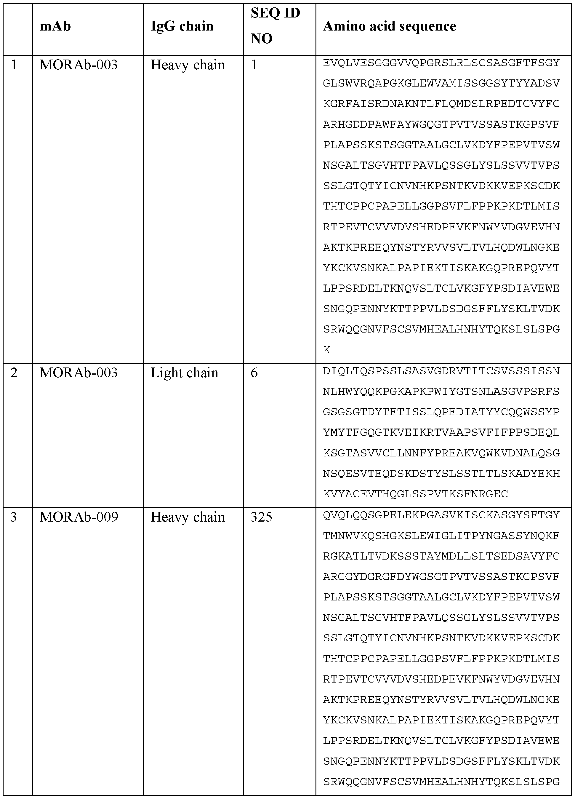


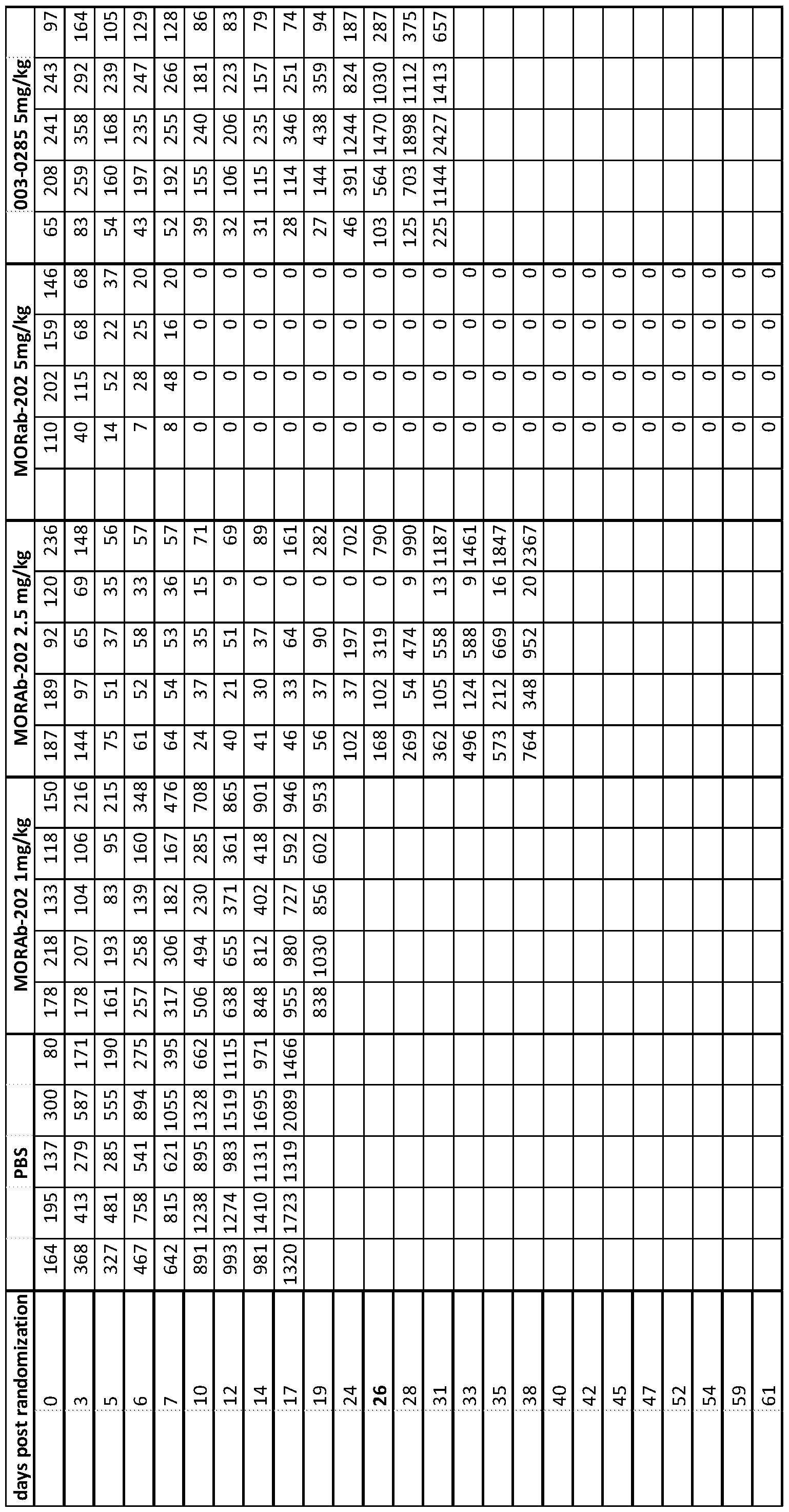


Claims
Priority Applications (36)
| Application Number | Priority Date | Filing Date | Title |
|---|---|---|---|
| KR1020227022690A KR102456433B1 (en) | 2016-03-02 | 2017-03-02 | Eribulin-based antibody-drug conjugates and methods of use |
| CN202111360555.3A CN114191428A (en) | 2016-03-02 | 2017-03-02 | Eribulin-based antibody-drug conjugates and methods of use |
| MYPI2018001424A MY189113A (en) | 2016-03-02 | 2017-03-02 | Eribulin-based antibody-drug conjugates and methods of use |
| BR112018067379A BR112018067379A2 (en) | 2016-03-02 | 2017-03-02 | antibody-drug conjugate, composition, method for treating a patient who has or is at risk of cancer, to produce the antibody-drug conjugate or composition, to determine if a patient will be responsive to treatment with the antibody-conjugate drug or composition, and use of an antibody-drug conjugate or composition. |
| IL292946A IL292946A (en) | 2016-03-02 | 2017-03-02 | Eribulin-based antibody-drug conjugates and methods of use |
| CN202111360563.8A CN114272389B (en) | 2016-03-02 | 2017-03-02 | Eribulin-based antibody-drug conjugates and methods of use |
| RU2018134331A RU2754369C2 (en) | 2016-03-02 | 2017-03-02 | Antibody-drug conjugates based on eribulin and application methods |
| JP2018545210A JP6599019B2 (en) | 2016-03-02 | 2017-03-02 | Eribulin-based antibody-drug conjugates and methods of use |
| DK17711475.8T DK3423105T3 (en) | 2016-03-02 | 2017-03-02 | ANTIBODY-Drug Conjugates Based on ERIBULIN AND PROCEDURES FOR USE |
| PL17711475T PL3423105T3 (en) | 2016-03-02 | 2017-03-02 | Eribulin-based antibody-drug conjugates and methods of use |
| AU2017225982A AU2017225982B2 (en) | 2016-03-02 | 2017-03-02 | Eribulin-based antibody-drug conjugates and methods of use |
| CN201780022381.5A CN108883198B (en) | 2016-03-02 | 2017-03-02 | Eribulin-based antibody-drug conjugates and methods of use |
| ES17711475T ES2880402T3 (en) | 2016-03-02 | 2017-03-02 | Eribulin-based antibody-drug conjugates and methods of use |
| MX2018010562A MX2018010562A (en) | 2016-03-02 | 2017-03-02 | Eribulin-based antibody-drug conjugates and methods of use. |
| PE2023001172A PE20231050A1 (en) | 2016-03-02 | 2017-03-02 | ERIBULIN-BASED ANTIBODY-DRUG CONJUGATES AND METHODS FOR THEIR USE |
| RS20210889A RS62108B1 (en) | 2016-03-02 | 2017-03-02 | Eribulin-based antibody-drug conjugates and methods of use |
| EP20204172.9A EP3824909A1 (en) | 2016-03-02 | 2017-03-02 | Eribulin-based antibody-drug conjugates and methods of use |
| KR1020187028407A KR102445255B1 (en) | 2016-03-02 | 2017-03-02 | Eribulin-Based Antibody-Drug Conjugates and Methods of Use |
| EP17711475.8A EP3423105B1 (en) | 2016-03-02 | 2017-03-02 | Eribulin-based antibody-drug conjugates and methods of use |
| PE2023001171A PE20231049A1 (en) | 2016-03-02 | 2017-03-02 | ERIBULIN-BASED ANTIBODY-DRUG CONJUGATES AND METHODS FOR THEIR USE |
| MDE20190061T MD3423105T2 (en) | 2016-03-02 | 2017-03-02 | Eribulin-based antibody-drug conjugates and methods of use |
| KR1020227022691A KR20220101204A (en) | 2016-03-02 | 2017-03-02 | Eribulin-based antibody-drug conjugates and methods of use |
| UAA201809739A UA125024C2 (en) | 2016-03-02 | 2017-03-02 | Eribulin-based antibody-drug conjugates and methods of use |
| SI201730837T SI3423105T1 (en) | 2016-03-02 | 2017-03-02 | Eribulin-based antibody-drug conjugates and methods of use |
| CN202111360093.5A CN114191563A (en) | 2016-03-02 | 2017-03-02 | Eribulin-based antibody-drug conjugates and methods of use |
| SG11201806515RA SG11201806515RA (en) | 2016-03-02 | 2017-03-02 | Eribulin-based antibody-drug conjugates and methods of use |
| LTEP17711475.8T LT3423105T (en) | 2016-03-02 | 2017-03-02 | Eribulin-based antibody-drug conjugates and methods of use |
| MA45280A MA45280B1 (en) | 2016-03-02 | 2017-03-02 | Eribulin-based antibody-drug conjugates and methods of use thereof |
| CA3013791A CA3013791A1 (en) | 2016-03-02 | 2017-03-02 | Eribulin-based antibody-drug conjugates and methods of use |
| KR1020247000482A KR20240007722A (en) | 2016-03-02 | 2017-03-02 | Eribulin-based antibody-drug conjugates and methods of use |
| CONC2018/0008667A CO2018008667A2 (en) | 2016-03-02 | 2018-08-16 | Eribulin-based antibody and drug conjugates and methods for use |
| IL261428A IL261428A (en) | 2016-03-02 | 2018-08-28 | Eribulin-based antibody-drug conjugates and methods of use |
| PH12018501847A PH12018501847A1 (en) | 2016-03-02 | 2018-08-30 | Eribulin-based antibody-drug conjugates and methods of use |
| HRP20211125TT HRP20211125T1 (en) | 2016-03-02 | 2021-07-14 | Eribulin-based antibody-drug conjugates and methods of use |
| CY20211100686T CY1124628T1 (en) | 2016-03-02 | 2021-08-02 | ERIBULIN-BASED ANTIBODY-DRUG CONJUGATIONS AND METHODS OF USE |
| AU2023285804A AU2023285804A1 (en) | 2016-03-02 | 2023-12-20 | Eribulin-based antibody-drug conjugates and methods of use |
Applications Claiming Priority (2)
| Application Number | Priority Date | Filing Date | Title |
|---|---|---|---|
| US201662302562P | 2016-03-02 | 2016-03-02 | |
| US62/302,562 | 2016-03-02 |
Publications (1)
| Publication Number | Publication Date |
|---|---|
| WO2017151979A1 true WO2017151979A1 (en) | 2017-09-08 |
Family
ID=58347964
Family Applications (1)
| Application Number | Title | Priority Date | Filing Date |
|---|---|---|---|
| PCT/US2017/020529 WO2017151979A1 (en) | 2016-03-02 | 2017-03-02 | Eribulin-based antibody-drug conjugates and methods of use |
Country Status (34)
| Country | Link |
|---|---|
| US (4) | US10548986B2 (en) |
| EP (2) | EP3423105B1 (en) |
| JP (5) | JP6599019B2 (en) |
| KR (4) | KR102445255B1 (en) |
| CN (4) | CN114272389B (en) |
| AR (3) | AR107787A1 (en) |
| AU (2) | AU2017225982B2 (en) |
| BR (1) | BR112018067379A2 (en) |
| CA (1) | CA3013791A1 (en) |
| CL (3) | CL2018002456A1 (en) |
| CO (1) | CO2018008667A2 (en) |
| CY (1) | CY1124628T1 (en) |
| DK (1) | DK3423105T3 (en) |
| ES (1) | ES2880402T3 (en) |
| HR (1) | HRP20211125T1 (en) |
| HU (1) | HUE054726T2 (en) |
| IL (2) | IL292946A (en) |
| JO (3) | JOP20170053B1 (en) |
| LT (1) | LT3423105T (en) |
| MA (1) | MA45280B1 (en) |
| MD (1) | MD3423105T2 (en) |
| MX (4) | MX2018010562A (en) |
| MY (1) | MY189113A (en) |
| PE (3) | PE20181953A1 (en) |
| PH (1) | PH12018501847A1 (en) |
| PL (1) | PL3423105T3 (en) |
| PT (1) | PT3423105T (en) |
| RS (1) | RS62108B1 (en) |
| RU (2) | RU2021125492A (en) |
| SG (2) | SG10202007520WA (en) |
| SI (1) | SI3423105T1 (en) |
| TW (2) | TWI825834B (en) |
| UA (1) | UA125024C2 (en) |
| WO (1) | WO2017151979A1 (en) |
Cited By (15)
| Publication number | Priority date | Publication date | Assignee | Title |
|---|---|---|---|---|
| WO2019232433A2 (en) | 2018-06-01 | 2019-12-05 | Eisai R&D Management Co., Ltd. | Methods of using splicing modulators |
| WO2021090062A1 (en) * | 2019-11-07 | 2021-05-14 | Eisai R&D Management Co., Ltd. | Anti-mesothelin eribulin antibody-drug conjugates and methods of use |
| WO2021132166A1 (en) | 2019-12-23 | 2021-07-01 | エーザイ・アール・アンド・ディー・マネジメント株式会社 | Method for producing eribulin-based antibody-drug conjugate |
| WO2021148003A1 (en) | 2020-01-22 | 2021-07-29 | 上海森辉医药有限公司 | Drug conjugate of eribulin derivative, preparation method therefor and application thereof in medicine |
| US11407762B2 (en) | 2017-11-15 | 2022-08-09 | President And Fellows Of Harvard College | Macrocyclic compounds and uses thereof |
| US11498892B2 (en) | 2017-07-06 | 2022-11-15 | President And Fellows Of Harvard College | Fe/Cu-mediated ketone synthesis |
| US11548898B2 (en) | 2017-07-06 | 2023-01-10 | President And Fellows Of Harvard College | Synthesis of halichondrins |
| WO2023001300A1 (en) | 2021-07-22 | 2023-01-26 | 上海森辉医药有限公司 | Drug conjugate of eribulin derivative |
| WO2023098691A1 (en) * | 2021-12-01 | 2023-06-08 | 上海生物制品研究所有限责任公司 | Antibody-drug conjugate and use thereof |
| WO2023103854A1 (en) * | 2021-12-09 | 2023-06-15 | 昆山新蕴达生物科技有限公司 | Antibody-drug conjugate having improved affinity, and preparation method therefor and application thereof |
| WO2023124963A1 (en) * | 2021-12-27 | 2023-07-06 | 昆山新蕴达生物科技有限公司 | Antibody-drug conjugate having reduced reversible reaction, and preparation method therefor and application thereof |
| US11725015B2 (en) | 2017-04-05 | 2023-08-15 | President And Fellows Of Harvard College | Macrocyclic compound and uses thereof |
| WO2023170247A1 (en) | 2022-03-11 | 2023-09-14 | Mablink Bioscience | Antibody-drug conjugates and their uses |
| WO2023200814A1 (en) | 2022-04-12 | 2023-10-19 | Eisai R & D Management Co., Ltd. | Eribulin-based antibody-drug conjugates and methods of use |
| JP7390315B2 (en) | 2018-06-01 | 2023-12-01 | エーザイ・アール・アンド・ディー・マネジメント株式会社 | Splicing modulator antibody-drug conjugates and methods of use thereof |
Families Citing this family (24)
| Publication number | Priority date | Publication date | Assignee | Title |
|---|---|---|---|---|
| EP2172487A1 (en) * | 2005-04-22 | 2010-04-07 | Morphotek Inc. | Antibodies with Immune Effector Activity that Internalize in Folate Receptor Alpha-Positive Cells |
| TWI769982B (en) * | 2015-06-24 | 2022-07-11 | 日商Jcr製藥股份有限公司 | Anti-human transferrin receptor antibody that passes through blood-brain barrier |
| EP3315515A4 (en) | 2015-06-24 | 2019-06-05 | JCR Pharmaceuticals Co., Ltd. | Fusion protein containing bdnf |
| AR110586A1 (en) | 2016-12-26 | 2019-04-10 | Japan Chem Res | HUMAN ANTITRANSFERRINE RECEPTOR ANTIBODY THAT PENETRATES THE HEMATOENCEPHALIC BARRIER |
| CA3081539A1 (en) * | 2017-11-08 | 2019-05-16 | Yafei Shanghai Biolog Medicine Science & Technology Co., Ltd. | Conjugates of biomolecule and use thereof |
| EP3834843A4 (en) | 2018-08-06 | 2022-05-11 | Daiichi Sankyo Company, Limited | Combination of antibody-drug conjugate and tubulin inhibitor |
| JOP20210159A1 (en) * | 2018-12-21 | 2023-01-30 | Novartis Ag | Antibodies to pmel17 and conjugates thereof |
| CN110568121A (en) * | 2019-07-11 | 2019-12-13 | 山东省药学科学院 | Eribulin and detection method of related substances in eribulin-containing preparation |
| KR20220018954A (en) * | 2020-08-07 | 2022-02-15 | 주식회사 피노바이오 | Conjugate comprising Deoxycytidine based anticancer drug and silyl ether-containing linker and uses thereof |
| US20240044920A1 (en) * | 2020-09-11 | 2024-02-08 | Alexion Pharmaceuticals, Inc. | Anti-ceruloplasmin antibodies and uses thereof |
| BR112023005593A2 (en) * | 2020-09-28 | 2023-05-09 | Navrogen Inc | COMPOSITION AND USE OF ALTERNATIVELY FORMATTED ANTI-MESOTHELIN ANTIBODIES FOR CANCER TREATMENT |
| WO2022082068A1 (en) * | 2020-10-18 | 2022-04-21 | Ardeagen Corporation | Anti-msln binding agents, conjugates thereof and methods of using the same |
| CN114560940B (en) * | 2020-11-27 | 2023-07-14 | 缔码生物科技(武汉)有限公司 | SIRP alpha resisting rabbit recombinant monoclonal antibody, and preparation method and application thereof |
| AU2022253902A1 (en) * | 2021-04-10 | 2023-11-02 | Profoundbio Us Co. | Folr1 binding agents, conjugates thereof and methods of using the same |
| CN115429893A (en) * | 2021-06-02 | 2022-12-06 | 百奥泰生物制药股份有限公司 | Drug conjugates and uses thereof |
| WO2023016488A1 (en) * | 2021-08-13 | 2023-02-16 | 昆山新蕴达生物科技有限公司 | Microtubule inhibitor-based antibody-drug conjugate |
| WO2023024949A1 (en) * | 2021-08-24 | 2023-03-02 | 昆山新蕴达生物科技有限公司 | Antibody-drug conjugate conjugated via breakable linker |
| CN115957339A (en) * | 2021-09-16 | 2023-04-14 | 正大天晴药业集团股份有限公司 | Antibody drug conjugates and compositions and uses thereof |
| WO2023055376A1 (en) * | 2021-09-30 | 2023-04-06 | Virtuoso Binco, Inc. | Multispecific antibodies for targeting cd47 and icam1 and methods of use thereof |
| TW202317088A (en) * | 2021-10-14 | 2023-05-01 | 大陸商江蘇恒瑞醫藥股份有限公司 | Preparation method of eribulin derivatives |
| WO2023143319A1 (en) * | 2022-01-28 | 2023-08-03 | 启德医药科技(苏州)有限公司 | Antibody-drug conjugate, and pharmaceutical composition and use thereof |
| WO2023143320A1 (en) * | 2022-01-28 | 2023-08-03 | 博瑞生物医药(苏州)股份有限公司 | Linker for preparing antibody-drug conjugate, compound, and use |
| WO2023155808A1 (en) * | 2022-02-16 | 2023-08-24 | 苏州宜联生物医药有限公司 | Conjugate of antibody-eribulin or derivative thereof, intermediate thereof, preparation method therefor, pharmaceutical composition thereof and use thereof |
| WO2023198079A1 (en) * | 2022-04-12 | 2023-10-19 | 百奥泰生物制药股份有限公司 | Method for treating her2-positive solid tumor |
Citations (21)
| Publication number | Priority date | Publication date | Assignee | Title |
|---|---|---|---|---|
| US4880078A (en) | 1987-06-29 | 1989-11-14 | Honda Giken Kogyo Kabushiki Kaisha | Exhaust muffler |
| US4880935A (en) | 1986-07-11 | 1989-11-14 | Icrf (Patents) Limited | Heterobifunctional linking agents derived from N-succinimido-dithio-alpha methyl-methylene-benzoates |
| WO1990005144A1 (en) | 1988-11-11 | 1990-05-17 | Medical Research Council | Single domain ligands, receptors comprising said ligands, methods for their production, and use of said ligands and receptors |
| US5122368A (en) | 1988-02-11 | 1992-06-16 | Bristol-Myers Squibb Company | Anthracycline conjugates having a novel linker and methods for their production |
| WO1992019244A2 (en) | 1991-05-01 | 1992-11-12 | Henry M. Jackson Foundation For The Advancement Of Military Medicine | A method for treating infectious respiratory diseases |
| US5622929A (en) | 1992-01-23 | 1997-04-22 | Bristol-Myers Squibb Company | Thioether conjugates |
| WO1997032572A2 (en) | 1996-03-04 | 1997-09-12 | The Penn State Research Foundation | Materials and methods for enhancing cellular internalization |
| WO1997044013A1 (en) | 1996-05-24 | 1997-11-27 | Massachusetts Institute Of Technology | Aerodynamically light particles for pulmonary drug delivery |
| WO1998031346A1 (en) | 1997-01-16 | 1998-07-23 | Massachusetts Institute Of Technology | Preparation of particles for inhalation |
| US5821337A (en) | 1991-06-14 | 1998-10-13 | Genentech, Inc. | Immunoglobulin variants |
| US5824805A (en) | 1995-12-22 | 1998-10-20 | King; Dalton | Branched hydrazone linkers |
| US5855913A (en) | 1997-01-16 | 1999-01-05 | Massachusetts Instite Of Technology | Particles incorporating surfactants for pulmonary drug delivery |
| US5934272A (en) | 1993-01-29 | 1999-08-10 | Aradigm Corporation | Device and method of creating aerosolized mist of respiratory drug |
| US5985309A (en) | 1996-05-24 | 1999-11-16 | Massachusetts Institute Of Technology | Preparation of particles for inhalation |
| WO1999066903A2 (en) | 1998-06-24 | 1999-12-29 | Advanced Inhalation Research, Inc. | Large porous particles emitted from an inhaler |
| US6019968A (en) | 1995-04-14 | 2000-02-01 | Inhale Therapeutic Systems, Inc. | Dispersible antibody compositions and methods for their preparation and use |
| US6214345B1 (en) | 1993-05-14 | 2001-04-10 | Bristol-Myers Squibb Co. | Lysosomal enzyme-cleavable antitumor drug conjugates |
| WO2006034488A2 (en) | 2004-09-23 | 2006-03-30 | Genentech, Inc. | Cysteine engineered antibodies and conjugates |
| WO2013173393A1 (en) | 2012-05-15 | 2013-11-21 | Concortis Biosystems, Corp | Drug-conjugates, conjugation methods, and uses thereof |
| US20140069822A1 (en) | 2012-09-10 | 2014-03-13 | Antec Leyden B.V. | Electrochemical reduction of disulfide bonds in proteinaceous substances and electrochemical cell for carrying out such reduction |
| WO2014089335A2 (en) * | 2012-12-07 | 2014-06-12 | Amgen Inc. | Bcma antigen binding proteins |
Family Cites Families (201)
| Publication number | Priority date | Publication date | Assignee | Title |
|---|---|---|---|---|
| US4816567A (en) | 1983-04-08 | 1989-03-28 | Genentech, Inc. | Recombinant immunoglobin preparations |
| US6019963A (en) | 1998-11-20 | 2000-02-01 | D.S.C. Products, Inc. | Deodorizing composition containing tea tree and eucalyptus oils |
| US7109019B2 (en) | 1999-01-06 | 2006-09-19 | The Regents Of The University Of California | Gene cluster for production of the enediyne antitumor antibiotic C-1027 |
| NZ529808A (en) | 2001-05-02 | 2007-02-23 | Purdue Research Foundation | Treatment and diagnosis of macrophage mediated disease |
| US7157277B2 (en) | 2001-11-28 | 2007-01-02 | Neose Technologies, Inc. | Factor VIII remodeling and glycoconjugation of Factor VIII |
| US7265084B2 (en) | 2001-10-10 | 2007-09-04 | Neose Technologies, Inc. | Glycopegylation methods and proteins/peptides produced by the methods |
| US7439043B2 (en) | 2001-10-10 | 2008-10-21 | Neose Technologies, Inc. | Galactosyl nucleotide sugars |
| US7297511B2 (en) | 2001-10-10 | 2007-11-20 | Neose Technologies, Inc. | Interferon alpha: remodeling and glycoconjugation of interferon alpha |
| US7696163B2 (en) | 2001-10-10 | 2010-04-13 | Novo Nordisk A/S | Erythropoietin: remodeling and glycoconjugation of erythropoietin |
| US7226903B2 (en) | 2001-10-10 | 2007-06-05 | Neose Technologies, Inc. | Interferon beta: remodeling and glycoconjugation of interferon beta |
| US7399613B2 (en) | 2001-10-10 | 2008-07-15 | Neose Technologies, Inc. | Sialic acid nucleotide sugars |
| US7173003B2 (en) | 2001-10-10 | 2007-02-06 | Neose Technologies, Inc. | Granulocyte colony stimulating factor: remodeling and glycoconjugation of G-CSF |
| US7795210B2 (en) | 2001-10-10 | 2010-09-14 | Novo Nordisk A/S | Protein remodeling methods and proteins/peptides produced by the methods |
| US7125843B2 (en) | 2001-10-19 | 2006-10-24 | Neose Technologies, Inc. | Glycoconjugates including more than one peptide |
| US7214660B2 (en) | 2001-10-10 | 2007-05-08 | Neose Technologies, Inc. | Erythropoietin: remodeling and glycoconjugation of erythropoietin |
| US8008252B2 (en) | 2001-10-10 | 2011-08-30 | Novo Nordisk A/S | Factor VII: remodeling and glycoconjugation of Factor VII |
| US7179617B2 (en) | 2001-10-10 | 2007-02-20 | Neose Technologies, Inc. | Factor IX: remolding and glycoconjugation of Factor IX |
| US7265085B2 (en) | 2001-10-10 | 2007-09-04 | Neose Technologies, Inc. | Glycoconjugation methods and proteins/peptides produced by the methods |
| WO2003072754A2 (en) | 2002-02-27 | 2003-09-04 | The Government Of The United States Of America, Represented By The Secretary, Department Of Health And Human Services | Conjugates of ligand, linker and cytotoxic agent and related compositions and methods of use |
| SI2357006T1 (en) * | 2002-07-31 | 2016-01-29 | Seattle Genetics, Inc. | Drug conjugates and their use for treating cancer, an autoimmune disease or an infectious disease |
| JP2004119755A (en) | 2002-09-27 | 2004-04-15 | Alps Electric Co Ltd | Magnetic detector and its manufacturing method |
| US20040071540A1 (en) | 2002-10-15 | 2004-04-15 | Lucas Philip J. | Disposable/recyclable pallet system and method |
| US20080025989A1 (en) | 2003-02-20 | 2008-01-31 | Seattle Genetics, Inc. | Anti-cd70 antibody-drug conjugates and their use for the treatment of cancer and immune disorders |
| SG155777A1 (en) | 2003-04-09 | 2009-10-29 | Neose Technologies Inc | Glycopegylation methods and proteins/peptides produced by the methods |
| BRPI0414568A (en) | 2003-09-18 | 2006-11-07 | Combinatorx Inc | drug combinations for the treatment of neoplasms |
| US6997024B2 (en) | 2003-10-01 | 2006-02-14 | Truth Hardware Corporation | Pull door lock |
| US8012944B2 (en) | 2003-10-30 | 2011-09-06 | Pharmascience Inc. | Method for treating cancer using IAP antisense oligomer and chemotherapeutic agent |
| US20050148535A1 (en) | 2003-10-30 | 2005-07-07 | Lacasse Eric | IAP nucleobase oligomers and oligomeric complexes and uses thereof |
| EP2316857A1 (en) | 2004-02-12 | 2011-05-04 | Morphotek, Inc. | Monoclonal antibodies that specifically bind to folate receptor alpha |
| US7117807B2 (en) | 2004-02-17 | 2006-10-10 | University Of Florida Research Foundation, Inc. | Dynamically modifiable polymer coatings and devices |
| EP1778298A4 (en) | 2004-04-01 | 2010-03-31 | Univ Pennsylvania | Lipoprotein nanoplatforms |
| CA2569003A1 (en) | 2004-05-07 | 2005-12-08 | Massachusetts Institute Of Technology | Methods and compositions for cancer treatment relating to brca1 brct domain recognition of phosphorylated bach1 |
| HUE046436T2 (en) | 2004-06-03 | 2020-02-28 | Eisai R&D Man Co Ltd | Intermediates for the preparation of halichondrin B |
| JO3000B1 (en) * | 2004-10-20 | 2016-09-05 | Genentech Inc | Antibody Formulations. |
| WO2006076100A2 (en) | 2004-12-09 | 2006-07-20 | Eisai Co. Ltd. | Tubulin isotype screening in cancer therapy using halichondrin b analogs |
| WO2006063135A2 (en) | 2004-12-09 | 2006-06-15 | Eisai R & D Management Co., Ltd. | Tubulin isotype screnning in cancer therapy using hemiasterlin analogs |
| EP3498289A1 (en) | 2005-07-07 | 2019-06-19 | Seattle Genetics, Inc. | Monomethylvaline compounds having phenylalanine side-chain modifications at the c-terminus |
| WO2007008848A2 (en) | 2005-07-07 | 2007-01-18 | Seattle Genetics, Inc. | Monomethylvaline compounds having phenylalanine carboxy modifications at the c-terminus |
| GB0515025D0 (en) | 2005-07-21 | 2005-08-31 | Novartis Ag | Organic compounds |
| JP5475992B2 (en) | 2005-08-19 | 2014-04-16 | エンドサイト,インコーポレイテッド | Multidrug ligand conjugate |
| ATE546160T1 (en) | 2005-09-14 | 2012-03-15 | Ucb Pharma Sa | ANTIBODY COMB POLYMER CONJUGATE |
| WO2007064691A1 (en) | 2005-12-02 | 2007-06-07 | Nabil Habib Lab | Treatment of cancer and other diseases |
| US7901885B2 (en) | 2006-05-09 | 2011-03-08 | Dsm Ip Assets B.V. | Genes and markers in type 2 diabetes and obesity |
| US20150030534A1 (en) | 2013-07-26 | 2015-01-29 | Rutgers, The State University Of New Jersey | Antibody cocktails for breast cancer radioimmunotherapy |
| US20140037539A1 (en) | 2012-07-27 | 2014-02-06 | Rutgers, The State University Of New Jersey | Antibody cocktails for breast cancer radioimmunotherapy |
| US8398956B2 (en) | 2007-01-11 | 2013-03-19 | Immunomedics, Inc. | In vivo copper-free click chemistry for delivery of therapeutic and/or diagnostic agents |
| WO2008101231A2 (en) | 2007-02-16 | 2008-08-21 | Endocyte, Inc. | Methods and compositions for treating and diagnosing kidney disease |
| AU2008224988A1 (en) | 2007-03-14 | 2008-09-18 | Endocyte, Inc. | Binding ligand linked drug delivery conjugates of tubulysins |
| EP1977765A1 (en) | 2007-04-03 | 2008-10-08 | Diatos | Peptide prodrugs |
| JP5735277B2 (en) | 2007-10-03 | 2015-06-17 | エーザイ・アール・アンド・ディー・マネジメント株式会社 | Intermediates and methods for the synthesis of halichondrin B analogues |
| CN101884058B (en) | 2007-10-30 | 2012-07-25 | 富士通株式会社 | Mobile terminal device |
| DK2842575T3 (en) | 2008-03-18 | 2017-11-27 | Seattle Genetics Inc | Auristatin drug linker conjugates |
| EP2977468B1 (en) | 2008-03-27 | 2019-06-19 | Vascular Biosciences, Inc. | Methods of novel therapeutic candidate identification through gene expression analysis in pulmonary arterial hypertension |
| US20100260668A1 (en) | 2008-04-29 | 2010-10-14 | Abbott Laboratories | Dual Variable Domain Immunoglobulins and Uses Thereof |
| MX2010011955A (en) | 2008-04-29 | 2011-01-21 | Abbott Lab | Dual variable domain immunoglobulins and uses thereof. |
| SG189817A1 (en) * | 2008-04-30 | 2013-05-31 | Immunogen Inc | Potent conjugates and hydrophilic linkers |
| US9035027B2 (en) | 2008-06-03 | 2015-05-19 | Abbvie Inc. | Dual variable domain immunoglobulins and uses thereof |
| NZ589436A (en) | 2008-06-03 | 2012-12-21 | Abbott Lab | Dual variable domain immunoglobulins and uses thereof |
| NZ590074A (en) | 2008-07-08 | 2012-12-21 | Abbott Lab | Prostaglandin e2 dual variable domain immunoglobulins and uses thereof |
| BRPI0924571A2 (en) | 2008-09-12 | 2016-08-09 | Santen Pharmaceutical Co Ltd | glucocorticoid receptor agonist, pharmaceutical composition, therapeutic agent for inflammatory disease, therapeutic agent for inflammatory eye disease, therapeutic agent for inflammatory bone joint disorder, method for treating inflammatory disease, method for treating inflammatory eye disease, and method for treating bone disorder inflammatory joint |
| CA2745271A1 (en) | 2008-12-04 | 2010-06-10 | Abbott Laboratories | Dual variable domain immunoglobulins and uses thereof |
| SG173705A1 (en) | 2009-03-05 | 2011-09-29 | Abbott Lab | Il-17 binding proteins |
| US20140339088A1 (en) | 2009-03-09 | 2014-11-20 | Virginia Tech Intellectual Properties, Inc. | Dielectrophoresis methods for determining a property of a plurality of cancer cells |
| TW201042040A (en) | 2009-05-01 | 2010-12-01 | Abbott Lab | Dual variable domain immunoglobulins and uses thereof |
| US8686520B2 (en) * | 2009-05-29 | 2014-04-01 | International Business Machines Corporation | Spin-torque magnetoresistive structures |
| TW201109438A (en) | 2009-07-29 | 2011-03-16 | Abbott Lab | Dual variable domain immunoglobulins and uses thereof |
| SG178419A1 (en) * | 2009-08-15 | 2012-04-27 | Genentech Inc | Anti-angiogenesis therapy for the treatment of previously treated breast cancer |
| CA2772628A1 (en) * | 2009-09-01 | 2011-03-10 | Abbott Laboratories | Dual variable domain immunoglobulins and uses thereof |
| GB0917044D0 (en) | 2009-09-29 | 2009-11-18 | Cytoguide As | Agents, uses and methods |
| AR078651A1 (en) | 2009-10-15 | 2011-11-23 | Abbott Lab | IMMUNOGLOBULINS WITH DUAL VARIABLE DOMAIN AND USES OF THE SAME |
| UY32979A (en) | 2009-10-28 | 2011-02-28 | Abbott Lab | IMMUNOGLOBULINS WITH DUAL VARIABLE DOMAIN AND USES OF THE SAME |
| AU2010315126B2 (en) | 2009-11-06 | 2015-06-25 | Plexxikon, Inc. | Compounds and methods for kinase modulation, and indications therefor |
| US8203010B2 (en) | 2010-01-26 | 2012-06-19 | Eisai R&D Management Co., Ltd. | Compounds useful in the synthesis of halichondrin B analogs |
| CN110386876A (en) | 2010-03-24 | 2019-10-29 | 俄亥俄州立大学 | The composition and method inhibited for glucose transport |
| US8710035B2 (en) | 2010-03-24 | 2014-04-29 | The University Of Chicago | Methods and compositions related to glucocorticoid receptor antagonists and breast cancer |
| AU2011252883B2 (en) | 2010-05-14 | 2015-09-10 | Abbvie Inc. | IL-1 binding proteins |
| HUE035599T2 (en) | 2010-06-15 | 2018-05-28 | Genmab As | Human antibody drug conjugates against tissue factor |
| UY33492A (en) | 2010-07-09 | 2012-01-31 | Abbott Lab | IMMUNOGLOBULINS WITH DUAL VARIABLE DOMAIN AND USES OF THE SAME |
| AU2011285852B2 (en) | 2010-08-03 | 2014-12-11 | Abbvie Inc. | Dual variable domain immunoglobulins and uses thereof |
| KR20130139884A (en) | 2010-08-26 | 2013-12-23 | 애브비 인코포레이티드 | Dual variable domain immunoglobulins and uses thereof |
| US9089570B2 (en) | 2010-09-03 | 2015-07-28 | Tactical Therapeutics Inc | Compositions for treating cancers having acquired resitance to prior chemotherapeutic and targeted drugs using carboxyamidotriazole orotate |
| US9192677B2 (en) | 2010-09-07 | 2015-11-24 | Johannes Kepler Universität Linz | Biodegradable, water soluble and pH responsive poly(organo)phosphazenes |
| WO2012065019A2 (en) | 2010-11-12 | 2012-05-18 | Exelixis, Inc. | Pyridopyrimidinone inhibitors of p13k alpha |
| WO2012065057A2 (en) | 2010-11-12 | 2012-05-18 | Exelixis, Inc. | Phosphatidylinositol 3-kinase inhibitors and methods of their use |
| WO2012075361A2 (en) | 2010-12-02 | 2012-06-07 | Immunomedics, Inc. | In vivo copper-free click chemistry for delivery of therapeutic and/or diagnostic agents |
| RU2627171C2 (en) | 2010-12-21 | 2017-08-03 | Эббви Инк. | Il-1 alpha and beta bispecific immunoglobulins with double variable domains and their application |
| EP2655415A4 (en) | 2010-12-22 | 2016-03-09 | Abbvie Inc | Tri-variable domain binding proteins and uses thereof |
| UY33827A (en) | 2010-12-22 | 2012-07-31 | Abbott Lab | MEDIUM-IMMUNOGLOBULIN UNION PROTEINS AND ITS USES |
| DK2675474T3 (en) | 2011-02-15 | 2019-04-23 | Vaxiion Therapeutics Llc | THERAPEUTIC COMPOSITIONS AND PROCEDURES FOR TARGETED ADMINISTRATION OF BIOACTIVE MOLECULES BY BACK-TERIAL MINICLES BASED ON ANTIBODY AND FC-CONTAINING TAR-GETING MOLECULES |
| WO2012116277A1 (en) | 2011-02-25 | 2012-08-30 | Arena Pharmaceuticals, Inc. | Cannabinoid receptor modulators |
| EP2680839A1 (en) * | 2011-03-02 | 2014-01-08 | Morphotek, Inc. | Co -administration of eribulin and farletuzumab for the treatment of breast cancer |
| WO2012118978A1 (en) | 2011-03-03 | 2012-09-07 | The Regents Of The University Of Colorado, A Body Corporate | Methods for treating oncovirus positive cancers |
| CN103502218B (en) | 2011-03-04 | 2016-08-17 | 生命科技公司 | Compounds and methods for for conjugating biomolecules |
| JP5971769B2 (en) | 2011-03-15 | 2016-08-17 | ザ ユニバーシティー オブ ノースカロライナ アット チャペル ヒル | Methods of treating breast cancer using anthracycline therapy |
| CN103562406B (en) | 2011-03-18 | 2018-03-27 | 卫材R&D管理有限公司 | For predicting the method and composition of the reaction to eribulin |
| EP2508525A1 (en) | 2011-04-05 | 2012-10-10 | Bayer Pharma Aktiengesellschaft | Substituted 2,3-dihydroimidazo[1,2-c]quinazoline salts |
| EP2670866A4 (en) | 2011-04-05 | 2015-09-02 | Translational Genomics Res Inst | Biomarkers and methods of use thereof |
| WO2012145098A1 (en) | 2011-04-21 | 2012-10-26 | Saint Louis University | Use of adenosine a3 receptor agonists for treatment of neuropathic pain |
| MX345538B (en) | 2011-05-27 | 2017-02-03 | Ambrx Inc | Compositions containing, methods involving, and uses of non-natural amino acid linked dolastatin derivatives. |
| KR102179369B1 (en) | 2011-05-27 | 2020-11-16 | 암브룩스, 인코포레이티드 | Compositions containing, methods involving, and uses of non-natural amino acid linked dolastatin derivatives |
| WO2012170640A1 (en) | 2011-06-07 | 2012-12-13 | The Trustees Of Columbia University In The City Of New York | Methods and compositions for trail-drug combination therapy |
| BR112013031819B1 (en) | 2011-06-10 | 2022-05-03 | Mersana Therapeutics, Inc | Polymeric support, pharmaceutical composition, compound and support use |
| US8815226B2 (en) | 2011-06-10 | 2014-08-26 | Mersana Therapeutics, Inc. | Protein-polymer-drug conjugates |
| PT2739649T (en) | 2011-08-05 | 2018-01-03 | Bioasis Technologies Inc | P97 fragments with transfer activity |
| US9181152B2 (en) | 2011-11-30 | 2015-11-10 | Alphora Research Inc. | Process for preparation of (3R)-2,4-di-leaving group-3-methylbut-1-ene |
| WO2013086634A1 (en) | 2011-12-16 | 2013-06-20 | Alphora Research Inc. | Process for preparation of 3-((2s,5s)-4-methylene-5-(3-oxopropyl)tetrahydrofuran-2-yl) propanol derivatives and intermediates useful thereof |
| EP3539982A3 (en) | 2011-12-23 | 2020-01-15 | Pfizer Inc | Engineered antibody constant regions for site-specific conjugation and methods and uses therefor |
| WO2013092998A1 (en) | 2011-12-23 | 2013-06-27 | Innate Pharma | Enzymatic conjugation of antibodies |
| US9062020B2 (en) | 2011-12-29 | 2015-06-23 | Alphora Research Inc. | 2-((2S,3S,4R,5R)-5-((S)-3-amino-2-hydroxyprop-1-yl)-4-methoxy-3-(phenylsulfonylmethyl)tetrahydrofuran-2-yl)acetaldehyde derivatives and process for their preparation |
| TWI573792B (en) | 2012-02-01 | 2017-03-11 | 歐陸斯迪公司 | Novel therapeutic agents |
| WO2013130093A1 (en) * | 2012-03-02 | 2013-09-06 | Genentech, Inc. | Biomarkers for treatment with anti-tubulin chemotherapeutic compounds |
| WO2013138371A1 (en) | 2012-03-12 | 2013-09-19 | Merrimack Pharmaceuticals, Inc. | Methods for treating pancreatic cancer using combination therapies comprising an anti-erbb3 antibody |
| US9358235B2 (en) | 2012-03-19 | 2016-06-07 | Plexxikon Inc. | Kinase modulation, and indications therefor |
| WO2013148337A1 (en) | 2012-03-29 | 2013-10-03 | Altor Bioscience Corporation | Methods for treating neoplasia |
| CA2868627C (en) | 2012-03-30 | 2021-02-16 | Alphora Research Inc. | Synthetic process for preparation of macrocyclic c1-keto analogs of halichondrin b and intermediates useful therein |
| US20150231219A1 (en) | 2012-04-02 | 2015-08-20 | Merrimack Pharmaceuticals, Inc. | Dosage and administration of monospecific and bispecific anti-igr-1r and anti-erbb3 antibodies |
| KR20150035537A (en) | 2012-05-15 | 2015-04-06 | 디아테크 온콜로지, 엘엘씨 | Tumor cell isolation/purification process and methods for use thereof |
| NZ713941A (en) | 2012-06-07 | 2017-02-24 | Ambrx Inc | Prostate-specific membrane antigen antibody drug conjugates |
| WO2013182668A1 (en) | 2012-06-08 | 2013-12-12 | F. Hoffmann-La Roche Ag | Mutant selectivity and combinations of a phosphoinositide 3 kinase inhibitor compound and chemotherapeutic agents for the treatment of cancer |
| MX359449B (en) | 2012-06-19 | 2018-09-28 | Ambrx Inc | Anti-cd70 antibody drug conjugates. |
| US9220695B2 (en) | 2012-06-29 | 2015-12-29 | The Research Foundation For The State University Of New York | Polyenolic zinc-binding agents (pezbins) actively promote inactivation of cancer stem cells and potentiate cytotoxic anti-tumor drug substances |
| JP2015530072A (en) | 2012-06-29 | 2015-10-15 | ナノストリング テクノロジーズ,インコーポレイティド | Breast cancer treatment with gemcitabine therapy |
| LT3210627T (en) | 2012-07-12 | 2023-04-11 | Hangzhou Dac Biotech Co., Ltd | Conjugates of cell binding molecules with cytotoxic agents |
| WO2014047199A1 (en) | 2012-09-19 | 2014-03-27 | The Research Foundation For The State University Of New York | Novel prodrugs for selective anticancer therapy |
| BR112015007144A2 (en) | 2012-10-04 | 2017-12-12 | Ab Science | use of masitinib for cancer treatment in patient subpopulations identified using predictive factors |
| EP2906712A1 (en) | 2012-10-10 | 2015-08-19 | Stichting Het Nederlands Kanker Instituut- Antoni van Leeuwenhoek Ziekenhuis | Methods and means for predicting resistance to anti-cancer treatment |
| WO2014063443A1 (en) | 2012-10-22 | 2014-05-01 | Zhang Yuliang | Self-cooled thermal work doing method |
| CN110201186A (en) | 2012-10-23 | 2019-09-06 | 西纳福克斯股份有限公司 | Modified antibody, antibody-conjugate and preparation method thereof |
| ES2656298T3 (en) | 2012-11-05 | 2018-02-26 | Pfizer Inc. | Ayustoestatin analogues |
| ES2701076T3 (en) | 2012-11-24 | 2019-02-20 | Hangzhou Dac Biotech Co Ltd | Hydrophilic linkers and their uses for the conjugation of drugs to molecules that bind to cells |
| AU2013356241A1 (en) | 2012-12-03 | 2015-07-09 | Calithera Biosciences, Inc. | Treatment of cancer with heterocyclic inhibitors of glutaminase |
| US9243058B2 (en) | 2012-12-07 | 2016-01-26 | Amgen, Inc. | BCMA antigen binding proteins |
| JP6334553B2 (en) | 2012-12-10 | 2018-05-30 | メルサナ セラピューティクス,インコーポレイティド | Protein-polymer-drug conjugate |
| WO2014093379A1 (en) | 2012-12-10 | 2014-06-19 | Mersana Therapeutics, Inc. | Auristatin compounds and conjugates thereof |
| US9872918B2 (en) | 2012-12-12 | 2018-01-23 | Mersana Therapeutics, Inc. | Hydroxyl-polymer-drug-protein conjugates |
| EP2934596A1 (en) | 2012-12-21 | 2015-10-28 | Glykos Finland Oy | Linker-payload molecule conjugates |
| US20150182634A1 (en) | 2012-12-28 | 2015-07-02 | Cody P. Coyne | Molecular Design and Chemical Synthesis of Pharmaceutical-Ligands and Pharmaceutical-Pharmaceutical Analogs with Multiple Mechanisms of Action |
| EP2964245A4 (en) | 2013-01-19 | 2016-09-21 | Univ New York | Hydrogen-bond surrogate peptides and peptidomimetics for p53 reactivation |
| CA2898329A1 (en) | 2013-01-19 | 2014-07-24 | New York University | Oligooxopiperazines for p53 reactivation |
| DK2950784T3 (en) | 2013-02-01 | 2021-07-05 | Zoneone Pharma Inc | REMOVAL OF WEAKLY WATER-SOLUBLE MEDICINES IN LIPOSOMES |
| WO2014121235A2 (en) | 2013-02-01 | 2014-08-07 | Zoneone Pharma, Inc. | Transformation of drug cyclodextrin complex compositions into compositions of mixtures of lipid vesicle encapsulated drug and cyclodextrin drug complexes |
| WO2014124322A1 (en) | 2013-02-08 | 2014-08-14 | University Of Louisville Research Foundation, Inc. | Nanoparticles for drug delivery |
| WO2014130776A1 (en) | 2013-02-22 | 2014-08-28 | University Of Houston | Phosphaplatins as neuroprotective agents |
| WO2015073072A1 (en) | 2013-11-15 | 2015-05-21 | Oncoceutics, Inc. | 7-benzyl-4-(2-methylbenzyl)-2,4,6,7,8,9-hexahydroimidazo[1,2-a]pyrido[3,4-e]pyrimidin-5(1h)-one |
| ME03470B (en) | 2013-03-13 | 2020-01-20 | Oncoceutics Inc | 7-benzyl-10-(2-methylbenzyl)-2,6,7,8,9,10-exahydroimidazo [1,2-a]pyrido[4,3-d]pyrimidin-5(3h)-on for use in the treatment of cancer |
| US9376437B2 (en) | 2013-03-13 | 2016-06-28 | Oncoceutics, Inc | 7-benzyl-4-(2-methylbenzyl)-2,4,6,7,8,9-hexahydroimidazo[1,2-a]pyrido[3,4-e]pyrimidin-5(1H)-one, salts thereof and methods of using the same in combination therapy |
| US20160022829A1 (en) | 2013-03-14 | 2016-01-28 | Mersana Therapeutics, Inc. | Tubulysin compounds and conjugates thereof |
| CN105377252A (en) | 2013-03-14 | 2016-03-02 | 佛罗里达大学研究基金会 | Regulation of cancer using natural compounds and/or diet |
| US20140271923A1 (en) | 2013-03-14 | 2014-09-18 | Christopher Brian Reid | Compositions & formulations for preventing and treating chronic diseases that cluster in patients such as cardiovascular disease, diabetes, obesity, polycystic ovary syndrome, hyperlipidemia and hypertension, as well as for preventing and treating other diseases and conditions |
| SG11201507619PA (en) | 2013-03-15 | 2015-10-29 | Ct For Drug Res And Dev | Cytotoxic and anti-mitotic compounds, and methods of using the same |
| US9295731B2 (en) | 2013-04-01 | 2016-03-29 | Mark Quang Nguyen | Cleavable drug conjugates, compositions thereof and methods of use |
| US10233219B2 (en) | 2013-04-12 | 2019-03-19 | Tufts Medical Center | Methods and systems for designing and/or characterizing soluble lipidated ligand agents |
| WO2014176284A1 (en) | 2013-04-22 | 2014-10-30 | Avelas Biosciences, Inc. | Selective drug delivery compositions and methods of use |
| GB2513405A (en) | 2013-04-26 | 2014-10-29 | Adc Biotechnology Ltd | Method of synthesising ADCs using affinity resins |
| ES2759503T3 (en) | 2013-05-02 | 2020-05-11 | Glykos Finland Oy | Conjugates of a glycoprotein or a glycan with a toxic payload |
| WO2014183211A1 (en) | 2013-05-15 | 2014-11-20 | Alphora Research Inc. | 3-((2s,5s)-4-methylene-5-(3-oxopropyl)tetrahydrofuran-2-yl)propanol derivatives, their preparation and intermediates useful thereof |
| GB201309807D0 (en) | 2013-05-31 | 2013-07-17 | Pharma Mar Sau | Antibody drug conjugates |
| US20140363454A1 (en) | 2013-06-06 | 2014-12-11 | Igenica Biotherapeutics, Inc. | Antibody-Drug Conjugates, Compositions and Methods of Use |
| US11229711B2 (en) | 2013-06-06 | 2022-01-25 | Magenta Therapeutics, Inc. | Linkers for antibody-drug conjugates and related compounds, compositions, and methods of use |
| US10071169B2 (en) | 2013-06-20 | 2018-09-11 | Innate Pharma | Enzymatic conjugation of polypeptides |
| AU2014283185B2 (en) * | 2013-06-21 | 2019-05-02 | Innate Pharma | Enzymatic conjugation of polypeptides |
| US20160299147A1 (en) | 2013-06-26 | 2016-10-13 | Afg Technologies S.À.R.L. | Screening, diagnosis, prognostication and treatment of ovarian cancer |
| CN105431438A (en) | 2013-07-03 | 2016-03-23 | 阿方拉研究股份有限公司 | Synthetic process for preparation of macrocyclic c1-keto analogs of halichondrin b and intermediates useful therein including intermediates containing -so2-(p-tolyl) groups |
| WO2015017729A1 (en) | 2013-07-31 | 2015-02-05 | Virginia Tech Intellectual Properties, Inc. | Dielectrophoresis methods for determining a property of a plurality of cancer cells |
| WO2015017034A1 (en) | 2013-08-02 | 2015-02-05 | Gourdie Robert G | Methods of treating a cancer through targeted disruption of alpha connexin 43-zonula occludens-1 (zo-1) interaction |
| CN105848671B (en) | 2013-08-28 | 2019-12-13 | 艾伯维施特姆森特克斯有限责任公司 | Site-specific antibody conjugation methods and compositions |
| US20150093399A1 (en) | 2013-08-28 | 2015-04-02 | Bioasis Technologies, Inc. | Cns-targeted conjugates having modified fc regions and methods of use thereof |
| WO2015031541A1 (en) | 2013-08-28 | 2015-03-05 | Stem Centrx, Inc. | Novel sez6 modulators and methods of use |
| JP2016537010A (en) | 2013-09-09 | 2016-12-01 | ブリティッシュ コロンビア キャンサー エージェンシー ブランチ | Method and kit for predicting prognosis, and method and kit for treating breast cancer using radiation therapy |
| WO2015038426A1 (en) | 2013-09-13 | 2015-03-19 | Asana Biosciences, Llc | Self-immolative linkers containing mandelic acid derivatives, drug-ligand conjugates for targeted therapies and uses thereof |
| US20150093331A1 (en) | 2013-10-01 | 2015-04-02 | OncoLock Co., Ltd. | Biomarkers for breast cancer |
| US9878049B2 (en) | 2013-10-09 | 2018-01-30 | The University Of Akron | High drug loading system to co-deliver anticancer drugs and nucleic acids for cancer therapy |
| CN105813654B (en) | 2013-10-11 | 2019-05-31 | 美国政府(由卫生和人类服务部的部长所代表) | TEM8 antibody and application thereof |
| CA2926586C (en) | 2013-10-11 | 2020-04-07 | Mersana Therapeutics, Inc. | Polymeric scaffold based on phf for targeted drug delivery |
| KR102355745B1 (en) | 2013-10-11 | 2022-01-26 | 아사나 바이오사이언시스 엘엘씨 | Protein-polymer-drug conjugates |
| WO2015061372A1 (en) | 2013-10-21 | 2015-04-30 | Hemoshear, Llc | In vitro model for a tumor microenvironment |
| CN114716453A (en) | 2013-11-04 | 2022-07-08 | 卫材R&D管理有限公司 | Macrocyclization reactions and intermediates useful in the synthesis of analogs of halichondrin B |
| US9457019B2 (en) | 2013-11-07 | 2016-10-04 | Deciphera Pharmaceuticals, Llc | Methods for inhibiting tie-2 kinase useful in the treatment of cancer |
| CA2929715A1 (en) | 2013-11-07 | 2015-05-14 | Deciphera Pharmaceuticals, Llc | Methods for inhibiting tie2 kinase useful in the treatment of cancer |
| HUE043291T2 (en) | 2013-11-15 | 2019-08-28 | Oncoceutics Inc | 7-benzyl-4-(2-methylbenzyl)-2,4,6,7,8,9-hexahydroimidazo[1,2-a]pyrido[3,4-e]pyrimidin-5(1h)-one, salts thereof and methods of use |
| US9493413B2 (en) | 2013-11-27 | 2016-11-15 | Redwood Bioscience, Inc. | Hydrazinyl-pyrrolo compounds and methods for producing a conjugate |
| US9303039B2 (en) | 2013-12-06 | 2016-04-05 | Eisai R&D Management Co., Ltd. | Methods useful in the synthesis of halichondrin B analogs |
| WO2015095784A1 (en) | 2013-12-19 | 2015-06-25 | Luminus Biosciences, Inc. | Solid nanoparticle formulation of microtubule inhibitors with reduced ostwald ripening for oral administration |
| EP3092254A4 (en) | 2014-01-10 | 2017-09-20 | Birdie Biopharmaceuticals Inc. | Compounds and compositions for treating her2 positive tumors |
| EP3092027A4 (en) | 2014-01-10 | 2017-09-06 | Atossa Genetics Inc. | Transpapillary methods and compositions for diagnosing and treating breast conditions |
| WO2015106164A1 (en) | 2014-01-10 | 2015-07-16 | Rgenix, Inc. | Lxr agonists and uses thereof |
| CN109316605B (en) | 2014-01-20 | 2023-07-14 | 博瑞生物医药(苏州)股份有限公司 | Folate receptor binding ligand-drug conjugates |
| JP5957637B2 (en) * | 2014-02-03 | 2016-07-27 | 国立研究開発法人国立がん研究センター | Anti-TissueFactor monoclonal antibody |
| CA3008111C (en) | 2014-02-03 | 2019-09-24 | Eidgenoessische Technische Hochschule Zurich | Low molecular weight drug conjugates for binding to carbonic anhydrase ix |
| EP2913064A1 (en) | 2014-02-26 | 2015-09-02 | celares GmbH | Branched drug-linker conjugates for the coupling to biological targeting molecules |
| TW201617326A (en) | 2014-03-06 | 2016-05-16 | Alphora研發股份有限公司 | Crystalline derivatives of (S)-1-((2R,3R,4S,5S)-5-allyl-3-methoxy-4-(tosylmethyl)tetrahydrofuran-2-yl)-3-aminopropan-2-ol |
| RU2016137150A (en) | 2014-03-13 | 2018-04-18 | Ф. Хоффманн-Ля Рош Аг | METHODS AND COMPOSITIONS FOR MODULATION OF MUTANTS OF ESTROGEN RECEPTORS |
| US9260478B2 (en) * | 2014-04-04 | 2016-02-16 | Shanghui Hu | Potent and efficient cytotoxic peptides and antibody-drug conjugates thereof and their synthesis |
| JP2017515800A (en) | 2014-04-08 | 2017-06-15 | ザ・メソジスト・ホスピタル | iNOS Inhibiting Composition and Its Use as a Breast Cancer Treatment |
| EP3131587A4 (en) | 2014-04-14 | 2017-11-01 | Endocyte, Inc. | Drug delivery conjugates for treating resistant cancer and for use in combination therapy |
| WO2015161247A1 (en) * | 2014-04-17 | 2015-10-22 | Igenica Biotherapeutics, Inc. | Humanized anti-c16orf54 antibodies and methods of use thereof |
| US9808528B2 (en) * | 2014-06-18 | 2017-11-07 | Mersana Therapeutics, Inc. | Protein-polymer-drug conjugates and methods of using same |
| TWI695011B (en) * | 2014-06-18 | 2020-06-01 | 美商梅爾莎納醫療公司 | Monoclonal antibodies against her2 epitope and methods of use thereof |
| EP3069734A1 (en) * | 2015-03-17 | 2016-09-21 | Exiris S.r.l. | Cryptophycin-based antibody-drug conjugates with novel self-immolative linkers |
| US10975112B2 (en) * | 2015-06-16 | 2021-04-13 | Hangzhou Dac Biotech Co., Ltd. | Linkers for conjugation of cell-binding molecules |
| WO2015151079A2 (en) | 2015-06-20 | 2015-10-08 | Hangzhou Dac Biotech Co, Ltd | Auristatin analogues and their conjugates with cell-binding molecules |
-
2017
- 2017-03-02 MY MYPI2018001424A patent/MY189113A/en unknown
- 2017-03-02 RS RS20210889A patent/RS62108B1/en unknown
- 2017-03-02 KR KR1020187028407A patent/KR102445255B1/en active IP Right Grant
- 2017-03-02 KR KR1020227022690A patent/KR102456433B1/en active IP Right Grant
- 2017-03-02 DK DK17711475.8T patent/DK3423105T3/en active
- 2017-03-02 WO PCT/US2017/020529 patent/WO2017151979A1/en active Application Filing
- 2017-03-02 TW TW111125421A patent/TWI825834B/en active
- 2017-03-02 PL PL17711475T patent/PL3423105T3/en unknown
- 2017-03-02 SG SG10202007520WA patent/SG10202007520WA/en unknown
- 2017-03-02 MX MX2018010562A patent/MX2018010562A/en unknown
- 2017-03-02 CN CN202111360563.8A patent/CN114272389B/en active Active
- 2017-03-02 CN CN201780022381.5A patent/CN108883198B/en active Active
- 2017-03-02 LT LTEP17711475.8T patent/LT3423105T/en unknown
- 2017-03-02 TW TW106106901A patent/TWI772288B/en active
- 2017-03-02 CA CA3013791A patent/CA3013791A1/en active Pending
- 2017-03-02 HU HUE17711475A patent/HUE054726T2/en unknown
- 2017-03-02 US US15/448,497 patent/US10548986B2/en active Active
- 2017-03-02 RU RU2021125492A patent/RU2021125492A/en unknown
- 2017-03-02 AR ARP170100524A patent/AR107787A1/en unknown
- 2017-03-02 JP JP2018545210A patent/JP6599019B2/en active Active
- 2017-03-02 CN CN202111360093.5A patent/CN114191563A/en active Pending
- 2017-03-02 JO JOP/2017/0053A patent/JOP20170053B1/en active
- 2017-03-02 EP EP17711475.8A patent/EP3423105B1/en active Active
- 2017-03-02 EP EP20204172.9A patent/EP3824909A1/en active Pending
- 2017-03-02 CN CN202111360555.3A patent/CN114191428A/en active Pending
- 2017-03-02 SG SG11201806515RA patent/SG11201806515RA/en unknown
- 2017-03-02 BR BR112018067379A patent/BR112018067379A2/en active Search and Examination
- 2017-03-02 RU RU2018134331A patent/RU2754369C2/en active
- 2017-03-02 PE PE2018001578A patent/PE20181953A1/en unknown
- 2017-03-02 KR KR1020247000482A patent/KR20240007722A/en not_active Application Discontinuation
- 2017-03-02 KR KR1020227022691A patent/KR20220101204A/en not_active Application Discontinuation
- 2017-03-02 PT PT177114758T patent/PT3423105T/en unknown
- 2017-03-02 PE PE2023001171A patent/PE20231049A1/en unknown
- 2017-03-02 ES ES17711475T patent/ES2880402T3/en active Active
- 2017-03-02 SI SI201730837T patent/SI3423105T1/en unknown
- 2017-03-02 MD MDE20190061T patent/MD3423105T2/en unknown
- 2017-03-02 PE PE2023001172A patent/PE20231050A1/en unknown
- 2017-03-02 UA UAA201809739A patent/UA125024C2/en unknown
- 2017-03-02 IL IL292946A patent/IL292946A/en unknown
- 2017-03-02 MA MA45280A patent/MA45280B1/en unknown
- 2017-03-02 AU AU2017225982A patent/AU2017225982B2/en active Active
-
2018
- 2018-02-09 US US15/892,921 patent/US10322192B2/en active Active
- 2018-08-16 CO CONC2018/0008667A patent/CO2018008667A2/en unknown
- 2018-08-28 IL IL261428A patent/IL261428A/en unknown
- 2018-08-28 CL CL2018002456A patent/CL2018002456A1/en unknown
- 2018-08-30 PH PH12018501847A patent/PH12018501847A1/en unknown
- 2018-08-31 MX MX2023003806A patent/MX2023003806A/en unknown
- 2018-08-31 MX MX2023003809A patent/MX2023003809A/en unknown
- 2018-08-31 MX MX2023003808A patent/MX2023003808A/en unknown
-
2019
- 2019-10-01 JP JP2019181469A patent/JP6870051B2/en active Active
- 2019-12-04 US US16/702,720 patent/US20200297860A1/en not_active Abandoned
-
2020
- 2020-05-13 JP JP2020084451A patent/JP2020128413A/en active Pending
-
2021
- 2021-01-07 CL CL2021000048A patent/CL2021000048A1/en unknown
- 2021-01-07 CL CL2021000049A patent/CL2021000049A1/en unknown
- 2021-02-10 AR ARP210100339A patent/AR121302A2/en unknown
- 2021-02-10 AR ARP210100338A patent/AR121301A2/en unknown
- 2021-04-13 JO JOP/2021/0074A patent/JOP20210074A1/en unknown
- 2021-04-13 JO JOP/2021/0073A patent/JOP20210073A1/en unknown
- 2021-07-14 HR HRP20211125TT patent/HRP20211125T1/en unknown
- 2021-08-02 CY CY20211100686T patent/CY1124628T1/en unknown
- 2021-08-25 JP JP2021137504A patent/JP7254861B2/en active Active
-
2022
- 2022-12-07 US US18/062,996 patent/US20230398228A1/en active Pending
-
2023
- 2023-03-29 JP JP2023053129A patent/JP2023082096A/en active Pending
- 2023-12-20 AU AU2023285804A patent/AU2023285804A1/en active Pending
Patent Citations (25)
| Publication number | Priority date | Publication date | Assignee | Title |
|---|---|---|---|---|
| US4880935A (en) | 1986-07-11 | 1989-11-14 | Icrf (Patents) Limited | Heterobifunctional linking agents derived from N-succinimido-dithio-alpha methyl-methylene-benzoates |
| US4880078A (en) | 1987-06-29 | 1989-11-14 | Honda Giken Kogyo Kabushiki Kaisha | Exhaust muffler |
| US5122368A (en) | 1988-02-11 | 1992-06-16 | Bristol-Myers Squibb Company | Anthracycline conjugates having a novel linker and methods for their production |
| WO1990005144A1 (en) | 1988-11-11 | 1990-05-17 | Medical Research Council | Single domain ligands, receptors comprising said ligands, methods for their production, and use of said ligands and receptors |
| WO1992019244A2 (en) | 1991-05-01 | 1992-11-12 | Henry M. Jackson Foundation For The Advancement Of Military Medicine | A method for treating infectious respiratory diseases |
| US5290540A (en) | 1991-05-01 | 1994-03-01 | Henry M. Jackson Foundation For The Advancement Of Military Medicine | Method for treating infectious respiratory diseases |
| US5821337A (en) | 1991-06-14 | 1998-10-13 | Genentech, Inc. | Immunoglobulin variants |
| US5622929A (en) | 1992-01-23 | 1997-04-22 | Bristol-Myers Squibb Company | Thioether conjugates |
| US5934272A (en) | 1993-01-29 | 1999-08-10 | Aradigm Corporation | Device and method of creating aerosolized mist of respiratory drug |
| US6214345B1 (en) | 1993-05-14 | 2001-04-10 | Bristol-Myers Squibb Co. | Lysosomal enzyme-cleavable antitumor drug conjugates |
| US6019968A (en) | 1995-04-14 | 2000-02-01 | Inhale Therapeutic Systems, Inc. | Dispersible antibody compositions and methods for their preparation and use |
| US5824805A (en) | 1995-12-22 | 1998-10-20 | King; Dalton | Branched hydrazone linkers |
| WO1997032572A2 (en) | 1996-03-04 | 1997-09-12 | The Penn State Research Foundation | Materials and methods for enhancing cellular internalization |
| US5985320A (en) | 1996-03-04 | 1999-11-16 | The Penn State Research Foundation | Materials and methods for enhancing cellular internalization |
| US5874064A (en) | 1996-05-24 | 1999-02-23 | Massachusetts Institute Of Technology | Aerodynamically light particles for pulmonary drug delivery |
| US5985309A (en) | 1996-05-24 | 1999-11-16 | Massachusetts Institute Of Technology | Preparation of particles for inhalation |
| WO1997044013A1 (en) | 1996-05-24 | 1997-11-27 | Massachusetts Institute Of Technology | Aerodynamically light particles for pulmonary drug delivery |
| US5855913A (en) | 1997-01-16 | 1999-01-05 | Massachusetts Instite Of Technology | Particles incorporating surfactants for pulmonary drug delivery |
| WO1998031346A1 (en) | 1997-01-16 | 1998-07-23 | Massachusetts Institute Of Technology | Preparation of particles for inhalation |
| WO1999066903A2 (en) | 1998-06-24 | 1999-12-29 | Advanced Inhalation Research, Inc. | Large porous particles emitted from an inhaler |
| WO2006034488A2 (en) | 2004-09-23 | 2006-03-30 | Genentech, Inc. | Cysteine engineered antibodies and conjugates |
| WO2013173393A1 (en) | 2012-05-15 | 2013-11-21 | Concortis Biosystems, Corp | Drug-conjugates, conjugation methods, and uses thereof |
| WO2013173391A1 (en) | 2012-05-15 | 2013-11-21 | Concortis Biosystems, Corp | Drug-conjugates, conjugation methods, and uses thereof |
| US20140069822A1 (en) | 2012-09-10 | 2014-03-13 | Antec Leyden B.V. | Electrochemical reduction of disulfide bonds in proteinaceous substances and electrochemical cell for carrying out such reduction |
| WO2014089335A2 (en) * | 2012-12-07 | 2014-06-12 | Amgen Inc. | Bcma antigen binding proteins |
Non-Patent Citations (63)
| Title |
|---|
| "Macromolecule Sequencing and Synthesis, Selected Methods and Applications", 1988, ALAN R. LISS, INC, article "Current Methods in Sequence Comparison and Analysis", pages: 127 - 149 |
| AB ET AL., MOL. CANCER THER, vol. 14, 2015, pages 1605 - 13 |
| ALTSCHUL ET AL., J. MOL. BIOL., vol. 215, 1990, pages 403 - 10 |
| ALTSCHUL ET AL., METHODS IN ENZYMOLOGY, vol. 266, 1996, pages 460 - 80 |
| ALTSCHUL ET AL., NUCL. ACIDS RES., vol. 25, 1993, pages 3389 - 402 |
| ALTSCHUL ET AL., NUCLEIC ACIDS RES., vol. 25, 1997, pages 3389 - 402 |
| BIOCHEMICA, 1999, pages 34 - 37 |
| BIRD ET AL., SCIENCE, vol. 242, 1988, pages 423 - 6 |
| CHRISTOPH ET AL., CLIN. LUNG CANCER, vol. 15, no. 5, 2014, pages 320 - 30 |
| CLACKSON ET AL., NATURE, vol. 352, 1991, pages 624 - 8 |
| DEVEREUX ET AL., NUCL. ACID RES., vol. 12, 1984, pages 387 - 95 |
| DORONINA ET AL., NAT. BIOTECHNOL., vol. 21, 2003, pages 778 - 84 |
| DUBOWCHIK ET AL., BIOCONJUGATE CHEM., vol. 13, 2002, pages 855 - 69 |
| DUBOWCHIK; WALKER, PHARM. THERAPEUTICS, vol. 83, 1999, pages 67 - 123 |
| DUCRY; STUMP, BIOCONJUGATE CHEM., vol. 21, 2010, pages 5 - 13 |
| DUKE; COHEN ET AL.: "Current Protocols in Immunology", 1992, pages: 3.17.1 - 3.17.16 |
| DUMONTET; JORDAN, NAT. REV. DRUG DISCOV., vol. 9, 2010, pages 790 - 803 |
| EBEL ET AL., CANCER IMMUNITY, vol. 7, 2007, pages 6 |
| ENGLISH ET AL., MOL. DIAGN. THER., vol. 17, 2013, pages 85 - 99 |
| FENG; DOOLITTLE, J. MOL. EVOL., vol. 35, 1987, pages 351 - 60 |
| FITTING ET AL., MABS, vol. 7, 2015, pages 390 - 402 |
| GAJRIA; CHANDARLAPATY, EXPERT REV. ANTICANCER THER., vol. 11, 2011, pages 263 - 75 |
| GERSHONI ET AL., EXPERT REV. PROTEOMICS, vol. 21, 2007, pages 145 - 56 |
| GOLDMACHER: "In Cancer Drug Discovery and Development: Antibody-Drug Conjugates and Immunotoxins", 2013, SPRINGER |
| HAGER-BRAUN; TOMER, EXPERT REV. PROTEOMICS, vol. 2, 2005, pages 745 - 56 |
| HAMANN ET AL., EXPERT OPIN. THER. PATENTS, vol. 15, 2005, pages 1087 - 103 |
| HARLOW; LANE: "Antibodies, A Laboratory Manual", 1988, COLD SPRING HARBOR LABORATORY |
| HE ET AL., J. NUCL. MED., vol. 51, 2010, pages 427 - 32 |
| HIGGINS; SHARP, CABIOS, vol. 5, 1989, pages 151 - 3 |
| HOLLIGER ET AL., PROC. NATL. ACAD. SCI. USA, vol. 90, 1993, pages 6444 - 8 |
| HUSTON ET AL., PROC. NATL. ACAD. SCI. USA, vol. 85, 1988, pages 5879 - 83 |
| JAIN ET AL., PHARM RES, vol. 32, 2015, pages 3526 - 40 |
| KABAT: "Sequences of Proteins of Immunological Interest", 1987, NATIONAL INSTITUTES OF HEALTH |
| KARIN ET AL., PROC. NATL. ACAD. SCI. USA, vol. 90, 1993, pages 5873 - 87 |
| KING ET AL., SCIENCE, vol. 229, 1985, pages 974 - 6 |
| KLEIN ET AL., NATURE MED., vol. 3, 1997, pages 402 - 8 |
| KOHLER ET AL., NATURE, vol. 256, 1975, pages 495 |
| LYON ET AL., METHODS ENZYMOL., vol. 502, 2012, pages 123 - 38 |
| MARKS ET AL., J. MOL. BIOL., vol. 222, 1991, pages 581 - 97 |
| MOORE ET AL., AMERICAN SOCIETY OF CLINICAL ONCOLOGY (ASCO) ANNUAL MEETING, 2015 |
| MUKHTAR ET AL., MOL. CANCER THER., vol. 13, 2014, pages 275 - 84 |
| NEEDLEMAN; WUNSCH, J. MOL. BIOL., vol. 48, 1970, pages 443 |
| NEVILLE ET AL., BIOL. CHEM., vol. 264, 1989, pages 14653 - 61 |
| NIKOLAOS DIAMANTIS ET AL: "Antibody-drug conjugates-an emerging class of cancer treatment", BRITISH JOURNAL OF CANCER, vol. 114, no. 4, 16 February 2016 (2016-02-16), GB, pages 362 - 367, XP055371017, ISSN: 0007-0920, DOI: 10.1038/bjc.2015.435 * |
| O'SHANNESSY ET AL., INT. J. GYNECOL. PATHOL., vol. 32, no. 3, 2013, pages 258 - 68 |
| O'SHANNESSY ET AL., ONCOTARGET, vol. 2, 2011, pages 1227 - 43 |
| PAGE ET AL., INTL. J. ONCOLOGY, vol. 3, 1993, pages 473 - 6 |
| PEARSON; LIPMAN, PROC. NAT. ACAD. SCI. USA, vol. 85, 1988, pages 2444 |
| POLJAK ET AL., STRUCTURE, vol. 2, 1994, pages 1121 - 3 |
| SIMON ET AL., PROC. NATL. ACAD. SCI. USA, vol. 89, 1992, pages 9367 |
| SKEHAN ET AL., J. NATL. CANCER INST., vol. 82, 1990, pages 1107 - 12 |
| SLAMON ET AL., SCIENCE, vol. 244, 1989, pages 707 - 12 |
| SMITH; WATERMAN, ADV. APPL. MATH., vol. 2, 1981, pages 482 |
| SRIDHAR NARAYAN ET AL: "Novel second generation analogs of eribulin. Part II: Orally available and active against resistant tumors in vivo", BIOORGANIC & MEDICINAL CHEMISTRY LETTERS, PERGAMON, AMSTERDAM, NL, vol. 21, no. 6, 20 January 2011 (2011-01-20), pages 1634 - 1638, XP028169543, ISSN: 0960-894X, [retrieved on 20110125], DOI: 10.1016/J.BMCL.2011.01.097 * |
| STEFANO ET AL., METHODS MOL. BIOL., vol. 1045, 2013, pages 145 - 71 |
| SUN ET AL., BIOORG. MED. CHEM. LETT., vol. 12, 2002, pages 2213 - 5 |
| SUN ET AL., BIOORG. MED. CHEM., vol. 11, 2003, pages 1761 - 8 |
| THORPE ET AL., CANCER RES., vol. 47, 1987, pages 5924 - 31 |
| TZARTOS: "Methods in Molecular Biology", vol. 66, 1998, pages: 55 - 66 |
| WANG ET AL., PLOS ONE, vol. 7, 2012, pages E33214 |
| WARD ET AL., NATURE, vol. 341, 1989, pages 544 - 6 |
| WAWRZYNCZAK ET AL.: "Immunoconjugates: Antibody Conjugates in Radioimagery and Therapy of Cancer", 1987, OXFORD U. PRESS |
| ZHENG W ET AL: "Macrocyclic ketone analogues of halichondrin B", BIOORGANIC & MEDICINAL CHEMISTRY LETTERS, PERGAMON, AMSTERDAM, NL, vol. 14, no. 22, 15 November 2004 (2004-11-15), pages 5551 - 5554, XP004598592, ISSN: 0960-894X, DOI: 10.1016/J.BMCL.2004.08.069 * |
Cited By (19)
| Publication number | Priority date | Publication date | Assignee | Title |
|---|---|---|---|---|
| US11725015B2 (en) | 2017-04-05 | 2023-08-15 | President And Fellows Of Harvard College | Macrocyclic compound and uses thereof |
| US11548898B2 (en) | 2017-07-06 | 2023-01-10 | President And Fellows Of Harvard College | Synthesis of halichondrins |
| US11498892B2 (en) | 2017-07-06 | 2022-11-15 | President And Fellows Of Harvard College | Fe/Cu-mediated ketone synthesis |
| US11814398B2 (en) | 2017-11-15 | 2023-11-14 | President And Fellows Of Harvard College | Macrocyclic compounds and uses thereof |
| US11407762B2 (en) | 2017-11-15 | 2022-08-09 | President And Fellows Of Harvard College | Macrocyclic compounds and uses thereof |
| JP7390315B2 (en) | 2018-06-01 | 2023-12-01 | エーザイ・アール・アンド・ディー・マネジメント株式会社 | Splicing modulator antibody-drug conjugates and methods of use thereof |
| WO2019232433A2 (en) | 2018-06-01 | 2019-12-05 | Eisai R&D Management Co., Ltd. | Methods of using splicing modulators |
| US11572414B2 (en) | 2019-11-07 | 2023-02-07 | Eisai R&D Management Co., Ltd. | Eribulin antibody-drug conjugates and methods of use |
| WO2021090062A1 (en) * | 2019-11-07 | 2021-05-14 | Eisai R&D Management Co., Ltd. | Anti-mesothelin eribulin antibody-drug conjugates and methods of use |
| KR20220120586A (en) | 2019-12-23 | 2022-08-30 | 에자이 알앤드디 매니지먼트 가부시키가이샤 | Methods for Generating Eribulin-Based Antibody-Drug Conjugates |
| WO2021132166A1 (en) | 2019-12-23 | 2021-07-01 | エーザイ・アール・アンド・ディー・マネジメント株式会社 | Method for producing eribulin-based antibody-drug conjugate |
| WO2021148003A1 (en) | 2020-01-22 | 2021-07-29 | 上海森辉医药有限公司 | Drug conjugate of eribulin derivative, preparation method therefor and application thereof in medicine |
| EP4094779A4 (en) * | 2020-01-22 | 2023-11-22 | Shanghai Senhui Medicine Co., Ltd. | Drug conjugate of eribulin derivative, preparation method therefor and application thereof in medicine |
| WO2023001300A1 (en) | 2021-07-22 | 2023-01-26 | 上海森辉医药有限公司 | Drug conjugate of eribulin derivative |
| WO2023098691A1 (en) * | 2021-12-01 | 2023-06-08 | 上海生物制品研究所有限责任公司 | Antibody-drug conjugate and use thereof |
| WO2023103854A1 (en) * | 2021-12-09 | 2023-06-15 | 昆山新蕴达生物科技有限公司 | Antibody-drug conjugate having improved affinity, and preparation method therefor and application thereof |
| WO2023124963A1 (en) * | 2021-12-27 | 2023-07-06 | 昆山新蕴达生物科技有限公司 | Antibody-drug conjugate having reduced reversible reaction, and preparation method therefor and application thereof |
| WO2023170247A1 (en) | 2022-03-11 | 2023-09-14 | Mablink Bioscience | Antibody-drug conjugates and their uses |
| WO2023200814A1 (en) | 2022-04-12 | 2023-10-19 | Eisai R & D Management Co., Ltd. | Eribulin-based antibody-drug conjugates and methods of use |
Also Published As
Similar Documents
| Publication | Publication Date | Title |
|---|---|---|
| AU2017225982B2 (en) | Eribulin-based antibody-drug conjugates and methods of use | |
| CN110248963B (en) | anti-CCR 7 antibody drug conjugates | |
| JP2020180152A (en) | Anti-axl antibodies, antibody fragments and their immunoconjugates and uses thereof | |
| CA2900755A1 (en) | Specific sites for modifying antibodies to make immunoconjugates | |
| CA2815277A1 (en) | Novel egfr-binding molecules and immunoconjugates thereof | |
| ES2930255T3 (en) | Anti-Ror2 antibodies, antibody fragments, their immunoconjugates and uses thereof | |
| CA3042054A1 (en) | Binding molecules specific for asct2 and uses thereof | |
| KR20160083949A (en) | Monovalent antigen binding constructs targeting egfr and/or her2 and uses thereof | |
| US11608375B2 (en) | Binding molecules specific for ASCT2 and uses thereof | |
| IL298053A (en) | Anti-bcma antibody-drug conjugates and methods of use | |
| CA3159541A1 (en) | Anti-mesothelin eribulin antibody-drug conjugates and methods of use | |
| KR20200131840A (en) | Anti-HER2 2-paratope antibody-drug conjugates and methods of use thereof | |
| CN113329770A (en) | Novel cancer antigen and antibody against said antigen | |
| NZ785232A (en) | Eribulin-based Antibody-Drug Conjugates and Methods of Use | |
| KR20220016482A (en) | Methods of using a bispecific antigen-binding construct targeting HER2 for the treatment of biliary tract cancer | |
| AU2018326877A1 (en) | Anti-EGFR antibody drug conjugates (ADC) and uses thereof | |
| WO2024062038A1 (en) | Novel binding molecules binding to l1cam | |
| WO2023169896A1 (en) | BINDING MOLECULES AGAINST FRα | |
| JP2021521872A (en) | Antibodies that specifically bind to C-MET and their uses |
Legal Events
| Date | Code | Title | Description |
|---|---|---|---|
| WWE | Wipo information: entry into national phase |
Ref document number: 3013791 Country of ref document: CA |
|
| ENP | Entry into the national phase |
Ref document number: 2017225982 Country of ref document: AU Date of ref document: 20170302 Kind code of ref document: A |
|
| WWE | Wipo information: entry into national phase |
Ref document number: 11201806515R Country of ref document: SG |
|
| ENP | Entry into the national phase |
Ref document number: 2018545210 Country of ref document: JP Kind code of ref document: A |
|
| WWE | Wipo information: entry into national phase |
Ref document number: 001578-2018 Country of ref document: PE |
|
| WWE | Wipo information: entry into national phase |
Ref document number: 261428 Country of ref document: IL |
|
| WWE | Wipo information: entry into national phase |
Ref document number: 122021025337 Country of ref document: BR Ref document number: MX/A/2018/010562 Country of ref document: MX |
|
| NENP | Non-entry into the national phase |
Ref country code: DE |
|
| ENP | Entry into the national phase |
Ref document number: 20187028407 Country of ref document: KR Kind code of ref document: A |
|
| WWE | Wipo information: entry into national phase |
Ref document number: 2017711475 Country of ref document: EP |
|
| ENP | Entry into the national phase |
Ref document number: 2017711475 Country of ref document: EP Effective date: 20181002 |
|
| REG | Reference to national code |
Ref country code: BR Ref legal event code: B01A Ref document number: 112018067379 Country of ref document: BR |
|
| 121 | Ep: the epo has been informed by wipo that ep was designated in this application |
Ref document number: 17711475 Country of ref document: EP Kind code of ref document: A1 |
|
| ENP | Entry into the national phase |
Ref document number: 112018067379 Country of ref document: BR Kind code of ref document: A2 Effective date: 20180831 |