WO2017181119A2 - Compositions and methods for selective protein expression - Google Patents
Compositions and methods for selective protein expression Download PDFInfo
- Publication number
- WO2017181119A2 WO2017181119A2 PCT/US2017/027778 US2017027778W WO2017181119A2 WO 2017181119 A2 WO2017181119 A2 WO 2017181119A2 US 2017027778 W US2017027778 W US 2017027778W WO 2017181119 A2 WO2017181119 A2 WO 2017181119A2
- Authority
- WO
- WIPO (PCT)
- Prior art keywords
- domain
- cell
- protein
- fusion protein
- expression
- Prior art date
Links
Classifications
-
- C—CHEMISTRY; METALLURGY
- C07—ORGANIC CHEMISTRY
- C07K—PEPTIDES
- C07K14/00—Peptides having more than 20 amino acids; Gastrins; Somatostatins; Melanotropins; Derivatives thereof
- C07K14/435—Peptides having more than 20 amino acids; Gastrins; Somatostatins; Melanotropins; Derivatives thereof from animals; from humans
- C07K14/705—Receptors; Cell surface antigens; Cell surface determinants
- C07K14/70503—Immunoglobulin superfamily
- C07K14/7051—T-cell receptor (TcR)-CD3 complex
-
- A—HUMAN NECESSITIES
- A61—MEDICAL OR VETERINARY SCIENCE; HYGIENE
- A61K—PREPARATIONS FOR MEDICAL, DENTAL OR TOILETRY PURPOSES
- A61K35/00—Medicinal preparations containing materials or reaction products thereof with undetermined constitution
- A61K35/12—Materials from mammals; Compositions comprising non-specified tissues or cells; Compositions comprising non-embryonic stem cells; Genetically modified cells
- A61K35/14—Blood; Artificial blood
- A61K35/15—Cells of the myeloid line, e.g. granulocytes, basophils, eosinophils, neutrophils, leucocytes, monocytes, macrophages or mast cells; Myeloid precursor cells; Antigen-presenting cells, e.g. dendritic cells
-
- A—HUMAN NECESSITIES
- A61—MEDICAL OR VETERINARY SCIENCE; HYGIENE
- A61P—SPECIFIC THERAPEUTIC ACTIVITY OF CHEMICAL COMPOUNDS OR MEDICINAL PREPARATIONS
- A61P17/00—Drugs for dermatological disorders
-
- A—HUMAN NECESSITIES
- A61—MEDICAL OR VETERINARY SCIENCE; HYGIENE
- A61P—SPECIFIC THERAPEUTIC ACTIVITY OF CHEMICAL COMPOUNDS OR MEDICINAL PREPARATIONS
- A61P19/00—Drugs for skeletal disorders
- A61P19/02—Drugs for skeletal disorders for joint disorders, e.g. arthritis, arthrosis
-
- A—HUMAN NECESSITIES
- A61—MEDICAL OR VETERINARY SCIENCE; HYGIENE
- A61P—SPECIFIC THERAPEUTIC ACTIVITY OF CHEMICAL COMPOUNDS OR MEDICINAL PREPARATIONS
- A61P21/00—Drugs for disorders of the muscular or neuromuscular system
- A61P21/04—Drugs for disorders of the muscular or neuromuscular system for myasthenia gravis
-
- A—HUMAN NECESSITIES
- A61—MEDICAL OR VETERINARY SCIENCE; HYGIENE
- A61P—SPECIFIC THERAPEUTIC ACTIVITY OF CHEMICAL COMPOUNDS OR MEDICINAL PREPARATIONS
- A61P25/00—Drugs for disorders of the nervous system
-
- A—HUMAN NECESSITIES
- A61—MEDICAL OR VETERINARY SCIENCE; HYGIENE
- A61P—SPECIFIC THERAPEUTIC ACTIVITY OF CHEMICAL COMPOUNDS OR MEDICINAL PREPARATIONS
- A61P29/00—Non-central analgesic, antipyretic or antiinflammatory agents, e.g. antirheumatic agents; Non-steroidal antiinflammatory drugs [NSAID]
-
- A—HUMAN NECESSITIES
- A61—MEDICAL OR VETERINARY SCIENCE; HYGIENE
- A61P—SPECIFIC THERAPEUTIC ACTIVITY OF CHEMICAL COMPOUNDS OR MEDICINAL PREPARATIONS
- A61P31/00—Antiinfectives, i.e. antibiotics, antiseptics, chemotherapeutics
- A61P31/12—Antivirals
-
- A—HUMAN NECESSITIES
- A61—MEDICAL OR VETERINARY SCIENCE; HYGIENE
- A61P—SPECIFIC THERAPEUTIC ACTIVITY OF CHEMICAL COMPOUNDS OR MEDICINAL PREPARATIONS
- A61P35/00—Antineoplastic agents
-
- A—HUMAN NECESSITIES
- A61—MEDICAL OR VETERINARY SCIENCE; HYGIENE
- A61P—SPECIFIC THERAPEUTIC ACTIVITY OF CHEMICAL COMPOUNDS OR MEDICINAL PREPARATIONS
- A61P35/00—Antineoplastic agents
- A61P35/02—Antineoplastic agents specific for leukemia
-
- A—HUMAN NECESSITIES
- A61—MEDICAL OR VETERINARY SCIENCE; HYGIENE
- A61P—SPECIFIC THERAPEUTIC ACTIVITY OF CHEMICAL COMPOUNDS OR MEDICINAL PREPARATIONS
- A61P37/00—Drugs for immunological or allergic disorders
- A61P37/02—Immunomodulators
-
- A—HUMAN NECESSITIES
- A61—MEDICAL OR VETERINARY SCIENCE; HYGIENE
- A61P—SPECIFIC THERAPEUTIC ACTIVITY OF CHEMICAL COMPOUNDS OR MEDICINAL PREPARATIONS
- A61P5/00—Drugs for disorders of the endocrine system
- A61P5/14—Drugs for disorders of the endocrine system of the thyroid hormones, e.g. T3, T4
-
- A—HUMAN NECESSITIES
- A61—MEDICAL OR VETERINARY SCIENCE; HYGIENE
- A61P—SPECIFIC THERAPEUTIC ACTIVITY OF CHEMICAL COMPOUNDS OR MEDICINAL PREPARATIONS
- A61P7/00—Drugs for disorders of the blood or the extracellular fluid
- A61P7/06—Antianaemics
-
- A—HUMAN NECESSITIES
- A61—MEDICAL OR VETERINARY SCIENCE; HYGIENE
- A61P—SPECIFIC THERAPEUTIC ACTIVITY OF CHEMICAL COMPOUNDS OR MEDICINAL PREPARATIONS
- A61P9/00—Drugs for disorders of the cardiovascular system
-
- A—HUMAN NECESSITIES
- A61—MEDICAL OR VETERINARY SCIENCE; HYGIENE
- A61P—SPECIFIC THERAPEUTIC ACTIVITY OF CHEMICAL COMPOUNDS OR MEDICINAL PREPARATIONS
- A61P9/00—Drugs for disorders of the cardiovascular system
- A61P9/12—Antihypertensives
-
- C—CHEMISTRY; METALLURGY
- C07—ORGANIC CHEMISTRY
- C07K—PEPTIDES
- C07K14/00—Peptides having more than 20 amino acids; Gastrins; Somatostatins; Melanotropins; Derivatives thereof
- C07K14/435—Peptides having more than 20 amino acids; Gastrins; Somatostatins; Melanotropins; Derivatives thereof from animals; from humans
- C07K14/705—Receptors; Cell surface antigens; Cell surface determinants
- C07K14/70503—Immunoglobulin superfamily
- C07K14/70517—CD8
-
- C—CHEMISTRY; METALLURGY
- C07—ORGANIC CHEMISTRY
- C07K—PEPTIDES
- C07K14/00—Peptides having more than 20 amino acids; Gastrins; Somatostatins; Melanotropins; Derivatives thereof
- C07K14/435—Peptides having more than 20 amino acids; Gastrins; Somatostatins; Melanotropins; Derivatives thereof from animals; from humans
- C07K14/705—Receptors; Cell surface antigens; Cell surface determinants
- C07K14/70575—NGF/TNF-superfamily, e.g. CD70, CD95L, CD153, CD154
-
- C—CHEMISTRY; METALLURGY
- C07—ORGANIC CHEMISTRY
- C07K—PEPTIDES
- C07K16/00—Immunoglobulins [IGs], e.g. monoclonal or polyclonal antibodies
- C07K16/18—Immunoglobulins [IGs], e.g. monoclonal or polyclonal antibodies against material from animals or humans
- C07K16/28—Immunoglobulins [IGs], e.g. monoclonal or polyclonal antibodies against material from animals or humans against receptors, cell surface antigens or cell surface determinants
- C07K16/2803—Immunoglobulins [IGs], e.g. monoclonal or polyclonal antibodies against material from animals or humans against receptors, cell surface antigens or cell surface determinants against the immunoglobulin superfamily
-
- A—HUMAN NECESSITIES
- A61—MEDICAL OR VETERINARY SCIENCE; HYGIENE
- A61K—PREPARATIONS FOR MEDICAL, DENTAL OR TOILETRY PURPOSES
- A61K38/00—Medicinal preparations containing peptides
-
- C—CHEMISTRY; METALLURGY
- C07—ORGANIC CHEMISTRY
- C07K—PEPTIDES
- C07K2317/00—Immunoglobulins specific features
- C07K2317/60—Immunoglobulins specific features characterized by non-natural combinations of immunoglobulin fragments
- C07K2317/62—Immunoglobulins specific features characterized by non-natural combinations of immunoglobulin fragments comprising only variable region components
- C07K2317/622—Single chain antibody (scFv)
-
- C—CHEMISTRY; METALLURGY
- C07—ORGANIC CHEMISTRY
- C07K—PEPTIDES
- C07K2319/00—Fusion polypeptide
- C07K2319/01—Fusion polypeptide containing a localisation/targetting motif
- C07K2319/03—Fusion polypeptide containing a localisation/targetting motif containing a transmembrane segment
-
- C—CHEMISTRY; METALLURGY
- C07—ORGANIC CHEMISTRY
- C07K—PEPTIDES
- C07K2319/00—Fusion polypeptide
- C07K2319/50—Fusion polypeptide containing protease site
-
- C—CHEMISTRY; METALLURGY
- C07—ORGANIC CHEMISTRY
- C07K—PEPTIDES
- C07K2319/00—Fusion polypeptide
- C07K2319/70—Fusion polypeptide containing domain for protein-protein interaction
-
- C—CHEMISTRY; METALLURGY
- C07—ORGANIC CHEMISTRY
- C07K—PEPTIDES
- C07K2319/00—Fusion polypeptide
- C07K2319/95—Fusion polypeptide containing a motif/fusion for degradation (ubiquitin fusions, PEST sequence)
Definitions
- the invention features fusion proteins including two protein domains separated by a heterologous protease cleavage site (e.g., a protease cleave site that is cleaved by a mammalian intracellular or extracellular protease), wherein a first of the protein domains is a conditional expression domain, e.g., a degradation domain or an aggregation domain, and the second protein is a protein of interest, e.g., a transmembrane protein (e.g., a CAR).
- a heterologous protease cleavage site e.g., a protease cleave site that is cleaved by a mammalian intracellular or extracellular protease
- the conditional expression domain is a degradation domain, e.g., a degradation domain as described herein.
- the degradation domain is unstable and/or unable to fold into a stable conformation in the absence of an expression compound, e.g., a stabilization compound.
- the misfolded/unfolded degradation domain can be degraded by intracellular degradation pathways along with the other domain(s) of the fusion protein (see, for example, Figure 25).
- the degradation domain is able to fold into a stable conformation and is less susceptible to intracellular degradation pathways, e.g., relative to the degradation domain in the absence of the expression compound.
- the level and/or rate of cell surface expression or extracellular expression of the fusion protein is enhanced, e.g., by at least 2-, 3-, 4-, 5-, 6-, 7-, 8-, 9-, 10-, 20-, or 30-fold, in the presence of the expression compound relative to the absence of the expression compound.
- the conditional expression domain is an aggregation domain.
- the aggregation domain of the fusion protein associates with one or more other aggregation domains into oligomers and aggregates (see, for example, Figure 26).
- the aggregated fusion protein can be sequestered in the cellular compartment in which it is aggregated.
- the aggregation domains dissociate from one another, and the fusion proteins are solubilized (e.g., assumes a monomeric configuration).
- the level and/or rate of cell surface expression or extracellular expression of the fusion protein is enhanced, e.g., by at least 2-, 3-, 4-, 5-, 6-, 7-, 8-, 9-, 10-, 20-, or 30-fold, in the presence of the expression compound relative to the absence of the expression compound.
- the heterologous cleavage site is exposed, leading to removal of the aggregation domain, and thus, freeing the second protein domain.
- the fusion protein includes a first protein domain that is or comprises a conditional expression domain, e.g., a degradation domain or an aggregation domain, and a second protein domain that is or comprises a protein of interest, e.g., a transmembrane protein (e.g., a CAR), wherein the first and the second domains of the fusion protein are separated by a heterologous protease cleavage site (e.g., a protease cleave site that is cleaved by a mammalian intracellular or extracellular protease).
- a heterologous protease cleavage site e.g., a protease cleave site that is cleaved by a mammalian intracellular or extracellular protease.
- conditional expression domain e.g., the degradation or the aggregation domain
- the conditional expression domain is located N-terminal to the second protein domain.
- the fusion protein further includes a signal peptide N-terminal to the degradation domain.
- the invention pertains to a fusion protein, comprising two protein domains separated by a heterologous protease cleavage site, wherein a first of said protein domains is a conditional expression domain, and a second of said protein domains is a transmembrane protein, wherein the conditional expression domain has a first state associated with a first level of surface expression and/or extracellular expression of the fusion protein and a second state associated with a second level of surface expression and/or extracellular expression of the fusion protein, wherein the second level is increased, e.g., by at least 2, 3, 4, 5, 10, 20 or 30 fold over the first level in the presence of an expression compound.
- the invention pertains to a fusion protein, comprising two protein domains separated by a heterologous protease cleavage site, wherein a first of said protein domains is a conditional expression domain, and a second of said protein domains is a transmembrane protein, wherein the heterologous protease cleavage site is a furin cleavage site, provided that the furin cleavage site does not comprise the amino acid sequence SARNRQKR (SEQ ID NO: 981).
- the invention pertains to a fusion protein, comprising two protein domains separated by a heterologous protease cleavage site, wherein a first of said protein domains is a conditional expression domain, and a second of said protein domains is a chimeric antigen receptor (CAR).
- the conditional expression domain is a degradation domain.
- the conditional expression domain is an aggregation domain.
- the fusion protein comprises two protein domains separated by a heterologous protease cleavage site, wherein the first of said protein domains (also referred to herein as the first protein domain) is or comprises a degradation domain, e.g., a degradation domain as described herein, and the second of said protein domains (also referred to herein as the second protein domain) is a protein of interest.
- the protein of interest is a transmembrane protein, e.g., a CAR.
- the degradation domain is chosen from an estrogen receptor (ER) domain, an FKB protein (FKBP) domain or an dihydrofolate reductase (DHFR).
- the degradation domain has a first state associated with a first level of surface expression and/or extracellular expression of the fusion protein and a second state associated with a second level of surface expression and/or extracellular expression of the fusion protein, wherein the second level is increased, e.g., by at least 2, 3, 4, 5, 10, 20 or 30 fold over the first level in the presence of a stabilization compound.
- the degradation domain is derived from an estrogen receptor.
- the degradation domain can comprise an amino acid sequence selected from SEQ ID NO: 58 or a sequence having at least 90%, 95%, 97%, 98%, or 99% identity thereto, or SEQ ID NO: 121 or a sequence having at least 90%, 95%, 97%, 98%, or 99% identity thereto.
- the degradation domain comprises an amino acid sequence selected from SEQ ID NO: 58 or SEQ ID NO: 121.
- the stabilization compound can be selected from Bazedoxifene or 4-hydroxy tamoxifen (4-OHT). In some embodiments, the stabilization compound is Bazedoxifene.
- the degradation domain is derived from an FKB protein (FKBP).
- FKBP FKB protein
- the degradation domain can comprise an amino acid sequence of SEQ ID NO: 56 or a sequence having at least 90%, 95%, 97%, 98%, or 99% identity thereto.
- the degradation domain comprises SEQ ID NO: 56.
- the stabilization compound can be Shield-1.
- the degradation domain is derived from dihydrofolate reductase (DHFR).
- the degradation domain comprises an amino acid sequence selected from SEQ ID NO: 57 or a sequence having at least 90%, 95%, 97%, 98%, or 99% identity thereto. In some embodiments, the degradation domain comprises SEQ ID NO: 57.
- the stabilization compound can be Trimethoprim. In other embodiments, the degradation domain is not derived from an FKB protein or estrogen receptor.
- the fusion protein comprises two protein domains separated by a heterologous protease cleavage site, wherein the first of said protein domains (also referred to herein as the first protein domain) is or comprises a aggregation domain, e.g., an aggregationdomain as described herein, and the second of said protein domains (also referred to herein as the second protein domain) is a protein of interest.
- the protein of interest is a transmembrane protein, e.g., a CAR.
- the fusion protein comprises two protein domains separated by a heterologous protease cleavage site, wherein a first of said protein domains is an aggregation domain, and a second of said protein domains is a transmembrane protein, wherein the aggregation domain has a first state associated with a first level of surface expression and/or extracellular expression of the fusion protein and a second state associated with a second level of surface expression and/or extracellular expression of the fusion protein, and wherein the second level is increased, e.g., by at least 2, 3, 4, 5, 10, 20 or 30 fold over the first level in the presence of a deaggregation compound.
- the fusion protein comprises two protein domains separated by a heterologous protease cleavage site, wherein a first of said protein domains is an aggregation domain, and a second of said protein domains is a transmembrane protein, wherein the heterologous protease cleavage site is a furin cleavage site, provided that the furin cleavage site does not comprise the amino acid sequence SARNRQKR (SEQ ID NO: 981).
- the fusion protein comprises two protein domains separated by a heterologous protease cleavage site, wherein a first of said protein domains is an aggregation domain, and a second of said protein domains is a chimeric antigen receptor (CAR).
- the aggregation domain comprises 1, 2, 3, 4 5, 6, 7, 8, or more repeats of a dimerization domain, e.g., a homodimerization or a heterodimerization domain.
- the aggregation domain is from an FKB protein (FKBP).
- the aggregation domain is an FKBP F36M domain.
- the aggregation domain is from an FKB protein (FKBP) and comprises an amino acid sequence that is at least 90, 95, 97, 98, 99, or 100% identical to either of SEQ ID NOs: 975 or 976.
- the fusion protein comprises a further 2nd, 3 rd , 4 th , 5 th , 6 th , 7 th , 8 th , 9 th , or 10 th aggregation domain.
- the 2nd, 3 rd , 4 th , 5 th , 6 th , 7 th , 8 th , 9 th , or 10 th aggregation domain is the same type of aggregation domain as the first aggregation domain.
- the aggregation domain forms homodimers with the same aggregation domain.
- the fusion protein comprises a plurality of aggregation domains, wherein the plurality comprises more than one, e.g., two, types of aggregation domains, and a first type of aggregation domain forms heterodimers with a second type of aggregation domain.
- the fusion protein comprises 2, 4, 6, 8, or 10 aggregation domains, wherein the fusion protein comprises equal numbers of the first type of aggregation domain and the second type of aggregation domain.
- the aggregation domains are disposed in the fusion protein in an alternating order of first type and second type, e.g., first, second, first, second, or second, first, second, first.
- said deaggregation compound is selected from FK506, rapamycin, AP22542, AP21998, and Shield-1 when the fusion protein comprises an aggregation domain derived from FKB protein (FKBP), e.g., FKBP F36M.
- FKBP FKB protein
- said heterologous cleavage site is cleaved by a mammalian intracellular protease.
- said cleavage site is cleaved by a protease selected from the group consisting of furin, PCSK1, PCSK5, PCSK6, PCSK7, cathepsin B, Granzyme B, Factor XA, Enterokinase, genenase, sortase, precission protease, thrombin, TEV protease, and elastase 1.
- said cleavage site comprises a polypeptide having an cleavage motif selected from the group consisting of RX(K/R)R consensus motif, RXXX[KR]R consensus motif, RRX consensus motif, I-E-P-D-X consensus motif (SEQ ID NO: 35), Glu/Asp-Gly-Arg, Asp-Asp-Asp-Asp-Lys (SEQ ID NO: 36), Pro-Gly-Ala-Ala-His-Tyr (SEQ ID NO: 37), LPXTG/A consensus motif, Leu-Glu-Val-Phe-Gln-Gly-Pro (SEQ ID NO: 38), Leu-Val-Pro-Arg-Gly-Ser (SEQ ID NO: 40), E-N-L-Y-F-Q-G (SEQ ID NO: 41), and [AGSV]-x (SEQ ID NO: 42).
- RX(K/R)R consensus motif RXXXX[KR]R consensus motif
- said cleavage site is cleaved by furin.
- the fusion protein comprises a furin cleavage site selected from RTKR (SEQ ID NO: 123); GTGAEDPRPSRKRRSLGDVG (SEQ ID NO: 125); GTGAEDPRPSRKRR (SEQ ID NO: 127); LQWLEQQVAKRRTKR (SEQ ID NO: 129); GTGAEDPRPSRKRRSLGG (SEQ ID NO: 131);
- GTGAEDPRPSRKRRSLG (SEQ ID NO: 133); SLNLTESHNSRKKR (SEQ ID NO: 135); or
- the fusion protein comprises a furin cleavage site selected from
- the fusion protein comprises the furin cleavage site of
- said heterologous protease cleavage site is cleaved by a mammalian extracellular protease.
- said mammalian extracellular protease is selected from the group consisting of Factor XA, Enterokinase, genenase, sortase, precission protease, thrombin, TEV protease, and elastase 1.
- said cleavage site comprises a polypeptide having an amino acid sequence selected from the group consisting of Glu/Asp-Gly-Arg, Asp-Asp-Asp-Asp-Lys (SEQ ID NO: 36), Pro-Gly- Ala-Ala-His-Tyr (SEQ ID NO: 37), LPXTG/A consensus motif, Leu-Glu-Val-Phe-Gln-Gly-Pro (SEQ ID NO: 38), Leu-Val-Pro-Arg-Gly-Ser (SEQ ID NO: 40), E-N-L-Y-F-Q-G (SEQ ID NO: 41), and [AGSV]-x (SEQ ID NO: 42).
- the heterologous cleavage site is cleaved by furin, PCSK1, PCSK5, PCSK6, PCSK7, cathepsin B, Granzyme B, Factor XA, Enterokinase, genenase, sortase, precission protease, thrombin, TEV protease, or elastase 1.
- the protease cleave site can include a polypeptide having an cleavage motif selected from RX(K/R)R consensus motif, RXXX[KR]R consensus motif, RRX consensus motif, I-E-P-D-X consensus motif (SEQ ID NO: 35), Glu/Asp-Gly-Arg, Asp-Asp-Asp-Asp-Lys (SEQ ID NO: 36), Pro-Gly-Ala-Ala-His-Tyr (SEQ ID NO: 37), LPXTG/A consensus motif, Leu-Glu-Val-Phe-Gln-Gly-Pro (SEQ ID NO: 38), Leu-Val-Pro-Arg-Gly-Ser (SEQ ID NO: 40), E-N-L-Y-F-Q-G (SEQ ID NO: 41), or [AGSV]-x (SEQ ID NO: 42).
- RX(K/R)R consensus motif RXXXX[KR]R consensus motif
- the mammalian extracellular protease is selected from Factor XA, Enterokinase, genenase, sortase, precission protease, thrombin, TEV protease, or elastase 1
- the cleavage site can include a polypeptide having an amino acid sequence selected from Glu/Asp-Gly-Arg, Asp-Asp-Asp- Asp-Lys (SEQ ID NO: 36), Pro-Gly-Ala-Ala-His-Tyr (SEQ ID NO: 37), LPXTG/A consensus motif, Leu- Glu-Val-Phe-Gln-Gly-Pro (SEQ ID NO: 38), Leu-Val-Pro-Arg-Gly-Ser (SEQ ID NO: 40), E-N-L-Y-F-Q-G (SEQ ID NO: 41), or [AGSV]-x (SEQ ID NO: 42)).
- the fusion protein described herein includes a furin cleavage site.
- the fusion proteins described herein include any one of furin cleavage sites listed in Table 20.
- the fusion proteins described herein include a furin cleavage site selected from RTKR (SEQ ID NO: 123) or a sequence having at least 90%, 95%, 97%, 98%, or 99% identity thereto; GTGAEDPRPSRKRRSLGDVG (SEQ ID NO: 125) or a sequence having at least 90%, 95%, 97%, 98%, or 99% identity thereto; GTGAEDPRPSRKRR (SEQ ID NO: 127) or a sequence having at least 90%, 95%, 97%, 98%, or 99% identity thereto; LQWLEQQVAKRRTKR (SEQ ID NO: 129) or a sequence having at least 90%, 95%, 97%, 98%, or 99% identity thereto; GTGAEDPRPSRKRRSLGG (SEQ ID NO: 123) or a sequence having at least
- GTGAEDPRPSRKRRSLG (SEQ ID NO: 133) or a sequence having at least 90%, 95%, 97%, 98%, or 99% identity thereto; SLNLTESHNSRKKR (SEQ ID NO: 135) or a sequence having at least 90%, 95%, 97%, 98%, or 99% identity thereto; or CKINGYPKRGRKRR (SEQ ID NO: 137) or a sequence having at least 90%, 95%, 97%, 98%, or 99% identity thereto.
- the fusion proteins described herein include a furin cleavage site selected from GTGAEDPRPSRKRRSLGDVG (SEQ ID NO: 125) or a sequence having at least 90%, 95%, 97%, 98%, or 99% identity thereto , or GTGAEDPRPSRKRR (SEQ ID NO: 127) or a sequence having at least 90%, 95%, 97%, 98%, or 99% identity thereto.
- the fusion proteins described herein include the furin cleavage site of
- the fusion proteins described herein include a furin cleavage site selected from RTKR (SEQ ID NO: 123); GTGAEDPRPSRKRRSLGDVG (SEQ ID NO: 125); GTGAEDPRPSRKRR (SEQ ID NO: 127); LQWLEQQVAKRRTKR (SEQ ID NO: 129); GTGAEDPRPSRKRRSLGG (SEQ ID NO: 131); GTGAEDPRPSRKRRSLG (SEQ ID NO: 133); SLNLTESHNSRKKR (SEQ ID NO: 135); or
- the fusion proteins described herein include a furin cleavage site selected from GTGAEDPRPSRKRRSLGDVG (SEQ ID NO: 125) or
- the fusion proteins described herein include the furin cleavage site of GTGAEDPRPSRKRRSLGDVG (SEQ ID NO: 125).
- the conditional expression domain e.g., aggregation domain or degradation domain, is located N-terminal to said second protein domain or C-terminal to said second protein domain.
- said fusion protein further comprises a signal peptide N-terminal to said conditional expression domain, e.g., aggregation domain or degradation domain.
- the fusion protein further comprises a linker positioned between the signal peptide and said conditional expression domain, e.g., aggregation domain or degradation domain.
- the linker is a linker in any fusion protein listed in Tables 23 and 24.
- the fusion protein comprises an amino acid sequence of any fusion protein listed in Tables 22, 23, or 24.
- a second of the protein domains is a transmembrane protein (e.g., a transmembrane receptor).
- the transmembrane receptor can be, e.g., a synthetic protein (e.g., a chimeric antigen receptor).
- Chimeric antigen receptors can include, e.g., in a N-terminal to C-terminal direction, an antigen binding domain, a transmembrane domain, and one or more intracellular signaling domains.
- the signaling domain may include one or more primary signaling domains (e.g., a CD3-zeta stimulatory domain) and, optionally, one or more costimulatory signaling domains (e.g., an intracellular domain from a costimulatory protein selected from CD27, CD28, 4-1BB (CD137), OX40, GITR, CD30, CD40, ICOS, BAFFR, HVEM, ICAM-1, lymphocyte function-associated antigen-1 (LFA-1), CD2, CDS, CD7, CD287, LIGHT, NKG2C, NKG2D, SLAMF7, NKp80, NKp30, NKp44, NKp46, CD160, B7-H3, or a ligand that specifically binds CD83).
- the antigen binding domain is an scFv.
- the antigen binding domain may binds an antigen selected from CD19; CD123; CD22; CD30; CD171; CS-1; C-type lectin-like molecule-1, CD33; epidermal growth factor receptor variant III (EGFRvIII); ganglioside G2 (GD2);
- ganglioside GD3 TNF receptor family member B cell maturation (BCMA); Tn antigen ((Tn Ag) or (GalNAc ⁇ -Ser/Thr)); prostate-specific membrane antigen (PSMA); Receptor tyrosine kinase-like orphan receptor 1 (ROR1); Fms-Like Tyrosine Kinase 3 (FLT3); Tumor-associated glycoprotein 72 (TAG72); CD38; CD44v6; Carcinoembryonic antigen (CEA); Epithelial cell adhesion molecule (EPCAM); B7H3 (CD276); KIT (CD117); Interleukin-13 receptor subunit alpha-2; Mesothelin; Interleukin 11 receptor alpha (IL-11Ra); prostate stem cell antigen (PSCA); Protease Serine 21; vascular endothelial growth factor receptor 2 (VEGFR2); Lewis(Y) antigen; CD24; Platelet-derived growth factor receptor beta (PDGFR- beta); Stage-specific embryonic
- ganglioside GM3 transglutaminase 5 (TGS5); high molecular weight-melanoma-associated antigen (HMWMAA); o-acetyl-GD2 ganglioside (OAcGD2); Folate receptor beta; tumor endothelial marker 1 (TEM1/CD248); tumor endothelial marker 7-related (TEM7R); claudin 6 (CLDN6); thyroid stimulating hormone receptor (TSHR); G protein-coupled receptor class C group 5, member D (GPRC5D); chromosome X open reading frame 61 (CXORF61); CD97; CD179a; anaplastic lymphoma kinase (ALK); Polysialic acid; placenta-specific 1 (PLAC1); hexasaccharide portion of globoH glycoceramide (GloboH); mammary gland differentiation antigen (NY-BR-1); uroplakin 2 (UPK2); Hepatitis A virus cellular receptor 1 (HAVCR1)
- the fusion protein comprises an antigen binding domain that binds CD19.
- the fusion protein comprises an antigen binding domain comprising an amino acid sequence selected from any one of SEQ ID NOs: 356-368 or 381.
- the fusion protein comprises a chimeric antigen receptor comprising an amino acid sequence selected from any one of SEQ ID NOs: 897, 902, 907, 912, 917, 922, 927, 932, 937, 942, 947, 952, 956.
- the fusion protein comprises an antigen binding domain that binds CD123. In some embodiments, the fusion protein comprises an antigen binding domain comprising an amino acid sequence selected from any one of SEQ ID NOs: 751, 756, 761, or 766. In some embodiments, the fusion protein comprises a chimeric antigen receptor comprising an amino acid sequence selected from any one of SEQ ID NOs: 750, 755, 760, or 765. In some embodiments, the fusion protein comprises an antigen binding domain that binds BCMA.
- the fusion protein comprises an antigen binding domain comprising an amino acid sequence selected from any one of SEQ ID NOs: 382, 386, 390, 394, 398, 402, 406, 410, 414, 418, 422, 426, 430, 434, 438, 442, 446, 450, 454, 458, 462, 466, 470, 474, 478, 482, 486, 490, 494, 498, 502, 506, 510, 514, 518, 522, 528, 531, 534, or 537.
- the fusion protein comprises a chimeric antigen receptor comprising an amino acid sequence selected from any one of SEQ ID NOs: 789, 791, 793, 795, 797, 799, 801, 803, 805, 807, 809, 811, 813, 815, 817, 819, 821, 823, 825, 827, 829, 831, 833, 835, 837, 839, 841, 843, 845, 847, 849, 851, 853, 855, 857, or 859.
- the fusion protein comprises an antigen binding domain that binds CD20.
- the fusion protein comprises an antigen binding domain comprising an amino acid sequence located at positions 470-712 or 470-939 of SEQ ID NO: 3033. In some embodiments, the fusion protein comprises a chimeric antigen receptor comprising an amino acid sequence of SEQ ID NO: 3033.
- Cells, nucleic acids and method of making the fusion proteins features a cell, e.g., a host cell including any one of the foregoing fusion proteins.
- the cell e.g., the host cell, is an immune cell, e.g., an immune effector cell.
- the cell is a T cell or an NK cell.
- the invention features a nucleic acid (e.g., an mRNA or DNA molecule) encoding any one of the foregoing fusion proteins.
- the invention features a vector (e.g., a viral vector (such as a lentiviral vector)) containing such a nucleic acid.
- the invention also features a viral particle including such a viral vector.
- the invention features a cell, e.g., host cell (e.g., a human T cell) containing any of the foregoing vectors, nucleic acids, or fusion proteins.
- the cell further includes a protease capable of cleaving the heterologous protease cleavage site.
- the host cell can further include a stabilization compound (e.g., Bazedoxifene, Shield1 or 1 ⁇ M 4-OHT (4-hydroxy tamoxifen)) wherein said degradation domain assumes a conformation permissive to cellular degradation in the absence of said stabilization compound.
- a stabilization compound e.g., Bazedoxifene, Shield1 or 1 ⁇ M 4-OHT (4-hydroxy tamoxifen)
- the fusion protein in the absence of an expression compound, e.g., a stabilization compound, the fusion protein is degraded by cellular degradation pathways, e.g., at least 50%, 60%, 70%, 80%, 90% or greater of the fusion protein is degraded.
- the fusion protein in the absence of an expression compound, e.g., deaggregation compound, is in an aggregated state in the cell, e.g., in the endoplasmic reticulum or the cytosol, e.g., at least 50%, 60%, 70%, 80%, 90% or greater is in the aggregated state.
- said cell further comprises an expression compound, e.g., a stabilization compound.
- the conditional expression domain e.g., degradation domain, assumes a conformation more resistant to cellular degradation in the presence of the expression compound, e.g., stabilization compound, relative to a conformation in the absence of the expression compound.
- the conformation of the fusion protein is more permissive to cleavage at the heterologous protease cleavage site in the presence of the expression compound, e.g., stabilization compound, relative to a conformation in the absence of the expression compound.
- the level of cell surface expression or extracellular expression of the fusion protein is greater, e.g., 2, 3, 4, 5, 6, 7, 8, 9, 10, 20, or 30 fold greater, than the level of cell surface expression or extracellular expression of the fusion protein in a cell not comprising an expression compound, e.g., a stabilization compound.
- said cell further comprises an expression compound, e.g., a deaggregation compound.
- conditional expression domain e.g., aggregation domain
- the conformation of the fusion protein is more permissive to cleavage at the heterologous protease cleavage site in the presence of the expression compound, e.g., deaggregation compound, relative to a conformation in the absence of the expression compound.
- the level of cell surface expression or extracellular expression of the fusion protein is greater, e.g., 2, 3, 4, 5, 6, 7, 8, 9, 10, 20, or 30 fold greater, than the level of cell surface expression or extracellular expression of the fusion protein in a cell not comprising an expression compound, e.g., a deaggregation compound.
- a method of making a fusion protein as described herein is disclosed. The method includes providing a cell, e.g., a host cell as described herein, e.g., a host cell comprising any of the foregoing vectors, nucleic acids, or fusion proteins, under conditions suitable for expression.
- the invention also features a method of conditionally expressing a protein of interest.
- the protein of interest is a transmembrane protein, e.g., a CAR.
- the invention also features a method for conditionally expressing a protein of interest, transmembrane protein, or CAR on the surface of a cell (e.g., an immune cell, e.g., a host cell).
- the method includes: providing a cell, e.g., an immune cell (e.g., a host cell) comprising a fusion protein or a nucleic acid encoding the fusion protein (e.g., any of the fusion proteins described herein);
- contacting the fusion protein or the cell comprising said fusion protein with an expression compound wherein: (a) in the presence of said expression compound, the surface expression of said protein of interest, transmembrane protein, or CAR is increased, e.g., 2, 3, 4, 5, 6, 7, 8, 9, 10, 20, or 30 fold greater, relative a reference value, e.g., relative to the level of surface expression of said protein of interest, transmembrane protein, or CAR in the absence of said expression compound; and
- the surface expression of said protein of interest, transmembrane protein, or CAR is substantially decreased, e.g., e.g., 2, 3, 4, 5, 6, 7, 8, 9, 10, 20, or 30 fold less, relative a reference value, e.g., relative relative to the level of surface expression of said protein of interest, transmembrane protein, or CAR in the presence of the expression compound.
- the presence of said expression compound is associated with, e.g., causes, a change in conformation of the conditional expression domain from a first folding state to second folding state, wherein the first folding state is more susceptible to degradation, e.g., cellular degradation, or aggregation relative to the second folding state.
- the presence of said expression compound exposes the heterologous protease cleavage site, e.g., to a greater extent, e.g., 2, 3, 4, 5, 6, 7, 8, 9, 10, 20, or 30 fold greater, relative the exposure of the protease cleavage site in the absence of said expression compound.
- the invention also features a method of conditionally expressing a protein of interest, transmembrane protein, or CAR, said method comprising contacting a cell, e.g., a host cell and/or a cell described herein, with a stabilization compound, wherein: (a) in the presence of said stabilization compound, (i) said degradation domain assumes a conformation more resistant to cellular degradation relative to a conformation in the absence of the stabilization compound, thereby resulting in cleavage of said degradation domain from said protein of interest, transmembrane protein, or CAR and the expression of said protein of interest, transmembrane protein, or CAR; and (b) in the absence of said stabilization compound, said degradation domain assumes a conformation more permissive to cellular degradation relative to a conformation in the presence of stabilization compound, thereby resulting in degradation of said protein of interest, transmembrane protein, or CAR.
- the invention also features a method of conditionally expressing a protein of interest, transmembrane protein, or CAR, said method comprising contacting a cell, e.g., a host cell and/or a cell described herein, with a deaggregation compound, wherein: (a) in the presence of said deaggregation compound, (i) said aggregation domain assumes a conformation more resistant to aggregation or oligomerization relative to a conformation in the absence of the deaggregation compound, thereby resulting in cleavage of said aggregation domain from said protein of interest, transmembrane protein, or CAR and the expression of said protein of interest, transmembrane protein, or CAR; and (b) in the absence of said deaggregation compound, said aggregation domain assumes a conformation more
- the invention features a method of treating a subject having a disease associated with expression of a tumor antigen, including administering to the subject an effective amount of any of the foregoing host cells, wherein the second protein is a chimeric antigen receptor and includes, in a N- terminal to C-terminal direction, an antigen binding domain, a transmembrane domain, and one or more intracellular signaling domains and the antigen binding domain specifically binds the tumor antigen.
- the invention features a method of treating an autoantibody or alloantibody disease or condition, the method comprising administering to the subject an effective amount of the foregoing host cell, wherein said second protein is a chimeric antigen receptor and comprises, in a N-terminal to C- terminal direction, an antigen binding domain, a transmembrane domain, and one or more intracellular signaling domains and said antigen binding domain specifically binds an antigen specific of said autoantibody or alloantibody disease.
- the host cell can be either autologous or non- autologous, e.g., allogeneic, to the subject.
- Such methods can further include the step of contacting the host cell, in vivo or ex vivo, with the foregoing stabilization compounds.
- the cell is contacted with an expression compound, and: (a) in the presence of said expression compound, (i) said conditional expression domain assumes a conformation more resistant to cellular degradation or aggregation relative to a conformation in the absence of said expression compound, thereby resulting in cleavage of said conditional expression domain from said chimeric antigen receptor (CAR) and the expression of said CAR; and (b) in the absence of said expression compound, said conditional expression domain assumes a conformation more permissive to cellular degradation or aggregation relative to a conformation in the presence of said expression compound, thereby resulting in degradation or aggregation of said fusion protein.
- CAR chimeric antigen receptor
- the cell e.g., host cell
- a stabilization compound in the presence of said stabilization compound, (i) said degradation domain assumes a conformation more resistant to cellular degradation relative to a conformation in the absence of said stabilization compound, thereby resulting in cleavage of said degradation domain from said chimeric antigen receptor (CAR)and the expression of said CAR; and (b) in the absence of said stabilization compound, said degradation domain assumes a conformation more permissive to cellular degradation relative to a conformation in the presence of said stabilization compound, thereby resulting in degradation of said fusion protein.
- CAR chimeric antigen receptor
- said stabilization compound is selected from Bazedoxifene or 4-hydroxy tamoxifen (4-OHT) when the fusion protein comprises a degradation domain derived from estrogen receptor. In some embodiments, said stabilization compound is Shield-1 when the fusion protein comprises a degradation domain derived from an FKB protein.
- the cell is contacted with a deaggregation compound, and: (a) in the presence of said deaggregation compound, (i) said aggregation domain assumes a conformation more resistant to aggregation or oligomerization relative to a conformation in the absence of said deaggregation compound, thereby resulting in cleavage of said aggregation domain from said chimeric antigen receptor (CAR) and the expression of said CAR; and (b) in the absence of said deaggregation compound, said aggregation domain assumes a conformation more permissive to aggregation or oligomerization relative to a conformation in the presence of said deaggregation compound, thereby resulting in aggregation of said fusion protein.
- a deaggregation compound in the presence of said deaggregation compound, (i) said aggregation domain assumes a conformation more resistant to aggregation or oligomerization relative to a conformation in the absence of said deaggregation compound, thereby resulting
- said deaggregation compound is selected from FK506, rapamycin, AP22542, and AP21998 when the fusion protein comprises an aggregation domain derived from FKB protein (FKBP), e.g., FKBP F36M.
- FKBP FKB protein
- the autoantibody disease or condition is selected from the group consisting of bullous pemphigoid, epidermolysis bullosa acquisita, p200 pemphigoid, linear IgA bullous dermatosis, other pemphigoid group diseases, dermatitis herpetiformis, celiac disease, myasthenia gravis,
- Goodpasture’s syndrome granulomatosis with polyangiitis and other ANCA+ vasculitidies, autoimmune limbic encephalitis, anti-N-methyl-D-aspartate receptor encephalitis, neuromyelitis optica, autoimmune hemolytic anemia, autoantibody-associated end-organ damage in lupus and other connective tissue diseases (due to anti-dsDNA, anti-Ro, and other autoantibodies), Graves‘ and Hashimoto’s thyroiditis, anti-insulin antibodies in diabetes, anti-insulin receptor antibodies in autoimmune hypoglycemia, cryoglobulinemia, rheumatoid arthritis, multiple sclerosis, Sjogren’s syndrome, dermatomyositis, anti-Fc- epsilon receptor antibodies in chronic idiopathic urticaria, anti-folate receptor antibodies, anti-endothelial receptor or anti-adrenergic receptor antibodies in pulmonary arterial hypertension, refractory
- the alloantibody disease or condition is an immune reaction in response to an organ transplant, blood transfusion, pregnancy, or protein replacement therapy.
- the cancer is mesothelioma (e.g., malignant pleural mesothelioma), e.g., in a subject who has progressed on at least one prior standard therapy; lung cancer (e.g., non-small cell lung cancer, small cell lung cancer, squamous cell lung cancer, or large cell lung cancer); pancreatic cancer (e.g., pancreatic ductal adenocarcinoma, or metastatic pancreatic ductal adenocarcinoma (PDA), e.g., in a subject who has progressed on at least one prior standard therapy); esophageal adenocarcinoma, ovarian cancer (e.g., serous epithelial ovarian cancer, e.g., in a subject who has progressed after at least one prior regimen of standard therapy), breast cancer, colorectal cancer, bladder cancer or any combination thereof.
- lung cancer e.g., non-small cell lung cancer, small cell
- the disease associated with expression of a tumor antigen is a cancer.
- the disease associated with expression of the tumor antigen is a hematological cancer, e.g., a hematological cancer chosen from a leukemia or lymphoma.
- the cancer is chosen from: chronic lymphocytic leukemia (CLL), mantle cell lymphoma (MCL), multiple myeloma, acute lymphoid leukemia (ALL), Hodgkin lymphoma, B-cell acute lymphoid leukemia (BALL), T-cell acute lymphoid leukemia (TALL), small lymphocytic leukemia (SLL), B cell prolymphocytic leukemia, blastic plasmacytoid dendritic cell neoplasm, Burkitt's lymphoma, diffuse large B cell lymphoma (DLBCL), DLBCL associated with chronic inflammation, chronic myeloid leukemia, myeloproliferative neoplasms, follicular lymphoma, pediatric follicular lymphoma, hairy cell leukemia, small cell- or a large cell-follicular lymphoma, malignant lymphoproliferative conditions, MALT lymphoma (extranodal marginal).
- the cancer is chosen from MCL, CLL, ALL, Hodgkin lymphoma, AML, or multiple myeloma.
- the invention features a fusion protein, cell, nucleic acid, viral particle, or vector described herein for use as a medicament.
- the invention features a fusion protein, cell, nucleic acid, vector, or method described herein for use in the treatment of a disease expressing a tumor antigen.
- the invention features methods of treating an autoantibody or alloantibody disease or condition in a subject in need thereof comprising administering an effective amount of a pharmaceutical composition comprising a modified T cell to the subject, wherein the modified T cell comprises a nucleic acid comprising a suicide gene and a nucleic acid encoding a chimeric antigen receptor (CAR) comprising an anti-B cell binding domain, a transmembrane domain, a costimulatory domain and an intracellular signaling domain.
- the invention features methods of treating an autoantibody or alloantibody disease or condition in a subject in need thereof comprising administering an effective amount of a pharmaceutical composition comprising a modified T cell to the subject, wherein the modified T cell comprises a nucleic acid encoding a dimerization domain and a chimeric antigen receptor (CAR) comprising an anti-B cell binding domain, a transmembrane domain, a costim
- the suicide gene encodes the amino acid sequence selected from the group consisting of SEQ ID NOs: 3005-3007.
- the suicide gene further comprises a dimerization domain comprising the amino acid sequence selected from the group consisting of SEQ ID NO: 3013 and 3014.
- the dimerization domain comprises the amino acid sequence of SEQ ID NO: 980.
- the dimerization domain further comprises a furin cleavage site comprising the amino acid sequence of SEQ ID NO: 980.
- the CAR further comprises a signal peptide.
- the signal peptide comprises the amino acid sequence of SEQ ID NO: 3035.
- administering the effective amount comprises activating the modified T cell to effect cytotoxic function against B cells.
- the method further comprises activating a suicide gene product of the suicide gene to induce cell death of the modified T cell.
- activating the suicide gene product further comprises administering a dimerization agent to promote dimerization of the suicide gene product.
- activating the suicide gene product occurs after the modified T cell exerts cytotoxic function against B cells.
- activating the suicide gene product occurs after an onset of an adverse reaction in the subject to the modified T cell.
- the method further comprises repressing activation of a suicide gene product of the suicide gene to repress cell death of the modified T cell.
- repressing activation of the suicide gene product further comprises administering a solubilizing agent to prevent dimerization of the suicide gene product.
- administering the solubilizing agent occurs concurrently with administration of the modified T cell and continues as the modified T cell exerts cytotoxic function against B cells.
- administering the solubilizing agent is ceased after an onset of an adverse reaction in the subject to the modified T cell.
- the anti-B cell binding domain of the CAR comprises an antibody selected from the group consisting of a monoclonal antibody, a polyclonal antibody, a synthetic antibody, human antibody, humanized antibody, single domain antibody, single chain variable fragment, and antigen- binding fragments thereof.
- the anti-B cell binding domain of the CAR specifically binds a B cell marker selected from the group consisting of CD19, BCMA, CD20, CD21, CD27, CD38, CD138 and any combination thereof. In some embodiments, the anti-B cell binding domain of the CAR specifically binds a B cell marker selected from the group consisting of CD20, CD21, CD27, CD38, CD138, any combination thereof, and at least one surface marker selectively found on a pro-B cell, pre-B cell, immature B cell, mature B cell, memory B cell, and plasma cell. In some embodiments, the intracellular domain of the CAR comprises dual signaling domains.
- the costimulatory domain is selected from the group consisting of CD3, CD27, CD28, CD83, CD86, CD127, 4-1BB, 4-1BBL, PD1, PD1L, T cell receptor (TCR), any derivative or variant thereof, any synthetic sequence thereof that has the same functional capability, and any combination thereof.
- the method further comprises administering a solubilizing agent to prevent dimerization of the CAR.
- administering the solubilizing agent occurs concurrently with administration of the modified T cell and continues as the modified T cell exerts cytotoxic function against B cells.
- administering the solubilizing agent is ceased after an onset of an adverse reaction in the subject to the modified T cell.
- the autoantibody disease or condition is selected from the group consisting of bullous pemphigoid, epidermolysis bullosa acquisita, p200 pemphigoid, linear IgA bullous dermatosis, other pemphigoid group diseases, dermatitis herpetiformis, celiac disease, myasthenia gravis,
- Goodpasture’s syndrome granulomatosis with polyangiitis and other ANCA+ vasculitidies, autoimmune limbic encephalitis, anti-N-methyl-D-aspartate receptor encephalitis, neuromyelitis optica, autoimmune hemolytic anemia, autoantibody-associated end-organ damage in lupus and other connective tissue diseases (due to anti-dsDNA, anti-Ro, and other autoantibodies), Graves‘ and Hashimoto’s thyroiditis, anti-insulin antibodies in diabetes, anti-insulin receptor antibodies in autoimmune hypoglycemia, cryoglobulinemia, rheumatoid arthritis, multiple sclerosis, Sjogren’s syndrome, dermatomyositis, anti-Fc- epsilon receptor antibodies in chronic idiopathic urticaria, anti-folate receptor antibodies, anti-endothelial receptor or anti-adrenergic receptor antibodies in pulmonary arterial hypertension, refractory
- the alloantibody disease or condition is an immune reaction in response to an organ transplant, blood transfusion, pregnancy, and protein replacement therapy.
- the modified T cell is further modified by deleting a gene selected from the group consisting of a T cell receptor (TCR) chain, a major histocompatibility complex protein, and any combination thereof.
- TCR T cell receptor
- the modified T cell is further modified before administration to the subject in need thereof.
- the modified T cell is further modified by inducing a CRISPR/Cas system.
- the invention features a pharmaceutical composition formulated for use in a method described herein, the composition comprising a modified T cell comprising a nucleic acid encoding a suicide gene and a nucleic acid encoding a chimeric antigen receptor (CAR) comprising an anti-B cell binding domain, a transmembrane domain, a costimulatory domain and an intracellular signaling domain.
- a modified T cell comprising a nucleic acid encoding a suicide gene and a nucleic acid encoding a chimeric antigen receptor (CAR) comprising an anti-B cell binding domain, a transmembrane domain, a costimulatory domain and an intracellular signaling domain.
- CAR chimeric antigen receptor
- the invention features a pharmaceutical composition formulated for use in a method described herein, the composition comprising a modified T cell comprising a nucleic acid encoding a dimerization domain and a chimeric antigen receptor (CAR) comprising an anti-B cell binding domain, a transmembrane domain, a costimulatory domain and an intracellular signaling domain.
- the suicide gene encodes the amino acid sequence selected from the group consisting of SEQ ID NOs: 3005-3007.
- the suicide gene further comprises a dimerization domain comprising the amino acid sequence selected from the group consisting of SEQ ID NOs: 3013 and 3014.
- the dimerization domain comprises the amino acid sequence of SEQ ID NO: 980.
- the dimerization domain further comprises a furin cleavage site comprising the amino acid sequence of SEQ ID NO: 980.
- the CAR further comprises a signal peptide.
- the signal peptide comprises the amino acid sequence of SEQ ID NO: 3035.
- the composition further comprises an inducing agent to induce activation of the suicide gene.
- the modified T cell lacks at least one gene encoding a T cell receptor (TCR) chain, and a major histocompatibility complex protein.
- the invention features an isolated nucleic acid sequence comprising a nucleic acid sequence comprising (i) a suicide gene comprising a nucleic acid sequence selected from the group consisting of SEQ ID NOs: 3001-3004; and (ii) a nucleic acid sequence encoding a chimeric antigen receptor (CAR) comprising an anti-B cell binding domain, a transmembrane domain, a costimulatory domain and an intracellular signaling domain.
- the isolated nucleic acid sequence comprises SEQ ID NO: 3018, 3020, 3024, 3026, 3028 or 3030.
- the invention features an isolated polypeptide comprising (i) an amino acid sequence encoded by a suicide gene wherein the amino acid sequence is selected from the group consisting of SEQ ID NOs: 3005-3007; and (ii) a chimeric antigen receptor (CAR) comprising an anti-B cell binding domain, a transmembrane domain, a costimulatory domain and an intracellular signaling domain.
- the isolated polypeptide comprises an amino acid sequence of SEQ ID NO: 3019, 3021, 3026, 3028, 3030 or 3034.
- the invention features an isolated nucleic acid sequence comprising (i) a nucleic acid encoding a dimerization domain; and (ii) a chimeric antigen receptor (CAR) comprising an anti-B cell binding domain, a transmembrane domain, a costimulatory domain and an intracellular signaling domain.
- the dimerization domain comprises the amino acid sequence of SEQ ID NO: 980.
- the isolated nucleic acid sequence comprises SEQ ID NO: 977 or 3032.
- the invention features an isolated polypeptide comprising (i) a dimerization domain; and (ii) a chimeric antigen receptor (CAR) comprising an anti-B cell binding domain, a transmembrane domain, a costimulatory domain and an intracellular signaling domain.
- the isolated polypeptide comprises an amino acid sequence of SEQ ID NO: 978 or 3033.
- FIG.1 is a graph showing expression of PCSK (proprotein convertase) family members in primary human T cells.
- the expression of the PCSK family members was measured by qRT-PCR.
- RNA was harvested from normal donor T cells on days 0, 4, and 11 following stimulation with anti-CD3/anti-CD28 activation beads. An additional group was supplemented with 100 U/mL IL-2 during culture and RNA was harvested on day 11.
- FIG.2 is a series of graphs showing compound-dependent CAR expression in Jurkat T cells transduced with an anti-CD19 scFv CAR construct fused to the indicated furin degradation domain (FKBP FD , ER ⁇ FD , or DHFR FD ) followed by treatment with the corresponding stabilizing compound.
- TMP Trimethoprim
- FIG.3 is a graph showing kinetics of CAR expression in Jurkat T cells transduced with an anti-CD19 scFv CAR construct fused to the indicated furin degradation domain (FKBP FD or ER ⁇ FD ) following addition of a stabilization compound.
- the FKBP FD transduced cells were treated with 1 ⁇ M Shield1 and the ER ⁇ FD transduced cells were treated with 1 ⁇ M 4-OHT (4-hydroxy tamoxifen) for the time indicated; and CAR expression was determined by FACS.
- FIG.4 is a series of histograms showing a furin degron domain ER ⁇ FD can regulate CAR19 expression in a Bazedoxifene -dependent manner in primary human T cells, and that stabilization is enhanced in the presence of IL-2 in vitro.
- Primary human T cells were transduced with ER ⁇ FD domain fused to an anti- CD19 scFv CAR construct.100 U/mL IL-2 was added on day 9 following activation with anti-CD3/CD28 stimulation beads.
- Bazedoxifene was added on day 10, and CAR expression was determined by FACS on day 11.
- FIG.5 is a pair of graphs showing kinetics of CAR expression following compound washout in primary T cells transduced with CAR construct fused to a furin degradation domain.
- FIG.6 is a series of plots showing multiple ER ⁇ targeting drugs stabilize FurOn CARTs.
- Jurkat T cells were transduced with ER ⁇ FD degradation domain fused to an anti-CD19 scFv CAR construct followed by treatment with the indicated compounds for 24hours.
- FIG.7 is a graph showing dose response of ER ⁇ FD fused CAR expression to Bazedoxifene.
- Primary human T cells were transduced with ER ⁇ FD degradation domain fused to an anti-CD19 scFv CAR construct or the parental CD19 CAR construct.100 U/mL IL-2 was added on day 9 following activation with anti-CD3/CD28 stimulation beads, and T cells were frozen on day 11. T cells were thawed and placed in culture with Bazedoxifene for 48 hours at the concentrations indicated. CAR expression was determined by FACS.
- FIG.8 is a pair of graphs showing compound-dependent target specific cell killing by ER-alpha based FurON CART.
- Primary human T cells were transduced with ER ⁇ FD degradation domain fused to an anti- CD19 scFv CAR construct or the parental CD19 CAR construct.
- 100 U/mL IL-2 and Bazedoxifene were added on day 9 following activation with anti-CD3/CD28 stimulation beads, and T cells were frozen on day 11.
- T cells were thawed and incubated for 20 hours with the indicated luciferized cell line targets, K562 (CD19-) or NALM6 (CD19+). Percent killing was determined by analysis of remaining luciferase activity.
- FIG.9 is a pair of graphs showing compound-dependent target specific cell killing by FKBP based FurON CART.
- Primary human T cells were transduced with FKBP FD furin degradation domain fused to an anti- CD19 scFv CAR construct or the parental CD19 CAR construct.100 U/mL IL-2 and Shield1 were added on day 9 following activation with anti-CD3/CD28 stimulation beads, and T cells were frozen on day 11. T cells were thawed and incubated for 20 hours with the indicated luciferized cell line targets, K562 (CD19-) or NALM6 (CD19+). Percent killing was determined by analysis of remaining luciferase activity.
- FIG.10 is a pair of graphs showing compound-dependent cytokine production of ER-alpha FurOn CART.
- Primary human T cells were transduced with ER ⁇ FD furin degradation domain fused to an anti-CD19 scFv CAR construct or the parental CD19 CAR construct.100 U/mL IL-2 and Bazedoxifene were added on day 9 following activation with anti-CD3/CD28 stimulation beads, and T cells were frozen on day 11. T cells were thawed and incubated with the indicated cell line targets for 20 hrs. Supernatants were harvested and analyzed by cytokine bead array.
- FIG.11 is a graph showing compound dependent proliferation of ER-alpha FurOn CART.
- FIGs.12A-12B are representative immunoblots showing the degree of furin cleavage across the various tested furin cleavage sites in ER ⁇ -FurON CAR19 constructs. The tested furin cleavage sites are:
- #105– LQWLEQQVAKRRTKR (SEQ ID NO: 129); #106– GTGAEDPRPSRKRRSLGDVG (SEQ ID NO: 125); #107– GTGAEDPRPSRKRRSLGG (SEQ ID NO: 131); #108– GTGAEDPRPSRKRRSLG (SEQ ID NO: 133); #102– SLNLTESHNSRKKR (SEQ ID NO: 135); #103– GTGAEDPRPSRKRR (SEQ ID NO: 127); #104– CKINGYPKRGRKRR (SEQ ID NO: 137); #73– RTKR (SEQ ID NO: 123).
- FIG.13 is a table showing the furon (furin degron) domain regulates CAR19 expression in a stabilizing compound-dependent manner in human primary T cells, but had no impact on cell viability and cell proliferation.
- FIG.14 is a series of line graphs showing that CAR19 with the furin degron domain kills CD19+ tumor cells in a stabilizing compound dose-dependent manner and is noninferior to the parental CAR construct.
- FIG. 15 is a bar graph showing primary human T cells expressing ER ⁇ FurON CAR19 secrete IFN ⁇ in the presence of CD19+ tumor cells, in a manner that is stabilizing compound dose-dependent and is noninferior to the parental CAR construct.
- FIG. 14 is a series of line graphs showing that CAR19 with the furin degron domain kills CD19+ tumor cells in a stabilizing compound dose-dependent manner and is noninferior to the parental CAR construct.
- FIG. 15 is a bar graph showing primary human T cells expressing ER ⁇ F
- FIG. 16 is a series of bar graphs showing primary human T cells expressing ER ⁇ FurON CAR19 proliferate in the presence of CD19+ tumor cells, in a manner that is stabilizing compound-dependent and is noninferior to the parental CAR19 construct.
- FIG. 17 is a series of graphs showing that the tested FurON domains regulate CAR19 expression in Jurkat cells in a compound-dependent manner, regardless of the number of mutations; and no CAR surface expression is detected in the absence of stabilizing compound.
- FIG.18 is a table showing that the FurON domain regulates CAR19 expression in a stabilizing compound- and IL-2-dependent manner in human primary T cells.
- FIGs.19A-19B are line graphs showing that CART cells expressing FurON CAR19, comprising a limited number of mutations in the degron domain, kill CD19+ tumor cells, in a stabilizing compound- and target- dependent manner and is noninferior to the parental CAR construct.
- FIG.20 is a bar graph showing that CART cells expressing FurON CAR19, comprising a limited number of mutations in the degron domain, secrete cytokines in the presence of CD19+ tumor cells, in a manner that is stabilizing compound-dependent and is noninferior to the parental CAR construct.
- FIG.21 is a bar graph showing that CD3+ T cells comprising FurON CAR19 proliferate in the presence of CD19+ tumor cells, in a manner that is stabilizing compound-dependent and is noninferior to the parental CAR construct.
- FIG. 22 is a series of graphs showing that the FurON domain regulates CAR123 expression in primary human T cells, in a stabilizing compound-dependent manner.
- FIG.23 is a series of bar graphs showing that CART cells expressing FurON CAR123 kill CD123+ tumor cells, in a stabilizing compound- and target-dependent manner and is noninferior to the parental CAR construct.
- FIG.24 is a series of bar graphs showing that CART cells expressing FurON CAR123 secrete cytokines in the presence of CD123+ tumor cells, in a manner that is stabilizing compound-dependent and is noninferior to the parental CAR construct.
- FIG.25 is a schematic showing an exemplary fusion protein comprising a degradation domain (degron), protease cleavage site, and a second protein domain (a CAR), and the change in degradation of the fusion protein in the presence of a drug, e.g., stabilization compound.
- a drug e.g., stabilization compound.
- FIG.26 is a schematic showing an exemplary fusion protein comprising four copies of an aggregation domain (FKBP12F36M), a protease cleavage site (Furin site), and a second protein domain (ScFv with cytoplasmic tail), and the change in aggregation of the fusion protein in the presence of a compound, e.g., deaggregation compound.
- FKBP12F36M an aggregation domain
- Furin site protease cleavage site
- ScFv with cytoplasmic tail second protein domain
- This figure is an exemplary illustration of a regulatory on-CAR system, which is yet another embodiment to control the cell surface expression and hence function of the CAR.
- the CAR is expressed downstream of modified FKBP12 domains, separated by a furin site.
- the CAR molecules spontaneously aggregate in the endoplasmic reticulum and a reduced number of CAR molecules, e.g., no CAR molecule, can egress to the surface of the T cell. This results in a reduced CAR mediated T cell function, e.g., no CAR mediated T cell function.
- the solubilizing compound is present, the CAR aggregation domains are separated and aggregation is prevented. A furin site which is located N-terminal of the scFv becomes accessible for cleavage. After removal of the aggregation domains by furin in the late Golgi apparatus, CAR molecules egress to the surface of the T cells and CAR mediated T cell function can occur.
- FIG.27 is an illustration of an anti-CD19 or anti-CD20 chimeric antigen receptor construct with a suicide switch (inducible caspase-9 or iCasp9 or iC9), collectively referred to as CD19 sCAR or CD20 sCAR, designed to cause complete but transient B cell depletion.
- CD19/CD20 sCAR T cells exert their therapeutic effect by depleting B cells upon infusion.
- Subsequent treatment with small molecule compounds allow caspase-9 to dimerize and activate caspase activity to induce suicide of CD19/CD20 sCAR T cells, thereby allowing B cell repopulation to occur.
- FIG.28 is an illustration of a reversible caspase-9 suicide cassette, which is another embodiment of a caspase-9 suicide cassette.
- caspase-9 is constitutively expressed and spontaneously dimerizes, thus inducing apoptosis as the default in CD19 revCAR T cells.
- caspase activity is inhibited.
- FIGS.29A-29F are a series of graphs showing efficient expression of CD19 sCAR, CD19revCAR, and CD20 CAR constructs in primary human T cells.
- FIG.30 is a graph showing specific killing in vitro by CD19 sCARs.
- Nalm6 is a B cell line that expresses CD19 (wt, wildtype).
- FIG.31 is a graph showing specific and robust elimination of CD19 sCAR T cells after activation of the caspase-9 suicide switch with AP20187.
- CAR T cells were incubated in the presence of the indicated concentrations of AP20187 for 16 hours at 37 0 C. Dead cells were detected with live/dead-violet dye and quantified by flow cytometry. Only T cells that expressed the inducible caspase were eliminated (FMC63 iC9 or CD19 sCAR, as well as iC9 control CAR).
- FIG.32 is a graph showing that CD19 sCAR T cells expressing the inducible caspase-9 are effective in vivo. NSG mice were injected with 1x10 6 CD19+ Nalm6 cells. Five days later, mice were treated either with CD19 CAR T cells that express an inducible caspase-9 or with non-transduced control T cells.
- FIG.33 is a graph showing detection of CD19 sCAR T cells expressing an inducible caspase-9 in the blood and spleen by flow cytometry at the conclusion of the in vivo experiment, indicating engraftment.
- FIG.34 is a series of graph depicting Killing assay: CD20sCAR, 20revCAR and CD20 onCAR.4h chromium release assay to assess killing of Nalm6 cell that were previously engineered to express CD20.
- Kwt K562 wild-type cells that are CD20 negative and serve as irrelevant target to demonstrate specific killing.
- CD20sCAR, 20revCAR and CD20 onCAR show equivalent and specific killing of the CD20+ target cells.
- On-CAR was tested in presence of 500nM Shield-1.
- FIG.35 is a series of histograms demonstrating that the absence of solubilizing FKBP ligand (e.g. shield- 1) inhibits CAR function.
- FIG.36 is series of graphs illustrating the modulation of CAR surface expression with FKBP ligand Shield- 1.
- CD20 on-CAR T cells were stained for CAR expression and the mean fluorescence intensity (MFI, assessed by flow cytometry) of the CAR signal was plotted against the Shield-1 concentration.
- Shield-1 results in a dose-dependent increase in CAR expression. Absence of Shield-1 results in ⁇ 60% reduction of CD20 on-CAR expression (bottom graph).
- FIG.37 is a series of flow cytometry graphs depicting the titration of Shield-1 in CD19rev CAR.
- CD19rev CAR T cells ( ⁇ 38% transduced) were incubated in different doses of Shield-1 for 24h to assess dose- dependent caspase-9 acitvation. The remaining CAR+ cells were quantified by flow cytometry.
- FIG.38 is a graph depicting the in vivo assessment of apoptosis efficiency in anti CD19 revCAR T cells.
- Nalm6 cells expressing click-beetle luciferase that are CD19 positive were injected (i.v.) into NSG mice on day 0. On day 4, the mice were injected with CD19 targeting revCAR, 19sCAR, or irrelevant
- FIG.39 is a series of a schematic timeline and a graph showing the in vivo efficacy of CD19 revCAR T cells.
- Nalm6 cells expressing click-beetle luciferase
- the osmotic infusion pumps were implanted into the mice to secrete aquashield- 1 at a dose of 20mg/kg/day.
- CD19rev CAR T cells were injected 4 hours after successful pump implantation (Bottom graph, black arrow). Bioluminescence signals of Nalm6 cells was quantified at indicated time points.
- FIG.40 is a series of flow cytometry graphs depicting that the suicide activation results in pheripheral depletion of the suicide CAR T cells.
- Mice were injected with Nalm6 cells and after 5 days were treated with 19s CAR T cells.
- CD19s CAR T cells were sorted before injection based on scFV expression.
- Flow plots show peripheral T cells (CD3+, CD45+) on day 8 and day26.
- CD19 sCAR T cells were depleted on day 10 by injection of AP1903 (10mg/kg).
- FIG.41 is a series of graphs demonstrating that suicide activation results in the peripheral depletion of sCAR T cells. The quantification of T cells was done from several mice. Mice were treated with AP1903 on day 10 to deplete sCAR T cells. After treatment the T cell percentages drop in the AP1903 group as opposed to the vehicle treated group.
- FIG.42 is a series of images and graphs demonstrating that suicide activation results in the depletion of sCAR T cells from lymphoid organs.
- FIG.43 is a series of a schematic timeline and graphs showing that using BT (bone marrow, liver, thymus) mice as host requires‘universal’ T cells lacking TCR and MHCI expression.
- T cells were transduced on day 1 after activation with CD19 sCAR encoding lentivirus, the cells were electroporated with Cas9 guide RNA on day 3 (10ug/10 ⁇ 6 cells) and again electroporated on day 4 with guide RNAs targeting the TCRbeta chain and beta-2 microglobulin.
- Flow plots show sCAR19 expression in 45.2% of the cells 10 days after activation and double knock-out for TCRbeta and beta-2 microglobulin in ⁇ 20% of the cells.
- the schematic outlines the timeline for T cell production.
- FIG.44 is a schematic timeline of the experimental design of the in vivo experiment of the present invention.
- BT bone marrow, liver, and thymus mice for assessment of universal sCAR function in a non-oncology mouse model.
- Human fetal bone marrow and thymus were implanted into NSG mice on day -91. Validation of engraftment was done by flow cytometry on day -27. Universal T cells expressing CD19 sCAR T cells were injected on day 0, B cell depletion was assessed on day 10 and mice were treated with AP1903 (3 daily injections with 10mg/kg). sCAR survival was assessed with qPCR.
- FIG.45 is a series of flow cytometry graphs depicting that‘Universal’ sCAR T cells deplete peripheral B cells in non-autologous BT (bone marrow, liver, and thymus) mice. Universal CAR T cells were able to deplete human B cells in a non-cancer humanized mouse model. Flow plots on day 0 and day 10 demonstrate complete absence of CD19 positive cells in the peripheral circulation.
- FIG.46 is a graph showing that the universal sCAR T cells can be depleted with
- FIG.47 is a graph showing that CD19+ NALM6 tumor growth was inhibited in mice infused with CART19, or infused with FurON CART19 and treated with BZA, with or without cotreatment with IL2.
- the invention features the regulated expression of recombinant fusion proteins.
- This regulation features expression of fusion proteins containing a protein of interest fused to a conditional expression domain, e.g., degradation domain (frequently termed a“degron” in the art) or an aggregation domain.
- a conditional expression domain e.g., degradation domain (frequently termed a“degron” in the art) or an aggregation domain.
- degradation domains fold into a stable confirmation only in the presence of a specific ligand (e.g., a soluble ligand or a ligand tethered to the fusion protein).
- a specific ligand e.g., a soluble ligand or a ligand tethered to the fusion protein.
- the degradation domain assumes a disorganized structure, leading to degradation of the entire fusion protein by native intracellular mechanisms.
- such aggregation domains associate into oligomers and/or aggregates in the absence of a specific ligand (e.g., a soluble ligand or a ligand tethered to the fusion protein), leading to the aggregation and/or sequestration of the entire fusion protein.
- a specific ligand e.g., a soluble ligand or a ligand tethered to the fusion protein
- the invention is based on the insight that the degradation domain or aggregation domain can be separated from the protein of interest by a cleavage site (e.g., a protease cleavage site) when expressed under stabilizing conditions (e.g., in the presence of a stabilization compound) or deaggregating conditions (e.g., in the presence of a deaggregation compound).
- stabilizing conditions e.g., in the presence of a stabilization compound
- deaggregating conditions e.g., in the presence of a deaggregat
- the fusion protein comprising a degradation domain will escape degradation only in the presence of the stabilizing compound and the fusion protein comprising an aggregation domain will escape aggregation only in the presence of the deaggregation compound.
- the presence of a stabilizing compound or deaggregating compound is no longer necessary to allow the protein of interest to escape degradation or aggregation because it is no longer associated with the degradation domain or aggregation domain.
- the fusion protein comprising a degradation domain is susceptible to degradation and the fusion protein comprising an aggregation domain is susceptible to aggregation, however, once the cleavage has occurred, the resulting protein of interest can be otherwise indistinguishable from its non-fusion protein counterpart.
- the fusion proteins of the invention thus have three essential elements: a conditional expression domain (e.g., degradation domain or aggregation domain), a domain containing the protein of interest, and a cleavage domain separating the two. Within each of these elements, the specific domains are interchangeable and discussed below.
- the fusion protein can be arranged such that conditional expression domain is located either N-terminal or C-terminal to the protein of interest.
- conditional expression domain is N-terminal to the protein of interest.
- conditional expression domain is a degradation domain
- the degradation domain where disorganized, targets the entire fusion protein for degradation prior to the cleavage of the cleavage domain.
- the protease cleavage site has to be oriented towards the compartment where the protease resides.
- the C-terminal degron needs to face the lumen of the endoplasmic reticulum and Golgi.
- conditional expression domain refers to a domain of a fusion protein that has a first state and a second state, e.g., states of aggregation or conformational states, e.g., states of stabilization/destabilization, or states of folding/misfolding.
- the first state is associated with, causes, or mediates cell surface expression or extracellular expression of one or more (e.g., all) portions of the fusion protein at a first rate or level
- the second state is associated with, causes, or mediates cell surface expression or extracellular expression of one or more (e.g., all) portions of the fusion protein at a second rate or level.
- a conditional expression domain is identifiable by the following
- a conditional expression domain is a degradation domain, for example, as described herein.
- a conditional expression domain is an aggregation domain, for example as described herein.
- the fusion protein comprising the conditional expression domain comprises a chimeric antigen receptor (CAR), for example, as described herein.
- the fusion protein comprises a heterologous protease cleavage site disposed between the conditional expression domain and a second domain.
- the protease cleavage site of a large fraction of the fusion proteins in the absence of an expression compound, is not accessible by the congnate protease, and the cellular fate of the fusion protein is directed by the conditional expression domain.
- the protease cleavage site of a large fraction of the fusion proteins become accessible to the cognate protease, the conditional expression domain is cleaved from the fusion protein, and the cellular fate of the remainder of the fusion protein proceeds as is uninterrupted by the conditional expression domain.
- aggregation domain refers to a domain of a fusion protein that causes intracellular aggregation of the fusion protein. Absent a deaggregation compound, when expressed in a cell of interest, a large fraction of the fusion protein comprising an aggregation domain is present in aggregates which, for example, are sequestered within the cell before reaching mature expression, for example, before being expressed on the surface of the cell or before being secreted extracellulary.
- an aggregation domain is identifiable by the following characteristics: (1) it is not naturally occurring in the context of the fusion protein; (2) surface expression and/or extracellular expression is regulated co- translationally or post-translationally; (3) the rate of surface expression and/or extracellular expression is substantially increased in the presence of a deaggregation compound.
- the aggregation domain comprises one or more, e.g., 1, 2, 3, 4, 5, 6, 7, or more repeats of a dimerization domain.
- the dimerization domain is a homodimerization domain.
- the dimerization domain is a heterodimerization domain.
- expression compound refers to a compound that, when added to a cell expressing a fusion protein comprising a conditional expression domain, binds to a conditional expression domain and results in increased surface expression and/or extracellular expression of one or more (e.g., all) domains of the fusion protein. Expression compounds can be naturally occurring or synthetic.
- the term“deaggregation compound” refers to a compound that, when added to a cell expressing a fusion protein comprising an aggregation domain, binds to an aggregation domain and results in increased surface expression and/or extracellular expression of one or more (e.g., all) domains of the fusion protein.
- Deaggregation compounds can be naturally occurring or synthetic.
- “degradation domain” is meant a domain of a fusion protein that assumes a stable conformation when expressed in the presence of a stabilizing compound. Absent the stable conformation when expressed in a cell of interest, a large fraction of degradation domains (and, typically, any protein to which they are fused) will be degraded by endogenous cellular machinery.
- a degradation domain is not a naturally occurring domain of a protein but is rather engineered to be unstable absent contact with the stabilizing compound.
- a degradation domain is identifiable by the following characteristics: (1) it is not naturally occurring; (2) its expression is regulated co-translationally or post-translationally through increased or decreased degradation rates; (3) the rate of degradation is substantially decreased in the presence of a stabilizing compound.
- the degradation domain does not lead to aggregation of the fusion proteins.
- absent a stabilizing compound the degradation domain or other domain of the fusion protein is not substantially detectable in or on the cell.
- the terms“deaggregation” and“disaggregation” are used interchangeably.
- a“fusion protein” or“chimeric protein” refers to a protein created through the joining of two or more heterologous protein domains into a single, continuous protein.
- heterologous is meant a domain (e.g., a protein domain) that has a different origin (i.e., does not naturally occur fused to at least one of the other referenced domains) than one or more protein domains to which it is fused.
- heterologous protease cleavage site is meant a protease cleavage site that is not cleaved by any domain of the fusion protein in which it is present.
- protease is meant a protein that cleaves another protein based on the presence of a cleavage site in the to-be-cleaved protein.
- intracellular protease is meant a protease that is natively expressed inside a cell of interest.
- extracellular protease is meant a protease that is natively expressed in an organism (e.g., a mammal) and secreted or exposed to the outside of cells (e.g., in the blood or the surface of the skin).
- stabilization compound or“stabilizing compound” is meant a compound that, when added to a cell expressing a degradation domain, stabilizes the degradation domain and decreases the rate at which it is subsequently degraded.
- Stabilization compounds or stabilizing compounds can be naturally occurring or synthetic.
- the term“a” and“an” refers to one or to more than one (i.e., to at least one) of the grammatical object of the article.
- “an element” means one element or more than one element.
- the term“about” when referring to a measurable value such as an amount, a temporal duration, and the like, is meant to encompass variations of ⁇ 20% or in some instances ⁇ 10%, or in some instances ⁇ 5%, or in some instances ⁇ 1%, or in some instances ⁇ 0.1% from the specified value, as such variations are appropriate to perform the disclosed methods.
- a“Chimeric Antigen Receptor” or alternatively a“CAR” refers to a recombinant polypeptide construct comprising at least an extracellular antigen binding domain, a transmembrane domain and a cytoplasmic signaling domain (also referred to herein as“an intracellular signaling domain”) comprising a functional signaling domain derived from a stimulatory molecule as defined below.
- a“CAR” refers to a recombinant polypeptide construct comprising at least an extracellular antigen binding domain, a transmembrane domain and a cytoplasmic signaling domain (also referred to herein as“an intracellular signaling domain”) comprising a functional signaling domain derived from a stimulatory molecule as defined below.
- the domains in the CAR polypeptide construct are in the same polypeptide chain, e.g., comprise a chimeric fusion protein. In some embodiments, the domains in the CAR polypeptide construct are not contiguous with each other, e.g., are in different polypeptide chains, e.g., in regulatable chimeric antigen receptor (RCAR).
- the stimulatory molecule is the zeta chain associated with the T cell receptor complex.
- the cytoplasmic signaling domain comprises a primary signaling domain (e.g., a primary signaling domain of CD3-zeta).
- the cytoplasmic signaling domain further comprises one or more functional signaling domains derived from at least one costimulatory molecule as defined below.
- the costimulatory molecule is chosen from 4-1BB (i.e., CD137), CD27, ICOS, and/or CD28.
- the CAR comprises a chimeric fusion protein comprising an extracellular antigen binding domain, a transmembrane domain and an intracellular signaling domain comprising a functional signaling domain derived from a stimulatory molecule.
- the CAR comprises a chimeric fusion protein comprising an extracellular antigen binding domain, a transmembrane domain and an intracellular signaling domain comprising a functional signaling domain derived from a co-stimulatory molecule and a functional signaling domain derived from a stimulatory molecule.
- the CAR comprises a chimeric fusion protein comprising an extracellular antigen binding domain, a transmembrane domain and an intracellular signaling domain comprising two functional signaling domains derived from one or more co-stimulatory molecule(s) and a functional signaling domain derived from a stimulatory molecule.
- the CAR comprises a chimeric fusion protein comprising an extracellular antigen binding domain, a transmembrane domain and an intracellular signaling domain comprising at least two functional signaling domains derived from one or more co-stimulatory molecule(s) and a functional signaling domain derived from a stimulatory molecule.
- the CAR comprises an optional leader sequence at the amino-terminus (N-ter) of the CAR fusion protein.
- the CAR further comprises a leader sequence at the N-terminus of the extracellular antigen binding domain, wherein the leader sequence is optionally cleaved from the antigen recognition domain (e.g., a scFv) during cellular processing and localization of the CAR to the cellular membrane.
- a CAR that comprises an antigen binding domain e.g., a scFv, or TCR
- a specific antigen X such as those described herein
- CD19CAR a CAR that comprises an antigen binding domain that targets CD19
- signaling domain refers to the functional portion of a protein which acts by transmitting information within the cell to regulate cellular activity via defined signaling pathways by generating second messengers or functioning as effectors by responding to such messengers.
- the signaling domain of the CAR described herein is derived from a stimulatory molecule or co-stimulatory molecule described herein, or is a synthesized or engineered signaling domain.
- the term“antibody,” as used herein, refers to a protein, or polypeptide sequence derived from an immunoglobulin molecule which specifically binds with an antigen.
- Antibodies can be polyclonal or monoclonal, multiple or single chain, or intact immunoglobulins, and may be derived from natural sources or from recombinant sources.
- Antibodies can be tetramers of immunoglobulin molecules.
- antibody fragment refers to at least one portion of an intact antibody, or recombinant variants thereof, and refers to the antigen binding domain, e.g., an antigenic determining variable region of an intact antibody, that is sufficient to confer recognition and specific binding of the antibody fragment to a target, such as an antigen.
- antibody fragments include, but are not limited to, Fab, Fab', F(ab') 2 , and Fv fragments, scFv antibody fragments, linear antibodies, single domain antibodies such as sdAb (either VL or VH), camelid VHH domains, and multi-specific antibodies formed from antibody fragments such as a bivalent fragment comprising two Fab fragments linked by a disulfide brudge at the hinge region, and an isolated CDR or other epitope binding fragments of an antibody.
- An antigen binding fragment can also be incorporated into single domain antibodies, maxibodies, minibodies, nanobodies, intrabodies, diabodies, triabodies, tetrabodies, v-NAR and bis-scFv (see, e.g., Hollinger and Hudson, Nature Biotechnology 23:1126-1136, 2005).
- Antigen binding fragments can also be grafted into scaffolds based on polypeptides such as a fibronectin type III (Fn3)(see U.S. Patent No.: 6,703,199, which describes fibronectin polypeptide minibodies).
- scFv refers to a fusion protein comprising at least one antibody fragment comprising a variable region of a light chain and at least one antibody fragment comprising a variable region of a heavy chain, wherein the light and heavy chain variable regions are contiguously linked via a short flexible polypeptide linker, and capable of being expressed as a single chain polypeptide, and wherein the scFv retains the specificity of the intact antibody from which it is derived.
- an scFv may have the VL and VH variable regions in either order, e.g., with respect to the N-terminal and C-terminal ends of the polypeptide, the scFv may comprise VL-linker-VH or may comprise VH-linker-VL.
- the precise amino acid sequence boundaries of a given CDR can be determined using any of a number of well-known schemes, including those described by Kabat et al. (1991),“Sequences of Proteins of Immunological Interest,” 5th Ed. Public Health Service, National Institutes of Health, Bethesda, MD (“Kabat” numbering scheme), Al-Lazikani et al., (1997) JMB 273,927-948 (“Chothia” numbering scheme), or a combination thereof.
- the CDR amino acid residues in the heavy chain variable domain (VH) are numbered 31-35 (HCDR1), 50-65 (HCDR2), and 95-102 (HCDR3); and the CDR amino acid residues in the light chain variable domain (VL) are numbered 24-34 (LCDR1), 50-56 (LCDR2), and 89-97 (LCDR3).
- the CDR amino acids in the VH are numbered 26-32 (HCDR1), 52-56 (HCDR2), and 95-102 (HCDR3); and the CDR amino acid residues in the VL are numbered 26-32 (LCDR1), 50-52 (LCDR2), and 91-96 (LCDR3).
- the CDRs correspond to the amino acid residues that are part of a Kabat CDR, a Chothia CDR, or both.
- the CDRs correspond to amino acid residues 26-35 (HCDR1), 50-65 (HCDR2), and 95-102 (HCDR3) in a VH, e.g., a mammalian VH, e.g., a human VH; and amino acid residues 24-34 (LCDR1), 50-56 (LCDR2), and 89-97 (LCDR3) in a VL, e.g., a mammalian VL, e.g., a human VL.
- the portion of the CAR of the invention comprising an antibody or antibody fragment thereof may exist in a variety of forms where the antigen binding domain is expressed as part of a contiguous polypeptide chain including, for example, scFv antibody fragments, linear antibodies, single domain antibodies such as sdAb (either VL or VH), camelid VHH domains ,a humanized antibody, a bispecific antibody, an antibody conjugate (Harlow et al., 1999, In: Using Antibodies: A Laboratory Manual, Cold Spring Harbor Laboratory Press, NY; Harlow et al., 1989, In: Antibodies: A Laboratory Manual, Cold Spring Harbor, New York; Houston et al., 1988, Proc. Natl. Acad. Sci.
- the antigen binding domain of a CAR of the invention comprises an antibody fragment.
- the CAR comprises an antibody fragment that comprises a scFv.
- the term“binding domain” or “antibody molecule” (also referred to herein as“anti-target (e.g., CD19) binding domain”) refers to a protein, e.g., an immunoglobulin chain or fragment thereof, comprising at least one immunoglobulin variable domain sequence.
- binding domain or “antibody molecule” encompasses antibodies and antibody fragments.
- an antibody molecule is a multispecific antibody molecule, e.g., it comprises a plurality of immunoglobulin variable domain sequences, wherein a first immunoglobulin variable domain sequence of the plurality has binding specificity for a first epitope and a second immunoglobulin variable domain sequence of the plurality has binding specificity for a second epitope.
- a multispecific antibody molecule is a bispecific antibody molecule.
- a bispecific antibody has specificity for no more than two antigens.
- a bispecific antibody molecule is characterized by a first immunoglobulin variable domain sequence which has binding specificity for a first epitope and a second immunoglobulin variable domain sequence that has binding specificity for a second epitope.
- antibody heavy chain refers to the larger of the two types of polypeptide chains present in antibody molecules in their naturally occurring conformations, and which normally determines the class to which the antibody belongs.
- antibody light chain refers to the smaller of the two types of polypeptide chains present in antibody molecules in their naturally occurring conformations.
- Kappa ( ⁇ ) and lambda ( ⁇ ) light chains refer to the two major antibody light chain isotypes.
- recombinant antibody refers to an antibody which is generated using recombinant DNA technology, such as, for example, an antibody expressed by a bacteriophage or yeast expression system.
- the term should also be construed to mean an antibody which has been generated by the synthesis of a DNA molecule encoding the antibody and which DNA molecule expresses an antibody protein, or an amino acid sequence specifying the antibody, wherein the DNA or amino acid sequence has been obtained using recombinant DNA or amino acid sequence technology which is available and well known in the art.
- the term“immunoglobulin” or“Ig,” as used herein is defined as a class of proteins, which function as antibodies. Antibodies expressed by B cells are sometimes referred to as the BCR (B cell receptor) or antigen receptor. The five members included in this class of proteins are IgA, IgG, IgM, IgD, and IgE.
- IgA is the primary antibody that is present in body secretions, such as saliva, tears, breast milk,
- IgG is the most common circulating antibody.
- IgM is the main immunoglobulin produced in the primary immune response in most subjects. It is the most efficient immunoglobulin in agglutination, complement fixation, and other antibody responses, and is important in defense against bacteria and viruses.
- IgD is the immunoglobulin that has no known antibody function, but may serve as an antigen receptor.
- IgE is the immunoglobulin that mediates immediate hypersensitivity by causing release of mediators from mast cells and basophils upon exposure to allergen.
- the term“antigen” or“Ag” refers to a molecule that provokes an immune response.
- This immune response may involve either antibody production, or the activation of specific immunologically-competent cells, or both.
- any macromolecule including virtually all proteins or peptides, can serve as an antigen.
- antigens can be derived from recombinant or genomic DNA.
- any DNA which comprises a nucleotide sequences or a partial nucleotide sequence encoding a protein that elicits an immune response therefore encodes an“antigen” as that term is used herein.
- an antigen need not be encoded solely by a full length nucleotide sequence of a gene.
- an antigen need not be encoded by a“gene” at all. It is readily apparent that an antigen can be generated or can be derived from a biological sample, or might be macromolecule besides a polypeptide.
- a biological sample can include, but is not limited to a tissue sample, a tumor sample, a cell or a fluid with other biological components.
- autoantigen means, in accordance with the present invention, any self-antigen which is recognized by the immune system as being foreign. Autoantigens comprise, but are not limited to, cellular proteins, phosphoproteins, cellular surface proteins, cellular lipids, nucleic acids, glycoproteins, including cell surface receptors. “Autoantibody” refers to an antibody that is produced by a B cell specific for an autoantigen.
- autoantibody disease as used herein is defined as a disorder that results from an autoantibody immune response. An autoantibody disease is the result of an inappropriate and excessive response production of autoantibodies.
- autoantibody diseases include but are not limited to, bullous pemphigoid, epidermolysis bullosa acquisita, p200 pemphigoid, linear IgA bullous dermatosis, other pemphigoid group diseases, dermatitis herpetiformis, celiac disease, myasthenia gravis, Goodpasture’s syndrome, granulomatosis with polyangiitis and other ANCA+ vasculitidies, autoimmune limbic encephalitis, anti-N-methyl-D-aspartate receptor encephalitis, neuromyelitis optica, autoimmune hemolytic anemia, autoantibody-associated end-organ damage in lupus and other connective tissue diseases (due to anti-dsDNA, anti-Ro, and other autoantibodies), Graves‘ and Hashimoto’s thyroiditis, anti-insulin antibodies in diabetes, anti-insulin receptor antibodies in autoimmune hypoglycemia, cryoglobulinemia, rheuma
- autoimmune disease as used herein is defined as a disorder or condition that results from an antibody mediated autoimmune response against autoantigens.
- An autoimmune disease results in the production of autoantibodies that are inappropriately produced and/or excessively produced to a self- antigen or autoantigen.
- autologous refers to any material derived from the same individual to whom it is later to be re- introduced into the individual.
- anti-tumor effect or“anti-tumor activity” refers to a biological effect which can be manifested by various means, including but not limited to, e.g., a decrease in tumor volume, a decrease in the number of tumor cells, a decrease in the number of metastases, an increase in life expectancy, decrease in tumor cell proliferation, decrease in tumor cell survival, or amelioration of various physiological symptoms associated with the cancerous condition.
- An“anti-tumor effect” can also be manifested by the ability of the peptides, polynucleotides, cells and antibodies of the invention in prevention of the occurrence of tumor in the first place.
- allogeneic refers to any material derived from a different animal of the same species as the individual to whom the material is introduced. Two or more individuals are said to be allogeneic to one another when the genes at one or more loci are not identical. In some aspects, allogeneic material from individuals of the same species may be sufficiently unlike genetically to interact antigenically
- xenogeneic refers to a graft derived from an animal of a different species.
- apheresis refers to an extracorporeal process by which the blood of a donor or patient is removed from the donor or patient and passed through an apparatus that separates out selected particular constituent(s) and returns the remainder to the circulation of the donor or patient, e.g., by retransfusion.
- an apheresis sample refers to a sample obtained using apheresis.
- cleavage refers to the breakage of covalent bonds, such as in the backbone of a nucleic acid molecule or the hydrolysis of peptide bonds.
- Cleavage can be initiated by a variety of methods, including, but not limited to, enzymatic or chemical hydrolysis of a phosphodiester bond. Both single-stranded cleavage and double-stranded cleavage are possible. Double-stranded cleavage can occur as a result of two distinct single-stranded cleavage events. DNA cleavage can result in the production of either blunt ends or staggered ends. In certain embodiments, fusion polypeptides may be used for targeting cleaved double-stranded DNA.
- the term“cancer” or“tumor” refers to a disease characterized by the uncontrolled growth of aberrant cells.
- Cancer includes all types of cancerous growths or oncogenic processes, metastatic tissues or malignantly transformed cells, tissues or organs irrespective of the histopathologic type or stage of invasiveness. Cancer cells can spread locally or through the bloodstream and lymphatic system to other parts of the body. Examples of various cancers are described herein and include but are not limited to, breast cancer, prostate cancer, ovarian cancer, cervical cancer, skin cancer, pancreatic cancer, colorectal cancer, renal cancer, liver cancer, brain cancer, lymphoma, leukemia, lung cancer and the like.
- the term“CRISPR/CAS,”“clustered regularly interspaced short palindromic repeats system,” or “CRISPR” refers to DNA loci containing short repetitions of base sequences.
- CRISPR-CRISPR–associated (Cas) systems that use short RNA to direct degradation of foreign nucleic acids.
- CRISPR-CRISPR–associated (Cas) systems that use short RNA to direct degradation of foreign nucleic acids.
- the CRISPR system provides acquired immunity against invading foreign DNA via RNA-guided DNA cleavage.
- spacers short segments of foreign DNA, termed“spacers” are integrated within the CRISPR genomic loci and transcribed and processed into short CRISPR RNA (crRNA).
- crRNAs anneal to trans-activating crRNAs (tracrRNAs) and direct sequence-specific cleavage and silencing of pathogenic DNA by Cas proteins.
- target recognition by the Cas9 protein requires a“seed” sequence within the crRNA and a conserved dinucleotide-containing protospacer adjacent motif (PAM) sequence upstream of the crRNA-binding region.
- PAM protospacer adjacent motif
- crRNA-tracrRNA fusion transcripts hereafter referred to as“guide RNAs” or“gRNAs” may be designed, from human U6 polymerase III promoter.
- CRISPR/CAS mediated genome editing and regulation highlighted its transformative potential for basic science, cellular engineering and therapeutics.
- CRISPRi refers to a CRISPR system for sequence specific gene repression or inhibition of gene expression, such as at the transcriptional level. “Derived from” as that term is used herein, indicates a relationship between a first and a second molecule. It generally refers to structural similarity between the first molecule and a second molecule and does not connotate or include a process or source limitation on a first molecule that is derived from a second molecule. For example, in the case of an intracellular signaling domain that is derived from a CD3zeta molecule, the intracellular signaling domain retains sufficient CD3zeta structure such that is has the required function, namely, the ability to generate a signal under the appropriate conditions.
- the phrase“disease associated with expression of a tumor antigen” includes, but is not limited to, a disease associated with expression of a tumor antigen as described herein or condition associated with cells which express a tumor antigen as described herein including, e.g., proliferative diseases such as a cancer or malignancy or a precancerous condition such as a myelodysplasia, a myelodysplastic syndrome or a preleukemia; or a noncancer related indication associated with cells which express a tumor antigen as described herein.
- a cancer associated with expression of a tumor antigen as described herein is a hematological cancer.
- a cancer associated with expression of a tumor antigen as described herein is a solid cancer.
- Further diseases associated with expression of a tumor antigen described herein include, but not limited to, e.g., atypical and/or non- classical cancers, malignancies, precancerous conditions or proliferative diseases associated with expression of a tumor antigen as described herein.
- Non-cancer related indications associated with expression of a tumor antigen as described herein include, but are not limited to, e.g., autoimmune disease, (e.g., lupus), inflammatory disorders (allergy and asthma) and transplantation.
- conservative sequence modifications refers to amino acid modifications that do not significantly affect or alter the binding characteristics of the antibody or antibody fragment containing the amino acid sequence. Such conservative modifications include amino acid substitutions, additions and deletions. Modifications can be introduced into an antibody or antibody fragment of the invention by standard techniques known in the art, such as site-directed mutagenesis and PCR-mediated
- Conservative amino acid substitutions are ones in which the amino acid residue is replaced with an amino acid residue having a similar side chain.
- Families of amino acid residues having similar side chains have been defined in the art. These families include amino acids with basic side chains (e.g., lysine, arginine, histidine), acidic side chains (e.g., aspartic acid, glutamic acid), uncharged polar side chains (e.g., glycine, asparagine, glutamine, serine, threonine, tyrosine, cysteine, tryptophan), nonpolar side chains (e.g., alanine, valine, leucine, isoleucine, proline, phenylalanine, methionine), beta-branched side chains (e.g., threonine, valine, isoleucine) and aromatic side chains (e.g., tyrosine, phenylalanine, tryptophan, histidine).
- basic side chains
- one or more amino acid residues within a CAR of the invention can be replaced with other amino acid residues from the same side chain family and the altered CAR can be tested using the functional assays described herein.
- stimulation refers to a primary response induced by binding of a stimulatory molecule (e.g., a TCR/CD3 complex or CAR) with its cognate ligand (or tumor antigen in the case of a CAR) thereby mediating a signal transduction event, such as, but not limited to, signal transduction via the TCR/CD3 complex or signal transduction via the appropriate NK receptor or signaling domains of the CAR.
- a stimulatory molecule e.g., a TCR/CD3 complex or CAR
- cognate ligand or tumor antigen in the case of a CAR
- Stimulation can mediate altered expression of certain molecules, such as downregulation of TGF- ⁇ , and/or reorganization of cytoskeletal structures, and the like.
- the term“stimulatory molecule,” refers to a molecule expressed by an immune effector cell (e.g., a T cell, NK cell, B cell) that provides the cytoplasmic signaling sequence(s) that regulate activation of the immune effector cell in a stimulatory way for at least some aspect of the immune effector cell signaling pathway, e.g., the T cell signaling pathway.
- the signal is a primary signal that is initiated by, for instance, binding of a TCR/CD3 complex with an MHC molecule loaded with peptide, and which leads to mediation of a T cell response, including, but not limited to, proliferation, activation, differentiation, and the like.
- a primary cytoplasmic signaling sequence (also referred to as a“primary signaling domain”) that acts in a stimulatory manner may contain a signaling motif which is known as immunoreceptor tyrosine- based activation motif or ITAM.
- ITAM containing primary cytoplasmic signaling sequence includes, but is not limited to, those derived from CD3 zeta, common FcR gamma (FCER1G), Fc gamma RIIa, FcR beta (Fc epsilon R1b), CD3 gamma, CD3 delta , CD3 epsilon, CD5, CD22, CD79a, CD79b, CD278 (also known as“ICOS”), Fc ⁇ RI, DAP10, DAP12, and CD66d.
- FCER1G common FcR gamma
- Fc gamma RIIa FcR beta
- CD3 gamma CD3 delta
- CD3 epsilon CD5, CD22, CD79a, CD79b, CD278 (also known as“ICOS”), Fc ⁇ RI, DAP10, DAP12, and CD66d.
- the intracellular signaling domain in any one or more CARs of the invention comprises an intracellular signaling sequence, e.g., a primary signaling sequence of CD3- zeta.
- the primary signaling sequence of CD3-zeta is the sequence provided as SEQ ID NO:18, or the equivalent residues from a non-human species, e.g., mouse, rodent, monkey, ape and the like.
- the primary signaling sequence of CD3-zeta is the sequence as provided in SEQ ID NO:20, or the equivalent residues from a non-human species, e.g., mouse, rodent, monkey, ape and the like.
- the term“antigen presenting cell” or“APC” refers to an immune system cell such as an accessory cell (e.g., a B-cell, a dendritic cell, and the like) that displays a foreign antigen complexed with major histocompatibility complexes (MHC's) on its surface. T-cells may recognize these complexes using their T-cell receptors (TCRs). APCs process antigens and present them to T-cells.
- the intracellular signaling domain generates a signal that promotes an immune effector function of the CAR-expressingcell, e.g., a CART cell or CAR-expressing NK cell.
- immune effector function e.g., in a CART cell or CAR-expressing NK cell
- cytolytic activity and helper activity include the secretion of cytokines.
- helper activity including the secretion of cytokines.
- the entire intracellular signaling domain can be employed, in many cases it is not necessary to use the entire chain. To the extent that a truncated portion of the intracellular signaling domain is used, such truncated portion may be used in place of the intact chain as long as it transduces the effector function signal.
- intracellular signaling domain is thus meant to include any truncated portion of the intracellular signaling domain sufficient to transduce the effector function signal.
- the intracellular signaling domain can comprise a primary intracellular signaling domain.
- Exemplary primary intracellular signaling domains include those derived from the molecules responsible for primary stimulation, or antigen dependent simulation.
- the intracellular signaling domain can comprise a costimulatory intracellular domain.
- Exemplary costimulatory intracellular signaling domains include those derived from molecules responsible for costimulatory signals, or antigen independent stimulation.
- the intracellular signaling domain is synthesized or engineered.
- a primary intracellular signaling domain can comprise a cytoplasmic sequence of a T cell receptor
- a primary intracellular signaling domain can comprise a cytoplasmic sequence of a T cell receptor
- a costimulatory intracellular signaling domain can comprise cytoplasmic sequence from co- receptor or costimulatory molecule.
- a primary intracellular signaling domain can comprise a signaling motif which is known as an
- ITAM immunoreceptor tyrosine-based activation motif
- ITAM containing primary cytoplasmic signaling sequences include, but are not limited to, those derived from CD3 zeta, common FcR gamma (FCER1G), Fc gamma RIIa, FcR beta, CD3 gamma, CD3 delta, CD3 epsilon, CD5, CD22, CD79a, CD79b, CD278 (“ICOS”), Fc ⁇ RI CD66d, DAP10 and DAP12.
- FCER1G common FcR gamma
- Fc RIIa FcR beta
- CD3 gamma CD3 delta
- CD3 epsilon CD5, CD22, CD79a, CD79b, CD278 (“ICOS”)
- Fc ⁇ RI CD66d DAP10 and DAP12.
- zeta or alternatively“zeta chain”,“CD3-zeta” or“TCR-zeta” is defined as
- BAG36664.1 or the equivalent residues from a non-human species, e.g., mouse, rodent, monkey, ape and the like, and a“zeta stimulatory domain” or alternatively a“CD3-zeta stimulatory domain” or a“TCR-zeta stimulatory domain” is defined as the amino acid residues from the cytoplasmic domain of the zeta chain that are sufficient to functionally transmit an initial signal necessary for T cell activation.
- the cytoplasmic domain of zeta comprises residues 52 through 164 of GenBank Acc. No.
- the“zeta stimulatory domain” or a“CD3-zeta stimulatory domain” is the sequence provided as SEQ ID NO:18.
- the“zeta stimulatory domain” or a“CD3-zeta stimulatory domain” is the sequence provided as SEQ ID NO:20.
- CD3 zeta domains comprising one or more mutations to the amino acid sequences described herein, e.g., SEQ ID NO: 20.
- costimulatory molecule refers to the cognate binding partner on a T cell that specifically binds with a costimulatory ligand, thereby mediating a costimulatory response by the T cell, such as, but not limited to, proliferation.
- Costimulatory molecules are cell surface molecules other than antigen receptors or their ligands that are required for an efficient immune response.
- Costimulatory molecules include, but are not limited to an MHC class I molecule, a TNF receptor protein, an Immunoglobulin-like protein, a cytokine receptor, an integrin, a signaling lymphocytic activation molecule (SLAM protein), an activating NK cell receptor, BTLA, a Toll ligand receptor, OX40, CD2, CD7, CD27, CD28, CD30, CD40, CDS, ICAM- 1, LFA-1 (CD11a/CD18), 4-1BB (CD137), B7-H3, CDS, ICAM-1, ICOS (CD278), GITR, BAFFR, LIGHT, HVEM (LIGHTR), KIRDS2, SLAMF7, NKp80 (KLRF1), NKp44, NKp30, NKp46, CD19, CD4, CD8alpha, CD8beta, IL2R beta, IL2R gamma, IL7R alpha, ITGA4, VLA1, CD49
- a costimulatory intracellular signaling domain or costimulatory signaling domain can be the intracellular portion of a costimulatory molecule.
- the intracellular signaling domain can comprise the entire intracellular portion, or the entire native intracellular signaling domain, of the molecule from which it is derived, or a functional fragment thereof.
- the term“4-1BB” refers to a member of the TNFR superfamily with an amino acid sequence provided as GenBank Acc. No. AAA62478.2, or the equivalent residues from a non-human species, e.g., mouse, rodent, monkey, ape and the like; and a“4-1BB costimulatory domain” is defined as amino acid residues 214-255 of GenBank Acc. No.
- the“4-1BB costimulatory domain” is the sequence provided as SEQ ID NO:14 or the equivalent residues from a non-human species, e.g., mouse, rodent, monkey, ape and the like.
- the term“immune response” as used herein is defined as a cellular response to an antigen that occurs when lymphocytes identify antigenic molecules as foreign and induce the formation of antibodies and/or activate lymphocytes to remove the antigen.
- Immuno effector cell refers to a cell that is involved in an immune response, e.g., in the promotion of an immune effector response.
- immune effector cells examples include T cells, e.g., alpha/beta T cells and gamma/delta T cells, B cells, natural killer (NK) cells, natural killer T (NKT) cells, mast cells, and myeloid-derived phagocytes.
- T cells e.g., alpha/beta T cells and gamma/delta T cells
- B cells natural killer (NK) cells, natural killer T (NKT) cells, mast cells, and myeloid-derived phagocytes.
- NK natural killer
- NKT natural killer T
- mast cells e.g., myeloid-derived phagocytes.
- myeloid-derived phagocytes myeloid-derived phagocytes.
- Immuno effector function or immune effector response refers to function or response, e.g., of an immune effector cell, that enhances or promotes an immune attack of a target cell.
- an immune effector function or response refers a property of a T or NK cell that promote
- effector function refers to a specialized function of a cell. Effector function of a T cell, for example, may be cytolytic activity or helper activity including the secretion of cytokines.
- encoding refers to the inherent property of specific sequences of nucleotides in a polynucleotide, such as a gene, a cDNA, or an mRNA, to serve as templates for synthesis of other polymers and macromolecules in biological processes having either a defined sequence of nucleotides (e.g., rRNA, tRNA and mRNA) or a defined sequence of amino acids and the biological properties resulting therefrom.
- a gene, cDNA, or RNA encodes a protein if transcription and translation of mRNA corresponding to that gene produces the protein in a cell or other biological system.
- Both the coding strand, the nucleotide sequence of which is identical to the mRNA sequence and is usually provided in sequence listings, and the non-coding strand, used as the template for transcription of a gene or cDNA, can be referred to as encoding the protein or other product of that gene or cDNA.
- a“nucleotide sequence encoding an amino acid sequence” includes all nucleotide sequences that are degenerate versions of each other and that encode the same amino acid sequence.
- nucleotide sequence that encodes a protein or a RNA may also include introns to the extent that the nucleotide sequence encoding the protein may in some version contain an intron(s).
- effective amount or“therapeutically effective amount” are used interchangeably herein, and refer to an amount of a compound, formulation, material, or composition, as described herein effective to achieve a particular biological result.
- endogenous refers to any material from or produced inside an organism, cell, tissue or system.
- exogenous refers to any material introduced from or produced outside an organism, cell, tissue or system.
- expression refers to the transcription and/or translation of a particular nucleotide sequence driven by a promoter.
- transfer vector refers to a composition of matter which comprises an isolated nucleic acid and which can be used to deliver the isolated nucleic acid to the interior of a cell.
- Numerous vectors are known in the art including, but not limited to, linear polynucleotides, polynucleotides associated with ionic or amphiphilic compounds, plasmids, and viruses.
- the term“transfer vector” includes an autonomously replicating plasmid or a virus.
- the term should also be construed to further include non- plasmid and non-viral compounds which facilitate transfer of nucleic acid into cells, such as, for example, a polylysine compound, liposome, and the like.
- viral transfer vectors examples include, but are not limited to, adenoviral vectors, adeno-associated virus vectors, retroviral vectors, lentiviral vectors, and the like.
- expression vector refers to a vector comprising a recombinant polynucleotide comprising expression control sequences operatively linked to a nucleotide sequence to be expressed.
- An expression vector comprises sufficient cis-acting elements for expression; other elements for expression can be supplied by the host cell or in an in vitro expression system.
- Expression vectors include all those known in the art, including cosmids, plasmids (e.g., naked or contained in liposomes) and viruses (e.g., lentiviruses, retroviruses, adenoviruses, and adeno-associated viruses) that incorporate the recombinant polynucleotide.
- viruses e.g., lentiviruses, retroviruses, adenoviruses, and adeno-associated viruses
- lentivirus refers to a genus of the Retroviridae family. Lentiviruses are unique among the retroviruses in being able to infect non-dividing cells; they can deliver a significant amount of genetic information into the DNA of the host cell, so they are one of the most efficient methods of a gene delivery vector.
- lentivirus vector refers to a vector derived from at least a portion of a lentivirus genome, including especially a self-inactivating lentiviral vector as provided in Milone et al., Mol. Ther.17(8): 1453– 1464 (2009).
- Other examples of lentivirus vectors that may be used in the clinic include but are not limited to, e.g., the LENTIVECTOR® gene delivery technology from Oxford BioMedica, the LENTIMAXTM vector system from Lentigen and the like. Nonclinical types of lentiviral vectors are also available and would be known to one skilled in the art.
- homologous or“identity” refers to the subunit sequence identity between two polymeric molecules, e.g., between two nucleic acid molecules, such as, two DNA molecules or two RNA molecules, or between two polypeptide molecules.
- two nucleic acid molecules such as, two DNA molecules or two RNA molecules
- two polypeptide molecules or between two polypeptide molecules.
- a subunit position in both of the two molecules is occupied by the same monomeric subunit; e.g., if a position in each of two DNA molecules is occupied by adenine, then they are homologous or identical at that position.
- the homology between two sequences is a direct function of the number of matching or homologous positions; e.g., if half (e.g., five positions in a polymer ten subunits in length) of the positions in two sequences are homologous, the two sequences are 50% homologous; if 90% of the positions (e.g., 9 of 10), are matched or homologous, the two sequences are 90% homologous.
- “Humanized” forms of non-human (e.g., murine) antibodies are chimeric immunoglobulins
- humanized antibodies and antibody fragments thereof are human immunoglobulins (recipient antibody or antibody fragment) in which residues from a complementary- determining region (CDR) of the recipient are replaced by residues from a CDR of a non-human species (donor antibody) such as mouse, rat or rabbit having the desired specificity, affinity, and capacity.
- CDR complementary- determining region
- donor antibody non-human species
- a humanized antibody/antibody fragment can comprise residues which are found neither in the recipient antibody nor in the imported CDR or framework sequences. These modifications can further refine and optimize antibody or antibody fragment performance.
- the humanized antibody or antibody fragment thereof will comprise substantially all of at least one, and typically two, variable domains, in which all or substantially all of the CDR regions correspond to those of a non-human immunoglobulin and all or a significant portion of the FR regions are those of a human immunoglobulin sequence.
- the humanized antibody or antibody fragment can also comprise at least a portion of an immunoglobulin constant region (Fc), typically that of a human immunoglobulin.
- Fc immunoglobulin constant region
- Fully human refers to an immunoglobulin, such as an antibody or antibody fragment, where the whole molecule is of human origin or consists of an amino acid sequence identical to a human form of the antibody or immunoglobulin.
- isolated means altered or removed from the natural state.
- nucleic acid or a peptide naturally present in a living animal is not“isolated,” but the same nucleic acid or peptide partially or completely separated from the coexisting materials of its natural state is“isolated.”
- An isolated nucleic acid or protein can exist in substantially purified form, or can exist in a non-native environment such as, for example, a host cell.
- the following abbreviations for the commonly occurring nucleic acid bases are used.“A” refers to adenosine,“C” refers to cytosine,“G” refers to guanosine,“T” refers to thymidine, and“U” refers to uridine.
- operably linked refers to functional linkage between a regulatory sequence and a heterologous nucleic acid sequence resulting in expression of the latter.
- a first nucleic acid sequence is operably linked with a second nucleic acid sequence when the first nucleic acid sequence is placed in a functional relationship with the second nucleic acid sequence.
- a promoter is operably linked to a coding sequence if the promoter affects the transcription or expression of the coding sequence.
- Operably linked DNA sequences can be contiguous with each other and, e.g., where necessary to join two protein coding regions, are in the same reading frame.
- nucleic acid or“polynucleotide” refers to deoxyribonucleic acids (DNA) or ribonucleic acids (RNA) and polymers thereof in either single- or double-stranded form. Unless specifically limited, the term encompasses nucleic acids containing known analogues of natural nucleotides that have similar binding properties as the reference nucleic acid and are metabolized in a manner similar to naturally occurring nucleotides.
- nucleic acid sequence also implicitly encompasses conservatively modified variants thereof (e.g., degenerate codon substitutions), alleles, orthologs, SNPs, and complementary sequences as well as the sequence explicitly indicated.
- degenerate codon substitutions may be achieved by generating sequences in which the third position of one or more selected (or all) codons is substituted with mixed-base and/or deoxyinosine residues (Batzer et al., Nucleic Acid Res.19:5081 (1991); Ohtsuka et al., J. Biol. Chem.260:2605-2608 (1985); and Rossolini et al., Mol. Cell. Probes 8:91-98 (1994)).
- the terms“peptide,”“polypeptide,” and“protein” are used interchangeably, and refer to a compound comprised of amino acid residues covalently linked by peptide bonds.
- a protein or peptide must contain at least two amino acids, and no limitation is placed on the maximum number of amino acids that can comprise a protein’s or peptide’s sequence.
- Polypeptides include any peptide or protein comprising two or more amino acids joined to each other by peptide bonds.
- Polypeptides include, for example, biologically active fragments, substantially homologous polypeptides, oligopeptides, homodimers, heterodimers, variants of polypeptides, modified polypeptides, derivatives, analogs, fusion proteins, among others.
- a polypeptide includes a natural peptide, a recombinant peptide, or a combination thereof.
- promoter refers to a DNA sequence recognized by the synthetic machinery of the cell, or introduced synthetic machinery, required to initiate the specific transcription of a polynucleotide sequence.
- promoter/regulatory sequence refers to a nucleic acid sequence which is required for expression of a gene product operably linked to the promoter/regulatory sequence. In some instances, this sequence may be the core promoter sequence and in other instances, this sequence may also include an enhancer sequence and other regulatory elements which are required for expression of the gene product.
- the promoter/regulatory sequence may, for example, be one which expresses the gene product in a tissue specific manner.
- the term“constitutive” promoter refers to a nucleotide sequence which, when operably linked with a polynucleotide which encodes or specifies a gene product, causes the gene product to be produced in a cell under most or all physiological conditions of the cell.
- the term“inducible” promoter refers to a nucleotide sequence which, when operably linked with a polynucleotide which encodes or specifies a gene product, causes the gene product to be produced in a cell substantially only when an inducer which corresponds to the promoter is present in the cell.
- tissue-specific promoter refers to a nucleotide sequence which, when operably linked with a polynucleotide encodes or specified by a gene, causes the gene product to be produced in a cell substantially only if the cell is a cell of the tissue type corresponding to the promoter.
- cancer associated antigen or“tumor antigen” interchangeably refers to a molecule (typically a protein, carbohydrate or lipid) that is expressed on the surface of a cancer cell, either entirely or as a fragment (e.g., MHC/peptide), and which is useful for the preferential targeting of a pharmacological agent to the cancer cell.
- a tumor antigen is a marker expressed by both normal cells and cancer cells, e.g., a lineage marker, e.g., CD19 on B cells.
- a tumor antigen is a cell surface molecule that is overexpressed in a cancer cell in comparison to a normal cell, for instance, 1-fold over expression, 2-fold overexpression, 3-fold overexpression or more in comparison to a normal cell.
- a tumor antigen is a cell surface molecule that is inappropriately synthesized in the cancer cell, for instance, a molecule that contains deletions, additions or mutations in comparison to the molecule expressed on a normal cell.
- a tumor antigen will be expressed exclusively on the cell surface of a cancer cell, entirely or as a fragment (e.g., MHC/peptide), and not synthesized or expressed on the surface of a normal cell.
- exemplary tumor antigens include: CD19; CD123; CD22; CD30; CD171; CS-1; C-type lectin-like molecule-1, CD33; epidermal growth factor receptor variant III (EGFRvIII); ganglioside G2 (GD2); ganglioside GD3; TNF receptor family member B cell maturation (BCMA); Tn antigen ((Tn Ag) or (GalNAc ⁇ -Ser/Thr)); prostate- specific membrane antigen (PSMA); Receptor tyrosine kinase-like orphan receptor 1 (ROR1); Fms-Like Tyrosine Kinase 3 (FLT3); Tumor-associated glycoprotein 72 (TAG72); CD38; CD44v6;
- Carcinoembryonic antigen CEA
- Epithelial cell adhesion molecule EPCAM
- B7H3 CD276
- KIT CD117
- Interleukin-13 receptor subunit alpha-2 Mesothelin
- Interleukin 11 receptor alpha IL-11Ra
- PSCA prostate stem cell antigen
- Protease Serine 21 vascular endothelial growth factor receptor 2 (VEGFR2)
- Lewis(Y) antigen CD24
- Platelet-derived growth factor receptor beta PDGFR-beta
- Stage-specific embryonic antigen-4 SESEA-4
- CD20 Folate receptor alpha; Receptor tyrosine-protein kinase ERBB2 (Her2/neu); Mucin 1, cell surface associated (MUC1); epidermal growth factor receptor (EGFR); neural cell adhesion molecule (NCAM); Prostase; prostatic acid phosphatase (PAP); elongation factor 2 mutated (ELF2M); Ephrin B2; fibroblast
- transglutaminase 5 TSS5; high molecular weight-melanoma-associated antigen (HMWMAA); o-acetyl- GD2 ganglioside (OAcGD2); Folate receptor beta; tumor endothelial marker 1 (TEM1/CD248); tumor endothelial marker 7-related (TEM7R); claudin 6 (CLDN6); thyroid stimulating hormone receptor (TSHR); G protein-coupled receptor class C group 5, member D (GPRC5D); chromosome X open reading frame 61 (CXORF61); CD97; CD179a; anaplastic lymphoma kinase (ALK); Polysialic acid; placenta-specific 1 (PLAC1); hexasaccharide portion of globoH glycoceramide (GloboH); mammary gland differentiation antigen (NY-BR-1); uroplakin 2 (UPK2); Hepatitis A virus cellular receptor 1 (HAVCR1); adrenoceptor
- the CARs of the present disclosure includes CARs comprising an antigen binding domain (e.g., antibody or antibody fragment) that binds to a MHC presented peptide.
- an antigen binding domain e.g., antibody or antibody fragment
- peptides derived from endogenous proteins fill the pockets of Major histocompatibility complex (MHC) class I molecules, and are recognized by T cell receptors (TCRs) on CD8 + T lymphocytes.
- MHC Major histocompatibility complex
- TCRs T cell receptors
- virus-specific and/or tumor-specific peptide/MHC complexes represent a unique class of cell surface targets for immunotherapy.
- TCR-like antibodies targeting peptides derived from viral or tumor antigens in the context of human leukocyte antigen (HLA)-A1 or HLA-A2 have been described (see, e.g., Sastry et al., J Virol.201185(5):1935-1942; Sergeeva et al., Blood, 2011
- TCR-like antibody can be identified from screening a library, such as a human scFv phage displayed library.
- the present disclosure provides CARs that comprise an antigen binding domain that binds to a MHC presented peptide of a molecule selected from the group of WT1, NY-ESO-1, LAGE-1a, MAGE-A1 and RAGE-1.
- the term“flexible polypeptide linker” or“linker” as used in the context of a scFv refers to a peptide linker that consists of amino acids such as glycine and/or serine residues used alone or in combination, to link variable heavy and variable light chain regions together.
- the flexible polypeptide linkers include, but are not limited to, (Gly4 Ser)4 (SEQ ID NO:29) or (Gly4 Ser)3 (SEQ ID NO:30).
- the linkers include multiple repeats of (Gly2Ser), (GlySer) or (Gly3Ser) (SEQ ID NO:31). Also included within the scope of the invention are linkers described in WO2012/138475, incorporated herein by reference).
- a 5' cap also termed an RNA cap, an RNA 7-methylguanosine cap or an RNA m 7 G cap
- the 5' cap consists of a terminal group which is linked to the first transcribed nucleotide.
- RNA Ribonucleic acid
- RNA polymerase RNA polymerase
- This enzymatic complex catalyzes the chemical reactions that are required for mRNA capping. Synthesis proceeds as a multi-step biochemical reaction.
- the capping moiety can be modified to modulate functionality of mRNA such as its stability or efficiency of translation.
- “in vitro transcribed RNA” refers to RNA, preferably mRNA, that has been synthesized in vitro.
- the in vitro transcribed RNA is generated from an in vitro transcription vector.
- the in vitro transcription vector comprises a template that is used to generate the in vitro transcribed RNA.
- a“poly(A)” is a series of adenosines attached by polyadenylation to the mRNA.
- the polyA is between 50 and 5000 (SEQ ID NO: 32), preferably greater than 64, more preferably greater than 100, most preferably greater than 300 or 400.
- Poly(A) sequences can be modified chemically or enzymatically to modulate mRNA functionality such as localization, stability or efficiency of translation.
- polyadenylation refers to the covalent linkage of a polyadenylyl moiety, or its modified variant, to a messenger RNA molecule.
- mRNA messenger RNA
- the 3' poly(A) tail is a long sequence of adenine nucleotides (often several hundred) added to the pre-mRNA through the action of an enzyme, polyadenylate polymerase.
- poly(A) tail is added onto transcripts that contain a specific sequence, the polyadenylation signal.
- Polyadenylation is also important for transcription termination, export of the mRNA from the nucleus, and translation. Polyadenylation occurs in the nucleus immediately after transcription of DNA into RNA, but additionally can also occur later in the cytoplasm.
- the mRNA chain is cleaved through the action of an endonuclease complex associated with RNA polymerase.
- the cleavage site is usually characterized by the presence of the base sequence AAUAAA near the cleavage site.
- adenosine residues are added to the free 3' end at the cleavage site.
- transient refers to expression of a non-integrated transgene for a period of hours, days or weeks, wherein the period of time of expression is less than the period of time for expression of the gene if integrated into the genome or contained within a stable plasmid replicon in the host cell.
- the terms“treat”,“treatment” and“treating” refer to the reduction or amelioration of the progression, severity and/or duration of a proliferative disorder, or the amelioration of one or more symptoms (preferably, one or more discernible symptoms) of a proliferative disorder resulting from the administration of one or more therapies (e.g., one or more therapeutic agents such as a CAR of the invention).
- the terms“treat,”“treatment” and“treating” refer to the amelioration of at least one measurable physical parameter of a proliferative disorder, such as growth of a tumor, not necessarily discernible by the patient.
- the terms“treat”,“treatment” and“treating” - refer to the inhibition of the progression of a proliferative disorder, either physically by, e.g., stabilization of a discernible symptom, physiologically by, e.g., stabilization of a physical parameter, or both.
- the terms“treat”,“treatment” and“treating” refer to the reduction or stabilization of tumor size or cancerous cell count.
- the term“signal transduction pathway” refers to the biochemical relationship between a variety of signal transduction molecules that play a role in the transmission of a signal from one portion of a cell to another portion of a cell.
- the phrase“cell surface receptor” includes molecules and complexes of molecules capable of receiving a signal and transmitting signal across the membrane of a cell.
- the term“subject” is intended to include living organisms in which an immune response can be elicited (e.g., mammals, human).
- A“subject” or“patient,” as used therein, may be a human or non-human mammal.
- Non-human mammals include, for example, livestock and pets, such as ovine, bovine, porcine, canine, feline and murine mammals.
- the subject is human.
- a“substantially purified” cell refers to a cell that is essentially free of other cell types.
- a substantially purified cell also refers to a cell which has been separated from other cell types with which it is normally associated in its naturally occurring state.
- a population of substantially purified cells refers to a homogenous population of cells. In other instances, this term refers simply to cell that have been separated from the cells with which they are naturally associated in their natural state.
- the cells are cultured in vitro. In other aspects, the cells are not cultured in vitro.
- the term“suicide gene” as used herein refers to a suicide or apoptosis-inducing gene operably linked to a promoter, which may be constitutive or inducible.
- Examples of a suicide gene include, but are not limited to, a herpes simplex virus thymidine kinase (HSV-TK), the cytoplasmic domain of Fas, a caspase such as caspase-8 or caspase-9, cytosine deaminase, E1A, FHIT, and other known suicide or apoptosis- inducing genes.
- HSV-TK herpes simplex virus thymidine kinase
- caspase such as caspase-8 or caspase-9
- cytosine deaminase E1A
- FHIT FHIT
- the term“suicide gene product” or“suicide domain” as used herein refers to the expression product of the suicide gene.
- therapeutic as used herein means a treatment. A therapeutic effect is obtained by reduction, suppression, remission, or eradication of a disease state.
- tolerance refers to a state in which a subject has a reduced or absent immune response to a specific antigen or group of antigens to which the subject is normally responsive to. Tolerance is achieved under conditions that suppress the immune reaction and is not just the absence of an immune response.
- tolerance in a subject can be characterized by one or more of the following: a decreased level of a specific immunological response (e.g., mediated by antigen-specific effector T lymphocytes, B lymphocytes, or antibody); a delay in the onset or progression of a specific immunological response; or a reduced risk of the onset or progression of a specific immunological response, as compared to untreated subjects.
- progeny means the prevention of or protective treatment for a disease or disease state.
- transfected or“transformed” or“transduced” refers to a process by which exogenous nucleic acid is transferred or introduced into the host cell.
- A“transfected” or“transformed” or“transduced” cell is one which has been transfected, transformed or transduced with exogenous nucleic acid.
- the cell includes the primary subject cell and its progeny.
- the term“specifically binds,” refers to an antibody, or a ligand, which recognizes and binds with a cognate binding partner (e.g., a stimulatory and/or costimulatory molecule present on a T cell) protein present in a sample, but which antibody or ligand, does not substantially recognize or bind other molecules in the sample.
- “Refractory” as used herein refers to a disease, e.g., cancer, that does not respond to a treatment.
- a refractory cancer can be resistant to a treatment before or at the beginning of the treatment. In other embodiments, the refractory cancer can become resistant during a treatment.
- a refractory cancer is also called a resistant cancer.
- Relapsed or“relapse” as used herein refers to the return or reappearance of a disease (e.g., cancer) or the signs and symptoms of a disease such as cancer after a period of improvement or responsiveness, e.g., after prior treatment of a therapy, e.g., cancer therapy.
- the initial period of responsiveness may involve the level of cancer cells falling below a certain threshold, e.g., below 20%, 1%, 10%, 5%, 4%, 3%, 2%, or 1%.
- the reappearance may involve the level of cancer cells rising above a certain threshold, e.g., above 20%, 1%, 10%, 5%, 4%, 3%, 2%, or 1%.
- the reappearance may involve, e.g., a reappearance of blasts in the blood, bone marrow (> 5%), or any extramedullary site, after a complete response.
- a complete response in this context, may involve ⁇ 5% BM blast.
- a response e.g., complete response or partial response
- the initial period of responsiveness lasts at least 1, 2, 3, 4, 5, or 6 days; at least 1, 2, 3, or 4 weeks; at least 1, 2, 3, 4, 6, 8, 10, or 12 months; or at least 1, 2, 3, 4, or 5 years.
- ranges throughout this disclosure, various aspects of the invention can be presented in a range format. It should be understood that the description in range format is merely for convenience and brevity and should not be construed as an inflexible limitation on the scope of the invention. Accordingly, the description of a range should be considered to have specifically disclosed all the possible subranges as well as individual numerical values within that range. For example, description of a range such as from 1 to 6 should be considered to have specifically disclosed subranges such as from 1 to 3, from 1 to 4, from 1 to 5, from 2 to 4, from 2 to 6, from 3 to 6 etc., as well as individual numbers within that range, for example, 1, 2, 2.7, 3, 4, 5, 5.3, and 6.
- a range such as 95-99% identity includes something with 95%, 96%, 97%, 98% or 99% identity, and includes subranges such as 96-99%, 96-98%, 96-97%, 97-99%, 97-98% and 98-99% identity. This applies regardless of the breadth of the range.
- FUSION PROTEINS Provided herein are fusion proteins including two protein domains separated by a heterologous protease cleavage site (e.g., a protease cleave site that is cleaved by a mammalian intracellular or extracellular protease), wherein a first of the protein domains is a conditional expression domain, e.g., a degradation domain or an aggregation domain.
- the fusion proteins described herein comprise three essential elements: a conditional expression domain, e.g., a degradation domain or an aggregation domain, a domain containing the protein of interest, and a protease cleavage domain separating the two.
- the conditional expression domain e.g., a degradation domain or an aggregation domain
- the fusion protein described herein includes a furin cleavage site.
- the fusion proteins described herein include any one of furin cleavage sites listed in Table 20.
- the fusion proteins described herein include a furin cleavage site selected from RTKR (SEQ ID NO: 123); GTGAEDPRPSRKRRSLGDVG (SEQ ID NO: 125) or a sequence having at least 90%, 95%, 97%, 98%, or 99% identity thereto; GTGAEDPRPSRKRR (SEQ ID NO: 127) or a sequence having at least 90%, 95%, 97%, 98%, or 99% identity thereto; LQWLEQQVAKRRTKR (SEQ ID NO: 129) or a sequence having at least 90%, 95%, 97%, 98%, or 99% identity thereto;
- GTGAEDPRPSRKRRSLGG (SEQ ID NO: 131) or a sequence having at least 90%, 95%, 97%, 98%, or 99% identity thereto; GTGAEDPRPSRKRRSLG (SEQ ID NO: 133) or a sequence having at least 90%, 95%, 97%, 98%, or 99% identity thereto; SLNLTESHNSRKKR (SEQ ID NO: 135) or a sequence having at least 90%, 95%, 97%, 98%, or 99% identity thereto; or CKINGYPKRGRKRR (SEQ ID NO: 137) or a sequence having at least 90%, 95%, 97%, 98%, or 99% identity thereto.
- the fusion proteins described herein include a furin cleavage site selected from
- GTGAEDPRPSRKRRSLGDVG (SEQ ID NO: 125) or a sequence having at least 90%, 95%, 97%, 98%, or 99% identity thereto, or GTGAEDPRPSRKRR (SEQ ID NO: 127) or a sequence having at least 90%, 95%, 97%, 98%, or 99% identity thereto.
- the fusion proteins described herein include the furin cleavage site of GTGAEDPRPSRKRRSLGDVG (SEQ ID NO: 125) or a sequence having at least 90%, 95%, 97%, 98%, or 99% identity thereto.
- the fusion proteins described herein include a furin cleavage site selected from RTKR (SEQ ID NO: 123); GTGAEDPRPSRKRRSLGDVG (SEQ ID NO: 125); GTGAEDPRPSRKRR (SEQ ID NO: 127); LQWLEQQVAKRRTKR (SEQ ID NO: 129); GTGAEDPRPSRKRRSLGG (SEQ ID NO: 131); GTGAEDPRPSRKRRSLG (SEQ ID NO: 133); SLNLTESHNSRKKR (SEQ ID NO: 135); or
- the fusion proteins described herein include a furin cleavage site selected from GTGAEDPRPSRKRRSLGDVG (SEQ ID NO: 125) or GTGAEDPRPSRKRR (SEQ ID NO: 127). In some embodiments, the fusion proteins described herein include the furin cleavage site of GTGAEDPRPSRKRRSLGDVG (SEQ ID NO: 125).
- the fusion protein described herein includes a furin degron (FurON) domain, which comprises two components: a degron or degradation domain, which is a mutated protein domain unable to acquire a proper conformation in the absence of a small molecule ligand, and a furin cleavage site.
- the furin degron domain can be fused to a protein of interest that is expressed in the endoplasmic reticulum.
- the N- and/ or C-termini of the protein of interest can be used as site of fusion, provided that the furin degron domain faces the lumen of the endoplasmic reticulum.
- the protein of interest can be either a membrane protein (regardless of the number of transmembrane domains) or a soluble protein.
- the orientation of the furin cleavage site relative to the degron domain should be such that the cleavage results in the separation of the furin degron domain and protein of interest.
- the furin degron domain leads to the destabilization or degradation of the entire fusion protein.
- the fusion protein is spared from degradation.
- the furin degron domain is mutated such that the affinity to its natural endogenous ligand is abolished; the affinity to the small molecule ligand or stabilization compound is preserved; and protein instability is conferred when the small molecule is absent.
- the degron or degradation domain is derived from a protein listed in Table 21.
- the degron or degradation domain is derived from an estrogen receptor.
- the degron or degradation domain comprises an amino acid sequence selected from SEQ ID NO: 58 or a sequence having at least 90%, 95%, 97%, 98%, or 99% identity thereto, or SEQ ID NO: 121 or a sequence having at least 90%, 95%, 97%, 98%, or 99% identity thereto.
- the degron or degradation domain comprises an amino acid sequence selected from SEQ ID NO: 58 or SEQ ID NO: 121. In some embodiments, the degron or degradation domain is derived from an FKB protein (FKBP). In some embodiments, the degradation domain comprises an amino acid sequence of SEQ ID NO: 56 or a sequence having at least 90%, 95%, 97%, 98%, or 99% identity thereto. In some embodiments, the degradation domain comprises an amino acid sequence of SEQ ID NO: 56. In some embodiments, the degron or degradation domain is derived from dihydrofolate reductase (DHFR).
- DHFR dihydrofolate reductase
- the degradation domain comprises an amino acid sequence selected from SEQ ID NO: 57 or a sequence having at least 90%, 95%, 97%, 98%, or 99% identity thereto. In some embodiments, the degradation domain comprises an amino acid sequence selected from SEQ ID NO: 57. In some embodiments, the degron or degradation domain is not derived from an FKB protein, estrogen receptor, or DHFR. PROTEINS OF INTEREST
- the fusion proteins of the invention can include virtually any protein of interest.
- the protein of interest can be a transmembrane protein (e.g., a transmembrane receptor).
- the format of the fusion proteins of the invention are of particular use in such transmembrane proteins because inclusion of an intact conditional expression domain, e.g., degradation domain or, e.g., aggregation domain, on the N-terminus of a transmembrane receptor would possibly result in disruption of the activity of the protein of interest, while inclusion on the C-terminus may result in poor regulation of the protein of interest (because, e.g., integration into the membrane may result in sufficient stabilization of the protein or degradation domain to avoid degradation and/or aggreggation).
- a class of proteins of interest is Chimeric Antigen T Cell Receptors as described in the section below.
- the protein of interest is a chimeric antigen receptor (CAR), wherein the CAR comprises an antigen binding domain (e.g., antibody or antibody fragment, TCR or TCR fragment) that binds to a tumor antigen as described herein, a transmembrane domain (e.g., a transmembrane domain described herein), and an intracellular signaling domain (e.g., an intracellular signaling domain described herein) (e.g., an intracellular signaling domain comprising a costimulatory domain (e.g., a costimulatory domain described herein) and/or a primary signaling domain (e.g., a primary signaling domain described herein).
- an antigen binding domain e.g., antibody or antibody fragment, TCR or TCR fragment
- TCR or TCR fragment binds to a tumor antigen as described herein
- a transmembrane domain e.g., a transmembrane domain described herein
- an intracellular signaling domain
- the protein of interest is a chimeric antigen receptor (CAR), wherein the CAR comprises an antigen binding domain (e.g., antibody or antibody fragment, TCR or TCR fragment) that binds to a tumor-supporting antigen (e.g., a tumor-supporting antigen as described herein), a tumor-supporting antigen (e.g., a tumor-supporting antigen as described herein), a tumor-supporting antigen as described herein), a tumor-supporting antigen binding domain (e.g., antibody or antibody fragment, TCR or TCR fragment) that binds to a tumor-supporting antigen (e.g., a tumor-supporting antigen as described herein), a tumor-supporting antigen binding domain (e.g., a tumor-supporting antigen as described herein), a tumor-supporting antigen binding domain (e.g., a tumor-supporting antigen as described herein), a tumor-supporting antigen binding domain (e.g., a
- the tumor- supporting antigen is an antigen present on a stromal cell or a myeloid-derived suppressor cell (MDSC).
- the invention features polypeptides encoded by such nucleic acids and host cells containing such nucleic acids and/or polypeptides.
- a CAR molecule comprises at least one intracellular signaling domain selected from a CD137 (4-1BB) signaling domain, a CD28 signaling domain, a CD27 signaling domain, an ICOS signaling domain, a CD3zeta signal domain, or any combination thereof.
- a CAR molecule comprises at least one intracellular signaling domain selected from one or more costimulatory molecule(s) selected from CD137 (4-1BB), CD28, CD27, or ICOS.
- the present invention in some aspects, also includes a method of modifying a T cell with a chimeric antigen receptor (CAR) and a suicide gene.
- CAR chimeric antigen receptor
- the present invention encompasses a nucleic acid encoding a CAR or a modified T cell comprising a CAR, wherein the CAR includes an antigen binding domain, a transmembrane domain and an intracellular domain.
- Example of CARs are described in U.S. Patent Nos.: 8,911,993, 8,906,682, 8,975,071, 8,916,381, 9,102,760, 9,101,584, and 9,102,761, all of which are incorporated herein by reference in their entireties. Sequences of non-limiting examples of various components that can be part of a CAR molecule are listed in Table 2, where“aa” stands for amino acids, and“na” stands for nucleic acids that encode the corresponding peptide. Table 2. Sequences of various components of CAR (aa– amino acids, na– nucleic acids that encodes the corresponding protein)
- a CAR construct comprises a scFv domain, wherein the scFv may be preceded by an optional leader sequence such as provided in SEQ ID NO: 2, and followed by an optional hinge sequence such as provided in SEQ ID NO:4 or SEQ ID NO:6 or SEQ ID NO:8 or SEQ ID NO:10, a transmembrane r egion such as provided in SEQ ID NO:12, an intracellular signalling domain that includes any one of SEQ ID NO:14, SEQ ID NO:16, SEQ ID NO: 120, or SEQ ID NO: 124, and a CD3 zeta sequence that includes SEQ ID NO: 18 or SEQ ID NO: 20, e.g., wherein the domains are contiguous with and in the same reading frame to form a single fusion protein.
- an optional leader sequence such as provided in SEQ ID NO: 2
- an optional hinge sequence such as provided in SEQ ID NO:4 or SEQ ID NO:6 or SEQ ID NO:8 or SEQ ID NO:10
- an exemplary CAR constructs comprise an optional leader sequence (e.g., a leader sequence described herein), an extracellular antigen binding domain (e.g., an antigen binding domain described herein), a hinge (e.g., a hinge region described herein), a transmembrane domain (e.g., a transmembrane domain described herein), and an intracellular stimulatory domain (e.g., an intracellular stimulatory domain described herein).
- an optional leader sequence e.g., a leader sequence described herein
- an extracellular antigen binding domain e.g., an antigen binding domain described herein
- a hinge e.g., a hinge region described herein
- a transmembrane domain e.g., a transmembrane domain described herein
- an intracellular stimulatory domain e.g., an intracellular stimulatory domain described herein
- an exemplary CAR construct comprises an optional leader sequence (e.g., a leader sequence described herein), an extracellular antigen binding domain (e.g., an antigen binding domain described herein), a hinge (e.g., a hinge region described herein), a transmembrane domain (e.g., a transmembrane domain described herein), an intracellular costimulatory signaling domain (e.g., a costimulatory signaling domain described herein) and/or an intracellular primary signaling domain (e.g., a primary signaling domain described herein).
- An exemplary leader sequence is provided as SEQ ID NO: 2.
- An exemplary hinge/spacer sequence is provided as SEQ ID NO: 4 or SEQ ID NO: 6 or SEQ ID NO: 8 or SEQ ID NO: 10.
- An exemplary transmembrane domain sequence is provided as SEQ ID NO: 12.
- An exemplary sequence of the intracellular signaling domain of the 4-1BB protein is provided as SEQ ID NO: 14.
- An exemplary sequence of the intracellular signaling domain of CD27 is provided as SEQ ID NO: 16.
- An exemplary sequence of the intracellular signaling domain of ICOS is provided as SEQ ID NO: 120.
- An exemplary sequence of the intracellular signaling domain of CD28 is provided as SEQ ID NO: 124.
- An exemplary CD3zeta domain sequence is provided as SEQ ID NO: 18 or SEQ ID NO: 20.
- a chimeric antigen receptor comprises a target-specific binding element otherwise referred to as an antigen binding domain. The choice of moiety depends upon the type and number of ligands that define the surface of a target cell.
- the antigen binding domain may be chosen or engineered to recognize a ligand that acts as a cell surface marker on target cells associated with a particular disease state, e.g., a tumor antigen associated with a particular cancer (e.g., an antigen binding domain that binds to a tumor antigen).
- the antigen binding domain is chosen or engineered to recognize normal B cells, or a subpopulation of B cells, for depleting normal B cells or a target B cell population (e.g., an antigen binding domain that binds to a B cell antigen).
- the antigen binding domain can be any domain that binds to the antigen including but not limited to a monoclonal antibody, a polyclonal antibody, a recombinant antibody, a bispecific antibody, a conjugated antibody, a human antibody, a humanized antibody, and a functional fragment thereof, including but not limited to a single-domain antibody such as a heavy chain variable domain (VH), a light chain variable domain (VL) and a variable domain (VHH) of camelid derived nanobody, and to an alternative scaffold known in the art to function as antigen binding domain, such as a recombinant fibronectin domain, a T cell receptor (TCR), a recombinant TCR with enhanced affinity, or a fragment there of, e.g., single chain TCR, and the like.
- VH heavy chain variable domain
- VL light chain variable domain
- VHH variable domain of camelid derived nanobody
- the antigen binding domain it is beneficial for the antigen binding domain to be derived from the same species in which the CAR will ultimately be used in.
- the antigen binding domain of the CAR may comprise human or humanized residues for the antigen binding domain of an antibody or antibody fragment.
- the antigen binding domain of the encoded CAR molecule comprises an antibody, an antibody fragment, an scFv, a Fv, a Fab, a (Fab’)2, a single domain antibody (SDAB), a VH or VL domain, a camelid VHH domain or a bi-functional (e.g. bi-specific) hybrid antibody (e.g., Lanzavecchia et al., Eur. J.
- the antigen binding domain of the encoded CAR molecule comprises an scFv.
- scFvs can be prepared according to method known in the art (see, for example, Bird et al., (1988) Science 242:423-426 and Huston et al., (1988) Proc. Natl. Acad. Sci. USA 85:5879-5883).
- ScFv molecules can be produced by linking VH and VL regions together using flexible polypeptide linkers.
- the antigen binding domain of the CAR is a scFv antibody fragment that is humanized compared to the murine sequence of the scFv from which it is derived.
- the antigen binding domain of a CAR of the invention is encoded by a nucleic acid molecule whose sequence has been codon optimized for expression in a mammalian cell.
- entire CAR construct of the invention is encoded by a nucleic acid molecule whose entire sequence has been codon optimized for expression in a mammalian cell. Codon optimization refers to the discovery that the frequency of occurrence of synonymous codons (i.e., codons that code for the same amino acid) in coding DNA is biased in different species. Such codon degeneracy allows an identical polypeptide to be encoded by a variety of nucleotide sequences. A variety of codon optimization methods is known in the art, and include, e.g., methods disclosed in at least US Patent Numbers
- the CAR comprises an antigen binding domain that binds to a B cell.
- Cell surface markers selectively found on B cells may act as an antigen that binds to the antigen binding domain of the CAR.
- the anti-B cell antigen binding domain can include any domain that binds to the B cell and may include, but is not limited to, a monoclonal antibody, a polyclonal antibody, a synthetic antibody, a human antibody, a humanized antibody, a non-human antibody, and any fragment thereof.
- the antigen binding domain portion comprises a mammalian antibody or a fragment thereof, such as a single chain variable fragment (scFv).
- the antigen binding domain may bind one or more antigens, such as, but not limited to, any surface marker selectively found on a B cell, such as a pro-B cell, pre-B cell, immature B cell, mature B cell, memory B cell, and plasma cell.
- the antigen binding domain binds at least one antigen, such as CD19, BCMA, and any combination thereof.
- the antigen binding domain binds at least one antigen, such as CD20, CD21, CD27, CD38, CD138, , and any combination thereof.
- tumor antigen binding domain of the CAR specifically targets a tumor antigen.
- tumor antigens There are two classes of tumor antigens (tumour antigens, TA) that can be targeted by CARs: (1) a tumor antigen that is expressed on the surface of cancer cells; and (2) a tumor antigen that itself is intracellular, however, a fragment of such antigen (peptide) is presented on the surface of the cancer cells by MHC (major histocompatibility complex).
- the tumor antigen is expressed on both normal cells and cancer cells, but is expressed at lower levels on normal cells.
- the method further comprises selecting a CAR that binds a tumor antigen with an affinity that allows the cell engineered to express the CAR to bind and kill the cancer cells expressing a tumor antigen but less than 30%, 25%, 20%, 15%, 10%, 5% or less of the normal cells expressing a tumor antigen are killed, e.g., as determined by an assay described herein.
- a killing assay such as flow cytometry based on Cr51 CTL can be used.
- the selected CAR has an antigen binding domain that has a binding affinity K D of 10 -4 M to 10 -8 M, e.g., 10 -5 M to 10 -7 M, e.g., 10 -6 M or 10 -7 M, for the target antigen.
- the selected antigen binding domain has a binding affinity that is at least five-fold, 10-fold, 20-fold, 30-fold, 50-fold, 100-fold or 1,000-fold less than a reference antibody, e.g., an antibody described herein.
- a cell can be engineered to express a CAR comprising an antigen binding domain that can target, e.g., bind to, any one of the exemplary tumor antigens (tumour antigens): CD123, CD30, CD171, CS-1, CLL-1 (CLECL1), CD33, EGFRvIII, GD2, GD3, Tn Ag , sTn Ag, Tn-O-Glycopeptides, Stn-O- Glycopeptides, PSMA, FLT3, FAP, TAG72, CD44v6, CEA, EPCAM, B7H3, KIT, IL-13Ra2, Mesothelin, IL-11Ra, PSCA, VEGFR2, LewisY, PDGFR-beta, PRSS21, SSEA-4, Folate receptor alpha, ERBB2 (Her2/neu), MUC1, EGFR, NCAM, Prostase, PAP, ELF2M, Ephrin B2, IGF-I receptor, CAIX,
- B cell antigens that can be targeted by a CAR described herein include: CD1a, CD1b, CD1c, CD1d, CD2, CD5, CD6, CD9, CD11a, CD11b, CD11c, CD17, CD18, CD26, CD27, CD29, CD30, CD31, CD32a, CD32b, CD35, CD38, CD39, CD40, CD44, CD45, CD45RA, CD45RB, CD45RC, CD45RO, CD46, CD47, CD48, CD49b, CD49c, CD49d, CD50, CD52, CD54, CD55, CD58, CD60a, CD62L, CD63, CD63, CD68 CD69, CD70, CD85E, CD85I, CD85J, CD92, CD95, CD97, CD98, CD99, CD100, CD102, CD108, CD119, CD120a, CD120b, CD121b, CD122, CD124, CD125, CD126, CD130, CD132, CD137, CD138, CD139
- the antigen binding domain of a CAR targets a tumor antigen that is associated with a solid tumor, e.g., expressed by a solid tumor cell, referred to herein as a solid tumor associated antigen, e.g., an antigen associated with mesothelioma (e.g., malignant pleural mesothelioma), lung cancer (e.g., non-small cell lung cancer, small cell lung cancer, squamous cell lung cancer, or large cell lung cancer), pancreatic cancer (e.g., pancreatic ductal adenocarcinoma), esophageal adenocarcinoma, ovarian cancer, breast cancer, colorectal cancer and bladder cancer or any combination thereof.
- a solid tumor associated antigen e.g., an antigen associated with mesothelioma (e.g., malignant pleural mesothelioma)
- lung cancer e.g., non-small cell lung cancer, small
- the disease is pancreatic cancer, e.g., metastatic pancreatic ductal adenocarcinoma (PDA), e.g., in a subject who has progressed on at least one prior standard therapy.
- the disease is mesothelioma (e.g., malignant pleural mesothelioma), e.g., in a subject who has progressed on at least one prior standard therapy.
- the disease is ovarian cancer, e.g., serous epithelial ovarian cancer, e.g., in a subject who has progressed after at least one prior regimen of standard therapy.
- tumor associated antigens include, without limitation: EGFRvIII, mesothelin, GD2, Tn antigen, sTn antigen, Tn-O-Glycopeptides, sTn-O-Glycopeptides, PSMA, CD97, TAG72, CD44v6, CEA, EPCAM, KIT, IL-13Ra2, leguman, GD3, CD171, IL-11Ra, PSCA, MAD-CT-1, MAD-CT-2, VEGFR2, LewisY, CD24, PDGFR-beta, SSEA-4, folate receptor alpha, ERBBs (e.g., ERBB2), Her2/neu, MUC1, EGFR, NCAM, Ephrin B2, CAIX, LMP2, sLe, HMWMAA, o-acetyl-GD2, folate receptor beta, TEM1/CD248, TEM7R, FAP, Legumain, HPV E6 or
- the antigen binding domain of a CAR binds to human mesothelin.
- the antigen binding domain is a murine scFv domain that binds to human mesothelin, e.g., SS1 as shown in Table 3.
- the antigen binding domain is a humanized antibody or antibody fragment, e.g., scFv domain, derived from the murine SS1 scFv.
- the antigen binding domain is a human antibody or antibody fragment that binds to human mesothelin.
- Exemplary human scFv domains (and their sequences) and the murine SS1 scFv that bind to mesothelin are provided in Table 3. CDR sequences are underlined.
- the scFv domain sequences provided in Table 3 include a light chain variable region (VL) and a heavy chain variable region (VH).
- the VL and VH are attached by a linker comprising the sequence GGGGSGGGGSGGGGS (SEQ ID NO: 30) (e.g., as shown in SS1 scFv domains) or GGGGSGGGGSGGGGSGGGGS (SEQ ID NO: 29) (e.g., as shown in M1, M2, M3, M4, M5, M6, M7, M8, M9, M10, M11, M12, M13, M14, M15, M16, M17, M18, M19, M20, M21, M22, M23, or M24 scFv domains).
- the scFv domains listed in Table 3 are in the following orientation: VL-linker-VH. Table 3. Antigen binding domains that bind to mesothelin
- the CDR sequences of the scFv domains of the mesothelin antigen binding domains provided in Table 3 are shown in Table 4 for the heavy chain variable domains and in Table 5 for the light chain variable domains. Table 4. Amino acid sequences for the heavy chain (HC) CDR1, CDR2, and CDR3 regions of human anti-mesothelin scFvs
- any known anti-mesothelin binding domain from, for example, a known antibody, bispecific molecule or CAR, may be suitable for use in the CAR of the present invention.
- the antigen binding domain against mesothelin is or may be derived from an antigen binding, e.g., CDRs or VH and VL, of an antibody, antigen-binding fragment or CAR described in, e.g., PCT publication WO2015/090230.
- the antigen binding domain against mesothelin is or is derived from an antigen binding portion, e.g., CDRs or VH and VL, of an antibody, antigen-binding fragment, or CAR described in, e.g., PCT publication WO1997/025068, WO1999/028471, WO2005/014652, WO2006/099141,
- the mesothelin binding domain comprises one or more (e.g., all three) light chain complementary determining region 1 (LC CDR1), light chain complementary determining region 2 (LC CDR2), and light chain complementary determining region 3 (LC CDR3) of a mesothelin binding domain described herein, e.g., provided in Table 3 or 5, and/or one or more (e.g., all three) heavy chain complementary determining region 1 (HC CDR1), heavy chain complementary determining region 2 (HC CDR2), and heavy chain complementary determining region 3 (HC CDR3) of a mesothelin binding domain described herein, e.g., provided in Table 3 or 4.
- LC CDR1 light chain complementary determining region 1
- HC CDR2 light chain complementary determining region 2
- HC CDR3 heavy chain complementary determining region 3
- the mesothelin binding domain comprises one, two, or all of LC CDR1, LC CDR2, and LC CDR3 of any amino acid sequences as provided in Table 5; and one, two or three of all of HC CDR1, HC CDR2 and HC CDR3, of any amino acid acid sequences as provided in Table 4.
- the mesothelin binding domain comprises a light chain variable region described herein (e.g., in Table 3) and/or a heavy chain variable region described herein (e.g., in Table 3).
- the mesothelin binding domain is a scFv comprising a light chain and a heavy chain of an amino acid sequence listed in Table 3.
- the mesothelin binding domain (e.g., an scFv) comprises: a light chain variable region comprising an amino acid sequence having at least one, two or three modifications (e.g., substitutions, e.g., conservative substitutions) but not more than 30, 20 or 10 modifications (e.g., substitutions, e.g., conservative substitutions) of an amino acid sequence of a light chain variable region provided in Table 3, or a sequence with 95-99% identity with an amino acid sequence provided in Table 3; and/or a heavy chain variable region comprising an amino acid sequence having at least one, two or three modifications (e.g., substitutions, e.g., conservative substitutions) but not more than 30, 20 or 10 modifications (e.g., substitutions, e.g., conservative substitutions) of an amino acid sequence of a heavy chain variable region provided in Table 3, or a sequence with 95-99% identity to an amino acid sequence provided in Table 3.
- a light chain variable region comprising an amino acid sequence having at least one, two or three
- the mesothelin binding domain comprises an amino acid sequence selected from any one of SEQ ID NOs: 201-225; or an amino acid sequence having at least one, two or three modifications (e.g., substitutions, e.g., conservative substitutions) but not more than 30, 20 or 10 modifications (e.g., substitutions, e.g., conservative substitutions) to any of the aforesaid sequences; or a sequence with 95-99% identity to any of the aforesaid sequences.
- modifications e.g., substitutions, e.g., conservative substitutions
- the mesothelin binding domain is a scFv, and a light chain variable region comprising an amino acid sequence described herein, e.g., in Table 3, is attached to a heavy chain variable region comprising an amino acid sequence described herein, e.g., in Table 3, via a linker, e.g., a linker described herein.
- the mesothelin binding domain includes a (Gly4-Ser)n linker, wherein n is 1, 2, 3, 4, 5, or 6, preferably 4 (SEQ ID NO: 967).
- the light chain variable region and heavy chain variable region of a scFv can be, e.g., in any of the following orientations: light chain variable region-linker-heavy chain variable region or heavy chain variable region-linker-light chain variable region.
- the antigen binding domain of a CAR e.g., a CAR expressed by a cell of the invention, binds to human EGFRvIII.
- the antigen binding domain is a murine scFv domain that binds to human EGFRvIII such as, e.g., mu310C.
- the antigen binding domain is a humanized antibody or antibody fragment, e.g., scFv domain, derived from the murine mu310C scFv.
- scFv domain derived from the murine mu310C scFv.
- Exemplary humanized scFv domains (and their sequences) and murine SS1 scFv that bind to EGFRvIII are provided in Table 6.
- the antigen binding domain of a CAR binds to human claudin 6 (CLDN6).
- the antigen binding domain is a murine scFv domain that binds to human CLDN6.
- the antigen binding domain is a humanized antibody or antibody fragment.
- scFv domains (and their sequences) that bind to CLDN6 are provided in Table 6.
- the scFv domain sequences provided in Table 6 include a light chain variable region (VL) and a heavy chain variable region (VH).
- the VL and VH are attached by a linker comprising the sequence GGGGSGGGGSGGGGSGGGGS (SEQ ID NO: 29), e.g., in the following orientation: VL-linker-VH.
- the EGFRvIII binding domain comprises one or more (e.g., all three) light chain complementary determining region 1 (LC CDR1), light chain complementary determining region 2 (LC CDR2), and light chain complementary determining region 3 (LC CDR3) of an EGFRvIII binding domain d escribed herein, e.g., provided in Table 6, and/or one or more (e.g., all three) heavy chain
- the EGFRvIII binding domain comprises a light chain variable region described herein (e.g., in Table 6) and/or a heavy chain variable region described herein (e.g., in Table 6).
- the EGFRvIII binding domain is a scFv comprising a light chain and a heavy chain of an amino acid sequence listed in Table 6.
- the EGFRvIII binding domain (e.g., an scFv) comprises: a light chain variable region comprising an amino acid sequence having at least one, two or three modifications (e.g., substitutions, e.g., conservative substitutions) but not more than 30, 20 or 10 modifications (e.g., substitutions, e.g., conservative substitutions) of an amino acid sequence of a light chain variable region provided in Table 6, or a sequence with 95-99% identity with an amino acid sequence provided in Table 6; and/or a heavy chain variable region comprising an amino acid sequence having at least one, two or three modifications (e.g., substitutions, e.g., conservative substitutions) but not more than 30, 20 or 10 modifications (e.g., substitutions, e.g., conservative substitutions) of an amino acid sequence of a heavy chain variable region provided in Table 6, or a sequence with 95-99% identity to an amino acid sequence provided in Table 6.
- a light chain variable region comprising an amino acid sequence having at least one, two or three modifications
- the EGFRvIII binding domain comprises an amino acid sequence selected from any one of SEQ ID NOs: 344-352; or an amino acid sequence having at least one, two or three modifications (e.g., substitutions, e.g., conservative substitutions) but not more than 30, 20 or 10 modifications (e.g., substitutions, e.g., conservative substitutions) to any of the aforesaid sequences; or a sequence with 95- 99% identity to any of the aforesaid sequences.
- modifications e.g., substitutions, e.g., conservative substitutions
- the EGFRvIII binding domain is a scFv, and a light chain variable region comprising an amino acid sequence described herein, e.g., in Table 6, is attached to a heavy chain variable region comprising an amino acid sequence described herein, e.g., in Table 6, via a linker, e.g., a linker described herein.
- the EGFRvIII binding domain includes a (Gly4-Ser)n linker, wherein n is 1, 2, 3, 4, 5, or 6, preferably 4 (SEQ ID NO: 967).
- the light chain variable region and heavy chain variable region of a scFv can be, e.g., in any of the following orientations: light chain variable region-linker-heavy chain variable region or heavy chain variable region-linker-light chain variable region.
- the Claudin6 binding domain comprises one or more (e.g., all three) light chain complementary determining region 1 (LC CDR1), light chain complementary determining region 2 (LC CDR2), and light chain complementary determining region 3 (LC CDR3) of a Claudin6 binding domain described herein, e.g., provided in Table 6, and/or one or more (e.g., all three) heavy chain
- the Claudin-6 binding domain comprises a light chain variable region described herein (e.g., in Table 6) and/or a heavy chain variable region described herein (e.g., in Table 6).
- the Claudin-6 binding domain is a scFv comprising a light chain and a heavy chain of an amino acid sequence listed in Table 6.
- the Claudin-6 binding domain (e.g., an scFv) comprises: a light chain variable region comprising an amino acid sequence having at least one, two or three modifications (e.g., substitutions, e.g., conservative substitutions) but not more than 30, 20 or 10 modifications (e.g., substitutions, e.g., conservative substitutions) of an amino acid sequence of a light chain variable region provided in Table 6, or a sequence with 95-99% identity with an amino acid sequence provided in Table 6; and/or a heavy chain variable region comprising an amino acid sequence having at least one, two or three modifications (e.g., substitutions, e.g., conservative substitutions) but not more than 30, 20 or 10 modifications (e.g., substitutions, e.g., conservative substitutions) of an amino acid sequence of a heavy chain variable region provided in Table 6, or a sequence with 95-99% identity to an amino acid sequence provided in Table 6.
- a light chain variable region comprising an amino acid sequence having at least one, two or three modifications (
- the Claudin6 binding domain comprises an amino acid sequence selected from any one of SEQ ID NOs: 353-355; or an amino acid sequence having at least one, two or three modifications (e.g., substitutions, e.g., conservative substitutions) but not more than 30, 20 or 10 modifications (e.g., substitutions, e.g., conservative substitutions) to any of the aforesaid sequences; or a sequence with 95- 99% identity to any of the aforesaid sequences.
- modifications e.g., substitutions, e.g., conservative substitutions
- the Claudin6 binding domain is a scFv, and a light chain variable region comprising an amino acid sequence described herein, e.g., in Table 6, is attached to a heavy chain variable region comprising an amino acid sequence described herein, e.g., in Table 6, via a linker, e.g., a linker described herein.
- the Claudin6 binding domain includes a (Gly4-Ser)n linker, wherein n is 1, 2, 3, 4, 5, or 6, preferably 4 (SEQ ID NO: 967).
- an antigen binding domain against GD2 is an antigen binding portion, e.g., CDRs, of an antibody described in, e.g., Mujoo et al., Cancer Res.47(4):1098-1104 (1987); Cheung et al., Cancer Res 45(6):2642-2649 (1985), Cheung et al., J Clin Oncol 5(9):1430-1440 (1987), Cheung et al., J Clin Oncol 16(9):3053-3060 (1998), Handgretinger et al., Cancer Immunol Immunother 35(3):199-204 (1992).
- an antigen binding domain against GD2 is an antigen binding portion of an antibody selected from mAb 14.18, 14G2a, ch14.18, hu14.18, 3F8, hu3F8, 3G6, 8B6, 60C3, 10B8, ME36.1, and 8H9, see e.g., WO2012033885, WO2013040371, WO2013192294, WO2013061273, WO2013123061, WO2013074916, and WO201385552.
- an antigen binding domain against GD2 is an antigen binding portion of an antibody described in US Publication No.:
- an antigen binding domain against the Tn antigen, the sTn antigen, a Tn-O- glycopeptide antigen, or a sTn-O-glycopeptide antigen is an antigen binding portion, e.g., CDRs, of an antibody described in, e.g., US 2014/0178365, US8,440,798, EP 2083868 A2, Brooks et al., PNAS 107(22):10056-10061 (2010), and Stone et al., OncoImmunology 1(6):863-873(2012).
- an antigen binding domain against PSMA is an antigen binding portion, e.g., CDRs, of an antibody described in, e.g., Parker et al., Protein Expr Purif 89(2):136-145 (2013), US 20110268656 (J591 ScFv); Frigerio et al, European J Cancer 49(9):2223-2232 (2013) (scFvD2B); WO 2006125481 (mAbs 3/A12, 3/E7 and 3/F11) and single chain antibody fragments (scFv A5 and D7).
- CDRs antigen binding portion
- an antigen binding domain against CD97 is an antigen binding portion, e.g., CDRs, of an antibody described in, e.g., US6,846,911;de Groot et al., J Immunol 183(6):4127-4134 (2009); or an antibody from R&D:MAB3734.
- an antigen binding domain against TAG72 is an antigen binding portion, e.g., CDRs, of an antibody described in, e.g., Hombach et al., Gastroenterology 113(4):1163-1170 (1997); and Abcam ab691.
- an antigen binding domain against CD44v6 is an antigen binding portion, e.g., CDRs, of an antibody described in, e.g., Casucci et al., Blood 122(20):3461-3472 (2013).
- an antigen binding domain against CEA is an antigen binding portion, e.g., CDRs, of an antibody described in, e.g., Chmielewski et al., Gastoenterology 143(4):1095-1107 (2012).
- an antigen binding domain against EPCAM is an antigen binding portion, e.g., CDRS, of an antibody selected from MT110, EpCAM-CD3 bispecific Ab (see, e.g.,
- an antigen binding domain against KIT is an antigen binding portion, e.g., CDRs, of an antibody described in, e.g., US7915391, US20120288506 , and several commercial catalog antibodies.
- an antigen binding domain against IL-13Ra2 is an antigen binding portion, e.g., CDRs, of an antibody described in, e.g., WO2008/146911, WO2004087758, several commercial catalog antibodies, and WO2004087758.
- an antigen binding domain against CD171 is an antigen binding portion, e.g., CDRs, of an antibody described in, e.g., Hong et al., J Immunother 37(2):93-104 (2014).
- an antigen binding domain against PSCA is an antigen binding portion, e.g., CDRs, of an antibody described in, e.g., Morgenroth et al., Prostate 67(10):1121-1131 (2007) (scFv 7F5); Nejatollahi et al., J of Oncology 2013(2013), article ID 839831 (scFv C5-II); and US Pat Publication No. 20090311181.
- an antigen binding domain against MAD-CT-2 is an antigen binding portion, e.g., CDRs, of an antibody described in, e.g., PMID: 2450952; US7635753.
- an antigen binding domain against Folate receptor alpha is an antigen binding portion, e.g., CDRs, of the antibody IMGN853, or an antibody described in US20120009181; US4851332, LK26: US5952484.
- an antigen binding domain against ERBB2 (Her2/neu) is an antigen binding portion, e.g., CDRs, of the antibody trastuzumab, or pertuzumab.
- an antigen binding domain against MUC1 is an antigen binding portion, e.g., CDRs, of the antibody SAR566658.
- the antigen binding domain against EGFR is antigen binding portion, e.g., CDRs, of the antibody cetuximab, panitumumab, zalutumumab, nimotuzumab, or matuzumab.
- an antigen binding domain against NCAM is an antigen binding portion, e.g., CDRs, of the antibody clone 2-2B: MAB5324 (EMD Millipore)
- an antigen binding domain against CAIX is an antigen binding portion, e.g., CDRs, of the antibody clone 303123 (R&D Systems).
- an antigen binding domain against Fos-related antigen 1 is an antigen binding portion, e.g., CDRs, of the antibody 12F9 (Novus Biologicals).
- an antigen binding domain against SSEA-4 is an antigen binding portion, e.g., CDRs, of antibody MC813 (Cell Signaling), or other commercially available antibodies.
- an antigen binding domain against PDGFR-beta is an antigen binding portion, e.g., CDRs, of an antibody Abcam ab32570.
- an antigen binding domain against ALK is an antigen binding portion, e.g., CDRs, of an antibody described in, e.g., Mino-Kenudson et al., Clin Cancer Res 16(5):1561-1571 (2010).
- an antigen binding domain against plysialic acid is an antigen binding portion, e.g., CDRs, of an antibody described in, e.g., Nagae et al., J Biol Chem 288(47):33784-33796 (2013).
- an antigen binding domain against PLAC1 is an antigen binding portion, e.g., CDRs, of an antibody described in, e.g., Ghods et al., Biotechnol Appl Biochem 2013 doi:10.1002/bab.1177.
- an antigen binding domain against GloboH is an antigen binding portion of the antibody VK9; or an antibody described in, e.g., Kudryashov V et al, Glycoconj J.15(3):243-9 ( 1998), Lou et al., Proc Natl Acad Sci USA 111(7):2482-2487 (2014) ; MBr1: Bremer E-G et al. J Biol Chem
- an antigen binding domain against NY-BR-1 is an antigen binding portion, e.g., CDRs of an antibody described in, e.g., Jager et al., Appl Immunohistochem Mol Morphol 15(1):77-83 (2007).
- an antigen binding domain against sperm protein 17 is an antigen binding portion, e.g., CDRs, of an antibody described in, e.g., Song et al., Target Oncol 2013 Aug 14 (PMID: 23943313); Song et al., Med Oncol 29(4):2923-2931 (2012).
- an antigen binding domain against TRP-2 is an antigen binding portion, e.g., CDRs, of an antibody described in, e.g., Wang et al, J Exp Med.184(6):2207-16 (1996).
- an antigen binding domain against CYP1B1 is an antigen binding portion, e.g., CDRs, of an antibody described in, e.g., Maecker et al, Blood 102 (9): 3287-3294 (2003).
- an antigen binding domain against RAGE-1 is an antigen binding portion, e.g., CDRs, of the antibody MAB5328 (EMD Millipore).
- an antigen binding domain against human telomerase reverse transcriptase is an antigen binding portion, e.g., CDRs, of the antibody cat no: LS-B95-100 (Lifespan Biosciences)
- an antigen binding domain against intestinal carboxyl esterase is an antigen binding portion, e.g., CDRs, of the antibody 4F12: cat no: LS-B6190-50 (Lifespan Biosciences).
- an antigen binding domain against mut hsp70-2 is an antigen binding portion, e.g., CDRs, of the antibody Lifespan Biosciences: monoclonal: cat no: LS-C133261-100 (Lifespan
- an antigen binding domain against MAD-CT-2 is an antigen binding portion, e.g., CDRs, of an antibody described in, e.g., PMID: 2450952; US7635753.
- the antigen binding domain comprises one, two three (e.g., all three) heavy chain CDRs, HC CDR1, HC CDR2 and HC CDR3, from an antibody listed above, and/or one, two, three (e.g., all three) light chain CDRs, LC CDR1, LC CDR2 and LC CDR3, from an antibody listed above.
- the antigen binding domain comprises a heavy chain variable region and/or a variable light chain region of an antibody listed above.
- the antigen binding domain of a CAR targets a tumor antigen that is an antigen expressed on a myeloid tumor (either a surface antigen or presented by MHC), and a cell comprising such a CAR recognizes a myeloid tumor antigen.
- the myeloid tumor antigen is an antigen that is preferentially or specifically expressed on the surface of a myeloid tumor cell.
- the antigen-binding domain of a CAR can be chosen such that a myeloid tumor population is targeted.
- an antigen binding domain that targets a myeloid tumor antigen that is expressed by more than one, e.g., all, of the myeloid tumors to be targeted can be selected.
- a CAR can target the following additional tumor antigens: CD123, CD34, Flt3, CD33 and CLL-1.
- the tumor antigen is selected from CD123, CD33 and CLL-1.
- the tumor antigen is CD123.
- the tumor antigen is CD33.
- the tumor antigen is CD34.
- the tumor antigen is Flt3.
- the tumor antigen is CLL-1.
- the antigen binding domain targets the human antigen.
- the antigen-binding domain of a CAR binds to CD123, e.g., human CD123. Any known CD123 binding domain may be used in the invention.
- an antigen binding domain against CD123 is an antigen binding portion, e.g., CDRs or VH and VL, of an antibody, antigen-binding fragment or CAR described in, e.g., PCT publication WO2014/130635.
- an antigen binding domain against CD123 is an antigen binding portion, e.g., CDRs or VH and VL, of an antibody, antigen-binding fragment or CAR described in, e.g., PCT publication WO/2017/028896.
- an antigen binding domain against CD123 is an antigen binding portion, e.g., CDRs, of an antibody, antigen-binding fragment, or CAR described in, e.g., PCT publication WO1997/024373, WO2008/127735 (e.g., a CD123 binding domain of 26292, 32701, 37716 or 32703), WO2014/138805 (e.g., a CD123 binding domain of CSL362), WO2014/138819, WO2013/173820, WO2014/144622, WO2001/66139, WO2010/126066 (e.g., the CD123 binding domain of any of Old4, Old5, Old17, Old19, New102, or Old6), WO2014/144622, WO2016/028896, or US2009/0252742.
- the antigen binding domain is or is derived from a murine anti-human CD123 binding domain.
- the antigen binding domain is a humanized antibody or antibody fragment, e.g., scFv domain.
- the antigen binding domain is a human antibody or antibody fragment that binds to human CD123.
- the antigen binding domain is an scFv domain which includes a light chain variable region (VL) and a heavy chain variable region (VH).
- VL and VH may attached by a linker described herein, e.g., comprising the sequence GGGGSGGGGSGGGGS (SEQ ID NO: 30), and may be in any orientation, e.g., VL-linker-VH, or VH-linker-VL.
- the antigen binding domain that binds to CD123 is a scFv domain. In some embodiments, the antigen binding domain that binds to CD123 is a murine scFv domain. In an embodiment, the antigen binding domain that binds to CD123 is a human or humanized scFv domain. Exemplary scFv domains (and their sequences) that bind to CLDN6 are provided in Table 19.
- the scFv domain sequences provided in Table 19 include a light chain variable region (VL) and a heavy chain variable region (VH). The VL and VH are attached by a linker comprising the sequence
- the CD123 binding domain comprises an amino acid sequence selected from any one of SEQ ID NOs: 751, 756, 761, or 766; or an amino acid sequence having at least one, two or three modifications (e.g., substitutions, e.g., conservative substitutions) but not more than 30, 20 or 10 modifications (e.g., substitutions, e.g., conservative substitutions) to any of the aforesaid sequences; or a sequence with 95-99% identity to an amino acid sequence selected from any one of SEQ ID NOs: 751, 756, 761, or 766.
- the antigen-binding domain of a CAR binds to CD33, e.g., human CD33. Any known CD33 binding domain may be used in the invention.
- an antigen binding domain against CD33 is an antigen binding portion, e.g., CDRs or VH and VL, of an antibody, antigen-binding fragment or CAR described in, e.g., PCT publication WO2016/014576 or US Patent Publication 2016- 0096892-A1, the contents of which are incorporated herein in their entirety.
- an antigen binding domain against CD33 is an antigen binding portion of or derived from Gemtuzumab ozogamicin (e.g., comprising an antigen binding domain comprising one or more, e.g., one, two, or three, CDRs of the heavy chain variable domain and/or one or more, e.g., one, two, or three, CDRs of the light chain variable domain, or the VH or VL, or the scFv sequence, of the scFv sequence of Gemtuzumab ozogamicin) (previously marketed as Mylotarg), e.g., Bross et al., Clin Cancer Res 7(6):1490-1496 (2001) (Gemtuzumab Ozogamicin, hP67.6).
- Gemtuzumab ozogamicin e.g., comprising an antigen binding domain comprising one or more, e.g., one, two, or three, CDRs of the heavy chain variable domain and
- an antigen binding domain against CD33 is an antigen binding portion of or derived from (e.g., comprising an antigen binding domain comprising one or more, e.g., one, two, or three, CDRs of the heavy chain variable domain and/or one or more, e.g., one, two, or three, CDRs of the light chain variable domain, or the VH or VL, or the scFv sequence) of the scFv sequence encoded by GenBank reference no. AM402974.1 (See, Wang et al., Mol. Ther., vol.23:1, pp. 184-191 (2015), hereby incorporated by reference.
- an antigen binding domain against CD33 is an antigen binding portion, e.g., CDRs, of an antibody described in,, e.g., Caron et al., Cancer Res 52(24):6761-6767 (1992) (Lintuzumab, HuM195), Lapusan et al., Invest New Drugs 30(3):1121-1131 (2012) (AVE9633), Aigner et al., Leukemia 27(5): 1107-1115 (2013) (AMG330, CD33 BiTE), Dutour et al., Adv hematol 2012:683065 (2012), and Pizzitola et al., Leukemia
- the antigen binding domain is or is derived from a murine anti-human CD33 binding domain.
- the antigen binding domain is a humanized antibody or antibody fragment, e.g., scFv domain.
- the antigen binding domain is a human antibody or antibody fragment that binds to human CD33.
- the antigen binding domain is an scFv domain which includes a light chain variable region (VL) and a heavy chain variable region (VH).
- the VL and VH may attached by a linker described herein, e.g., comprising the sequence GGGGSGGGGSGGGGS (SEQ ID NO: 30), and may be in any orientation, e.g., VL-linker-VH, or VH- linker-VL.
- the antigen-binding domain of a CAR binds to CLL-1, e.g., human CLL-1. Any known CLL-1 binding domain may be used in the invention.
- an antigen binding domain against CLL-1 is an antigen binding portion, e.g., CDRs or VH and VL, of an antibody, antigen- binding fragment or CAR described in, e.g., PCT publication WO2016/014535 or US Patent Publication 2016-0051651-A1, the contents of which are incorporated herein in their entirety.
- an antigen binding domain against CLL-1 is an antigen binding portion, e.g., CDRs, of an antibody available from R&D, ebiosciences, Abcam, for example, PE-CLL1-hu Cat# 353604 (BioLegend); and PE-CLL1 (CLEC12A) Cat# 562566 (BD).
- the antigen binding domain is or is derived from a murine anti-human CLL-1 binding domain.
- the antigen binding domain is a humanized antibody or antibody fragment, e.g., scFv domain.
- the antigen binding domain is a human antibody or antibody fragment that binds to human CLL-1.
- the antigen binding domain is an scFv domain which includes a light chain variable region (VL) and a heavy chain variable region (VH).
- the VL and VH may attached by a linker described herein, e.g., comprising the sequence GGGGSGGGGSGGGGS (SEQ ID NO: 30), and may be in any orientation, e.g., VL-linker-VH, or VH- linker-VL.
- the antigen binding domain of a CAR targets a B-Cell antigen.
- the B cell antigen is an antigen that is preferentially or specifically expressed on the surface of the B cell.
- the antigen can be expressed on the surface of any one of the following types of B cells: progenitor B cells (e.g., pre-B cells or pro-B cells), early pro-B cells, late pro-B cells, large pre-B cells, small pre-B cells, immature B cells, e.g., na ⁇ ve B cells, mature B cells, plama B cells, plasmablasts, memory B cells, B-1 cells, B-2 cells, marginal-zone B cells, follicular B cells, germinal center B cells, or regulatory B cells (Bregs).
- progenitor B cells e.g., pre-B cells or pro-B cells
- early pro-B cells e.g., late pro-B cells
- large pre-B cells e.g., large pre-B cells
- small pre-B cells e.g., immature B cells, e.g., na ⁇ ve B cells, mature B cells, plama B cells, plasmablasts, memory B cells, B-1
- CARs that can target the following antigens: CD19; CD123; CD22; CD30; CD171; CS-1; C-type lectin-like molecule-1, CD33; epidermal growth factor receptor variant III (EGFRvIII); ganglioside G2 (GD2); ganglioside GD3; TNF receptor family member B cell maturation (BCMA); Tn antigen ((Tn Ag) or (GalNAc ⁇ -Ser/Thr)); prostate-specific membrane antigen (PSMA); Receptor tyrosine kinase-like orphan receptor 1 (ROR1); Fms-Like Tyrosine Kinase 3 (FLT3); Tumor- associated glycoprotein 72 (TAG72); CD38; CD44v6; Carcinoembryonic antigen (CEA); Epithelial cell adhesion molecule (EPCAM); B7H3 (CD276); KIT (CD117); Interleukin-13 receptor subunit alpha-2; Mesothelin; Interleukin
- Cancer/testis antigen 1 (NY-ESO-1); Cancer/testis antigen 2 (LAGE-1a); Melanoma-associated antigen 1 (MAGE-A1); ETS translocation-variant gene 6, located on chromosome 12p (ETV6-AML); sperm protein 17 (SPA17); X Antigen Family, Member 1A (XAGE1); angiopoietin-binding cell surface receptor 2 (Tie 2); melanoma cancer testis antigen-1 (MAD-CT-1); melanoma cancer testis antigen-2 (MAD-CT-2); Fos- related antigen 1; tumor protein p53 (p53); p53 mutant; prostein; surviving; telomerase; prostate carcinoma tumor antigen-1, melanoma antigen recognized by T cells 1; Rat sarcoma (Ras) mutant;
- hTERT human Telomerase reverse transcriptase
- ML-IAP melanoma inhibitor of apoptosis
- ERG transmembrane protease, serine 2 (TMPRSS2) ETS fusion gene
- N-Acetyl glucosaminyl-transferase V NA17
- PAX3 paired box protein Pax-3
- Androgen receptor Cyclin B1; v-myc avian myelocytomatosis viral oncogene neuroblastoma derived homolog (MYCN); Ras Homolog Family Member C (RhoC); Tyrosinase-related protein 2 (TRP-2); Cytochrome P4501B1 (CYP1B1); CCCTC-Binding Factor (Zinc Finger Protein)-Like, Squamous Cell Carcinoma Antigen Recognized By T Cells 3 (SART3); Paired box protein Pax-5 (PAX5)
- B cell antigens that can be targeted by a CAR described herein include: CD1a, CD1b, CD1c, CD1d, CD2, CD5, CD6, CD9, CD11a, CD11b, CD11c, CD17, CD18, CD26, CD27, CD29, CD30, CD31, CD32a, CD32b, CD35, CD38, CD39, CD40, CD44, CD45, CD45RA, CD45RB, CD45RC, CD45RO, CD46, CD47, CD48, CD49b, CD49c, CD49d, CD50, CD52, CD54, CD55, CD58, CD60a, CD62L, CD63, CD63, CD68 CD69, CD70, CD85E, CD85I, CD85J, CD92, CD95, CD97, CD98, CD99, CD100, CD102, CD108, CD119, CD120a, CD120b, CD121b, CD122, CD124, CD125, CD126, CD130, CD132, CD137, CD138, CD139
- the antigen targeted by the CAR is chosen from CD19, BCMA, CD20, CD22, FcRn5, FcRn2, CS-1 and CD138.
- the antigen targeted by the CAR is CD19.
- the antigen targeted by the CAR is CD20.
- the antigen targeted by the CAR is CD22.
- the antigen targeted by the CAR is BCMA.
- the antigen targeted by the CAR is FcRn5.
- the antigen targeted by the CAR is FcRn2.
- the antigen targeted by the CAR is CS-1.
- the antigen targeted by the CAR is CD138.
- the antigen-binding domain of a CAR e.g., the CAR expressed by a cell of the invention (e.g., a cell that also expresses a CAR)
- a preferred B cell population is targeted.
- an antigen binding domain is selected that targets an antigen that is expressed on regulatory B cells and not on other B cell populations, e.g., plasma B cells and memory B cells.
- Cell surface markers expressed on regulatory B cells include: CD19, CD24, CD25, CD38, or CD86, or markers described in He et al., 2014, J Immunology Research, Article ID 215471.
- an antigen binding domain that targets an antigen that is expressed by all of the B cells to be targeted can be selected.
- the antigen-binding domain of a CAR e.g., the CAR expressed by a cell of the invention, binds to CD19.
- CD19 is found on B cells throughout differentiation of the lineage from the pro/pre-B cell stage through the terminally differentiated plasma cell stage.
- the antigen binding domain is a murine scFv domain that binds to human CD19, e.g., CTL019 (e.g., SEQ ID NO: 356).
- the antigen binding domain is a humanized antibody or antibody fragment, e.g., scFv domain, derived from the murine CTL019 scFv.
- the antigen binding domain is a human antibody or antibody fragment that binds to human CD19.
- Exemplary scFv domains (and their sequences, e.g., CDRs, VL and VH sequences) that bind to CD19 are provided in Table 7 and Table 15A.
- the scFv domain sequences provided in Table 7 include a light chain variable region (VL) and a heavy chain variable region (VH).
- VL and VH are attached by a linker comprising the sequence GGGGSGGGGSGGGGS (SEQ ID NO: 30), e.g., in the following orientation: VL-linker-VH.
- Table 7 Antigen Binding domains that bind CD19
- the antigen binding domain comprises an anti-CD19 antibody, or fragment thereof, e.g., an scFv.
- the antigen binding domain comprises a variable heavy chain and a variable light chain listed in Table 10.
- the linker sequence joining the variable heavy and variable light chains can be any of the linker sequences described herein, or alternatively, can be GSTSGSGKPGSGEGSTKG (SEQ ID NO: 378).
- the light chain variable region and heavy chain variable region of a scFv can be, e.g., in any of the following orientations: light chain variable region-linker-heavy chain variable region or heavy c hain variable region-linker-light chain variable region.
- the CD19 binding domain comprises one or more (e.g., all three) light chain complementary determining region 1 (LC CDR1), light chain complementary determining region 2 (LC CDR2), and light chain complementary determining region 3 (LC CDR3) of a CD19 binding domain described herein, e.g., provided in Table 7 or 9, and/or one or more (e.g., all three) heavy chain complementary determining region 1 (HC CDR1), heavy chain complementary determining region 2 (HC CDR2), and heavy chain complementary determining region 3 (HC CDR3) of a CD19 binding domain described herein, e.g., provided in Table 7 or 8.
- LC CDR1 light chain complementary determining region 1
- HC CDR2 light chain complementary determining region 2
- HC CDR3 light chain complementary determining region 3
- the CD19 binding domain comprises one, two, or all of LC CDR1, LC CDR2, and LC CDR3 of any amino acid sequences as provided in Table 9, incorporated herein by reference; and one, two or all of HC CDR1, HC CDR2, and HC CDR3 of any amino acid sequences as provided in Table 8.
- the CD19 binding domain comprises a light chain variable region described herein (e.g., in Table 7 or 10) and/or a heavy chain variable region described herein (e.g., in Table 7 or 10).
- the CD19 binding domain is a scFv comprising a light chain and a heavy chain of an amino acid sequence listed in Table 7 or 10.
- the CD19 binding domain (e.g., an scFv) comprises: a light chain variable region comprising an amino acid sequence having at least one, two or three modifications (e.g., substitutions, e.g., conservative substitutions) but not more than 30, 20 or 10 modifications (e.g., substitutions, e.g., conservative substitutions) of an amino acid sequence of a light chain variable region provided in Table 7 or 10, or a sequence with 95-99% identity with an amino acid sequence provided in Table 7 or 10; and/or a heavy chain variable region comprising an amino acid sequence having at least one, two or three modifications (e.g., substitutions, e.g., conservative substitutions) but not more than 30, 20 or 10 modifications (e.g., substitutions, e.g., conservative substitutions) of an amino acid sequence of a heavy chain variable region provided in Table 7 or 10, or a sequence with 95-99% identity to an amino acid sequence provided in Table 7 or 10.
- a light chain variable region comprising an amino acid sequence having at least one
- the CD19 binding domain comprises an amino acid sequence selected from any one of SEQ ID NOs: 356-368 and 381; or an amino acid sequence having at least one, two or three modifications (e.g., substitutions, e.g., conservative substitutions) but not more than 30, 20 or 10 modifications (e.g., substitutions, e.g., conservative substitutions) to any of the aforesaid sequences; or a sequence with 95-99% identity to any of the aforesaid sequences.
- modifications e.g., substitutions, e.g., conservative substitutions
- the CD19 binding domain is a scFv, and a light chain variable region comprising an amino acid sequence described herein, e.g., in Table 7 or 10, is attached to a heavy chain variable region comprising an amino acid sequence described herein, e.g., in Table 7 or 10, via a linker, e.g., a linker described herein.
- the CD19 binding domain includes a (Gly4-Ser)n linker, wherein n is 1, 2, 3, 4, 5, or 6, preferably 4 (SEQ ID NO: 967).
- the light chain variable region and heavy chain variable region of a scFv can be, e.g., in any of the following orientations: light chain variable region-linker-heavy chain variable region or heavy chain variable region-linker-light chain variable region.
- Any known CAR e.g., the CD19 antigen binding domain of any known CAR, in the art can be used in accordance with the instant invention to construct a CAR.
- LG-740 CAR described in the US Pat. No.8,399,645; US Pat.
- an antigen binding domain against CD19 is an antigen binding portion, e.g., CDRs, of a CAR, antibody or antigen-binding fragment thereof described in, e.g., PCT publication WO2012/079000; PCT publication WO2014/153270; Kochenderfer, J.N. et al., J. Immunother.32 (7), 689-702
- the antigen-binding domain of a CAR binds to BCMA.
- BCMA is found preferentially expressed in mature B lymphocytes.
- the antigen binding domain is a murine scFv domain that binds to human BCMA.
- the antigen binding domain is a humanized antibody or antibody fragment, e.g., scFv domain, that binds human BCMA.
- the antigen binding domain is a human antibody or antibody fragment that binds to human BCMA.
- Exemplary scFv domains (and their sequences, e.g., CDRs, VL and VH sequences) that bind to BCMA are provided in Table 11, Table 12, Table 13 and Table 14.
- the scFv domain sequences provided in Table 11 and Table 12 include a light chain variable region (VL) and a heavy chain variable region (VH). The VL and VH are attached by a linker, e.g., in the following orientation: VH-linker-VL.
- Linker e.g., in the following orientation: VH-linker-VL. Table 11.
- amino acid sequences variable heavy chain and variable light chain sequences for each scFv is also provided.
- additional exemplary CAR constructs are generated using the VH and VL sequences from PCT Publication WO2012/0163805 (the contents of which are hereby incorporated by reference in its entirety). In some embodiments, additional exemplary CAR constructs are generated using the VH and VL sequences from PCT Publication WO2016/014565 (the contents of which are hereby incorporated by reference in its entirety). In some embodiments, additional exemplary CAR constructs are generated using the VH and VL sequences from PCT Publication WO2014/122144 (the contents of which are hereby incorporated by reference in its entirety).
- additional exemplary CAR constructs are generated using the CAR molecules, and/or the VH and VL sequences from PCT Publication WO2016/014789 (the contents of which are hereby incorporated by reference in its entirety). In some embodiments, additional exemplary CAR constructs are generated using the CAR molecules, and/or the VH and VL sequences from PCT Publication WO2014/089335 (the contents of which are hereby incorporated by reference in its entirety). In some embodiments, additional exemplary CAR constructs are generated using the CAR molecules, and/or the VH and VL sequences from PCT Publication WO2014/140248 (the contents of which are hereby incorporated by reference in its entirety).
- additional exemplary CAR constructs can also be generated using the VH and VL sequences found in Table 12.
- the amino acid sequences of exemplary scFv domains comprising the VH and VL domains and a linker sequence, and full-length CARs are also found in Table 12.
- the human CDR sequences of the scFv domains are shown in Table 13 for the heavy chain variable domains and in Table 14 for the light chain variable domains.“ID” stands for the respective SEQ ID NO for each CDR.
- the CDRs are shown according to the Kabat definition, however, the CDRs under other convention, for example, Chothia or the combined Kabat/Chothia definitions may be readily deduced based on the VH and VL sequences above. Table 13.
- Heavy Chain Variable Domain CDRs according to the Kabat numbering scheme (Kabat et al. (1991),“Sequences of Proteins of Immunological Interest,” 5th Ed. Public Health Service, National Institutes of Health, Bethesda, MD)
- the BCMA binding domain comprises one or more (e.g., all three) light chain complementary determining region 1 (LC CDR1 ), light chain complementary determining region 2 (LC CDR2), and light chain complementary determining region 3 (LC CDR3) of a BCMA binding domain described herein, e.g., provided in Table 1 1 , 12 or 14, and/or one or more (e.g., all three) heavy chain complementary determining region 1 (HC CDR1 ), heavy chain complementary determining region 2 (HC CDR2), and heavy chain complementary determining region 3 (HC CDR3) of a BCMA binding domain described herein, e.g., provided in Table 1 1 , 12 or 13.
- LC CDR1 light chain complementary determining region 1
- LC CDR2 light chain complementary determining region 2
- LC CDR3 light chain complementary determining region 3
- the BCMA binding domain comprises one, two, or all of LC CDR1 , LC CDR2, and LC CDR3 of any amino acid sequences as provided in Table 1 1 , incorporated herein by reference; and one, two or all of HC CDR1 , HC CDR2, and HC CDR3 of any amino acid sequences as provided in Table 1 1 .
- the BCMA binding domain comprises a light chain variable region described herein (e.g., in Table 1 1 or 12) and/or a heavy chain variable region described herein (e.g., in Table 1 1 or 12).
- the BCMA binding domain is a scFv comprising a light chain and a heavy chain of an amino acid sequence listed in Table 1 1 or 12.
- the BCMA binding domain (e.g., an scFv) comprises: a light chain variable region comprising an amino acid sequence having at least one, two or three modifications (e.g., substitutions, e.g., conservative substitutions) but not more than 30, 20 or 10 modifications (e.g., substitutions, e.g., conservative substitutions) of an amino acid sequence of a light chain variable region provided in Table 1 1 or 12, or a sequence with 95-99% identity with an amino acid sequence provided in Table 1 1 or 12; and/or a heavy chain variable region comprising an amino acid sequence having at least one, two or three modifications (e.g., substitutions, e.g., conservative substitutions) but not more than 30, 20 or 10 modifications (e.g., substitutions, e.g., conservative substitutions) of an amino acid sequence of a heavy chain variable region provided in Table 11 or 12, or a sequence with 95-99% identity to an amino acid sequence provided in Table 11 or 12.
- a light chain variable region comprising an amino acid sequence having at
- the BCMA binding domain comprises an amino acid sequence selected from any one of SEQ ID NOs: 382, 386, 390, 394, 398, 402, 406, 410, 414, 418, 422, 426, 430, 434, 438, 442, 446, 450, 454, 458, 462, 466, 470, 474, 478, 482, 486, 490, 494, 498, 502, 506, 510, 514, 518, 522, 528, 531, 534, or 537; or an amino acid sequence having at least one, two or three modifications (e.g., substitutions, e.g., conservative substitutions) but not more than 30, 20 or 10 modifications (e.g., substitutions, e.g., conservative substitutions) to any of the aforesaid sequences; or a sequence with 95- 99% identity to any of the aforesaid sequences.
- the BCMA binding domain is a scFv, and a light chain variable region comprising an amino acid sequence described herein, e.g., in Table 11 or 12, is attached to a heavy chain variable region comprising an amino acid sequence described herein, e.g., in Table 11 or 12, via a linker, e.g., a linker described herein.
- the BCMA binding domain includes a (Gly4-Ser)n linker, wherein n is 1, 2, 3, 4, 5, or 6, preferably 4 (SEQ ID NO: 967).
- the light chain variable region and heavy chain variable region of a scFv can be, e.g., in any of the following orientations: light chain variable region-linker-heavy chain variable region or heavy chain variable region-linker-light chain variable region.
- Any known CAR e.g., the BCMA antigen binding domain of any known CAR, in the art can be used in accordance with the instant invention to construct a CAR. For example, those described herein.
- an antigen binding domain against ROR1 is an antigen binding portion, e.g., CDRs, of an antibody described in, e.g., Hudecek et al., Clin Cancer Res 19(12):3153-3164 (2013); WO 2011159847; and US20130101607.
- an antigen binding domain against CD22 is an antigen binding portion, e.g., CDRs, of an antibody described in, e.g., Haso et al., Blood, 121(7): 1165-1174 (2013); Wayne et al., Clin Cancer Res 16(6): 1894-1903 (2010); Kato et al., Leuk Res 37(1):83-88 (2013); Creative BioMart
- an antigen binding domain against CD20 is an antigen binding portion, e.g., CDRs, of the antibody Rituximab, Ofatumumab, Ocrelizumab, Veltuzumab, or GA101, or derivatives thereof.
- an antigen binding domain against CD20 is an antigen binding portion as described in WO2016/164731, hereby incorporated by reference in its entirety.
- the antigen binding domain comprises one, two three (e.g., all three) heavy chain CDRs, HC CDR1, HC CDR2 and HC CDR3, from an antibody listed above, and/or one, two, three (e.g., all three) light chain CDRs, LC CDR1, LC CDR2 and LC CDR3, from an antibody that binds a tumor antigen listed above.
- the antigen binding domain comprises a heavy chain variable region and/or a variable light chain region of an antibody that binds a tumor antigen listed above.
- the antigen binding domain of a CAR described herein is a scFv antibody fragment.
- such antibody fragments are functional in that they retain the equivalent binding affinity, e.g., they bind the same antigen with comparable efficacy, as the IgG antibody from which it is derived.
- the antibody fragment has a lower binding affinity, e.g., it binds the same antigen with a lower binding affinity than the antibody from which it is derived, but is functional in that it provides a biological response described herein.
- the CAR molecule comprises an antibody fragment that has a binding affinity K D of 10 -4 M to 10 -8 M, e.g., 10 -5 M to 10 -7 M, e.g., 10 -6 M or 10 -7 M, for the target antigen.
- the antibody fragment has a binding affinity that is at least five- fold, 10-fold, 20-fold, 30-fold, 50-fold, 100-fold or 1,000-fold less than a reference antibody, e.g., an antibody described herein.
- the antigen binding domain comprises a non-human antibody or antibody fragment, e.g., a mouse antibody or antibody fragment.
- the antigen binding domain comprises a humanized antibody or an antibody fragment.
- a non-human antibody is humanized, where specific sequences or regions of the antibody are modified to increase similarity to an antibody naturally produced in a human or fragment thereof.
- the antigen binding domain is humanized compared to the murine sequence of the antibody or antibody fragment, e.g., scFv, from which it is derived.
- a humanized antibody can be produced using a variety of techniques known in the art, including but not limited to, CDR-grafting (see, e.g., European Patent No.
- EP 239,400 International Publication No. WO 91/09967; and U.S. Pat. Nos.5,225,539, 5,530,101, and 5,585,089, each of which is incorporated herein in its entirety by reference
- veneering or resurfacing see, e.g., European Patent Nos. EP 592,106 and EP 519,596; Padlan, 1991, Molecular Immunology, 28(4/5):489-498; Studnicka et al., 1994, Protein Engineering, 7(6):805-814; and Roguska et al., 1994, PNAS, 91:969-973, each of which is incorporated herein by its entirety by reference
- chain shuffling see, e.g., U.S.
- framework residues in the framework regions will be substituted with the corresponding residue from the CDR donor antibody to alter, for example improve, antigen binding.
- framework substitutions are identified by methods well-known in the art, e.g., by modeling of the interactions of the CDR and framework residues to identify framework residues important for antigen binding and sequence comparison to identify unusual framework residues at particular positions. (See, e.g., Queen et al., U.S. Pat.
- a humanized antibody or antibody fragment has one or more amino acid residues remaining in it from a source which is nonhuman. These nonhuman amino acid residues are often referred to as“import” residues, which are typically taken from an“import” variable domain.
- humanized antibodies or antibody fragments comprise one or more CDRs from nonhuman immunoglobulin molecules and framework regions wherein the amino acid residues comprising the framework are derived completely or mostly from human germline.
- humanized antibodies and antibody fragments substantially less than an intact human variable domain has been substituted by the corresponding sequence from a nonhuman species.
- Humanized antibodies are often human antibodies in which some CDR residues and possibly some framework (FR) residues are substituted by residues from analogous sites in rodent antibodies.
- Humanization of antibodies and antibody fragments can also be achieved by veneering or resurfacing (EP 592,106; EP 519,596; Padlan, 1991, Molecular Immunology, 28(4/5):489-498; Studnicka et al., Protein Engineering, 7(6):805-814 (1994); and Roguska et al., PNAS, 91:969-973 (1994)) or chain shuffling (U.S. Pat. No. 5,565,332), the contents of which are incorporated herein by reference herein in their entirety.
- the choice of human variable domains, both light and heavy, to be used in making the humanized antibodies is to reduce antigenicity.
- the sequence of the variable domain of a rodent antibody is screened against the entire library of known human variable- domain sequences.
- the human sequence which is closest to that of the rodent is then accepted as the human framework (FR) for the humanized antibody (Sims et al., J. Immunol., 151:2296 (1993); Chothia et al., J. Mol. Biol., 196:901 (1987), the contents of which are incorporated herein by reference herein in their entirety).
- Another method uses a particular framework derived from the consensus sequence of all human antibodies of a particular subgroup of light or heavy chains.
- the same framework may be used for several different humanized antibodies (see, e.g., Nicholson et al. Mol. Immun.34 (16-17): 1157-1165 (1997); Carter et al., Proc. Natl. Acad. Sci. USA, 89:4285 (1992); Presta et al., J. Immunol., 151:2623 (1993), the contents of which are incorporated herein by reference herein in their entirety).
- the framework region e.g., all four framework regions, of the heavy chain variable region are derived from a VH4_4-59 germline sequence.
- the framework region can comprise, one, two, three, four or five modifications, e.g., substitutions, e.g., from the amino acid at the corresponding murine sequence.
- the framework region e.g., all four framework regions of the light chain variable region are derived from a VK3_1.25 germline sequence.
- the framework region can comprise, one, two, three, four or five modifications, e.g., substitutions, e.g., from the amino acid at the corresponding murine sequence.
- the antibody fragment of a CAR is humanized with retention of high affinity for the target antigen and other favorable biological properties.
- humanized antibodies and antibody fragments are prepared by a process of analysis of the parental sequences and various conceptual humanized products using three-dimensional models of the parental and humanized sequences.
- Three-dimensional immunoglobulin models are commonly available and are familiar to those skilled in the art.
- Computer programs are available which illustrate and display probable three- dimensional conformational structures of selected candidate immunoglobulin sequences. Inspection of these displays permits analysis of the likely role of the residues in the functioning of the candidate immunoglobulin sequence, e.g., the analysis of residues that influence the ability of the candidate immunoglobulin to bind the target antigen.
- FR residues can be selected and combined from the recipient and import sequences so that the desired antibody or antibody fragment characteristic, such as increased affinity for the target antigen, is achieved.
- the CDR residues are directly and most substantially involved in influencing antigen binding.
- a humanized antibody or antibody fragment may retain a similar antigenic specificity as the original antibody, e.g., in the present disclosure, the ability to bind human a tumor antigen as described herein.
- a humanized antibody or antibody fragment may have improved affinity and/or specificity of binding to a tumor antigen as described herein.
- a humanized antibody or antibody fragment may have lower affinity and/or specificity of a tumor antigen as described herein or a B cell antigen as described herein.
- the antigen binding domain of the invention is characterized by particular functional features or properties of an antibody or antibody fragment.
- the portion of a CAR of the invention that comprises an antigen binding domain specifically binds a tumor antigen as described herein.
- the antigen binding domain is a fragment, e.g., a single chain variable fragment (scFv).
- the anti- tumor antigen as described herein binding domain is a Fv, a Fab, a (Fab')2, or a bi- functional (e.g. bi-specific) hybrid antibody (e.g., Lanzavecchia et al., Eur. J. Immunol.17, 105 (1987)).
- the antibodies and fragments thereof of the invention binds a tumor antigen as described herein protein with wild-type or enhanced affinity.
- scFvs can be prepared according to method known in the art (see, for example, Bird et al., (1988) Science 242:423-426 and Huston et al., (1988) Proc. Natl. Acad. Sci. USA 85:5879-5883).
- ScFv molecules can be produced by linking VH and VL regions together using flexible polypeptide linkers.
- the scFv molecules comprise a linker (e.g., a Ser-Gly linker) with an optimized length and/or amino acid composition.
- the linker length can greatly affect how the variable regions of a scFv fold and interact. In fact, if a short polypeptide linker is employed (e.g., between 5-10 amino acids) intrachain folding is prevented. Interchain folding is also required to bring the two variable regions together to form a functional epitope binding site.
- linker orientation and size see, e.g., Hollinger et al.1993 Proc Natl Acad. Sci. U.S.A.90:6444-6448, U.S. Patent Application Publication Nos.2005/0100543, 2005/0175606, 2007/0014794, and PCT publication Nos. WO2006/020258 and WO2007/024715, is incorporated herein by reference.
- An scFv can comprise a linker of at least 1, 2, 3, 4, 5, 6, 7, 8, 9, 10, 11, 12, 13, 14, 15, 16, 17, 18, 19, 20, 25, 30, 35, 40, 45, 50, or more amino acid residues between its VL and VH regions.
- the linker sequence may comprise any naturally occurring amino acid.
- the linker sequence comprises amino acids glycine and serine.
- the linker sequence comprises sets of glycine and serine repeats such as (Gly 4 Ser)n, where n is a positive integer equal to or greater than 1 (SEQ ID NO:22).
- the linker can be (Gly 4 Ser) 4 (SEQ ID NO:29) or (Gly 4 Ser) 3 (SEQ ID NO:30).
- the antigen binding domain is a T cell receptor (“TCR”), an engineered TCR, or a fragment thereof, for example, a single chain TCR (scTCR).
- TCR T cell receptor
- scTCR single chain TCR
- scTCR can be engineered that contains the V ⁇ and V ⁇ genes from a T cell clone linked by a linker (e.g., a flexible peptide).
- a linker e.g., a flexible peptide.
- the antigen binding domain of the CAR comprises an amino acid sequence that is homologous to an antigen binding domain amino acid sequence described herein, and the antigen binding domain retains the desired functional properties of the antigen binding domain described herein.
- the antigen binding domain of the CAR comprises an antibody fragment.
- the antibody fragment comprises a scFv.
- the antibody fragment comprises a variable heavy chain (VH) only.
- the antigen binding domain of the CAR is engineered by modifying one or more amino acids within one or both variable regions (e.g., VH and/or VL), for example within one or more CDR regions and/or within one or more framework regions.
- the CAR composition of the invention comprises an antibody fragment.
- the antibody fragment comprises an scFv. It will be understood by one of ordinary skill in the art that the antibody or antibody fragment of the invention may further be modified such that they vary in amino acid sequence (e.g., from wild-type), but not in desired activity. For example, additional nucleotide substitutions leading to amino acid
- substitutions at "non-essential" amino acid residues may be made to the protein.
- a nonessential amino acid residue in a molecule may be replaced with another amino acid residue from the same side chain family.
- a string of amino acids can be replaced with a structurally similar string that differs in order and/or composition of side chain family members, e.g., a conservative substitution, in which an amino acid residue is replaced with an amino acid residue having a similar side chain, may be made.
- Families of amino acid residues having similar side chains have been defined in the art, including basic side chains (e.g., lysine, arginine, histidine), acidic side chains (e.g., aspartic acid, glutamic acid), uncharged polar side chains (e.g., glycine, asparagine, glutamine, serine, threonine, tyrosine, cysteine), nonpolar side chains (e.g., alanine, valine, leucine, isoleucine, proline, phenylalanine, methionine, tryptophan), beta-branched side chains (e.g., threonine, valine, isoleucine) and aromatic side chains (e.g., tyrosine, phenylalanine, tryptophan, histidine).
- basic side chains e.g., lysine, arginine, histidine
- acidic side chains e.g., aspartic acid
- Percent identity in the context of two or more nucleic acids or polypeptide sequences refers to two or more sequences that are the same. Two sequences are "substantially identical" if two sequences have a specified percentage of amino acid residues or nucleotides that are the same (e.g., 60% identity, optionally 70%, 71%, 72%, 73%, 74%, 75%, 76%, 77%, 78%, 79%, 80%, 81%, 82%, 83%, 84%, 85%, 86%, 87%, 88%, 89%, 90%, 91%, 92%, 93%, 94%, 95%, 96%, 97%, 98%, 99% identity over a specified region, or, when not specified, over the entire sequence), when compared and aligned for maximum correspondence over a comparison window, or designated region as measured using one of the following sequence comparison algorithms or by manual alignment and visual inspection.
- the identity exists over a region that is at least about 50 nucleotides (or 10 amino acids) in length, or more preferably over a region that is 100 to 500 or 1000 or more nucleotides (or 20, 50, 200 or more amino acids) in length.
- sequence comparison typically one sequence acts as a reference sequence, to which test sequences are compared.
- test and reference sequences are entered into a computer, subsequence coordinates are designated, if necessary, and sequence algorithm program parameters are designated. Default program parameters can be used, or alternative parameters can be designated.
- the sequence comparison algorithm then calculates the percent sequence identities for the test sequences relative to the reference sequence, based on the program parameters. Methods of alignment of sequences for comparison are well known in the art.
- Optimal alignment of sequences for comparison can be conducted, e.g., by the local homology algorithm of Smith and Waterman, (1970) Adv. Appl. Math. 2:482c, by the homology alignment algorithm of Needleman and Wunsch, (1970) J. Mol. Biol.48:443, by the search for similarity method of Pearson and Lipman, (1988) Proc. Nat’l. Acad. Sci. USA 85:2444, by computerized implementations of these algorithms (GAP, BESTFIT, FASTA, and TFASTA in the Wisconsin Genetics Software Package, Genetics Computer Group, 575 Science Dr., Madison, WI), or by manual alignment and visual inspection (see, e.g., Brent et al., (2003) Current Protocols in Molecular Biology).
- BLAST and BLAST 2.0 algorithms Two examples of algorithms that are suitable for determining percent sequence identity and sequence similarity are the BLAST and BLAST 2.0 algorithms, which are described in Altschul et al., (1977) Nuc. Acids Res.25:3389-3402; and Altschul et al., (1990) J. Mol. Biol.215:403-410, respectively.
- Software for performing BLAST analyses is publicly available through the National Center for Biotechnology
- the percent identity between two amino acid sequences can also be determined using the algorithm of E. Meyers and W. Miller, (1988) Comput. Appl. Biosci.4:11-17) which has been incorporated into the ALIGN program (version 2.0), using a PAM120 weight residue table, a gap length penalty of 12 and a gap penalty of 4.
- the percent identity between two amino acid sequences can be determined using the Needleman and Wunsch (1970) J. Mol.
- Biol.48:444-453 algorithm which has been incorporated into the GAP program in the GCG software package (available at www.gcg.com), using either a Blossom 62 matrix or a PAM250 matrix, and a gap weight of 16, 14, 12, 10, 8, 6, or 4 and a length weight of 1, 2, 3, 4, 5, or 6.
- the present disclosure contemplates modifications of the starting antibody or fragment (e.g., scFv) amino acid sequence that generate functionally equivalent molecules.
- the VH or VL of an antigen binding domain to a tumor antigen described herein, e.g., scFv, comprised in the CAR can be modified to retain at least about 70%, 71%.72%.73%, 74%, 75%, 76%, 77%, 78%, 79%, 80%,81%, 82%, 83%, 84%, 85%, 86%, 87%, 88%, 89%, 90%, 91%, 92%, 93%, 94%, 95%, 96%, 97%, 98%, 99% identity of the starting VH or VL framework region of the antigen binding domain to the tumor antigen described herein, e.g., scFv.
- the present disclosure contemplates modifications of the entire CAR construct, e.g., modifications in one or more amino acid sequences of the various domains of the CAR construct in order to generate functionally equivalent molecules.
- the CAR construct can be modified to retain at least about 70%, 71%.72%.73%, 74%, 75%, 76%, 77%, 78%, 79%, 80%, 81%, 82%, 83%, 84%, 85%, 86%, 87%, 88%, 89%, 90%, 91%, 92%, 93%, 94%, 95%, 96%, 97%, 98%, 99% identity of the starting CAR construct.
- Bispecific CARs In an embodiment a multispecific antibody molecule is a bispecific antibody molecule.
- a bispecific antibody has specificity for no more than two antigens.
- a bispecific antibody molecule is characterized by a first immunoglobulin variable domain sequence which has binding specificity for a first epitope and a second immunoglobulin variable domain sequence that has binding specificity for a second epitope.
- the first and second epitopes are on the same antigen, e.g., the same protein (or subunit of a multimeric protein).
- the first and second epitopes overlap.
- the first and second epitopes do not overlap.
- the first and second epitopes are on different antigens, e.g., different proteins (or different subunits of a multimeric protein).
- a bispecific antibody molecule comprises a heavy chain variable domain sequence and a light chain variable domain sequence which have binding specificity for a first epitope and a heavy chain variable domain sequence and a light chain variable domain sequence which have binding specificity for a second epitope.
- a bispecific antibody molecule comprises a half antibody having binding specificity for a first epitope and a half antibody having binding specificity for a second epitope.
- a bispecific antibody molecule comprises a half antibody, or fragment thereof, having binding specificity for a first epitope and a half antibody, or fragment thereof, having binding specificity for a second epitope.
- a bispecific antibody molecule comprises a scFv, or fragment thereof, have binding specificity for a first epitope and a scFv, or fragment thereof, have binding specificity for a second epitope.
- the antibody molecule is a multi-specific (e.g., a bispecific or a trispecific) antibody molecule.
- Protocols for generating bispecific or heterodimeric antibody molecules are known in the art; including but not limited to, for example, the“knob in a hole” approach described in, e.g., US 5731168; the electrostatic steering Fc pairing as described in, e.g., WO 09/089004, WO 06/106905 and WO 2010/129304; Strand Exchange Engineered Domains (SEED) heterodimer formation as described in, e.g., WO 07/110205; Fab arm exchange as described in, e.g., WO 08/119353, WO 2011/131746, and WO 2013/060867; double antibody conjugate, e.g., by antibody cross-linking to generate a bi-specific structure using a heterobifunctional reagent having an amine-reactive group and a sulfhydryl reactive group as described in, e.g., US 4433059; bispecific antibody determinants generated by recombining half antibodies (heavy-light chain pairs or
- trifunctional antibodies e.g., three Fab' fragments cross-linked through sulfhdryl reactive groups, as described in, e.g., US5273743; biosynthetic binding proteins, e.g., pair of scFvs cross-linked through C- terminal tails preferably through disulfide or amine-reactive chemical cross-linking, as described in, e.g., US5534254; bifunctional antibodies, e.g., Fab fragments with different binding specificities dimerized through leucine zippers (e.g., c-fos and c-jun) that have replaced the constant domain, as described in, e.g., US5582996; bispecific and oligospecific mono-and oligovalent receptors, e.g., VH-CH1 regions of two antibodies (two Fab fragments) linked through a polypeptide spacer between the CH1 region of one antibody and the VH region of the other antibody typically with associated light chains, as described in,
- the VH can be upstream or downstream of the VL.
- the upstream antibody or antibody fragment e.g., scFv
- the downstream antibody or antibody fragment is arranged with its VL (VL 2 ) upstream of its VH (VH 2 ), such that the overall bispecific antibody molecule has the arrangement VH 1 -VL 1 -VL 2 -VH 2 .
- the upstream antibody or antibody fragment (e.g., scFv) is arranged with its VL (VL 1 ) upstream of its VH (VH 1 ) and the downstream antibody or antibody fragment (e.g., scFv) is arranged with its VH (VH 2 ) upstream of its VL (VL 2 ), such that the overall bispecific antibody molecule has the arrangement VL 1 -VH 1 -VH 2 -VL 2 .
- a linker is disposed between the two antibodies or antibody fragments (e.g., scFvs), e.g., between VL 1 and VL 2 if the construct is arranged as VH 1 -VL 1 -VL 2 -VH 2 , or between VH 1 and VH 2 if the construct is arranged as VL 1 -VH 1 -VH 2 -VL 2 .
- the linker may be a linker as described herein, e.g., a (Gly 4 - Ser)n linker, wherein n is 1, 2, 3, 4, 5, or 6, preferably 4 (SEQ ID NO: 967).
- the linker between the two scFvs should be long enough to avoid mispairing between the domains of the two scFvs.
- a linker is disposed between the VL and VH of the first scFv.
- a linker is disposed between the VL and VH of the second scFv.
- any two or more of the linkers can be the same or different.
- a bispecific CAR comprises VLs, VHs, and optionally one or more linkers in an arrangement as described herein.
- the chimeric antigen receptor comprises a bispecific antigen binding domain, a
- the bispecific antigen binding domain comprises a first
- immunoglobulin variable domain sequence e.g., an scFv (or comprises the light chain CDRs and/or heavy chain CDRs from a scFv described herein), which binds an antigen, e.g., as described herein, e.g., (a CD19 binding domain or BCMA binding domain described herein, e.g., in Table 6 or Table 12), and a second immunoglobulin variable domain sequence, e.g., a scFv (or comprises the light chain CDRs and/or heavy chain CDRs from a scFv described herein), which has binding specificity for one or more antigens described herein, e.g., comprises a scFv as described herein, e.g., comprising a mesothelin binding domain or EGFRvIII binding domain (e.g., as described in Table 2 or Table 5).
- an antigen e.g., as described herein, e.g., (
- the bispecific antigen binding domain comprises a CD19 binding domain described herein and a mesothelin binding domain described herein. In embodiments, the bispecific antigen binding domain comprises a BCMA binding domain described herein and a mesothelin binding domain described herein. In embodiments, the bispecific antigen binding domain comprises a CD19 binding domain described herein and a EGFRvIII binding domain described herein. In embodiments, the bispecific antigen binding domain comprises a BCMA binding domain described herein and a EGFRvIII binding domain described herein.
- the invention provides a cell (e.g., a population of cells), e.g., an immune effector cell, e.g., a T cell or NK cell, e.g., as described herein, which is engineered to express (e.g., comprises) a bispecific CAR as described herein, e.g., a bispecific CAR comprising two antigen binding domains described herein.
- a bispecific CAR as described herein
- cells expressing such bispecific CARs are useful in the methods and compositions described herein.
- the antigen binding domains described herein e.g., the antibodies and antibody fragments, e.g., provided in the Tables herein, can be grafted to one or more constant domain of a T cell receptor (“TCR”) chain, for example, a TCR alpha or TCR beta chain, to create an chimeric TCR that binds specificity to a tumor antigen described herein.
- TCR T cell receptor
- chimeric TCRs will signal through the TCR complex upon antigen binding.
- a mesothelin or CD19 scFv or a fragment there of, e.g., a VL domain, or VH domain, as disclosed herein can be grafted to the constant domain, e.g., at least a portion of the extracellular constant domain, the transmembrane domain and the cytoplasmic domain, of a TCR chain, for example, the TCR alpha chain and/or the TCR beta chain.
- the CDRs of an antibody or antibody fragment may be grafted into a TCR alpha and/or beta chain to create a chimeric TCR that binds specifically to a tumor antigen described herein.
- the LCDRs disclosed herein may be grafted into the variable domain of a TCR alpha chain and the HCDRs disclosed herein may be grafted to the variable domain of a TCR beta chain, or vice versa.
- chimeric TCRs may be produced by methods known in the art (For example, Willemsen RA et al, Gene Therapy 2000; 7: 1369–1377; Zhang T et al, Cancer Gene Ther 2004; 11: 487–496; Aggen et al, Gene Ther.2012 Apr;19(4):365-74).
- Transmembrane Domain With respect to the transmembrane domain, in various embodiments, a CAR can be designed to comprise a transmembrane domain that is attached to the extracellular domain of the CAR, e.g., the antigen binding domain.
- a transmembrane domain can include one or more additional amino acids adjacent to the transmembrane region, e.g., one or more amino acid associated with the extracellular region of the protein from which the transmembrane was derived (e.g., 1, 2, 3, 4, 5, 6, 7, 8, 9, 10 up to 15 amino acids of the extracellular region) and/or one or more additional amino acids associated with the intracellular region of the protein from which the transmembrane protein is derived (e.g., 1, 2, 3, 4, 5, 6, 7, 8, 9, 10 up to 15 amino acids of the intracellular region).
- one or more amino acid associated with the extracellular region of the protein from which the transmembrane was derived e.g., 1, 2, 3, 4, 5, 6, 7, 8, 9, 10 up to 15 amino acids of the extracellular region
- additional amino acids associated with the intracellular region of the protein from which the transmembrane protein is derived e.g., 1, 2, 3, 4, 5, 6, 7, 8, 9, 10 up to 15 amino acids of the intracellular region
- the transmembrane domain is one that is associated with one of the other domains of the CAR, for example, the transmembrane domain is from the same protein as the intracellular signalling domain, e.g., the costimulatory domain.
- the transmembrane domain can be selected or modified by amino acid substitution to avoid binding of such domains to the transmembrane domains of the same or different surface membrane proteins, e.g., to minimize interactions with other members of the receptor complex.
- the transmembrane domain is capable of homodimerization with another CAR on the cell surface of a CAR- expressing cell.
- the amino acid sequence of the transmembrane domain may be modified or substituted so as to minimize interactions with the binding domains of the native binding partner present in the same CAR-expressing cell.
- the transmembrane domain may be derived either from a natural or from a recombinant source. Where the source is natural, the domain may be derived from any membrane-bound or transmembrane protein. In one aspect the transmembrane domain is capable of signaling to the intracellular domain(s) whenever the CAR has bound to a target.
- a transmembrane domain of particular use in this invention may include at least the transmembrane region(s) of e.g., the alpha, beta or zeta chain of the T-cell receptor, CD28, CD27, CD3 epsilon, CD45, CD4, CD5, CD8, CD9, CD16, CD22, CD33, CD37, CD64, CD80, CD86, CD134, CD137, CD154.
- a transmembrane domain may include at least the transmembrane region(s) of, e.g., KIRDS2, OX40, CD2, CD27, LFA-1 (CD11a, CD18), ICOS (CD278), 4- 1BB (CD137), GITR, CD40, BAFFR, HVEM (LIGHTR), SLAMF7, NKp80 (KLRF1), NKp44, NKp30, NKp46, CD160, CD19, IL2R beta, IL2R gamma, IL7R ⁇ , ITGA1, VLA1, CD49a, ITGA4, IA4, CD49D, ITGA6, VLA-6, CD49f, ITGAD, CD11d, ITGAE, CD103, ITGAL, CD11a, LFA-1, ITGAM, CD11b, ITGAX, CD11c, ITGB1, CD29, ITGB2, CD18, LFA-1, ITGB7, TNFR2, DNAM1 (CD22
- the transmembrane domain can be attached to the extracellular region of the CAR, e.g., the antigen binding domain of the CAR, via a hinge, e.g., a hinge from a human protein.
- the hinge can be a human Ig (immunoglobulin) hinge, e.g., an IgG4 hinge, or a CD8a hinge.
- the hinge or spacer comprises (e.g., consists of) the amino acid sequence of SEQ ID NO:4.
- the transmembrane domain comprises (e.g., consists of) a transmembrane domain of SEQ ID NO: 12.
- the hinge or spacer comprises an IgG4 hinge.
- the hinge or spacer comprises a hinge of the amino acid sequence SEQ ID NO: 6.
- the hinge or spacer comprises a hinge encoded by a nucleotide sequence of SEQ ID NO: 7.
- the hinge or spacer comprises an IgD hinge.
- the hinge or spacer comprises a hinge of the amino acid sequence SEQ ID NO: 8.
- the hinge or spacer comprises a hinge encoded by a nucleotide sequence of SEQ ID NO: 9.
- the transmembrane domain may be recombinant, in which case it will comprise predominantly hydrophobic residues such as leucine and valine.
- a triplet of phenylalanine, tryptophan and valine can be found at each end of a recombinant transmembrane domain.
- a short oligo- or polypeptide linker between 2 and 10 amino acids in length may form the linkage between the transmembrane domain and the cytoplasmic region of the CAR.
- a glycine-serine doublet provides a particularly suitable linker.
- the linker comprises the amino acid sequence of GGGGSGGGGS (SEQ ID NO:10).
- the linker is encoded by a nucleotide sequence of GGTGGCGGAGGTTCTGGAGGTGGAGGTTCC (SEQ ID NO:11).
- the hinge or spacer comprises a KIR2DS2 hinge.
- Cytoplasmic Domain The cytoplasmic domain or region of the CAR includes an intracellular signaling domain.
- An intracellular signaling domain is generally responsible for activation of at least one of the normal effector functions of the immune cell in which the CAR has been introduced.
- effector function refers to a specialized function of a cell. Effector function of a T cell, for example, may be cytolytic activity or helper activity including the secretion of cytokines.
- intracellular signaling domain refers to the portion of a protein which transduces the effector function signal and directs the cell to perform a specialized function.
- intracellular signaling domain While usually the entire intracellular signaling domain can be employed, in many cases it is not necessary to use the entire chain. To the extent that a truncated portion of the intracellular signaling domain is used, such truncated portion may be used in place of the intact chain as long as it transduces the effector function signal.
- the term intracellular signaling domain is thus meant to include any truncated portion of the intracellular signaling domain sufficient to transduce the effector function signal.
- intracellular signaling domains for use in the CAR of the invention include the cytoplasmic sequences of the T cell receptor (TCR) and co-receptors that act in concert to initiate signal transduction following antigen receptor engagement, as well as any derivative or variant of these sequences and any recombinant sequence that has the same functional capability. It is known that signals generated through the TCR alone are insufficient for full activation of the T cell and that a secondary and/or costimulatory signal is also required.
- TCR T cell receptor
- T cell activation can be said to be mediated by two distinct classes of cytoplasmic signaling sequences: those that initiate antigen- dependent primary activation through the TCR (primary intracellular signaling domains) and those that act in an antigen-independent manner to provide a secondary or costimulatory signal (secondary cytoplasmic domain, e.g., a costimulatory domain).
- primary signaling domain regulates primary activation of the TCR complex either in a stimulatory way, or in an inhibitory way.
- Primary intracellular signaling domains that act in a stimulatory manner may contain signaling motifs which are known as immunoreceptor tyrosine-based activation motifs or ITAMs.
- a CAR of the invention comprises an intracellular signaling domain, e.g., a primary signaling domain of CD3-zeta, e.g., a CD3-zeta sequence described herein.
- a primary signaling domain comprises a modified ITAM domain, e.g., a mutated ITAM domain which has altered (e.g., increased or decreased) activity as compared to the native ITAM domain.
- a primary signaling domain comprises a modified ITAM-containing primary intracellular signaling domain, e.g., an optimized and/or truncated ITAM-containing primary intracellular signaling domain.
- a primary signaling domain comprises one, two, three, four or more ITAM motifs.
- the intracellular signaling domain of the CAR can comprise the CD3-zeta signaling domain by itself or it can be combined with any other desired intracellular signaling domain(s) useful in the context of a CAR of the invention.
- the intracellular signaling domain of the CAR can comprise a CD3 zeta chain portion and a costimulatory signaling domain.
- the costimulatory signaling domain refers to a portion of the CAR comprising the intracellular domain of a costimulatory molecule.
- a costimulatory molecule is a cell surface molecule other than an antigen receptor or its ligands that is required for an efficient response of lymphocytes to an antigen.
- LFA-1 lymphocyte function-associated antigen-1
- CD2 CD7
- LIGHT NKG2C
- B7-H3 B7-H3
- a ligand that specifically binds with CD83 and the like.
- CD27 costimulation has been demonstrated to enhance expansion, effector function, and survival of human CART cells in vitro and augments human T cell persistence and antitumor activity in vivo (Song et al. Blood.2012; 119(3):696-706).
- costimulatory molecules include an MHC class I molecule, a TNF receptor protein, an Immunoglobulin-like protein, a cytokine receptor, an integrin, a signaling lymphocytic activation molecule (SLAM protein), an activating NK cell receptor, BTLA, a Toll ligand receptor, OX40, CD2, CD7, CD27, CD28, CD30, CD40, CDS, ICAM-1, LFA-1 (CD11a/CD18), 4- 1BB (CD137), B7-H3, CDS, ICAM-1, ICOS (CD278), GITR, BAFFR, LIGHT, HVEM (LIGHTR), KIRDS2, SLAMF7, NKp80 (KLRF1), NKp44, NKp30, NKp46, CD19, CD4, CD8alpha, CD8beta, IL2R beta, IL2R gamma, IL7R alpha, ITGA4, VLA1, CD49a, IT
- the intracellular signaling sequences within the cytoplasmic portion of the CAR of the invention may be linked to each other in a random or specified order.
- a short oligo- or polypeptide linker for example, between 2 and 10 amino acids (e.g., 2, 3, 4, 5, 6, 7, 8, 9, or 10 amino acids) in length may form the linkage between intracellular signaling sequence.
- a glycine-serine doublet can be used as a suitable linker.
- a single amino acid e.g., an alanine, a glycine, can be used as a suitable linker.
- the intracellular signaling domain is designed to comprise two or more, e.g., 2, 3, 4, 5, or more, costimulatory signaling domains.
- the two or more, e.g., 2, 3, 4, 5, or more, costimulatory signaling domains are separated by a linker molecule, e.g., a linker molecule described herein.
- the intracellular signaling domain comprises two costimulatory signaling domains.
- the linker molecule is a glycine residue.
- the linker is an alanine residue.
- the intracellular signaling domain is designed to comprise the signaling domain of CD3- zeta and the signaling domain of CD28.
- the intracellular signaling domain is designed to comprise the signaling domain of CD3-zeta and the signaling domain of 4-1BB.
- the signaling domain of 4-1BB is a signaling domain of SEQ ID NO: 14.
- the signaling domain of CD3-zeta is a signaling domain of SEQ ID NO: 18.
- the intracellular signaling domain is designed to comprise the signaling domain of CD3- zeta and the signaling domain of CD27.
- the signaling domain of CD27 comprises an amino acid sequence of SEQ ID NO:16.
- the signalling domain of CD27 is encoded by a nucleic acid sequence of SEQ ID NO:17.
- the intracellular is designed to comprise the signaling domain of CD3-zeta and the signaling domain of CD28.
- the signaling domain of CD28 comprises an amino acid sequence of SEQ ID NO: 124.
- the signaling domain of CD28 is encoded by a nucleic acid sequence of SEQ ID NO: 1113.
- the intracellular is designed to comprise the signaling domain of CD3-zeta and the signaling domain of ICOS.
- the signaling domain of ICOS comprises an amino acid sequence of SEQ ID NO: 120.
- the signaling domain of ICOS is encoded by a nucleic acid sequence of SEQ ID NO: 1111.
- the cell of the invention e.g., described herein, e.g., a cell expressing two different CARs, includes CARs that each include an antigen binding domain that binds a target antigen described herein, a transmembrane domain, a primary signaling domain, and a costimulatory signaling domain.
- the costimulatory domain of the two different CARs may be the same or different.
- one or more of the two different CARs comprise more than one costimulatory signalling domains.
- the cell of the invention e.g., described herein, e.g., a cell expressing two different CARs, includes a first CAR that includes an antigen binding domain that binds a target antigen described herein, a transmembrane domain, a primary signaling domain, but does not include a costimulatory signaling domain, and a second CAR that includes an antigen binding domain that binds a target antigen described herein, a transmembrane domain, and a costimulatory signaling domain, but does not include a primary signaling domain.
- the CAR-expressing cell described herein e.g.
- a cell expressing two different CARs can further comprise another, e.g., a third, CAR, e.g., another CAR that includes a different antigen binding domain, e.g., to the same target or a different target (e.g., a target other than a tumor antigen described herein or a different tumor antigen described herein).
- a third CAR comprising an antigen binding domain that binds a second target tumor antigen
- the third CAR includes an antigen binding domain to a target expressed the same cancer cell type as the tumor antigen targeted by the first or second CAR.
- the CAR- expressing cell comprises a first CAR that targets a first tumor antigen and includes an intracellular signaling domain having a costimulatory signaling domain but not a primary signaling domain, and a third CAR that targets a second, different, tumor antigen and includes an intracellular signaling domain having a primary signaling domain but not a costimulatory signaling domain.
- placement of a costimulatory signaling domain, e.g., 4-1BB, CD28, CD27 or OX-40, onto the first CAR, and the primary signaling domain, e.g., CD3 zeta, on the third CAR can limit the CAR activity to cells where both targets are expressed.
- the cell of the invention comprises a first CAR that includes an antigen binding domain that binds a target antigen described herein, a
- transmembrane domain and a costimulatory domain and a second CAR that targets a different target antigen e.g., an antigen expressed on that same cancer cell type as the first target antigen
- a different target antigen e.g., an antigen expressed on that same cancer cell type as the first target antigen
- the cell of the invention comprises (i.e., is genetically engineered to express) a first CAR that includes an antigen binding domain that binds a target antigen described herein, a transmembrane domain and a primary signaling domain and a second CAR that targets a tumor antigen other than the first target antigen (e.g., an antigen expressed on the same cancer cell type as the first target antigen) and includes an antigen binding domain to the antigen, a transmembrane domain and a costimulatory signaling domain.
- a first CAR that includes an antigen binding domain that binds a target antigen described herein, a transmembrane domain and a primary signaling domain
- a second CAR that targets a tumor antigen other than the first target antigen (e.g., an antigen expressed on the same cancer cell type as the first target antigen) and includes an antigen binding domain to the antigen, a transmembrane domain and a costimulatory signaling domain.
- the cell of the invention comprises (i.e., is genetically engineered to express) a first CAR that includes an antigen binding domain that binds a target antigen described herein, a transmembrane domain, a costimulatory signaling domain and a primary signaling domain, and a second CAR that targets a tumor antigen other than the first target antigen (e.g., an antigen expressed on the same cancer cell type as the first target antigen) and includes an antigen binding domain to the antigen, a transmembrane domain, a costimulatory signaling domain and a primary signaling domain.
- a tumor antigen other than the first target antigen e.g., an antigen expressed on the same cancer cell type as the first target antigen
- the costimulatory signaling domain of the first CAR and the second CAR may be derived from the same protein, e.g., from a costimulatory protein described herein, e.g., 4-1BB, CD28, or ICOS.
- the costimulatory signaling domain of the first CAR and the second CAR may be derived from the different proteins, e.g., the first CAR includes a costimulatory signaling domain described herein, e.g., of 4-1BB, and the second CAR includes a different costimulatory signaling domain described herein, e.g., of CD28.
- the CAR-expressing cell comprises a CAR comprising antigen binding domain that bind target antigens described herein and an inhibitory CAR.
- the inhibitory CAR comprises an antigen binding domain that binds an antigen found on normal cells but not cancer cells, e.g., normal cells that also express the tumor antigens targeted by the CAR comprising an antigen binding domain that binds a target antigen.
- the inhibitory CAR comprises the antigen binding domain, a transmembrane domain and an intracellular domain of an inhibitory molecule.
- the intracellular domain of the inhibitory CAR can be an intracellular domain of PD1, PD-L1, CTLA4, TIM3, LAG3, VISTA, BTLA, TIGIT, LAIR1, CD160, 2B4, CD80, CD86, B7-H3 (CD276), B7-H4 (VTCN1), HVEM (TNFRSF14 or CD270), KIR, A2aR, MHC class I, MHC class II, GAL9, adenosine, or TGFR beta.
- the antigen binding domains of the different CARs can be such that the antigen binding domains do not interact with one another.
- a cell expressing a first and second CAR can have an antigen binding domain of the first CAR, e.g., as a fragment, e.g., an scFv, that does not form an association with the antigen binding domain of the second CAR, e.g., the antigen binding domain of the second CAR is a VHH.
- the antigen binding domain comprises a single domain antigen binding (SDAB) molecules include molecules whose complementary determining regions are part of a single domain polypeptide. Examples include, but are not limited to, heavy chain variable domains, binding molecules naturally devoid of light chains, single domains derived from conventional 4-chain antibodies, engineered domains and single domain scaffolds other than those derived from antibodies.
- SDAB single domain antigen binding
- SDAB molecules may be any of the art, or any future single domain molecules.
- SDAB molecules may be derived from any species including, but not limited to mouse, human, camel, llama, lamprey, fish, shark, goat, rabbit, and bovine. This term also includes naturally occurring single domain antibody molecules from species other than Camelidae and sharks.
- an SDAB molecule can be derived from a variable region of the immunoglobulin found in fish, such as, for example, that which is derived from the immunoglobulin isotype known as Novel Antigen Receptor (NAR) found in the serum of shark.
- NAR Novel Antigen Receptor
- an SDAB molecule is a naturally occurring single domain antigen binding molecule known as heavy chain devoid of light chains.
- Such single domain molecules are disclosed in WO 9404678 and Hamers-Casterman, C. et al. (1993) Nature 363:446-448, for example.
- this variable domain derived from a heavy chain molecule naturally devoid of light chain is known herein as a VHH or nanobody to distinguish it from the conventional VH of four chain
- VHH immunoglobulins.
- VHH molecule can be derived from Camelidae species, for example in camel, llama, dromedary, alpaca and guanaco. Other species besides Camelidae may produce heavy chain molecules naturally devoid of light chain; such VHHs are within the scope of the invention.
- the SDAB molecules can be recombinant, CDR-grafted, humanized, camelized, de-immunized and/or in vitro generated (e.g., selected by phage display).
- cells having a plurality of chimeric membrane embedded receptors comprising an antigen binding domain that interactions between the antigen binding domain of the receptors can be undesirable, e.g., because it inhibits the ability of one or more of the antigen binding domains to bind its cognate antigen.
- cells having a first and a second non-naturally occurring chimeric membrane embedded receptor comprising antigen binding domains that minimize such interactions are also disclosed herein.
- nucleic acids encoding a first and a second non- naturally occurring chimeric membrane embedded receptor comprising a antigen binding domains that minimize such interactions, as well as methods of making and using such cells and nucleic acids.
- the antigen binding domain of one of said first said second non-naturally occurring chimeric membrane embedded receptor comprises an scFv
- the other comprises a single VH domain, e.g., a camelid, shark, or lamprey single VH domain, or a single VH domain derived from a human or mouse sequence.
- the CAR-expressing cell described herein can further express another agent, e.g., an agent which enhances the activity of a CAR-expressing cell.
- the agent can be an agent which inhibits an inhibitory molecule.
- Inhibitory molecules, e.g., PD1 can, in some embodiments, decrease the ability of a CAR-expressing cell to mount an immune effector response.
- inhibitory molecules include PD1, PD-L1, CTLA4, TIM3, CEACAM (e.g., CEACAM-1, CEACAM-3 and/or CEACAM-5), LAG3, VISTA, BTLA, TIGIT, LAIR1, CD160, 2B4, CD80, CD86, B7-H3 (CD276), B7-H4 (VTCN1), HVEM (TNFRSF14 or CD270), KIR, A2aR, MHC class I, MHC class II, GAL9, adenosine, and TGFR beta.
- CEACAM e.g., CEACAM-1, CEACAM-3 and/or CEACAM-5
- LAG3, VISTA e.g., VISTA, BTLA, TIGIT, LAIR1, CD160, 2B4, CD80, CD86, B7-H3 (CD276), B7-H4 (VTCN1), HVEM (TNFRSF14 or CD270), KIR, A2aR, MHC class I
- the agent which inhibits an inhibitory molecule is a molecule described herein, e.g., an agent that comprises a first polypeptide, e.g., an inhibitory molecule, associated with a second polypeptide that provides a positive signal to the cell, e.g., an intracellular signaling domain described herein.
- the agent comprises a first polypeptide, e.g., of an inhibitory molecule such as PD1, PD-L1, CTLA4, TIM3, CEACAM (e.g., CEACAM-1, CEACAM-3 and/or CEACAM-5), LAG3, VISTA, BTLA, TIGIT, LAIR1, CD160, 2B4, CD80, CD86, B7-H3 (CD276), B7-H4 (VTCN1), HVEM (TNFRSF14 or CD270), KIR, A2aR, MHC class I, MHC class II, GAL9, adenosine, and TGFR beta, or a fragment of any of these (e.g., at least a portion of an extracellular domain of any of these), and a second polypeptide which is an intracellular signaling domain described herein (e.g., comprising a costimulatory domain (e.g., 41BB, CD27 or CD28, e.g., as described herein) and/
- the agent comprises a first polypeptide of PD1 or a fragment thereof (e.g., at least a portion of an extracellular domain of PD1), and a second polypeptide of an intracellular signaling domain described herein (e.g., a CD28 signaling domain described herein and/or a CD3 zeta signaling domain described herein).
- PD1 is an inhibitory member of the CD28 family of receptors that also includes CD28, CTLA-4, ICOS, and BTLA.
- PD-1 is expressed on activated B cells, T cells and myeloid cells (Agata et al.1996 Int. Immunol 8:765-75).
- PD-L1 Two ligands for PD1, PD-L1 and PD-L2 have been shown to downregulate T cell activation upon binding to PD1 (Freeman et a.2000 J Exp Med 192:1027-34; Latchman et al.2001 Nat Immunol 2:261-8; Carter et al. 2002 Eur J Immunol 32:634-43).
- PD-L1 is abundant in human cancers (Dong et al.2003 J Mol Med 81:281-7; Blank et al.2005 Cancer Immunol. Immunother 54:307-314; Konishi et al.2004 Clin Cancer Res 10:5094). Immune suppression can be reversed by inhibiting the local interaction of PD1 with PD- L1.
- the agent comprises the extracellular domain (ECD) of an inhibitory molecule, e.g., Programmed Death 1 (PD1), fused to a transmembrane domain and intracellular signaling domains such as 41BB and CD3 zeta (also referred to herein as a PD1 CAR).
- the PD1 CAR when used incombinations with a XCAR described herein, improves the persistence of the T cell.
- the CAR is a PD1 CAR comprising the extracellular domain of PD1 indicated as underlined in SEQ ID NO: 26.
- the PD1 CAR comprises the amino acid sequence of SEQ ID NO:26.
- the PD1 CAR comprises the amino acid sequence of SEQ ID NO:39).
- the agent comprises a nucleic acid sequence encoding the PD1 CAR, e.g., the PD1 CAR described herein.
- the nucleic acid sequence for the PD1 CAR is shown as SEQ ID NO: 27 in Table 1, with the sequence for PD1 ECD underlined.
- the present disclosure provides a population of CAR-expressing cells. In some embodiments, the population of CAR-expressing cells comprises a mixture of cells expressing different CARs.
- the population of CART cells can include a first cell expressing a CAR having an antigen binding domain to a antigen described herein, and a second cell expressing a CAR having a different antigen binding domain, e.g., an antigen binding domain to a different antigen described herein, e.g., an antigen binding domain to a tumor antigen described herein that differs from the tumor antigen bound by the antigen binding domain of the CAR expressed by the first cell.
- the population of CAR-expressing cells can include a first cell expressing a CAR that includes an antigen binding domain to a tumor antigen described herein, and a second cell expressing a CAR that includes an antigen binding domain to a target other than a tumor antigen as described herein.
- the population of CAR-expressing cells includes, e.g., a first cell expressing a CAR that includes a primary intracellular signaling domain, and a second cell expressing a CAR that includes a secondary signaling domain.
- the present disclosure provides a population of cells wherein at least one cell in the population expresses a CAR having an antigen binding domain to a tumor antigen described herein, and a second cell expressing another agent, e.g., an agent which enhances the activity of a CAR-expressing cell.
- the agent can be an agent which inhibits an inhibitory molecule.
- Inhibitory molecules e.g., PD-1, can, in some embodiments, decrease the ability of a CAR-expressing cell to mount an immune effector response.
- inhibitory molecules include PD-1, PD-L1, CTLA4, TIM3, CEACAM (e.g., CEACAM-1, CEACAM-3 and/or CEACAM-5), LAG3, VISTA, BTLA, TIGIT, LAIR1, CD160, 2B4, CD80, CD86, B7-H3 (CD276), B7-H4 (VTCN1), HVEM (TNFRSF14 or CD270), KIR, A2aR, MHC class I, MHC class II, GAL9, adenosine, and TGFR beta.
- CEACAM e.g., CEACAM-1, CEACAM-3 and/or CEACAM-5
- LAG3, VISTA e.g., VISTA, BTLA, TIGIT, LAIR1, CD160, 2B4, CD80, CD86, B7-H3 (CD276), B7-H4 (VTCN1), HVEM (TNFRSF14 or CD270)
- KIR KIR
- A2aR
- the agent which inhibits an inhibitory molecule is a molecule described herein, e.g., an agent that comprises a first polypeptide, e.g., an inhibitory molecule, associated with a second polypeptide that provides a positive signal to the cell, e.g., an intracellular signaling domain described herein.
- the agent comprises a first polypeptide, e.g., of an inhibitory molecule such as PD-1, PD-L1, CTLA4, TIM3, CEACAM (e.g., CEACAM-1, CEACAM-3 and/or CEACAM-5), LAG3, VISTA, BTLA, TIGIT, LAIR1, CD160, 2B4, CD80, CD86, B7-H3 (CD276), B7-H4 (VTCN1), HVEM (TNFRSF14 or CD270), KIR, A2aR, MHC class I, MHC class II, GAL9, adenosine, and TGFR beta, or a fragment of any of these, and a second polypeptide which is an intracellular signaling domain described herein (e.g., comprising a costimulatory domain (e.g., 41BB, CD27, OX40 or CD28, e.g., as described herein) and/or a primary signaling domain (e.g., a
- the agent comprises a first polypeptide of PD-1 or a fragment thereof, and a second polypeptide of an intracellular signaling domain described herein (e.g., a CD28 signaling domain described herein and/or a CD3 zeta signaling domain described herein).
- the present disclosure provides methods comprising administering a population of CAR- expressing cells, e.g., a mixture of cells expressing different CARs, in combination with another agent, e.g., a kinase inhibitor, such as a kinase inhibitor described herein.
- the present disclosure provides methods comprising administering a population of cells wherein at least one cell in the population expresses a CAR having an antigen binding domain of a tumor antigen described herein, and a second cell expressing another agent, e.g., an agent which enhances the activity of a CAR- expressing cell, in combination with another agent, e.g., a kinase inhibitor, such as a kinase inhibitor described herein.
- another agent e.g., an agent which enhances the activity of a CAR- expressing cell
- the CAR molecules disclosed herein can comprise a binding domain that binds to a target, e.g., a target as described herein; a transmembrane domain, e.g., a transmembrane domain as described herein; and an intracellular signaling domain, e.g., an intracellular domain as described herein.
- the binding domain comprises a heavy chain complementary determining region 1 (HC CDR1), a heavy chain complementary determining region 2 (HC CDR2), and a heavy chain complementary determining region 3 (HC CDR3) of a heavy chain binding domain described herein, and/or a light chain complementary determining region 1 (LC CDR1), a light chain complementary determining region 2 (LC CDR2), and a light chain complementary determining region 3 (LC CDR3) of a light chain binding domain described herein.
- the CAR molecule comprises a CD19 CAR molecule described herein, e.g., a CD19 CAR molecule described in US-2015-0283178-A1, e.g., CTL019.
- the CD19 CAR comprises an amino acid, or has a nucleotide sequence shown in US-2015-0283178-A1, incorporated herein by reference, or a sequence substantially identical thereto (e.g., at least 85%, 90%, 95% or more identical thereto).
- the CAR T cell that specifically binds to CD19 has the USAN designation
- CTL019 is made by a gene modification of T cells is mediated by stable insertion via transduction with a self-inactivating, replication deficient Lentiviral (LV) vector containing the CTL019 transgene under the control of the EF-1 alpha promoter.
- CTL019 can be a mixture of transgene positive and negative T cells that are delivered to the subject on the basis of percent transgene positive T cells.
- the CAR19 molecule comprises (e.g., consists of) an amino acid sequence as provided in Table 15A, or in Table 3 of International Publication No. WO2014/153270, filed March 15, 2014; incorporated herein by reference.
- the amino acid and nucleotide sequences encoding the CD19 CAR molecules and antigen binding domains are specified in WO2014/153270.
- the CD19 CAR comprises an amino acid, or has a nucleotide sequence shown in WO2014/153270 incorporated herein by reference, or a sequence substantially identical to any of the aforesaid sequences (e.g., at least 85%, 90%, 95% or more identical to any of the aforesaid CD19 CAR sequences).
- the CAR molecule comprises (e.g., consists of) an amino acid sequence selected from any one of SEQ ID NOs: 897, 902, 907, 912, 917, 922, 927, 932, 937, 942, 947, 952, 956; or an amino acid sequence having at least one, two, three, four, five, 10, 15, 20 or 30 modifications (e.g., substitutions, e.g., conservative substitutions) but not more than 60, 50, or 40 modifications (e.g., substitutions, e.g., conservative substitutions) of an amino acid sequence selected from any one of SEQ ID NOs: 897, 902, 907, 912, 917, 922, 927, 932, 937, 942, 947, 952, 956; or an amino acid sequence having 85%, 90%, 95%, 96%, 97%, 98%, 99% identity to an amino acid sequence selected from any one of SEQ ID NOs: 897, 902, 907, 912, 917, 917, 9
- the parental murine scFv sequence is the CAR19 construct provided in PCT publication WO2012/079000 (incorporated herein by reference) and provided herein in Table 15A.
- the anti-CD19 binding domain is a scFv described in WO2012/079000 and provided herein in Table 15A.
- the CD19 CAR comprises an amino acid sequence provided as SEQ ID NO: 12 in PCT publication WO2012/079000.
- the amino acid sequence is: MALPVTALLLPLALLLHAARPdiqmtqttsslsaslgdrvtiscrasqdiskylnwyqqkpdgtvklliyhtsrlhsgvpsrfsgsgtdyslti snleqediatyfcqqgntlpytfgggtkleitggggsggggsggggsevklqesgpglvapsqslsvtctvsgvslpdygvswirqpprkglewlgvi wgsettyynsalksrltiikdnsksqvflkmnslqtddtaiyycakhyyyggsyamdywgqgtsvtvssttpaprpptpaptiasqplslrpeacrpa agg
- the amino acid sequence is: diqmtqttsslsaslgdrvtiscrasqdiskylnwyqqkpdgtvklliyhtsrlhsgvpsrfsgsgtdysltisnleqediatyfcqqgntlpytfgggtkle itggggsggggsggggsevklqesgpglvapsqslsvtctvsgvslpdygvswirqpprkglewlgviwgsettyynsalksrltiikdnsksqvflkm nslqtddtaiyycakhyyyggsyamdywgqgtsvtvssttpaprpptpaptiasqplslrpeacrpaaggavhtrgldfacdiyi
- the CAR molecule is a CD19 CAR molecule described herein, e.g., a humanized CAR molecule described herein, e.g., a humanized CD19 CAR molecule of Table 15A or having CDRs as set out in Tables 15B and 15C.
- the CAR molecule is a CD19 CAR molecule described herein, e.g., a murine CAR molecule described herein, e.g., a murine CD19 CAR molecule of Table 15A or having CDRs as set out in Tables 15B and 15C.
- the CAR molecule comprises one, two, and/or three CDRs from the heavy chain variable region and/or one, two, and/or three CDRs from the light chain variable region of the murine or humanized CD19 CAR of Tables 15B and 15C.
- the antigen binding domain comprises one, two three (e.g., all three) heavy chain CDRs, HC CDR1, HC CDR2 and HC CDR3, from an antibody listed herein, and/or one, two, three (e.g., all three) light chain CDRs, LC CDR1, LC CDR2 and LC CDR3, from an antibody listed herein.
- the antigen binding domain comprises a heavy chain variable region and/or a variable light chain region of an antibody listed or described herein.
- Exemplary CD19 CARs include any of the CD19 CARs or anti-CD19 binding domains described herein, e.g., in one or more tables (e.g., Table 15A) described herein (e.g., , or an anti-CD19 CAR described in Xu et al. Blood 123.24(2014):3750-9; Kochenderfer et al. Blood 122.25(2013):4129-39, Cruz et al.
- NCT00709033 NCT02081937, NCT00924326, NCT02735083, NCT02794246, NCT02746952,
- NCT01593696 NCT02134262, NCT01853631, NCT02443831, NCT02277522, NCT02348216, NCT02614066, NCT02030834, NCT02624258, NCT02625480, NCT02030847, NCT02644655,
- NCT02349698 NCT02813837, NCT02050347, NCT01683279, NCT02529813, NCT02537977,
- NCT02799550 NCT02672501, NCT02819583, NCT02028455, NCT01840566, NCT01318317,
- NCT02431988 NCT01815749, NCT02153580, NCT01865617, NCT02208362, NCT02685670,
- NCT02535364 NCT02631044, NCT02728882, NCT02735291, NCT01860937, NCT02822326,
- NCT02247609 NCT01029366, NCT01626495, NCT02721407, NCT01044069, NCT00422383,
- NCT01680991, NCT02794961, or NCT02456207 are incorporated herein by reference in its entirety.
- CD19 CAR and antigen binding domain constructs that can be used in the methods described herein are shown in Table 15A.
- the light and heavy chain CDR sequences according to Kabat are shown by the bold and underlined text, and are also summarized in Tables 15A-C.
- the location of the signal sequence and histidine tag are also underlined.
- the CD19 CAR sequences and antigen binding fragments thereof do not include the signal sequence and/or histidine tag sequences.
- the CD19 CAR comprises an anti- CD19 binding domain (e.g., murine or humanized anti- CD19 binding domain), a transmembrane domain, and an intracellular signaling domain, and wherein said anti- CD19 binding domain comprises a heavy chain complementary determining region 1 (HC CDR1), a heavy chain complementary determining region 2 (HC CDR2), and a heavy chain complementary determining region 3 (HC CDR3) of any anti- CD19 heavy chain binding domain amino acid sequences listed in Table 15A-C, or a sequence at least 85%, 90%, 95% or more identical thereto (e.g., having less than 5, 4, 3, 2 or 1 amino acid substitutions, e.g., conservative substitutions).
- HC CDR1 heavy chain complementary determining region 1
- HC CDR2 heavy chain complementary determining region 2
- HC CDR3 heavy chain complementary determining region 3
- the anti- CD19 binding domain comprises a light chain variable region described herein (e.g., in Table 15A) and/or a heavy chain variable region described herein (e.g., in Table 15A), or a sequence at least 85%, 90%, 95% or more identical thereto.
- the encoded anti- CD19 binding domain is a scFv comprising a light chain and a heavy chain of an amino acid sequence of Tables 5, or a sequence at least 85%, 90%, 95% or more identical thereto.
- the human or humanized anti- CD19 binding domain comprises: a light chain variable region comprising an amino acid sequence having at least one, two or three modifications (e.g., substitutions, e.g., conservative substitutions) but not more than 30, 20 or 10 modifications (e.g., substitutions, e.g., conservative substitutions) of an amino acid sequence of a light chain variable region provided in Table 15A, or a sequence at least 85%, 90%, 95% or more identical thereto; and/or a heavy chain variable region comprising an amino acid sequence having at least one, two or three modifications (e.g., substitutions, e.g., conservative substitutions) but not more than 30, 20 or 10 modifications (e.g., substitutions, e.g., conservative substitutions) of an amino acid sequence of a heavy chain variable region provided in Table 15A, or a sequence at least 85%, 90%, 95% or more identical thereto.
- a light chain variable region comprising an amino acid sequence having at least one, two or three modifications (e.g.
- the CAR molecule comprises a CD19 antigen binding domain (e.g., a murine, human or humanized antibody or antibody fragment that specifically binds to CD19), a transmembrane domain, and an intracellular signaling domain (e.g., an intracellular signaling domain comprising a costimulatory domain and/or a primary signaling domain).
- a CD19 antigen binding domain e.g., a murine, human or humanized antibody or antibody fragment that specifically binds to CD19
- a transmembrane domain e.g., a transmembrane domain
- an intracellular signaling domain e.g., an intracellular signaling domain comprising a costimulatory domain and/or a primary signaling domain.
- Exemplary CAR19 molecules are provided in Table 15A (and also Table 7).
- the CAR molecules in Table 15A comprise a CD19 antigen binding domain, e.g., an amino acid sequence of any CD19 antigen binding domain provided in Table 7 or 10. Table
- the CD19 CAR or binding domain includes the amino acid sequence of CTL019, or is encoded by the nucleotide sequence of CTL019 according to Table 5 with or without the leader sequence or the his tag, or a sequence substantially identical thereto (e.g., at least 85%, 90%, 95% or higher identity).
- the CDRs are defined according to the Kabat numbering scheme, the Chothia numbering scheme, or a combination thereof. The sequences of humanized CDR sequences of the scFv domains are shown in Table 15B for the heavy c hain variable domains and in Table 15C for the light chain variable domains.“ID” stands for the respective SEQ ID NO for each CDR. Table 15B. Heavy Chain Variable Domain CDRs (according to Kabat)
- the CAR comprises a CAR molecule comprising a BCMA antigen binding domain (e.g., a murine, human or humanized antibody or antibody fragment that specifically binds to BCMA, e.g., human BCMA), a transmembrane domain, and an intracellular signaling domain (e.g., an intracellular signaling domain comprising a costimulatory domain and/or a primary signaling domain).
- BCMA antigen binding domain e.g., a murine, human or humanized antibody or antibody fragment that specifically binds to BCMA, e.g., human BCMA
- an intracellular signaling domain e.g., an intracellular signaling domain comprising a costimulatory domain and/or a primary signaling domain.
- Exemplary CAR molecules are provided in Table 16, or Table 1 of WO2016/014565, or as otherwise described herein.
- the CAR molecules in Table 16 comprise a BCMA antigen binding domain, e.g., an amino acid sequence of any BCMA anti
- the CAR comprises (e.g., consists of) an amino acid sequence provided in Table 16, or Table 1 of WO2016/014565, or as otherwise described herein.
- the CAR comprises (e.g., consists of) an amino acid sequence selected from any one of SEQ ID NOs: 789, 791, 793, 795, 797, 799, 801, 803, 805, 807, 809, 811, 813, 815, 817, 819, 821, 823, 825, 827, 829, 831, 833, 835, 837, 839, 841, 843, 845, 847, 849, 851, 853, 855, 857, 859; or an amino acid sequence having at least one, two, three, four, five, 10, 15, 20 or 30 modifications (e.g., substitutions, e.g., conservative substitutions) but not more than 60, 50, or 40 modifications (e.g., substitutions, e.g., conservative substitutions) of
- CAR molecules that target mesothelin are described herein, and are provided in Table 17.
- the CAR molecules in Table 17 comprise a mesothelin antigen binding domain, e.g., an amino acid sequence of any mesothelin antigen binding domain provided in Table 3.
- the leader sequence is in bold and underlined, CDRs are underlined, and the linker sequence between the heavy and light chain of the antigen binding region is shaded in grey.
- a CAR molecule that binds mesothelin comprises (e.g., consists of) an amino acid sequence as provided in Table 17, or Table 2 of International Publication No. WO2015/090230, filed December 19, 2014; incorporated herein by reference.
- the CAR molecule comprises ( e.g., consists of) an amino acid sequence selected from any one of SEQ ID NOs: 724-748; or an amino acid sequence having at least one, two, three, four, five, 10, 15, 20 or 30 modifications (e.g.,
- the CAR molecule comprises an EGFRvIII antigen binding domain (e.g., a murine, human or humanized antibody or antibody fragment that specifically binds to EGFRvIII), a
- transmembrane domain and an intracellular signaling domain (e.g., an intracellular signaling domain comprising a costimulatory domain and/or a primary signaling domain).
- intracellular signaling domain e.g., an intracellular signaling domain comprising a costimulatory domain and/or a primary signaling domain.
- Exemplary CAR molecules that target EGFRvIII are described herein, and are provided in Table 18, or in Table 2 of WO/2014/130657 or as described in WO2016/014789. Table 18.
- Exemplary Humanized CAR Constructs Sequences are provided with a leader, and the CDRs are underlined. Nt stands for nucleic acid and aa stands for amino acid
- a CAR molecule that binds EGFRvIII comprises (e.g., consists of) an amino acid sequence as provided in Table 18.
- the CAR that binds EGFRvIII comprises (e.g., consists of) an amino acid sequence selected from any one of SEQ ID NOs: 770, 772, 774, 776, 778, 780, 782, 784, 786, 788; or an amino acid sequence having at least one, two, three, four, five, 10, 15, 20 or 30 modifications (e.g., substitutions, e.g., conservative substitutions) but not more than 60, 50, or 40 modifications (e.g., substitutions, e.g., conservative substitutions) of an amino acid sequence selected from any one of SEQ ID NOs: 770, 772, 774, 776, 778, 780, 782, 784, 786, 788; or an amino acid sequence having 85%, 90%, 95%, 96%, 97%, 98%, 99%
- a CAR molecule comprises a CD123 antigen binding domain (e.g., a murine, human or humanized antibody or antibody fragment that specifically binds to CD123), a transmembrane domain, and an intracellular signaling domain (e.g., an intracellular signaling domain comprising a costimulatory domain and/or a primary signaling domain).
- CD123 antigen binding domain e.g., a murine, human or humanized antibody or antibody fragment that specifically binds to CD123
- a transmembrane domain e.g., an intracellular signaling domain comprising a costimulatory domain and/or a primary signaling domain.
- an intracellular signaling domain e.g., an intracellular signaling domain comprising a costimulatory domain and/or a primary signaling domain.
- Exemplary CAR molecules that target CD123 include those p rovided in Table 19, and those provided in Tables 2, 6 and 9 of WO2016/028896.
- a CAR molecule that binds CD123 comprises (e.g., consists of) an amino acid sequence as provided in Table 19.
- the CAR that binds CD123 comprises (e.g., consists of) an amino acid sequence selected from any one of SEQ ID NOs: 750, 755, 760, 765; or an amino acid sequence having at least one, two, three, four, five, 10, 15, 20 or 30 modifications (e.g., substitutions, e.g., conservative substitutions) but not more than 60, 50, or 40 modifications (e.g., substitutions, e.g., conservative substitutions) of an amino acid sequence selected from any one of SEQ ID NOs: 750, 755, 760, 765; or an amino acid sequence having 85%, 90%, 95%, 96%, 97%, 98%, 99% identity to an amino acid sequence selected from any one of SEQ ID NOs: 750, 755,
- the CAR molecule comprises a CD33 antigen binding domain (e.g., a murine, human or humanized antibody or antibody fragment that specifically binds to CD33), a transmembrane domain, and an intracellular signaling domain (e.g., an intracellular signaling domain comprising a costimulatory d omain and/or a primary signaling domain).
- CD33 antigen binding domain e.g., a murine, human or humanized antibody or antibody fragment that specifically binds to CD33
- a transmembrane domain e.g., a transmembrane domain
- an intracellular signaling domain e.g., an intracellular signaling domain comprising a costimulatory d omain and/or a primary signaling domain.
- intracellular signaling domain e.g., an intracellular signaling domain comprising a costimulatory d omain and/or a primary signaling domain.
- the CAR molecule comprises a CLL-1 antigen binding domain (e.g., a murine, human or humanized antibody or antibody fragment that specifically binds to CLL-1), a transmembrane domain, and an intracellular signaling domain (e.g., an intracellular signaling domain comprising a costimulatory domain and/or a primary signaling domain).
- a CLL-1 antigen binding domain e.g., a murine, human or humanized antibody or antibody fragment that specifically binds to CLL-1
- a transmembrane domain e.g., a transmembrane domain
- an intracellular signaling domain e.g., an intracellular signaling domain comprising a costimulatory domain and/or a primary signaling domain.
- Exemplary CAR molecules that target CLL-1 are described herein, and are provided in WO/2017/014535, e.g., in Table 2 of WO2016/014535.
- the NKR component can be a transmembrane domain, a hinge domain, or a cytoplasmic domain from any of the following natural killer cell receptors: killer cell immunoglobulin-like receptor (KIR), e.g., KIR2DL1, KIR2DL2/L3, KIR2DL4, KIR2DL5A, KIR2DL5B, KIR2DS1, KIR2DS2, KIR2DS3, KIR2DS4, DIR2DS5, KIR3DL1/S1, KIR3DL2, KIR3DL3, KIR2DP1, and KIR3DP1; natural cyotoxicity receptor (NCR), e.g., NKp30, NKp44, NKp46; signaling lymphocyte activation molecule (SLAM) family of immune cell receptors, e.g., CD48, CD229, 2B4, CD84, NTB-A, CRACC, BLAME, and CD2F-10; Fc receptor (FcR), e.g., CD16, and CD64;
- the CAR molecules comprising components of NKRs described herein may interact with an adaptor molecule or intracellular signaling domain, e.g., DAP12.
- an adaptor molecule or intracellular signaling domain e.g., DAP12.
- Exemplary configurations and sequences of CAR molecules comprising NKR components are described in International Publication No. WO2014/145252, the contents of which are hereby incorporated by reference.
- Split CAR In some embodiments, a CAR-expressing cell described herein uses a split CAR. The split CAR approach is described in more detail in publications WO2014/055442 and WO2014/055657, incorporated herein by reference.
- a split CAR system comprises a cell expressing a first CAR having a first antigen binding domain and a costimulatory domain (e.g., 41BB), and the cell also expresses a second CAR having a second antigen binding domain and an intracellular signaling domain (e.g., CD3 zeta).
- a costimulatory domain e.g. 41BB
- an intracellular signaling domain e.g. CD3 zeta
- the first antigen binding domain recognizes an antigen described herein, e.g., comprises an antigen binding domain described herein, and the second antigen binding domain recognizes a second antigen described herein.
- the antigen binding domain is operably linked to another domain of the CAR, such as the transmembrane domain or the intracellular domain, both described elsewhere herein, for expression in the cell.
- a nucleic acid encoding the antigen binding domain is operably linked to a nucleic acid encoding a transmembrane domain and a nucleic acid encoding an intracellular domain.
- a spacer domain may be incorporated.
- the term“spacer domain” generally means any oligo- or polypeptide that functions to link the transmembrane domain to, either the antigen binding domain or, the intracellular domain in the polypeptide chain.
- the spacer domain may comprise up to 300 amino acids, preferably 10 to 100 amino acids and most preferably 25 to 50 amino acids.
- a short oligo- or polypeptide linker preferably between 2 and 10 amino acids in length may form the linkage between the transmembrane domain and the intracellular domain of the CAR.
- An example of a linker includes a glycine-serine doublet.
- the CAR further comprises a signal peptide.
- the signal peptide comprises a nucleic acid sequence comprising
- the signal peptide comprises the amino acid sequence comprising MALPVTALLLPLALLLHAARP or SEQ ID NO: 3035.
- the CAR further comprises a dimerization domain, such a dimerization domain from FKBP or similar molecule. In such an embodiment, the presence of CAR molecules on the surface of the modified T cell is prevented by spontaneous aggregation of the CAR molecules in the cytoplasm or other internal location in the cell.
- the dimerization domain comprises the nucleic acid sequence comprising CGGGGCGTGCAGGTTGAGACAATTTCCCCAGGAGATGGGCGAACGTTCCCCAAGCGCGGACAGAC ATGCGTTGTGCACTACACAGGAATGTTGGAGGACGGAAAGAAAATGGACAGTTCAAGAGATCGGAA CAAACCATTCAAATTCATGTTGGGAAAACAGGAAGTGATACGGGGCTGGGAAGAGGGTGTAGCGCA AATGTCCGTTGGTCAACGAGCAAAACTCACGATAAGTCCCGATTATGCTTACGGCGCAACCGGTCAC CCGGGCATCATACCGCCTCATGCGACTTTGGTCTTTGATGTGGAGCTGTTGAAACTTGAAACTCGCG GAGTACAGGTTGAAACAATATCACCCGGGGACGGGCGGACTTTTCCGAAGAGAGGTCAGACCTGCG TCGTCCATTATACCGGTATGCTGGAGGACGGAAAGAAAATGGACAGCTCACGGGACCGAAATAAAC CATTCAAATTTATGTTGGGGAA
- the dimerization domain comprises the amino acid sequence comprising RGVQVETISPGDGRTFPKRGQTCVVHYTGMLEDGKKMDSSRDRNKPFKFMLGKQEVIRGWEEGVAQM SVGQRAKLTISPDYAYGATGHPGIIPPHATLVFDVELLKLETRGVQVETISPGDGRTFPKRGQTCVVHYTG MLEDGKKMDSSRDRNKPFKFMLGKQEVIRGWEEGVAQMSVGQRAKLTISPDYAYGATGHPGIIPPHATL VFDVELLKLETRGVQVETISPGDGRTFPKRGQTCVVHYTGMLEDGKKMDSSRDRNKPFKFMLGKQEVIR GWEEGVAQMSVGQRAKLTISPDYAYGATGHPGIIPPHATLVFDVELLKLETRGVQVETISPGDGRTFPKRGQTCVVHYTGMLEDGKKMDSSRDRNKPFKFMLGKQEVIR GWEEGVAQMSVGQRAKLTISPDYAYGATGHPGIIPPHATLVFDVEL
- Solubilization of the dimerization domains with a solubilizing agent prevents dimerization and allows the construct to egress through the secretory system, where it is processed via furin cleavage to express a functional cell surface CAR.
- a solubilizing agent In the presence of a solubilizing agent, the CAR molecules are separated and aggregation is prevented.
- Administration of a solubilizing agent would prevent dimerization or aggregation of the CAR molecules and allow the CAR molecules to be presented on the surface of the T cell.
- the furin cleavage site comprises the nucleic acid sequence comprising TCAGCCCGGAACAGGCGGAAGAGA or SEQ ID NO:3016.
- the furin cleavage site comprises the amino acid sequence SARNRRKR or SEQ ID NO: 3017.
- the invention includes an isolated nucleic acid sequence comprising a nucleic acid sequence comprising a suicide gene comprising a nucleic acid sequence selected from the group consisting of SEQ ID NOs: 3001-3004; and a nucleic acid sequence encoding a CAR comprising an anti- B cell binding domain, a transmembrane domain, a costimulatory domain and an intracellular signaling domain.
- the isolated nucleic acid sequence comprises SEQ ID NO: 3018, 3020, 3024, 3026, 3028 or 3030.
- the invention includes an isolated polypeptide comprising an amino acid sequence encoded by a suicide gene wherein the amino acid sequence is selected from the group consisting of SEQ ID NOs: 3005-3007; and a CAR comprising an anti-B cell binding domain, a transmembrane domain, a costimulatory domain and an intracellular signaling domain.
- the isolated polypeptide comprises an amino acid sequence of SEQ ID NO: 3019, 3021, 3026, 3028, 3030 or 3034.
- the invention includes an isolated nucleic acid sequence comprising a nucleic acid encoding a dimerization domain; and a CAR comprising an anti-B cell binding domain, a transmembrane domain, a costimulatory domain and an intracellular signaling domain.
- the dimerization domain comprises the amino acid sequence of SEQ ID NO: 980.
- the isolated nucleic acid sequence comprises SEQ ID NO: 977 or 3032.
- the invention includes an isolated polypeptide comprising a dimerization domain; and a CAR comprising an anti-B cell binding domain, a transmembrane domain, a costimulatory domain and an intracellular signaling domain.
- the isolated polypeptide comprises an amino acid sequence of SEQ ID NO: 978 or 3033.
- SIGNAL PEPTIDE the fusion proteins of the invention further include a signal peptide. Signal peptides are useful if it is desirable to have the protein follow the secretory pathway. In preferred embodiments, this signal peptide will be engineered to be present at the very N-terminus of the fusion protein.
- CD8 MALPVTALLLPLALLLHAARP (SEQ ID NO: 2)
- GMCSFR MLLLVTSLLLCELPHPAFLLIP (SEQ ID NO: 43)
- IL2 MYRMQLLSCIALSLALVTNS (SEQ ID NO: 44)
- IgK chain MAQVKLQESGTELAKPGAAVK (SEQ ID NO: 45)
- NPC2 MRFLAATFLLLALSTAAQA (SEQ ID NO: 46)
- LAMB1 MGLLQLLAFSFLALCRARVRA (SEQ ID NO: 1114)
- P3IP1 MLLAWVQAFLVSNMLLAEAYG (SEQ ID NO: 1115)
- DMKN MKFQGPLACLLLALCLGSGEA (SEQ ID NO: 1116)
- TPA MDAMKRGLCCVLLLCGAVFVSP (SEQ ID NO: 1117)
- PCSK9 MGTVSSRRSWWPLPLLLLLLLLLGPAGARAQEDEDEDEDED
- Proteins of interest can be engineered to include glycosylation patterns for internalization via mannose-6- phosphate receptor and targeting to the endosomal/ lysosomal system. These should be included in the protein of interest itself, if this is a protein resident in that compartment. Consensus for N-glycosylation is Asn-X-Ser/Thr, where X is any aminoacid except proline (Pro), serine (Ser), and threonine (Thr).
- the fusion protein can be engineered to include a C-terminal peroxisomal targeting signal (e.g., PTS1: -SKL).
- Each fusion protein of the invention includes a cleavage site.
- the cleavage site can either be self- cleavage sites and/or protease cleavage sites.
- the cleavage site can be designed to be cleaved by any site-specific protease that is expressed in a cell of interest (either through recombinant expression or endogenous expression) at adequate levels to cleave off the conditional expression domain, e.g., a degradation domain or an aggregation domain.
- the protease cleavage site is chosen to correspond to a protease natively (or by virtue of cell engineering) to be present in a cellular compartment relevant to the expression of the protein of interest.
- the intracellular trafficking of the protease should overlap or partially overlap with the intracellular trafficking of the protein of interest that contains the conditional expression domain, e.g., a degradation domain or an aggregation domain employed.
- the enzyme to cleave it can be added exogenous to the cell.
- a protease cleavage site for an enzyme resident in those compartments can be used.
- protease/consensus motifs include, e.g., Furin: RX(K/R)R consensus motif PCSK1: RX(K/R)R consensus motif PCSK5: RX(K/R)R consensus motif PCSK6: RX(K/R)R consensus motif PCSK7: RXXX[KR]R consensus motif Cathepsin B : RRX Granzyme B : I-E-P-D-X (SEQ ID NO: 35) Factor XA: Ile– Glu/Asp-Gly-Arg Enterokinase: Asp-Asp-Asp-Asp-Lys (SEQ ID NO: 36) Genenase: Pro-Gly-Ala-Ala-His-Tyr (SEQ ID NO: 37) Sortase: LPXTG/A PreScission protease: Leu-Glu-Val-Phe-Gln-Gly-Pro (SEQ ID NO: 38)
- the fusion protein described herein includes a furin cleavage site.
- the fusion proteins described herein include any one of furin cleavage sites listed in Table 20.
- the fusion proteins described herein include a furin cleavage site selected from RTKR (SEQ ID NO: 123) or a sequence having at least 90%, 95%, 97%, 98%, or 99% identity thereto; GTGAEDPRPSRKRRSLGDVG (SEQ ID NO: 125) or a sequence having at least 90%, 95%, 97%, 98%, or 99% identity thereto; GTGAEDPRPSRKRR (SEQ ID NO: 127) or a sequence having at least 90%, 95%, 97%, 98%, or 99% identity thereto; LQWLEQQVAKRRTKR (SEQ ID NO: 129) or a sequence having at least 90%, 95%, 97%, 98%, or 99% identity thereto; GTGAEDPRPSRKRRSLGG (SEQ ID NO: 123) or a sequence having at least
- GTGAEDPRPSRKRRSLG (SEQ ID NO: 133) or a sequence having at least 90%, 95%, 97%, 98%, or 99% identity thereto; SLNLTESHNSRKKR (SEQ ID NO: 135) or a sequence having at least 90%, 95%, 97%, 98%, or 99% identity thereto; or CKINGYPKRGRKRR (SEQ ID NO: 137) or a sequence having at least 90%, 95%, 97%, 98%, or 99% identity thereto.
- the fusion proteins described herein include a furin cleavage site selected from RTKR (SEQ ID NO: 123); GTGAEDPRPSRKRRSLGDVG (SEQ ID NO: 125); GTGAEDPRPSRKRR (SEQ ID NO: 127); LQWLEQQVAKRRTKR (SEQ ID NO: 129); GTGAEDPRPSRKRRSLGG (SEQ ID NO: 131); GTGAEDPRPSRKRRSLG (SEQ ID NO: 133); SLNLTESHNSRKKR (SEQ ID NO: 135); or
- the fusion proteins described herein include a furin cleavage site selected from GTGAEDPRPSRKRRSLGDVG (SEQ ID NO: 125) or a sequence having at least 90%, 95%, 97%, 98%, or 99% identity thereto, or GTGAEDPRPSRKRR (SEQ ID NO: 127) or a sequence having at least 90%, 95%, 97%, 98%, or 99% identity thereto.
- the fusion proteins described herein include a furin cleavage site selected from GTGAEDPRPSRKRRSLGDVG (SEQ ID NO: 125) or GTGAEDPRPSRKRR (SEQ ID NO: 127).
- the fusion proteins described herein include the furin cleavage site of
- the fusion protein of the invention can include a conditional expression domain.
- a conditional expression domain has a first state and a second state, e.g., states of aggregation or conformational states, e.g., states of stabilization/destabilization, or states of folding/misfolding.
- the first state is associated with, causes, or mediates cell surface expression or extracellular expression of one or more (e.g., all) portions of the fusion protein at a first rate or level
- the second state is associated with, causes, or mediates cell surface expression or extracellular expression of one or more (e.g., all) portions of the fusion protein at a second rate or level.
- the second state has a level or rate that is greater, e.g., 2, 3, 4, 5, 6, 7, 8, 9, 10, 20, or 30 fold greater, than the rate or level of the first state.
- one of the states, e.g., the second state is associated with, maintained by, or caused by the presence of an expression compound.
- the presence of the expression compound can be associated with, cause, or mediate a change in a physical property, e.g., the second state is associated with an altered level of the physical property, e.g., presence of the expression compound is associated with, causes, or mediates the transformation of a first state with first value for a physical property to a second state with a second value for a physical property, e.g., wherein the property can comprise molecular weight, stability, absorbance at a preselected wavelength, the ability to interact with another molecule, or solubility.
- the presence of the expression compound can be associated with, cause, or mediate the transformation of a first folding state to a second folding state, e.g., from misfolded to more properly folded state, e.g., a first state susceptable to degradation to a second state less susceptable to degradation than the first state; or from a first folding state that has a first level of degradation to a second folding state what has a second, lessor, level of degradation, e.g., in a cell of interest.
- the presence of the expression compound can be associated with, cause, or mediate disassociation or disaggregation of a first and a second conditional expression domains, e.g., from a higher order association to a lower order association, e.g., from a dimer to monomers, or from tetramers to lower order structures, e.g., dimers or monomers.
- the presence of the expression compound can be associated with, cause, or mediate a change in solubility, e.g., the second state is associated with a higher level of solubility, e.g., presence of the expression compound is associated with, causes, or mediates the transformation of a first state with lower solubility to a second state with higher solubility.
- the presence of the expression compound can be associated with, cause, or mediate the transformation of a first state of localization or
- compartmentalization of one or more or all portions of the fusion protein to a second state of localization or compartmentalization of one or more or all portions of the fusion protein, e.g., from the golgi or ER to another compartment or localization, e.g., the cytosol, a membrane, the cell surface, or extracellular space.
- a second state of localization or compartmentalization of one or more or all portions of the fusion protein e.g., from the golgi or ER to another compartment or localization, e.g., the cytosol, a membrane, the cell surface, or extracellular space.
- the presence of the expression compound can be associated with, cause, or mediate, the transformation from or of a first folding state to a second folding state, e.g., from a misfolded state to a more properly folded state, e.g., a first state susceptable to degradation to a second state less susceptable to degradation than the first state; or from a first folding state that has a first level of degradation to a second folding state that has a second, lessor, level of degradation, e.g., in a cell of interest.
- one of the states e.g., the second state, is associated with, causes, or mediates cleavage at the cleavage site.
- cleavage results or is associated with cell surface expression or extracellular expresson of one or more (e.g., all) portions of the fusion protein.
- fusion protein e.g., a first level, e.g., a large fraction, of the fusion protein, comprising a conditional expression domain is not detectable either on the cell surface or extracellularly, e.g., by steric hinderance of the cleavage by the another entity, e.g., the conditional expression domain.
- surface expression and/or extracellular expression of one or more (e.g., all) domains of the fusion protein is transformed to a second level which is higher than the first level, e.g., substantially increased, e.g., by 2, 3, 4, 5, or 10 fold over the first level or increased, e.g., by at least 10, 20, 30, 40, 50, 60, 70, 80, 90, 100, 150, 200, 300, 400, 500, 600, 700, 800, 900, or 1000% over the first level.
- a conditional expression domain is identifiable or characterized by the following characteristics: (1) it is not naturally occurring in the context of the fusion protein, e.g., it is recombinant or engineered; (2) surface expression and/or extracellular expression is regulated co-translationally or post-translationally; (3) the rate of surface expression and/or extracellular expression is substantially increased in the presence of an expression compound.
- addition of expression compound, e.g., deaggregation compound or stabilization compound, to a plurality of cells, e.g., host cells or cells comprising fusion proteins described herein, causes a transformation of a sub-plurality of cells from the first state to the second state, e.g., states of aggregation or conformational states, e.g., states of stabilization/destabilization, or states of
- a conditional expression domain is a degradation domain, for example, as described herein. In other embodiments, a conditional expression domain is an aggregation domain, for example as described herein.
- a conditional expression domain does not comprise a degradation domain. In an embodiment, a conditional expression domain does not comprise an aggregation domain.
- the fusion protein comprising the conditional expression domain comprises a transmembrane protein, e.g., a chimeric antigen receptor (CAR), e.g., as described herein.
- the conditional expression domain is a degradation domain. In some embodiments, the conditional expression domain is an aggregation domain. AGGREGATION DOMAINS In some embodiments, the conditional expression domain is an aggregation domain.
- fusion proteins of the invention will include an aggregation domain for which no deaggregation compound is natively expressed or provided in the cell compartments of interest.
- the fusion protein is designed for expression in T-cells, it is preferable to select an aggregation domain which is paired with a deaggregation compound that is not naturally present in T-cells.
- the aggregation domain when expressed in the cell of interest, will only be deaggregated in the presence of
- this property can be engineered by either engineering the aggregation domain to no longer bind a natively expressed/present deaggregation compound (in which case the aggregation domain will only deaggregate in the presence of a synthetic compound) or by expressing the aggregation domain in a compartment where the natively expressed/present ligand does not occur (e.g., the aggregation domain can be derived from a species other than the species in which the fusion protein will be expressed).
- Aggregation domain–deaggregation compound pairs can be derived from any naturally occurring or synthetically developed protein.
- Deaggregation compounds can be any naturally occurring or synthetic compounds. In certain embodiments, the deaggregation compounds will be existing prescription or over- the-counter medicines.
- the aggregation domain is a dimerization domain, e.g., is derived from an an FKB protein (FKBP).
- FKBP FKB protein
- the aggregation domain comprises an amino acid sequence selected from SEQ ID NO: 975 or a sequence having at least 90%, 95%, 97%, 98%, or 99% identity thereto, or SEQ ID NO: 976 or a sequence having at least 90%, 95%, 97%, 98%, or 99% identity thereto.
- the aggregation domain comprises an amino acid sequence selected from SEQ ID NO: 979 or a sequence having at least 90%, 95%, 97%, 98%, or 99% identity thereto, or SEQ ID NO: 980 or a sequence having at least 90%, 95%, 97%, 98%, or 99% identity thereto.
- the deaggregation compound can be selected from AP21998 or AP22542, known in the art and described in Rollins et al 2000 PNAS vol.97 no. 13, 7096-7101,
- FKBP domains comprising F36M substitutions associate into dimers.
- the FKBP F36M dimers can be disassociated, e.g., deaggregated, by addition of ligand, e.g., FK506, rapamycin, AP22542, AP21998, or Shield-1.
- GVQVETISPGDGRTFPKRGQTCVVHYTGMLEDGKKFDSSRDRNKPFKFMLGKQEVIRGWEEGVAQMSV GQRAKLTISPDYAYGATGHPGIIPPHATLVFDVELLKLE SEQ ID NO: 975
- GVQVETISPGDGRTFPKRGQTCVVHYTGMLEDGKKMDSSRDRNKPFKFMLGKQEVIRGWEEGVAQMS VGQRAKLTISPDYAYGATGHPGIIPPHATLVFDVELLKLE SEQ ID NO: 976
- UVR8 constitutively forms photolabile homodimers which dissociate upon absorption of ultraviolet B (UVB) radiation (280-320 nm).
- UVB ultraviolet B
- the aggregation domain is derived from a UVR8 domain, and the deaggregation compound is a suitable dose of UVB radiation.
- the suitable dose of UVB radiation is sufficient to dissociate at least 25, 50, 75, or 100% of the UVR8 homodimers.
- a fusion protein of the present invention comprises multiple, e.g., 2, 3, 4, 5, 6, 7, 8, 9, 10, or more, copies of a conditional expression domain, e.g., an aggregation domain or degradation domain.
- a fusion protein of the present invention comprises multiple, e.g., 2, 3, 4, 5, 6, 7, 8, 9, 10, or more, aggregation domains.
- each of the multiple aggregation domains is a copy of the same aggregation domain.
- a fusion protein of the present invention comprises a plurality, e.g., more than one, of aggregation domains, wherein the plurality comprises more than one type, e.g., a first type and a second type, of aggregation domain.
- the first type of aggregation domain associates, e.g., binds to and promotes oligomerization or aggregation, with the second type of aggregation domain; such association represents heteromeric association or heterooligomerization of aggregation domains, e.g., into heterodimers.
- a fusion protein of the present invention comprises two, four, six, or eight aggregation domains, wherein the fusion protein comprises at least one of each type, e.g., a first type and a second type, of aggregation domain.
- a fusion protein comprising a plurality of aggregation domains, wherein the plurality comprises more than one type, e.g., a first type and a second type, of aggregation domain comprises equal numbers of each type of aggregation domain.
- a fusion protein comprises four or more (e.g., six, eight, ten, or more) aggregation domains
- the aggregation domains are disposed in an alternating pattern in the fusion protein, e.g., type 1, type 2, type 1, type 2.
- the aggregation domains are disposed in a block pattern in the fusion protein, e.g., type 1, type 1, type 2, type 2.
- the first type of aggregation domain does not appreciably associate with other copies of the first type of aggregation domain and the second type of aggregation domain does not appreciably associate with other copies of the second type of aggregation domain.
- the first type of aggregation domain only appreciably associates with other copies of the first type of aggregation domain and the second type of aggregation domain only appreciably associates with other copies of the second type of aggregation domain.
- the expression compound e.g., deaggregation compound
- AP21998 and AP22542 Preparation and use of AP21998 and AP22542 can be found in the art and in Rollins et al.2000, Amara, J. F., et al. (1997) Proc. Natl. Acad. Sci. USA 94, 10618–10623, Clackson, T., et al. (1998) Proc. Natl. Acad. Sci. USA 95, 10437–10442, and Yang, W., et al. (2000) J. Med. Chem.43, 1135–1142.
- Each of these references is incorporated by reference in its entirety.
- the expression compound e.g., deaggregation compound
- the conditional expression domain e.g., aggregation domain
- the expression compound, e.g., deaggregation compound is FK506 (tacrolimus) or rapamycin (sirolimus). Preparation and use of FK506 and rapamycin can be found in the art.
- the expression compound, e.g., deaggregation compound inhibits interaction between aggregation domains.
- the deaggregation compound is FK506, rapamycin, AP22542, or AP21998, the deaggregation compound inhibits interaction between fusion proteins comprising an aggregation domain derived from FKBP F36M.
- An exemplary aggregation domain-deaggregation compound pair has been generated and is featured in the present invention (SEQ ID NOs: 977 (nucleic acid) and 978 (amino acid)).
- This aggregation domain comprises an FKBP F36M domain as described in:“A ligand-reversible dimerization system for controlling protein–protein interactions.”
- Rollins et al. PNAS vol.97 no.13, 7096-7101, and WO/2000/023600 Each of these references is incorporated by reference in its entirety.
- CD19bbz on-CAR amino acid sequence (SEQ ID NO:978)
- the conditional expression domain is a degradation domain.
- Methods of generating degradation domains that are selectively stable in the presence of a stabilization compound are well known in the art and discussed further below.
- Several such domain-stabilization compound pairs have been generated to date and are featured in the present invention. These include degradation domains based on FKBP (e.g., using a“Shield” stabilization compound) as described in: A Rapid, Reversible, and Tunable Method to Regulate Protein Function in Living Cells Using Synthetic Small Molecules.” Banaszynski, L. A.; Chen, L.-C.; Maynard-Smith, L. A.; Ooi, A. G. L.; Wandless, T. J.
- domains based on DHFR e.g., using trimethoprim as a stabilization compound
- DHFR e.g., using trimethoprim as a stabilization compound
- domains based on estrogen receptor alpha e.g., where 4OHT is used as a stabilization compound
- fusion proteins of the invention will include a degradation domain for which there is no ligand natively expressed in the cell compartments of interest.
- the fusion protein is designed for expression in T-cells, it is preferable to select a degradation domain for which there is no naturally occurring ligand present in T cells.
- the degradation domain when expressed in the cell of interest, will only be stabilized in the presence of an exogenously added compound.
- this property can be engineered by either engineering the degradation domain to no longer bind a natively expressed ligand (in which case the degradation domain will only be stable in the presence of a synthetic compound) or by expressing the degradation domain in a compartment where the natively expressed ligand does not occur (e.g., the degradation domain can be derived from a species other than the species in which the fusion protein will be expressed).
- Degradation domain–stabilization compound pairs can be derived from any naturally occurring or synthetically developed protein.
- Stabilization compounds can be any naturally occurring or synthetic compounds. In certain embodiments, the stabilization compounds will be existing prescription or over- the-counter medicines. Examples of proteins that can be engineered to possess the properties of a degradation domain are set forth in Table 21 below along with a corresponding stabilization compound.
- the degradation domain is derived from a protein listed in Table 21. In some embodiments, the degradation domain is derived from an estrogen receptor (ER). In some embodiments, the degradation domain comprises an amino acid sequence selected from SEQ ID NO: 58 or a sequence having at least 90%, 95%, 97%, 98%, or 99% identity thereto, or SEQ ID NO: 121 or a sequence having at least 90%, 95%, 97%, 98%, or 99% identity thereto. In some embodiments, the degradation domain comprises an amino acid sequence selected from SEQ ID NO: 58, or SEQ ID NO: 121. When the degradation domain is derived from an estrogen receptor, the stabilization compound can be selected from Bazedoxifene or 4-hydroxy tamoxifen (4-OHT).
- the stabilization compound is Bazedoxifene. Tamoxifen and Bazedoxifene are FDA approved drugs, and thus are safe to use in human.
- the degradation domain is derived from an FKB protein (FKBP).
- FKBP FKB protein
- the degradation domain comprises an amino acid sequence of SEQ ID NO: 56 or a sequence having at least 90%, 95%, 97%, 98%, or 99% identity thereto.
- the degradation domain comprises an amino acid sequence of SEQ ID NO: 56.
- the stabilization compound can be Shield-1.
- the degradation domain is derived from dihydrofolate reductase (DHFR).
- the degradation domain comprises an amino acid sequence selected from SEQ ID NO: 57 or a sequence having at least 90%, 95%, 97%, 98%, or 99% identity thereto.
- the degradation domain comprises an amino acid sequence selected from SEQ ID NO: 57.
- the stabilization compound can be Trimethoprim.
- the degradation domain is not derived from an FKB protein, estrogen receptor, or DHFR. Table 21. Exemplary proteins for generating degradation domains
- Degradation domains can be engineered from known proteins (e.g., those proteins set forth in the above table) through any of a variety of routine methods known in the art. Generally, such methods employ first creating a library of interest including proteins derived from the, e.g., naturally occurring protein. Second, 5 cells or cell populations expressing proteins from individual library constructs will be selected on the basis of whether the expression of the derived protein is dependent on the presence of the desired stabilization compound. The process of derivation and selection can be repeated in as many cycles necessary to identify a suitable candidate. For example, a library can be created through rational protein design based on sampling different structures and putative affinities of the protein domain to the selected compound. Alternatively, a library can be generated by random mutagenesis of the target protein.
- Jurkat cells can be transduced with a lentiviral library generated from the constructs.
- Jurkat cells can then undergo a round of FACS sorting, to eliminate cells that constitutively express the protein of interest.
- the sorted cells are incubated with the compound of choice for 24 hrs and positive cells are FACS sorted. These are expanded through single cell cloning. From there, individual transduced clones will be assessed for the ability to induce expression of the protein of interest in a compound-dependent manner.
- a modified CAR T cell is selectively ablated in the subject by inducing activation of a suicide domain in the modified T cell.
- a modified T cell is selectively activated in the subject by actively preventing the apoptosis of modified CAR T cell during therapy.
- the modified T cell is selectively activated in the subject by allowing cell-surface expression of the CAR construct.
- the CAR T cells can be selectively ablated after targeting B cells for depletion to treat autoantibody or alloantibody diseases or conditions.
- the invention includes an isolated nucleic acid comprising a suicide gene.
- suicide genes include, but are not limited to, herpes simplex virus thymidine kinase (HSV-TK), the cytoplasmic domain of Fas, a caspase such as caspase-8 or caspase-9, cytosine deaminase, E1A, FHIT, and other known suicide or apoptosis-inducing genes (Straathof et al., 2005, Blood 105:4247-4254; Cohen et al., 1999, Leuk.
- HSV-TK herpes simplex virus thymidine kinase
- the suicide gene may be operably linked to a promoter, such as an inducible promoter sequence.
- a promoter such as an inducible promoter sequence.
- inducible promoters include, but are not limited to, a heat shock promoter, a tetracycline- regulated promoter, a steroid-regulated promoter, a metal-regulated promoter, an estrogen receptor- regulated promoter, and others known in the art.
- the invention includes an isolated nucleic acid sequence comprising a nucleic acid sequence comprising a suicide gene and a nucleic acid encoding a chimeric antigen receptor.
- the invention includes an isolated nucleic acid sequence comprising a nucleic acid sequence comprising a suicide gene and a nucleic acid encoding a chimeric antigen receptor.
- the suicide gene comprises the nucleic acid selected from the group consisting of
- the present invention includes a vector comprising a nucleic acid sequence comprising a suicide gene comprising SEQ ID NOs: 3001, 3002, 3003 or 3004.
- the expression vector may also include other genes, such as a chimeric antigen receptor and/or CRISPR system disclosed elsewhere herein.
- the invention also includes a cell comprising the suicide gene.
- the present invention includes a modified cell comprising a nucleic acid comprising a suicide gene encoding an amino acid sequence selected from the group consisting of SEQ ID NO: 3005, 3006, and 3007 and a nucleic acid encoding a chimeric antigen receptor.
- the present invention includes a modified cell comprising a nucleic acid comprising a suicide gene selected from the group consisting of SEQ ID NO: 3001, 3002, 3003 and 3004 and a nucleic acid encoding a chimeric antigen receptor.
- the suicide gene encodes an amino acid sequence selected from the group consisting of SEQ ID NO: 3005, 3006, and 3007.
- the suicide gene has an inducible promoter.
- the CAR modified T cell comprises nucleic acids encoding a suicide gene as a separate nucleic acid sequence from the CAR construct described elsewhere herein.
- HSV-TK, i-Casp9, the cytoplasmic domain of Fas, or a caspase can be incorporated into genetically engineered T cells separate from the CAR construct.
- the CAR modified T cell comprises a suicide gene in the same construct as the nucleic acids encoding the CAR.
- the nucleic acids comprising the suicide gene may be upstream or downstream of the nucleic acids encoding the CAR.
- expression of the suicide gene is activated in the cell by contacting the cell with an inducing agent administered to the cell or to a mammal comprising the cell. The inducing agent then activates an inducible promoter to express the suicide gene.
- the inducing agent is administered to the subject to induce expression of the suicide gene.
- a suicide gene product that is expressed from the suicide gene is activated by an activating agent, such as a dimerization agent.
- the dimerization agent such as AP20187, promotes dimerization and activation of caspase-9 molecules.
- expression of the suicide gene may be turned off in the cell by contacting the cell with an inhibiting agent administered to the cell or to a mammal comprising the cell. The inhibiting agent selectively turns off expression.
- caspase-9 is constitutively expressed in the cell and the addition of an inhibiting agent represses expression or activation of caspase-9.
- the inhibiting agent is administered to the subject to repress expression of the suicide gene.
- activation of the suicide gene product may be repressed in the cell by contacting the cell with an inhibiting agent, such as a solubilizing agent, administered to the cell or to a mammal comprising the cell.
- the inhibiting agent represses activation of the suicide gene product, such as by preventing dimerization of the caspase-9 molecules.
- the solubilizing agent is administered to the subject to repress activation of the suicide gene product.
- the suicide gene is not immunogenic to the cell comprising the suicide gene or host harboring the suicide gene.
- TK thymidine kinase
- suicide genes that are not immunogenic to the host include caspase-9, caspase-8, and cytosine deaminase.
- suicide gene expression is linked in tandem to one or more dimerization domains, which cause aggregation of the fusion protein containing the suicide domain and the dimerization domain, preventing cell-surface expression and hence function of the suicide gene.
- the nucleic acid sequence encoding a dimerization domain, linked to a suicide gene comprises the nucleic acid sequence selected from the group consisting of
- the dimerization domain, linked to a suicide domain comprises the amino acid sequence selected from the group consisting of
- the present invention includes a method of modifying a T cell with a chimeric antigen receptor (CAR) and a suicide gene.
- CAR chimeric antigen receptor
- the present invention encompasses a nucleic acid encoding a CAR or a modified T cell comprising a CAR, wherein the CAR includes an antigen binding domain, a transmembrane domain and an intracellular domain.
- One or more domains or a fragment of a domain of the CAR may be human.
- the present invention includes a fully human CAR.
- the nucleic acid sequences coding for the desired domains can be obtained using recombinant methods known in the art, such as, for example by screening libraries from cells expressing the gene, by deriving the gene from a vector known to include the same, or by isolating directly from cells and tissues containing the same, using standard techniques.
- the gene of interest can be produced synthetically, rather than as a cloned molecule.
- Example of CARs are described in U.S. Patent Nos.: 8,911,993, 8,906,682, 8,975,071,
- the invention pertains to a nucleic acid encoding any of the fusion proteins described herein, or a vector comprising such a nucleic acid.
- the vector is chosen from a DNA vector, an RNA vector, a plasmid, a lentivirus vector, adenoviral vector, or a retrovirus vector.
- the vector is a lentivirus vector.
- the nucleic acids described herein include a sequence encoding a furin cleavage site.
- the nucleic acids described herein include any one of SEQ ID NOs: 1112, 126, 128, 130, 132, 134, 136, or 138, or a sequence having at least 90%, 95%, 97%, 98%, or 99% identity thereto. In some embodiments, the nucleic acids described herein include any one of SEQ ID NOs: 1112, 126, 128, 130, 132, 134, 136, or 138. In some embodiments, the nucleic acids described herein include SEQ ID NO: 126 or SEQ ID NO: 128. In some embodiments, the nucleic acids described herein include SEQ ID NO: 126.
- the nucleic acids described herein include a sequence encoding a conditional expression domain, e.g., an aggregation domain or a degradation domain. In some embodiments, the nucleic acids described herein include a sequence encoding an aggregation domain derived from FKBP, e.g., FKBP F36M. In some embodiments, the nucleic acids described herein include a sequence encoding a degradation domain derived from an estrogen receptor (ER). In some embodiments, the nucleic acids described herein include SEQ ID NO: 1110 or SEQ ID NO: 122, or a sequence having at least 90%, 95%, 97%, 98%, or 99% identity thereto.
- ER estrogen receptor
- the nucleic acids described herein include SEQ ID NO: 1110 or SEQ ID NO: 122. In some embodiments, the nucleic acids described herein include SEQ ID NO: 122.
- the present disclosure also provides vectors in which a DNA of the present disclosureis inserted.
- Vectors derived from retroviruses such as the lentivirus are suitable tools to achieve long-term gene transfer since they allow long-term, stable integration of a transgene and its propagation in daughter cells.
- Lentiviral vectors have the added advantage over vectors derived from onco-retroviruses such as murine leukemia viruses in that they can transduce non-proliferating cells, such as hepatocytes. They also have the added advantage of low immunogenicity.
- a retroviral vector may also be, e.g., a gammaretroviral vector.
- a gammaretroviral vector may include, e.g., a promoter, a packaging signal ( ⁇ ), a primer binding site (PBS), one or more (e.g., two) long terminal repeats (LTR), and a transgene of interest, e.g., a gene encoding a CAR.
- a gammaretroviral vector may lack viral structural gens such as gag, pol, and env.
- Exemplary gammaretroviral vectors include Murine Leukemia Virus (MLV), Spleen-Focus Forming Virus (SFFV), and Myeloproliferative Sarcoma Virus (MPSV), and vectors derived therefrom.
- MMV Murine Leukemia Virus
- SFFV Spleen-Focus Forming Virus
- MPSV Myeloproliferative Sarcoma Virus
- Other gammaretroviral vectors are described, e.g., in Tobias Maetzig et al.,“Gammaretroviral Vectors: Biology, Technology and
- the vector comprising the nucleic acid encoding the desired fusion protein of the invention is an adenoviral vector (A5/35).
- the expression of nucleic acids encoding chimeric molecules can be accomplished using of transposons such as sleeping beauty, crisper, CAS9, and zinc finger nucleases. See below June et al.2009 Nature Reviews Immunology 9.10: 704-716, is incorporated herein by reference.
- the nucleic acid can be cloned into a number of types of vectors.
- the nucleic acid can be cloned into a vector including, but not limited to a plasmid, a phagemid, a phage derivative, an animal virus, and a cosmid.
- Vectors of particular interest include expression vectors, replication vectors, probe generation vectors, and sequencing vectors.
- Disclosed herein are methods for producing an in vitro transcribed RNA chimeric molecule.
- the present disclosure also includes a chimeric molecule encoding RNA construct that can be directly transfected into a cell.
- a method for generating mRNA for use in transfection can involve in vitro transcription (IVT) of a template with specially designed primers, followed by polyA addition, to produce a construct containing 3' and 5' untranslated sequence (“UTR”), a 5' cap and/or Internal Ribosome Entry Site (IRES), the nucleic acid to be expressed, and a polyA tail, typically 50-5000 bases in length (SEQ ID NO:32).
- RNA so produced can efficiently transfect different kinds of cells.
- the template includes sequences for the CAR.
- Nucleic Acid Constructs Encoding a fusion protein comprising a CAR
- the present disclosure also provides nucleic acid molecules encoding a fusion protein, e.g., as described herein, comprising a domain that includes one or more of the CAR constructs targeting an antigen described herein.
- the nucleic acid molecule is provided as a messenger RNA transcript.
- the nucleic acid molecule is provided as a DNA construct.
- the invention pertains to a nucleic acid molecule encoding a fusion protein, e.g., as described herein, comprising a chimeric antigen receptor (CAR), wherein the CAR comprises an antigen binding domain that binds to an antigen described herein, a transmembrane domain (e.g., a transmembrane domain described herein), and an intracellular signaling domain (e.g., an intracellular signaling domain described herein) comprising a stimulatory domain, e.g., a costimulatory signaling domain (e.g., a costimulatory signaling domain described herein) and/or a primary signaling domain (e.g., a primary signaling domain described herein, e.g., a zeta chain described herein).
- CAR chimeric antigen receptor
- the transmembrane domain is transmembrane domain of a protein selected from the group consisting of the alpha, beta or zeta chain of the T-cell receptor, CD28, CD3 epsilon, CD45, CD4, CD5, CD8, CD9, CD16, CD22, CD33, CD37, CD64, CD80, CD86, CD134, CD137 and CD154.
- a transmembrane domain may include at least the transmembrane region(s) of, e.g., KIRDS2, OX40, CD2, CD27, LFA-1 (CD11a, CD18), ICOS (CD278), 4-1BB (CD137), GITR, CD40, BAFFR, HVEM (LIGHTR), SLAMF7, NKp80 (KLRF1), NKp44, NKp30, NKp46, CD160, CD19, IL2R beta, IL2R gamma, IL7R ⁇ , ITGA1, VLA1, CD49a, ITGA4, IA4, CD49D, ITGA6, VLA-6, CD49f, ITGAD, CD11d, ITGAE, CD103, ITGAL, CD11a, LFA-1, ITGAM, CD11b, ITGAX, CD11c, ITGB1, CD29, ITGB2, CD18, LFA-1, ITGB7, NKG2D, NKG2C,
- the transmembrane domain comprises a sequence of SEQ ID NO: 12, or a sequence with 95-99% identity thereof.
- the antigen binding domain is connected to the transmembrane domain by a hinge region, e.g., a hinge described herein.
- the hinge region comprises SEQ ID NO:4 or SEQ ID NO:6 or SEQ ID NO:8 or SEQ ID NO:10, or a sequence with 95-99% identity thereof.
- the isolated nucleic acid molecule further comprises a sequence encoding a costimulatory domain.
- the costimulatory domain is a functional signaling domain of a protein selected from the group consisting of OX40, CD27, CD28, CDS, ICAM-1, LFA-1 (CD11a/CD18), ICOS (CD278), and 4-1BB (CD137).
- costimulatory molecules include CDS, ICAM-1, GITR, BAFFR, HVEM (LIGHTR), SLAMF7, NKp80 (KLRF1), NKp44, NKp30, NKp46, CD160, CD19, CD4, CD8alpha, CD8beta, IL2R beta, IL2R gamma, IL7R alpha, ITGA4, VLA1, CD49a, ITGA4, IA4, CD49D, ITGA6, VLA-6, CD49f, ITGAD, CD11d, ITGAE, CD103, ITGAL, CD11a, LFA-1, ITGAM, CD11b, ITGAX, CD11c, ITGB1, CD29, ITGB2, CD18, LFA-1, ITGB7, NKG2D, NKG2C, TNFR2, TRANCE/RANKL, DNAM1 (CD226), SLAMF4 (CD244, 2B4), CD84, CD96 (Tactile), CEACAM1, CRT
- the intracellular signaling domain comprises a functional signaling domain of 4-1BB and a functional signaling domain of CD3 zeta.
- the intracellular signaling domain comprises the sequence of SEQ ID NO: 14, 16, 120, or 124, or a sequence with 95-99% identity thereof, and the sequence of SEQ ID NO: 18 or SEQ ID NO:20, or a sequence with 95-99% identity thereof, wherein the sequences comprising the intracellular signaling domain are expressed in the same frame and as a single polypeptide chain.
- the invention pertains to an isolated nucleic acid molecule encoding a fusion protein, e.g., as described herein, comprising a domain that includes a CAR construct comprising a leader sequence of SEQ ID NO: 2, a scFv domain as described herein, a hinge region of SEQ ID NO:4 or SEQ ID NO:6 or SEQ ID NO:8 or SEQ ID NO:10 (or a sequence with 95-99% identity thereof), a
- transmembrane domain having a sequence of SEQ ID NO: 12 (or a sequence with 95-99% identity thereof), a 4-1BB costimulatory domain having a sequence of SEQ ID NO:14, a CD27 costimulatory domain having a sequence of SEQ ID NO:16 (or a sequence with 95-99% identity thereof), a ICOS costimulatory domain having a sequence of SEQ ID NO: 120 (or a sequence with 95-99% identity thereof) or a CD28 costimulatory domain having a sequence of SEQ ID NO:124, and a CD3 zeta stimulatory domain having a sequence of SEQ ID NO:18 or SEQ ID NO:20 (or a sequence with 95-99% identity thereof).
- the nucleic acid sequences coding for the desired molecules can be obtained using recombinant methods known in the art, such as, for example by screening libraries from cells expressing the gene, by deriving the gene from a vector known to include the same, or by isolating directly from cells and tissues containing the same, using standard techniques. Alternatively, the gene of interest can be produced synthetically, rather than cloned.
- the present disclosure also provides vectors in which a nucleic acid of the present disclosure is inserted. Vectors derived from retroviruses such as the lentivirus are suitable tools to achieve long-term gene transfer since they allow long-term, stable integration of a transgene and its propagation in daughter cells.
- Lentiviral vectors have the added advantage over vectors derived from onco-retroviruses such as murine leukemia viruses in that they can transduce non-proliferating cells, such as hepatocytes. They also have the added advantage of low immunogenicity.
- the vector comprising the nucleic acid encoding a fusion protein, e.g., as described herein, comprising a domain that includes aCAR of the invention is an adenoviral vector (A5/35).
- the expression of nucleic acids encoding CARs can be accomplished using of transposons such as sleeping beauty, crispr/CAS9, and zinc finger nucleases.
- nucleic acids encoding a fusion protein e.g., as described herein, comprising a domain that includes CARs
- expression of natural or synthetic nucleic acids encoding a fusion protein, e.g., as described herein, comprising a domain that includes CARs is typically achieved by operably linking a nucleic acid encoding the CAR polypeptide or portions thereof to a promoter, and incorporating the construct into an expression vector.
- the vectors can be suitable for replication and integration eukaryotes.
- Typical cloning vectors contain transcription and translation terminators, initiation sequences, and promoters useful for regulation of the expression of the desired nucleic acid sequence.
- the expression constructs of the present disclosure may also be used for nucleic acid immunization and gene therapy, using standard gene delivery protocols. Methods for gene delivery are known in the art. See, e.g., U.S. Pat. Nos.5,399,346, 5,580,859, 5,589,466, incorporated by reference herein in their entireties.
- the invention provides a gene therapy vector.
- the nucleic acid can be cloned into a number of types of vectors.
- the nucleic acid can be cloned into a vector including, but not limited to a plasmid, a phagemid, a phage derivative, an animal virus, and a cosmid.
- Vectors of particular interest include expression vectors, replication vectors, probe generation vectors, and sequencing vectors. Further, the expression vector may be provided to a cell in the form of a viral vector.
- Viral vector technology is well known in the art and is described, for example, in Sambrook et al., 2012, MOLECULAR CLONING: A LABORATORY MANUAL, volumes 1 -4, Cold Spring Harbor Press, NY), and in other virology and molecular biology manuals.
- Viruses, which are useful as vectors include, but are not limited to, retroviruses, adenoviruses, adeno- associated viruses, herpes viruses, and lentiviruses.
- a suitable vector contains an origin of replication functional in at least one organism, a promoter sequence, convenient restriction endonuclease sites, and one or more selectable markers, (e.g., WO 01/96584; WO 01/29058; and U.S. Pat. No.6,326,193).
- a number of viral based systems have been developed for gene transfer into mammalian cells.
- retroviruses provide a convenient platform for gene delivery systems.
- a selected gene can be inserted into a vector and packaged in retroviral particles using techniques known in the art.
- the recombinant virus can then be isolated and delivered to cells of the subject either in vivo or ex vivo.
- retroviral systems are known in the art.
- adenovirus vectors are used.
- a number of adenovirus vectors are known in the art.
- lentivirus vectors are used. Additional promoter elements, e.g., enhancers, regulate the frequency of transcriptional initiation.
- promoters typically contain functional elements downstream of the start site as well.
- the spacing between promoter elements frequently is flexible, so that promoter function is preserved when elements are inverted or moved relative to one another.
- tk thymidine kinase
- the spacing between promoter elements can be increased to 50 bp apart before activity begins to decline.
- individual elements can function either cooperatively or independently to activate transcription.
- promoters include the CMV IE gene, EF-1 ⁇ , ubiquitin C, or
- PGK phosphoglycerokinase
- An example of a promoter that is capable of expressing a fusion protein, e.g., as described herein, comprising a domain that includes aCAR encoding nucleic acid molecule in a mammalian T cell is the EF1a promoter.
- the native EF1a promoter drives expression of the alpha subunit of the elongation factor-1 complex, which is responsible for the enzymatic delivery of aminoacyl tRNAs to the ribosome.
- the EF1a promoter has been extensively used in mammalian expression plasmids and has been shown to be effective in driving expression of a fusion protein, e.g., as described herein, a fusion protein comprising a domain that includes a CAR, from nucleic acid molecules cloned into a lentiviral vector. See, e.g., Milone et al., Mol. Ther.17(8): 1453–1464 (2009).
- the EF1a promoter comprises the sequence provided as SEQ ID NO:1.
- Another example of a promoter is the immediate early cytomegalovirus (CMV) promoter sequence.
- This promoter sequence is a strong constitutive promoter sequence capable of driving high levels of expression of any polynucleotide sequence operatively linked thereto.
- other constitutive promoter sequences may also be used, including, but not limited to the simian virus 40 (SV40) early promoter, mouse mammary tumor virus (MMTV), human immunodeficiency virus (HIV) long terminal repeat (LTR) promoter, MoMuLV promoter, an avian leukemia virus promoter, an Epstein-Barr virus immediate early promoter, a Rous sarcoma virus promoter, as well as human gene promoters such as, but not limited to, the actin promoter, the myosin promoter, the elongation factor-1 ⁇ promoter, the hemoglobin promoter, and the creatine kinase promoter.
- SV40 simian virus 40
- MMTV mouse mammary tumor virus
- HSV human immunodeficiency virus
- LTR long terminal repeat
- inducible promoters are also contemplated as part of the invention.
- the use of an inducible promoter provides a molecular switch capable of turning on expression of the polynucleotide sequence which it is operatively linked when such expression is desired, or turning off the expression when expression is not desired.
- inducible promoters include, but are not limited to a metallothionine promoter, a glucocorticoid promoter, a progesterone promoter, and a tetracycline promoter.
- PGK phosphoglycerate kinase
- a truncated PGK promoter e.g., a PGK promoter with one or more, e.g., 1, 2, 5, 10, 100, 200, 300, or 400, nucleotide deletions when compared to the wild-type PGK promoter sequence
- a truncated PGK promoter e.g., a PGK promoter with one or more, e.g., 1, 2, 5, 10, 100, 200, 300, or 400, nucleotide deletions when compared to the wild-type PGK promoter sequence
- the nucleotide sequences of exemplary PGK promoters are provided below.
- a vector may also include, e.g., a signal sequence to facilitate secretion, a poly
- the expression vector to be introduced into a cell can also contain either a selectable marker gene or a reporter gene or both to facilitate identification and selection of expressing cells from the population of cells sought to be transfected or infected through viral vectors.
- the selectable marker may be carried on a separate piece of DNA and used in a co- transfection procedure.
- Both selectable markers and reporter genes may be flanked with appropriate regulatory sequences to enable expression in the host cells.
- Useful selectable markers include, for example, antibiotic-resistance genes, such as neo and the like.
- Reporter genes are used for identifying potentially transfected cells and for evaluating the functionality of regulatory sequences.
- a reporter gene is a gene that is not present in or expressed by the recipient organism or tissue and that encodes a polypeptide whose expression is manifested by some easily detectable property, e.g., enzymatic activity. Expression of the reporter gene is assayed at a suitable time after the DNA has been introduced into the recipient cells.
- Suitable reporter genes may include genes encoding luciferase, beta-galactosidase, chloramphenicol acetyl transferase, secreted alkaline phosphatase, or the green fluorescent protein gene (e.g., Ui-Tei et al., 2000 FEBS Letters 479: 79-82).
- Suitable expression systems are well known and may be prepared using known techniques or obtained commercially.
- the construct with the minimal 5' flanking region showing the highest level of expression of reporter gene is identified as the promoter.
- Such promoter regions may be linked to a reporter gene and used to evaluate agents for the ability to modulate promoter- driven transcription.
- the a vector comprising a nucleic acid sequence encoding a fusion protein described herein can further comprise a second nucleic acid sequence encoding a polypeptide, e.g., an agent that increases the activity of thefusion protein, e.g., as described herein, comprising a domain that includes CAR molecule.
- a single nucleic acid molecule, or vector comprising said nucleic acid molecule encodes multiple fusion proteins, e.g., as described herein, each comprising domains that include a CAR, described herein.
- the nucleic acid encoding the a first fusion protein is under separate regulatory control (e.g., by a promoter described herein) from the nucleic acid endocing a second fusion protein (e.g., by a promoter described herein).
- the two or more nucleic acid sequences are encoded by a single nucleic molecule in the same frame and as a single polypeptide chain.
- the two or more fusion proteins e.g., as described herein, each comprising a domain that includes a CAR, can, e.g., be separated by one or more peptide cleavage sites (e.g., an auto-cleavage site or a substrate for an intracellular protease).
- peptide cleavage sites e.g., an auto-cleavage site or a substrate for an intracellular protease.
- Examples of peptide cleavage sites include the following, wherein the GSG residues are optional: T2A: (GSG) E G R G S L L T C G D V E E N P G P (SEQ ID NO: 190) P2A: (GSG) A T N F S L L K Q A G D V E E N P G P (SEQ ID NO: 191) E2A: (GSG) Q C T N Y A L L K L A G D V E S N P G P (SEQ ID NO: 192) F2A: (GSG) V K Q T L N F D L L K L A G D V E S N P G P (SEQ ID NO: 193)
- Methods of introducing and expressing genes into a cell are known in the art.
- the vector can be readily introduced into a host cell, e.g., mammalian, bacterial, yeast, or insect cell by any method in the art.
- the expression vector can be transferred into a host cell by physical, chemical, or biological means.
- Physical methods for introducing a polynucleotide into a host cell include calcium phosphate precipitation, lipofection, particle bombardment, microinjection, electroporation, and the like.
- Methods for producing cells comprising vectors and/or exogenous nucleic acids are well-known in the art. See, for example, Sambrook et al., 2012, MOLECULAR CLONING: A LABORATORY MANUAL, volumes 1 -4, Cold Spring Harbor Press, NY).
- a preferred method for the introduction of a polynucleotide into a host cell is calcium phosphate transfection or electroporation.
- Biological methods for introducing a polynucleotide of interest into a host cell include the use of DNA and RNA vectors.
- Viral vectors, and especially retroviral vectors have become the most widely used method for inserting genes into mammalian, e.g., human cells.
- Other viral vectors can be derived from lentivirus, poxviruses, herpes simplex virus I, adenoviruses and adeno-associated viruses, and the like. See, for example, U.S. Pat. Nos.5,350,674 and 5,585,362.
- Chemical means for introducing a polynucleotide into a host cell include colloidal dispersion systems, such as macromolecule complexes, nanocapsules, microspheres, beads, and lipid-based systems including oil-in-water emulsions, micelles, mixed micelles, and liposomes.
- An exemplary colloidal system for use as a delivery vehicle in vitro and in vivo is a liposome (e.g., an artificial membrane vesicle).
- Other methods of state-of-the-art targeted delivery of nucleic acids are available, such as delivery of polynucleotides with targeted nanoparticles or other suitable sub-micron sized delivery system.
- an exemplary delivery vehicle is a liposome.
- lipid formulations is contemplated for the introduction of the nucleic acids into a host cell (in vitro, ex vivo or in vivo).
- the nucleic acid may be associated with a lipid.
- the nucleic acid associated with a lipid may be encapsulated in the aqueous interior of a liposome, interspersed within the lipid bilayer of a liposome, attached to a liposome via a linking molecule that is associated with both the liposome and the oligonucleotide, entrapped in a liposome, complexed with a liposome, dispersed in a solution containing a lipid, mixed with a lipid, combined with a lipid, contained as a suspension in a lipid, contained or complexed with a micelle, or otherwise associated with a lipid.
- Lipid, lipid/DNA or lipid/expression vector associated compositions are not limited to any particular structure in solution.
- Lipids are fatty substances which may be naturally occurring or synthetic lipids.
- lipids include the fatty droplets that naturally occur in the cytoplasm as well as the class of compounds which contain long-chain aliphatic hydrocarbons and their derivatives, such as fatty acids, alcohols, amines, amino alcohols, and aldehydes.
- Lipids suitable for use can be obtained from commercial sources. For example, dimyristyl phosphatidylcholine (“DMPC”) can be obtained from Sigma, St. Louis, MO; dicetyl phosphate (“DCP”) can be obtained from K & K Laboratories (Plainview, NY); cholesterol (“Choi”) can be obtained from
- DMPG dimyristyl phosphatidylglycerol
- Stock solutions of lipids in chloroform or chloroform/methanol can be stored at about -20oC. Chloroform is used as the only solvent since it is more readily evaporated than methanol.
- “Liposome” is a generic term encompassing a variety of single and multilamellar lipid vehicles formed by the generation of enclosed lipid bilayers or aggregates. Liposomes can be characterized as having vesicular structures with a phospholipid bilayer membrane and an inner aqueous medium.
- Multilamellar liposomes have multiple lipid layers separated by aqueous medium. They form spontaneously when phospholipids are suspended in an excess of aqueous solution. The lipid components undergo self-rearrangement before the formation of closed structures and entrap water and dissolved solutes between the lipid bilayers (Ghosh et al., 1991 Glycobiology 5: 505-10). However, compositions that have different structures in solution than the normal vesicular structure are also encompassed. For example, the lipids may assume a micellar structure or merely exist as nonuniform aggregates of lipid molecules. Also contemplated are lipofectamine-nucleic acid complexes.
- assays include, for example, “molecular biological” assays well known to those of skill in the art, such as Southern and Northern blotting, RT-PCR and PCR;“biochemical” assays, such as detecting the presence or absence of a particular peptide, e.g., by immunological means (ELISAs and Western blots) or by assays described herein to identify agents falling within the scope of the invention.
- “molecular biological” assays well known to those of skill in the art, such as Southern and Northern blotting, RT-PCR and PCR
- biochemical assays such as detecting the presence or absence of a particular peptide, e.g., by immunological means (ELISAs and Western blots) or by assays described herein to identify agents falling within the scope of the invention.
- the present disclosure further provides a vector comprising a fusion protein, e.g., as described herein, comprising a domain that includes a CAR, -encoding nucleic acid molecule.
- the vector comprises a CAR encoding nucleic acid molecule, e.g., as described herein.
- the vector comprises twoCAR encoding nucleic acid molecules.
- the one or more CAR vectors can be directly transduced into a cell, e.g., a T cell or a NK cell.
- the vector is a cloning or expression vector, e.g., a vector including, but not limited to, one or more plasmids (e.g., expression plasmids, cloning vectors, minicircles, minivectors, double minute
- the vector is capable of expressing the CAR construct in mammalian immune effector cells (e.g., T cells, NK cells).
- mammalian immune effector cells e.g., T cells, NK cells.
- a vector comprising one or more (e.g., one or two) CAR-encoding nucleic acid molecules is transduced into an immune effector cell.
- immune effector cells with stable expression of two fusion proteins can be generated using lentiviral vectors.
- Cells that exhibit stable expression of a two fusion proteins, e.g., as described herein, each comprising a domain that includes a CAR express the CARs for at least 1 week, 2 weeks, 3 weeks, 4 weeks, 5 weeks, 6 weeks, 7 weeks, 8 weeks, 3 months, 6 months, 9 months, or 12 months after transduction.
- one or more (e.g., one or two) fusion proteins e.g., as described herein, comprising a domain that includes a CAR
- one or more (e.g., one or two)a fusion protein -encoding nucleic acid molecules is transfected into an immune effector cell.
- the one or more (e.g., one or two) fusion proteins, e.g., as described herein, comprising a domain that includes a CAR, -encoding nucleic acid molecules may be a vector comprising a one or more (e.g., one or two) CAR encoding nucleic acid molecules, or an in vitro transcribed RNA one or more (e.g., one or two) CARs.
- RNA CARs In vitro transcribed RNA CARs and methods for transfection into immune effector cells are further described below.
- RNA Transfection Disclosed herein are methods for producing an in vitro transcribed RNA encoding a fusion protein, e.g., as described herein, comprising a domain that includes a CAR.
- the present disclosure also includes a fusion protein, e.g., as described herein, comprising a domain that includes CAR, -encoding RNA construct that can be directly transfected into a cell.
- a method for generating mRNA for use in transfection can involve in vitro transcription (IVT) of a template with specially designed primers, followed by polyA addition, to produce a construct containing 3' and 5' untranslated sequence (“UTR”), a 5' cap and/or Internal Ribosome Entry Site (IRES), the nucleic acid to be expressed, and a polyA tail, typically 50-5000 bases in length (SEQ ID NO: 32).
- RNA so produced can efficiently transfect different kinds of cells.
- the template includes sequences for the CAR.
- a fusion protein e.g., as described herein, comprising a domain that includes a CAR, of the present disclosure is encoded by a messenger RNA (mRNA).
- the mRNA encoding a CAR described herein is introduced into a T cell or a NK cell.
- the in vitro transcribed RNA encoding a fusion protein, e.g., as described herein, comprising a domain that includes a CAR can be introduced to a cell as a form of transient transfection.
- the RNA is produced by in vitro transcription using a polymerase chain reaction (PCR)-generated template. DNA of interest from any source can be directly converted by PCR into a template for in vitro mRNA synthesis using appropriate primers and RNA polymerase.
- PCR polymerase chain reaction
- the source of the DNA can be, for example, genomic DNA, plasmid DNA, phage DNA, cDNA, synthetic DNA sequence or any other appropriate source of DNA.
- the desired template for in vitro transcription is a fusion protein, e.g., as described herein, comprising a domain that includes a CAR,described herein.
- the template for the RNA CAR comprises an extracellular region comprising a single chain variable domain of an antibody to an antigen described herein; a hinge region (e.g., a hinge region described herein), a transmembrane domain (e.g., a transmembrane domain described herein such as a transmembrane domain of CD8a); and a cytoplasmic region that includes an intracellular signaling domain, e.g., an intracellular signaling domain described herein, e.g., comprising the signaling domain of CD3-zeta and the signaling domain of 4-1BB.
- the DNA to be used for PCR contains an open reading frame. The DNA can be from a naturally occurring DNA sequence from the genome of an organism.
- the nucleic acid can include some or all of the 5' and/or 3' untranslated regions (UTRs).
- the nucleic acid can include exons and introns.
- the DNA to be used for PCR is a human nucleic acid sequence.
- the DNA to be used for PCR is a human nucleic acid sequence including the 5' and 3' UTRs.
- the DNA can alternatively be an artificial DNA sequence that is not normally expressed in a naturally occurring organism.
- An exemplary artificial DNA sequence is one that contains portions of genes that are ligated together to form an open reading frame that encodes a fusion protein. The portions of DNA that are ligated together can be from a single organism or from more than one organism.
- PCR is used to generate a template for in vitro transcription of mRNA which is used for transfection.
- Methods for performing PCR are well known in the art.
- Primers for use in PCR are designed to have regions that are substantially complementary to regions of the DNA to be used as a template for the PCR.
- “Substantially complementary,” as used herein, refers to sequences of nucleotides where a majority or all of the bases in the primer sequence are complementary, or one or more bases are non-complementary, or mismatched. Substantially complementary sequences are able to anneal or hybridize with the intended DNA target under annealing conditions used for PCR.
- the primers can be designed to be substantially complementary to any portion of the DNA template.
- the primers can be designed to amplify the portion of a nucleic acid that is normally transcribed in cells (the open reading frame), including 5' and 3' UTRs.
- the primers can also be designed to amplify a portion of a nucleic acid that encodes a particular domain of interest.
- the primers are designed to amplify the coding region of a human cDNA, including all or portions of the 5' and 3' UTRs.
- Primers useful for PCR can be generated by synthetic methods that are well known in the art.
- “Forward primers” are primers that contain a region of nucleotides that are substantially complementary to nucleotides on the DNA template that are upstream of the DNA sequence that is to be amplified.
- “Upstream” is used herein to refer to a location 5, to the DNA sequence to be amplified relative to the coding strand.
- “Reverse primers” are primers that contain a region of nucleotides that are substantially complementary to a double-stranded DNA template that are downstream of the DNA sequence that is to be amplified.
- “Downstream” is used herein to refer to a location 3' to the DNA sequence to be amplified relative to the coding strand.
- Any DNA polymerase useful for PCR can be used in the methods disclosed herein. The reagents and polymerase are commercially available from a number of sources.
- the RNA preferably has 5' and 3' UTRs.
- the 5' UTR is between one and 3000 nucleotides in length.
- the length of 5' and 3' UTR sequences to be added to the coding region can be altered by different methods, including, but not limited to, designing primers for PCR that anneal to different regions of the UTRs. Using this approach, one of ordinary skill in the art can modify the 5' and 3' UTR lengths required to achieve optimal translation efficiency following transfection of the transcribed RNA.
- the 5' and 3' UTRs can be the naturally occurring, endogenous 5' and 3' UTRs for the nucleic acid of interest.
- UTR sequences that are not endogenous to the nucleic acid of interest can be added by incorporating the UTR sequences into the forward and reverse primers or by any other modifications of the template.
- the use of UTR sequences that are not endogenous to the nucleic acid of interest can be useful for modifying the stability and/or translation efficiency of the RNA. For example, it is known that AU-rich elements in 3' UTR sequences can decrease the stability of mRNA. Therefore, 3'
- UTRs can be selected or designed to increase the stability of the transcribed RNA based on properties of UTRs that are well known in the art.
- the 5' UTR can contain the Kozak sequence of the endogenous nucleic acid.
- a consensus Kozak sequence can be redesigned by adding the 5' UTR sequence.
- Kozak sequences can increase the efficiency of translation of some RNA transcripts, but does not appear to be required for all RNAs to enable efficient translation. The requirement for Kozak sequences for many mRNAs is known in the art.
- the 5' UTR can be 5'UTR of an RNA virus whose RNA genome is stable in cells.
- various nucleotide analogues can be used in the 3' or 5' UTR to impede exonuclease degradation of the mRNA.
- a promoter of transcription should be attached to the DNA template upstream of the sequence to be transcribed.
- the RNA polymerase promoter becomes incorporated into the PCR product upstream of the open reading frame that is to be transcribed.
- the promoter is a T7 polymerase promoter, as described elsewhere herein.
- Other useful promoters include, but are not limited to, T3 and SP6 RNA polymerase promoters. Consensus nucleotide sequences for T7, T3 and SP6 promoters are known in the art.
- the mRNA has both a cap on the 5' end and a 3' poly(A) tail which determine ribosome binding, initiation of translation and stability mRNA in the cell.
- RNA polymerase produces a long concatameric product which is not suitable for expression in eukaryotic cells.
- the transcription of plasmid DNA linearized at the end of the 3' UTR results in normal sized mRNA which is not effective in eukaryotic transfection even if it is polyadenylated after transcription.
- phage T7 RNA polymerase can extend the 3' end of the transcript beyond the last base of the template (Schenborn and Mierendorf, Nuc Acids Res., 13:6223-36 (1985); Nacheva and Berzal-Herranz, Eur. J. Biochem., 270:1485-65 (2003).
- the conventional method of integration of polyA/T stretches into a DNA template is molecular cloning.
- polyA/T sequence integrated into plasmid DNA can cause plasmid instability, which is why plasmid DNA templates obtained from bacterial cells are often highly contaminated with deletions and other aberrations. This makes cloning procedures not only laborious and time consuming but often not reliable.
- the polyA/T segment of the transcriptional DNA template can be produced during PCR by using a reverse primer containing a polyT tail, such as 100T tail (SEQ ID NO: 968) (size can be 50-5000 T (SEQ ID NO: 974)), or after PCR by any other method, including, but not limited to, DNA ligation or in vitro recombination.
- Poly(A) tails also provide stability to RNAs and reduce their degradation. Generally, the length of a poly(A) tail positively correlates with the stability of the transcribed RNA.
- the poly(A) tail is between 100 and 5000 adenosines (SEQ ID NO: 82).
- Poly(A) tails of RNAs can be further extended following in vitro transcription with the use of a poly(A) polymerase, such as E. coli polyA polymerase (E-PAP).
- E-PAP E. coli polyA polymerase
- increasing the length of a poly(A) tail from 100 nucleotides to between 300 and 400 nucleotides (SEQ ID NO: 969) results in about a two-fold increase in the translation efficiency of the RNA.
- the attachment of different chemical groups to the 3' end can increase mRNA stability. Such attachment can contain
- RNAs produced by the methods disclosed herein include a 5' cap.
- the 5' cap is provided using techniques known in the art and described herein (Cougot, et al., Trends in Biochem. Sci., 29:436-444 (2001); Stepinski, et al., RNA, 7:1468-95 (2001); Elango, et al., Biochim. Biophys. Res.
- RNAs produced by the methods disclosed herein can also contain an internal ribosome entry site (IRES) sequence.
- IRES sequence may be any viral, chromosomal or artificially designed sequence which initiates cap-independent ribosome binding to mRNA and facilitates the initiation of translation. Any solutes suitable for cell electroporation, which can contain factors facilitating cellular permeability and viability such as sugars, peptides, lipids, proteins, antioxidants, and surfactants can be included.
- RNA can be introduced into target cells using any of a number of different methods, for instance, commercially available methods which include, but are not limited to, electroporation (Amaxa)
- Nucleofector-II (Amaxa Biosystems, Cologne, Germany)), (ECM 830 (BTX) (Harvard Instruments, Boston, Mass.) or the Gene Pulser II (BioRad, Denver, Colo.), Multiporator (Eppendort, Hamburg Germany), cationic liposome mediated transfection using lipofection, polymer encapsulation, peptide mediated transfection, or biolistic particle delivery systems such as“gene guns” (see, for example, Nishikawa, et al. Hum Gene Ther., 12(8):861-70 (2001).
- non-viral methods can be used to deliver a nucleic acid encoding a chimeric molecule or fusion protein described herein into a cell or tissue or a subject.
- the non-viral method includes the use of a transposon (also called a transposable element).
- a transposon is a piece of DNA that can insert itself at a location in a genome, for example, a piece of DNA that is capable of self-replicating and inserting its copy into a genome, or a piece of DNA that can be spliced out of a longer nucleic acid and inserted into another place in a genome.
- a transposon comprises a DNA sequence made up of inverted repeats flanking genes for transposition.
- cells e.g., T or NK cells
- a chimeric molecule or fusion protein e.g., as described herein, by using a combination of gene insertion using the SBTS and genetic editing using a nuclease (e.g., Zinc finger nucleases (ZFNs), Transcription Activator-Like Effector Nucleases (TALENs), the CRISPR/Cas system, or engineered meganuclease re-engineered homing endonucleases).
- ZFNs Zinc finger nucleases
- TALENs Transcription Activator-Like Effector Nucleases
- CRISPR/Cas system or engineered meganuclease re-engineered homing endonucleases
- use of a non-viral method of delivery permits reprogramming of cells, e.g., T or NK cells, and direct infusion of the cells into a subject.
- Advantages of non-viral vectors include but are not limited to the ease and relatively low cost of producing sufficient amounts required to meet a patient population, stability during storage, and lack of immunogenicity.
- HOST CELLS Also provided herein are cells, e.g., immune effector cells (e.g., a population of cells, e.g., a population of immune effector cells) comprising a nucleic acid molecule, a fusion protein molecule, or a vector, e.g., as described herein.
- the provided cells comprise a fusion protein, e.g., as described herein, comprising a domain that includes a CAR, a nucleic acid molecule encoding a fusion protein comprising a domain that includes a CAR, or a vector comprising the same.
- immune effector cells e.g., T cells or NK cells
- T cells or NK cells can be obtained from a unit of blood collected from a subject using any number of techniques known to the skilled artisan, such as FicollTM separation.
- cells from the circulating blood of an individual are obtained by apheresis.
- the apheresis product typically contains lymphocytes, including T cells, monocytes, granulocytes, B cells, other nucleated white blood cells, red blood cells, and platelets.
- the cells collected by apheresis may be washed to remove the plasma fraction and, optionally, to place the cells in an appropriate buffer or media for subsequent processing steps.
- the cells are washed with phosphate buffered saline (PBS).
- the wash solution lacks calcium and may lack magnesium or may lack many if not all divalent cations. Initial activation steps in the absence of calcium can lead to magnified activation.
- a washing step may be accomplished by methods known to those in the art, such as by using a semi-automated“flow-through” centrifuge (for example, the Cobe 2991 cell processor, the Baxter CytoMate, or the Haemonetics Cell Saver 5) according to the manufacturer’s instructions.
- the cells may be resuspended in a variety of biocompatible buffers, such as, for example, Ca-free, Mg-free PBS, PlasmaLyte A, or other saline solution with or without buffer.
- the undesirable components of the apheresis sample may be removed and the cells directly resuspended in culture media.
- culture media conditions comprising 5% or less, for example 2%, human AB serum, and employ known culture media conditions and compositions, for example those described in Smith et al.,“Ex vivo expansion of human T cells for adoptive
- T cells are isolated from peripheral blood lymphocytes by lysing the red blood cells and depleting the monocytes, for example, by centrifugation through a PERCOLL TM gradient or by counterflow centrifugal elutriation.
- the methods described herein can include, e.g., selection of a specific subpopulation of immune effector cells, e.g., T cells, that are a T regulatory cell-depleted population, CD25+ depleted cells, using, e.g., a negative selection technique, e.g., described herein.
- the population of T regulatory depleted cells contains less than 30%, 25%, 20%, 15%, 10%, 5%, 4%, 3%, 2%, 1% of CD25+ cells.
- T regulatory cells e.g., CD25+ T cells
- the anti- CD25 antibody, or fragment thereof, or CD25-binding ligand is conjugated to a substrate, e.g., a bead, or is otherwise coated on a substrate, e.g., a bead.
- the anti-CD25 antibody, or fragment thereof is conjugated to a substrate as described herein.
- the T regulatory cells are removed from the population using CD25 depletion reagent from Miltenyi TM .
- the ratio of cells to CD25 depletion reagent is 1e7 cells to 20 uL, or 1e7 cells to15 uL, or 1e7 cells to 10 uL, or 1e7 cells to 5 uL, or 1e7 cells to 2.5 uL, or 1e7 cells to 1.25 uL.
- greater than 500 million cells/ml is used.
- a concentration of cells of 600, 700, 800, or 900 million cells/ml is used.
- the population of immune effector cells to be depleted includes about 6 x 10 9 CD25+ T cells.
- the population of immune effector cells to be depleted include about 1 x 10 9 to 1x 10 10 CD25+ T cell, and any integer value in between.
- the resulting population T regulatory depleted cells has 2 x 10 9 T regulatory cells, e.g., CD25+ cells, or less (e.g., 1 x 10 9 , 5 x 10 8 , 1 x 10 8 , 5 x 10 7 , 1 x 10 7 , or less CD25+ cells).
- the T regulatory cells e.g., CD25+ cells
- a depletion tubing set such as, e.g., tubing 162-01.
- the CliniMAC system is run on a depletion setting such as, e.g., DEPLETION2.1.
- decreasing the level of negative regulators of immune cells e.g., decreasing the number of unwanted immune cells, e.g., T REG cells
- a fusion protein e.g., as described herein, comprising a domain that includes a CAR, -expressing cell product
- methods of depleting T REG cells are known in the art. Methods of decreasing T REG cells include, but are not limited to, cyclophosphamide, anti-GITR antibody (an anti-GITR antibody described herein), CD25-depletion, and combinations thereof.
- the manufacturing methods comprise reducing the number of (e.g., depleting) T REG cells prior to manufacturing of the fusion protein, e.g., as described herein, comprising a domain that includes a CAR, -expressing cell.
- manufacturing methods comprise contacting the sample, e.g., the apheresis sample, with an anti-GITR antibody and/or an anti-CD25 antibody (or fragment thereof, or a CD25-binding ligand), e.g., to deplete T REG cells prior to manufacturing of the CAR- expressing cell (e.g., T cell, NK cell) product.
- a subject is pre-treated with one or more therapies that reduce T REG cells prior to collection of cells for fusion protein, e.g., as described herein, comprising a domain that includes a CAR, - expressing cell product manufacturing, thereby reducing the risk of subject relapse to fusion protein, e.g., as described herein, comprising a domain that includes a CAR, -expressing cell treatment.
- methods of decreasing T REG cells include, but are not limited to, administration to the subject of one or more of cyclophosphamide, anti-GITR antibody, CD25-depletion, or a combination thereof.
- cyclophosphamide administered before, during or after an infusion of the CAR-expressing cell product.
- a subject is pre-treated with cyclophosphamide prior to collection of cells for CAR- expressing cell product manufacturing, thereby reducing the risk of subject relapse to fusion protein, e.g., as described herein, comprising a domain that includes a CAR, -expressing cell treatment.
- a subject is pre-treated with an anti-GITR antibody prior to collection of cells for CAR- expressing cell product manufacturing, thereby reducing the risk of subject relapse to CAR-expressing cell treatment.
- the population of cells to be removed are neither the regulatory T cells or tumor cells, but cells that otherwise negatively affect the expansion and/or function of CART cells, e.g. cells expressing CD14, CD11b, CD33, CD15, or other markers expressed by potentially immune suppressive cells.
- such cells are envisioned to be removed concurrently with regulatory T cells and/or tumor cells, or following said depletion, or in another order.
- the methods described herein can include more than one selection step, e.g., more than one depletion step. Enrichment of a T cell population by negative selection can be accomplished, e.g., with a combination of antibodies directed to surface markers unique to the negatively selected cells.
- One method is cell sorting and/or selection via negative magnetic immunoadherence or flow cytometry that uses a cocktail of monoclonal antibodies directed to cell surface markers present on the cells negatively selected.
- a monoclonal antibody cocktail can include antibodies to CD14, CD20, CD11b, CD16, HLA-DR, and CD8.
- the methods described herein can further include removing cells from the population which express a tumor antigen, e.g., a tumor antigen that does not comprise CD25, e.g., CD19, CD30, CD38, CD123, CD20, CD14 or CD11b, to thereby provide a population of T regulatory depleted, e.g., CD25+ depleted, and tumor antigen depleted cells that are suitable for expression of a fusion protein, e.g., as described herein, comprising a domain that includes a CAR, e.g., a CAR described herein.
- tumor antigen expressing cells are removed simultaneously with the T regulatory, e.g., CD25+ cells.
- an anti-CD25 antibody, or fragment thereof, and an anti-tumor antigen antibody, or fragment thereof can be attached to the same substrate, e.g., bead, which can be used to remove the cells or an anti-CD25 antibody, or fragment thereof, or the anti-tumor antigen antibody, or fragment thereof, can be attached to separate beads, a mixture of which can be used to remove the cells.
- the removal of T regulatory cells, e.g., CD25+ cells, and the removal of the tumor antigen expressing cells is sequential, and can occur, e.g., in either order.
- a check point inhibitor e.g., a check point inhibitor described herein, e.g., one or more of PD1+ cells, LAG3+ cells, and TIM3+ cells
- check point inhibitors include B7-H1, B7-1, CD160, P1H, 2B4, PD1, TIM3, CEACAM (e.g., CEACAM-1, CEACAM-3 and/or CEACAM-5), LAG3, TIGIT, CTLA-4, BTLA and LAIR1.
- check point inhibitor expressing cells are removed simultaneously with the T regulatory, e.g., CD25+ cells.
- an anti-CD25 antibody, or fragment thereof, and an anti-check point inhibitor antibody, or fragment thereof can be attached to the same bead which can be used to remove the cells, or an anti- CD25 antibody, or fragment thereof, and the anti-check point inhibitor antibody, or fragment there, can be attached to separate beads, a mixture of which can be used to remove the cells.
- the removal of T regulatory cells, e.g., CD25+ cells, and the removal of the check point inhibitor expressing cells is sequential, and can occur, e.g., in either order. Methods described herein can include a positive selection step.
- T cells can isolated by incubation with anti-CD3/anti-CD28 (e.g., 3x28)-conjugated beads, such as DYNABEADS® M-450 CD3/CD28 T, for a time period sufficient for positive selection of the desired T cells.
- the time period is about 30 minutes.
- the time period ranges from 30 minutes to 36 hours or longer and all integer values there between.
- the time period is at least 1, 2, 3, 4, 5, or 6 hours.
- the time period is 10 to 24 hours, e.g., 24 hours.
- TIL tumor infiltrating lymphocytes
- T cell population can be selected that expresses one or more of IFN- ⁇ , TNF ⁇ , IL- 17A, IL-2, IL-3, IL-4, GM-CSF, IL-10, IL-13, granzyme B, and perforin, or other appropriate molecules, e.g., other cytokines.
- Methods for screening for cell expression can be determined, e.g., by the methods described in PCT Publication No.: WO 2013/126712.
- the concentration of cells and surface can be varied.
- it may be desirable to significantly decrease the volume in which beads and cells are mixed together e.g., increase the concentration of cells, to ensure maximum contact of cells and beads.
- a concentration of 10 billion cells/ml, 9 billion/ml, 8 billion/ml, 7 billion/ml, 6 billion/ml, or 5 billion/ml is used.
- a concentration of 1 billion cells/ml is used.
- a concentration of cells from 75, 80, 85, 90, 95, or 100 million cells/ml is used.
- concentrations of 125 or 150 million cells/ml can be used.
- Using high concentrations can result in increased cell yield, cell activation, and cell expansion.
- use of high cell concentrations allows more efficient capture of cells that may weakly express target antigens of interest, such as CD28-negative T cells, or from samples where there are many tumor cells present (e.g., leukemic blood, tumor tissue, etc.). Such populations of cells may have therapeutic value and would be desirable to obtain.
- using high concentration of cells allows more efficient selection of CD8+ T cells that normally have weaker CD28 expression.
- T cells By significantly diluting the mixture of T cells and surface (e.g., particles such as beads), interactions between the particles and cells is minimized. This selects for cells that express high amounts of desired antigens to be bound to the particles. For example, CD4+ T cells express higher levels of CD28 and are more efficiently captured
- the concentration of cells used is 5 x 10 /ml. In other aspects, the concentration used can be from about 1 x 10 5 /ml to 1 x 10 6 /ml, and any integer value in between.
- the cells may be incubated on a rotator for varying lengths of time at varying speeds at either 2-10 o C or at room temperature. T cells for stimulation can also be frozen after a washing step. Wishing not to be bound by theory, the freeze and subsequent thaw step provides a more uniform product by removing granulocytes and to some extent monocytes in the cell population. After the washing step that removes plasma and platelets, the cells may be suspended in a freezing solution.
- one method involves using PBS containing 20% DMSO and 8% human serum albumin, or culture media containing 10% Dextran 40 and 5% Dextrose, 20% Human Serum Albumin and 7.5% DMSO, or 31.25% Plasmalyte-A, 31.25% Dextrose 5%, 0.45% NaCl, 10% Dextran 40 and 5% Dextrose, 20% Human Serum Albumin, and 7.5% DMSO or other suitable cell freezing media containing for example, Hespan and PlasmaLyte A, the cells then are frozen to -80°C at a rate of 1° per minute and stored in the vapor phase of a liquid nitrogen storage tank.
- cryopreserved cells are thawed and washed as described herein and allowed to rest for one hour at room temperature prior to activation using the methods of the present invention.
- collection of blood samples or apheresis product from a subject at a time period prior to when the expanded cells as described herein might be needed.
- the source of the cells to be expanded can be collected at any time point necessary, and desired cells, such as T cells, isolated and frozen for later use in immune effector cell therapy for any number of diseases or conditions that would benefit from immune effector cell therapy, such as those described herein.
- a blood sample or an apheresis is taken from a generally healthy subject.
- a blood sample or an apheresis is taken from a generally healthy subject who is at risk of developing a disease, but who has not yet developed a disease, and the cells of interest are isolated and frozen for later use.
- the T cells may be expanded, frozen, and used at a later time.
- samples are collected from a patient shortly after diagnosis of a particular disease as described herein but prior to any treatments.
- the cells are isolated from a blood sample or an apheresis from a subject prior to any number of relevant treatment modalities, including but not limited to treatment with agents such as natalizumab, efalizumab, antiviral agents, chemotherapy, radiation, immunosuppressive agents, such as cyclosporin, azathioprine, methotrexate, mycophenolate, and FK506, antibodies, or other immunoablative agents such as CAMPATH, anti-CD3 antibodies, cytoxan, fludarabine, cyclosporin, FK506, rapamycin, mycophenolic acid, steroids, FR901228, and irradiation.
- agents such as natalizumab, efalizumab, antiviral agents, chemotherapy, radiation, immunosuppressive agents, such as cyclosporin, azathioprine, methotrexate, mycophenolate, and FK506, antibodies, or other immunoablative agents such as CAMPATH, anti-CD3
- T cells are obtained from a patient directly following treatment that leaves the subject with functional T cells.
- the quality of T cells obtained may be optimal or improved for their ability to expand ex vivo.
- these cells may be in a preferred state for enhanced engraftment and in vivo expansion.
- mobilization for example, mobilization with GM-CSF
- conditioning regimens can be used to create a condition in a subject wherein repopulation, recirculation, regeneration, and/or expansion of particular cell types is favored, especially during a defined window of time following therapy.
- Illustrative cell types include T cells, B cells, dendritic cells, and other cells of the immune system.
- the immune effector cells expressing a fusion protein, e.g., as described herein, comprising a domain that includes a CAR molecule, e.g., a CAR molecule described herein, are obtained from a subject that has received a low, immune enhancing dose of an mTOR inhibitor.
- the population of immune effector cells, e.g., T cells, to be engineered to express a CAR are harvested after a sufficient time, or after sufficient dosing of the low, immune enhancing, dose of an mTOR inhibitor, such that the level of PD1 negative immune effector cells, e.g., T cells, or the ratio of PD1 negative immune effector cells, e.g., T cells/ PD1 positive immune effector cells, e.g., T cells, in the subject or harvested from the subject has been, at least transiently, increased.
- population of immune effector cells e.g., T cells, which have, or will be engineered to express a fusion protein, e.g., as described herein, comprising a domain that includes a CAR
- a fusion protein e.g., as described herein, comprising a domain that includes a CAR
- an mTOR inhibitor that increases the number of PD1 negative immune effector cells, e.g., T cells or increases the ratio of PD1 negative immune effector cells, e.g., T cells/ PD1 positive immune effector cells, e.g., T cells.
- a T cell population is diaglycerol kinase (DGK)-deficient.
- DGK diaglycerol kinase
- DGK-deficient cells include cells that do not express DGK RNA or protein, or have reduced or inhibited DGK activity.
- DGK-deficient cells can be generated by genetic approaches, e.g., administering RNA-interfering agents, e.g., siRNA, shRNA, miRNA, to reduce or prevent DGK expression.
- RNA-interfering agents e.g., siRNA, shRNA, miRNA
- DGK-deficient cells can be generated by treatment with DGK inhibitors described herein.
- a T cell population is Ikaros-deficient.
- Ikaros-deficient cells include cells that do not express Ikaros RNA or protein, or have reduced or inhibited Ikaros activity
- Ikaros-deficient cells can be generated by genetic approaches, e.g., administering RNA-interfering agents, e.g., siRNA, shRNA, miRNA, to reduce or prevent Ikaros expression.
- RNA-interfering agents e.g., siRNA, shRNA, miRNA
- Ikaros-deficient cells can be generated by treatment with Ikaros inhibitors, e.g., lenalidomide.
- a T cell population is DGK-deficient and Ikaros-deficient, e.g., does not express DGK and Ikaros, or has reduced or inhibited DGK and Ikaros activity.
- DGK and Ikaros-deficient cells can be generated by any of the methods described herein.
- the NK cells are obtained from the subject.
- the NK cells are an NK cell line, e.g., NK-92 cell line (Conkwest).
- a fusion protein comprising a domain that includes a CAR, -expressing immune effector cell described herein can further express another agent, e.g., an agent which enhances the activity of a CAR-expressing cell.
- the agent can be an agent which inhibits an inhibitory molecule.
- inhibitory molecules include PD-1, PD-L1, CTLA-4, TIM-3, CEACAM (e.g., CEACAM-1, CEACAM-3 and/or CEACAM-5), LAG-3, VISTA, BTLA, TIGIT, LAIR1, CD160, 2B4 and TGFR beta, e.g., as described herein.
- the agent that inhibits an inhibitory molecule comprises a first polypeptide, e.g., an inhibitory molecule, associated with a second polypeptide that provides a positive signal to the cell, e.g., an intracellular signaling domain described herein.
- the agent comprises a first polypeptide, e.g., of an inhibitory molecule such as PD-1, PD-L1, CTLA-4, TIM-3, CEACAM (e.g., CEACAM-1, CEACAM-3 and/or CEACAM-5), LAG-3, VISTA, BTLA, TIGIT, LAIR1, CD160, 2B4 or TGFR beta, or a fragment of any of these, and a second polypeptide which is an intracellular signaling domain described herein (e.g., comprising a costimulatory domain (e.g., 41BB, CD27 or CD28, e.g., as described herein) and/or a primary signaling domain (e.g., a CD3 zeta signaling domain described herein).
- an inhibitory molecule such as PD-1, PD-L1, CTLA-4, TIM-3, CEACAM (e.g., CEACAM-1, CEACAM-3 and/or CEACAM-5), LAG-3, VIS
- the agent comprises a first polypeptide of PD-1 or a fragment thereof, and a second polypeptide of an intracellular signaling domain described herein (e.g., a CD28, CD27, OX40 or 4-IBB signaling domain described herein and/or a CD3 zeta signaling domain described herein).
- a first polypeptide of PD-1 or a fragment thereof and a second polypeptide of an intracellular signaling domain described herein (e.g., a CD28, CD27, OX40 or 4-IBB signaling domain described herein and/or a CD3 zeta signaling domain described herein).
- the fusion protein e.g., as described herein, comprising a domain that includes a CAR
- -expressing immune effector cell described herein can further comprise a second fusion protein, e.g., as described herein, comprising a domain that includes a CAR, e.g., a second CAR that includes a different antigen binding domain, e.g., to the same target (e.g., a target described above) or a different target.
- the second CAR includes an antigen binding domain to a target expressed on the same cancer cell type as the target of the first CAR.
- the CAR-expressing immune effector cell comprises a first CAR that targets a first antigen and includes an intracellular signaling domain having a costimulatory signaling domain but not a primary signaling domain, and a second CAR that targets a second, different, antigen and includes an intracellular signaling domain having a primary signaling domain but not a costimulatory signaling domain.
- a costimulatory signaling domain e.g., 4-1BB, CD28, CD27 or OX-40
- the primary signaling domain e.g., CD3 zeta
- the CAR expressing immune effector cell comprises a first CAR that includes an antigen binding domain that targets, e.g., a target described above, a transmembrane domain and a costimulatory domain and a second CAR that targets an antigen other than antigen targeted by the first CAR (e.g., an antigen expressed on the same cancer cell type as the first target) and includes an antigen binding domain, a transmembrane domain and a primary signaling domain.
- a first CAR that includes an antigen binding domain that targets, e.g., a target described above, a transmembrane domain and a costimulatory domain
- a second CAR that targets an antigen other than antigen targeted by the first CAR e.g., an antigen expressed on the same cancer cell type as the first target
- the CAR expressing immune effector cell comprises a first CAR that includes an antigen binding domain that targets, e.g., a target described above, a transmembrane domain and a primary signaling domain and a second CAR that targets an antigen other than antigen targeted by the first CAR (e.g., an antigen expressed on the same cancer cell type as the first target) and includes an antigen binding domain to the antigen, a transmembrane domain and a costimulatory signaling domain.
- the CAR-expressing immune effector cell comprises a CAR described herein, e.g., a CAR to a target described above, and an inhibitory CAR.
- the inhibitory CAR comprises an antigen binding domain that binds an antigen found on normal cells but not cancer cells, e.g., normal cells that also express the target.
- the inhibitory CAR comprises the antigen binding domain, a transmembrane domain and an intracellular domain of an inhibitory molecule.
- the intracellular domain of the inhibitory CAR can be an intracellular domain of PD1, PD-L1, CTLA-4, TIM-3, CEACAM (e.g., CEACAM-1, CEACAM-3 and/or CEACAM-5), LAG-3, VISTA, BTLA, TIGIT, LAIR1, CD160, 2B4 or TGFR beta.
- an immune effector cell (e.g., T cell, NK cell) comprises a first CAR comprising an antigen binding domain that binds to a tumor antigen as described herein, and a second CAR comprising a PD1 extracellular domain or a fragment thereof.
- the cell further comprises an inhibitory molecule as described above.
- the second CAR in the cell is an inhibitory CAR, wherein the inhibitory CAR comprises an antigen binding domain, a transmembrane domain, and an intracellular domain of an inhibitory molecule.
- the inhibitory molecule can be chosen from one or more of: PD1, PD-L1, CTLA-4, TIM-3, LAG-3, VISTA, BTLA, TIGIT, LAIR1, CD160, 2B4, TGFR beta, CEACAM-1, CEACAM-3, and CEACAM-5.
- the second CAR molecule comprises the extracellular domain of PD1 or a fragment thereof.
- the second CAR molecule in the cell further comprises an intracellular signaling domain comprising a primary signaling domain and/or an intracellular signaling domain.
- the intracellular signaling domain in the cell comprises a primary signaling domain comprising the functional domain of CD3 zeta and a costimulatory signaling domain comprising the functional domain of 4-1BB.
- the second CAR molecule in the cell comprises the amino acid sequence of SEQ ID NO: 26.
- the antigen binding domain of the first CAR molecule comprises a scFv and the antigen binding domain of the second CAR molecule does not comprise a scFv.
- the antigen binding domain of the first CAR molecule comprises a scFv and the antigen binding domain of the second CAR molecule comprises a camelid VHH domain.
- a split CAR system comprises a cell expressing a first CAR having a first antigen binding domain and a costimulatory domain (e.g., 41BB), and the cell also expresses a second CAR having a second antigen binding domain and an intracellular signaling domain (e.g., CD3 zeta).
- a costimulatory domain e.g. 41BB
- an intracellular signaling domain e.g. CD3 zeta
- the fusion protein e.g., as described herein, comprising a domain that includes a CAR
- - expressing cell described herein can further comprise a second fusion protein, e.g., as described herein, comprising a domain that includes a CAR, e.g., a second CAR that includes a different antigen binding domain, e.g., to the same target or a different target (e.g., a target other than a cancer associated antigen described herein or a different cancer associated antigen described herein).
- the second CAR includes an antigen binding domain to a target expressed the same cancer cell type as the cancer associated antigen.
- the CAR-expressing cell comprises a first CAR that targets a first antigen and includes an intracellular signaling domain having a costimulatory signaling domain but not a primary signaling domain, and a second CAR that targets a second, different, antigen and includes an intracellular signaling domain having a primary signaling domain but not a costimulatory signaling domain.
- a costimulatory signaling domain e.g., 4-1BB, CD28, CD27 or OX-40
- the CAR expressing cell comprises a first cancer associated antigen CAR that includes an antigen binding domain that binds a target antigen described herein, a transmembrane domain and a costimulatory domain and a second CAR that targets a different target antigen (e.g., an antigen expressed on that same cancer cell type as the first target antigen) and includes an antigen binding domain, a transmembrane domain and a primary signaling domain.
- a target antigen e.g., an antigen expressed on that same cancer cell type as the first target antigen
- the CAR expressing cell comprises a first CAR that includes an antigen binding domain that binds a target antigen described herein, a transmembrane domain and a primary signaling domain and a second CAR that targets an antigen other than the first target antigen (e.g., an antigen expressed on the same cancer cell type as the first target antigen) and includes an antigen binding domain to the antigen, a
- the claimed invention comprises a first and second CAR, wherein the antigen binding domain of one of said first CAR said second CAR does not comprise a variable light domain and a variable heavy domain.
- the antigen binding domain of one of said first CAR said second CAR is an scFv, and the other is not an scFv.
- the antigen binding domain of one of said first CAR said second CAR comprises a single VH domain, e.g., a camelid, shark, or lamprey single VH domain, or a single VH domain derived from a human or mouse sequence.
- the antigen binding domain of one of said first CAR said second CAR comprises a nanobody.
- the antigen binding domain of one of said first CAR said second CAR comprises a camelid VHH domain. Allogeneic cells
- the immune effector cell can be an allogeneic immune effector cell, e.g., T cell or NK cell.
- the cell can be an allogeneic T cell, e.g., an allogeneic T cell lacking expression of a functional T cell receptor (TCR) and/or human leukocyte antigen (HLA), e.g., HLA class I and/or HLA class II or beta 2 microglobulin (B2M).
- TCR T cell receptor
- HLA human leukocyte antigen
- B2M beta 2 microglobulin
- a T cell lacking a functional TCR can be, e.g., engineered such that it does not express any functional TCR on its surface, engineered such that it does not express one or more subunits that comprise a functional TCR, e.g., TRAC, TRBC1, TRBC2, CD3E, CD3G, or CD3D, or engineered such that it produces very little functional TCR on its surface.
- the T cell can express a substantially impaired TCR, e.g., by expression of mutated or truncated forms of one or more of the subunits of the TCR.
- substantially impaired TCR means that this TCR will not elicit an adverse immune reaction in a host.
- a T cell described herein can be, e.g., engineered such that it does not express a functional HLA on its surface.
- a T cell described herein can be engineered such that cell surface expression HLA, e.g., HLA class 1 and/or HLA class II, or subunit or regulator of HLA expression, e.g., B2M, is downregulated.
- a T cell described herein can be, e.g., engineered such that it does not express a functional B2M on its surface.
- a T cell described herein can be engineered such that cell surface expression of B2M is downregulated.
- the T cell can lack a functional TCR and a functional HLA, e.g., HLA class I and/or HLA class II.
- Modified T cells that lack expression of a functional TCR and/or HLA can be obtained by any suitable means, including a knock out or knock down of one or more subunit of TCR or HLA.
- the T cell can include a knock down of TCR and/or HLA using siRNA, shRNA, clustered regularly interspaced short palindromic repeats (CRISPR) transcription-activator like effector nuclease (TALEN), or zinc finger endonuclease (ZFN).
- siRNA siRNA
- shRNA clustered regularly interspaced short palindromic repeats
- TALEN clustered regularly interspaced short palindromic repeats
- ZFN zinc finger endonuclease
- the allogeneic cell can be a cell which does not express or expresses at low levels an inhibitory molecule, e.g. by any mehod described herein.
- the cell can be a cell that does not express or expresses at low levels an inhibitory molecule, e.g., that can decrease the ability of a CAR- expressing cell to mount an immune effector response.
- inhibitory molecules include PD1, PD-L1, CTLA4, TIM3, CEACAM (e.g., CEACAM-1, CEACAM-3 and/or CEACAM-5), LAG3, VISTA, BTLA, TIGIT, LAIR1, CD160, 2B4 and TGFR beta.
- an inhibitory nucleic acid e.g., an inhibitory nucleic acid, e.g., a dsRNA, e.g., an siRNA or shRNA, a clustered regularly interspaced short palindromic repeats (CRISPR), a transcription-activator like effector nuclease (TALEN), or a zinc finger endonuclease (ZFN), e.g., as described herein, can be used.
- an inhibitory nucleic acid e.g., an inhibitory nucleic acid, e.g., a dsRNA, e.g., an siRNA or shRNA, a clustered regularly interspaced short palindromic repeats (CRISPR), a transcription-activator like effector nuclease (TALEN), or a zinc finger endonuclease (ZFN), e.g., as described herein, can be used.
- CRISPR clustered regularly interspaced short palindromic repeat
- TCR expression and/or HLA or B2M expression can be inhibited using siRNA or shRNA that targets a nucleic acid encoding a TCR and/or HLA in a T cell.
- CRISPR or“CRISPR to TCR and/or HLA” or“CRISPR to inhibit TCR and/or HLA” as used herein refers to a set of clustered regularly interspaced short palindromic repeats, or a system comprising such a set of repeats.“Cas”, as used herein, refers to a CRISPR-associated protein.
- A“CRISPR/Cas” system refers to a system derived from CRISPR and Cas which can be used to silence or mutate a TCR and/or HLA or B2M gene.
- Artificial CRISPR/Cas systems can be generated which inhibit TCR and/or HLA, using technology known in the art, e.g., that described in U.S.
- TALEN or“TALEN to HLA and/or TCR” or“TALEN to inhibit HLA and/or TCR” refers to a transcription activator-like effector nuclease, an artificial nuclease which can be used to edit the HLA or B2M and/or TCR gene.
- TALENs are produced artificially by fusing a TAL effector DNA binding domain to a DNA cleavage domain.
- Transcription activator-like effects (TALEs) can be engineered to bind any desired DNA sequence, including a portion of the HLA or TCR gene. By combining an engineered TALE with a DNA cleavage domain, a restriction enzyme can be produced which is specific to any desired DNA sequence, including a HLA or TCR sequence.
- ZFN or“Zinc Finger Nuclease” or“ZFN to HLA and/or TCR” or“ZFN to inhibit HLA and/or TCR” refer to a zinc finger nuclease, an artificial nuclease which can be used to edit the HLA and/or TCR or B2M gene.
- ZFNs specific to sequences in HLA AND/OR TCR can be constructed using any method known in the art. See, e.g., Provasi (2011) Nature Med. 18: 807-815; Torikai (2013) Blood 122: 1341-1349; Cathomen et al. (2008) Mol. Ther.16: 1200-7; Guo et al. (2010) J. Mol. Biol.400: 96; U.S. Patent Publication
- a therapeutic T cell has short term persistence in a patient, due to shortened telomeres in the T cell; accordingly, transfection with a telomerase gene can lengthen the telomeres of the T cell and improve persistence of the T cell in the patient. See Carl June,“Adoptive T cell therapy for cancer in the clinic”, Journal of Clinical Investigation, 117:1466-1476 (2007).
- an immune effector cell e.g., a T cell, ectopically expresses a telomerase subunit, e.g., the catalytic subunit of telomerase, e.g., TERT, e.g., hTERT.
- this disclosure provides a method of producing a CAR-expressing cell, comprising contacting a cell with a nucleic acid encoding a telomerase subunit, e.g., the catalytic subunit of telomerase, e.g., TERT, e.g., hTERT.
- the cell may be contacted with the nucleic acid before, simultaneous with, or after being contacted with a construct encoding a CAR. Expansion and Activation
- Immune effector cells such as T cells may be activated and expanded generally using methods as described, for example, in U.S. Patents 6,352,694; 6,534,055; 6,905,680; 6,692,964; 5,858,358;
- a population of immune effector cells e.g., T regulatory cell depleted cells, may be expanded by contact with a surface having attached thereto an agent that stimulates a CD3/TCR complex associated signal and a ligand that stimulates a costimulatory molecule on the surface of the T cells.
- T cell populations may be stimulated as described herein, such as by contact with an anti-CD3 antibody, or antigen-binding fragment thereof, or an anti-CD2 antibody immobilized on a surface, or by contact with a protein kinase C activator (e.g., bryostatin) in conjunction with a calcium ionophore.
- a protein kinase C activator e.g., bryostatin
- a ligand that binds the accessory molecule is used for co- stimulation of an accessory molecule on the surface of the T cells.
- a population of T cells can be contacted with an anti-CD3 antibody and an anti-CD28 antibody, under conditions appropriate for stimulating proliferation of the T cells.
- an anti-CD3 antibody and an anti-CD28 antibody can be used.
- an anti-CD28 antibody include 9.3, B-T3, XR-CD28 (Diaclone, Besançon, France) can be used as can other methods commonly known in the art (Berg et al., Transplant Proc.30(8):3975-3977, 1998; Haanen et al., J. Exp. Med.190(9):13191328, 1999; Garland et al., J. Immunol Meth.227(1-2):53-63, 1999).
- the primary stimulatory signal and the costimulatory signal for the T cell may be provided by different protocols.
- the agents providing each signal may be in solution or coupled to a surface.
- the agents When coupled to a surface, the agents may be coupled to the same surface (i.e., in “cis” formation) or to separate surfaces (i.e., in“trans” formation).
- one agent may be coupled to a surface and the other agent in solution.
- the agent providing the costimulatory signal is bound to a cell surface and the agent providing the primary activation signal is in solution or coupled to a surface.
- both agents can be in solution.
- the agents may be in soluble form, and then cross-linked to a surface, such as a cell expressing Fc receptors or an antibody or other binding agent which will bind to the agents.
- the two agents are immobilized on beads, either on the same bead, i.e.,“cis,” or to separate beads, i.e.,“trans.”
- the agent providing the primary activation signal is an anti-CD3 antibody or an antigen-binding fragment thereof and the agent providing the costimulatory signal is an anti-CD28 antibody or antigen-binding fragment thereof; and both agents are co-immobilized to the same bead in equivalent molecular amounts.
- a 1:1 ratio of each antibody bound to the beads for CD4+ T cell expansion and T cell growth is used.
- a ratio of anti CD3:CD28 antibodies bound to the beads is used such that an increase in T cell expansion is observed as compared to the expansion observed using a ratio of 1:1. In one particular aspect an increase of from about 1 to about 3 fold is observed as compared to the expansion observed using a ratio of 1:1.
- the ratio of CD3:CD28 antibody bound to the beads ranges from 100:1 to 1:100 and all integer values there between.
- more anti-CD28 antibody is bound to the particles than anti-CD3 antibody, i.e., the ratio of CD3:CD28 is less than one.
- the ratio of anti CD28 antibody to anti CD3 antibody bound to the beads is greater than 2:1.
- a 1:100 CD3:CD28 ratio of antibody bound to beads is used.
- a 1:75 CD3:CD28 ratio of antibody bound to beads is used.
- a 1:50 CD3:CD28 ratio of antibody bound to beads is used.
- a 1:30 CD3:CD28 ratio of antibody bound to beads is used.
- a 1:10 CD3:CD28 ratio of antibody bound to beads is used.
- a 1:3 CD3:CD28 ratio of antibody bound to the beads is used.
- a 3:1 CD3:CD28 ratio of antibody bound to the beads is used.
- Ratios of particles to cells from 1:500 to 500:1 and any integer values in between may be used to stimulate T cells or other target cells.
- the ratio of particles to cells may depend on particle size relative to the target cell. For example, small sized beads could only bind a few cells, while larger beads could bind many.
- the ratio of cells to particles ranges from 1:100 to 100:1 and any integer values in-between and in further aspects the ratio comprises 1:9 to 9:1 and any integer values in between, can also be used to stimulate T cells.
- the ratio of anti-CD3- and anti-CD28-coupled particles to T cells that result in T cell stimulation can vary as noted above, however certain preferred values include 1:100, 1:50, 1:40, 1:30, 1:20, 1:10, 1:9, 1:8, 1:7, 1:6, 1:5, 1:4, 1:3, 1:2, 1:1, 2:1, 3:1, 4:1, 5:1, 6:1, 7:1, 8:1, 9:1, 10:1, and 15:1 with one preferred ratio being at least 1:1 particles per T cell.
- a ratio of particles to cells of 1:1 or less is used.
- a preferred particle: cell ratio is 1:5.
- the ratio of particles to cells can be varied depending on the day of stimulation.
- the ratio of particles to cells is from 1:1 to 10:1 on the first day and additional particles are added to the cells every day or every other day thereafter for up to 10 days, at final ratios of from 1:1 to 1:10 (based on cell counts on the day of addition).
- the ratio of particles to cells is 1:1 on the first day of stimulation and adjusted to 1:5 on the third and fifth days of stimulation.
- particles are added on a daily or every other day basis to a final ratio of 1:1 on the first day, and 1:5 on the third and fifth days of stimulation.
- the ratio of particles to cells is 2:1 on the first day of stimulation and adjusted to 1:10 on the third and fifth days of stimulation.
- particles are added on a daily or every other day basis to a final ratio of 1:1 on the first day, and 1:10 on the third and fifth days of stimulation.
- ratios will vary depending on particle size and on cell size and type.
- the most typical ratios for use are in the neighborhood of 1:1, 2:1 and 3:1 on the first day.
- the cells such as T cells, are combined with agent-coated beads, the beads and the cells are subsequently separated, and then the cells are cultured.
- the agent-coated beads and cells are not separated but are cultured together.
- the beads and cells are first concentrated by application of a force, such as a magnetic force, resulting in increased ligation of cell surface markers, thereby inducing cell stimulation.
- a force such as a magnetic force
- cell surface proteins may be ligated by allowing paramagnetic beads to which anti- CD3 and anti-CD28 are attached (3x28 beads) to contact the T cells.
- the cells for example, 10 4 to 10 9 T cells
- beads for example, DYNABEADS® M-450 CD3/CD28 T paramagnetic beads at a ratio of 1:1
- a buffer for example PBS (without divalent cations such as, calcium and magnesium).
- the target cell may be very rare in the sample and comprise only 0.01% of the sample or the entire sample (i.e., 100%) may comprise the target cell of interest. Accordingly, any cell number is within the context of the present invention.
- a concentration of about 10 billion cells/ml, 9 billion/ml, 8 billion/ml, 7 billion/ml, 6 billion/ml, 5 billion/ml, or 2 billion cells/ml is used. In one aspect, greater than 100 million cells/ml is used.
- a concentration of cells of 10, 15, 20, 25, 30, 35, 40, 45, or 50 million cells/ml is used. In yet one aspect, a concentration of cells from 75, 80, 85, 90, 95, or 100 million cells/ml is used. In further aspects, concentrations of 125 or 150 million cells/ml can be used.
- Using high concentrations can result in increased cell yield, cell activation, and cell expansion. Further, use of high cell concentrations allows more efficient capture of cells that may weakly express target antigens of interest, such as CD28-negative T cells. Such populations of cells may have therapeutic value and would be desirable to obtain in certain aspects. For example, using high concentration of cells allows more efficient selection of CD8+ T cells that normally have weaker CD28 expression.
- cells transduced with a nucleic acid encoding a fusion protein, e.g., as described herein, comprising a domain that includes a CAR, e.g., a CAR described herein are expanded, e.g., by a method described herein.
- the cells are expanded in culture for a period of several hours (e.g., about 2, 3, 4, 5, 6, 7, 8, 9, 10, 15, 18, 21 hours) to about 14 days (e.g., 1, 2, 3, 4, 5, 6, 7, 8, 9, 10, 11, 12, 13 or 14 days).
- the cells are expanded for a period of 4 to 9 days.
- the cells are expanded for a period of 8 days or less, e.g., 7, 6 or 5 days.
- the cells are expanded in culture for 5 days, and the resulting cells are more potent than the same cells expanded in culture for 9 days under the same culture conditions. Potency can be defined, e.g., by various T cell functions, e.g. proliferation, target cell killing, cytokine production, activation, migration, or combinations thereof.
- the cells are expanded for 5 days show at least a one, two, three or four fold increase in cells doublings upon antigen stimulation as compared to the same cells expanded in culture for 9 days under the same culture conditions.
- the cells are expanded in culture for 5 days, and the resulting cells exhibit higher proinflammatory cytokine production, e.g., IFN- ⁇ and/or GM-CSF levels, as compared to the same cells expanded in culture for 9 days under the same culture conditions.
- the cells expanded for 5 days show at least a one, two, three, four, five, ten fold or more increase in pg/ml of proinflammatory cytokine production, e.g., IFN- ⁇ and/or GM-CSF levels, as compared to the same cells expanded in culture for 9 days under the same culture conditions.
- Several cycles of stimulation may also be desired such that culture time of T cells can be 60 days or more.
- Conditions appropriate for T cell culture include an appropriate media (e.g., Minimal Essential Media or RPMI Media 1640 or, X-vivo 15, (Lonza)) that may contain factors necessary for proliferation and viability, including serum (e.g., fetal bovine or human serum), interleukin-2 (IL-2), insulin, IFN- ⁇ , IL-4, IL-7, GM-CSF, IL-10, IL-12, IL-15, TGF ⁇ , and TNF- ⁇ or any other additives for the growth of cells known to the skilled artisan.
- Other additives for the growth of cells include, but are not limited to, surfactant, plasmanate, and reducing agents such as N-acetyl-cysteine and 2-mercaptoethanol.
- Media can include RPMI 1640, AIM-V, DMEM, MEM, ⁇ -MEM, F-12, X-Vivo 15, and X-Vivo 20, Optimizer, with added amino acids, sodium pyruvate, and vitamins, either serum-free or supplemented with an appropriate amount of serum (or plasma) or a defined set of hormones, and/or an amount of cytokine(s) sufficient for the growth and expansion of T cells.
- Antibiotics e.g., penicillin and streptomycin, are included only in experimental cultures, not in cultures of cells that are to be infused into a subject.
- the target cells are maintained under conditions necessary to support growth, for example, an appropriate temperature (e.g., 37° C) and atmosphere (e.g., air plus 5% CO 2 ).
- the cells are expanded in an appropriate media (e.g., media described herein) that includes one or more interleukin that result in at least a 200-fold (e.g., 200-fold, 250-fold, 300-fold, 350- fold) increase in cells over a 14 day expansion period, e.g., as measured by a method described herein such as flow cytometry.
- the cells are expanded in the presence of IL-15 and/or IL-7 (e.g., IL-15 and IL-7).
- methods described herein e.g., fusion protein, e.g., as described herein, comprising a domain that includes a CAR, -expressing cell manufacturing methods, comprise removing T regulatory cells, e.g., CD25+ T cells, from a cell population, e.g., using an anti-CD25 antibody, or fragment thereof, or a CD25-binding ligand, IL-2.
- T regulatory cells e.g., CD25+ T cells
- the methods further comprise contacting a cell population (e.g., a cell population in which T regulatory cells, such as CD25+ T cells, have been depleted; or a cell population that has previously contacted an anti-CD25 antibody, fragment thereof, or CD25-binding ligand) with IL-15 and/or IL-7.
- a cell population e.g., a cell population in which T regulatory cells, such as CD25+ T cells, have been depleted; or a cell population that has previously contacted an anti-CD25 antibody, fragment thereof, or CD25-binding ligand
- the cell population e.g., that has previously contacted an anti-CD25 antibody, fragment thereof, or CD25-binding ligand
- a fusion protein comprising a domain that includes a CAR, -expressing cell described herein is contacted with a composition comprising a interleukin-15 (IL- 15) polypeptide, a interleukin-15 receptor alpha (IL-15Ra) polypeptide, or a combination of both a IL-15 polypeptide and a IL-15Ra polypeptide e.g., hetIL-15, during the manufacturing of the CAR-expressing cell, e.g., ex vivo.
- IL- 15 interleukin-15
- IL-15Ra interleukin-15 receptor alpha
- a CAR-expressing cell described herein is contacted with a composition comprising a IL-15 polypeptide during the manufacturing of the CAR-expressing cell, e.g., ex vivo.
- a CAR-expressing cell described herein is contacted with a composition comprising a combination of both a IL-15 polypeptide and a IL-15 Ra polypeptide during the
- a CAR-expressing cell described herein is contacted with a composition comprising hetIL-15 during the manufacturing of the CAR-expressing cell, e.g., ex vivo.
- the fusion protein, e.g., as described herein, comprising a domain that includes a CAR, -expressing cell described herein is contacted with a composition comprising hetlL-15 during ex vivo expansion.
- the CAR-expressing cell described herein is contacted with a composition comprising an IL-15 polypeptide during ex vivo expansion.
- the CAR- expressing cell described herein is contacted with a composition comprising both an IL-1 5 polypeptide and an IL-15Ra polypeptide during ex vivo expansion.
- the contacting results in the survival and proliferation of a lymphocyte subpopulation, e.g., CD8+ T cells.
- T cells that have been exposed to varied stimulation times may exhibit different characteristics.
- typical blood or apheresed peripheral blood mononuclear cell products have a helper T cell population (TH, CD4+) that is greater than the cytotoxic or suppressor T cell population (TC, CD8+).
- TH, CD4+ helper T cell population
- TC cytotoxic or suppressor T cell population
- Ex vivo expansion of T cells by stimulating CD3 and CD28 receptors produces a population of T cells that prior to about days 8-9 consists predominately of TH cells, while after about days 8-9, the population of T cells comprises an increasingly greater population of TC cells.
- infusing a subject with a T cell population comprising predominately of TH cells may be advantageous.
- an antigen-specific subset of TC cells has been isolated it may be beneficial to expand this subset to a greater degree.
- phenotypic markers vary significantly, but in large part, reproducibly during the course of the cell expansion process. Thus, such reproducibility enables the ability to tailor an activated T cell product for specific purposes.
- a fusion protein e.g., as described herein, comprising a domain that includes a CAR, described herein is constructed
- various assays can be used to evaluate the activity of the molecule, such as but not limited to, the ability to expand T cells following antigen stimulation, sustain T cell expansion in the absence of re-stimulation, and anti-cancer activities in appropriate in vitro and animal models.
- CARs containing the full length TCR- ⁇ cytoplasmic domain and the endogenous TCR- ⁇ chain are detected by western blotting using an antibody to the TCR- ⁇ chain.
- the same T cell subsets are used for SDS-PAGE analysis under non-reducing conditions to permit evaluation of covalent dimer formation.
- T cells following antigen stimulation can be measured by flow cytometry.
- a mixture of CD4 + and CD8 + T cells are stimulated with aCD3/aCD28 aAPCs followed by transduction with lentiviral vectors expressing GFP under the control of the promoters to be analyzed.
- promoters include the CMV IE gene, EF-1 ⁇ , ubiquitin C, or phosphoglycerokinase (PGK) promoters.
- GFP fluorescence is evaluated on day 6 of culture in the CD4 + and/or CD8 + T cell subsets by flow cytometry. See, e.g., Milone et al., Molecular Therapy 17(8): 1453-1464 (2009).
- a mixture of CD4 + and CD8 + T cells are stimulated with ⁇ CD3/ ⁇ CD28 coated magnetic beads on day 0, and transduced with CAR on day 1 using a bicistronic lentiviral vector expressing CAR along with eGFP using a 2A ribosomal skipping sequence.
- Cultures are re-stimulated with either a cancer associated antigen as described herein + K562 cells (K562 expressing a cancer associated antigen as described herein), wild- type K562 cells (K562 wild type) or K562 cells expressing hCD32 and 4-1BBL in the presence of antiCD3 and anti-CD28 antibody (K562-BBL-3/28) following washing.
- Exogenous IL-2 is added to the cultures every other day at 100 IU/ml.
- GFP + T cells are enumerated by flow cytometry using bead-based counting. See, e.g., Milone et al., Molecular Therapy 17(8): 1453-1464 (2009).
- Sustained fusion protein + e.g., as described herein, comprising a domain that includes a CAR, e.g., CAR +
- T cell expansion in the absence of re-stimulation can also be measured.
- mean T cell volume (fl) is measured on day 8 of culture using a Coulter Multisizer III particle counter, a Nexcelom Cellometer Vision or Millipore Scepter, following stimulation with ⁇ CD3/ ⁇ CD28 coated magnetic beads on day 0, and transduction with the indicated CAR on day 1.
- Animal models can also be used to measure a CART activity.
- mice are randomized as to treatment groups. Different numbers of a cancer associated antigen -specific CARengineered T cells are coinjected at a 1:1 ratio into NOD-SCID- ⁇ ⁇ / ⁇ mice bearing B-ALL. The number of copies of a cancer associated antigen - specific CAR vector in spleen DNA from mice is evaluated at various times following T cell injection. Animals are assessed for leukemia at weekly intervals.
- Peripheral blood a cancer associate antigen as described herein + B-ALL blast cell counts are measured in mice that are injected with a cancer associated antigen described herein- ⁇ CAR + T cells or mock-transduced T cells. Survival curves for the groups are compared using the log-rank test.
- absolute peripheral blood CD4 + and CD8 + T cell counts 4 weeks following T cell injection in NOD-SCID- ⁇ ⁇ / ⁇ mice can also be analyzed. Mice are injected with leukemic cells and 3 weeks later are injected with T cells engineered to express CAR by a bicistronic lentiviral vector that encodes the CAR linked to eGFP.
- T cells are normalized to 45–50% input GFP + T cells by mixing with mock-transduced cells prior to injection, and confirmed by flow cytometry. Animals are assessed for leukemia at 1-week intervals. Survival curves for the CAR + T cell groups are compared using the log-rank test.
- Dose dependent fusion protein e.g., as described herein, comprising a domain that includes a CAR, treatment response can be evaluated. See, e.g., Milone et al., Molecular Therapy 17(8): 1453-1464 (2009).
- peripheral blood is obtained 35–70 days after establishing leukemia in mice injected on day 21 with CAR T cells, an equivalent number of mock-transduced T cells, or no T cells.
- mice from each group are randomly bled for determination of peripheral blood a cancer associate antigen as described herein + ALL blast counts and then killed on days 35 and 49. The remaining animals are evaluated on days 57 and 70.
- Assessment of cell proliferation and cytokine production has been previously described, e.g., at Milone et al., Molecular Therapy 17(8): 1453-1464 (2009). Briefly, assessment of CAR-mediated proliferation is performed in microtiter plates by mixing washed T cells with K562 cells expressing a cancer associated antigen described herein (K19) or CD32 and CD137 (KT32-BBL) for a final T-cell:K562 ratio of 2:1. K562 cells are irradiated with gamma-radiation prior to use.
- Anti-CD3 (clone OKT3) and anti- CD28 (clone 9.3) monoclonal antibodies are added to cultures with KT32-BBL cells to serve as a positive control for stimulating T-cell proliferation since these signals support long-term CD8 + T cell expansion ex vivo.
- T cells are enumerated in cultures using CountBrightTM fluorescent beads (Invitrogen, Carlsbad, CA) and flow cytometry as described by the manufacturer.
- CAR + T cells are identified by GFP expression using T cells that are engineered with eGFP-2A linked CAR-expressing lentiviral vectors.
- CAR+ T cells not expressing GFP the CAR+ T cells are detected with biotinylated recombinant a cancer associate antigen as described herein protein and a secondary avidin-PE conjugate.
- CD4+ and CD8 + expression on T cells are also simultaneously detected with specific monoclonal antibodies (BD Biosciences). Cytokine measurements are performed on supernatants collected 24 hours following re-stimulation using the human TH1/TH2 cytokine cytometric bead array kit (BD Biosciences, San Diego, CA) according the manufacturer’s instructions. Fluorescence is assessed using a FACScalibur flow cytometer, and data is analyzed according to the manufacturer’s instructions. Cytotoxicity can be assessed by a standard 51Cr-release assay.
- target cells K562 lines and primary pro-B-ALL cells
- 51Cr as NaCrO4, New England Nuclear, Boston, MA
- Effector T cells are mixed with target cells in the wells in complete RPMI at varying ratios of effector cell:target cell (E:T).
- Additional wells containing media only (spontaneous release, SR) or a 1% solution of triton-X 100 detergent (total release, TR) are also prepared.
- % Lysis (ER ⁇ SR) / (TR– SR), where ER represents the average 51Cr released for each experimental condition.
- Imaging technologies can be used to evaluate specific trafficking and proliferation of CARs in tumor- bearing animal models. Such assays have been described, for example, in Barrett et al., Human Gene Therapy 22:1575-1586 (2011).
- NSG mice are injected IV with Nalm-6 cells followed 7 days later with T cells 4 hour after electroporation with the CAR constructs.
- the T cells are stably transfected with a lentiviral construct to express firefly luciferase, and mice are imaged for bioluminescence.
- therapeutic efficacy and specificity of a single injection of CAR + T cells in Nalm-6 xenograft model can be measured as the following: NSG mice are injected with Nalm-6 transduced to stably express firefly luciferase, followed by a single tail-vein injection of T cells electroporated with cars of the present disclosure7 days later.
- the invention pertains to a method of making a cell (e.g., an immune effector cell or population thereof) comprising introducing into (e.g., transducing) a cell, e.g., a T cell or a NK cell described herein, with a vector of comprising a nucleic acid encoding a fusion protein, e.g., as described herein, comprising a domain that includes a CAR, e.g., a CAR described herein; or a nucleic acid encoding a CAR molecule e.g., a CAR described herein.
- the cell in the methods is an immune effector cell (e.g., aT cell or a NK cell, or a combination thereof).
- the cell in the methods is diaglycerol kinase (DGK) and/or Ikaros deficient.
- the introducing the nucleic acid molecule encoding a CAR comprises transducing a vector comprising the nucleic acid molecule encoding a fusion protein, e.g., as described herein, comprising a domain that includes a CAR, or transfecting the nucleic acid molecule encoding a CAR, wherein the nucleic acid molecule is an in vitro transcribed RNA.
- the method further comprises: providing a population of immune effector cells (e.g., T cells or NK cells); and removing T regulatory cells from the population, thereby providing a population of T regulatory-depleted cells; wherein steps a) and b) are performed prior to introducing the nucleic acid encoding the fusion protein, e.g., as described herein, comprising a domain that includes a CAR, to the population.
- the T regulatory cells comprise CD25+ T cells, and are removed from the cell population using an anti-CD25 antibody, or fragment thereof.
- the anti-CD25 antibody, or fragment thereof can be conjugated to a substrate, e.g., a bead.
- the population of T regulatory-depleted cells provided from step (b) contains less than 30%, 25%, 20%, 15%, 10%, 5%, 4%, 3%, 2%, 1% of CD25+ cells.
- the method further comprises removing cells from the population which express a tumor antigen that does not comprise CD25 to provide a population of T regulatory-depleted and tumor antigen depleted cells prior to introducing the nucleic acid encoding a fusion protein, e.g., as described herein, comprising a domain that includes a CAR, to the population.
- the tumor antigen can be selected from CD19, CD30, CD38, CD123, CD20, CD14 or CD11b, or a combination thereof.
- the method further comprises removing cells from the population which express a checkpoint inhibitor, to provide a population of T regulatory-depleted and inhibitory molecule depleted cells prior to introducing the nucleic acid encoding a fusion protein, e.g., as described herein, comprising a domain that includes a CAR, to the population.
- the checkpoint inhibitor can be chosen from PD-1, LAG-3, TIM3, B7-H1, CD160, P1H, 2B4, CEACAM (e.g., CEACAM-1, CEACAM-3, and/or CEACAM-5), TIGIT, CTLA-4, BTLA, and LAIR1.
- Further embodiments disclosed herein encompass providing a population of immune effector cells.
- the population of immune effector cells provided can be selected based upon the expression of one or more of CD3, CD28, CD4, CD8, CD45RA, and/or CD45RO.
- the population of immune effector cells provided are CD3+ and/or CD28+.
- the method further comprises expanding the population of cells after the nucleic acid molecule encoding a fusion protein, e.g., as described herein, comprising a domain that includes a CAR, has been introduced.
- the population of cells is expanded for a period of 8 days or less.
- the population of cells is expanded in culture for 5 days, and the resulting cells are more potent than the same cells expanded in culture for 9 days under the same culture conditions.
- the population of cells is expanded in culture for 5 days show at least a one, two, three or four fold increase in cell doublings upon antigen stimulation as compared to the same cells expanded in culture for 9 days under the same culture conditions.
- the population of cells is expanded in culture for 5 days, and the resulting cells exhibit higher proinflammatory IFN- ⁇ and/or GM-CSF levels, as compared to the same cells expanded in culture for 9 days under the same culture conditions.
- the population of cells is expanded by culturing the cells in the presence of an agent that stimulates a CD3/TCR complex associated signal and/or a ligand that stimulates a costimulatory molecule on the surface of the cells.
- the agent can be a bead conjugated with anti-CD3 antibody, or a fragment thereof, and/or anti-CD28 antibody, or a fragment thereof.
- the population of cells is expanded in an appropriate media that includes one or more interleukin that result in at least a 200-fold, 250-fold, 300-fold, or 350-fold increase in cells over a 14 day expansion period, as measured by flow cytometry.
- the population of cells is expanded in the presence IL-15 and/or IL-7.
- the method further includes cryopresercing he population of the cells after the appropriate expansion period.
- the method of making discosed herein further comprises contacting the population of immune effector cells with a nucleic acid encoding a telomerase subunit, e.g., hTERT.
- the the nucleic acid encoding the telomerase subunit can be DNA.
- the present disclosure also provides a method of generating a population of RNA-engineered cells, e.g., cells described herein, e.g., immune effector cells (e.g., T cells, NK cells), transiently expressing exogenous RNA.
- the method comprises introducing an in vitro transcribed RNA or synthetic RNA into a cell, where the RNA comprises a nucleic acid encoding a fusion protein, e.g., as described herein, comprising a domain that includes a CAR molecule, described herein.
- the invention pertains to a method of providing an anti-tumor immunity in a subject comprising administering to the subject an effective amount of a cell comprising a fusion protein, e.g., as described herein, comprising a domain that includes a CAR molecule, e.g., a cell expressing a CAR molecule described herein.
- the cell is an autologous T cell or NK cell.
- the cell is an allogeneic T cell or NK cell.
- the subject is a human.
- the invention includes a population of autologous cells that are transfected or transduced with a vector comprising a nucleic acid molecule encoding a fusion protein, e.g., as described herein, comprising a domain that includes a CAR molecule, e.g., as described herein.
- the vector is a retroviral vector.
- the vector is a self-inactivating lentiviral vector as described elsewhere herein.
- the vector is delivered (e.g., by transfecting or electroporating) to a cell, e.g., a T cell or a NK cell, wherein the vector comprises a nucleic acid molecule encoding a CAR of the present disclosureas described herein, which is transcribed as an mRNA molecule, and the CARs of the present disclosureis translated from the RNA molecule and expressed on the surface of the cell.
- the present disclosure provides a population of fusion protein, e.g., as described herein, comprising a domain that includes a CAR, -expressing cells, e.g., CAR-expressing immune effector cells (e.g., T cells or NK cells).
- the population of CAR-expressing cells comprises a mixture of cells expressing different CARs.
- the population of CAR- expressing immune effector cells e.g., T cells or NK cells
- the population of CAR-expressing immune effector cells can include a first cell expressing a CAR having an antigen binding domain that binds to a first tumor antigen as described herein, and a second cell expressing a CAR having a different antigen binding domain that binds to a second tumor antigen as described herein.
- the population of CAR-expressing cells can include a first cell expressing a CAR that includes an antigen binding domain that binds to a tumor antigen as described herein, and a second cell expressing a CAR that includes an antigen binding domain to a target other than a tumor antigen as described herein.
- the population of CAR-expressing cells includes, e.g., a first cell expressing a CAR that includes a primary intracellular signaling domain, and a second cell expressing a CAR that includes a secondary signaling domain, e.g., a costimulatory signaling domain.
- the present disclosure provides a population of cells wherein at least one cell in the population expresses a fusion protein, e.g., as described herein, comprising a domain that includes a CAR having an antigen binding domain that binds to a tumor antigen as described herein, and a second cell expressing another agent, e.g., an agent which enhances the activity of a CAR-expressing cell.
- a fusion protein e.g., as described herein
- the agent can be an agent which inhibits an inhibitory molecule.
- inhibitory molecules examples include PD-1, PD-L1, CTLA-4, TIM-3, CEACAM (e.g., CEACAM-1, CEACAM-3 and/or CEACAM-5), LAG-3, VISTA, BTLA, TIGIT, LAIR1, CD160, 2B4 and TGFR beta.
- the agent which inhibits an inhibitory molecule e.g., is a molecule described herein, e.g., an agent that comprises a first polypeptide, e.g., an inhibitory molecule, associated with a second polypeptide that provides a positive signal to the cell, e.g., an intracellular signaling domain described herein.
- the agent comprises a first polypeptide, e.g., of an inhibitory molecule such as PD-1, LAG-3, CTLA-4, CD160, BTLA, LAIR1, TIM-3, CEACAM (e.g., CEACAM-1, CEACAM-3 and/or CEACAM-5), 2B4 and TIGIT, or a fragment of any of these, and a second polypeptide which is an intracellular signaling domain described herein (e.g., comprising a costimulatory domain (e.g., 41BB, CD27 or CD28, e.g., as described herein) and/or a primary signaling domain (e.g., a CD3 zeta signaling domain described herein).
- an inhibitory molecule such as PD-1, LAG-3, CTLA-4, CD160, BTLA, LAIR1, TIM-3, CEACAM (e.g., CEACAM-1, CEACAM-3 and/or CEACAM-5), 2B4 and TIGIT, or a
- the agent comprises a first polypeptide of PD-1 or a fragment thereof, and a second polypeptide of an intracellular signaling domain described herein (e.g., a CD28, CD27, OX40 or 4-IBB signaling domain described herein and/or a CD3 zeta signaling domain described herein).
- the nucleic acid molecule encoding a fusion protein, e.g., as described herein, comprising a domain that includes a CAR of the present disclosuremolecule, e.g., as described herein is expressed as an mRNA molecule.
- the genetically modified CAR of the present invention-expressing cells e.g., immune effector cells (e.g., T cells, NK cells)
- immune effector cells e.g., T cells, NK cells
- a CAR of the present disclosuremolecule is translated from the RNA molecule once it is incorporated and expressed on the surface of the recombinant cell.
- a method for generating mRNA for use in transfection involves in vitro transcription (IVT) of a template with specially designed primers, followed by polyA addition, to produce a construct containing 3’ and 5’ untranslated sequence (“UTR”) (e.g., a 3’ and/or 5’ UTR described herein), a 5’ cap (e.g., a 5’ cap described herein) and/or Internal Ribosome Entry Site (IRES) (e.g., an IRES described herein), the nucleic acid to be expressed, and a polyA tail, typically 50-5000 bases in length (SEQ ID NO:32). RNA so produced can efficiently transfect different kinds of cells.
- ITR untranslated sequence
- IRES Internal Ribosome Entry Site
- the template includes sequences for the fusion protein, e.g., as described herein, comprising a domain that includes a CAR.
- an RNA fusion protein, e.g., as described herein, comprising a domain that includes a CAR, vector is transduced into a cell, e.g., a T cell or a NK cell, by electroporation.
- the fusion protein e.g., as described herein, comprising a domain that includes a CAR
- immune effector cells e.g., T cells, NK cells
- the subject e.g., human
- receives an initial administration of fusion protein e.g., as described herein, comprising a domain that includes a CAR, immune effector cells (e.g., T cells, NK cells) of the invention, and one or more subsequent administrations of the fusion protein, e.g., as described herein, comprising a domain that includes a CAR, immune effector cells (e.g., T cells, NK cells) of the invention, wherein the one or more subsequent administrations are administered less than 15 days, e.g., 14, 13, 12, 11, 10, 9, 8, 7, 6, 5, 4, 3, or 2 days after the previous administration. In one embodiment, more than one
- administration of the CAR immune effector cells (e.g., T cells, NK cells) of the invention are administered to the subject (e.g., human) per week, e.g., 2, 3, or 4 administrations of the CAR immune effector cells (e.g., T cells, NK cells) of the invention are administered per week.
- the subject e.g., human
- administrations of the CAR immune effector cells (e.g., T cells, NK cells) of the invention are administered per week.
- the subject receives more than one administration of the CAR immune effector cells (e.g., T cells, NK cells) per week (e.g., 2, 3 or 4 administrations per week) (also referred to herein as a cycle), followed by a week of no CAR immune effector cells (e.g., T cells, NK cells) administrations, and then one or more additional administration of the CAR immune effector cells (e.g., T cells, NK cells) (e.g., more than one administration of the CAR immune effector cells (e.g., T cells, NK cells) per week) is administered to the subject.
- the CAR immune effector cells e.g., T cells, NK cells
- the subject receives more than one cycle of CAR immune effector cells (e.g., T cells, NK cells), and the time between each cycle is less than 10, 9, 8, 7, 6, 5, 4, or 3 days.
- the CAR immune effector cells e.g., T cells, NK cells
- the CAR immune effector cells are administered every other day for 3 administrations per week.
- the CAR immune effector cells (e.g., T cells, NK cells) of the invention are administered for at least two, three, four, five, six, seven, eight or more weeks.
- CAR-expressing cells are generated using lentiviral viral vectors, such as lentivirus.
- CAR-expressing cells e.g., CARTs
- CAR-expressing cells are generated using a viral vector such as a gammaretroviral vector, e.g., a gammaretroviral vector described herein.
- CARTs generated using these vectors can have stable CAR expression.
- CARTs transiently express CAR vectors for 4, 5, 6, 7, 8, 9, 10, 11, 12, 13, 14, 15 days after transduction.
- Transient expression of CARs can be effected by RNA CAR vector delivery.
- the CAR RNA is transduced into the T cell by electroporation.
- a potential issue that can arise in patients being treated using transiently expressing CAR immune effector cells is anaphylaxis after multiple treatments.
- CAR immune effector cells e.g., T cells, NK cells
- anaphylaxis after multiple treatments.
- an anaphylactic response might be caused by a patient developing humoral anti-CAR response, i.e., anti-CAR antibodies having an anti-IgE isotype.
- a patient’s antibody producing cells undergo a class switch from IgG isotype (that does not cause anaphylaxis) to IgE isotype when there is a ten to fourteen day break in exposure to antigen.
- CART infusion breaks should not last more than ten to fourteen days.
- a fusion protein e.g., comprising a CAR polypeptide
- Such methods can include contacting a host cell comprising any of the fusion proteins described herein or a nucleic acid encoding such a fusion protein with a expression compound, e.g., a deaggregation compound or stabilization compound.
- the degradation domain of the fusion protein assumes a conformation resistant to cellular degradation, and a protease cleaves the protease cleavage site within the fusion protein, thereby resulting in cleavage of the degradation domain from the desired protein and the expression of the desired protein.
- the degradation domain of the fusion protein assumes a conformation permissive to cellular degradation, which results in degradation of the fusion protein.
- the host cell is contacted with the stabilization compound in vivo. In some embodiments, the host cell is contacted with the stabilization compound ex vivo.
- the deaggregation domain of the fusion protein assumes a conformation resistant to oligomerization (e.g., dimerization), causing the fusion protein to enter a monomeric state accessible to a protease, and a protease cleaves the protease cleavage site within the fusion protein, thereby resulting in cleavage of the aggregation domain from the desired protein and the expression of the desired protein.
- the deaggregation domain of the fusion protein assumes a conformation permissive to dimerization, causing the fusion protein to enter into an oligomeric, e.g., dimeric, e.g., aggregated state.
- the oligomeric, e.g., dimeric, e.g., aggregated state prevents the fusion protein from moving between cellular compartments or from being secreted.
- the oligomeric, e.g., dimeric, e.g., aggregated state is inaccessible to protease.
- the host cell is contacted with the deaggregation compound in vivo.
- the host cell is contacted with the deaggregation compound ex vivo.
- the stabilization compound can be selected based on the degradation domain. For example, when the degradation domain is derived from an estrogen receptor, the stabilization compound can be selected from Bazedoxifene or 4-hydroxy tamoxifen (4-OHT).
- the stabilization compound can be Shield1.
- the deaggregation compound can be selected based on the deaggregation domain. For example, when the deaggregation domain is derived from FKBP, the deaggregation compound can be selected from FK506, rapamycin, AP22542, or AP21998.
- the present disclosure provides methods comprising administering a fusion protein of the invention as a therapy. Typically, such administration will be in the form of host cells (e.g., autologous or allogeneic host cells) expressing the fusion protein of invention to the subject. Accordingly, through administration of a corresponding stabilization compound (either in vivo or ex vivo), the expression of the therapeutic (i.e., the protein remaining after cleavage of the degradation domain) can be regulated.
- host cells e.g., autologous or allogeneic host cells
- the expression of the therapeutic i.e., the protein remaining after cleavage of the aggregation domain
- expression of known synthetic therapeutic proteinsor transmembrane receptors e.g., a fusion protein, e.g., as described herein, comprising a domain that includes a CAR molecule described herein
- the subject has a disorder described herein, e.g., the subject has cancer, e.g., the subject has a cancer which expresses a target antigen described herein.
- the subject is a human.
- the fusion protein comprises a chimeric antigen receptor (CAR), which comprises, in an N-terminal to C- terminal direction, an antigen binding domain that specifically binds the tumor antigen, a transmembrane domain, and one or more intracellular signaling domains.
- CAR chimeric antigen receptor
- the host cell is autologous to the subject. In some embodiments, the host cell is allogenic to said subject.
- the host cell is contacted with an expression compound, e.g., stabilization compound or deaggregation compound.
- an expression compound e.g., stabilization compound or deaggregation compound.
- the degradation domain of the fusion protein assumes a conformation resistant to cellular degradation, and a protease cleaves the protease cleavage site within the fusion protein, thereby results in cleavage of the degradation domain from the desired protein and the expression of the desired protein.
- the degradation domain of the fusion protein assumes a conformation permissive to cellular degradation, which results in degradation of the fusion protein.
- the aggregation domain of the fusion protein assumes a conformation resistant to
- oligomerization or aggregation and a protease cleaves the protease cleavage site within the fusion protein, thereby resulting in cleavage of the aggregation domain from the desired protein and the expression of the desired protein.
- the aggregation domain of the fusion protein assumes a conformation permissive to oligomerization or aggregation, which results in aggregation of the fusion protein.
- the invention pertains to a method of treating a subject having a disease associated with expression of a cancer associated antigen as described herein comprising administering to the subject an effective amount of a cell comprising a fusion protein comprising a CAR molecule, e.g., a fusion protein comprising a CAR molecule described herein.
- the invention features a method of treating a subject having a disease associated with expression of a tumor antigen (e.g., an antigen described herein), comprising administering to the subject an effective amount of a cell, e.g., an immune effector cell (e.g., a population of immune effector cells) comprising a fusion protein comprising a CAR molecule, wherein the CAR molecule comprises an antigen binding domain, a transmembrane domain, and an intracellular domain, said intracellular domain comprises a costimulatory domain and/or a primary signaling domain, wherein said antigen binding domain binds to the tumor antigen associated with the disease, e.g. a tumor antigen as described herein.
- a tumor antigen e.g., an antigen described herein
- the invention features a method of treating a subject having a disease associated with expression of a tumor antigen.
- the method comprises administering to the subject an effective amount of a cell, e.g., an immune effector cell (e.g., a population of immune effector cells), comprising a fusion protein comprising a CAR molecule, in combination with an agent that increases the efficacy of the immune cell, wherein: the agent that increases the efficacy of the immune cell is chosen from one or more of: (i) a protein phosphatase inhibitor; (ii) a kinase inhibitor; (iii) a cytokine; (iv) an inhibitor of an immune inhibitory molecule; or (v) an agent that decreases the level or activity of a T REG cell.
- a cell e.g., an immune effector cell (e.g., a population of immune effector cells), comprising a fusion protein comprising a CAR molecule, in combination with an agent that increases the effic
- the invention features a composition comprising an immune effector cell (e.g., a population of immune effector cells) comprising a fusion protein comprising a CAR molecule (e.g., a fusion protein comprising a CAR molecule as described herein) for use in the treatment of a subject having a disease associated with expression of a tumor antigen, e.g., a disorder as described herein.
- an immune effector cell e.g., a population of immune effector cells
- a fusion protein comprising a CAR molecule e.g., a fusion protein comprising a CAR molecule as described herein
- the disease associated with a tumor antigen is selected from a proliferative disease such as a cancer or malignancy or a precancerous condition such as a myelodysplasia, a myelodysplastic syndrome or a preleukemia, or is a non-cancer related indication associated with expression of a tumor antigen described herein.
- the disease is a cancer described herein, e.g., a cancer described herein as being associated with a target described herein.
- the disease is a hematologic cancer.
- the hematologic cancer is leukemia.
- the cancer is selected from the group consisting of one or more acute leukemias including but not limited to B-cell acute lymphoid leukemia (“BALL”), T-cell acute lymphoid leukemia (“TALL”), acute lymphoid leukemia (ALL); one or more chronic leukemias including but not limited to chronic myelogenous leukemia (CML), chronic lymphocytic leukemia (CLL); additional hematologic cancers or hematologic conditions including, but not limited to B cell prolymphocytic leukemia, blastic plasmacytoid dendritic cell neoplasm, Burkitt's lymphoma, diffuse large B cell lymphoma, follicular lymphoma, hairy cell leukemia, small cell- or a large cell-follicular lymphoma, malignant lymphoproliferative conditions, MALT lymphoma, mantle cell lymphoma, Marginal zone lymphoma, multiple myeloma, myelody
- the disease associated with a tumor antigen described herein is a solid tumor.
- the methods or uses are carried out in combination with an agent that increases the efficacy of the immune effector cell, e.g., an agent as described herein.
- the disease associated with expression of the tumor antigen is selected from the group consisting of a proliferative disease, a precancerous condition, a cancer, and a non-cancer related indication associated with expression of the tumor antigen.
- the cancer can be a hematologic cancer, e.g., a cancer chosen from one or more of chronic lymphocytic leukemia (CLL), acute leukemias, acute lymphoid leukemia (ALL), B-cell acute lymphoid leukemia (B- ALL), T-cell acute lymphoid leukemia (T-ALL), chronic myelogenous leukemia (CML), B cell
- CLL chronic lymphocytic leukemia
- ALL acute lymphoid leukemia
- B- ALL B-cell acute lymphoid leukemia
- T-ALL T-cell acute lymphoid leukemia
- CML chronic myelogenous leukemia
- prolymphocytic leukemia blastic plasmacytoid dendritic cell neoplasm, Burkitt's lymphoma, diffuse large B cell lymphoma, follicular lymphoma, hairy cell leukemia, small cell- or a large cell-follicular lymphoma, malignant lymphoproliferative conditions, MALT lymphoma, mantle cell lymphoma, marginal zone lymphoma, multiple myeloma, myelodysplasia and myelodysplastic syndrome, non-Hodgkin’s lymphoma, Hodgkin’s lymphoma, plasmablastic lymphoma, plasmacytoid dendritic cell neoplasm, Waldenstrom macroglobulinemia, or pre-leukemia.
- the cancer can also be chosen from colon cancer, rectal cancer, renal-cell carcinoma, liver cancer, non- small cell carcinoma of the lung, cancer of the small intestine, cancer of the esophagus, melanoma, bone cancer, pancreatic cancer, skin cancer, cancer of the head or neck, cutaneous or intraocular malignant melanoma, uterine cancer, ovarian cancer, rectal cancer, cancer of the anal region, stomach cancer, testicular cancer, uterine cancer, carcinoma of the fallopian tubes, carcinoma of the endometrium, carcinoma of the cervix, carcinoma of the vagina, carcinoma of the vulva, Hodgkin's Disease, non- Hodgkin's lymphoma, cancer of the endocrine system, cancer of the thyroid gland, cancer of the parathyroid gland, cancer of the adrenal gland, sarcoma of soft tissue, cancer of the urethra, cancer of the penis, solid tumors of childhood, cancer of the bladder, cancer of the kidney or ureter, carcinoma of
- the cell is administered in combination with an agent that increases the efficacy of the cell, e.g., one or more of a protein phosphatase inhibitor, a kinase inhibitor, a cytokine, an inhibitor of an immune inhibitory molecule; or an agent that decreases the level or activity of a T REG cell.
- an agent that increases the efficacy of the cell e.g., one or more of a protein phosphatase inhibitor, a kinase inhibitor, a cytokine, an inhibitor of an immune inhibitory molecule; or an agent that decreases the level or activity of a T REG cell.
- the protein phosphatase inhibitor is a SHP-1 inhibitor and/or an SHP-2 inhibitor.
- kinase inhibitor is chosen from one or more of a CDK4 inhibitor, a CDK4/6 inhibitor (e.g., palbociclib), a BTK inhibitor (e.g., ibrutinib or RN-486), an mTOR inhibitor (e.g., rapamycin or everolimus (RAD001)), an MNK inhibitor, or a dual P13K/mTOR inhibitor.
- the BTK inhibitor does not reduce or inhibit the kinase activity of interleukin- 2-inducible kinase (ITK).
- the agent that inhibits the immune inhibitory molecule comprises an antibody or antibody fragment, an inhibitory nucleic acid, a clustered regularly interspaced short palindromic repeats (CRISPR), a transcription-activator like effector nuclease (TALEN), or a zinc finger endonuclease (ZFN) that inhibits the expression of the inhibitory molecule.
- CRISPR clustered regularly interspaced short palindromic repeats
- TALEN transcription-activator like effector nuclease
- ZFN zinc finger endonuclease
- the agent that decreases the level or activity of the TREG cells is chosen from cyclophosphamide, anti-GITR antibody, CD25-depletion, or a combination thereof.
- the immune inhibitory molecule is selected from the group consisting of PD1, PD-L1, CTLA-4, TIM-3, LAG-3, VISTA, BTLA, TIGIT, LAIR1, CD160, 2B4, TGFR beta, CEACAM-1, CEACAM-3, and CEACAM-5.
- the agent that inhibits the inhibitory molecule comprises a first polypeptide comprising an inhibitory molecule or a fragment thereof and a second polypeptide that provides a positive signal to the cell, and wherein the first and second polypeptides are expressed on the CAR-containing immune cells, wherein (i) the first polypeptide comprises PD1, PD-L1, CTLA-4, TIM-3, LAG3, VISTA, BTLA, TIGIT, LAIR1, CD160, 2B4, TGFR beta, CEACAM-1, CEACAM-3, and CEACAM-5 or a fragment thereof; and/or (ii) the second polypeptide comprises an intracellular signaling domain comprising a primary signaling domain and/or a costimulatory signaling domain.
- the primary signaling domain comprises a functional domain of CD3 zeta; and/or the costimulatory signaling domain comprises a functional domain of a protein selected from 41BB, CD27 and CD28.
- the cytokine is chosen from IL-7, IL-15, IL-18, or IL-21, or a combination thereof.
- the immune effector cell comprising the fusion protein and a second, e.g., any of the combination therapies disclosed herein (e.g., the agent that that increases the efficacy of the immune effector cell) are administered substantially simultaneously or sequentially.
- the immune cell comprising the fusion protein is administered in combination with a molecule that targets GITR and/or modulates GITR function.
- the molecule targeting GITR and/or modulating GITR function is administered prior to the CAR-expressing cell or population of cells, or prior to apheresis.
- lymphocyte infusion for example allogeneic lymphocyte infusion, is used in the treatment of the cancer, wherein the lymphocyte infusion comprises at least one CAR-expressing cell of the present invention.
- autologous lymphocyte infusion is used in the treatment of the cancer, wherein the autologous lymphocyte infusion comprises at least one CAR-expressing cell described herein.
- the cell is a T cell and the T cell is diaglycerol kinase (DGK) deficient.
- DGK diaglycerol kinase
- the cell is a T cell and the T cell is Ikaros deficient. In one embodiment, the cell is a T cell and the T cell is both DGK and Ikaros deficient. In one embodiment, the method includes administering a cell expressing the fusion protein comprising a CAR molecule, as described herein, in combination with an agent which enhances the activity of a CAR- expressing cell, wherein the agent is a cytokine, e.g., IL-7, IL-15, IL-18, IL-21, or a combination thereof. The cytokine can be delivered in combination with, e.g., simultaneously or shortly after, administration of the CAR-expressing cell.
- a cytokine e.g., IL-7, IL-15, IL-18, IL-21, or a combination thereof.
- the cytokine can be delivered in combination with, e.g., simultaneously or shortly after, administration of the CAR-expressing cell.
- the cytokine can be delivered after a prolonged period of time after administration of the CAR-expressing cell, e.g., after assessment of the subject’s response to the CAR-expressing cell.
- the cytokine is administered to the subject simultaneously (e.g., administered on the same day) with or shortly after administration (e.g., administered 1 day, 2 days, 3 days, 4 days, 5 days, 6 days, or 7 days after administration) of the cell or population of cells of any of claims 61-80.
- the cytokine is administered to the subject after a prolonged period of time (e.g., e.g., at least 2 weeks, 3 weeks, 4 weeks, 6 weeks, 8 weeks, 10 weeks, or more) after administration of the cell or population of cells of any of claims 61-80, or after assessment of the subject’s response to the cell.
- the cells expressing a fusion protein comprising a CAR molecule are administered in combination with an agent that ameliorates one or more side effects associated with administration of a cell expressing a CAR molecule.
- Side effects associated with the CAR-expressing cell can be chosen from cytokine release syndrome (CRS) or hemophagocytic lymphohistiocytosis (HLH).
- the cells expressing the CAR molecule are administered in combination with an agent that treats the disease associated with expression of the tumor antigen, e.g., any of the second or third therapies disclosed herein. Additional exemplary combinations include one or more of the following.
- the cell expressing the CAR molecule e.g., as described herein, can be administered in combination with another agent, e.g., a kinase inhibitor and/or checkpoint inhibitor described herein.
- a cell expressing the CAR molecule can further express another agent, e.g., an agent which enhances the activity of a CAR-expressing cell.
- the agent that enhances the activity of a CAR-expressing cell can be an agent which inhibits an inhibitory molecule (e.g., an immune inhibitor molecule).
- inhibitory molecules include PD1, PD-L1, CTLA-4, TIM-3, CEACAM (e.g., CEACAM-1, CEACAM-3 and/or CEACAM-5), LAG-3, VISTA, BTLA, TIGIT, LAIR1, CD160, 2B4 and TGFR beta.
- the agent that inhibits the inhibitory molecule is an inhibitory nucleic acid is a dsRNA, a siRNA, or a shRNA.
- the inhibitory nucleic acid is linked to the nucleic acid that encodes a component of the CAR molecule.
- the inhibitory molecule can be expressed on the CAR-expressing cell.
- the agent which inhibits an inhibitory molecule e.g., is a molecule described herein, e.g., an agent that comprises a first polypeptide, e.g., an inhibitory molecule, associated with a second polypeptide that provides a positive signal to the cell, e.g., an intracellular signaling domain described herein.
- the agent comprises a first polypeptide, e.g., of an inhibitory molecule such as PD-1, PD-L1, CTLA-4, TIM-3, CEACAM (e.g., CEACAM-1, CEACAM-3 and/or
- CEACAM-5 LAG-3, VISTA, BTLA, TIGIT, LAIR1, CD160, 2B4 or TGFR beta, or a fragment of any of these (e.g., at least a portion of the extracellular domain of any of these), and a second polypeptide which is an intracellular signaling domain described herein (e.g., comprising a costimulatory domain (e.g., 41BB, CD27 or CD28, e.g., as described herein) and/or a primary signaling domain (e.g., a CD3 zeta signaling domain described herein).
- a costimulatory domain e.g., 41BB, CD27 or CD28, e.g., as described herein
- a primary signaling domain e.g., a CD3 zeta signaling domain described herein.
- the agent comprises a first polypeptide of PD1 or a fragment thereof (e.g., at least a portion of the extracellular domain of PD1), and a second polypeptide of an intracellular signaling domain described herein (e.g., a CD28 signaling domain described herein and/or a CD3 zeta signaling domain described herein).
- the cell of the present invention e.g., T cell or NK cell
- a previous stem cell transplantation e.g., autologous stem cell transplantation.
- the cell of the present invention e.g., T cell or NK cells, is administered to a subject that has received a previous dose of melphalan.
- the cell expressing a fusion protein comprising a CAR molecule is administered in combination with an agent that increases the efficacy of a cell expressing a CAR molecule, e.g., an agent described herein.
- the cells expressing a fusion protein comprising a CAR molecule are administered in combination with a low, immune enhancing dose of an mTOR inhibitor.
- a low, immune enhancing, dose e.g., a dose that is insufficient to completely suppress the immune system but sufficient to improve immune function
- a decrease in PD-1 positive T cells or an increase in PD- 1 negative cells e.g., PD-1 positive T cells, but not PD-1 negative T cells, can be exhausted by engagement with cells which express a PD-1 ligand, e.g., PD-L1 or PD-L2.
- this approach can be used to optimize the performance of CAR cells described herein in the subject.
- cells e.g., T cells or NK cells, which have, or will be engineered to express a CAR
- an mTOR inhibitor that increases the number of PD1 negative immune effector cells, e.g., T cells or increases the ratio of PD1 negative immune effector cells, e.g., T cells/ PD1 positive immune effector cells, e.g., T cells.
- administering is initiated prior to administration of an CAR expressing cell described herein, e.g., T cells or NK cells.
- an mTOR inhibitor e.g., an allosteric inhibitor, e.g., RAD001, or a catalytic inhibitor
- the CAR cells are administered after a sufficient time, or sufficient dosing, of an mTOR inhibitor, such that the level of PD1 negative immune effector cells, e.g., T cells or NK cells, or the ratio of PD1 negative immune effector cells, e.g., T cells/ PD1 positive immune effector cells, e.g., T cells, has been, at least transiently, increased.
- the cell e.g., T cell or NK cell, to be engineered to express a CAR
- the cell is harvested after a sufficient time, or after sufficient dosing of the low, immune enhancing, dose of an mTOR inhibitor, such that the level of PD1 negative immune effector cells, e.g., T cells, or the ratio of PD1 negative immune effector cells, e.g., T cells/ PD1 positive immune effector cells, e.g., T cells, in the subject or harvested from the subject has been, at least transiently, increased.
- the cell expressing a fusion protein comprising a CAR molecule is administered in combination with an agent that ameliorates one or more side effect associated with administration of a cell expressing a CAR molecule, e.g., an agent described herein.
- the cell expressing a fusion protein comprising a CAR molecule is administered in combination with an agent that treats the disease associated with a cancer associated antigen as described herein, e.g., an agent described herein.
- a cell expressing two or more fusion proteins comprising CAR molecules is administered to a subject in need thereof to treat cancer.
- a population of cells including a fusion protein comprising a CAR expressing cell is administered to a subject in need thereof to treat cancer.
- the cell expressing a fusion protein comprising a CAR molecule is administered at a dose and/or dosing schedule described herein.
- the fusion protein comprising a CAR molecule is introduced into immune effector cells (e.g., T cells, NK cells), e.g., using in vitro transcription, and the subject (e.g., human) receives an initial administration of cells comprising a fusion protein comprising a CAR molecule, and one or more subsequent administrations of cells comprising a fusion protein comprising a CAR molecule, wherein the one or more subsequent administrations are administered less than 15 days, e.g., 14, 13, 12, 11, 10, 9, 8, 7, 6, 5, 4, 3, or 2 days after the previous administration. In one embodiment, more than one
- administration of cells comprising a fusion protein comprising a CAR molecule are administered to the subject (e.g., human) per week, e.g., 2, 3, or 4 administrations of cells comprising a fusion protein comprising a CAR molecule are administered per week.
- the subject receives more than one administration of cells comprising a fusion protein comprising a CAR molecule per week (e.g., 2, 3 or 4 administrations per week) (also referred to herein as a cycle), followed by a week of no administration of cells comprising a fusion protein comprising a CAR molecule, and then one or more additional administration of cells comprising a CAR molecule (e.g., more than one administration of the cells comprising a CAR molecule per week) is administered to the subject.
- a fusion protein comprising a CAR molecule per week e.g., 2, 3 or 4 administrations per week
- additional administration of cells comprising a CAR molecule e.g., more than one administration of the cells comprising a CAR molecule per week
- the subject receives more than one cycle of cells comprising a fusion protein comprising a CAR molecule, and the time between each cycle is less than 10, 9, 8, 7, 6, 5, 4, or 3 days.
- the cells comprising a fusion protein comprising a CAR molecule are administered every other day for 3 administrations per week.
- the cells comprising a fusion protein comprising a CAR molecule are administered for at least two, three, four, five, six, seven, eight or more weeks.
- the cells expressing a fusion protein comprising a CAR molecule are administered as a first line treatment for the disease, e.g., the cancer, e.g., the cancer described herein.
- the cells expressing a fusion protein comprising a CAR molecule are administered as a second, third, fourth line treatment for the disease, e.g., the cancer, e.g., the cancer described herein.
- a population of cells described herein is administered.
- the invention pertains to a cell expressing a fusion protein comprising a CAR molecule described herein for use as a medicament in combination with a kinase inhibitor and/or a checkpoint inhibitor as described herein.
- the invention pertains to a kinase inhibitor and/or a checkpoint inhibitor described herein for use as a medicament in combination with a cell expressing a CAR molecule described herein.
- the invention pertains to a cell expressing a fusion protein comprising a CAR molecule described herein for use in combination with a cytokine, e.g., IL-7, IL-15 and/or IL-21 as described herein, in the treatment of a disease expressing a tumor antigen targeted by the CAR.
- a cytokine e.g., IL-7, IL-15 and/or IL-21 as described herein
- the invention pertains to a cytokine described herein for use in combination with a cell expressing a fusion protein comprising a CAR molecule described herein, in the treatment of a disease expressing a tumor antigen targeted by the CAR.
- the invention pertains to a cell expressing a fusion protein comprising a CAR molecule described herein for use in combination with a kinase inhibitor and/or a checkpoint inhibitor as described herein, in the treatment of a disease expressing a tumor antigen targeted by the CAR.
- the invention pertains to a kinase inhibitor and/or a checkpoint inhibitor described herein for use in combination with a cell expressing a fusion protein comprising a CAR molecule described herein, in the treatment of a disease expressing a tumor antigen targeted by the CAR.
- the present disclosure provides a method comprising administering a fusion protein comprising a CAR molecule, e.g., a CAR molecule described herein, or a cell comprising a nucleic acid encoding a fusion protein comprising a CAR molecule, e.g., a CAR molecule described herein.
- the subject has a disorder described herein, e.g., the subject has cancer, e.g., the subject has a cancer and has tumor-supporting cells which express a tumor-supporting antigen described herein.
- the subject is a human.
- the fusion protein comprising CAR molecule is administered in combination with another agent.
- the agent can be a kinase inhibitor, e.g., a CDK4/6 inhibitor, a BTK inhibitor, an mTOR inhibitor, a MNK inhibitor, or a dual kinase inhibitor, e.g., a CDK4/6 inhibitor, a BTK inhibitor, an mTOR inhibitor
- the kinase inhibitor is a CDK4 inhibitor, e.g., a CDK4 inhibitor described herein, e.g., a CD4/6 inhibitor, such as, e.g., 6-Acetyl-8- cyclopentyl-5-methyl-2-(5-piperazin-1-yl-pyridin-2-ylamino)-8H-pyrido[2,3-d]pyrimidin-7-one, hydrochloride (also referred to as palbociclib or PD0332991).
- a CDK4 inhibitor described herein e.g., a CD4/6 inhibitor, such as, e.g., 6-Acetyl-8- cyclopentyl-5-methyl-2-(5-piperazin-1-yl-pyridin-2-ylamino)-8H-pyrido[2,3-d]pyrimidin-7-one, hydrochloride (also referred to as palbociclib or PD0332991).
- the kinase inhibitor is a BTK inhibitor, e.g., a BTK inhibitor described herein, such as, e.g., ibrutinib.
- the kinase inhibitor is an mTOR inhibitor, e.g., an mTOR inhibitor described herein, such as, e.g., rapamycin, a rapamycin analog, OSI-027.
- the mTOR inhibitor can be, e.g., an mTORC1 inhibitor and/or an mTORC2 inhibitor, e.g., an mTORC1 inhibitor and/or mTORC2 inhibitor described herein.
- the kinase inhibitor is a MNK inhibitor, e.g., a MNK inhibitor described herein, such as, e.g., 4-amino-5-(4- fluoroanilino)-pyrazolo [3,4-d] pyrimidine.
- the MNK inhibitor can be, e.g., a MNK1a, MNK1b, MNK2a and/or MNK2b inhibitor.
- the dual PI3K/mTOR inhibitor can be, e.g., PF-04695102.
- the kinase inhibitor is a CDK4 inhibitor selected from aloisine A; flavopiridol or HMR-1275, 2-(2-chlorophenyl)-5,7-dihydroxy-8-[(3S,4R)-3- hydroxy-1-methyl-4-piperidinyl]-4-chromenone; crizotinib (PF-02341066; 2-(2-Chlorophenyl)-5,7- dihydroxy-8-[(2R,3S)-2-(hydroxymethyl)-1-methyl-3-pyrrolidinyl]- 4H-1-benzopyran-4-one, hydrochloride (P276-00); 1-methyl-5-[[2-[5-(trifluoromethyl)-1H-imidazol-2-yl]-4-pyridinyl]oxy]-N-[4- (trifluoromethyl)phenyl]-1H-benzimidazol-2-amine (RAF265); indisulam (E70)
- the kinase inhibitor is a CDK4 inhibitor, e.g., palbociclib (PD0332991), and the palbociclib is administered at a dose of about 50 mg, 60 mg, 70 mg, 75 mg, 80 mg, 90 mg, 100 mg, 105 mg, 110 mg, 115 mg, 120 mg, 125 mg, 130 mg, 135 mg (e.g., 75 mg, 100 mg or 125 mg) daily for a period of time, e.g., daily for 14-21 days of a 28 day cycle, or daily for 7-12 days of a 21 day cycle. In one embodiment, 1, 2, 3, 4, 5, 6, 7, 8, 9, 10, 11, 12 or more cycles of palbociclib are administered.
- the kinase inhibitor is a BTK inhibitor selected from ibrutinib (PCI-32765); GDC-0834; RN-486; CGI-560; CGI-1764; HM-71224; CC-292; ONO- 4059; CNX-774; and LFM-A13.
- the BTK inhibitor does not reduce or inhibit the kinase activity of interleukin-2-inducible kinase (ITK), and is selected from GDC-0834; RN-486; CGI-560; CGI-1764; HM-71224; CC-292; ONO-4059; CNX-774; and LFM-A13.
- the kinase inhibitor is a BTK inhibitor, e.g., ibrutinib (PCI-32765), and the ibrutinib is administered at a dose of about 250 mg, 300 mg, 350 mg, 400 mg, 420 mg, 440 mg, 460 mg, 480 mg, 500 mg, 520 mg, 540 mg, 560 mg, 580 mg, 600 mg (e.g., 250 mg, 420 mg or 560 mg) daily for a period of time, e.g., daily for 21 day cycle, or daily for 28 day cycle. In one embodiment, 1, 2, 3, 4, 5, 6, 7, 8, 9, 10, 11, 12 or more cycles of ibrutinib are administered.
- ibrutinib PCI-32765
- the kinase inhibitor is a BTK inhibitor that does not inhibit the kinase activity of ITK, e.g., RN-486, and RN-486 is administered at a dose of about 100 mg, 110 mg, 120 mg, 130 mg, 140 mg, 150 mg, 160 mg, 170 mg, 180 mg, 190 mg, 200 mg, 210 mg, 220 mg, 230 mg, 240 mg, 250 mg (e.g., 150 mg, 200 mg or 250 mg) daily for a period of time, e.g., daily a 28 day cycle. In one embodiment, 1, 2, 3, 4, 5, 6, 7, or more cycles of RN-486 are administered.
- the kinase inhibitor is an mTOR inhibitor selected from temsirolimus; ridaforolimus (1R,2R,4S)-4-[(2R)-2 [(1R,9S,12S,15R,16E,18R,19R,21R, 23S,24E,26E,28Z,30S,32S,35R)-1,18-dihydroxy-19,30-dimethoxy-15,17,21,23, 29,35-hexamethyl- 2,3,10,14,20-pentaoxo-11,36-dioxa-4-azatricyclo[30.3.1.0 4,9 ] hexatriaconta-16,24,26,28-tetraen-12- yl]propyl]-2-methoxycyclohexyl dimethylphosphinate, also known as AP23573 and MK8669; everolimus (RAD001); rapamycin (AY22989); simapi
- the kinase inhibitor is an mTOR inhibitor, e.g., rapamycin, and the rapamycin is administered at a dose of about 3 mg, 4 mg, 5 mg, 6 mg, 7 mg, 8 mg, 9 mg, 10 mg (e.g., 6 mg) daily for a period of time, e.g., daily for 21 day cycle, or daily for 28 day cycle.
- rapamycin is administered at a dose of about 3 mg, 4 mg, 5 mg, 6 mg, 7 mg, 8 mg, 9 mg, 10 mg (e.g., 6 mg) daily for a period of time, e.g., daily for 21 day cycle, or daily for 28 day cycle.
- 1, 2, 3, 4, 5, 6, 7, 8, 9, 10, 11, 12 or more cycles of rapamycin are examples of 1, 2, 3, 4, 5, 6, 7, 8, 9, 10, 11, 12 or more cycles of rapamycin.
- the kinase inhibitor is an mTOR inhibitor, e.g., everolimus and the everolimus is administered at a dose of about 2 mg, 2.5 mg, 3 mg, 4 mg, 5 mg, 6 mg, 7 mg, 8 mg, 9 mg, 10 mg, 11 mg, 12 mg, 13 mg, 14 mg, 15 mg (e.g., 10 mg) daily for a period of time, e.g., daily for 28 day cycle. In one embodiment, 1, 2, 3, 4, 5, 6, 7, 8, 9, 10, 11, 12 or more cycles of everolimus are administered.
- the kinase inhibitor is an MNK inhibitor selected from CGP052088; 4-amino-3-(p-fluorophenylamino)-pyrazolo [3,4-d] pyrimidine (CGP57380); cercosporamide; ETC-1780445-2; and 4-amino-5-(4-fluoroanilino)-pyrazolo [3,4-d] pyrimidine.
- the kinase inhibitor is a dual kinase inhibitor
- PI3K phosphatidylinositol 3-kinase
- mTOR inhibitor selected from 2-Amino-8-[trans-4-(2- hydroxyethoxy)cyclohexyl]-6-(6-methoxy-3-pyridinyl)-4-methyl-pyrido[2,3-d]pyrimidin-7(8H)-one (PF- 04691502); N-[4-[[4-(Dimethylamino)-1-piperidinyl]carbonyl]phenyl]-N'-[4-(4,6-di-4-morpholinyl-1,3,5- triazin-2-yl)phenyl]urea (PF-05212384, PKI-587); 2-Methyl-2- ⁇ 4-[3-methyl-2-oxo-8-(quinolin-3-yl)-2,3- dihydro-1H-imidazo[4,5-c]quinolin-1-yl]phenyl ⁇ propanenitrile (BEZ-
- a cell comprising a fusion protein described herein is administered to a subject in combination with a protein tyrosine phosphatase inhibitor, e.g., a protein tyrosine phosphatase inhibitor described herein.
- a protein tyrosine phosphatase inhibitor e.g., a protein tyrosine phosphatase inhibitor described herein.
- the protein tyrosine phosphatase inhibitor is an SHP-1 inhibitor, e.g., an SHP-1 inhibitor described herein, such as, e.g., sodium stibogluconate.
- the protein tyrosine phosphatase inhibitor is an SHP-2 inhibitor.
- a cell comprising a fusion protein described herein is administered in combination with another agent, and the agent is a cytokine.
- the cytokine can be, e.g., IL-7, IL-15, IL-21, or a combination thereof.
- a cell comprising a fusion protein described herein is administered in combination with a checkpoint inhibitor, e.g., a checkpoint inhibitor described herein.
- the check point inhibitor inhibits an inhibitory molecule selected from PD-1, PD-L1, CTLA-4, TIM-3, CEACAM (e.g., CEACAM-1, CEACAM-3 and/or CEACAM-5), LAG-3, VISTA, BTLA, TIGIT, LAIR1, CD160, 2B4 and TGFR beta.
- the fusion protein described herein can be used to eradicate a normal cell that express a tumor antigen as described herein, thereby applicable for use as a cellular conditioning therapy prior to cell transplantation.
- the normal cell that expresses a tumor antigen as described herein is a normal stem cell and the cell transplantation is a stem cell transplantation.
- the present invention includes methods for treating autoantibody or alloantibody diseases or conditions in a subject by targeting B cells, including the autoantibody or alloantibody producing B cells, with anti-B cell modified T cells.
- B cells including the autoantibody or alloantibody producing B cells
- anti-B cell modified T cells after B cell depletion by the modified T cells, or following the onset of an adverse reaction to the modified T cells, the modified T cells are then selectively ablated in the subject by activating a suicide gene that has been inserted into the modified T cells.
- the invention includes a method for treating an autoantibody or alloantibody disease or condition in a subject in need thereof comprising administering an effective amount of a pharmaceutical composition comprising a modified T cell to the subject.
- the modified T cell comprises a nucleic acid comprising a suicide gene and a nucleic acid encoding a chimeric antigen receptor comprising an anti-B cell binding domain, a transmembrane domain, a costimulatory domain and an intracellular signaling domain.
- the method further comprises inducing expression of the suicide gene to produce a suicide gene product that induces cell death of the modified T cell.
- administering an inducing agent induces expression of the suicide gene.
- inducing expression of the suicide gene occurs after the modified T cell exerts cytotoxic function against B cells or after an onset of an adverse reaction in the subject to the modified T cell.
- the method further comprises activating a suicide gene product of the suicide gene to induce cell death of the modified T cell.
- activating the suicide gene product occurs after the modified T cell exerts cytotoxic function against B cells or after an onset of an adverse reaction in the subject to the modified T cell or after therapeutic effect is achieved.
- the method further comprises inhibiting expression of the suicide gene to inhibit cell death of the modified T cell. In one such embodiment, administering an inhibiting agent inhibits expression of the suicide gene.
- administering the inhibiting agent occurs concurrently with administration of the modified T cell and continues as the modified T cell exerts cytotoxic function against B cells and may be ceased after an onset of an adverse reaction in the subject to the modified T cell.
- the method comprises repressing activation of a suicide gene product of the suicide gene to repress cell death of the modified T cell.
- administering a solubilizing agent prevents dimerization of the suicide gene product to prevent activation the suicide gene product.
- administering the solubilizing agent occurs concurrently with administration of the modified T cell and continues as the modified T cell exerts cytotoxic function against B cells and may be ceased after an onset of an adverse reaction in the subject to the modified T cell or after therapeutic effect is achieved.
- the method further comprises administering a solubilizing agent to prevent dimerization of the CAR, wherein the CAR is linked to a dimerization domain, such as a dimerization domain of FKBP or similar molecule.
- the CAR molecules spontaneously aggregate in the cytoplasm or other internal location, thereby preventing presentation of the CAR molecules on the surface of the modified T cell.
- administering the solubilizing agent occurs concurrently with administration of the modified T cell and continues as the modified T cell exerts cytotoxic function against B cells and may be ceased after an onset of an adverse reaction in the subject to the modified T cell or after therapeutic effect is achieved.
- the modified T cells can be administered to an animal, preferably a mammal, even more preferably a human, to suppress an immune reaction, such as those common to an autoimmune disease or condition or an alloantibody disease or condition.
- the modified T cells of the present invention can be used for the treatment of any condition in which a diminished or otherwise inhibited immune response, especially a cell-mediated immune response, is desired in order to treat or alleviate the disease.
- the invention includes treating a condition, such as an autoantibody or alloantibody disease, in a subject, comprising administering to the subject a therapeutically effective amount of a pharmaceutical composition comprising the modified T cell described herein.
- autoantibody diseases or conditions include, but are not limited to, bullous pemphigoid, epidermolysis bullosa acquisita, p200 pemphigoid, linear IgA bullous dermatosis, other pemphigoid group diseases, dermatitis herpetiformis, celiac disease, myasthenia gravis, Goodpasture’s syndrome, granulomatosis with polyangiitis and other ANCA+ vasculitidies, autoimmune limbic encephalitis, anti-N- methyl-D-aspartate receptor encephalitis, neuromyelitis optica, autoimmune hemolytic anemia, autoantibody-associated end-organ damage in lupus and other connective tissue diseases (due to anti- ds
- An example of alloantibody disease or condition includes, but are not limited to, an immune reaction resulting from organ transplant, blood transfusion and production of donor-specific HLA and/or b2- microglobulin antibodies, pregnancy sensitization against blood group or Rh antigens or protein replacement therapy, such as with Factor VIII, ⁇ -L-iduronidase, type VII collagen, etc. including, but not limited to, the replacement proteins listed in Gorzelany, et al. (Sci. Transl. Med., 5(178):178s10, 2013).
- the disease or condition is pemphigus.
- this same approach should also be effective for any B-cell or antibody-mediated disease; for example diseases caused by alloantibodies resulting from blood transfusion or sensitization during pregnancy, donor-specific HLA alloantibodies that complicate organ transplant, and alloantibodies resulting from protein replacement therapy for genetic diseases. Even if alloantibodies recur due to re- exposure to alloantigen, intermittent re-treatment to completely, but transiently, eliminate B cells (and consequently alloantibodies) will be beneficial in these diseases.
- compositions of the present disclosure may comprise any fusion protein, nucleic acid encoding such a fusion protein, as described herein, and one or more pharmaceutically or physiologically acceptable carriers, diluents or excipients.
- Such compositions may comprise buffers such as neutral buffered saline, phosphate buffered saline and the like; carbohydrates such as glucose, mannose, sucrose or dextrans, mannitol; proteins; polypeptides or amino acids such as glycine; antioxidants;
- compositions of the present disclosure are in one aspect formulated for intravenous administration.
- Pharmaceutical compositions of the present disclosure may be administered in a manner appropriate to the disease to be treated (or prevented). The quantity and frequency of administration will be determined by such factors as the condition of the patient, and the type and severity of the patient’s disease, although appropriate dosages may be determined by clinical trials.
- the invention includes a pharmaceutical composition formulated for use in the method as described herein, the composition comprising a modified T cell comprising a nucleic acid encoding a suicide gene and a nucleic acid encoding a chimeric antigen receptor comprising an anti-B cell binding domain, a transmembrane domain, a costimulatory domain and an intracellular signaling domain.
- the invention includes a pharmaceutical composition formulated for use in the method as described herein, the composition comprising a modified T cell comprising a nucleic acid encoding a dimerization domain and a chimeric antigen receptor (CAR) comprising an anti-B cell binding domain, a transmembrane domain, a costimulatory domain and an intracellular signaling domain
- the pharmaceutical composition is substantially free of, e.g., there are no detectable levels of a contaminant, e.g., selected from the group consisting of endotoxin, mycoplasma, replication competent lentivirus (RCL), p24, VSV-G nucleic acid, HIV gag, residual anti-CD3/anti-CD28 coated beads, mouse antibodies, pooled human serum, bovine serum albumin, bovine serum, culture media components, vector packaging cell or plasmid components, a bacterium and a fungus.
- a contaminant e.g., selected from the group consisting of endotoxin, my
- the bacterium is at least one selected from the group consisting of Alcaligenes faecalis, Candida albicans, Escherichia coli, Haemophilus influenza, Neisseria meningitides, Pseudomonas aeruginosa, Staphylococcus aureus, Streptococcus pneumonia, and Streptococcus pyogenes group A.
- compositions of the present disclosureto be administered can be determined by a physician with consideration of individual differences in age, weight, tumor size, extent of infection or metastasis, and condition of the patient (subject). It can generally be stated that a pharmaceutical composition comprising the immune effector cells (e.g., T cells, NK cells) described herein may be administered at a dosage of 10 4 to 10 9 cells/kg body weight, in some instances 10 5 to 10 6 cells/kg body weight, including all integer values within those ranges. T cell compositions may also be administered multiple times at these dosages.
- T cells e.g., T cells, NK cells
- the cells can be administered by using infusion techniques that are commonly known in immunotherapy (see, e.g., Rosenberg et al., New Eng. J. of Med.319:1676, 1988).
- activated immune effector cells e.g., T cells, NK cells
- activate immune effector cells e.g., T cells, NK cells
- This process can be carried out multiple times every few weeks.
- immune effector cells e.g., T cells, NK cells
- immune effector cells e.g., T cells, NK cells
- the administration of the subject compositions may be carried out in any convenient manner, including by aerosol inhalation, injection, ingestion, transfusion, implantation or transplantation.
- the compositions described herein may be administered to a patient transarterially, subcutaneously, intradermally, intratumorally, intranodally, intramedullary, intramuscularly, by intravenous (i.v.) injection, or
- the cell compositions of the present disclosure are administered to a patient by intradermal or subcutaneous injection. In one aspect, the cell compositions of the present disclosure are administered by i.v. injection.
- the compositions of immune effector cells e.g., T cells, NK cells
- T cells may be injected directly into a tumor, lymph node, or site of infection.
- subjects may undergo leukapheresis, wherein leukocytes are collected, enriched, or depleted ex vivo to select and/or isolate the cells of interest, e.g., T cells.
- T cell isolates may be expanded by methods known in the art and treated such that one or more CAR c onstructs of the invention may be introduced, thereby creating a CAR T cell of the invention.
- Subjects in need thereof may subsequently undergo standard treatment with high dose chemotherapy followed by peripheral blood stem cell transplantation.
- subjects receive an infusion of the expanded CAR T cells of the present invention.
- expanded cells are administered before or following surgery.
- the dosage of the above treatments to be administered to a patient will vary with the precise nature of the condition being treated and the recipient of the treatment. The scaling of dosages for human
- administration can be performed according to art-accepted practices.
- EXAMPLE 1 MATERIALS AND METHODS FOR EXAMPLES 1-13. Constructs The following constructs shown in Table 22 were used in Examples 1-13. Note that the invention encompasses both orientations of heavy and light chain.
- T he scFvs used in the CAR constructs that were assessed in Examples 3 were independently synthesized based on an anti-CD19 scFv sequence described above.
- the scFv was cloned in a heavy-to- light chain direction, with a flexible linker connecting the VH and VL domains (e.g.,
- Jurkat cells were obtained from the ATCC and maintained in media according to supplier’s
- Jurkat cell lines were transduced by spinoculation with lentiviral supernatant from 293T cells transfected with pELPS using the protein-furin domain regulated plasmid DNA.
- NALM6 and K562 cell lines were obtained from the ATCC and maintained in media according to supplier’s recommendations.
- To produce a bioluminescent model for T cell killing assays all cells were transduced with a luciferase lentiviral construct and kept under antibiotic selection. Flow cytometry
- luciferase transduced target cell lines were incubated at the indicated ratios with effector CD19CAR or CD19CAR-FurON T cells for 18 hours. Remaining luciferase activity was measured by adding Bright-Glo (Promega) on a luminescence reader (Envision). Percentage killing was calculated using negative controls. Cytokine secretion
- Effector and target cells were incubated at a 3:1 ratio in RPMI containing 10% FBS for 18 hours.
- FURIN IS THE MOST HIGHLY EXPRESSED PROPROTEIN CONVERTASE IN PRIMARY HUMAN T CELLS Literature sources indicated that FURIN (FUR; PACE; PCSK3; SPC1) has a broad expression profile (Seidah, et al Nature Reviews Drug Discovery 11, 367-383 (May 2012)).
- the expression of the proprotein convertase family members was analysed by qRT-PCR. RNA was harvested from normal donor T cells on days 0, 4, and 11 following stimulation with anti-CD3/anti-CD28 activation beads. An additional group was supplemented with 100U/mL IL-2 during culture and RNA was harvested on day 11. These data demonstrate that following activation with anti-CD3/anti-CD28 beads, FURIN mRNA is expressed more highly than other members of the proprotein convertase family (FIG.1).
- EXAMPLE 3 EXPRESSION OF CD19 CAR CONTROLLED BY COMPOUND ADDITION
- Jurkat T cells were transduced with a furin degron domain (FKBP FD , ER ⁇ FD , or DHFR FD ) fused to an anti- CD19 scFv CAR construct followed by treatment with the corresponding compound.
- FKBP FD transduced cells treated with 1 ⁇ M Shield1.
- DHFR FD transduced cells treated with 1mM TMP.
- EXAMPLE 4 KINETICS OF CAR EXPRESSION IN JURKAT CELLS FOLLOWING COMPOUND ADDITION
- Jurkat T cells were transduced with the indicated furin degron domain (FKBP FD or ER ⁇ FD ) fused to an anti-CD19 scFv CAR construct. These cells were treated with either 1 ⁇ M Shield1 or 1 ⁇ M 4-OHT for the time indicated and CAR expression was determined by FACS. These data demonstrate that induction of expression using a furin degron CAR 19 occurs rapidly following compound addition and is stable throughout the period of compound treatment (FIG.3).
- EXAMPLE 5 COMPOUND INDUCIBLE CAR EXPRESSION IN PRIMARY HUMAN T CELLS Primary human T cells were transduced with ER ⁇ furin degron domain (ER ⁇ FD domain) fused to an anti- CD19 scFv CAR construct. 100 U/mL IL-2 was added on day 9 following activation with anti-CD3/CD28 stimulation beads. Bazedoxifene was added on day 10, and CAR expression was determined by FACS on day 11.
- furin degron domain e.g., ER ⁇ FD domain
- CAR 19 expression in a compound-dependent manner in primary human T cells (FIG.4), and that stabilization is enhanced in the presence of IL-2 in vitro (FIG.4).
- EXAMPLE 6 KINETICS OF CAR EXPRESSION FOLLOWING COMPOUND WASHOUT IN PRIMARY T CELLS
- Primary human T cells were transduced with the indicated furin degron domain (FKBP FD or ER ⁇ FD ) fused to an anti-CD19 scFv CAR construct.100 U/mL IL-2 was added on day 9 following activation with anti- CD3/CD28 stimulation beads. Bazedoxifene was added on day 10, and T cells were frozen on day 11. T cells were thawed, extensively washed, and placed in culture for the times indicated followed by determination of CAR expression by FACS.
- EXAMPLE 7 MULTIPLE ER-ALPHA TARGETING DRUGS STABILIZE ER-ALPHA FURON CARTS Jurkat T cells were transduced with the ER ⁇ FD degradation domain fused to an anti-CD19 scFv CAR construct followed by treatment with the indicated compounds for 24 hours. Compounds were used at 10 ⁇ M for 4-OHT, 1 ⁇ M for Bazedoxifene, or 1 ⁇ M for Lasofoxifene. These data demonstrate that multiple ER-alpha targeting compounds can induce CAR 19 expression using an estrogen receptor-based furin degron (FurON) system (FIG.6).
- EXAMPLE 8 DOSE RESPONSE WITH BAZEDOXIFENE IN HUMAN T CELLS Primary human T cells were transduced with ER ⁇ FD furin degron domain fused to an anti-CD19 scFv CAR construct or the parental CD19 CAR construct.100 U/mL IL-2 was added on day 9 following activation with anti-CD3/CD28 stimulation beads, and T cells were frozen on day 11. T cells were thawed and placed in culture for 48 hrs with Bazedoxifene at the concenrations indicated. CAR expression was determined by FACS. These data demonstrate that physiologically relevant concentrations of
- Bazedoxifene can stabilize furin degron CAR 19 expression in a level that is similar to parental CAR 19 (FIG.7).
- EXAMPLE 9 COMPOUND-DEPENDENT TARGET SPECIFIC CELL KILLING BY ER-ALPHA FURON CART Primary human T cells were transduced with ER ⁇ FD furin degron domain fused to an anti-CD19 scFv CAR construct or the parental CD19 CAR construct.100 U/mL IL-2 and Bazedoxifene were added on day 9 following activation with anti-CD3/CD28 stimulation beads, and T cells were frozen on day 11. T cells were thawed and incubated for 20 hours with the indicated luciferized cell line targets, K562 (CD19-) or NALM6 (CD19+). Percent killing was determined by analysis of remaining luciferase activity. These data demonstrate that CAR19 with an estrogen receptor-based furin degron domain specifically kills CD19+ tumor cells in a manner that is compound-dependent and is noninferior to the parental CAR19 (FIG.8).
- EXAMPLE 10 COMPOUND-DEPENDENT TARGET SPECIFIC CELL KILLING BY FKBP FURON CART Primary human T cells were transduced with FKBP FD furin degron domain fused to an anti-CD19 scFv CAR construct or the parental CD19 CAR construct.100U/mL IL-2 and Shield1 were added on day 9 following activation with anti-CD3/CD28 stimulation beads, and T cells were frozen on day 11. T cells were thawed and incubated for 20 hours with the indicated luciferized cell line targets, K562 (CD19-) or NALM6 (CD19+). Percent killing was determined by analysis of remaining luciferase activity. These data demonstrate that CAR19 with an FKBP-based furin degron domain specifically kills CD19+ tumor cells in a manner that is compound-dependent and is noninferior to the parental CAR19 (FIG.9).
- EXAMPLE 11 COMPOUND-DEPENDENT CYTOKINE PRODUCTION OF ER-ALPHA FURON CART
- Primary human T cells were transduced with ER ⁇ FD furin degron domain fused to an anti-CD19 scFv CAR construct or the parental CD19 CAR construct.100 U/mL IL-2 and Bazedoxifene were added on day 9 following activation with anti-CD3/CD28 stimulation beads, and T cells were frozen on day 11. T cells were thawed and incubated with the indicated cell line targets for 20 hours. Supernatants were harvested and analyzed by cytokine bead array. These data demonstrate that CAR19 with an estrogen receptor- based furin degron domain specifically produces cytokines in the presence of CD19+ tumor cells in a manner that is compound-dependent and is noninferior to the parental CAR19 (FIG.10).
- EXAMPLE 12 COMPOUND-DEPENDENT PROLIFERATION OF ER-ALPHA FURON CART Primary human T cells were transduced with ER ⁇ FD furin degron domain fused to an anti-CD19 scFv CAR construct or the parental CD19 CAR construct. 100 U/mL IL-2 and Bazedoxifene were added on day 9 following activation with anti-CD3/CD28 stimulation beads, and T cells were frozen on day 11. T cells were thawed and incubated with the indicated cell line targets for 4 days. T cell FurON-CAR numbers were analyzed by FACS. These data demonstrate that CAR19 with an estrogen receptor-based furin degron domain specifically proliferates in the presence of CD19+ tumor cells in a manner that is compound-dependent and is noninferior to the parental CAR19 (FIG.11).
- the fusion protein described herein includes a furin degron (FurON) domain, which comprises two components: a degron or degradation domain, which is a mutated protein domain unable to acquire a proper conformation in the absence of a small molecule ligand, and a furin cleavage site.
- the furin degron domain can be fused to a protein of interest that is expressed in the endoplasmic reticulum.
- the N- and/ or C-termini of the protein of interest can be used as site of fusion, provided that the furin degron domain faces the lumen of the endoplasmic reticulum.
- the protein of interest can be either a membrane protein (regardless of the number of transmembrane domains) or a soluble protein .
- the orientation of the furin cleavage site relative to the degron domain should be such that the cleavage results in the separation of the furin degron domain and protein of interest.
- the furin degron domain leads to the destabilization or degradation of the entire fusion protein.
- the fusion protein is spared from degradation.
- the furin degron domain is mutated such that the affinity to its natural endogenous ligand is abolished; the affinity to the small molecule ligand or stabilization compound is preserved; and protein instability is conferred when the small molecule is absent.
- Several constructs comprising different mutations and different furin cleavage sites within the furin degron domain were created and fused to the N-terminus of the anti-CD19 scFV CAR or anti-CD123 scFV CAR. Regardless the orientation of the scFV heavy and light chains, the fusion of the furin degron domain occurs between the signal peptide leader and the scFV.
- the mutated protein selected within the furin degron domain was a truncated version of the estrogen receptor alpha.
- the small molecule ligands chosen to stabilize the furin degron domain belong to the family of selective estrogen receptor modulators or downregulators.
- the efficacy of the furin degron domain at destabilizing or stabilizing the CAR structure was assessed, in the absence or presence of a small molecule ligand.
- the potency of the CART cell effector function, when the furin degron domain was fused, was also evaluated in the absence or presence of a small molecule ligand, and compared to the parental CAR19 construct.
- Cells were normalized to the same percentage of CAR expressing cells, with the addition of untransduced T cells (UTDs) from the same donor T cell expansion. The normalized populations were then used in co-cultured assays to measure CAR-mediated cytolytic function, cytokine production, and proliferation.
- UTDs untransduced T cells
- the scFvs used in the CAR constructs assessed in the examples were independently synthesized based on anti-CD19 or anti-CD123 scFV sequences described above.
- the scFVs were cloned in a light-to- heavy chain direction (anti-CD19) or heavy-to-light chain direction (anti-CD123), with a flexible linker connecting the VH and VL domains (e.g., GGGGSGGGGSGGGGSGGGGS, SEQ ID NO: 29), into a vector backbone containing the CD8 hinge region along with the 4-1BB molecule and the CD3zeta molecule.
- Jurkat cell lines were transduced by spinoculation with lentiviral supernatant from 293T cells transfected with the various plasmid DNAs.
- Cancer Cells NALM6, K562, Molm13 cell lines were obtained from the ATCC and maintained in media according to supplier’s recommendations.
- To produce a bioluminescent model for T cell killing assays all cells were transduced with a luciferase lentiviral construct and kept under antibiotic selection.
- Compound treatments 4-hydroxytamoxifen (4-OHT) was resuspended in ethanol. Bazedoxifene and raloxifene were
- Flow cytometry Cells were isolated from in vitro culture, washed once in PBS supplemented with 0.5% bovine serum albumin, and stained on ice using either biotinylated Protein L followed by incubated with a fluorochrome- conjugated streptavidin reagent or an antibody recognizing the given target antigen. In all analyses, the population of interest was gated based on forward vs side scatter characteristics, followed by singlet gating, and live cells were gated. Flow cytometry was performed on a four laser Fortessa (Becton- Dickinson).
- luciferase transduced target cell lines were co-cultured at the indicated ratios with effector CD19CAR, FurON-CD19CAR, CD123CAR, FurON-CD123 CAR T cells for 20 hours, in the presence or absence of bazedoxifene. Remaining luciferase activity was measured by adding Bright-Glo (Promega) on a luminescence reader (Envision). Percentage killing was calculated using negative controls. Cytokine secretion Effector and luciferase transduced target cell lines were co-cultured at a 3:1 ratio in RPMI containing 10% FBS for 20 hours, in the presence or absence of apeledoxifene.
- EXAMPLE 14 EVALUATION OF DIFFERENT FURIN CLEAVAGE SITES WITHIN THE FURIN
- FIGs.12A-12B showed the degree of furin cleavage across the various tested furin cleavage sites, in the CAR19 context.
- the tested furin cleavage sites are: #105– LQWLEQQVAKRRTKR (SEQ ID NO: 129); #106– GTGAEDPRPSRKRRSLGDVG (SEQ ID NO: 125); #107– GTGAEDPRPSRKRRSLGG (SEQ ID NO: 131); #108– GTGAEDPRPSRKRRSLG (SEQ ID NO: 133); #102– SLNLTESHNSRKKR (SEQ ID NO: 135); #103– GTGAEDPRPSRKRR (SEQ ID NO: 127); #104– CKINGYPKRGRKRR (SEQ ID NO: 137); #73– RTKR (SEQ ID NO: 123).
- EXAMPLE 15 COMPOUND INDUCIBLE CAR EXPRESSION IN PRIMARY HUMAN T CELLS Primary human T cells were transduced with a furin degron domain fused to an anti-CD19 scFV CAR construct (construct #106). Untransduced T cells (UTD) and the parental CAR19 were used as controls. Cells were expanded for 10 days in the presence of anti-CD3/CD28 stimulation beads. 100U/mL IL-2 was added on day 7, apeledoxifene (BZA) was added on day 8.
- Untransduced T cells were used as control.
- the parental CAR19 construct was included as a benchmark for full CAR functional activity.
- Cells are expanded for 10 days in the presence of anti-CD3/CD28 stimulation beads. 100U/mL IL-2 was added on day 7, bazedoxifene was added on day 8 (samples CD19CAR+ and FurON 106+) or omitted (samples CD19CAR and FurON 106). Cells were frozen on day 10.
- T cells were thawed and co-cultured with the luciferized target cell lines (CD19+ NALM6 cells or CD19- K562 cells) and apeledoxifene, for 20 hours.
- CART cells were normalized to the same percentage of CAR expressing cells.
- EXAMPLE 17 COMPOUND- AND TUMOR-DEPENDENT CYTOKINE SECRETION BY THE FURON CAR Primary human T cells were transduced with a furin degron domain fused to an anti-CD19 scFV CAR (construct 106). Untransduced T cells (UTD) were used as control.
- the parental CAR19 construct was included as a benchmark for full CAR functional activity.
- EXAMPLE 18 COMPOUND- AND TUMOR-DEPENDENT PROLIFERATION OF THE FURON CAR Primary human T cells were transduced with a furin degron domain fused to an anti-CD19 scFV CAR (construct 106). Untransduced T cells (UTD) were used as control. The parental CAR19 construct was included as a benchmark for full CAR functional activity. Cells were expanded for 10 days in the presence of anti-CD3/CD28 stimulation beads.
- IL-2 100U/mL IL-2 was added on day 7, bazedoxifene (Baz) was added on day 8 (samples CD19CAR+BAZ and FurON 106+BAZ) or omitted (samples CD19CAR and FurON 106).
- Cells were frozen on day 10. T cells were thawed and co-cultured with the luciferized target cell lines (CD19+ NALM6 cells or CD19- K562 cells) and apeledoxifene. CART cells were normalized to the same percentage of CAR expressing cells. After 4 days, the samples were analyzed by FACS and 3000 events in the counting beads gate were collected for all samples.
- EXAMPLE 19 COMPOUND INDUCIBLE CAR EXPRESSION IN JURKAT T CELLS Jurkat T cells were transduced with one of two furin degron domains fused to an anti-CD19 scFV CAR constructs. The two constructs share the same furin cleavage sequence but differ in the number of mutations in the degradation domain (ERmut1 or ERmut 2 shown in Table 23).
- the construct with more mutations was FurONCART19 #106 (with ERmut1); and the construct with fewer was FurONCART19 #130 (with ERmut2).
- the transduced cells were incubated with 4-OH tamoxifen, apeledoxifene, or raloxifene, at the indicated concentrations, for 42 hours.
- Expression of the anti-CD19 scFV was determined by FACS, using protein L.
- EXAMPLE 20 COMPOUND INDUCIBLE CAR EXPRESSION IN HUMAN PRIMARY T CELLS
- Primary human T cells were transduced with a furin degron domain fused to an anti-CD19 scFV CAR construct (construct #130).
- Untransduced T cells (UTDs) and the parental CAR19 were used as controls.
- Cells were expanded for 10 days in the presence of anti-CD3/CD28 stimulation beads. 100U/mL IL-2 was added on day 7 or omitted, bazedoxifene (BZA) was added on day 8. Prior to freezing down the cells, CAR expression was determined on day 10 by FACS, using protein L staining.
- the parental CAR19 construct was included as a benchmark for full CAR functional activity.
- Cells were expanded for 10 days in the presence of anti-CD3/CD28 stimulation beads. 100U/mL IL-2 was added on day 7.
- Bazedoxifene (BZA) was added on day 8 to a fraction of the FurON 130 BZA sample, so CAR expression can be determined on day 10 by FACS, using protein L staining, prior to freezing down the cells.
- T cells were thawed and co-cultured with the luciferized target cell lines (CD19+ NALM6 cells or CD19- K562 cells) for 20 hourrs, in the presence or absence of apeldoxifene.
- CART cells were normalized to the same percentage of CAR expressing cells. Percent tumor lysis was determined by analysis of remaining luciferase activity.
- EXAMPLE 22 COMPOUND- AND TUMOR-DEPENDENT CYTOKINE SECRETION BY THE FURON CAR
- Primary human T cells were transduced with a furin degron domain fused to an anti-CD19 scFV CAR construct (construct #130). Untransduced T cells (UTDs) were used as control.
- the parental CAR19 construct was included as a benchmark for full CAR functional activity. Cells were expanded for 10 days in the presence of anti-CD3/CD28 stimulation beads. 100U/mL IL-2 was added on day 7.
- Bazedoxifene was added on day 8 to a fraction of the FurON 130 BZA sample, so CAR expression can be determined on day 10 by FACS, using protein L staining, prior to freezing down the cells. Cells were frozen on day 10. T cells were thawed and co-cultured with the luciferized target cell lines (CD19+ NALM6 cells) for 20 hours, at a 5:1 effector:target ratio, in the presence or absence of apeledoxifene. CART cells were normalized to the same percentage of CAR expressing cells. Supernatants were harvested and evaluated for levels of IFN ⁇ .
- CART cells expressing FurON CAR19 comprising a limited number of mutations in the degron domain, secrete cytokines in the presence of CD19+ tumor cells, in a manner that is stabilizing compound-dependent and is noninferior to the parental CAR construct (FIG.20).
- EXAMPLE 23 COMPOUND- AND TUMOR-DEPENDENT T CELL PROLIFERATION Primary human T cells were transduced with a furin degron domain fused to an anti-CD19 scFV CAR (construct #130). Untransduced T cells (UTD) were used as control. The parental CAR19 construct was included as a benchmark for full CAR functional activity.
- Cells were expanded for 10 days in the presence of anti-CD3/CD28 stimulation beads. 100U/mL IL-2 was added on day 7. Bazedoxifene was added on day 8 to a fraction of the FurON 130 BZA sample, so CAR expression can be determined on day 10 by FACS, using protein L staining, prior to freezing down the cells. Cells were frozen on day 10. T cells were thawed and co-cultured with the indicated luciferized target cell lines for 4 days, in the presence or absence of apeledoxifene. CART cells were normalized to the same percentage of CAR expressing cells. After 4 days, the samples were analyzed by FACS and 3000 events in the counting beads gate are collected for all samples.
- EXAMPLE 24 COMPOUND INDUCIBLE CAR123 EXPRESSION IN PRIMARY HUMAN T CELLS
- Primary human T cells were transduced with a furin degron domain fused to an anti-CD123 scFV CAR construct.
- Untransduced T cells (UTD) and the parental CAR123 were used as controls. Cells were expanded for 10 days in the presence of anti-CD3/CD28 stimulation beads.
- EXAMPLE 25 COMPOUND-DEPENDENT CAR123-MEDIATED TUMOR KILLING Primary human T cells were transduced with a furin degron domain fused to an anti-CD123 scFV CAR construct. Untransduced T cells (UTD) were used as control.
- the parental CAR123 construct was included as a benchmark for full CAR functional activity.
- Cells were expanded for 10 days in the presence of anti-CD3/CD28 stimulation beads. 100U/mL IL-2 was added on day 7, bazedoxifene (BZA) was added on day 8 (FurON123+ BZA) or omitted (FurON123).
- BZA cipronifene
- Molm13 is a CD123+ cell line; very low expression of CD123+ observed in K562cells.
Abstract
Description


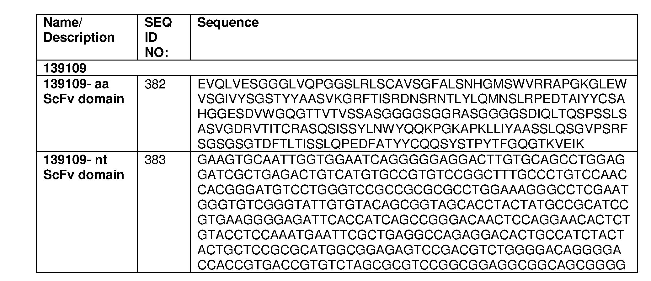


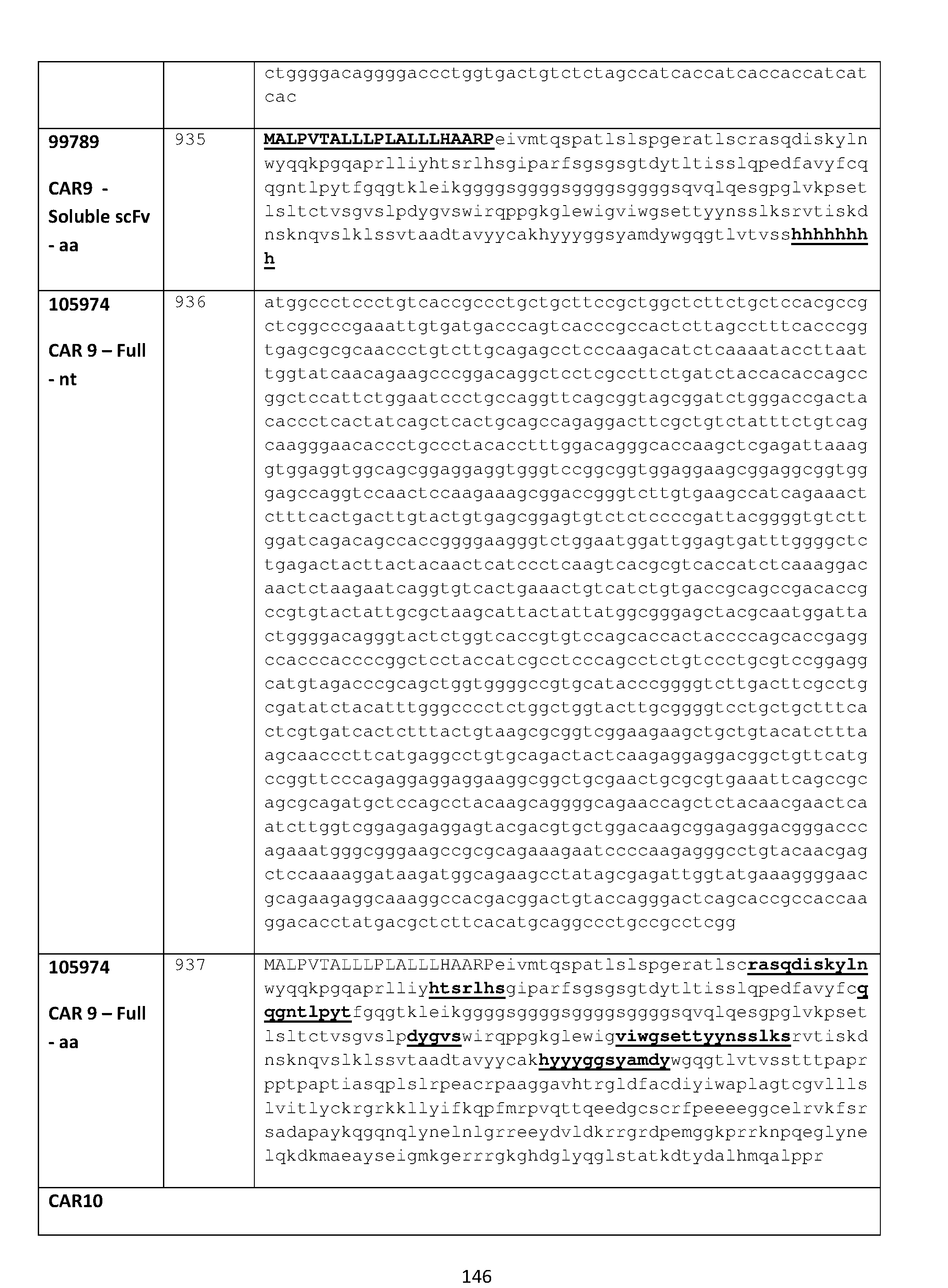


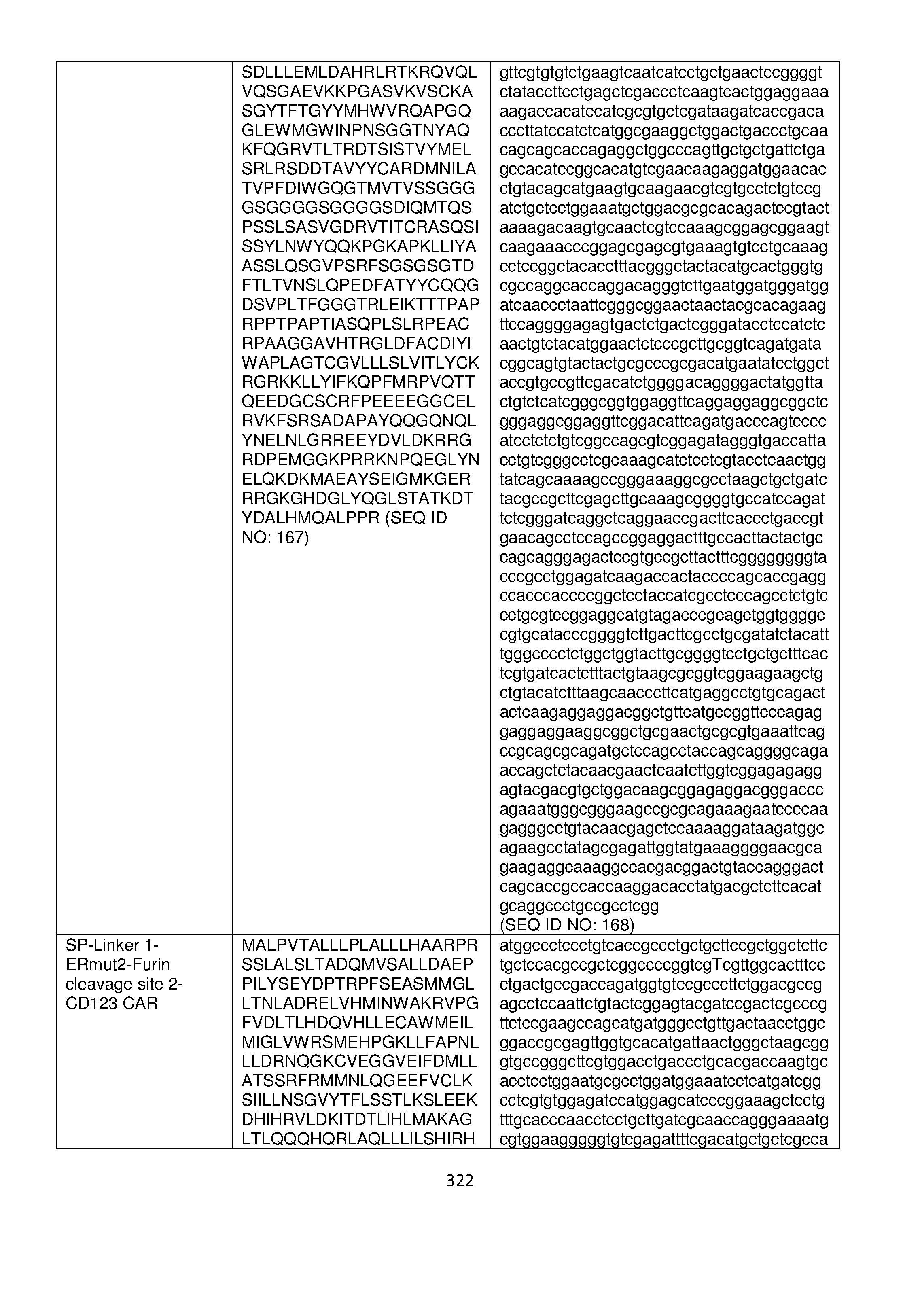


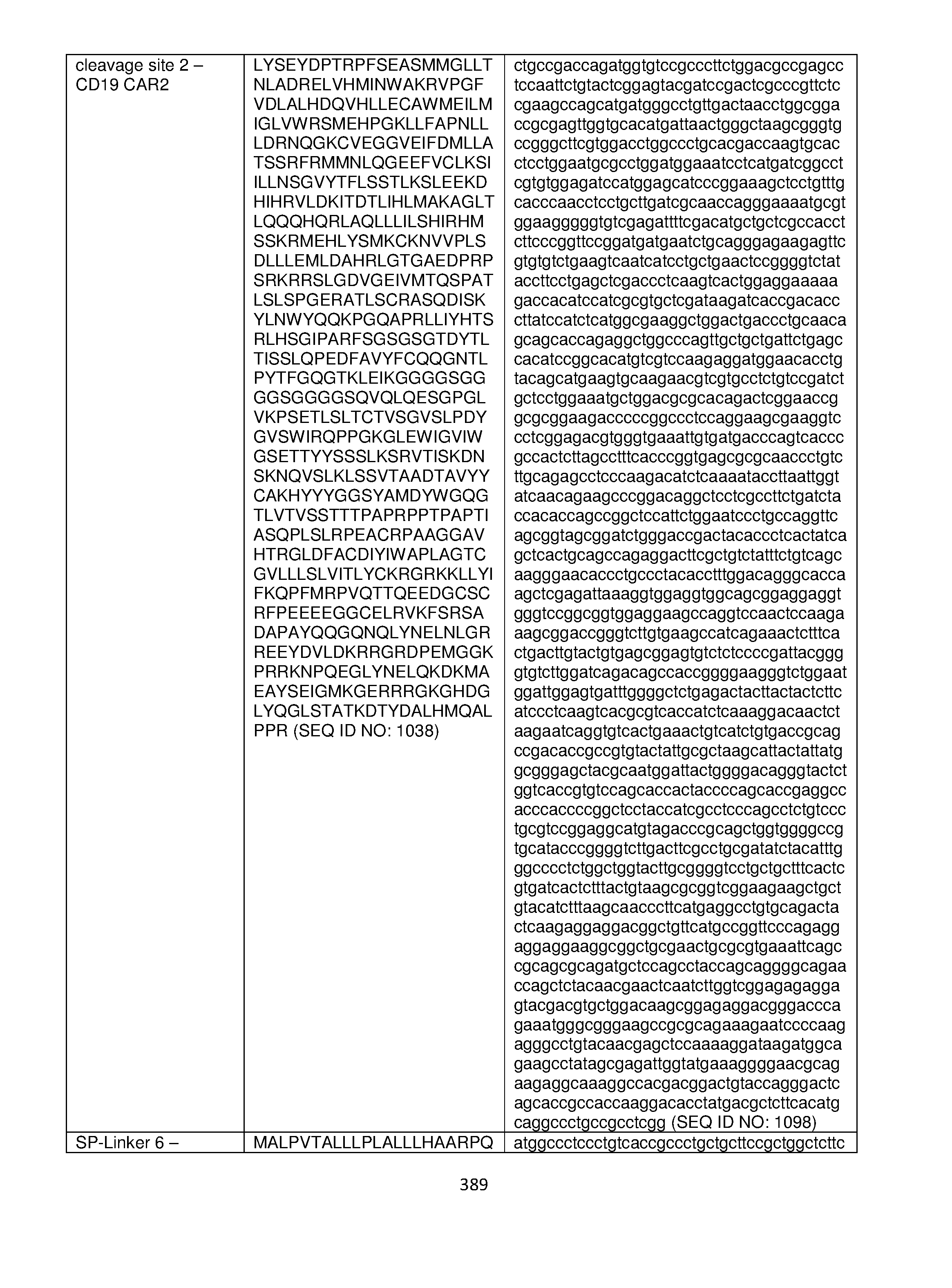


Claims
Priority Applications (21)
| Application Number | Priority Date | Filing Date | Title |
|---|---|---|---|
| ES17737079T ES2944607T3 (en) | 2016-04-15 | 2017-04-14 | Compositions and methods for the selective expression of chimeric receptors for the antigen |
| BR112018071096-2A BR112018071096A2 (en) | 2016-04-15 | 2017-04-14 | compositions and methods for selective protein expression |
| DK17737079.8T DK3443096T3 (en) | 2016-04-15 | 2017-04-14 | COMPOSITIONS AND METHODS FOR SELECTIVE EXPRESSION OF CHIMERIC ANTIGEN RECEPTORS |
| HRP20230457TT HRP20230457T1 (en) | 2016-04-15 | 2017-04-14 | Compositions and methods for selective expression of chimeric antigen receptors |
| MX2018012615A MX2018012615A (en) | 2016-04-15 | 2017-04-14 | Compositions and methods for selective protein expression. |
| SG11201809041TA SG11201809041TA (en) | 2016-04-15 | 2017-04-14 | Compositions and methods for selective protein expression |
| SI201731347T SI3443096T1 (en) | 2016-04-15 | 2017-04-14 | Compositions and methods for selective expression of chimeric antigen receptors |
| KR1020187032686A KR20180134385A (en) | 2016-04-15 | 2017-04-14 | Compositions and methods for selective protein expression |
| EP17737079.8A EP3443096B1 (en) | 2016-04-15 | 2017-04-14 | Compositions and methods for selective expression of chimeric antigen receptors |
| CN201780036531.8A CN109715808A (en) | 2016-04-15 | 2017-04-14 | Composition and method for selective protein expression |
| CA3021027A CA3021027A1 (en) | 2016-04-15 | 2017-04-14 | Compositions and methods for selective expression of chimeric antigen receptors |
| KR1020227032817A KR20220133318A (en) | 2016-04-15 | 2017-04-14 | Compositions and methods for selective protein expression |
| US16/093,758 US20230071283A1 (en) | 2016-04-15 | 2017-04-14 | Compositions and methods for selective protein expression |
| EP23151319.3A EP4219721A3 (en) | 2016-04-15 | 2017-04-14 | Compositions and methods for selective protein expression |
| AU2017250304A AU2017250304B2 (en) | 2016-04-15 | 2017-04-14 | Compositions and methods for selective protein expression |
| PL17737079.8T PL3443096T3 (en) | 2016-04-15 | 2017-04-14 | Compositions and methods for selective expression of chimeric antigen receptors |
| RU2018140063A RU2795467C2 (en) | 2016-04-15 | 2017-04-14 | Compositions and methods for selective protein expression |
| JP2018553989A JP2019518424A (en) | 2016-04-15 | 2017-04-14 | Compositions and methods for selective protein expression |
| IL262321A IL262321A (en) | 2016-04-15 | 2018-10-11 | Compositions and methods for selective protein expression |
| PH12018502191A PH12018502191A1 (en) | 2016-04-15 | 2018-10-12 | Compositions and methods for selective protein expression |
| JP2022110679A JP2022160429A (en) | 2016-04-15 | 2022-07-08 | Compositions and methods for selective protein expression |
Applications Claiming Priority (4)
| Application Number | Priority Date | Filing Date | Title |
|---|---|---|---|
| US201662322931P | 2016-04-15 | 2016-04-15 | |
| US62/322,931 | 2016-04-15 | ||
| US201762481094P | 2017-04-03 | 2017-04-03 | |
| US62/481,094 | 2017-04-03 |
Publications (2)
| Publication Number | Publication Date |
|---|---|
| WO2017181119A2 true WO2017181119A2 (en) | 2017-10-19 |
| WO2017181119A3 WO2017181119A3 (en) | 2018-01-04 |
Family
ID=59297341
Family Applications (1)
| Application Number | Title | Priority Date | Filing Date |
|---|---|---|---|
| PCT/US2017/027778 WO2017181119A2 (en) | 2016-04-15 | 2017-04-14 | Compositions and methods for selective protein expression |
Country Status (19)
| Country | Link |
|---|---|
| US (1) | US20230071283A1 (en) |
| EP (2) | EP3443096B1 (en) |
| JP (2) | JP2019518424A (en) |
| KR (2) | KR20180134385A (en) |
| CN (1) | CN109715808A (en) |
| AU (1) | AU2017250304B2 (en) |
| BR (1) | BR112018071096A2 (en) |
| CA (1) | CA3021027A1 (en) |
| DK (1) | DK3443096T3 (en) |
| ES (1) | ES2944607T3 (en) |
| HR (1) | HRP20230457T1 (en) |
| IL (1) | IL262321A (en) |
| MX (2) | MX2018012615A (en) |
| PH (1) | PH12018502191A1 (en) |
| PL (1) | PL3443096T3 (en) |
| PT (1) | PT3443096T (en) |
| SG (1) | SG11201809041TA (en) |
| SI (1) | SI3443096T1 (en) |
| WO (1) | WO2017181119A2 (en) |
Cited By (35)
| Publication number | Priority date | Publication date | Assignee | Title |
|---|---|---|---|---|
| CN109971724A (en) * | 2017-12-28 | 2019-07-05 | 上海细胞治疗研究院 | It targets ErbB receptor family and expresses the CAR-T cell and application thereof of PD-1 antibody certainly |
| WO2019118518A3 (en) * | 2017-12-11 | 2019-08-08 | Senti Biosciences, Inc. | Inducible cell receptors for cell-based therapeutics |
| WO2019213276A1 (en) | 2018-05-02 | 2019-11-07 | Novartis Ag | Regulators of human pluripotent stem cells and uses thereof |
| WO2019237035A1 (en) | 2018-06-08 | 2019-12-12 | Intellia Therapeutics, Inc. | Compositions and methods for immunooncology |
| WO2020072390A1 (en) * | 2018-10-01 | 2020-04-09 | Caribou Biosciences, Inc. | Suicide module compositions and methods |
| WO2019143961A3 (en) * | 2018-01-19 | 2020-04-16 | The Trustees Of The University Of Pennsylvania | Compositions and methods for targeting gamma delta t cells with chimeric antigen receptors |
| WO2020086742A1 (en) | 2018-10-24 | 2020-04-30 | Obsidian Therapeutics, Inc. | Er tunable protein regulation |
| CN111094358A (en) * | 2018-02-11 | 2020-05-01 | 江苏恒瑞医药股份有限公司 | Separated chimeric antigen receptor, modified T cell containing same and application |
| WO2020102709A1 (en) * | 2018-11-16 | 2020-05-22 | The Regents Of The University Of California | Compositions and methods for delivering crispr/cas effector polypeptides |
| WO2020185628A1 (en) | 2019-03-08 | 2020-09-17 | Obsidian Therapeutics, Inc. | Cd40l compositions and methods for tunable regulation |
| WO2020219742A1 (en) | 2019-04-24 | 2020-10-29 | Novartis Ag | Compositions and methods for selective protein degradation |
| JP2020536527A (en) * | 2018-02-01 | 2020-12-17 | 南京馴鹿医療技術有限公司 | Chimeric antigen receptor (CAR) that binds to BCMA and its applications |
| JP2021501605A (en) * | 2017-11-01 | 2021-01-21 | ジュノー セラピューティクス インコーポレイテッド | Antibodies and chimeric antigen receptors specific for B cell maturation antigens |
| WO2021055530A1 (en) * | 2019-09-18 | 2021-03-25 | Dana-Farber Cancer Institute, Inc. | Protein tag to induce ligand dependent degradation of protein/protein-fusions |
| WO2021061778A1 (en) * | 2019-09-23 | 2021-04-01 | The Trustees Of The Univeristy Of Pennsylvania | Disrupting tumor tissues by targeting fibroblast activation protein (fap) |
| WO2021080427A1 (en) * | 2019-10-23 | 2021-04-29 | Stichting Het Nederlands Kanker Instituut-Antoni van Leeuwenhoek Ziekenhuis | Chimeric polypeptide for regulating immune cells |
| CN112805371A (en) * | 2018-09-21 | 2021-05-14 | 佧珐药业有限公司 | Method for carrying out gene editing on cells based on CRISPR/Cas system |
| WO2021142376A1 (en) | 2020-01-08 | 2021-07-15 | Obsidian Therapeutics, Inc. | Compositions and methods for tunable regulation of transcription |
| WO2021262773A1 (en) | 2020-06-22 | 2021-12-30 | Obsidian Therapeutics, Inc. | Compositions and methods for tunable regulation of cas nucleases |
| US11266690B2 (en) | 2018-02-01 | 2022-03-08 | Nanjing Iaso Biotherapeutics Co., Ltd. | Chimeric antigen receptor (CAR) binding to BCMA, and uses thereof |
| WO2022099040A1 (en) * | 2020-11-09 | 2022-05-12 | The General Hospital Corporation | Methods of anchoring or reconstituting active molecules on metabolically labeled cells |
| WO2022113075A1 (en) * | 2020-11-25 | 2022-06-02 | Carmon Lior Binyamin | Chimeric polypeptide compositions and encoding polynucleotides thereof |
| WO2022159939A1 (en) | 2021-01-19 | 2022-07-28 | Obsidian Therapeutics, Inc. | Tumor-infiltrating lymphocytes with membrane bound interleukin 15 and uses thereof |
| US11446398B2 (en) | 2016-04-11 | 2022-09-20 | Obsidian Therapeutics, Inc. | Regulated biocircuit systems |
| WO2023069418A2 (en) | 2021-10-18 | 2023-04-27 | Obsidian Therapeutics, Inc. | Compositions and systems for regulation of function/abundance and delivery of polypeptide payloads |
| WO2023137472A2 (en) | 2022-01-14 | 2023-07-20 | Tune Therapeutics, Inc. | Compositions, systems, and methods for programming t cell phenotypes through targeted gene repression |
| WO2023137471A1 (en) | 2022-01-14 | 2023-07-20 | Tune Therapeutics, Inc. | Compositions, systems, and methods for programming t cell phenotypes through targeted gene activation |
| WO2023141436A1 (en) | 2022-01-18 | 2023-07-27 | Obsidian Therapeutics, Inc. | Methods for identifying and using allogeneic tumor infiltrating lymphocytes to treat cancer |
| US11786551B2 (en) | 2017-09-26 | 2023-10-17 | The Trustees Of The University Of Pennsylvania | Methods for treating heart disease via redirected T cell immunotherapies |
| WO2023225512A3 (en) * | 2022-05-17 | 2023-12-28 | The Trustees Of The University Of Pennsylvania | Methods for optimizing t cell immunotherapeutic effector and memory function |
| WO2024026407A1 (en) * | 2022-07-28 | 2024-02-01 | The Board Of Regents Of The University Of Oklahoma | Biomarkers, diagnostic methods, treatments, and therapeutics for autoimmune disorders and diseases |
| US11897937B2 (en) | 2017-01-05 | 2024-02-13 | Kahr Medical Ltd. | SIRPalpha-41BBL fusion protein and methods of use thereof |
| WO2024046468A1 (en) * | 2022-09-02 | 2024-03-07 | Nanjing Legend Biotech Co., Ltd. | Fusion proteins targeting lysosomal degradation pathway |
| US11938153B2 (en) | 2017-03-31 | 2024-03-26 | The Board Of Trustees Of The Leland Stanford Junior University | Methods of treating T cell exhaustion by inhibiting or modulating T cell receptor signaling |
| WO2024064642A2 (en) | 2022-09-19 | 2024-03-28 | Tune Therapeutics, Inc. | Compositions, systems, and methods for modulating t cell function |
Families Citing this family (14)
| Publication number | Priority date | Publication date | Assignee | Title |
|---|---|---|---|---|
| SG11201505896YA (en) | 2013-02-20 | 2015-09-29 | Novartis Ag | Treatment of cancer using humanized anti-egfrviii chimeric antigen receptor |
| US9745368B2 (en) | 2013-03-15 | 2017-08-29 | The Trustees Of The University Of Pennsylvania | Targeting cytotoxic cells with chimeric receptors for adoptive immunotherapy |
| EP4234685A3 (en) | 2015-04-17 | 2023-09-06 | Novartis AG | Methods for improving the efficacy and expansion of chimeric antigen receptor-expressing cells |
| US10525083B2 (en) | 2016-10-07 | 2020-01-07 | Novartis Ag | Nucleic acid molecules encoding chimeric antigen receptors comprising a CD20 binding domain |
| SG11202011830SA (en) | 2018-06-13 | 2020-12-30 | Novartis Ag | Bcma chimeric antigen receptors and uses thereof |
| CN110229849B (en) * | 2019-05-31 | 2021-09-17 | 中国科学院深圳先进技术研究院 | Time-air-conditioning control type uPA gene expression non-viral vector and preparation method and application thereof |
| CN110592014A (en) * | 2019-08-14 | 2019-12-20 | 广东美赛尔细胞生物科技有限公司 | Method for continuously removing feeder cells in vitro and in vivo without irradiation in NK cell therapy |
| CN111548422B (en) * | 2020-05-26 | 2023-04-07 | 郑州伊美诺生物技术有限公司 | Human thyroid stimulating hormone receptor fusion protein and preparation method and application thereof |
| CN112063608B (en) * | 2020-08-27 | 2022-06-17 | 浙江工业大学 | Fatty acid light decarboxylase mutant and application thereof in synthesis of L-glufosinate-ammonium |
| CN113284553B (en) * | 2021-05-28 | 2023-01-10 | 南昌大学 | Method for testing binding capacity of drug target for treating drug addiction |
| US20220403379A1 (en) * | 2021-05-28 | 2022-12-22 | The Regents Of The University Of California | Compositions and methods for targeted delivery of crispr-cas effector polypeptides and transgenes |
| CN113481169B (en) * | 2021-08-11 | 2023-06-16 | 南方医科大学南方医院 | Cell activation dependent secretion system and application |
| CN113782103A (en) * | 2021-10-26 | 2021-12-10 | 大连大学 | DNA matrix processing method based on combined enzyme digestion mechanism |
| WO2023070527A1 (en) * | 2021-10-29 | 2023-05-04 | 上海鑫湾生物科技有限公司 | Condition-controlled spliceable chimeric antigen receptor molecule and application thereof |
Citations (216)
| Publication number | Priority date | Publication date | Assignee | Title |
|---|---|---|---|---|
| US4433059A (en) | 1981-09-08 | 1984-02-21 | Ortho Diagnostic Systems Inc. | Double antibody conjugate |
| US4444878A (en) | 1981-12-21 | 1984-04-24 | Boston Biomedical Research Institute, Inc. | Bispecific antibody determinants |
| EP0239400A2 (en) | 1986-03-27 | 1987-09-30 | Medical Research Council | Recombinant antibodies and methods for their production |
| US4816567A (en) | 1983-04-08 | 1989-03-28 | Genentech, Inc. | Recombinant immunoglobin preparations |
| US4851332A (en) | 1985-04-01 | 1989-07-25 | Sloan-Kettering Institute For Cancer Research | Choriocarcinoma monoclonal antibodies and antibody panels |
| EP0346087A2 (en) | 1988-06-09 | 1989-12-13 | Snow Brand Milk Products Co., Ltd. | Hybrid antibody and process for the production thereof |
| WO1991003493A1 (en) | 1989-08-29 | 1991-03-21 | The University Of Southampton | Bi-or trispecific (fab)3 or (fab)4 conjugates |
| WO1991009967A1 (en) | 1989-12-21 | 1991-07-11 | Celltech Limited | Humanised antibodies |
| EP0519596A1 (en) | 1991-05-17 | 1992-12-23 | Merck & Co. Inc. | A method for reducing the immunogenicity of antibody variable domains |
| US5225539A (en) | 1986-03-27 | 1993-07-06 | Medical Research Council | Recombinant altered antibodies and methods of making altered antibodies |
| WO1993017105A1 (en) | 1992-02-19 | 1993-09-02 | Scotgen Limited | Altered antibodies, products and processes relating thereto |
| WO1993023537A1 (en) | 1992-05-08 | 1993-11-25 | Creative Biomolecules | Chimeric multivalent protein analogues and methods of use thereof |
| US5273743A (en) | 1990-03-09 | 1993-12-28 | Hybritech Incorporated | Trifunctional antibody-like compounds as a combined diagnostic and therapeutic agent |
| WO1994004678A1 (en) | 1992-08-21 | 1994-03-03 | Casterman Cecile | Immunoglobulins devoid of light chains |
| EP0592106A1 (en) | 1992-09-09 | 1994-04-13 | Immunogen Inc | Resurfacing of rodent antibodies |
| WO1994009131A1 (en) | 1992-10-15 | 1994-04-28 | Scotgen Limited | Recombinant specific binding protein |
| WO1994012625A2 (en) | 1992-11-23 | 1994-06-09 | Zeneca Limited | LIGAND BINDING VARIABLE DOMAIN (V-MIN) COMPRISING A FRAMEWORK REGION WITH A CYCLICALLY PERMUTED CENTRAL β-BARREL |
| US5350674A (en) | 1992-09-04 | 1994-09-27 | Becton, Dickinson And Company | Intrinsic factor - horse peroxidase conjugates and a method for increasing the stability thereof |
| US5399346A (en) | 1989-06-14 | 1995-03-21 | The United States Of America As Represented By The Department Of Health And Human Services | Gene therapy |
| WO1995009917A1 (en) | 1993-10-07 | 1995-04-13 | The Regents Of The University Of California | Genetically engineered bispecific tetravalent antibodies |
| US5530101A (en) | 1988-12-28 | 1996-06-25 | Protein Design Labs, Inc. | Humanized immunoglobulins |
| US5534254A (en) | 1992-02-06 | 1996-07-09 | Chiron Corporation | Biosynthetic binding proteins for immuno-targeting |
| US5565332A (en) | 1991-09-23 | 1996-10-15 | Medical Research Council | Production of chimeric antibodies - a combinatorial approach |
| WO1996037621A2 (en) | 1995-05-23 | 1996-11-28 | Morphosys Gesellschaft Für Proteinoptimierung Mbh | Multimeric proteins |
| US5580859A (en) | 1989-03-21 | 1996-12-03 | Vical Incorporated | Delivery of exogenous DNA sequences in a mammal |
| US5582996A (en) | 1990-12-04 | 1996-12-10 | The Wistar Institute Of Anatomy & Biology | Bifunctional antibodies and method of preparing same |
| US5585362A (en) | 1989-08-22 | 1996-12-17 | The Regents Of The University Of Michigan | Adenovirus vectors for gene therapy |
| US5591828A (en) | 1989-06-22 | 1997-01-07 | Behringwerke Aktiengesellschaft | Bispecific and oligospecific mono-and oligovalent receptors, the preparation and use thereof |
| US5635602A (en) | 1993-08-13 | 1997-06-03 | The Regents Of The University Of California | Design and synthesis of bispecific DNA-antibody conjugates |
| US5637481A (en) | 1993-02-01 | 1997-06-10 | Bristol-Myers Squibb Company | Expression vectors encoding bispecific fusion proteins and methods of producing biologically active bispecific fusion proteins in a mammalian cell |
| WO1997024373A1 (en) | 1995-12-29 | 1997-07-10 | Medvet Science Pty. Limited | Monoclonal antibody antagonists to haemopoietic growth factors |
| WO1997025068A2 (en) | 1996-01-05 | 1997-07-17 | The Government Of The United States Of America, Represented By The Secretary, Department Of Health And Human Services | Mesothelin antigen and methods and kits for targeting it |
| US5731168A (en) | 1995-03-01 | 1998-03-24 | Genentech, Inc. | Method for making heteromultimeric polypeptides |
| US5766886A (en) | 1991-12-13 | 1998-06-16 | Xoma Corporation | Modified antibody variable domains |
| US5786464A (en) | 1994-09-19 | 1998-07-28 | The General Hospital Corporation | Overexpression of mammalian and viral proteins |
| US5837821A (en) | 1992-11-04 | 1998-11-17 | City Of Hope | Antibody construct |
| US5837242A (en) | 1992-12-04 | 1998-11-17 | Medical Research Council | Multivalent and multispecific binding proteins, their manufacture and use |
| US5844094A (en) | 1992-09-25 | 1998-12-01 | Commonwealth Scientific And Industrial Research Organization | Target binding polypeptide |
| US5858358A (en) | 1992-04-07 | 1999-01-12 | The United States Of America As Represented By The Secretary Of The Navy | Methods for selectively stimulating proliferation of T cells |
| US5864019A (en) | 1990-06-11 | 1999-01-26 | Celltech Limited | Multivalent antigen-binding proteins |
| US5869620A (en) | 1986-09-02 | 1999-02-09 | Enzon, Inc. | Multivalent antigen-binding proteins |
| US5910573A (en) | 1992-01-23 | 1999-06-08 | Merck Patent Gesellschaft Mit Beschrankter Haftung | Monomeric and dimeric antibody-fragment fusion proteins |
| WO1999028471A2 (en) | 1997-12-01 | 1999-06-10 | The Government Of The United States Of America, Represented By The Secretary, Department Of Health And Human Services | ANTIBODIES, INCLUDING Fv MOLECULES, AND IMMUNOCONJUGATES HAVING HIGH BINDING AFFINITY FOR MESOTHELIN AND METHODS FOR THEIR USE |
| US5932448A (en) | 1991-11-29 | 1999-08-03 | Protein Design Labs., Inc. | Bispecific antibody heterodimers |
| US5952484A (en) | 1994-03-08 | 1999-09-14 | Sloan-Kettering Cancer Center | Recombinant human anti-LK26 antibodies |
| US5959083A (en) | 1991-06-03 | 1999-09-28 | Behringwerke Aktiengellschaft | Tetravalent bispecific receptors, the preparation and use thereof |
| US5989830A (en) | 1995-10-16 | 1999-11-23 | Unilever Patent Holdings Bv | Bifunctional or bivalent antibody fragment analogue |
| WO1999064460A1 (en) | 1998-06-10 | 1999-12-16 | Celltech Therapeutics Limited | Divalent antibody fragments |
| US6005079A (en) | 1992-08-21 | 1999-12-21 | Vrije Universiteit Brussels | Immunoglobulins devoid of light chains |
| WO2000006605A2 (en) | 1998-07-28 | 2000-02-10 | Micromet Ag | Heterominibodies |
| WO2000023600A1 (en) | 1998-10-19 | 2000-04-27 | Ariad Gene Therapeutics, Inc. | Materials and methods involving conditional aggregation domains |
| US6114148A (en) | 1996-09-20 | 2000-09-05 | The General Hospital Corporation | High level expression of proteins |
| WO2001029058A1 (en) | 1999-10-15 | 2001-04-26 | University Of Massachusetts | Rna interference pathway genes as tools for targeted genetic interference |
| US6239259B1 (en) | 1996-04-04 | 2001-05-29 | Unilever Patent Holdings B.V. | Multivalent and multispecific antigen-binding protein |
| WO2001066139A1 (en) | 2000-03-06 | 2001-09-13 | University Of Kentucky Research Foundation | Use of a compound that selectively binds to cd123 to impair hematologic cancer progenitor cell |
| US6294353B1 (en) | 1994-10-20 | 2001-09-25 | Morphosys Ag | Targeted hetero-association of recombinant proteins to multi-functional complexes |
| US6326193B1 (en) | 1999-11-05 | 2001-12-04 | Cambria Biosciences, Llc | Insect control agent |
| WO2001096584A2 (en) | 2000-06-12 | 2001-12-20 | Akkadix Corporation | Materials and methods for the control of nematodes |
| US6333396B1 (en) | 1998-10-20 | 2001-12-25 | Enzon, Inc. | Method for targeted delivery of nucleic acids |
| US20020004587A1 (en) | 2000-04-11 | 2002-01-10 | Genentech, Inc. | Multivalent antibodies and uses therefor |
| US6352694B1 (en) | 1994-06-03 | 2002-03-05 | Genetics Institute, Inc. | Methods for inducing a population of T cells to proliferate using agents which recognize TCR/CD3 and ligands which stimulate an accessory molecule on the surface of the T cells |
| US6407213B1 (en) | 1991-06-14 | 2002-06-18 | Genentech, Inc. | Method for making humanized antibodies |
| US20020076406A1 (en) | 2000-07-25 | 2002-06-20 | Leung Shui-On | Multivalent target binding protein |
| US20020103345A1 (en) | 2000-05-24 | 2002-08-01 | Zhenping Zhu | Bispecific immunoglobulin-like antigen binding proteins and method of production |
| WO2002072635A2 (en) | 2001-03-13 | 2002-09-19 | University College London | Specific binding members |
| US6476198B1 (en) | 1993-07-13 | 2002-11-05 | The Scripps Research Institute | Multispecific and multivalent antigen-binding polypeptide molecules |
| US6511663B1 (en) | 1991-06-11 | 2003-01-28 | Celltech R&D Limited | Tri- and tetra-valent monospecific antigen-binding proteins |
| WO2003014161A2 (en) | 2001-08-10 | 2003-02-20 | Aberdeen University | Antigen binding domains from fish |
| US6534055B1 (en) | 1988-11-23 | 2003-03-18 | Genetics Institute, Inc. | Methods for selectively stimulating proliferation of T cells |
| US6548640B1 (en) | 1986-03-27 | 2003-04-15 | Btg International Limited | Altered antibodies |
| US20030207346A1 (en) | 1997-05-02 | 2003-11-06 | William R. Arathoon | Method for making multispecific antibodies having heteromultimeric and common components |
| US20030211078A1 (en) | 2001-12-07 | 2003-11-13 | Heavner George A. | Pseudo-antibody constructs |
| US6670453B2 (en) | 1997-10-27 | 2003-12-30 | Unilever Patent Holdings B.V. | Multivalent antigen-binding proteins |
| US6692964B1 (en) | 1995-05-04 | 2004-02-17 | The United States Of America As Represented By The Secretary Of The Navy | Methods for transfecting T cells |
| US6703199B1 (en) | 1997-06-12 | 2004-03-09 | Research Corporation Technologies, Inc. | Artificial antibody polypeptides |
| US20040101519A1 (en) | 2002-01-03 | 2004-05-27 | The Trustees Of The University Of Pennsylvania | Activation and expansion of T-cells using an engineered multivalent signaling platform as a research tool |
| US6743896B2 (en) | 1997-04-30 | 2004-06-01 | Enzon, Inc. | Single-chain antigen-binding proteins capable of glycosylation, production and uses thereof |
| WO2004081051A1 (en) | 2003-03-12 | 2004-09-23 | The University Of Birmingham | Bispecific antibodies |
| US6797514B2 (en) | 2000-02-24 | 2004-09-28 | Xcyte Therapies, Inc. | Simultaneous stimulation and concentration of cells |
| WO2004087758A2 (en) | 2003-03-26 | 2004-10-14 | Neopharm, Inc. | Il 13 receptor alpha 2 antibody and methods of use |
| US6809185B1 (en) | 1998-01-23 | 2004-10-26 | Vlaams Interuniversitair Instituut Voor Biotechnologie | Multipurpose antibody derivatives |
| US20040220388A1 (en) | 2000-06-30 | 2004-11-04 | Nico Mertens | Novel heterodimeric fusion proteins |
| US20040219643A1 (en) | 2001-06-28 | 2004-11-04 | Greg Winter | Dual-specific ligand |
| US20040242847A1 (en) | 2000-10-20 | 2004-12-02 | Naoshi Fukushima | Degraded agonist antibody |
| US6833441B2 (en) | 2001-08-01 | 2004-12-21 | Abmaxis, Inc. | Compositions and methods for generating chimeric heteromultimers |
| US20050004352A1 (en) | 1998-04-09 | 2005-01-06 | Roland Kontermann | Single-chain multiple antigen-binding molecule, its preparation and use |
| US20050003403A1 (en) | 2003-04-22 | 2005-01-06 | Rossi Edmund A. | Polyvalent protein complex |
| US6846911B2 (en) | 1996-10-25 | 2005-01-25 | The United States Of America As Represented By The Department Of Health And Human Services | Methods and compositions for inhibiting inflammation and angiogenesis comprising a mammalian CD97 α subunit |
| WO2005014652A1 (en) | 2003-08-05 | 2005-02-17 | Morphotek, Inc. | A variant cell surface molecule associated with cancer |
| US20050042664A1 (en) | 2003-08-22 | 2005-02-24 | Medimmune, Inc. | Humanization of antibodies |
| US20050048617A1 (en) | 2003-08-18 | 2005-03-03 | Medimmune, Inc. | Humanization of antibodies |
| US6867041B2 (en) | 2000-02-24 | 2005-03-15 | Xcyte Therapies, Inc. | Simultaneous stimulation and concentration of cells |
| US20050069552A1 (en) | 2003-07-28 | 2005-03-31 | Bleck Gregory T. | Fusion antibodies |
| US20050079170A1 (en) | 2001-09-14 | 2005-04-14 | Fabrice Le Gall | Dimeric and multimeric antigen binding structure |
| US20050100543A1 (en) | 2003-07-01 | 2005-05-12 | Immunomedics, Inc. | Multivalent carriers of bi-specific antibodies |
| US6905874B2 (en) | 2000-02-24 | 2005-06-14 | Xcyte Therapies, Inc. | Simultaneous stimulation and concentration of cells |
| US6905680B2 (en) | 1988-11-23 | 2005-06-14 | Genetics Institute, Inc. | Methods of treating HIV infected subjects |
| US20050136051A1 (en) | 2003-12-22 | 2005-06-23 | Bernard Scallon | Methods for generating multimeric molecules |
| US20050136049A1 (en) | 2001-01-17 | 2005-06-23 | Ledbetter Jeffrey A. | Binding constructs and methods for use thereof |
| US20050163782A1 (en) | 2003-06-27 | 2005-07-28 | Biogen Idec Ma Inc. | Modified binding molecules comprising connecting peptides |
| US20050175606A1 (en) | 2001-04-11 | 2005-08-11 | Hua-Liang Huang | Cyclic single-chain trispecific antibody |
| US20050266425A1 (en) | 2003-12-31 | 2005-12-01 | Vaccinex, Inc. | Methods for producing and identifying multispecific antibodies |
| US20060034810A1 (en) | 2004-05-27 | 2006-02-16 | The Trustees Of The University Of Pennsylvania | Novel artificial antigen presenting cells and uses therefor |
| WO2006020258A2 (en) | 2004-07-17 | 2006-02-23 | Imclone Systems Incorporated | Novel tetravalent bispecific antibody |
| US20060083747A1 (en) | 2002-12-27 | 2006-04-20 | Domantis Limited | Fc fusion |
| US20060120960A1 (en) | 2004-01-30 | 2006-06-08 | Sergey Deyev | Multivalent complexes, their production and method of use |
| US20060121005A1 (en) | 2000-02-24 | 2006-06-08 | Xcyte Therapies, Inc. | Activation and expansion of cells |
| US7067318B2 (en) | 1995-06-07 | 2006-06-27 | The Regents Of The University Of Michigan | Methods for transfecting T cells |
| US20060204493A1 (en) | 2004-09-02 | 2006-09-14 | Genentech, Inc. | Heteromultimeric molecules |
| WO2006099141A2 (en) | 2005-03-10 | 2006-09-21 | Morphotek, Inc. | Anti-mesothelin antibodies |
| WO2006106905A1 (en) | 2005-03-31 | 2006-10-12 | Chugai Seiyaku Kabushiki Kaisha | Process for production of polypeptide by regulation of assembly |
| US7129330B1 (en) | 1998-05-05 | 2006-10-31 | Deutsches Krebsforschungszentrum Stiftung Des Offentlichen Rechts | Multivalent antibody constructs |
| US20060263367A1 (en) | 2005-05-23 | 2006-11-23 | Fey Georg H | Bispecific antibody devoid of Fc region and method of treatment using same |
| WO2006125481A1 (en) | 2005-05-27 | 2006-11-30 | Universitätsklinikum Freiburg | Monoclonal antibodies and single chain antibody fragments against cell-surface prostate specific membrane antigen |
| US20070004909A1 (en) | 2005-04-15 | 2007-01-04 | Macrogenics, Inc. | Covalent diabodies and uses thereof |
| US7175843B2 (en) | 1994-06-03 | 2007-02-13 | Genetics Institute, Llc | Methods for selectively stimulating proliferation of T cells |
| US7183076B2 (en) | 1997-05-02 | 2007-02-27 | Genentech, Inc. | Method for making multispecific antibodies having heteromultimeric and common components |
| WO2007024715A2 (en) | 2005-08-19 | 2007-03-01 | Abbott Laboratories | Dual variable domain immunoglobin and uses thereof |
| US20070087381A1 (en) | 2002-04-15 | 2007-04-19 | Tetsuo Kojima | Methods for constructing scdb libraries |
| WO2007044887A2 (en) | 2005-10-11 | 2007-04-19 | Transtarget, Inc. | Method for producing a population of homogenous tetravalent bispecific antibodies |
| US20070128150A1 (en) | 2003-12-23 | 2007-06-07 | Norman Timothy J | Branched molecular scaffolds for linking polymer residues to biologically active moieties |
| US20070141049A1 (en) | 2005-08-26 | 2007-06-21 | Reinhard Bredehorst | Bivalent IgY antibody constructs for diagnostic and therapeutic applications |
| US20070154901A1 (en) | 1997-06-11 | 2007-07-05 | Protein Engineering Technology Aps | Trimerising module |
| WO2007095338A2 (en) | 2006-02-15 | 2007-08-23 | Imclone Systems Incorporated | Functional antibodies |
| WO2007110205A2 (en) | 2006-03-24 | 2007-10-04 | Merck Patent Gmbh | Engineered heterodimeric protein domains |
| US20070274985A1 (en) | 2006-05-26 | 2007-11-29 | Stefan Dubel | Antibody |
| WO2007137760A2 (en) | 2006-05-25 | 2007-12-06 | Bayer Schering Pharma Aktiengesellschaft | Dimeric molecular complexes |
| US20080050370A1 (en) | 2006-03-17 | 2008-02-28 | Scott Glaser | Stabilized polypeptide compositions |
| US20080069820A1 (en) | 2006-08-30 | 2008-03-20 | Genentech, Inc. | Multispecific antibodies |
| US20080152645A1 (en) | 2006-08-18 | 2008-06-26 | Armagen Technologies, Inc. | Genetically Encoded Multifunctional Compositions Bidrectionally Transported Between Peripheral Blood and the CNS |
| US20080241884A1 (en) | 2003-10-08 | 2008-10-02 | Kenya Shitara | Fused Protein Composition |
| WO2008119353A1 (en) | 2007-03-29 | 2008-10-09 | Genmab A/S | Bispecific antibodies and methods for production thereof |
| US20080254512A1 (en) | 2006-11-02 | 2008-10-16 | Capon Daniel J | Hybrid immunoglobulins with moving parts |
| US20080260738A1 (en) | 2007-04-18 | 2008-10-23 | Moore Margaret D | Single chain fc, methods of making and methods of treatment |
| WO2008127735A1 (en) | 2007-04-13 | 2008-10-23 | Stemline Therapeutics, Inc. | Il3ralpha antibody conjugates and uses thereof |
| US7446190B2 (en) | 2002-05-28 | 2008-11-04 | Sloan-Kettering Institute For Cancer Research | Nucleic acids encoding chimeric T cell receptors |
| WO2008146911A1 (en) | 2007-06-01 | 2008-12-04 | Sapporo Medical University | Antibody directed against il13ra2, and diagnostic/therapeutic agent comprising the antibody |
| WO2009021754A2 (en) | 2007-08-15 | 2009-02-19 | Bayer Schering Pharma Aktiengesellschaft | Monospecific and multispecific antibodies and method of use |
| WO2009045957A1 (en) | 2007-10-01 | 2009-04-09 | Medarex, Inc. | Human antibodies that bind mesothelin, and uses thereof |
| US7521056B2 (en) | 2005-04-06 | 2009-04-21 | Ibc Pharmaceuticals, Inc. | Stably tethered structures of defined compositions with multiple functions or binding specificities |
| US7527787B2 (en) | 2005-10-19 | 2009-05-05 | Ibc Pharmaceuticals, Inc. | Multivalent immunoglobulin-based bioactive assemblies |
| US7534866B2 (en) | 2005-10-19 | 2009-05-19 | Ibc Pharmaceuticals, Inc. | Methods and compositions for generating bioactive assemblies of increased complexity and uses |
| US20090130106A1 (en) | 2005-11-29 | 2009-05-21 | The University Of Sydney | Demibodies: dimerization-activated therapeutic agents |
| WO2009068630A1 (en) | 2007-11-27 | 2009-06-04 | Ablynx N.V. | Immunoglobulin constructs |
| WO2009068204A1 (en) | 2007-11-26 | 2009-06-04 | Bayer Schering Pharma Aktiengesellschaft | Anti-mesothelin antibodies and uses therefor |
| US20090148905A1 (en) | 2007-11-30 | 2009-06-11 | Claire Ashman | Antigen-binding constructs |
| US20090155275A1 (en) | 2007-07-31 | 2009-06-18 | Medimmune, Llc | Multispecific epitope binding proteins and uses thereof |
| US20090162359A1 (en) | 2007-12-21 | 2009-06-25 | Christian Klein | Bivalent, bispecific antibodies |
| US20090162360A1 (en) | 2007-12-21 | 2009-06-25 | Christian Klein | Bivalent, bispecific antibodies |
| US20090175867A1 (en) | 2006-06-12 | 2009-07-09 | Trubion Pharmaceuticals, Inc. | Single-Chain Multivalent Binding Proteins with Effector Function |
| US20090175851A1 (en) | 2007-12-21 | 2009-07-09 | Christian Klein | Bivalent, bispecific antibodies |
| WO2009089004A1 (en) | 2008-01-07 | 2009-07-16 | Amgen Inc. | Method for making antibody fc-heterodimeric molecules using electrostatic steering effects |
| EP2083868A2 (en) | 2006-10-04 | 2009-08-05 | Københavns Universitet | Generation of a cancer-specific immune response toward muc1 and cancer specific muc1 antibodies |
| US20090232811A1 (en) | 2007-12-21 | 2009-09-17 | Christian Klein | Bivalent, bispecific antibodies |
| US20090234105A1 (en) | 2006-03-24 | 2009-09-17 | The Regents Of The University Of California | Construction of a Multivalent SCFV Through Alkyne-Azide 1,3-Dipolar Cycloaddition |
| US20090263392A1 (en) | 2006-03-31 | 2009-10-22 | Chugai Seiyaku Kabushiki Kaisha | Methods of modifying antibodies for purification of bispecific antibodies |
| US7612181B2 (en) | 2005-08-19 | 2009-11-03 | Abbott Laboratories | Dual variable domain immunoglobulin and uses thereof |
| US20090274649A1 (en) | 2002-03-01 | 2009-11-05 | Immunomedics, Inc. | Bispecific Antibody Point Mutations for Enhancing Rate of Clearance |
| US20090311181A1 (en) | 2006-03-20 | 2009-12-17 | The Regents Of The University Of California | Engineered Anti-Prostate Stem Cell Antigen (PSCA) Antibodies for Cancer Targeting |
| US7635753B2 (en) | 2007-02-19 | 2009-12-22 | Wisconsin Alumni Research Foundation | Prostate cancer and melanoma antigens |
| US20100150910A1 (en) | 2006-10-10 | 2010-06-17 | Universite De Nantes | Use of monoclonal antibodies specific to the o-acetylated form of gd2 ganglioside for treatment of certain cancers |
| WO2010126066A1 (en) | 2009-04-27 | 2010-11-04 | 協和発酵キリン株式会社 | Anti-il-3rα antibody for use in treatment of blood tumor |
| WO2010129304A2 (en) | 2009-04-27 | 2010-11-11 | Oncomed Pharmaceuticals, Inc. | Method for making heteromultimeric molecules |
| US7915391B2 (en) | 2006-04-24 | 2011-03-29 | Amgen Inc. | Humanized c-Kit antibody |
| US20110158957A1 (en) | 2009-11-10 | 2011-06-30 | Sangamo Biosciences, Inc. | Targeted disruption of T cell receptor genes using engineered zinc finger protein nucleases |
| WO2011131746A2 (en) | 2010-04-20 | 2011-10-27 | Genmab A/S | Heterodimeric antibody fc-containing proteins and methods for production thereof |
| US20110268656A1 (en) | 2009-12-02 | 2011-11-03 | David Ho | J591 minibodies and cys-diabodies for targeting human prostate specific membrane antigen (psma) and methods for their use |
| WO2011160119A2 (en) | 2010-06-19 | 2011-12-22 | Memorial Sloan-Kettering Cancer Center | Anti-gd2 antibodies |
| WO2011159847A2 (en) | 2010-06-15 | 2011-12-22 | The Regents Of The University Of California | Receptor tyrosine kinase-like orphan receptor 1 (ror1) single chain fv antibody fragment conjugates and methods of use thereof |
| US20120009181A1 (en) | 2010-02-24 | 2012-01-12 | Ab Olga | Folate Receptor 1 Antibodies and Immunoconjugates and Uses Thereof |
| US20120060230A1 (en) | 2010-07-21 | 2012-03-08 | Trevor Collingwood | Methods and compositions for modification of a hla locus |
| WO2012033885A1 (en) | 2010-09-08 | 2012-03-15 | Baylor College Of Medicine | Immunotherapy of cancer using genetically engineered gd2-specific t cells |
| WO2012079000A1 (en) | 2010-12-09 | 2012-06-14 | The Trustees Of The University Of Pennsylvania | Use of chimeric antigen receptor-modified t cells to treat cancer |
| WO2012138475A1 (en) | 2011-04-08 | 2012-10-11 | The United States Of America, As Represented By The Secretary, Department Of Health And Human Services | Anti-epidermal growth factor receptor variant iii chimeric antigen receptors and use of same for the treatment of cancer |
| US20120288506A1 (en) | 2011-05-12 | 2012-11-15 | Imclone Llc | C-kit antibodies and uses thereof |
| WO2012163805A1 (en) | 2011-05-27 | 2012-12-06 | Glaxo Group Limited | Bcma (cd269/tnfrsf17) -binding proteins |
| US8399645B2 (en) | 2003-11-05 | 2013-03-19 | St. Jude Children's Research Hospital, Inc. | Chimeric receptors with 4-1BB stimulatory signaling domain |
| WO2013040371A2 (en) | 2011-09-16 | 2013-03-21 | Baylor College Of Medicine | Targeting the tumor microenvironment using manipulated nkt cells |
| WO2013040557A2 (en) | 2011-09-16 | 2013-03-21 | The Trustees Of The University Of Pennsylvania | Rna engineered t cells for the treatment of cancer |
| WO2013063419A2 (en) | 2011-10-28 | 2013-05-02 | The Trustees Of The University Of Pennsylvania | A fully human, anti-mesothelin specific chimeric immune receptor for redirected mesothelin-expressing cell targeting |
| WO2013061273A1 (en) | 2011-10-25 | 2013-05-02 | Massimo Dominici | A modified effector cell (or chimeric receptor) for treating disialoganglioside gd2 -expressing neoplasia |
| WO2013060867A2 (en) | 2011-10-27 | 2013-05-02 | Genmab A/S | Production of heterodimeric proteins |
| WO2013074916A1 (en) | 2011-11-18 | 2013-05-23 | Board Of Regents, The University Of Texas System | Car+ t cells genetically modified to eliminate expression of t- cell receptor and/or hla |
| WO2013085552A1 (en) | 2011-12-08 | 2013-06-13 | Cleveland Clinic Foundation | Glenoid vault fixation |
| WO2013123061A1 (en) | 2012-02-13 | 2013-08-22 | Seattle Children's Hospital D/B/A Seattle Children's Research Institute | Bispecific chimeric antigen receptors and therapeutic uses thereof |
| WO2013126712A1 (en) | 2012-02-22 | 2013-08-29 | The Trustees Of The University Of Pennsylvania | Compositions and methods for generating a persisting population of t cells useful for the treatment of cancer |
| WO2013142034A1 (en) | 2012-03-23 | 2013-09-26 | The United States Of America, As Represented By The Secretary, Department Of Health And Human Services | Anti-mesothelin chimeric antigen receptors |
| WO2013173820A2 (en) | 2012-05-18 | 2013-11-21 | Scott & White Healthcare | Bispecific scfv immunofusion (bif) |
| WO2013192294A1 (en) | 2012-06-20 | 2013-12-27 | Boston 3T Biotechnologies, Inc. | Cellular therapies for treating and preventing cancers and other immune system disorders |
| WO2014031687A1 (en) | 2012-08-20 | 2014-02-27 | Jensen, Michael | Method and compositions for cellular immunotherapy |
| US20140068797A1 (en) | 2012-05-25 | 2014-03-06 | University Of Vienna | Methods and compositions for rna-directed target dna modification and for rna-directed modulation of transcription |
| WO2014055442A2 (en) | 2012-10-01 | 2014-04-10 | The Trustees Of The University Of Pennsylvania | Compositions and methods for targeting stromal cells for the treatment of cancer |
| WO2014055657A1 (en) | 2012-10-05 | 2014-04-10 | The Trustees Of The University Of Pennsylvania | Use of a trans-signaling approach in chimeric antigen receptors |
| US8697359B1 (en) | 2012-12-12 | 2014-04-15 | The Broad Institute, Inc. | CRISPR-Cas systems and methods for altering expression of gene products |
| WO2014089335A2 (en) | 2012-12-07 | 2014-06-12 | Amgen Inc. | Bcma antigen binding proteins |
| US20140178365A1 (en) | 2012-04-04 | 2014-06-26 | Sialix Inc | Glycan-interacting compounds |
| US8795965B2 (en) | 2012-12-12 | 2014-08-05 | The Broad Institute, Inc. | CRISPR-Cas component systems, methods and compositions for sequence manipulation |
| WO2014122144A1 (en) | 2013-02-05 | 2014-08-14 | Engmab Ag | BISPECIFIC ANTIBODIES AGAINST CD3ε AND BCMA |
| WO2014130635A1 (en) | 2013-02-20 | 2014-08-28 | Novartis Ag | Effective targeting of primary human leukemia using anti-cd123 chimeric antigen receptor engineered t cells |
| WO2014130657A1 (en) | 2013-02-20 | 2014-08-28 | The Trustees Of The University Of Pennsylvania | Treatment of cancer using humanized anti-egfrviii chimeric antigen receptor |
| WO2014145252A2 (en) | 2013-03-15 | 2014-09-18 | Milone Michael C | Targeting cytotoxic cells with chimeric receptors for adoptive immunotherapy |
| WO2014138805A1 (en) | 2013-03-14 | 2014-09-18 | Csl Limited | Anti il-3r alpha agents and uses thereof |
| WO2014138819A1 (en) | 2013-03-14 | 2014-09-18 | Csl Limited | Agents that neutralize il-3 signaling and uses thereof |
| WO2014140248A1 (en) | 2013-03-15 | 2014-09-18 | Amgen Research (Munich) Gmbh | Binding molecules for bcma and cd3 |
| WO2014144622A2 (en) | 2013-03-15 | 2014-09-18 | Stephen Forman | Cd123-specific chimeric antigen receptor redirected t cells and methods of their use |
| WO2014153270A1 (en) | 2013-03-16 | 2014-09-25 | Novartis Ag | Treatment of cancer using humanized anti-cd19 chimeric antigen receptor |
| US8865406B2 (en) | 2012-12-12 | 2014-10-21 | The Broad Institute Inc. | Engineering and optimization of improved systems, methods and enzyme compositions for sequence manipulation |
| WO2015090230A1 (en) | 2013-12-19 | 2015-06-25 | Novartis Ag | Human mesothelin chimeric antigen receptors and uses thereof |
| WO2015142675A2 (en) | 2014-03-15 | 2015-09-24 | Novartis Ag | Treatment of cancer using chimeric antigen receptor |
| US20150283178A1 (en) | 2014-04-07 | 2015-10-08 | Carl H. June | Treatment of cancer using anti-cd19 chimeric antigen receptor |
| WO2016014565A2 (en) | 2014-07-21 | 2016-01-28 | Novartis Ag | Treatment of cancer using humanized anti-bcma chimeric antigen receptor |
| WO2016014576A1 (en) | 2014-07-21 | 2016-01-28 | Novartis Ag | Treatment of cancer using a cd33 chimeric antigen receptor |
| WO2016014535A1 (en) | 2014-07-21 | 2016-01-28 | Novartis Ag | Treatment of cancer using a cll-1 chimeric antigen receptor |
| WO2016014789A2 (en) | 2014-07-24 | 2016-01-28 | Bluebird Bio, Inc. | Bcma chimeric antigen receptors |
| WO2016028896A1 (en) | 2014-08-19 | 2016-02-25 | Novartis Ag | Anti-cd123 chimeric antigen receptor (car) for use in cancer treatment |
| WO2016164731A2 (en) | 2015-04-08 | 2016-10-13 | Novartis Ag | Cd20 therapies, cd22 therapies, and combination therapies with a cd19 chimeric antigen receptor (car) - expressing cell |
Family Cites Families (10)
| Publication number | Priority date | Publication date | Assignee | Title |
|---|---|---|---|---|
| IL142137A0 (en) * | 1998-10-19 | 2002-03-10 | Ariad Gene Therapeutics Inc | Materials and methods involving conditional retention domains |
| US7262005B1 (en) * | 2000-02-04 | 2007-08-28 | Aurora Biosciences Corporation | Methods of protein destabilization and uses thereof |
| AU2013243951A1 (en) * | 2012-04-02 | 2014-10-30 | Moderna Therapeutics, Inc. | Modified polynucleotides for the production of secreted proteins |
| US20150290244A1 (en) * | 2012-07-13 | 2015-10-15 | The Trustees Of The University Of Pennsylvania | Use of cart19 to deplete normal b cells to induce tolerance |
| CA2940460A1 (en) * | 2014-03-07 | 2015-09-11 | Bellicum Pharmaceuticals, Inc. | Caspase polypeptides having modified activity and uses thereof |
| EP3166968B1 (en) * | 2014-05-02 | 2021-09-22 | The Trustees of the University of Pennsylvania | Compositions and methods of chimeric autoantibody receptor t cells |
| LT3151672T (en) * | 2014-06-06 | 2021-02-10 | Bluebird Bio, Inc. | Improved t cell compositions |
| US10550379B2 (en) * | 2015-06-29 | 2020-02-04 | The Board Of Trustees Of The Leland Stanford Junior University | Degron fusion constructs and methods for controlling protein production |
| EP3331906A1 (en) * | 2015-08-06 | 2018-06-13 | Dana-Farber Cancer Institute, Inc. | Tunable endogenous protein degradation |
| JP6894430B2 (en) * | 2015-08-24 | 2021-06-30 | セレクティスCellectis | Chimeric antigen receptor with integrated regulatory function |
-
2017
- 2017-04-14 HR HRP20230457TT patent/HRP20230457T1/en unknown
- 2017-04-14 SG SG11201809041TA patent/SG11201809041TA/en unknown
- 2017-04-14 EP EP17737079.8A patent/EP3443096B1/en active Active
- 2017-04-14 PL PL17737079.8T patent/PL3443096T3/en unknown
- 2017-04-14 CN CN201780036531.8A patent/CN109715808A/en active Pending
- 2017-04-14 JP JP2018553989A patent/JP2019518424A/en active Pending
- 2017-04-14 KR KR1020187032686A patent/KR20180134385A/en not_active Application Discontinuation
- 2017-04-14 US US16/093,758 patent/US20230071283A1/en active Pending
- 2017-04-14 PT PT177370798T patent/PT3443096T/en unknown
- 2017-04-14 ES ES17737079T patent/ES2944607T3/en active Active
- 2017-04-14 CA CA3021027A patent/CA3021027A1/en active Pending
- 2017-04-14 DK DK17737079.8T patent/DK3443096T3/en active
- 2017-04-14 KR KR1020227032817A patent/KR20220133318A/en not_active Application Discontinuation
- 2017-04-14 AU AU2017250304A patent/AU2017250304B2/en active Active
- 2017-04-14 MX MX2018012615A patent/MX2018012615A/en unknown
- 2017-04-14 EP EP23151319.3A patent/EP4219721A3/en active Pending
- 2017-04-14 BR BR112018071096-2A patent/BR112018071096A2/en unknown
- 2017-04-14 SI SI201731347T patent/SI3443096T1/en unknown
- 2017-04-14 WO PCT/US2017/027778 patent/WO2017181119A2/en active Application Filing
-
2018
- 2018-10-11 IL IL262321A patent/IL262321A/en unknown
- 2018-10-12 PH PH12018502191A patent/PH12018502191A1/en unknown
- 2018-10-15 MX MX2023000082A patent/MX2023000082A/en unknown
-
2022
- 2022-07-08 JP JP2022110679A patent/JP2022160429A/en active Pending
Patent Citations (243)
| Publication number | Priority date | Publication date | Assignee | Title |
|---|---|---|---|---|
| US4433059A (en) | 1981-09-08 | 1984-02-21 | Ortho Diagnostic Systems Inc. | Double antibody conjugate |
| US4444878A (en) | 1981-12-21 | 1984-04-24 | Boston Biomedical Research Institute, Inc. | Bispecific antibody determinants |
| US6331415B1 (en) | 1983-04-08 | 2001-12-18 | Genentech, Inc. | Methods of producing immunoglobulins, vectors and transformed host cells for use therein |
| US4816567A (en) | 1983-04-08 | 1989-03-28 | Genentech, Inc. | Recombinant immunoglobin preparations |
| US4851332A (en) | 1985-04-01 | 1989-07-25 | Sloan-Kettering Institute For Cancer Research | Choriocarcinoma monoclonal antibodies and antibody panels |
| US5225539A (en) | 1986-03-27 | 1993-07-06 | Medical Research Council | Recombinant altered antibodies and methods of making altered antibodies |
| EP0239400A2 (en) | 1986-03-27 | 1987-09-30 | Medical Research Council | Recombinant antibodies and methods for their production |
| US6548640B1 (en) | 1986-03-27 | 2003-04-15 | Btg International Limited | Altered antibodies |
| US5869620A (en) | 1986-09-02 | 1999-02-09 | Enzon, Inc. | Multivalent antigen-binding proteins |
| EP0346087A2 (en) | 1988-06-09 | 1989-12-13 | Snow Brand Milk Products Co., Ltd. | Hybrid antibody and process for the production thereof |
| US5883223A (en) | 1988-11-23 | 1999-03-16 | Gray; Gary S. | CD9 antigen peptides and antibodies thereto |
| US6534055B1 (en) | 1988-11-23 | 2003-03-18 | Genetics Institute, Inc. | Methods for selectively stimulating proliferation of T cells |
| US6887466B2 (en) | 1988-11-23 | 2005-05-03 | Genetics Institute, Inc. | Methods for selectively stimulating proliferation of T cells |
| US6905680B2 (en) | 1988-11-23 | 2005-06-14 | Genetics Institute, Inc. | Methods of treating HIV infected subjects |
| US7232566B2 (en) | 1988-11-23 | 2007-06-19 | The United States As Represented By The Secretary Of The Navy | Methods for treating HIV infected subjects |
| US7144575B2 (en) | 1988-11-23 | 2006-12-05 | The Regents Of The University Of Michigan | Methods for selectively stimulating proliferation of T cells |
| US5585089A (en) | 1988-12-28 | 1996-12-17 | Protein Design Labs, Inc. | Humanized immunoglobulins |
| US5530101A (en) | 1988-12-28 | 1996-06-25 | Protein Design Labs, Inc. | Humanized immunoglobulins |
| US5589466A (en) | 1989-03-21 | 1996-12-31 | Vical Incorporated | Induction of a protective immune response in a mammal by injecting a DNA sequence |
| US5580859A (en) | 1989-03-21 | 1996-12-03 | Vical Incorporated | Delivery of exogenous DNA sequences in a mammal |
| US5399346A (en) | 1989-06-14 | 1995-03-21 | The United States Of America As Represented By The Department Of Health And Human Services | Gene therapy |
| US5591828A (en) | 1989-06-22 | 1997-01-07 | Behringwerke Aktiengesellschaft | Bispecific and oligospecific mono-and oligovalent receptors, the preparation and use thereof |
| US5585362A (en) | 1989-08-22 | 1996-12-17 | The Regents Of The University Of Michigan | Adenovirus vectors for gene therapy |
| WO1991003493A1 (en) | 1989-08-29 | 1991-03-21 | The University Of Southampton | Bi-or trispecific (fab)3 or (fab)4 conjugates |
| WO1991009967A1 (en) | 1989-12-21 | 1991-07-11 | Celltech Limited | Humanised antibodies |
| US5273743A (en) | 1990-03-09 | 1993-12-28 | Hybritech Incorporated | Trifunctional antibody-like compounds as a combined diagnostic and therapeutic agent |
| US5864019A (en) | 1990-06-11 | 1999-01-26 | Celltech Limited | Multivalent antigen-binding proteins |
| US5582996A (en) | 1990-12-04 | 1996-12-10 | The Wistar Institute Of Anatomy & Biology | Bifunctional antibodies and method of preparing same |
| EP0519596A1 (en) | 1991-05-17 | 1992-12-23 | Merck & Co. Inc. | A method for reducing the immunogenicity of antibody variable domains |
| US5959083A (en) | 1991-06-03 | 1999-09-28 | Behringwerke Aktiengellschaft | Tetravalent bispecific receptors, the preparation and use thereof |
| US6511663B1 (en) | 1991-06-11 | 2003-01-28 | Celltech R&D Limited | Tri- and tetra-valent monospecific antigen-binding proteins |
| US6407213B1 (en) | 1991-06-14 | 2002-06-18 | Genentech, Inc. | Method for making humanized antibodies |
| US5565332A (en) | 1991-09-23 | 1996-10-15 | Medical Research Council | Production of chimeric antibodies - a combinatorial approach |
| US5932448A (en) | 1991-11-29 | 1999-08-03 | Protein Design Labs., Inc. | Bispecific antibody heterodimers |
| US5766886A (en) | 1991-12-13 | 1998-06-16 | Xoma Corporation | Modified antibody variable domains |
| US5910573A (en) | 1992-01-23 | 1999-06-08 | Merck Patent Gesellschaft Mit Beschrankter Haftung | Monomeric and dimeric antibody-fragment fusion proteins |
| US5534254A (en) | 1992-02-06 | 1996-07-09 | Chiron Corporation | Biosynthetic binding proteins for immuno-targeting |
| WO1993017105A1 (en) | 1992-02-19 | 1993-09-02 | Scotgen Limited | Altered antibodies, products and processes relating thereto |
| US5858358A (en) | 1992-04-07 | 1999-01-12 | The United States Of America As Represented By The Secretary Of The Navy | Methods for selectively stimulating proliferation of T cells |
| WO1993023537A1 (en) | 1992-05-08 | 1993-11-25 | Creative Biomolecules | Chimeric multivalent protein analogues and methods of use thereof |
| WO1994004678A1 (en) | 1992-08-21 | 1994-03-03 | Casterman Cecile | Immunoglobulins devoid of light chains |
| US6005079A (en) | 1992-08-21 | 1999-12-21 | Vrije Universiteit Brussels | Immunoglobulins devoid of light chains |
| US5350674A (en) | 1992-09-04 | 1994-09-27 | Becton, Dickinson And Company | Intrinsic factor - horse peroxidase conjugates and a method for increasing the stability thereof |
| EP0592106A1 (en) | 1992-09-09 | 1994-04-13 | Immunogen Inc | Resurfacing of rodent antibodies |
| US5844094A (en) | 1992-09-25 | 1998-12-01 | Commonwealth Scientific And Industrial Research Organization | Target binding polypeptide |
| WO1994009131A1 (en) | 1992-10-15 | 1994-04-28 | Scotgen Limited | Recombinant specific binding protein |
| US5837821A (en) | 1992-11-04 | 1998-11-17 | City Of Hope | Antibody construct |
| WO1994012625A2 (en) | 1992-11-23 | 1994-06-09 | Zeneca Limited | LIGAND BINDING VARIABLE DOMAIN (V-MIN) COMPRISING A FRAMEWORK REGION WITH A CYCLICALLY PERMUTED CENTRAL β-BARREL |
| US5837242A (en) | 1992-12-04 | 1998-11-17 | Medical Research Council | Multivalent and multispecific binding proteins, their manufacture and use |
| US5637481A (en) | 1993-02-01 | 1997-06-10 | Bristol-Myers Squibb Company | Expression vectors encoding bispecific fusion proteins and methods of producing biologically active bispecific fusion proteins in a mammalian cell |
| US6476198B1 (en) | 1993-07-13 | 2002-11-05 | The Scripps Research Institute | Multispecific and multivalent antigen-binding polypeptide molecules |
| US5635602A (en) | 1993-08-13 | 1997-06-03 | The Regents Of The University Of California | Design and synthesis of bispecific DNA-antibody conjugates |
| WO1995009917A1 (en) | 1993-10-07 | 1995-04-13 | The Regents Of The University Of California | Genetically engineered bispecific tetravalent antibodies |
| US5952484A (en) | 1994-03-08 | 1999-09-14 | Sloan-Kettering Cancer Center | Recombinant human anti-LK26 antibodies |
| US7175843B2 (en) | 1994-06-03 | 2007-02-13 | Genetics Institute, Llc | Methods for selectively stimulating proliferation of T cells |
| US6352694B1 (en) | 1994-06-03 | 2002-03-05 | Genetics Institute, Inc. | Methods for inducing a population of T cells to proliferate using agents which recognize TCR/CD3 and ligands which stimulate an accessory molecule on the surface of the T cells |
| US6905681B1 (en) | 1994-06-03 | 2005-06-14 | Genetics Institute, Inc. | Methods for selectively stimulating proliferation of T cells |
| US5786464A (en) | 1994-09-19 | 1998-07-28 | The General Hospital Corporation | Overexpression of mammalian and viral proteins |
| US5786464C1 (en) | 1994-09-19 | 2012-04-24 | Gen Hospital Corp | Overexpression of mammalian and viral proteins |
| US6294353B1 (en) | 1994-10-20 | 2001-09-25 | Morphosys Ag | Targeted hetero-association of recombinant proteins to multi-functional complexes |
| US20070014794A1 (en) | 1995-03-01 | 2007-01-18 | Genentech, Inc. | Method for making heteromultimeric polypeptides |
| US5731168A (en) | 1995-03-01 | 1998-03-24 | Genentech, Inc. | Method for making heteromultimeric polypeptides |
| US6692964B1 (en) | 1995-05-04 | 2004-02-17 | The United States Of America As Represented By The Secretary Of The Navy | Methods for transfecting T cells |
| US7172869B2 (en) | 1995-05-04 | 2007-02-06 | The United States Of America As Represented By The Secretary Of The Navy | Methods for transfecting T cells |
| WO1996037621A2 (en) | 1995-05-23 | 1996-11-28 | Morphosys Gesellschaft Für Proteinoptimierung Mbh | Multimeric proteins |
| US7067318B2 (en) | 1995-06-07 | 2006-06-27 | The Regents Of The University Of Michigan | Methods for transfecting T cells |
| US5989830A (en) | 1995-10-16 | 1999-11-23 | Unilever Patent Holdings Bv | Bifunctional or bivalent antibody fragment analogue |
| WO1997024373A1 (en) | 1995-12-29 | 1997-07-10 | Medvet Science Pty. Limited | Monoclonal antibody antagonists to haemopoietic growth factors |
| WO1997025068A2 (en) | 1996-01-05 | 1997-07-17 | The Government Of The United States Of America, Represented By The Secretary, Department Of Health And Human Services | Mesothelin antigen and methods and kits for targeting it |
| US6239259B1 (en) | 1996-04-04 | 2001-05-29 | Unilever Patent Holdings B.V. | Multivalent and multispecific antigen-binding protein |
| US6114148A (en) | 1996-09-20 | 2000-09-05 | The General Hospital Corporation | High level expression of proteins |
| US6114148C1 (en) | 1996-09-20 | 2012-05-01 | Gen Hospital Corp | High level expression of proteins |
| US6846911B2 (en) | 1996-10-25 | 2005-01-25 | The United States Of America As Represented By The Department Of Health And Human Services | Methods and compositions for inhibiting inflammation and angiogenesis comprising a mammalian CD97 α subunit |
| US6743896B2 (en) | 1997-04-30 | 2004-06-01 | Enzon, Inc. | Single-chain antigen-binding proteins capable of glycosylation, production and uses thereof |
| US7183076B2 (en) | 1997-05-02 | 2007-02-27 | Genentech, Inc. | Method for making multispecific antibodies having heteromultimeric and common components |
| US20030207346A1 (en) | 1997-05-02 | 2003-11-06 | William R. Arathoon | Method for making multispecific antibodies having heteromultimeric and common components |
| US20070154901A1 (en) | 1997-06-11 | 2007-07-05 | Protein Engineering Technology Aps | Trimerising module |
| US6703199B1 (en) | 1997-06-12 | 2004-03-09 | Research Corporation Technologies, Inc. | Artificial antibody polypeptides |
| US6670453B2 (en) | 1997-10-27 | 2003-12-30 | Unilever Patent Holdings B.V. | Multivalent antigen-binding proteins |
| WO1999028471A2 (en) | 1997-12-01 | 1999-06-10 | The Government Of The United States Of America, Represented By The Secretary, Department Of Health And Human Services | ANTIBODIES, INCLUDING Fv MOLECULES, AND IMMUNOCONJUGATES HAVING HIGH BINDING AFFINITY FOR MESOTHELIN AND METHODS FOR THEIR USE |
| US6809185B1 (en) | 1998-01-23 | 2004-10-26 | Vlaams Interuniversitair Instituut Voor Biotechnologie | Multipurpose antibody derivatives |
| US20050004352A1 (en) | 1998-04-09 | 2005-01-06 | Roland Kontermann | Single-chain multiple antigen-binding molecule, its preparation and use |
| US7129330B1 (en) | 1998-05-05 | 2006-10-31 | Deutsches Krebsforschungszentrum Stiftung Des Offentlichen Rechts | Multivalent antibody constructs |
| WO1999064460A1 (en) | 1998-06-10 | 1999-12-16 | Celltech Therapeutics Limited | Divalent antibody fragments |
| WO2000006605A2 (en) | 1998-07-28 | 2000-02-10 | Micromet Ag | Heterominibodies |
| WO2000023600A1 (en) | 1998-10-19 | 2000-04-27 | Ariad Gene Therapeutics, Inc. | Materials and methods involving conditional aggregation domains |
| US6333396B1 (en) | 1998-10-20 | 2001-12-25 | Enzon, Inc. | Method for targeted delivery of nucleic acids |
| WO2001029058A1 (en) | 1999-10-15 | 2001-04-26 | University Of Massachusetts | Rna interference pathway genes as tools for targeted genetic interference |
| US6326193B1 (en) | 1999-11-05 | 2001-12-04 | Cambria Biosciences, Llc | Insect control agent |
| US6905874B2 (en) | 2000-02-24 | 2005-06-14 | Xcyte Therapies, Inc. | Simultaneous stimulation and concentration of cells |
| US6867041B2 (en) | 2000-02-24 | 2005-03-15 | Xcyte Therapies, Inc. | Simultaneous stimulation and concentration of cells |
| US6797514B2 (en) | 2000-02-24 | 2004-09-28 | Xcyte Therapies, Inc. | Simultaneous stimulation and concentration of cells |
| US20060121005A1 (en) | 2000-02-24 | 2006-06-08 | Xcyte Therapies, Inc. | Activation and expansion of cells |
| WO2001066139A1 (en) | 2000-03-06 | 2001-09-13 | University Of Kentucky Research Foundation | Use of a compound that selectively binds to cd123 to impair hematologic cancer progenitor cell |
| US20020004587A1 (en) | 2000-04-11 | 2002-01-10 | Genentech, Inc. | Multivalent antibodies and uses therefor |
| US20020103345A1 (en) | 2000-05-24 | 2002-08-01 | Zhenping Zhu | Bispecific immunoglobulin-like antigen binding proteins and method of production |
| WO2001096584A2 (en) | 2000-06-12 | 2001-12-20 | Akkadix Corporation | Materials and methods for the control of nematodes |
| US20040220388A1 (en) | 2000-06-30 | 2004-11-04 | Nico Mertens | Novel heterodimeric fusion proteins |
| US20020076406A1 (en) | 2000-07-25 | 2002-06-20 | Leung Shui-On | Multivalent target binding protein |
| US20040242847A1 (en) | 2000-10-20 | 2004-12-02 | Naoshi Fukushima | Degraded agonist antibody |
| US20050136049A1 (en) | 2001-01-17 | 2005-06-23 | Ledbetter Jeffrey A. | Binding constructs and methods for use thereof |
| WO2002072635A2 (en) | 2001-03-13 | 2002-09-19 | University College London | Specific binding members |
| US20050175606A1 (en) | 2001-04-11 | 2005-08-11 | Hua-Liang Huang | Cyclic single-chain trispecific antibody |
| US20040219643A1 (en) | 2001-06-28 | 2004-11-04 | Greg Winter | Dual-specific ligand |
| US6833441B2 (en) | 2001-08-01 | 2004-12-21 | Abmaxis, Inc. | Compositions and methods for generating chimeric heteromultimers |
| WO2003014161A2 (en) | 2001-08-10 | 2003-02-20 | Aberdeen University | Antigen binding domains from fish |
| US20050079170A1 (en) | 2001-09-14 | 2005-04-14 | Fabrice Le Gall | Dimeric and multimeric antigen binding structure |
| US20030211078A1 (en) | 2001-12-07 | 2003-11-13 | Heavner George A. | Pseudo-antibody constructs |
| US20040101519A1 (en) | 2002-01-03 | 2004-05-27 | The Trustees Of The University Of Pennsylvania | Activation and expansion of T-cells using an engineered multivalent signaling platform as a research tool |
| US20090274649A1 (en) | 2002-03-01 | 2009-11-05 | Immunomedics, Inc. | Bispecific Antibody Point Mutations for Enhancing Rate of Clearance |
| US20070087381A1 (en) | 2002-04-15 | 2007-04-19 | Tetsuo Kojima | Methods for constructing scdb libraries |
| US7446190B2 (en) | 2002-05-28 | 2008-11-04 | Sloan-Kettering Institute For Cancer Research | Nucleic acids encoding chimeric T cell receptors |
| US20060083747A1 (en) | 2002-12-27 | 2006-04-20 | Domantis Limited | Fc fusion |
| WO2004081051A1 (en) | 2003-03-12 | 2004-09-23 | The University Of Birmingham | Bispecific antibodies |
| WO2004087758A2 (en) | 2003-03-26 | 2004-10-14 | Neopharm, Inc. | Il 13 receptor alpha 2 antibody and methods of use |
| US20080171855A1 (en) | 2003-04-22 | 2008-07-17 | Ibc Pharmaceuticals, Inc. | Polyvalent protein complex |
| US20050003403A1 (en) | 2003-04-22 | 2005-01-06 | Rossi Edmund A. | Polyvalent protein complex |
| US20050163782A1 (en) | 2003-06-27 | 2005-07-28 | Biogen Idec Ma Inc. | Modified binding molecules comprising connecting peptides |
| US20050100543A1 (en) | 2003-07-01 | 2005-05-12 | Immunomedics, Inc. | Multivalent carriers of bi-specific antibodies |
| US20050069552A1 (en) | 2003-07-28 | 2005-03-31 | Bleck Gregory T. | Fusion antibodies |
| WO2005014652A1 (en) | 2003-08-05 | 2005-02-17 | Morphotek, Inc. | A variant cell surface molecule associated with cancer |
| US20050048617A1 (en) | 2003-08-18 | 2005-03-03 | Medimmune, Inc. | Humanization of antibodies |
| US20050042664A1 (en) | 2003-08-22 | 2005-02-24 | Medimmune, Inc. | Humanization of antibodies |
| US20080241884A1 (en) | 2003-10-08 | 2008-10-02 | Kenya Shitara | Fused Protein Composition |
| US8399645B2 (en) | 2003-11-05 | 2013-03-19 | St. Jude Children's Research Hospital, Inc. | Chimeric receptors with 4-1BB stimulatory signaling domain |
| US20050136051A1 (en) | 2003-12-22 | 2005-06-23 | Bernard Scallon | Methods for generating multimeric molecules |
| US20070128150A1 (en) | 2003-12-23 | 2007-06-07 | Norman Timothy J | Branched molecular scaffolds for linking polymer residues to biologically active moieties |
| US20050266425A1 (en) | 2003-12-31 | 2005-12-01 | Vaccinex, Inc. | Methods for producing and identifying multispecific antibodies |
| US20060120960A1 (en) | 2004-01-30 | 2006-06-08 | Sergey Deyev | Multivalent complexes, their production and method of use |
| US20060034810A1 (en) | 2004-05-27 | 2006-02-16 | The Trustees Of The University Of Pennsylvania | Novel artificial antigen presenting cells and uses therefor |
| WO2006020258A2 (en) | 2004-07-17 | 2006-02-23 | Imclone Systems Incorporated | Novel tetravalent bispecific antibody |
| US20060204493A1 (en) | 2004-09-02 | 2006-09-14 | Genentech, Inc. | Heteromultimeric molecules |
| WO2006099141A2 (en) | 2005-03-10 | 2006-09-21 | Morphotek, Inc. | Anti-mesothelin antibodies |
| WO2006106905A1 (en) | 2005-03-31 | 2006-10-12 | Chugai Seiyaku Kabushiki Kaisha | Process for production of polypeptide by regulation of assembly |
| US7521056B2 (en) | 2005-04-06 | 2009-04-21 | Ibc Pharmaceuticals, Inc. | Stably tethered structures of defined compositions with multiple functions or binding specificities |
| US20070004909A1 (en) | 2005-04-15 | 2007-01-04 | Macrogenics, Inc. | Covalent diabodies and uses thereof |
| US20060263367A1 (en) | 2005-05-23 | 2006-11-23 | Fey Georg H | Bispecific antibody devoid of Fc region and method of treatment using same |
| WO2006125481A1 (en) | 2005-05-27 | 2006-11-30 | Universitätsklinikum Freiburg | Monoclonal antibodies and single chain antibody fragments against cell-surface prostate specific membrane antigen |
| WO2007024715A2 (en) | 2005-08-19 | 2007-03-01 | Abbott Laboratories | Dual variable domain immunoglobin and uses thereof |
| US7612181B2 (en) | 2005-08-19 | 2009-11-03 | Abbott Laboratories | Dual variable domain immunoglobulin and uses thereof |
| US20070141049A1 (en) | 2005-08-26 | 2007-06-21 | Reinhard Bredehorst | Bivalent IgY antibody constructs for diagnostic and therapeutic applications |
| WO2007044887A2 (en) | 2005-10-11 | 2007-04-19 | Transtarget, Inc. | Method for producing a population of homogenous tetravalent bispecific antibodies |
| US7527787B2 (en) | 2005-10-19 | 2009-05-05 | Ibc Pharmaceuticals, Inc. | Multivalent immunoglobulin-based bioactive assemblies |
| US7534866B2 (en) | 2005-10-19 | 2009-05-19 | Ibc Pharmaceuticals, Inc. | Methods and compositions for generating bioactive assemblies of increased complexity and uses |
| US20090130106A1 (en) | 2005-11-29 | 2009-05-21 | The University Of Sydney | Demibodies: dimerization-activated therapeutic agents |
| WO2007095338A2 (en) | 2006-02-15 | 2007-08-23 | Imclone Systems Incorporated | Functional antibodies |
| US20080050370A1 (en) | 2006-03-17 | 2008-02-28 | Scott Glaser | Stabilized polypeptide compositions |
| US20090311181A1 (en) | 2006-03-20 | 2009-12-17 | The Regents Of The University Of California | Engineered Anti-Prostate Stem Cell Antigen (PSCA) Antibodies for Cancer Targeting |
| US20090234105A1 (en) | 2006-03-24 | 2009-09-17 | The Regents Of The University Of California | Construction of a Multivalent SCFV Through Alkyne-Azide 1,3-Dipolar Cycloaddition |
| WO2007110205A2 (en) | 2006-03-24 | 2007-10-04 | Merck Patent Gmbh | Engineered heterodimeric protein domains |
| US20090263392A1 (en) | 2006-03-31 | 2009-10-22 | Chugai Seiyaku Kabushiki Kaisha | Methods of modifying antibodies for purification of bispecific antibodies |
| US7915391B2 (en) | 2006-04-24 | 2011-03-29 | Amgen Inc. | Humanized c-Kit antibody |
| WO2007137760A2 (en) | 2006-05-25 | 2007-12-06 | Bayer Schering Pharma Aktiengesellschaft | Dimeric molecular complexes |
| US20070274985A1 (en) | 2006-05-26 | 2007-11-29 | Stefan Dubel | Antibody |
| US20090175867A1 (en) | 2006-06-12 | 2009-07-09 | Trubion Pharmaceuticals, Inc. | Single-Chain Multivalent Binding Proteins with Effector Function |
| US20080152645A1 (en) | 2006-08-18 | 2008-06-26 | Armagen Technologies, Inc. | Genetically Encoded Multifunctional Compositions Bidrectionally Transported Between Peripheral Blood and the CNS |
| US20080069820A1 (en) | 2006-08-30 | 2008-03-20 | Genentech, Inc. | Multispecific antibodies |
| US8440798B2 (en) | 2006-10-04 | 2013-05-14 | Københavns Universitet | Generation of a cancer-specific immune response toward MUC1 and cancer specific MUC1 antibodies |
| EP2083868A2 (en) | 2006-10-04 | 2009-08-05 | Københavns Universitet | Generation of a cancer-specific immune response toward muc1 and cancer specific muc1 antibodies |
| US20100150910A1 (en) | 2006-10-10 | 2010-06-17 | Universite De Nantes | Use of monoclonal antibodies specific to the o-acetylated form of gd2 ganglioside for treatment of certain cancers |
| US20080254512A1 (en) | 2006-11-02 | 2008-10-16 | Capon Daniel J | Hybrid immunoglobulins with moving parts |
| US7635753B2 (en) | 2007-02-19 | 2009-12-22 | Wisconsin Alumni Research Foundation | Prostate cancer and melanoma antigens |
| WO2008119353A1 (en) | 2007-03-29 | 2008-10-09 | Genmab A/S | Bispecific antibodies and methods for production thereof |
| WO2008127735A1 (en) | 2007-04-13 | 2008-10-23 | Stemline Therapeutics, Inc. | Il3ralpha antibody conjugates and uses thereof |
| US20090252742A1 (en) | 2007-04-13 | 2009-10-08 | Ivan Bergstein | IL3Ralpha antibody conjugates and uses thereof |
| US20080260738A1 (en) | 2007-04-18 | 2008-10-23 | Moore Margaret D | Single chain fc, methods of making and methods of treatment |
| WO2008146911A1 (en) | 2007-06-01 | 2008-12-04 | Sapporo Medical University | Antibody directed against il13ra2, and diagnostic/therapeutic agent comprising the antibody |
| US20090155275A1 (en) | 2007-07-31 | 2009-06-18 | Medimmune, Llc | Multispecific epitope binding proteins and uses thereof |
| WO2009021754A2 (en) | 2007-08-15 | 2009-02-19 | Bayer Schering Pharma Aktiengesellschaft | Monospecific and multispecific antibodies and method of use |
| WO2009045957A1 (en) | 2007-10-01 | 2009-04-09 | Medarex, Inc. | Human antibodies that bind mesothelin, and uses thereof |
| WO2009068204A1 (en) | 2007-11-26 | 2009-06-04 | Bayer Schering Pharma Aktiengesellschaft | Anti-mesothelin antibodies and uses therefor |
| WO2009068630A1 (en) | 2007-11-27 | 2009-06-04 | Ablynx N.V. | Immunoglobulin constructs |
| US20090148905A1 (en) | 2007-11-30 | 2009-06-11 | Claire Ashman | Antigen-binding constructs |
| US20090162359A1 (en) | 2007-12-21 | 2009-06-25 | Christian Klein | Bivalent, bispecific antibodies |
| US20090175851A1 (en) | 2007-12-21 | 2009-07-09 | Christian Klein | Bivalent, bispecific antibodies |
| US20090232811A1 (en) | 2007-12-21 | 2009-09-17 | Christian Klein | Bivalent, bispecific antibodies |
| US20090162360A1 (en) | 2007-12-21 | 2009-06-25 | Christian Klein | Bivalent, bispecific antibodies |
| WO2009089004A1 (en) | 2008-01-07 | 2009-07-16 | Amgen Inc. | Method for making antibody fc-heterodimeric molecules using electrostatic steering effects |
| WO2010126066A1 (en) | 2009-04-27 | 2010-11-04 | 協和発酵キリン株式会社 | Anti-il-3rα antibody for use in treatment of blood tumor |
| WO2010129304A2 (en) | 2009-04-27 | 2010-11-11 | Oncomed Pharmaceuticals, Inc. | Method for making heteromultimeric molecules |
| US20110158957A1 (en) | 2009-11-10 | 2011-06-30 | Sangamo Biosciences, Inc. | Targeted disruption of T cell receptor genes using engineered zinc finger protein nucleases |
| US20110268656A1 (en) | 2009-12-02 | 2011-11-03 | David Ho | J591 minibodies and cys-diabodies for targeting human prostate specific membrane antigen (psma) and methods for their use |
| US20120009181A1 (en) | 2010-02-24 | 2012-01-12 | Ab Olga | Folate Receptor 1 Antibodies and Immunoconjugates and Uses Thereof |
| WO2011131746A2 (en) | 2010-04-20 | 2011-10-27 | Genmab A/S | Heterodimeric antibody fc-containing proteins and methods for production thereof |
| US20130101607A1 (en) | 2010-06-15 | 2013-04-25 | Thomas J. Kipps | Receptor Tyrosine Kinase-Like Orphan Receptor 1 (ROR1) Single Chain FV Antibody Fragment Conjugates and Methods of Use Thereof |
| WO2011159847A2 (en) | 2010-06-15 | 2011-12-22 | The Regents Of The University Of California | Receptor tyrosine kinase-like orphan receptor 1 (ror1) single chain fv antibody fragment conjugates and methods of use thereof |
| WO2011160119A2 (en) | 2010-06-19 | 2011-12-22 | Memorial Sloan-Kettering Cancer Center | Anti-gd2 antibodies |
| US20120060230A1 (en) | 2010-07-21 | 2012-03-08 | Trevor Collingwood | Methods and compositions for modification of a hla locus |
| WO2012033885A1 (en) | 2010-09-08 | 2012-03-15 | Baylor College Of Medicine | Immunotherapy of cancer using genetically engineered gd2-specific t cells |
| US9102760B2 (en) | 2010-12-09 | 2015-08-11 | The Trustees Of The University Of Pennsylvania | Compositions for treatment of cancer |
| US9101584B2 (en) | 2010-12-09 | 2015-08-11 | The Trustees Of The University Of Pennsylvania | Methods for treatment of cancer |
| US9102761B2 (en) | 2010-12-09 | 2015-08-11 | The Trustees Of The University Of Pennsylvania | Compositions for treatment of cancer |
| WO2012079000A1 (en) | 2010-12-09 | 2012-06-14 | The Trustees Of The University Of Pennsylvania | Use of chimeric antigen receptor-modified t cells to treat cancer |
| US8975071B1 (en) | 2010-12-09 | 2015-03-10 | The Trustees Of The University Of Pennsylvania | Compositions for treatment of cancer |
| US8916381B1 (en) | 2010-12-09 | 2014-12-23 | The Trustees Of The University Of Pennyslvania | Methods for treatment of cancer |
| US8911993B2 (en) | 2010-12-09 | 2014-12-16 | The Trustees Of The University Of Pennsylvania | Compositions for treatment of cancer |
| US8906682B2 (en) | 2010-12-09 | 2014-12-09 | The Trustees Of The University Of Pennsylvania | Methods for treatment of cancer |
| WO2012138475A1 (en) | 2011-04-08 | 2012-10-11 | The United States Of America, As Represented By The Secretary, Department Of Health And Human Services | Anti-epidermal growth factor receptor variant iii chimeric antigen receptors and use of same for the treatment of cancer |
| US20120288506A1 (en) | 2011-05-12 | 2012-11-15 | Imclone Llc | C-kit antibodies and uses thereof |
| WO2012163805A1 (en) | 2011-05-27 | 2012-12-06 | Glaxo Group Limited | Bcma (cd269/tnfrsf17) -binding proteins |
| WO2013040557A2 (en) | 2011-09-16 | 2013-03-21 | The Trustees Of The University Of Pennsylvania | Rna engineered t cells for the treatment of cancer |
| WO2013040371A2 (en) | 2011-09-16 | 2013-03-21 | Baylor College Of Medicine | Targeting the tumor microenvironment using manipulated nkt cells |
| WO2013061273A1 (en) | 2011-10-25 | 2013-05-02 | Massimo Dominici | A modified effector cell (or chimeric receptor) for treating disialoganglioside gd2 -expressing neoplasia |
| WO2013060867A2 (en) | 2011-10-27 | 2013-05-02 | Genmab A/S | Production of heterodimeric proteins |
| WO2013063419A2 (en) | 2011-10-28 | 2013-05-02 | The Trustees Of The University Of Pennsylvania | A fully human, anti-mesothelin specific chimeric immune receptor for redirected mesothelin-expressing cell targeting |
| WO2013074916A1 (en) | 2011-11-18 | 2013-05-23 | Board Of Regents, The University Of Texas System | Car+ t cells genetically modified to eliminate expression of t- cell receptor and/or hla |
| WO2013085552A1 (en) | 2011-12-08 | 2013-06-13 | Cleveland Clinic Foundation | Glenoid vault fixation |
| WO2013123061A1 (en) | 2012-02-13 | 2013-08-22 | Seattle Children's Hospital D/B/A Seattle Children's Research Institute | Bispecific chimeric antigen receptors and therapeutic uses thereof |
| WO2013126712A1 (en) | 2012-02-22 | 2013-08-29 | The Trustees Of The University Of Pennsylvania | Compositions and methods for generating a persisting population of t cells useful for the treatment of cancer |
| WO2013142034A1 (en) | 2012-03-23 | 2013-09-26 | The United States Of America, As Represented By The Secretary, Department Of Health And Human Services | Anti-mesothelin chimeric antigen receptors |
| US20140178365A1 (en) | 2012-04-04 | 2014-06-26 | Sialix Inc | Glycan-interacting compounds |
| WO2013173820A2 (en) | 2012-05-18 | 2013-11-21 | Scott & White Healthcare | Bispecific scfv immunofusion (bif) |
| US20140068797A1 (en) | 2012-05-25 | 2014-03-06 | University Of Vienna | Methods and compositions for rna-directed target dna modification and for rna-directed modulation of transcription |
| WO2013192294A1 (en) | 2012-06-20 | 2013-12-27 | Boston 3T Biotechnologies, Inc. | Cellular therapies for treating and preventing cancers and other immune system disorders |
| WO2014031687A1 (en) | 2012-08-20 | 2014-02-27 | Jensen, Michael | Method and compositions for cellular immunotherapy |
| WO2014055442A2 (en) | 2012-10-01 | 2014-04-10 | The Trustees Of The University Of Pennsylvania | Compositions and methods for targeting stromal cells for the treatment of cancer |
| WO2014055657A1 (en) | 2012-10-05 | 2014-04-10 | The Trustees Of The University Of Pennsylvania | Use of a trans-signaling approach in chimeric antigen receptors |
| WO2014089335A2 (en) | 2012-12-07 | 2014-06-12 | Amgen Inc. | Bcma antigen binding proteins |
| US8795965B2 (en) | 2012-12-12 | 2014-08-05 | The Broad Institute, Inc. | CRISPR-Cas component systems, methods and compositions for sequence manipulation |
| US8771945B1 (en) | 2012-12-12 | 2014-07-08 | The Broad Institute, Inc. | CRISPR-Cas systems and methods for altering expression of gene products |
| US8865406B2 (en) | 2012-12-12 | 2014-10-21 | The Broad Institute Inc. | Engineering and optimization of improved systems, methods and enzyme compositions for sequence manipulation |
| US8871445B2 (en) | 2012-12-12 | 2014-10-28 | The Broad Institute Inc. | CRISPR-Cas component systems, methods and compositions for sequence manipulation |
| US8697359B1 (en) | 2012-12-12 | 2014-04-15 | The Broad Institute, Inc. | CRISPR-Cas systems and methods for altering expression of gene products |
| WO2014122144A1 (en) | 2013-02-05 | 2014-08-14 | Engmab Ag | BISPECIFIC ANTIBODIES AGAINST CD3ε AND BCMA |
| WO2014130657A1 (en) | 2013-02-20 | 2014-08-28 | The Trustees Of The University Of Pennsylvania | Treatment of cancer using humanized anti-egfrviii chimeric antigen receptor |
| WO2014130635A1 (en) | 2013-02-20 | 2014-08-28 | Novartis Ag | Effective targeting of primary human leukemia using anti-cd123 chimeric antigen receptor engineered t cells |
| WO2014138819A1 (en) | 2013-03-14 | 2014-09-18 | Csl Limited | Agents that neutralize il-3 signaling and uses thereof |
| WO2014138805A1 (en) | 2013-03-14 | 2014-09-18 | Csl Limited | Anti il-3r alpha agents and uses thereof |
| WO2014144622A2 (en) | 2013-03-15 | 2014-09-18 | Stephen Forman | Cd123-specific chimeric antigen receptor redirected t cells and methods of their use |
| WO2014145252A2 (en) | 2013-03-15 | 2014-09-18 | Milone Michael C | Targeting cytotoxic cells with chimeric receptors for adoptive immunotherapy |
| WO2014140248A1 (en) | 2013-03-15 | 2014-09-18 | Amgen Research (Munich) Gmbh | Binding molecules for bcma and cd3 |
| WO2014153270A1 (en) | 2013-03-16 | 2014-09-25 | Novartis Ag | Treatment of cancer using humanized anti-cd19 chimeric antigen receptor |
| WO2015090230A1 (en) | 2013-12-19 | 2015-06-25 | Novartis Ag | Human mesothelin chimeric antigen receptors and uses thereof |
| WO2015142675A2 (en) | 2014-03-15 | 2015-09-24 | Novartis Ag | Treatment of cancer using chimeric antigen receptor |
| US20150283178A1 (en) | 2014-04-07 | 2015-10-08 | Carl H. June | Treatment of cancer using anti-cd19 chimeric antigen receptor |
| WO2016014565A2 (en) | 2014-07-21 | 2016-01-28 | Novartis Ag | Treatment of cancer using humanized anti-bcma chimeric antigen receptor |
| WO2016014576A1 (en) | 2014-07-21 | 2016-01-28 | Novartis Ag | Treatment of cancer using a cd33 chimeric antigen receptor |
| WO2016014535A1 (en) | 2014-07-21 | 2016-01-28 | Novartis Ag | Treatment of cancer using a cll-1 chimeric antigen receptor |
| US20160051651A1 (en) | 2014-07-21 | 2016-02-25 | The Trustees Of The University Of Pennsylvania | Treatment of cancer using a cll-1 chimeric antigen receptor |
| US20160096892A1 (en) | 2014-07-21 | 2016-04-07 | The Trustees Of The University Of Pennsylvania | Treatment of cancer using a cd33 chimeric antigen receptor |
| WO2016014789A2 (en) | 2014-07-24 | 2016-01-28 | Bluebird Bio, Inc. | Bcma chimeric antigen receptors |
| WO2016028896A1 (en) | 2014-08-19 | 2016-02-25 | Novartis Ag | Anti-cd123 chimeric antigen receptor (car) for use in cancer treatment |
| WO2016164731A2 (en) | 2015-04-08 | 2016-10-13 | Novartis Ag | Cd20 therapies, cd22 therapies, and combination therapies with a cd19 chimeric antigen receptor (car) - expressing cell |
Non-Patent Citations (167)
| Title |
|---|
| 16TH ANNU MEET AM SOC GEN CELL THER (ASGCT, 15 May 2013 (2013-05-15) |
| AGATA ET AL., INT. IMMUNOL, vol. 8, 1996, pages 765 - 75 |
| AGGEN ET AL., GENE THER, vol. 19, no. 4, 2012, pages 365 - 74 |
| AGGEN ET AL., GENE THER., vol. 19, no. 4, April 2012 (2012-04-01), pages 365 - 74 |
| AIGNER ET AL., LEUKEMIA, vol. 27, no. 5, 2013, pages 1107 - 1115 |
| AL-LAZIKANI ET AL., JMB, vol. 273, 1997, pages 927 - 948 |
| ALTSCHUL ET AL., J. MOL. BIOL., vol. 215, 1990, pages 403 - 410 |
| ALTSCHUL ET AL., NUC. ACIDS RES., vol. 25, 1977, pages 3389 - 3402 |
| AMARA, J. F. ET AL., PROC. NATL. ACAD. SCI. USA, vol. 94, 1997, pages 10618 - 10623 |
| BACA ET AL., J. BIOL. CHEM., vol. 272, no. 16, 1997, pages 10678 - 84 |
| BANASZYNSKI, L. A.; CHEN, L.-C.; MAYNARD-SMITH, L. A.; OOI, A. G. L.; WANDLESS, T. J.: "A Rapid, Reversible, and Tunable Method to Regulate Protein Function in Living Cells Using Synthetic Small Molecules", CELL, vol. 126, 2006, pages 995 - 1004, XP008119027, DOI: doi:10.1016/j.cell.2006.07.025 |
| BARRETT ET AL., HUMAN GENE THERAPY, vol. 22, 2011, pages 1575 - 1586 |
| BATZER ET AL., NUCLEIC ACID RES., vol. 19, 1991, pages 5081 |
| BEJCEK, CANCER RESEARCH, vol. 55, 1995, pages 2346 - 2351 |
| BERG ET AL., TRANSPLANT PROC., vol. 30, no. 8, 1998, pages 3975 - 3977 |
| BIRD ET AL., SCIENCE, vol. 242, 1988, pages 423 - 426 |
| BIRD ET AL., SCIENCE, vol. 242, pages 423 - 426 |
| BLANK ET AL., CANCER IMMUNOL. IMMUNOTHER, vol. 54, 2005, pages 307 - 314 |
| BOCH ET AL., SCIENCE, vol. 326, 2009, pages 1509 - 12 |
| BOCH, NATURE BIOTECH, vol. 29, 2011, pages 135 - 6 |
| BREMER E-G ET AL., J BIOL CHEM, vol. 259, 1984, pages 14773 - 14777 |
| BRENT ET AL., CURRENT PROTOCOLS IN MOLECULAR BIOLOGY, 2003 |
| BRENTJENS ET AL., BLOOD, vol. 118, no. 18, 2011, pages 4817 - 4828 |
| BROOKS ET AL., PNAS, vol. 107, no. 22, 2010, pages 10056 - 10061 |
| BROSS ET AL., CLIN CANCER RES, vol. 7, no. 6, 2001, pages 1490 - 1496 |
| CALDAS ET AL., PROTEIN ENG., vol. 13, no. 5, 2000, pages 353 - 60 |
| CARL JUNE: "Adoptive T cell therapy for cancer in the clinic", JOURNAL OF CLINICAL INVESTIGATION, vol. 117, 2007, pages 1466 - 1476, XP055287551, DOI: doi:10.1172/JCI32446 |
| CARON ET AL., CANCER RES, vol. 52, no. 24, 1992, pages 6761 - 6767 |
| CARTER ET AL., EUR J IMMUNOL, vol. 32, 2002, pages 634 - 43 |
| CARTER ET AL., PROC. NATL. ACAD. SCI. USA, vol. 89, 1992, pages 4285 |
| CASUCCI ET AL., BLOOD, vol. 122, no. 20, 2013, pages 3461 - 3472 |
| CATHOMEN ET AL., MOL. THER, vol. 16, 2008, pages 1200 - 7 |
| CHEN ET AL., J. CELL. BIOL., vol. 201, no. 4, 2013, pages 631 - 640 |
| CHEUNG ET AL., CANCER RES, vol. 45, no. 6, 1985, pages 2642 - 2649 |
| CHEUNG ET AL., J CLIN ONCOL, vol. 16, no. 9, 1998, pages 3053 - 3060 |
| CHEUNG ET AL., J CLIN ONCOL, vol. 5, no. 9, 1987, pages 1430 - 1440 |
| CHMIELEWSKI ET AL., GASTOENTEROLOGY, vol. 143, no. 4, 2012, pages 1095 - 1107 |
| CHOTHIA ET AL., J. MOL. BIOL., vol. 196, 1987, pages 901 |
| CHRISTIE, J.M. ET AL., SCIENCE, vol. 335, 2012, pages 1492 - 1496 |
| CLACKSON, T. ET AL., PROC. NATL. ACAD. SCI. USA, vol. 95, 1998, pages 10437 - 10442 |
| COHEN ET AL., LEUK. LYMPHOMA, vol. 34, 1999, pages 473 - 480 |
| CONG, SCIENCE, vol. 339, 2013, pages 819 - 823 |
| COUGOT ET AL., TRENDS IN BIOCHEM. SCI., vol. 29, 2001, pages 436 - 444 |
| COUT ET AL., CANCER RES., vol. 55, no. 8, 1995, pages 1717 - 22 |
| COUTO ET AL., CANCER RES., vol. 55, no. 23, 1995, pages 5973s - 5977s |
| CRUZ ET AL., BLOOD, vol. 122, no. 17, 2013, pages 2965 - 2973 |
| CRUZ ET AL., BLOOD, vol. 122.17, 2013, pages 2965 - 73 |
| DAO ET AL., SCI TRANSL MED, vol. 5, no. 176, 2013, pages 176ra33 |
| DE GROOT ET AL., J IMMUNOL, vol. 183, no. 6, 2009, pages 4127 - 4134 |
| DI STASI ET AL., N. ENGL. J. MED., vol. 365, 2011, pages 1673 - 1683 |
| DONG ET AL., J MOL MED, vol. 81, 2003, pages 281 - 7 |
| DUTOUR ET AL., ADV HEMATOL, 2012, pages 683065 |
| E. MEYERS; W. MILLER, COMPUT. APPL. BIOSCI, vol. 4, 1988, pages 11 - 17 |
| ELANGO ET AL., BIOCHIM. BIOPHYS. RES. COMMUN, vol. 330, 2005, pages 958 - 966 |
| FREEMAN, J EXP MED, vol. 192, 2000, pages 1027 - 34 |
| FRIGERIO, EUROPEAN J CANCER, vol. 49, no. 9, 2013, pages 2223 - 2232 |
| GARLAND ET AL., J. IMMUNOL METH., vol. 227, no. 1-2, 1999, pages 53 - 63 |
| GEIBLER ET AL., PLOS ONE, vol. 6, 2011, pages e19509 |
| GHODS ET AL., BIOTECHNOL APPL BIOCHEM, 2013 |
| GHOSH ET AL., GLYCOBIOLOGY, vol. 5, 1991, pages 505 - 10 |
| GORZELANY ET AL., SCI. TRANS . MED., vol. 5, no. 178, 2013, pages 178s1 0 |
| GUO ET AL., J. MOL. BIOL., vol. 400, 2010, pages 96 |
| HAANEN ET AL., J. EXP. MED., vol. 190, no. 9, 1999, pages 13191328 |
| HAMERS-CASTERMAN, C. ET AL., NATURE, vol. 363, 1993, pages 446 - 448 |
| HANDGRETINGER ET AL., CANCER IMMUNOL IMMUNOTHER, vol. 35, no. 3, 1992, pages 199 - 204 |
| HARLOW ET AL.: "Antibodies: A Laboratory Manual", 1989, COLD SPRING HARBOR |
| HARLOW ET AL.: "Using Antibodies: A Laboratory Manual", 1999, COLD SPRING HARBOR LABORATORY PRESS |
| HASO ET AL., BLOOD, vol. 121, no. 7, 2013, pages 1165 - 1174 |
| HE ET AL., J IMMUNOLOGY RESEARCH, 2014 |
| HOLLINGER ET AL., PROC NATL ACAD. SCI. U.S.A., vol. 90, 1993, pages 6444 - 6448 |
| HOMBACH, GASTROENTEROLOGY, vol. 113, no. 4, 1997, pages 1163 - 1170 |
| HONG ET AL., J IMMUNOTHER, vol. 37, no. 2, 2014, pages 93 - 104 |
| HOUSTON ET AL., PROC. NATL. ACAD. SCI. USA, vol. 85, 1988, pages 5879 - 5883 |
| HUDECEK ET AL., CLIN CANCER RES, vol. 19, no. 12, 2013, pages 3153 - 3164 |
| HUSTON ET AL., PROC. NATL. ACAD. SCI. USA, vol. 85, 1988, pages 5879 - 5883 |
| IWAMOTO, M.; BJORKLUND, T.; LUNDBERG, C.; ; KIRIK, D.; WANDLESS, T.: "A general chemical method to regulate protein stability in the mammalian central nervous system", J. CHEMISTRY & BIOLOGY, vol. 17, 2010, pages 981 - 988, XP027299452 |
| JAGER ET AL., APPL IMMUNOHISTOCHEM MOL MORPHOL, vol. 15, no. 1, 2007, pages 77 - 83 |
| JONES ET AL., NATURE, vol. 321, 1986, pages 522 - 525 |
| JUNE ET AL., NATURE REVIEWS IMMUNOLOGY, vol. 9.10, 2009, pages 704 - 716 |
| KABAT ET AL.: "Sequences of Proteins of Immunological Interest, 5th ed.", 1991, NATIONAL INSTITUTES OF HEALTH |
| KATO ET AL., LEUK RES, vol. 37, no. 1, 2013, pages 83 - 88 |
| KOCHENDERFER ET AL., BLOOD, vol. 116, no. 20, 2010, pages 4099 - 102 |
| KOCHENDERFER ET AL., BLOOD, vol. 122, no. 25, 2013, pages 4129 - 39 |
| KOCHENDERFER ET AL., BLOOD, vol. 122.25, 2013, pages 4129 - 39 |
| KOCHENDERFER, J.N. ET AL., BLOOD, vol. 116, no. 20, 2010, pages 4099 - 4102 |
| KOCHENDERFER, J.N. ET AL., J. IMMUNOTHER, vol. 32, no. 7, 2009, pages 689 - 702 |
| KONISHI ET AL., CLIN CANCER RES, vol. 10, 2004, pages 5094 |
| KUDRYASHOV V ET AL., GLYCOCONJ J, vol. 15, no. 3, 1998, pages 243 - 9 |
| LANZAVECCHIA ET AL., EUR. J. IMMUNOL., vol. 17, 1987, pages 105 |
| LAPUSAN ET AL., INVEST NEW DRUGS, vol. 30, no. 3, 2012, pages 1121 - 1131 |
| LATCHMAN ET AL., NAT IMMUNOL, vol. 2, 2001, pages 261 - 8 |
| LOU ET AL., PROC NATL ACAD SCI USA, vol. 111, no. 7, 2014, pages 2482 - 2487 |
| MAECKER ET AL., BLOOD, vol. 102, no. 9, 2003, pages 3287 - 3294 |
| MILONE ET AL., MOL. THER, vol. 17, no. 8, 2009, pages 1453 - 1464 |
| MILONE ET AL., MOL. THER., vol. 17, no. 8, 2009, pages 1453 - 1464 |
| MILONE ET AL., MOLECULAR THERAPY, vol. 17, no. 8, 2009, pages 1453 - 1464 |
| MINO-KENUDSON ET AL., CLIN CANCER RES, vol. 16, no. 5, 2010, pages 1561 - 1571 |
| MOREA ET AL., METHODS, vol. 20, no. 3, 2000, pages 267 - 79 |
| MORGENROTH ET AL., PROSTATE, vol. 67, no. 10, 2007, pages 1121 - 1131 |
| MOSCOU ET AL., SCIENCE, vol. 326, 2009, pages 3501 |
| MUJOO ET AL., CANCER RES., vol. 47, no. 4, 1987, pages 1098 - 1104 |
| NACHEVA; BERZAL-HERRANZ, EUR. J. BIOCHEM., vol. 270, 2003, pages 1485 - 65 |
| NAGAE ET AL., J BIOL CHEM, vol. 288, no. 47, 2013, pages 33784 - 33796 |
| NATURE BIOTECHNOLOGY, vol. 23, 2005, pages 1126 - 1136 |
| NEEDLEMAN; WUNSCH, J. MOL. BIOL., vol. 48, 1970, pages 443 |
| NEEDLEMAN; WUNSCH, J. MOL. BIOL., vol. 48, 1970, pages 444 - 453 |
| NEJATOLLAHI ET AL., J OF ONCOLOGY, 2013 |
| NICHOLSON ET AL., MOL. IMMUN., vol. 34, no. 16-17, 1997, pages 1157 - 1165 |
| NISHIKAWA ET AL., HUM GENE THER, vol. 12, no. 8, 2001, pages 861 - 70 |
| OHTSUKA ET AL., J. BIOL. CHEM., vol. 260, 1985, pages 2605 - 2608 |
| PADLAN, MOLECULAR IMMUNOLOGY, vol. 28, no. 4/5, 1991, pages 489 - 498 |
| PARKER ET AL., PROTEIN EXPR PURIF, vol. 89, no. 2, 2013, pages 136 - 145 |
| PEARSON; LIPMAN, PROC. NAT'L. ACAD. SCI. USA, vol. 85, 1988, pages 2444 |
| PEDERSEN ET AL., J. MOL. BIOL., vol. 235, no. 3, 1994, pages 959 - 73 |
| PIZZITOLA ET AL., LEUKEMIA, 2014 |
| PRESTA ET AL., J. IMMUNOL., vol. 151, 1993, pages 2623 |
| PRESTA, CURR. OP. STRUCT. BIOL., vol. 2, 1992, pages 593 - 596 |
| PROVASI, NATURE MED, vol. 18, 2011, pages 807 - 815 |
| REICHMANN ET AL., NATURE, vol. 332, 1988, pages 323 - 329 |
| RIECHMANN ET AL., NATURE, vol. 332, 1988, pages 323 |
| RIECHMANN ET AL., NATURE, vol. 332, 1988, pages 323 - 327 |
| RIVERA, V. M. ET AL.: "Regulation of protein secretion through controlled aggregation in the endoplasmic reticulum", SCIENCE, vol. 287, 2000, pages 826 - 30, XP002714215, DOI: doi:10.1126/science.287.5454.826 |
| RIZZINI, L ET AL., SCIENCE, vol. 332, 2011, pages 103 - 106 |
| ROGUSKA ET AL., PNAS, vol. 91, 1994, pages 969 - 973 |
| ROGUSKA ET AL., PROTEIN ENG, vol. 9, no. 10, 1996, pages 895 - 904 |
| ROLLINS ET AL., PNAS, vol. 97, no. 13, 2000, pages 7096 - 7101 |
| ROLLINS ET AL.: "A ligand-reversible dimerization system for controlling protein-protein interactions", PNAS, vol. 97, no. 13, pages 7096 - 7101 |
| ROSENBERG ET AL., NEW ENG. J. OF MED., vol. 319, 1988, pages 1676 |
| ROSSOLINI ET AL., MOL. CELL. PROBES, vol. 8, 1994, pages 91 - 98 |
| SAMBROOK ET AL.: "MOLECULAR CLONING: A LABORATORY MANUAL", vol. 1 -4, 2012, COLD SPRING HARBOR PRESS |
| SANDHU J S, GENE, vol. 150, no. 2, 1994, pages 409 - 10 |
| SASTRY ET AL., J VIROL., vol. 85, no. 5, 2011, pages 1935 - 1942 |
| SCHENBORN; MIERENDORF, NUC ACIDS RES., vol. 13, 1985, pages 6223 - 36 |
| SERGEEVA ET AL., BLOOD, vol. 117, no. 16, 2011, pages 4262 - 4272 |
| SIMS ET AL., J. IMMUNOL., vol. 151, 1993, pages 2296 |
| SMITH ET AL.: "Ex vivo expansion of human T cells for adoptive immunotherapy using the novel Xeno-free CTS Immune Cell Serum Replacement", CLINICAL & TRANSLATIONAL IMMUNOLOGY, vol. 4, 2015, pages e31 |
| SMITH; WATERMAN, ADV. APPL. MATH., vol. 2, 1970, pages 482c |
| SONG ET AL., BLOOD, vol. 119, no. 3, 2012, pages 696 - 706 |
| SONG ET AL., MED ONCOL, vol. 29, no. 4, 2012, pages 2923 - 2931 |
| SONG ET AL., TARGET ONCOL, 14 August 2013 (2013-08-14) |
| STEPINSKI ET AL., RNA, vol. 7, 2001, pages 1468 - 95 |
| STONE ET AL., ONCOLMMUNOLOGY, vol. 1, no. 6, 2012, pages 863 - 873 |
| STRAATHOF ET AL., BLOOD, vol. 105, 2005, pages 4247 - 4254 |
| STRELTSOV, PROTEIN SCI., vol. 14, 2005, pages 2901 - 2909 |
| STUDNICKA ET AL., PROTEIN ENGINEERING, vol. 7, no. 6, 1994, pages 805 - 814 |
| TAN ET AL., J. IMMUNOL., vol. 169, 2002, pages 1119 - 25 |
| TASSEV ET AL., CANCER GENE THER, vol. 19, no. 2, 2012, pages 84 - 100 |
| TEY ET AL., BIOL. BLOOD MARROW TRANSPLANT, vol. 13, 2007, pages 913 - 924 |
| THOMIS ET AL., BLOOD, vol. 97, 2001, pages 1249 - 1257 |
| TOBIAS MAETZIG ET AL.: "Gammaretroviral Vectors: Biology, Technology and Application", VIRUSES, vol. 3, no. 6, June 2011 (2011-06-01), pages 677 - 713 |
| TORIKAI, BLOOD, vol. 122, 2013, pages 1341 - 1349 |
| TSAI, NATURE BIOTECHNOL., vol. 32, no. 6, 2014, pages 569 - 576 |
| UI-TEI ET AL., FEBS LETTERS, vol. 479, 2000, pages 79 - 82 |
| VERHOEYEN ET AL., SCIENCE, vol. 239, 1988, pages 1534 - 1536 |
| VERMA ET AL., J IMMUNOL, vol. 184, no. 4, 2010, pages 2156 - 2165 |
| WANG ET AL., J EXP MED., vol. 184, no. 6, 1996, pages 2207 - 16 |
| WANG ET AL., MOL. THER., vol. 23, no. 1, 2015, pages 184 - 191 |
| WAYNE ET AL., CLIN CANCER RES, vol. 16, no. 6, 2010, pages 1894 - 1903 |
| WILLEMSEN ET AL., GENE THER, vol. 8, no. 21, 2001, pages 1601 - 1608 |
| WILLEMSEN RA ET AL., GENE THERAPY, vol. 7, 2000, pages 1369 - 1377 |
| WU, D. ET AL., NATURE, vol. 484, 2012, pages 214 - 219 |
| XU ET AL., BLOOD, vol. 123.24, 2014, pages 3750 - 9 |
| XU ET AL., LEUK LYMPHOMA, vol. 54, no. 2, 2012, pages 255 - 260 |
| Y MIYAZAKI; H IMOTO; L-C CHEN; TJ WANDLESS: "Destabilizing domains derived from the human estrogen receptor", J. AM. CHEM. SOC., vol. 134, 2012, pages 3942 - 3945, XP055387238, DOI: doi:10.1021/ja209933r |
| YANG, W. ET AL., J. MED. CHEM., vol. 43, 2000, pages 1135 - 1142 |
| ZHANG ET AL., NATURE BIOTECH, vol. 29, 2011, pages 149 - 53 |
| ZHANG T ET AL., CANCER GENE THER, vol. 11, 2004, pages 487 - 496 |
Cited By (44)
| Publication number | Priority date | Publication date | Assignee | Title |
|---|---|---|---|---|
| US11446398B2 (en) | 2016-04-11 | 2022-09-20 | Obsidian Therapeutics, Inc. | Regulated biocircuit systems |
| US11897937B2 (en) | 2017-01-05 | 2024-02-13 | Kahr Medical Ltd. | SIRPalpha-41BBL fusion protein and methods of use thereof |
| US11938153B2 (en) | 2017-03-31 | 2024-03-26 | The Board Of Trustees Of The Leland Stanford Junior University | Methods of treating T cell exhaustion by inhibiting or modulating T cell receptor signaling |
| US11786551B2 (en) | 2017-09-26 | 2023-10-17 | The Trustees Of The University Of Pennsylvania | Methods for treating heart disease via redirected T cell immunotherapies |
| JP2021501605A (en) * | 2017-11-01 | 2021-01-21 | ジュノー セラピューティクス インコーポレイテッド | Antibodies and chimeric antigen receptors specific for B cell maturation antigens |
| JP7256197B2 (en) | 2017-11-01 | 2023-04-11 | ジュノー セラピューティクス インコーポレイテッド | Antibodies and chimeric antigen receptors specific for B-cell maturation antigens |
| US11623961B2 (en) | 2017-11-01 | 2023-04-11 | Juno Therapeutics, Inc. | Antibodies and chimeric antigen receptors specific for B-cell maturation antigen |
| WO2019118518A3 (en) * | 2017-12-11 | 2019-08-08 | Senti Biosciences, Inc. | Inducible cell receptors for cell-based therapeutics |
| CN109971724B (en) * | 2017-12-28 | 2023-10-31 | 上海细胞治疗研究院 | CAR-T cells targeting the ErbB receptor family and self-expressing PD-1 antibodies and uses thereof |
| CN109971724A (en) * | 2017-12-28 | 2019-07-05 | 上海细胞治疗研究院 | It targets ErbB receptor family and expresses the CAR-T cell and application thereof of PD-1 antibody certainly |
| WO2019143961A3 (en) * | 2018-01-19 | 2020-04-16 | The Trustees Of The University Of Pennsylvania | Compositions and methods for targeting gamma delta t cells with chimeric antigen receptors |
| US11266690B2 (en) | 2018-02-01 | 2022-03-08 | Nanjing Iaso Biotherapeutics Co., Ltd. | Chimeric antigen receptor (CAR) binding to BCMA, and uses thereof |
| US11026975B2 (en) | 2018-02-01 | 2021-06-08 | Nanjing Iaso Biotherapeutics Co., Ltd. | Chimeric antigen receptor (CAR) binding to BCMA, and uses thereof |
| JP7246647B2 (en) | 2018-02-01 | 2023-03-28 | 南京馴鹿医療技術有限公司 | Chimeric antigen receptor (CAR) binding to BCMA and its application |
| JP2020536527A (en) * | 2018-02-01 | 2020-12-17 | 南京馴鹿医療技術有限公司 | Chimeric antigen receptor (CAR) that binds to BCMA and its applications |
| JP2021118736A (en) * | 2018-02-01 | 2021-08-12 | 南京馴鹿医療技術有限公司 | Chimeric antigen receptor (car) binding to bcma, and applications thereof |
| EP3757133A4 (en) * | 2018-02-11 | 2021-12-01 | Jiangsu Hengrui Medicine Co., Ltd. | Isolated chimeric antigen receptor, modified t cell comprising same and use thereof |
| CN111094358A (en) * | 2018-02-11 | 2020-05-01 | 江苏恒瑞医药股份有限公司 | Separated chimeric antigen receptor, modified T cell containing same and application |
| WO2019213276A1 (en) | 2018-05-02 | 2019-11-07 | Novartis Ag | Regulators of human pluripotent stem cells and uses thereof |
| WO2019237035A1 (en) | 2018-06-08 | 2019-12-12 | Intellia Therapeutics, Inc. | Compositions and methods for immunooncology |
| CN112805371A (en) * | 2018-09-21 | 2021-05-14 | 佧珐药业有限公司 | Method for carrying out gene editing on cells based on CRISPR/Cas system |
| WO2020072390A1 (en) * | 2018-10-01 | 2020-04-09 | Caribou Biosciences, Inc. | Suicide module compositions and methods |
| WO2020086742A1 (en) | 2018-10-24 | 2020-04-30 | Obsidian Therapeutics, Inc. | Er tunable protein regulation |
| WO2020102709A1 (en) * | 2018-11-16 | 2020-05-22 | The Regents Of The University Of California | Compositions and methods for delivering crispr/cas effector polypeptides |
| WO2020185628A1 (en) | 2019-03-08 | 2020-09-17 | Obsidian Therapeutics, Inc. | Cd40l compositions and methods for tunable regulation |
| WO2020219742A1 (en) | 2019-04-24 | 2020-10-29 | Novartis Ag | Compositions and methods for selective protein degradation |
| WO2021055530A1 (en) * | 2019-09-18 | 2021-03-25 | Dana-Farber Cancer Institute, Inc. | Protein tag to induce ligand dependent degradation of protein/protein-fusions |
| WO2021061778A1 (en) * | 2019-09-23 | 2021-04-01 | The Trustees Of The Univeristy Of Pennsylvania | Disrupting tumor tissues by targeting fibroblast activation protein (fap) |
| WO2021080427A1 (en) * | 2019-10-23 | 2021-04-29 | Stichting Het Nederlands Kanker Instituut-Antoni van Leeuwenhoek Ziekenhuis | Chimeric polypeptide for regulating immune cells |
| WO2021142376A1 (en) | 2020-01-08 | 2021-07-15 | Obsidian Therapeutics, Inc. | Compositions and methods for tunable regulation of transcription |
| WO2021262773A1 (en) | 2020-06-22 | 2021-12-30 | Obsidian Therapeutics, Inc. | Compositions and methods for tunable regulation of cas nucleases |
| WO2022099040A1 (en) * | 2020-11-09 | 2022-05-12 | The General Hospital Corporation | Methods of anchoring or reconstituting active molecules on metabolically labeled cells |
| WO2022113075A1 (en) * | 2020-11-25 | 2022-06-02 | Carmon Lior Binyamin | Chimeric polypeptide compositions and encoding polynucleotides thereof |
| WO2022159935A1 (en) | 2021-01-19 | 2022-07-28 | Obsidian Therapeutics, Inc. | Compositions and methods for expansion of t cells and tumor infiltrating lymphocytes |
| WO2022159939A1 (en) | 2021-01-19 | 2022-07-28 | Obsidian Therapeutics, Inc. | Tumor-infiltrating lymphocytes with membrane bound interleukin 15 and uses thereof |
| WO2023069418A3 (en) * | 2021-10-18 | 2023-06-01 | Obsidian Therapeutics, Inc. | Compositions and systems for regulation of function/abundance and delivery of polypeptide payloads |
| WO2023069418A2 (en) | 2021-10-18 | 2023-04-27 | Obsidian Therapeutics, Inc. | Compositions and systems for regulation of function/abundance and delivery of polypeptide payloads |
| WO2023137472A2 (en) | 2022-01-14 | 2023-07-20 | Tune Therapeutics, Inc. | Compositions, systems, and methods for programming t cell phenotypes through targeted gene repression |
| WO2023137471A1 (en) | 2022-01-14 | 2023-07-20 | Tune Therapeutics, Inc. | Compositions, systems, and methods for programming t cell phenotypes through targeted gene activation |
| WO2023141436A1 (en) | 2022-01-18 | 2023-07-27 | Obsidian Therapeutics, Inc. | Methods for identifying and using allogeneic tumor infiltrating lymphocytes to treat cancer |
| WO2023225512A3 (en) * | 2022-05-17 | 2023-12-28 | The Trustees Of The University Of Pennsylvania | Methods for optimizing t cell immunotherapeutic effector and memory function |
| WO2024026407A1 (en) * | 2022-07-28 | 2024-02-01 | The Board Of Regents Of The University Of Oklahoma | Biomarkers, diagnostic methods, treatments, and therapeutics for autoimmune disorders and diseases |
| WO2024046468A1 (en) * | 2022-09-02 | 2024-03-07 | Nanjing Legend Biotech Co., Ltd. | Fusion proteins targeting lysosomal degradation pathway |
| WO2024064642A2 (en) | 2022-09-19 | 2024-03-28 | Tune Therapeutics, Inc. | Compositions, systems, and methods for modulating t cell function |
Also Published As
| Publication number | Publication date |
|---|---|
| IL262321A (en) | 2018-11-29 |
| ES2944607T3 (en) | 2023-06-22 |
| DK3443096T3 (en) | 2023-05-22 |
| WO2017181119A3 (en) | 2018-01-04 |
| EP4219721A2 (en) | 2023-08-02 |
| RU2018140063A3 (en) | 2020-10-02 |
| JP2022160429A (en) | 2022-10-19 |
| MX2018012615A (en) | 2019-05-30 |
| SI3443096T1 (en) | 2023-07-31 |
| CA3021027A1 (en) | 2017-10-19 |
| SG11201809041TA (en) | 2018-11-29 |
| PL3443096T3 (en) | 2023-06-19 |
| AU2017250304A1 (en) | 2018-11-08 |
| PH12018502191A1 (en) | 2019-09-09 |
| KR20180134385A (en) | 2018-12-18 |
| CN109715808A (en) | 2019-05-03 |
| AU2017250304B2 (en) | 2023-08-17 |
| HRP20230457T1 (en) | 2023-07-21 |
| JP2019518424A (en) | 2019-07-04 |
| KR20220133318A (en) | 2022-10-04 |
| EP3443096B1 (en) | 2023-03-01 |
| MX2023000082A (en) | 2023-02-09 |
| EP4219721A3 (en) | 2023-09-06 |
| EP3443096A2 (en) | 2019-02-20 |
| US20230071283A1 (en) | 2023-03-09 |
| PT3443096T (en) | 2023-05-30 |
| BR112018071096A2 (en) | 2019-02-26 |
| RU2018140063A (en) | 2020-05-15 |
Similar Documents
| Publication | Publication Date | Title |
|---|---|---|
| AU2017250304B2 (en) | Compositions and methods for selective protein expression | |
| TWI790213B (en) | Compositions and methods for tcr reprogramming using fusion proteins | |
| US11896614B2 (en) | Methods for improving the efficacy and expansion of chimeric antigen receptor-expressing cells | |
| US20200399383A1 (en) | Chimeric antigen receptor therapy in combination with il-15r and il15 | |
| CN105392888B (en) | Treatment of cancer using humanized anti-CD 19 chimeric antigen receptor | |
| AU2021203219A1 (en) | Methods for improving the efficacy and expansion of immune cells | |
| CA3057306A1 (en) | Biomarkers and car t cell therapies with enhanced efficacy | |
| US20210347851A1 (en) | Cd19 chimeric antigen receptor (car) and cd22 car combination therapies | |
| US20200368268A1 (en) | Immune-enhancing rnas for combination with chimeric antigen receptor therapy | |
| CA2999070A1 (en) | Car t cell therapies with enhanced efficacy | |
| US20230074800A1 (en) | Car-t cell therapies with enhanced efficacy | |
| WO2016126608A1 (en) | Car-expressing cells against multiple tumor antigens and uses thereof | |
| EP3286211A1 (en) | Treatment of cancer using chimeric antigen receptor and protein kinase a blocker | |
| EP3856779A1 (en) | Cd22 chimeric antigen receptor (car) therapies | |
| US20210038659A1 (en) | Combination therapy using a chimeric antigen receptor | |
| WO2018111340A1 (en) | Methods for determining potency and proliferative function of chimeric antigen receptor (car)-t cells | |
| RU2795467C2 (en) | Compositions and methods for selective protein expression |
Legal Events
| Date | Code | Title | Description |
|---|---|---|---|
| ENP | Entry into the national phase |
Ref document number: 2018553989 Country of ref document: JP Kind code of ref document: A Ref document number: 3021027 Country of ref document: CA |
|
| NENP | Non-entry into the national phase |
Ref country code: DE |
|
| REG | Reference to national code |
Ref country code: BR Ref legal event code: B01A Ref document number: 112018071096 Country of ref document: BR |
|
| ENP | Entry into the national phase |
Ref document number: 2017250304 Country of ref document: AU Date of ref document: 20170414 Kind code of ref document: A |
|
| ENP | Entry into the national phase |
Ref document number: 20187032686 Country of ref document: KR Kind code of ref document: A |
|
| WWE | Wipo information: entry into national phase |
Ref document number: 2017737079 Country of ref document: EP |
|
| ENP | Entry into the national phase |
Ref document number: 2017737079 Country of ref document: EP Effective date: 20181115 |
|
| 121 | Ep: the epo has been informed by wipo that ep was designated in this application |
Ref document number: 17737079 Country of ref document: EP Kind code of ref document: A2 |
|
| 121 | Ep: the epo has been informed by wipo that ep was designated in this application |
Ref document number: 17737079 Country of ref document: EP Kind code of ref document: A2 |
|
| ENP | Entry into the national phase |
Ref document number: 112018071096 Country of ref document: BR Kind code of ref document: A2 Effective date: 20181011 |